the impact of microgravity on bone in humanszaf.biol.pmf.unizg.hr/predavanja/predavanje - labeštije...
TRANSCRIPT

�������� ����� ��
The impact of microgravity on bone in humans
Daniela Grimm, Jirka Grosse, Markus Wehland, Vivek Mann, JanneElin Reseland, Alamelu Sundaresan, Thomas Juhl Corydon
PII: S8756-3282(16)30078-3DOI: doi: 10.1016/j.bone.2015.12.057Reference: BON 10996
To appear in: Bone
Received date: 25 June 2015Revised date: 17 November 2015Accepted date: 18 December 2015
Please cite this article as: Grimm Daniela, Grosse Jirka, Wehland Markus, Mann Vivek,Reseland Janne Elin, Sundaresan Alamelu, Corydon Thomas Juhl, The impact of micro-gravity on bone in humans, Bone (2016), doi: 10.1016/j.bone.2015.12.057
This is a PDF file of an unedited manuscript that has been accepted for publication.As a service to our customers we are providing this early version of the manuscript.The manuscript will undergo copyediting, typesetting, and review of the resulting proofbefore it is published in its final form. Please note that during the production processerrors may be discovered which could affect the content, and all legal disclaimers thatapply to the journal pertain.

ACC
EPTE
D M
ANU
SCR
IPT
ACCEPTED MANUSCRIPT
1
THE IMPACT OF MICROGRAVITY ON BONE IN HUMANS
Daniela Grimm*1, Jirka Grosse
+, Markus Wehland
±, Vivek Mann
§, Janne Elin
Reseland#, Alamelu Sundaresan
§, Thomas Juhl Corydon*
*Department of Biomedicine, Aarhus University, DK-8000 Aarhus C, Denmark
+Department of Nuclear Medicine Germany, University of Regensburg, D-93042 Regensburg,
Germany
±Clinic for Plastic, Aesthetic and Hand Surgery, Otto-von-Guericke University, D-39120
Magdeburg, Germany
§Department of Biology, Texas Southern University, 3100 Cleburne, Houston, TX 77004,
USA
#Department of Biomaterials, Faculty of Dentistry, University of Oslo, N-0317 Oslo, Norway
Running Title: Microgravity and bone
Word count: 14009
Tables: 2
Figures: 2
1Correspondence: Department of Biomedicine, Aarhus University, Wilhelm Meyers Allé 4,
DK-8000 Aarhus C, Denmark, E-mail: [email protected]

ACC
EPTE
D M
ANU
SCR
IPT
ACCEPTED MANUSCRIPT
2
THE IMPACT OF MICROGRAVITY ON BONE IN HUMANS
Daniela Grimm*1, Jirka Grosse
+, Markus Wehland
±, Vivek Mann
§, Janne Elin
Reseland#, Alamelu Sundaresan
§, Thomas Juhl Corydon*
*Department of Biomedicine, Aarhus University, DK-8000 Aarhus C, Denmark
+Department of Nuclear Medicine Germany, University of Regensburg, D-93042 Regensburg,
Germany
±Clinic for Plastic, Aesthetic and Hand Surgery, Otto-von-Guericke University, D-39120
Magdeburg, Germany
§Department of Biology, Texas Southern University, 3100 Cleburne, Houston, TX 77004,
USA
#Department of Biomaterials, Faculty of Dentistry, University of Oslo, N-0317 Oslo, Norway
Running Title: Microgravity and bone
Word count: 14009
Tables: 2
Figures: 2
1Correspondence: Department of Biomedicine, Aarhus University, Wilhelm Meyers Allé 4,
DK-8000 Aarhus C, Denmark, E-mail: [email protected]
ACC
EPTE
D M
ANU
SCR
IPT
ACCEPTED MANUSCRIPT
3
ABSTRACT
Experiencing real weightlessness in space is a dream for many of us who are interested in
space research. Although space traveling fascinates us, it can cause both short-term and long-
term health problems. Microgravity is the most important influence on the human organism in
space. The human body undergoes dramatic changes during a long-term spaceflight. In this
review, we will mainly focus on changes in calcium, sodium and bone metabolism of space
travelers. Moreover, we report on the current knowledge on the mechanisms of bone loss in
space, available models to simulate the effects of microgravity on bone on Earth as well as the
combined effects of microgravity and cosmic radiation on bone. The available
countermeasures applied in space will also be evaluated.
Words: 122
Keywords: Microgravity, metabolism, calcium, bone loss, countermeasures
ACC
EPTE
D M
ANU
SCR
IPT
ACCEPTED MANUSCRIPT
4
BACKGROUND
Since the first manned spaceflight in 1961 (Vostok 1), astronauts, cosmonauts, and taikonauts
have been spending increasingly longer time periods on board orbiting spaceships. In that
time, the health and safety of humans has been extensively monitored before, during and after
spaceflight. These medical examinations have revealed several health problems for space
travelers, including bone and muscle loss, cardiovascular dysfunction, and reduced immune
function [1-5]. Consequently, the field of space medicine developed as an addendum to space
missions. From the beginning, the success of the missions depended on a sometimes difficult,
but close collaboration between medicine and engineering [6]. The designs of the early
vehicles only allowed for a more general systemic space medicine investigation of the flight
crew, whereas subsequent space stations facilitated more sophisticated research, including
animal and in vitro studies.
The International Space Station (ISS) and the Shenzhou programs are the currently important
research platforms for experimental cell [7-9] and space medicine research. They are used for
studies on the health of astronauts (e.g. bone and muscle loss, the cardiovascular system,
immune system, radiation studies, and more) [5], in the field of gravitational biology on cells
as well as for investigations on mechanisms for control of cell growth related to cancer
research on Earth [10].
Nowadays, researchers aim to investigate the mechanisms of the physiological alterations that
occur when the human body is exposed to microgravity during spaceflight and to develop
countermeasures accordingly. Hence, space exploration is linked to considerable efforts in
aerospace medical research. Studies on human physiology and health in space require
integrated and synergistic expertise and resources from many disciplines. Therefore, a careful
and comprehensive preparation of each flight experiment is indispensable.
ACC
EPTE
D M
ANU
SCR
IPT
ACCEPTED MANUSCRIPT
5
Long-term spaceflights can dramatically influence the health of humans in space. This is due
to the absence of gravity. In fact, experiencing weightlessness triggers a shift in body fluids,
particularly the redistribution of blood and lymph towards the head, which results in a
syndrome called “puffy face-bird legs” [11].
In addition, space motion sickness is very common. Even though this can be a difficult
experience, space motion sickness lasts only a few days [12]. Another problem is the post-
flight orthostatic intolerance and reduced exercise capacity, which are well substantiated by
spaceflight and ground experiments [13-15]. Another major system that is altered by a
microgravity environment is the immune system. Immunosuppression occurs in space [5, 16].
The majority of immune cell types are hampered and the secretion of immunologic proteins,
such as cytokines, is changed. Changes in T lymphocytes in vitro have been extensively
investigated by a variety of researchers [3, 17-19]. Due to the negation of weight, muscles
lose both mass and strength and the bone starts to demineralize, which can eventually result in
osteoporosis [20-22].
In this review, we present the current knowledge on sodium and calcium metabolism of
humans in space. Moreover, we will focus on bone metabolism, the mechanisms of bone loss
in space, the combined effects of radiation and microgravity on bone and available
countermeasures to protect crewmembers against bone loss.

ACC
EPTE
D M
ANU
SCR
IPT
ACCEPTED MANUSCRIPT
6
CALCIUM AND BONE METABOLISM OF HUMANS IN SPACE
Calcium metabolism in space
Astronauts have an increased risk of renal stone formation, largely because of elevated
calcium excretion secondary to bone loss [23] (Fig. 1). Bone mineral loss occurs as secondary
osteoporosis due to the unloading of weight-bearing bones, observed during both bed-rest
experiments and spaceflight.
Spaceflight or a condition requiring long-term bed-rest both increase bone resorption,
inducing the rapid decrease of bone minerals and osteoporosis [21, 24-27]. The lack of
gravitation and the resulting decreased mechanical load on the weight-bearing bones induced
during spaceflight results in an increase in bone resorption and a decrease in bone formation.
The longer the mission, generally the more bone and calcium is lost [28]. In addition to
mechanical stress, the diet or individual nutrients exert both positive (e.g. calcium, vitamin D,
vitamin K) and negative effects (for example sodium chloride, vitamin A overdose) on bone.
Complete calcium balance studies were performed during the Skylab missions in the early
1970s [29].
Smith et al. [30] showed that, during long-term spaceflight (115 days – MIR 18), calcium
intake and absorption decreased by up to 50%, urinary calcium excretion increased by up to
50%, and bone resorption (determined by kinetics or bone markers) increased by over 50%
[30]. The three male subjects lost 250 mg of bone calcium per day during flight and regained
bone calcium at a slower rate of 100 mg/day for up to three months after landing [30].
The loss of calcium is a recurrent observation on spaceflights, mainly via urinary and fecal
excretion [20, 31-37], which gives rise to an increased risk of renal stone formation during
and after the mission [34, 35, 38]. Nonetheless, the human body retains its ability to regulate

ACC
EPTE
D M
ANU
SCR
IPT
ACCEPTED MANUSCRIPT
7
circulating calcium levels. Highly increased urinary concentrations of uric acid, calcium
oxalate, brushite, and sodium urate have been observed on a multitude of space missions [39].
Additional factors that can promote the formation of kidney stones in space include reduced
fluid intake, hypercalciuria, and the presence of nanobacteria [40]. In a study comprising a
total of 42 astronauts (33 men and 9 women) on long-duration missions to the ISS, it was
shown that the effect of spaceflight on bone density in different exercise groups was the same.
The urinary supersaturation risk, as well as biochemical markers of bone formation and
resorption, did not differ significantly between men and women during spaceflight [23]. In
addition, this study showed that, although there are documented sex differences on Earth
regarding bone mineral density, content and renal stone risk, the response to spaceflight is
similar in men and women [23].
In real microgravity (spaceflight) and simulated microgravity (bed-rest models), changes in
bone biochemistry occur rapidly after humans enter the altered environment. As a reaction to
unloading, calcium is released from bone, which suppresses parathyroid hormone. This
induces a drop in circulating 1,25-dihydroxyvitamin D (1,25(OH)2D) and leads to a decrease
in calcium absorption [30, 37, 41-43] as indicated by increased fecal and urinary calcium
excretion during flight [33, 44, 45]. Bed-rest studies have shown effects similar to those
during spaceflight [46-53], albeit to a lesser extent.
Pre- and postflight data obtained during the Shuttle-Mir Science Program showed that, after
landing, bone resorption was increased. Moreover, urinary calcium and collagen cross-links
(N-telopeptide, pyridinoline, and deoxypyridinoline) were all increased above preflight levels
[43]. Smith et al. demonstrated in 2005 that 1,25(OH)2D levels were reduced early inflight
after 14 days in space, as were parathyroid hormone levels, which decreased during flight and
were elevated again after returning from the space mission (intact PTH was decreased during
the flight: preflight 26±10 pg/mL; early inflight (14 d): 16±4 pg/mL; late inflight 1: 16±8

ACC
EPTE
D M
ANU
SCR
IPT
ACCEPTED MANUSCRIPT
8
pg/mL; late inflight 2: 15±6 pg/mL and after return 0/1: 25±12 pg/mL) [43]. The authors
hypothesized that the decreases in circulating 1,25(OH)2D are related to lower PTH levels,
rather than to increased disposal of vitamin D metabolites. They concluded that calcium
kinetic studies indicated that bone loss in space is a consequence of elevated bone resorption
and reduced intestinal calcium absorption [43].
Bone metabolism in space
Humans in space are at high risk for bone loss. Living in microgravity results in elevated bone
resorption and reduced bone formation [5]. The impact of spaceflight on calcium and bone
metabolism is shown in Fig. 1.
Throughout life, bone remodeling or bone metabolism is an ongoing process where bone
resorption occurs and new bone tissue is formed. During adult life, bone structure and
function are maintained through a constant remodeling process [54, 55]. This active process
helps the bone to adapt to changing loads. In healthy people, there is a balance between bone
formation and bone resorption. Bone remodeling occurs through the interaction of two central
cell types: osteoblasts, responsible for bone matrix formation, and osteoclasts, which mediate
bone matrix resorption. The balance of bone metabolism at the cellular level is regulated by
molecular mechanisms. Cytokines, growth factors (IGF, FGF, TGF-1), hormones, age and
gender (estrogen deficiency), the extent of physical activity and some drugs play a key roles
in these processes. The Wnt family and bone morphogenetic proteins (BMP) are involved in
bone and chondrocyte remodeling; for example, when human chondrocytes are exposed to
short-term real microgravity [56]. In general, chondrocytes are very robust under conditions
of parabolic flight maneuvers [57].

ACC
EPTE
D M
ANU
SCR
IPT
ACCEPTED MANUSCRIPT
9
Bone cell culture experiments under conditions of microgravity have shown that cultures of
MC3T3-E1 bone cells undergo changes in the actin cytoskeleton [58]. Integrins bind to actin
to form a link between the cytoskeleton and the extracellular matrix (ECM). Thus, any
changes in actin can affect these relationships and influence bone cell function [59-62]. In
addition, changes in the ECM may be responsible for altered cell function. However, MC3T3-
E1 cell synthesis and the orientation of fibronectin are unaltered by microgravity [63]. These
findings suggest that changes in the ECM protein fibronectin are not responsible for
alterations in osteoblast cell function in microgravity. Furthermore, a recent study revealed
that the responsiveness of runt-related transcription factor (Runx2), a major transcription
factor involved in osteoblast differentiation, to bone morphogenetic protein 2 (BMP2)
stimulation is reduced under conditions of simulated microgravity due to the disruption of the
F-action microfilament network [64]. In a follow-up study, Dai et al. [65] also showed that
gravity- and IGF-1-dependent Runx2 transcription is regulated by integrin αvβ3. While
further investigation into the possible roles of other inter- and extracellular components in
altering bone cell function is warranted, it appears, at least, that changes in the cytoskeletal
structure of some components are central to the modification of bone cell function. Apart
from the role of the ECM and the cytoskeleton, cell culture experiments in simulated
microgravity have provided further insight into the possible mechanisms involved in bone
loss in space. In a recent study, Sun et al. observed that microgravity inhibits L-type voltage
sensitive calcium channel currents, which are central for the cellular responses of osteoblasts
to mechanical stimuli. In addition, they also reported a parallel down-regulation of Cav1.2
protein [66]. The authors concluded that these effects were triggered by an up-regulation of
the microRNA miR-103, offering a novel mechanism for bone formation dysregulation under
microgravity. Furthermore, it has been shown that preosteoblast mineralized nodule formation
is significantly suppressed under simulated microgravity using a random positioning machine
(RPM) [67]. Both alkaline phosphatase as well as extracellular signal-regulated kinases
ACC
EPTE
D M
ANU
SCR
IPT
ACCEPTED MANUSCRIPT
10
(ERK) signaling seemed to be involved in the down-regulation of osteocalcin, collagen I α1,
dentin matrix protein 1, and Runx2, which are all important markers and regulators of
osteoblast differentiation [67].
Bone loss in weight-bearing bone during spaceflights
The loss of normal weight-bearing activity occurs during bed-rest, limb immobilization, and
spaceflight. The underlying bone loss is detected first in weight-bearing bones and later in less
weight-bearing bones [68]. The result is a catabolic response in muscles and bones.
Significant losses of bone mass and bone mineral density in weight-bearing bone are mainly
observed in the spine and lower limbs. Early studies demonstrated mean losses in the spine,
femur neck, trochanter, and pelvis of about 1%-1.6%, with considerable variation between
individuals [69, 70]. These findings are a result of gravitational unloading in a microgravity
environment [68, 70]. The result is a condition that is suggested to be similar to disuse
osteoporosis or secondary osteoporosis, which result from a decrease in weight-bearing [71].
An increase in bone resorption and a decrease in bone formation have been found [71].
During ISS missions, seven of eight cosmonauts revealed a reduction in BMD (2.5-10.6%) in
the lumbar vertebrae, all eight showed decreased BMD in the femur (3-10%), and four of the
eight showed a 1.7-10.5% decrease in BMD in the femoral neck [72].
It has been shown that bone formation is reduced partly as a result of impaired osteoblast
function [73]. This deficit in bone mass can be replaced, but the time span for restoration
exceeds the period of unloading. During the post-flight period, the time required to recover
lost bone mass is greater than the mission length (by a factor of three or four times longer)
[74]. Thus, the post-flight period deserves attention and increased research effort.

ACC
EPTE
D M
ANU
SCR
IPT
ACCEPTED MANUSCRIPT
11
Determination of the areal bone mineral density (aBMD) is currently used to measure the
effects of spaceflight on bones. Clear declines in aBMD with losses >10% had been measured
in the hip and spine in humans following a typical six-month spaceflight [75]. Rates of bone
loss in astronauts exposed to a long-term space missions are about 10-fold greater than those
observed in postmenopausal women [76]. Possible mechanisms for bone loss in space have
been investigated received using the hindlimb unloading animal model.
Serotonin (5-hydroxytryptamine (5-HT) produced in the central nervous system and in the gut
is mainly known to be involved in the control of mood, sleep, and intestinal function, but
circulating serotonin may also have effects on bone metabolism [77-79]. The molecular
mechanisms of direct and/or indirect effects of serotonin on bone metabolism are still unclear,
and gender- and age-specific factors may play important roles in humans [80]. Yoashioka and
coworkers demonstrated that, during a parabolic flight experiment with mice, the serotonergic
system was affected, and presynaptic synthetic pathways were activated in the central nervous
system by microgravity-mediated stress [81]. In contrast, Popova and coworkers found no
effect of one month of spaceflight on the expression of genes encoding the main regulators of
serotonergic activity in mice [82]. Radiation has been shown to induce elevated serotonin
levels in the intestinal tissue in mice [83]. 5-hydroxy-tryptophan (5-HTP) levels were
increased in the intestinal tissue after γ radiation exposure, with long-term differential changes
in mouse intestinal metabolomics after γ and heavy ion radiation exposure [83]. Studies on the
effects of weightlessness or radiation alone or in combination on circulating serotonin or
serotonin receptors in bone has to our knowledge not been reported.
ACC
EPTE
D M
ANU
SCR
IPT
ACCEPTED MANUSCRIPT
12
Animal models to study the effects of microgravity on bone metabolism
Animal models are a relatively easily accessible means to study the effects of microgravity on
bone metabolism. The hind-limb unloading (HLU) technique has been used to mimic aspects
of microgravity, such as unloading of weight-bearing parts of the skeleton and fluid shifts.
Apart from these effects, it has been shown that this method does not exert any more stress on
the animals, as weight gain and eating habits remained normal [84]. Overall, HLU induces
bone loss similar to those effects observed during spaceflight and can be considered a
relatively accurate approximation of exposure to real weightlessness for the unloaded limbs
[71]. The rat tail suspension method was originally designed to examine the effects of skeletal
unloading and reloading occurring during and following spaceflight [84]. HLU mimics the
characteristics of microgravity by removing weight-bearing loads from the hindquarters and
inducing a cephalic fluid shift [84]. The method was reviewed in detail concerning the history
of the technique, comparison with spaceflight data, technical details, and extension of the
model to mice by Morey-Holton and Globus in 2002 [85]. The hindlimbs of rats or mice are
elevated to produce a 30˚ head-down tilt. The forelimbs remain loaded in the model and
provide a good internal control to distinguish between the local and systemic effects of HLU
[84]. The HLU rodent model is a good analog to examine the physiological, cellular or
molecular mechanisms of the skeletal response to weight-bearing loads, and has been shown
to be a model for simulating a spaceflight and its effects on tissues, like bone [84, 86]. Today
the model is widely used for different periods of unloading in rodents to study, for instance,
the molecular mechanisms of cortical bone loss [87] or to investigate the effects of vibration
on bone loss [88] as well as to examine the combined effects of radiation and microgravity
[89]. The HLU model also seems to provide new information not only on the molecular
mechanisms involved in bone loss, but also on potential countermeasures [90, 91].

ACC
EPTE
D M
ANU
SCR
IPT
ACCEPTED MANUSCRIPT
13
One of the more recent results from this kind of study was the finding that 1,25-
dihydroxyvitamin D3 supplementation may prevent the cellular processes of osteoporosis
induced by weightlessness [92]. Furthermore, HLU demonstrated that mechanical unloading
of the limbs resulted in a decrease in early B-cell differentiation, which showed similarities to
age-related modifications in B lymphopoiesis [93]. In hind-limb-suspended rats, localized
vibration of 35 Hz was able to counteract microgravity-induced musculoskeletal loss [88] and
a specially designed stepper-device preserved the bone and muscle structure [94].
Space Shuttle flights have also provided the possibility of expose rodents to real microgravity
for a limited period of time. Two recently published studies conducted during the STS-131
mission demonstrated that non-weight-bearing bones are also affected by microgravity,
probably induced by fluid shifts alone [95]. The pelvic and femoral regions in mice were the
most active sites of rapid bone loss, which was not limited to osteoclastic degeneration but
was also mediated by cell cycle inhibition in osteoblasts [96].
Human bed-rest studies as an experimental analog for spaceflight
In human test subjects bed-rest studies have been used to examine muscle and bone changes
due to simulated microgravity [97]. The ground-based analogue bed-rest is a good model of
microgravity-induced bone loss [49, 98], which occurs in space. In study participants, bed-rest
induces metabolic changes as well as bone loss comparable to event occurring during a stay in
orbit. An overview of the most recent bed-rest studies is presented in Table 1. The amount of
bone loss in bed-rest is less than that observed in astronauts [98, 99]. Related to bone
biochemistry, short-term bed-rest studies fulfill the requirements of simulating spaceflight
[49, 100]. A combined analysis of a total of five head-down-tilt bed-rest studies showed that
men and women do not have substantially different responses to skeletal unloading, but that
ACC
EPTE
D M
ANU
SCR
IPT
ACCEPTED MANUSCRIPT
14
these changes may predispose men to higher renal stone risk due to their elevated excretion of
bone resorption biomarkers and calcium [101]. Cervinka et al. found that the greatest mean
bone loss occurs in the cortical compartment, but only during the first 60 days of bed-rest.
Later, a trabecular loss was observed [97]. Continued disuse, i.e. longer than 2 months, would
result in bone loss in the trabecular compartment [97]. Therefore, more long-term bed-rest
studies should be performed for confirmation [97].
In a number of studies in animals, reduced bone formation and increased bone resorption have
been repeatedly demonstrated [102]. These findings have not been consistently shown in
humans. However, in general, bone formation decreases slightly during spaceflight and bone
resorption increases in humans. Frequently measured bone formation markers are bone-
specific alkaline phosphatase (BSAP) and osteocalcin [103, 104]. The rate of formation
cannot be quantified without proper bone morphometric analysis of biopsies, e.g. after
tetracycline administration. During bone resorption, elevated urinary calcium excretion is
often observed during and post-spaceflight [33]. Urine hydroxyproline is another marker of
bone resorption, but the metabolism of dietary collagen can also produce hydroxyproline [33].
In addition, collagen crosslinks, appearing in the urine because of collagen degradation, are
associated with bone resorption. Pyridinoline, deoxypyridinoline, N-telopeptide, and C-
telopeptide are formed only in mature collagen. The release of these compounds reflects the
breakdown of mature collagen, as they are not absorbed from the gut, and they are stable in
frozen urine samples [30, 103-105].
Hormones also have an influence on bone remodeling processes. The absence of ultraviolet
light and reduced dietary intake of vitamin D during a stay on the ISS can diminish vitamin D
pools in the body. Circulating parathyroid hormone concentrations decrease during flight,
albeit with some variability [30, 43, 45, 103]. A reduced concentration of 1,25-

ACC
EPTE
D M
ANU
SCR
IPT
ACCEPTED MANUSCRIPT
15
dihydroxyvitamin D is observed during spaceflight and seems to be related to decreased
production secondary to decreased parathyroid hormone concentrations.
In addition, several nutrients affect bone and calcium homeostasis, such as calcium, vitamin
D, vitamin K, protein, sodium, and phosphorus. Supplemental calcium is important in the
treatment of osteoporosis patients [106]. For periods when calcium intake is insufficient, it
does not correct the problem of bone loss during spaceflight. A high calcium diet (>1
mg/day), vitamin D, and alendronate together with exercise (advance resistance exercise
device, ARED) may be useful to prevent the decline in bone mass in astronauts during long-
term spaceflight [107, 108]. Alendronate Supplementation with calcium and vitamin D, as
well as agents with potent effects on cancellous and cortical bone, are important to stabilize
calcium balance and bone metabolism and prevent bone loss in astronauts during long-term
spaceflight [106].
SODIUM METABOLISM OF HUMANS IN SPACE
High sodium intake has bone-resorbing effects during periods of inactivity such as bed-rest.
When a very high NaCl intake is consumed during bed-rest, the increase in the excretion of
bone resorption markers is higher than it would have been because of immobility alone [109].
High NaCl intake has been shown to enhance the excretion of calcium, resulting in 43–50%
greater excretion of the bone resorption markers COOH- (CTX) and NH2- (NTX) terminal
telopeptide of type I collagen in the bed-rest group [109].
During several space missions, it was found that the sodium intake of astronauts was very
high. Prepackaged food for the ISS was originally high in sodium (5300 mg/d), but NASA
has reformulated 90 foods to reduce sodium intake to 3000 mg/d [110].
ACC
EPTE
D M
ANU
SCR
IPT
ACCEPTED MANUSCRIPT
16
Water-salt homeostasis plays an important role in the adaptation of the human body to
microgravity. Conditions of microgravity induce a negative balance of liquids and basic
electrolytes [111]. In the early periods of spaceflight, hypohydration of the body occurs as an
adaptive response. Directly after spaceflight, cosmonauts have shown reduced body weight
compared to the preflight value, but after the third post-flight day, their body weight was
normalized, indicating that this deficit may be due to hypohydration of the body [111].
In general, the adaptation to microgravity is known to occur in two phases. The initial
response phase takes place during the first 3-6 weeks of spaceflight, when gradual
readjustments occur in all body systems [112]. Changes in neurovestibular receptors,
sympathetic and parasympathetic function, fluid volume, metabolism, hemodynamics, and
endocrine regulation have been found [112]. In the subsequent time period, changes such as
regional bone and muscle loss occur, changes in neurotransmitter and receptor functions,
further dysfunction of the immune system, as well as alterations in the central nervous system,
which are known health problems of humans in space [5, 112, 113].
MOLECULAR MECHANISMS OF BONE LOSS IN SPACE
Bed-rest has a similar effect as spaceflight and microgravity on bone mineral density, bone
markers, and calcium balance and excretion, although of lower magnitude (close to two-fold
lower) [76, 99, 114]. A key role has been found for sclerostin in the regulation of the Wnt/β-
catenin signaling pathway and subsequent bone loss during disuse [76]. The Wnt/β-catenin
signaling pathway is an important regulator of osteoblastogenesis [115]. Canonical Wnt
signaling induces the differentiation of osteoblast precursors toward mature osteoblasts and
prevents osteoblast and osteocyte apoptosis [115]. The glycoproteins sclerostin and Dkk-1
inhibit the canonical Wnt cascade by binding to Wnt co-receptors, low-density lipoprotein

ACC
EPTE
D M
ANU
SCR
IPT
ACCEPTED MANUSCRIPT
17
receptor-related protein (LRP) 5 and 6 [116-119]. One of the mechanisms by which
microgravity depresses bone formation is through effects on the Wnt/β-catenin signaling
pathway. Recent studies have shown that conditions like microgravity increase the expression
of sclerostin and Dkk-1 [120].
The Wnt-signaling pathway not only increases osteoblastic cell differentiation and bone
formation, but also inhibits bone resorption by blocking the receptor activator of nuclear
factor-κB-ligand (RANKL)/RANK interaction [121]. Preosteoblasts secrete RANKL, so if
Wnt signaling is inhibited, there are more preosteoblasts and more RANKL, but less
osteoprotegerin, and fewer mature osteoblasts capable of refilling resorption cavities. A recent
histochemical study of human iliac crest biopsies has shown that the sclerostin is not present
in osteocytes near the bone surface, but only in the more mature osteocytes that lie deeper in
the bone [122]. Osteocytes are terminally differentiated osteoblasts and form a network in the
bone reminiscent of the neural network of the body. These osteocytes are able to detect
mechanical strain by sensing fluid movement within the canaliculi, and they also secrete
sclerostin. Osteocytes tonically suppress the lining cells via sclerostin and then stop secreting
it when the need arises to form new bone. When larger stresses are applied to bone, small
cracks appear, followed by bone resorption that removes the damaged bone. Although the
exact mechanism is not known, it seems possible that sclerostin plays a significant role in
skeletal adaptation to mechanical forces. In support of this notion, a recent study investigating
the effects of microgravity on osteoblasts and osteoclasts during a five-day spaceflight found
an increase in bone resorption by osteoclasts as well as a decrease in osteoblast cellular
integrity [123]. Interestingly, results from bed-rest studies, showed increases in sclerostin,
suggesting that the Wnt signaling pathway is involved in disuse-induced bone loss in humans
[124]; this is consistent with findings showing that long-term sclerostin deficiency inhibits
bone loss normally induced by decreased mechanical load [125]. A recent study by Spatz and

ACC
EPTE
D M
ANU
SCR
IPT
ACCEPTED MANUSCRIPT
18
coworkers investigated the expression sclerostin in osteocytes in vitro and showed that
sclerostin is upregulated by mechanical unloading, suggesting that mechanical loading
regulates intrinsic osteocyte responses [126]. Together, these findings support the importance
of the Wnt/beta-catenin pathway as a potential target for strategies to prevent bone loss in
space.
COMBINED EFFECTS OF RADIATION AND MICROGRAVITY
Besides the risks of spaceflight to bone health, ionizing radiation (IR) can also damage bone.
Galactic cosmic rays (containing heavy ions and protons) and solar particle events (protons)
are radiation sources that influence the health of astronauts, who may be exposed to 2 Gy of
radiation in space [127, 128]. Little is known about the combined effect of weightlessness and
IR. It is a challenge to simulate both conditions of microgravity and cosmic space radiation in
a laboratory, but ground-based facilities such as the random positioning machine (RPM) or
the rotating wall vessel (RWV) for the simulation of spaceflight conditions exist for in vitro
cell culture experiments [7, 129, 130]. Both devices can be used for microgravity experiments
on different cell types [131-133] and for tissue engineering of three-dimensional aggregates
(spheroids) [134], such as bone [135, 136] or cartilage [137].
Beck et al. assessed fetal mouse fibroblasts in the RPM with or without IR. Gene array data
revealed that a large number of genes altered in either RPM or IR were not changed following
combined treatment, indicating a complex interaction of radiation and microgravity on the
cellular level [138, 139]. It is known that the inflammatory cytokine IL-6 plays an important
role in different biological processes in microgravity [7, 129, 140, 141]. Alterations in
cytokine signaling have also been reported in different cell types after radiation exposure
[142, 143]. A recent paper showed that radiation in combination with microgravity using the

ACC
EPTE
D M
ANU
SCR
IPT
ACCEPTED MANUSCRIPT
19
NASA-developed rotary cell culture system increased IL-6 production in the MC3T3-E1
subclone 4 mouse pre-osteoblast cell line [144].
A widely used model for the simulation of microgravity is hindlimb unloading (HU) by tail
traction. The combination of HU and radiation has been shown to increase bone loss [89,
145]. Yumoto et al. [146] investigated mice using the HU model for seven days and exposed
them to 56
Fe radiation for three days, starting from day 4. HU and radiation caused a 126%
increase in tartrate-resistant acid phosphatase (TRAP) positive osteoclasts on the cancellous
bone surface. The combined effect of HU and IR led to greater suppression of
osteoblastogenesis ex vivo than either treatment alone [146]. In addition, in vitro heavy ion
irradiation acutely increased osteoclast numbers in cancellous tissue, and with
musculoskeletal disuse, it enhanced the sensitivity of ostoprogenitors [146].
In a further study, the effect of proton irradiation and hindlimb suspension on bone in mice
was studied [147]. IR exposure was performed at day 1 and a day later the hindlimb
suspension treatment started for 4 weeks. The authors found that radiation alone resulted in a
20% loss of trabecular bone volume (tibia, femur) and had no effect on the cortical bone
compartment. HU induced a greater loss of trabecular bone (60-70%) and cortical bone (20%)
in the tibia and femur [147]. Moreover, the combined effect appeared to be additive for
certain parameters, such as an increase in TRAP, reduction in osteocalcin, and decreased bone
volume fraction in the femur and tibia. The combination resulted in significantly greater
alterations in bone structural properties [147]. A recent study [148] investigated the effects of
4 Gy of X-ray radiation following HU (4 weeks) in rats. The combination induced a combined
effect on bone mass with a reduction of 64.8%. Radiation alone resulted in a 30.7%-reduction
and HU alone in a 56.6 reduction of bone mass [148]. Moreover, nuclear factor of activated
T-cells, cytoplasmic 1 (NFATC1) and RANKL were clearly increased in rats with combined
treatment compared with corresponding controls [148].

ACC
EPTE
D M
ANU
SCR
IPT
ACCEPTED MANUSCRIPT
20
Taking these findings together, the combination of HU and radiation causes cumulative,
adverse alterations in the structure and function of bone and results in severe bone loss. Future
studies are necessary to investigate the cellular and molecular mechanisms in more detail.
Such investigations should focus on osteoprogenitor cells and test whether, for example,
antioxidants can effectively protect these cells and thus influence the bone response to
radiation and microgravity.
BONE LOSS COUNTERMEASURES
Since the early days of manned spaceflight, the negative impact of microgravity on skeletal
health had been mentioned as a major concern. The recommendations from the NASA Bone
Summit have been thoroughly described in a recent paper by Orwoll and coworkers [24]. In
this section, we review the available physical countermeasures. The relevant papers are listed
and described in Table 2.
Bed-rest studies have included exercise, vibration, and centrifugation. Shackelford et al.
investigated nine subjects who participated in a supine maximal resistance exercise-training
program during bed-rest and concluded that resistance exercise had a positive treatment effect
and may be a useful countermeasure to prevent bone changes after long-term spaceflight
[149]. The training group was compared with 18 control subjects who followed the same bed-
rest protocol without exercise. Heavy resistance exercise induced increased bone formation,
but had little or no effect on resorption, while maintaining bone mineral density [51, 149].
The duration of the experiment was 17 weeks. These experiments indicated that resistance
exercise prevented bone mineral density loss and significantly increased bone metabolism
markers and net calcium balance, suggesting that exercise is a countermeasure against disuse-
induced bone loss [149]. The WISE study set out to investigate whether a regime of combined

ACC
EPTE
D M
ANU
SCR
IPT
ACCEPTED MANUSCRIPT
21
resistive and aerobic exercise during bed-rest would prevent bone loss. Sixteen women were
involved in a 60-day bed-rest study. Half of the participants performed training alternating
between supine treadmill exercise (3-4 times per week) and flywheel resistive exercise (2-3
times per week). The other half of the subjects employed no countermeasures. The data
showed that exercise treatment attenuated the loss of bone mineral density. The WISE study
also revealed that the combination of resistive and aerobic exercise did not prevent bone
resorption, but did promote bone formation [51]. In a parallel study, it was shown that a
resistive vibration exercise countermeasure program prevented bone loss more effectively
than resistive exercise alone [150]. The 20 male subjects in this study underwent 56 days of
bed-rest and were prescribed to either a countermeasure group performing high-load resistive
exercise with whole-body vibration or an inactive control group. The utilized exercise device
permitted exercise in the supine position during the bed-rest period, and targeted the lower leg
muscle groups with resistive loading and muscular stimuli. The resistive loading was applied
at the feet by whole-body vibrations. The exercise session was given twice per day, 5 days
every week. In total, 89 resistive vibration exercises were given to each of the involved
subjects.
Exercise as a countermeasure against bone loss had also been studied for the past four decades
of spaceflights [42]. Hypergravity resistance exercise represents an important countermeasure
to microgravity; although it has proven beneficial for the muscular and cardiovascular
systems [151], clear effects on bone have yet to be shown. Recently, Rittweger et al. [100]
scrutinized human short-arm centrifugation as a musculoskeletal countermeasure. Eleven
participants were subjected head-down tilt bed-rest (HDT) for five days. Bed-rest without
artificial gravity was used as the control condition, and artificial gravity provided by means of
centrifugation (1 g) once per day (30 min bouts or in 6 bouts of 5 min) was the experimental
condition. The study demonstrated clear catabolic effects upon muscle and bone metabolism,

ACC
EPTE
D M
ANU
SCR
IPT
ACCEPTED MANUSCRIPT
22
which were not prevented by human centrifugation. Apart from exercise, lower body negative
pressure (LBNP) has been found to prevent increased tibial microvascular flow induced by
head-down tilt; the authors of this study suggested that LBNP may be a suitable
countermeasure for dysregulated microvascular flow during spaceflight [152]. The group of
Professor Dieter Felsenberg from the Charité-University Hospital in Berlin, Germany, showed
in 2010 that high-load resistive exercise, with or without whole body vibration, performed
three days/week, can reduce lumbar muscle atrophy in bed-rest subjects [153]. The group
showed that combining whole-body vibration and high-load resistance exercise seems to be
more efficient than high-load resistive exercise alone in preventing bone loss at some skeletal
sites during and after prolonged bed-rest [154].
The effects of exercise during bed-rest impact upon bone recovery up to three months after
bed-rest [154]. Taken together, the results have shown that vibration of higher intensity or a
combination of vibration and resistance exercise delivered promising results for bone and
muscle [51, 149, 150, 153-156]. Artificial gravity itself (1 hour of centrifugation daily) did
not negatively affect nutritional status during a bed-rest study [157]. The authors
demonstrated that artificial gravity also had no protective effect on nutritional status during
bed-rest [48, 157].
Heavy resistance exercise plus good nutritional and vitamin D status have been demonstrated
to reduce the loss of bone mineral density on long-duration ISS missions [27]. Smith et al.
showed in 2012 [42] that resistance exercise coupled with adequate energy and vitamin D
intake could maintain bone mass in most regions during four- to six-month missions in
microgravity. In this study, involving 13 crewmembers on ISS missions from 2006 to 2009,
eight members had access to an interim resistive exercise device (iRED) and five had access
to an advanced resistive exercise device (ARED). All crewmembers were provided with a
specific program prescribing 2.5 hours of exercise per day (6 days per week), including

ACC
EPTE
D M
ANU
SCR
IPT
ACCEPTED MANUSCRIPT
23
aerobic training and resistance exercise. The diet was designed to provide adequate intake of
seven nutrients of interest (protein, water, energy, sodium, calcium, iron, and potassium).
Astronauts receiving vitamin D were provided with a daily supplement of 800 IU. The authors
concluded that improved nutrition and resistance exercise during spaceflight could attenuate
the expected BMD deficits observed on prolonged missions [42]. A recent study reviewed
space missions flown between 2000 and 2013 [23]. The authors analyzed data based on the
available resistance exercise equipment of 42 ISS astronauts. Astronauts who launched before
November 14, 2008 were trained with iRED and the other group who launched on November
14, 2008 or later performed their training with the ARED. Men and women lost proportional
amounts of bone under iRED, but the ARED program protected them against severe bone
loss. The current ISS exercise program (ARED) seems to be beneficial for astronaut bone
health [42]. An important concern in disfavor of ARED is the notion that it is unlikely that a
long-duration mission to Mars will have room for an ARED device.
An international study, as a collaboration between the space agencies NASA and JAXA, has
investigated the potential value of anti-resorptive agents like bisphosphonates (alendronate) to
mitigate the well-established bone changes associated with long-duration spaceflight. LeBlanc
et al. [107] reported in 2013 the results of alendronate ingestion plus exercise in preventing
the declines in bone mass and strength and elevated levels of urinary calcium and bone
resorption in astronauts during 5.5 months of spaceflight [107]. Ten crewmembers were
enrolled in the bisphosphonate study. Of these ten, seven completed the treatment. Subjects
were given an oral dose of 70 mg of alendronate weekly starting three weeks before lift-off
and continuing during the entire flight mission. The in-flight exercise program consisted of
2.5 h of training divided into 1.5 h of resistance training (iRED or ARED) and 1 h of aerobic
training, 6 days/week. Combination of exercise plus an anti-resorptive drug may be useful for
protecting bone health and preventing renal stone formation during long-duration spaceflight

ACC
EPTE
D M
ANU
SCR
IPT
ACCEPTED MANUSCRIPT
24
and long-term bed-rest [107]. In support of this study, a previous simulated microgravity
experiment showed that alendronate attenuated most of the changes in bone occurring during
bed-rest [158]. In a study by LeBlanc and coworkers, eight male subjects received a daily oral
dose of 10 mg alendronate during 17 weeks of bed-rest [158, 159]. However, findings from
another bed-rest study investigating the effects of flywheel resistive exercise and pamidronate
(a nitrogen-containing bisphosphonate) on muscle atrophy and bone loss suggested that both
countermeasures were only partial effective at preserving bone mineral content [160].
Twenty-five male subjects participated in the study and underwent strict bed-rest with head-
down tilt for 90 days. Participants were divided into three groups: An exercise group that
practiced resistive exercise with a flywheel device every second or third day, a pamidronate
group that received i.v. injections of 60 mg pamidronate two weeks before bed-rest, and a
control group that received no countermeasures [160].
Several pharmacological studies focusing on bisphosphonates have been conducted in bed-
rest studies [158, 160, 161], animal studies [162], and also in studies on quadriplegic or
paraplegic subjects with anti-resorptive drugs [163]. Schapiro et al. [163] demonstrated that a
single administration of zoledronic acid could ameliorate bone loss and maintain parameters
of bone strength at three proximal femur sites for six months and at femur intertrochanteric
and shaft sites for twelve months in patients with acute spinal cord injury [163].
DISCUSSION
Today, humans in space have to undergo daily time-consuming workouts using special
equipment during their stay in orbit on the ISS. The aim of this training is to reduce the
detrimental effects of microgravity. However, the crewmembers will return to Earth with an

ACC
EPTE
D M
ANU
SCR
IPT
ACCEPTED MANUSCRIPT
25
average overall bone mass loss of one to two percent for each month spent on station. It is
important to say that exercise is beneficial for numerous body systems, but crewmembers
need to have additional options available to them to prevent bone loss on long-duration
flights. Bisphosphonates are such an option [107]. This is very important when an astronaut
has an injury and cannot perform the recommended exercise program. Another point is that
resistance exercise has risks. It has been shown that a large number of minor back injuries had
been experienced in the course of using exercise equipment on the ISS [164]. The majority of
the musculoskeletal injuries occurring in space were attributable to the use aerobic and/or
resistance exercise equipment [164].
Astronauts have a higher post-flight risk for disk herniation, and high resistance exercise
loading could exacerbate this problem during a spaceflight [165]. Despite intensive research,
calcium and bone metabolism as well as muscle atrophy remain key health problems for
astronauts. Physical, pharmacological, and nutritional means have been used to counteract
these changes. Uncertainty still continues to exist as to whether the bone is as strong after
spaceflight as it was before spaceflight and whether nutritional and exercise prescriptions can
be optimized during spaceflight. The findings from the studies reviewed here will not only
help future space explorers but will also broaden our understanding of the regulation of bone
and calcium homeostasis and muscle atrophy on Earth.
Recommendations for important research challenges for the field of bone loss during
spaceflight
Summarizing the available data, the following are our recommendations for research
challenges to address in the field of counteracting bone loss.

ACC
EPTE
D M
ANU
SCR
IPT
ACCEPTED MANUSCRIPT
26
The bone density changes in the hip and back during spaceflight are comparable to
osteoporosis seen in women after menopause. Hip and spine fractures can occur similar to
what is observed in patients suffering from osteoporosis. Studies on teriparatide would be
interesting in astronauts. Teriparatide is an anabolic bone-growing drug and is indicated for
use in postmenopausal women with osteoporosis at a high risk of fracture or with a history of
osteoporotic fracture, in patients with multiple risk factors for fracture, and for patients who
have failed or are intolerant to other available osteoporosis therapies. Because of these
anabolic properties, teriparatide should be studied in future spaceflights.
During the NASA Bone Summit [24], the experts recommended that quantitative computed
tomography (QCT) and hip strength assessments pre- and post-flight should be performed in
order to detect changes in bone density and strength. In addition, dual-energy X-ray
absorptiometry (DXA) should be applied to analyze the bone density of astronauts. Risk
factors for osteoporosis such as physical activity and nutrition should be optimized in
astronauts and drug interventions with bisphosphonates should be evaluated in future studies.
Bisphosphonates are applied in the treatment of osteoporosis and have proven to be effective
in increasing bone mass and in reducing the occurrence of bone fractures. During spaceflight,
the combination of exercise plus an antiresoptive drug is useful for protecting bone health
during long-duration spaceflight [107].
It is suggested that astronauts can reduce their risk of bone loss and renal stones with the
proper intake of appropriate nutrients, such as calcium and vitamin D, an effective exercise
program, and minimal amounts of medication. The drug alendronate was used weekly in the
study by Leblanc and coworkers. Future studies should be performed using other
bisphosphonates such as ibandronate as a monthly treatment regimen [166]. Because
spaceflights are rare, bed-rest studies may be good models to test the drugs ibandronate and
teriparatide in advance.

ACC
EPTE
D M
ANU
SCR
IPT
ACCEPTED MANUSCRIPT
27
As already mentioned, it is important to study the combined effects of cosmic radiation and
microgravity. This is very difficult as only cellular models and animal experiments are
currently available, and only few reports can be found today. Therefore, future studies are
necessary to test the effects of radiation and microgravity on osteoblasts, osteoclasts and
progenitor cells in vitro, but also in vivo using the HLU model. Furthermore, it has to be taken
into account that animals do not have a similar load on their limbs as humans, and that cell
studies in vitro are not as complex as in vivo studies concerning the interpretations of
mechanisms in tissue and in the organism concerning communication between tissues.
Therefore, it would be of interest to use the model of three-dimensional spheroids. These 3D
aggregates are very similar to tissues and are closer to the in vivo situation, as has already
been shown in space [8, 131, 136].
Tissue engineering of bone is currently a hot topic. Preosteoblasts cultured in a RWV could
be engineered into osseous-like tissue [167, 168]. These bone tissue pieces may be useful for
testing the combined effects of radiation and microgravity using the RWV. A similar
technical approach using 3D bone constructs was recently published by Beck et al. in 2014
[138], and is demonstrated in Fig. 2.
Besides exercise and dietary supplementation, several regulatory pathways are targets for
therapeutic strategies to combat against bone loss (e.g. Wnt pathway, estrogen, RANKL).
MicroRNAs regulate the expression of specific mRNA species and consequently the
concentration of encoded proteins, and miRNA-based therapeutic strategies might have
potential in various tissues, including bone [169, 170].
In a recent paper Smith and coworkers investigated the growth factor IGF-1 during
spaceflights [171]. The level of IGF-1 was increased in astronauts having access to restrictive
exercise countermeasure hardware. These finding is in support of the growing body of
evidence showing that exercise, together with nutritional supplementation, increase bone
ACC
EPTE
D M
ANU
SCR
IPT
ACCEPTED MANUSCRIPT
28
formation. However, the consequence of exercise-induced changes in bone metabolism on
bone strength as well as fracture risk still needs to be determined.
Osteoprotegerin-Fc (OPG-Fc) is a decoy receptor for receptor activation of RANKL and it has
been shown that this monoclonal antibody prevent loss of bone mass [172]. To investigate
whether RANKL inhibitors like OPG-Fc should be used to mitigate bone loss in astronauts,
mice were flown for a 12-day mission with and without OPG-Fc treatment. Using this set up
Lloyd and colleagues found that a single prophylactic injection of OPG-Fc effectively could
prevent adverse effects on mouse bone related to spaceflight [173], thereby paving the way
for using osteoprotegerin as a countermeasure for bone loss in future spaceflights.
Another recent study has investigated the effect of spaceflight on non-weight bearing bones of
the head [174]. In this study Ghosh and coworkers demonstrated that during spaceflight
mandibular bones experience skeletal alterations, suggesting that non-weight bearing bones
are altered in a weightless environment [174]. Notably, they showed that the adaptation of
mandible to spaceflight to some extent is different to that of the cranium.
CONCLUSIONS
The flight of STS-135 was the final mission of the American Space Shuttle program [16].
Currently, the SpaceX Falcon rocket and its capsule, Dragon, are delivering supplies and
experiments to the ISS. Researchers are able to send their experiments with the Dragon to the
ISS. We had this opportunity last year with the SpaceX CRS-3 mission [9]. The same
hardware suitable for cell experiments in space was flown earlier during the Sino-German
Shenzhou-8/SIMBOX spaceflight in 2011 [175]. Future studies on spaceflight-induced
osteoporosis may well yield new information and pharmaceutical targets to treat Earth-bound

ACC
EPTE
D M
ANU
SCR
IPT
ACCEPTED MANUSCRIPT
29
human diseases [16].
Today, we know more about the unique differences between humans because of advances in
omics technologies like proteomics, genomics, transcriptomics, and metabolomics [165],
which can be used in spaceflight to develop a personalized countermeasure package for each
space traveler. For instance, applying pharmacogenomics and targeted metabolomics to
personalize drug prescribing is one preventive method to improve the safety and efficacy of
drugs used in space [165]. The development of a comprehensive omics-based countermeasure
repertoire may help to protect astronaut health in the future. Since omics investigations have
revealed that unique differences exist between individuals, personalized medicine may
become a standard of care for astronauts in space.
ACKNOWLEDGMENTS
The authors would like to thank the German Space Agency (DLR; (DG) BMWi projects
50WB1124 and 50WB1524) and Aarhus University, Denmark (DG, TJC) for funding our
research. Proof-Reading-Service (PRS, Hertfordshire, UK) is thanked for professionally
proofreading of the paper.

ACC
EPTE
D M
ANU
SCR
IPT
ACCEPTED MANUSCRIPT
30
REFERENCES
[1] Berry CA. Effects of weightlessness in man. Life Sci Space Res 1973;11: 187-199.
[2] Charles JB, Lathers CM. Cardiovascular adaptation to spaceflight. J Clin Pharmacol 1991;31:
1010-1023.
[3] Cogoli A. Gravitational physiology of human immune cells: a review of in vivo, ex vivo and in
vitro studies. J Gravit Physiol 1996;3: 1-9.
[4] Leach CS, Cintron NM, Krauhs JM. Metabolic changes observed in astronauts. J Clin
Pharmacol 1991;31: 921-927.
[5] White RJ, Averner M. Humans in space. Nature 2001;409: 1115-1118.
[6] Berry CA, Hoffler GW, Jernigan CA, Kerwin JP, Mohler SR. History of space medicine: the
formative years at NASA. Aviat Space Environ Med 2009;80: 345-352.
[7] Grimm D, Pietsch J, Wehland M, Richter P, Strauch SM, Lebert M, Magnusson NE, Wise P,
Bauer J. The impact of microgravity-based proteomics research. Expert Rev Proteomics
2014;11: 465-476.
[8] Pietsch J, Ma X, Wehland M, Aleshcheva G, Schwarzwalder A, Segerer J, Birlem M, Horn A,
Bauer J, Infanger M, Grimm D. Spheroid formation of human thyroid cancer cells in an
automated culturing system during the Shenzhou-8 Space mission. Biomaterials 2013;34:
7694-7705.
[9] Riwaldt S, Pietsch J, Sickmann A, Bauer J, Braun M, Segerer J, Schwarzwalder A, Aleshcheva G,
Corydon TJ, Infanger M, Grimm D. Identification of proteins involved in inhibition of spheroid
formation under microgravity. Proteomics 2015;15: 2945-2952.
[10] Becker JL, Souza GR. Using space-based investigations to inform cancer research on Earth.
Nat Rev Cancer 2013;13: 315-327.
[11] Williams D, Kuipers A, Mukai C, Thirsk R. Acclimation during space flight: effects on human
physiology. Cmaj 2009;180: 1317-1323.
[12] Kohn SR. A brief history of trying: man, NASA, and medicine. Cutis 1991;48: 309-314.

ACC
EPTE
D M
ANU
SCR
IPT
ACCEPTED MANUSCRIPT
31
[13] Sides MB, Vernikos J, Convertino VA, Stepanek J, Tripp LD, Draeger J, Hargens AR, Kourtidou-
Papadeli C, Pavy-LeTraon A, Russomano T, Wong JY, Buccello RR, Lee PH, Nangalia V, Saary
MJ. The Bellagio Report: Cardiovascular risks of spaceflight: implications for the future of
space travel. Aviat Space Environ Med 2005;76: 877-895.
[14] Cogoli A. The effect of hypogravity and hypergravity on cells of the immune system. J Leukoc
Biol 1993;54: 259-268.
[15] Grenon SM, Saary J, Gray G, Vanderploeg JM, Hughes-Fulford M. Can I take a space flight?
Considerations for doctors. Bmj 2012;345: e8124.
[16] Hughes-Fulford M. To infinity ... and beyond! Human spaceflight and life science. Faseb j
2011;25: 2858-2864.
[17] Maccarrone M, Battista N, Meloni M, Bari M, Galleri G, Pippia P, Cogoli A, Finazzi-Agro A.
Creating conditions similar to those that occur during exposure of cells to microgravity
induces apoptosis in human lymphocytes by 5-lipoxygenase-mediated mitochondrial
uncoupling and cytochrome c release. J Leukoc Biol 2003;73: 472-481.
[18] Battista N, Meloni MA, Bari M, Mastrangelo N, Galleri G, Rapino C, Dainese E, Agro AF, Pippia
P, Maccarrone M. 5-Lipoxygenase-dependent apoptosis of human lymphocytes in the
International Space Station: data from the ROALD experiment. Faseb j 2012;26: 1791-1798.
[19] Martinez EM, Yoshida MC, Candelario TL, Hughes-Fulford M. Spaceflight and simulated
microgravity cause a significant reduction of key gene expression in early T-cell activation.
Am J Physiol Regul Integr Comp Physiol 2015;308: R480-488.
[20] Leach CS, Rambaut PC. Biochemical responses of the Skylab crewmen. In: Johnson RS,
Dietlein LF, editors. Biomedical results from Slylab. Washington, DC: NASA; 1977.
[21] Cavanagh PR, Licata AA, Rice AJ. Exercise and pharmacological countermeasures for bone
loss during long-duration space flight. Gravit Space Biol Bull 2005;18: 39-58.
[22] Fitts RH, Riley DR, Widrick JJ. Functional and structural adaptations of skeletal muscle to
microgravity. J Exp Biol 2001;204: 3201-3208.
ACC
EPTE
D M
ANU
SCR
IPT
ACCEPTED MANUSCRIPT
32
[23] Smith SM, Zwart SR, Heer M, Hudson EK, Shackelford L, Morgan JL. Men and women in
space: bone loss and kidney stone risk after long-duration spaceflight. J Bone Miner Res
2014;29: 1639-1645.
[24] Orwoll ES, Adler RA, Amin S, Binkley N, Lewiecki EM, Petak SM, Shapses SA, Sinaki M, Watts
NB, Sibonga JD. Skeletal health in long-duration astronauts: nature, assessment, and
management recommendations from the NASA Bone Summit. J Bone Miner Res 2013;28:
1243-1255.
[25] Sibonga J, Cavanagh P, Lang T, LeBlanc A, Schneider V, Shackelford L, Smith S, Vico L.
Adaptation of the Skeletal System During Long-Duration Spaceflight. Clinic. Rev. Bone Miner.
Metab. 2007;5: 249-261.
[26] Sibonga JD, Evans HJ, Sung HG, Spector ER, Lang TF, Oganov VS, Bakulin AV, Shackelford LC,
LeBlanc AD. Recovery of spaceflight-induced bone loss: bone mineral density after long-
duration missions as fitted with an exponential function. Bone 2007;41: 973-978.
[27] Smith SM, Abrams SA, Davis-Street JE, Heer M, O'Brien KO, Wastney ME, Zwart SR. Fifty years
of human space travel: implications for bone and calcium research. Annu Rev Nutr 2014;34:
377-400.
[28] Smith SM, McCoy T, Gazda D, Morgan JL, Heer M, Zwart SR. Space flight calcium: implications
for astronaut health, spacecraft operations, and Earth. Nutrients 2012;4: 2047-2068.
[29] Whedon GD, Rambaut PC. Effects of long-duration space flight on calcium metabolism:
Review of human studies from Skylab to the present. Acta Astronaut 2006;58: 59-81.
[30] Smith SM, Wastney ME, Morukov BV, Larina IM, Nyquist LE, Abrams SA, Taran EN, Shih CY,
Nillen JL, Davis-Street JE, Rice BL, Lane HW. Calcium metabolism before, during, and after a 3-
mo spaceflight: kinetic and biochemical changes. Am J Physiol 1999;277: R1-10.
[31] Smith MC, Rambaut PC, Vogel JM, Whittle MW. Bone mineral measurement (Experiment
M078). In: Johnston RS, Dietlein LF, editors. Biomedical results of Skylab. NASA SP-377.
Washington D.C.: NASA; 1977.

ACC
EPTE
D M
ANU
SCR
IPT
ACCEPTED MANUSCRIPT
33
[32] Rambaut PC, Goode AW. Skeletal changes during space flight. Lancet 1985;2: 1050-1052.
[33] Whedon GD, Lutwak L, Rambaut PC, Whittle MW, Smith MC, Reid J, Leach C, Stadler CR,
Sanford DD. Mineral and nitrogen metabolic studies experiment M071. In: Johnston RS,
Dietlein LF, editors. Biomedical results from Skylab. NASA SP-377. Washington, DC: NASA;
1977.
[34] Whitson PA, Pietrzyk RA, Pak CY, Cintron NM. Alterations in renal stone risk factors after
space flight. J Urol 1993;150: 803-807.
[35] Whitson PA, Pietrzyk RA, Morukov BV, Sams CF. The risk of renal stone formation during and
after long duration space flight. Nephron 2001;89: 264-270.
[36] Zittermann A, Heer M, Caillot-Augusso A, Rettberg P, Scheld K, Drummer C, Alexandre C,
Horneck G, Vorobiev D, Stehle P. Microgravity inhibits intestinal calcium absorption as shown
by a stable strontium test. Eur J Clin Invest 2000;30: 1036-1043.
[37] Smith SM, Davis-Street JE, Fontenot TB, Lane HW. Assessment of a portable clinical blood
analyzer during space flight. Clin Chem 1997;43: 1056-1065.
[38] Whitson PA, Pietrzyk RA, Pak CY. Renal stone risk assessment during Space Shuttle flights. J
Urol 1997;158: 2305-2310.
[39] Monga M, Macias B, Groppo E, Kostelec M, Hargens A. Renal stone risk in a simulated
microgravity environment: impact of treadmill exercise with lower body negative pressure. J
Urol 2006;176: 127-131.
[40] Liakopoulos V, Leivaditis K, Eleftheriadis T, Dombros N. The kidney in space. Int Urol Nephrol
2012;44: 1893-1901.
[41] Caillot-Augusseau A, Lafage-Proust MH, Margaillan P, Vergely N, Faure S, Paillet S, Lang F,
Alexandre C, Estour B. Weight gain reverses bone turnover and restores circadian variation of
bone resorption in anorexic patients. Clin Endocrinol (Oxf) 2000;52: 113-121.

ACC
EPTE
D M
ANU
SCR
IPT
ACCEPTED MANUSCRIPT
34
[42] Smith SM, Heer MA, Shackelford LC, Sibonga JD, Ploutz-Snyder L, Zwart SR. Benefits for bone
from resistance exercise and nutrition in long-duration spaceflight: Evidence from
biochemistry and densitometry. J Bone Miner Res 2012;27: 1896-1906.
[43] Smith SM, Wastney ME, O'Brien KO, Morukov BV, Larina IM, Abrams SA, Davis-Street JE,
Oganov V, Shackelford LC. Bone markers, calcium metabolism, and calcium kinetics during
extended-duration space flight on the mir space station. J Bone Miner Res 2005;20: 208-218.
[44] Lutwak L, Whedon GD, Lachance PA, Reid JM, Lipscomb HS. Mineral, electrolyte and nitrogen
balance studies of the Gemini-VII fourteen-day orbital space flight. J Clin Endocrinol Metab
1969;29: 1140-1156.
[45] Whedon GD, Lutwak L, Reid J, Rambaut P, Whittle M, Smith M, Leach C. Mineral and nitrogen
balance study: results of metabolic observations on Skylab II 28-day orbital mission. Acta
Astronaut 1975;2: 297-309.
[46] LeBlanc A, Schneider V, Spector E, Evans H, Rowe R, Lane H, Demers L, Lipton A. Calcium
absorption, endogenous excretion, and endocrine changes during and after long-term bed
rest. Bone 1995;16: 301s-304s.
[47] Baecker N, Frings-Meuthen P, Smith SM, Heer M. Short-term high dietary calcium intake
during bedrest has no effect on markers of bone turnover in healthy men. Nutrition 2010;26:
522-527.
[48] Smith SM, Zwart SR, Heer MA, Baecker N, Evans HJ, Feiveson AH, Shackelford LC, Leblanc AD.
Effects of artificial gravity during bed rest on bone metabolism in humans. J Appl Physiol
(1985) 2009;107: 47-53.
[49] Morgan JL, Zwart SR, Heer M, Ploutz-Snyder R, Ericson K, Smith SM. Bone metabolism and
nutritional status during 30-day head-down-tilt bed rest. J Appl Physiol (1985) 2012;113:
1519-1529.
[50] Smith SM, Davis-Street JE, Fesperman JV, Calkins DS, Bawa M, Macias BR, Meyer RS, Hargens
AR. Evaluation of treadmill exercise in a lower body negative pressure chamber as a

ACC
EPTE
D M
ANU
SCR
IPT
ACCEPTED MANUSCRIPT
35
countermeasure for weightlessness-induced bone loss: a bed rest study with identical twins.
J Bone Miner Res 2003;18: 2223-2230.
[51] Smith SM, Zwart SR, Heer M, Lee SM, Baecker N, Meuche S, Macias BR, Shackelford LC,
Schneider S, Hargens AR. WISE-2005: supine treadmill exercise within lower body negative
pressure and flywheel resistive exercise as a countermeasure to bed rest-induced bone loss
in women during 60-day simulated microgravity. Bone 2008;42: 572-581.
[52] Armbrecht G, Belavy DL, Gast U, Bongrazio M, Touby F, Beller G, Roth HJ, Perschel FH,
Rittweger J, Felsenberg D. Resistive vibration exercise attenuates bone and muscle atrophy in
56 days of bed rest: biochemical markers of bone metabolism. Osteoporos Int 2010;21: 597-
607.
[53] Baecker N, Tomic A, Mika C, Gotzmann A, Platen P, Gerzer R, Heer M. Bone resorption is
induced on the second day of bed rest: results of a controlled crossover trial. J Appl Physiol
(1985) 2003;95: 977-982.
[54] Farr JN, Khosla S. Skeletal changes through the lifespan-from growth to senescence. Nat Rev
Endocrinol 2015.
[55] Farr JN, Amin S, LeBrasseur NK, Atkinson EJ, Achenbach SJ, McCready LK, Joseph Melton L,
3rd, Khosla S. Body composition during childhood and adolescence: relations to bone
strength and microstructure. J Clin Endocrinol Metab 2014;99: 4641-4648.
[56] Aleshcheva G, Wehland M, Sahana J, Bauer J, Corydon TJ, Hemmersbach R, Frett T, Egli M,
Infanger M, Grosse J, Grimm D. Moderate alterations of the cytoskeleton in human
chondrocytes after short-term microgravity produced by parabolic flight maneuvers could be
prevented by up-regulation of BMP-2 and SOX-9. Faseb j 2015;29: 2303-2314.
[57] Wehland M, Aleshcheva G, Schulz H, Saar K, Hubner N, Hemmersbach R, Braun M, Ma X,
Frett T, Warnke E, Riwaldt S, Pietsch J, Corydon TJ, Infanger M, Grimm D. Differential gene
expression of human chondrocytes cultured under short-term altered gravity conditions
during parabolic flight maneuvers. Cell Commun Signal 2015;13: 18.

ACC
EPTE
D M
ANU
SCR
IPT
ACCEPTED MANUSCRIPT
36
[58] Hughes-Fulford M, Lewis ML. Effects of microgravity on osteoblast growth activation. Exp Cell
Res 1996;224: 103-109.
[59] Demais V, Audrain C, Mabilleau G, Chappard D, Basle MF. Diversity of bone matrix adhesion
proteins modulates osteoblast attachment and organization of actin cytoskeleton.
Morphologie 2014;98: 53-64.
[60] Iwamoto DV, Calderwood DA. Regulation of integrin-mediated adhesions. Curr Opin Cell Biol
2015;36: 41-47.
[61] Chiquet M, Gelman L, Lutz R, Maier S. From mechanotransduction to extracellular matrix
gene expression in fibroblasts. Biochim Biophys Acta 2009;1793: 911-920.
[62] Klein-Nulend J, Bacabac RG, Bakker AD. Mechanical loading and how it affects bone cells: the
role of the osteocyte cytoskeleton in maintaining our skeleton. Eur Cell Mater 2012;24: 278-
291.
[63] Hughes-Fulford M, Gilbertson V. Osteoblast fibronectin mRNA, protein synthesis, and matrix
are unchanged after exposure to microgravity. Faseb j 1999;13 Suppl: S121-127.
[64] Dai Z, Wu F, Chen J, Xu H, Wang H, Guo F, Tan Y, Ding B, Wang J, Wan Y, Li Y. Actin
microfilament mediates osteoblast Cbfa1 responsiveness to BMP2 under simulated
microgravity. PLoS One 2013;8: e63661.
[65] Dai Z, Guo F, Wu F, Xu H, Yang C, Li J, Liang P, Zhang H, Qu L, Tan Y, Wan Y, Li Y. Integrin
alphavbeta3 mediates the synergetic regulation of core-binding factor alpha1 transcriptional
activity by gravity and insulin-like growth factor-1 through phosphoinositide 3-kinase
signaling. Bone 2014;69: 126-132.
[66] Sun Z, Cao X, Zhang Z, Hu Z, Zhang L, Wang H, Zhou H, Li D, Zhang S, Xie M. Simulated
microgravity inhibits L-type calcium channel currents partially by the up-regulation of miR-
103 in MC3T3-E1 osteoblasts. Sci Rep 2015;5: 8077.
[67] Hu LF, Li JB, Qian AR, Wang F, Shang P. Mineralization initiation of MC3T3-E1 preosteoblast is
suppressed under simulated microgravity condition. Cell Biol Int 2015;39: 364-372.

ACC
EPTE
D M
ANU
SCR
IPT
ACCEPTED MANUSCRIPT
37
[68] Vico L, Lafage-Proust MH, Alexandre C. Effects of gravitational changes on the bone system in
vitro and in vivo. Bone 1998;22: 95S-100S.
[69] LeBlanc A, Schneider V, Shackelford L, West S, Oganov V, Bakulin A, Voronin L. Bone mineral
and lean tissue loss after long duration space flight. J Musculoskelet Neuronal Interact
2000;1: 157-160.
[70] LeBlanc A, Shackelford L, Schneider V. Future human bone research in space. Bone 1998;22:
113S-116S.
[71] Nagaraja MP, Risin D. The current state of bone loss research: data from spaceflight and
microgravity simulators. J Cell Biochem 2013;114: 1001-1008.
[72] Kozlovskaya IB, Grigoriev AI. Russian system of countermeasures on board of the
International Space Station (ISS): the first results. Acta Astronaut 2004;55: 233-237.
[73] Carmeliet G, Vico L, Bouillon R. Space flight: a challenge for normal bone homeostasis. Crit
Rev Eukaryot Gene Expr 2001;11: 131-144.
[74] Ohshima H. Secondary osteoporosis UPDATE. Bone loss due to bed rest and human space
flight study. Clin Calcium 2010;20: 709-716.
[75] Sibonga JD. Spaceflight-induced bone loss: is there an osteoporosis risk? Curr Osteoporos
Rep 2013;11: 92-98.
[76] Bloomfield SA. Disuse osteopenia. Curr Osteoporos Rep 2010;8: 91-97.
[77] Bliziotes MM, Eshleman AJ, Zhang XW, Wiren KM. Neurotransmitter action in osteoblasts:
expression of a functional system for serotonin receptor activation and reuptake. Bone
2001;29: 477-486.
[78] Gustafsson BI, Thommesen L, Stunes AK, Tommeras K, Westbroek I, Waldum HL, Slordahl K,
Tamburstuen MV, Reseland JE, Syversen U. Serotonin and fluoxetine modulate bone cell
function in vitro. J Cell Biochem 2006;98: 139-151.
[79] Gustafsson BI, Westbroek I, Waarsing JH, Waldum H, Solligard E, Brunsvik A, Dimmen S, van
Leeuwen JP, Weinans H, Syversen U. Long-term serotonin administration leads to higher
ACC
EPTE
D M
ANU
SCR
IPT
ACCEPTED MANUSCRIPT
38
bone mineral density, affects bone architecture, and leads to higher femoral bone stiffness in
rats. J Cell Biochem 2006;97: 1283-1291.
[80] Wang Q, Chen D, Nicholson P, Cheng S, Alen M, Mao L, Cheng S. The associations of serum
serotonin with bone traits are age- and gender-specific. PLoS One 2014;9: e109028.
[81] Yoshioka M, Yamaguchi T, Ohta H, Gyotoku J, Miyamoto M, Ochiai T. Effects of Parabolic
Flight on Serotonin-Related Gene Expression in the Mouse Brain. Gravitational and Space
Biology 2012;26.
[82] Popova NK, Kulikov AV, Kondaurova EM, Tsybko AS, Kulikova EA, Krasnov IB, Shenkman BS,
Bazhenova EY, Sinyakova NA, Naumenko VS. Risk neurogenes for long-term spaceflight:
dopamine and serotonin brain system. Mol Neurobiol 2015;51: 1443-1451.
[83] Cheema AK, Suman S, Kaur P, Singh R, Fornace AJ, Jr., Datta K. Long-term differential changes
in mouse intestinal metabolomics after gamma and heavy ion radiation exposure. PLoS One
2014;9: e87079.
[84] Morey-Holton ER, Globus RK. Hindlimb unloading of growing rats: a model for predicting
skeletal changes during space flight. Bone 1998;22: 83s-88s.
[85] Morey-Holton ER, Globus RK. Hindlimb unloading rodent model: technical aspects. J Appl
Physiol (1985) 2002;92: 1367-1377.
[86] Dehority W, Halloran BP, Bikle DD, Curren T, Kostenuik PJ, Wronski TJ, Shen Y, Rabkin B,
Bouraoui A, Morey-Holton E. Bone and hormonal changes induced by skeletal unloading in
the mature male rat. Am J Physiol 1999;276: E62-69.
[87] Gerbaix M, Vico L, Ferrari SL, Bonnet N. Periostin expression contributes to cortical bone loss
during unloading. Bone 2015;71: 94-100.
[88] Sun LW, Luan HQ, Huang YF, Wang Y, Fan YB. Effects of local vibration on bone loss in -tail-
suspended rats. Int J Sports Med 2014;35: 615-624.

ACC
EPTE
D M
ANU
SCR
IPT
ACCEPTED MANUSCRIPT
39
[89] Kondo H, Yumoto K, Alwood JS, Mojarrab R, Wang A, Almeida EA, Searby ND, Limoli CL,
Globus RK. Oxidative stress and gamma radiation-induced cancellous bone loss with
musculoskeletal disuse. J Appl Physiol (1985) 2010;108: 152-161.
[90] Swift JM, Nilsson MI, Hogan HA, Sumner LR, Bloomfield SA. Simulated resistance training
during hindlimb unloading abolishes disuse bone loss and maintains muscle strength. J Bone
Miner Res 2010;25: 564-574.
[91] Swift JM, Swift SN, Nilsson MI, Hogan HA, Bouse SD, Bloomfield SA. Cancellous bone
formation response to simulated resistance training during disuse is blunted by concurrent
alendronate treatment. J Bone Miner Res 2011;26: 2140-2150.
[92] Hashemian SJ, Rismanchi M, Esfahani EN, Khoshvaghti A, Razi F. Effect of calcitriol
supplementation and tail suspension on serum biomarkers of bone formation in rats. J
Diabetes Metab Disord 2015;14: 14.
[93] Lescale C, Schenten V, Djeghloul D, Bennabi M, Gaignier F, Vandamme K, Strazielle C, Kuzniak
I, Petite H, Dosquet C, Frippiat JP, Goodhardt M. Hind limb unloading, a model of spaceflight
conditions, leads to decreased B lymphopoiesis similar to aging. Faseb j 2015;29: 455-463.
[94] Sun LW, Blottner D, Luan HQ, Salanova M, Wang C, Niu HJ, Felsenberg D, Fan YB. Bone and
muscle structure and quality preserved by active versus passive muscle exercise on a new
stepper device in 21 days tail-suspended rats. J Musculoskelet Neuronal Interact 2013;13:
166-177.
[95] Zhang B, Cory E, Bhattacharya R, Sah R, Hargens AR. Fifteen days of microgravity causes
growth in calvaria of mice. Bone 2013;56: 290-295.
[96] Blaber EA, Dvorochkin N, Lee C, Alwood JS, Yousuf R, Pianetta P, Globus RK, Burns BP,
Almeida EA. Microgravity induces pelvic bone loss through osteoclastic activity, osteocytic
osteolysis, and osteoblastic cell cycle inhibition by CDKN1a/p21. PLoS One 2013;8: e61372.

ACC
EPTE
D M
ANU
SCR
IPT
ACCEPTED MANUSCRIPT
40
[97] Cervinka T, Sievanen H, Hyttinen J, Rittweger J. Bone loss patterns in cortical, subcortical, and
trabecular compartments during simulated microgravity. J Appl Physiol (1985) 2014;117: 80-
88.
[98] Pavy-Le Traon A, Heer M, Narici MV, Rittweger J, Vernikos J. From space to Earth: advances in
human physiology from 20 years of bed rest studies (1986-2006). Eur J Appl Physiol
2007;101: 143-194.
[99] LeBlanc AD, Spector ER, Evans HJ, Sibonga JD. Skeletal responses to space flight and the bed
rest analog: a review. J Musculoskelet Neuronal Interact 2007;7: 33-47.
[100] Rittweger J, Bareille MP, Clement G, Linnarsson D, Paloski WH, Wuyts F, Zange J, Angerer O.
Short-arm centrifugation as a partially effective musculoskeletal countermeasure during 5-
day head-down tilt bed rest-results from the BRAG1 study. Eur J Appl Physiol 2015.
[101] Morgan JL, Heer M, Hargens AR, Macias BR, Hudson EK, Shackelford LC, Zwart SR, Smith SM.
Sex-specific responses of bone metabolism and renal stone risk during bed rest. Physiol Rep
2014;2.
[102] Shahnazari M, Kurimoto P, Boudignon BM, Orwoll BE, Bikle DD, Halloran BP. Simulated
spaceflight produces a rapid and sustained loss of osteoprogenitors and an acute but
transitory rise of osteoclast precursors in two genetic strains of mice. Am J Physiol Endocrinol
Metab 2012;303: E1354-1362.
[103] Caillot-Augusseau A, Lafage-Proust MH, Soler C, Pernod J, Dubois F, Alexandre C. Bone
formation and resorption biological markers in cosmonauts during and after a 180-day space
flight (Euromir 95). Clin Chem 1998;44: 578-585.
[104] Collet P, Uebelhart D, Vico L, Moro L, Hartmann D, Roth M, Alexandre C. Effects of 1- and 6-
month spaceflight on bone mass and biochemistry in two humans. Bone 1997;20: 547-551.
[105] Smith SM, Nillen JL, Leblanc A, Lipton A, Demers LM, Lane HW, Leach CS. Collagen cross-link
excretion during space flight and bed rest. J Clin Endocrinol Metab 1998;83: 3584-3591.
ACC
EPTE
D M
ANU
SCR
IPT
ACCEPTED MANUSCRIPT
41
[106] Devine A, Criddle RA, Dick IM, Kerr DA, Prince RL. A longitudinal study of the effect of sodium
and calcium intakes on regional bone density in postmenopausal women. Am J Clin Nutr
1995;62: 740-745.
[107] Leblanc A, Matsumoto T, Jones J, Shapiro J, Lang T, Shackelford L, Smith SM, Evans H, Spector
E, Ploutz-Snyder R, Sibonga J, Keyak J, Nakamura T, Kohri K, Ohshima H. Bisphosphonates as a
supplement to exercise to protect bone during long-duration spaceflight. Osteoporos Int
2013;24: 2105-2114.
[108] Iwamoto J, Takeda T, Sato Y. Interventions to prevent bone loss in astronauts during space
flight. Keio J Med 2005;54: 55-59.
[109] Frings-Meuthen P, Buehlmeier J, Baecker N, Stehle P, Fimmers R, May F, Kluge G, Heer M.
High sodium chloride intake exacerbates immobilization-induced bone resorption and
protein losses. J Appl Physiol (1985) 2011;111: 537-542.
[110] Lane HW, Bourland C, Barrett A, Heer M, Smith SM. The role of nutritional research in the
success of human space flight. Adv Nutr 2013;4: 521-523.
[111] Noskov VB. Adaptation of the water-electrolyte metabolism to space flight and at its
imitation. Human Physiology 2013;39: 551-556.
[112] Graebe A, Schuck EL, Lensing P, Putcha L, Derendorf H. Physiological, pharmacokinetic, and
pharmacodynamic changes in space. J Clin Pharmacol 2004;44: 837-853.
[113] Nicogossian AE, Sawin C, Huntoon C. Overall physiologic response to spaceflight. In:
Nicogossian AE, Huntoon C, Pool S, editors. Space Physiology and Medicine. Philadelphia Lea
and Febiger; 1994.
[114] Morey-Holton ER, Turner RT. Laboratory models of adult human bone loss: ground based
models that mimic spaceflights. In: Edited by Cavanagh PRaR, A.J., editor. In Bone loss During
Spaceflights: etiology, countermeasures and implications for bone health on earth. .
Cleveland: Cleveland clinical press; 2007, p. 17-24.

ACC
EPTE
D M
ANU
SCR
IPT
ACCEPTED MANUSCRIPT
42
[115] Regard JB, Zhong Z, Williams BO, Yang Y. Wnt signaling in bone development and disease:
making stronger bone with Wnts. Cold Spring Harb Perspect Biol 2012;4.
[116] Bonucci E. The osteocyte: the underestimated conductor of the bone orchestra. Rend. Fis.
Acc. Lincei 2009;20: 237-254.
[117] Burgers TA, Williams BO. Regulation of Wnt/beta-catenin signaling within and from
osteocytes. Bone 2013;54: 244-249.
[118] Li X, Zhang Y, Kang H, Liu W, Liu P, Zhang J, Harris SE, Wu D. Sclerostin binds to LRP5/6 and
antagonizes canonical Wnt signaling. J Biol Chem 2005;280: 19883-19887.
[119] Semenov M, Tamai K, He X. SOST is a ligand for LRP5/LRP6 and a Wnt signaling inhibitor. J
Biol Chem 2005;280: 26770-26775.
[120] Yang X, Sun L-W, Liang M, Wang X-N, Fan Y-B. The Response of wnt/β-Catenin Signaling
Pathway in Osteocytes Under Simulated Microgravity. Microgravity Sci. Technol. 2015: DOI
10.1007/s12217-12015-19439-12218.
[121] Jackson A, Vayssiere B, Garcia T, Newell W, Baron R, Roman-Roman S, Rawadi G. Gene array
analysis of Wnt-regulated genes in C3H10T1/2 cells. Bone 2005;36: 585-598.
[122] Poole KE, van Bezooijen RL, Loveridge N, Hamersma H, Papapoulos SE, Lowik CW, Reeve J.
Sclerostin is a delayed secreted product of osteocytes that inhibits bone formation. FASEB J
2005;19: 1842-1844.
[123] Nabavi N, Khandani A, Camirand A, Harrison RE. Effects of microgravity on osteoclast bone
resorption and osteoblast cytoskeletal organization and adhesion. Bone 2011;49: 965-974.
[124] Frings-Meuthen P, Boehme G, Liphardt AM, Baecker N, Heer M, Rittweger J. Sclerostin and
DKK1 levels during 14 and 21 days of bed rest in healthy young men. J Musculoskelet
Neuronal Interact 2013;13: 45-52.
[125] Morse A, McDonald MM, Kelly NH, Melville KM, Schindeler A, Kramer I, Kneissel M, van der
Meulen MC, Little DG. Mechanical load increases in bone formation via a sclerostin-
independent pathway. J Bone Miner Res 2014;29: 2456-2467.

ACC
EPTE
D M
ANU
SCR
IPT
ACCEPTED MANUSCRIPT
43
[126] Spatz JM, Wein MN, Gooi JH, Qu Y, Garr JL, Liu S, Barry KJ, Uda Y, Lai F, Dedic C, Balcells-
Camps M, Kronenberg HM, Babij P, Pajevic PD. The Wnt Inhibitor Sclerostin Is Up-regulated
by Mechanical Unloading in Osteocytes in Vitro. J Biol Chem 2015;290: 16744-16758.
[127] Stephens DL, Jr., Townsend LW, Hoff JL. Interplanetary crew dose estimates for worst case
solar particle events based on historical data for the Carrington flare of 1859. Acta Astronaut
2005;56: 969-974.
[128] Townsend LW. Implications of the space radiation environment for human exploration in
deep space. Radiat Prot Dosimetry 2005;115: 44-50.
[129] Svejgaard B, Wehland M, Ma X, Kopp S, J. S, Warnke E, Aleshcheva G, Hemmersbach R,
Hauslage J, Grosse J, Bauer J, Corydon TJ, Islam T, Infanger M, Grimm D. Common effects on
cancer cells exerted by a Random Positioning Machine and a 2D Clinostat. PLoS One 2015;10:
e0135157.
[130] Warnke E, Pietsch J, Wehland M, Bauer J, Infanger M, Gorog M, Hemmersbach R, Braun M,
Ma X, Sahana J, Grimm D. Spheroid formation of human thyroid cancer cells under simulated
microgravity: a possible role of CTGF and CAV1. Cell Commun Signal 2014;12: 32.
[131] Grimm D, Wehland M, Pietsch J, Aleshcheva G, Wise P, van Loon J, Ulbrich C, Magnusson NE,
Infanger M, Bauer J. Growing tissues in real and simulated microgravity: new methods for
tissue engineering. Tissue Eng Part B Rev 2014;20: 555-566.
[132] Grimm D, Wise P, Lebert M, Richter P, Baatout S. How and why does the proteome respond
to microgravity? Expert Rev Proteomics 2011;8: 13-27.
[133] Grosse J, Wehland M, Pietsch J, Schulz H, Saar K, Hubner N, Eilles C, Bauer J, Abou-El-Ardat K,
Baatout S, Ma X, Infanger M, Hemmersbach R, Grimm D. Gravity-sensitive signaling drives 3-
dimensional formation of multicellular thyroid cancer spheroids. FASEB J 2012;26: 5124-
5140.

ACC
EPTE
D M
ANU
SCR
IPT
ACCEPTED MANUSCRIPT
44
[134] Ma X, Wehland M, Schulz H, Saar K, Hubner N, Infanger M, Bauer J, Grimm D. Genomic
approach to identify factors that drive the formation of three-dimensional structures by
EA.hy926 endothelial cells. PLoS One 2013;8: e64402.
[135] Clarke MS, Sundaresan A, Vanderburg CR, Banigan MG, Pellis NR. A three-dimensional tissue
culture model of bone formation utilizing rotational co-culture of human adult osteoblasts
and osteoclasts. Acta Biomater 2013;9: 7908-7916.
[136] Ulbrich C, Wehland M, Pietsch J, Aleshcheva G, Wise P, van Loon J, Magnusson N, Infanger M,
Grosse J, Eilles C, Sundaresan A, Grimm D. The impact of simulated and real microgravity on
bone cells and mesenchymal stem cells. Biomed Res Int 2014;2014: 928507.
[137] Ulbrich C, Westphal K, Pietsch J, Winkler HD, Leder A, Bauer J, Kossmehl P, Grosse J,
Schoenberger J, Infanger M, Egli M, Grimm D. Characterization of human chondrocytes
exposed to simulated microgravity. Cell Physiol Biochem 2010;25: 551-560.
[138] Beck M, Moreels M, Quintens R, Abou-El-Ardat K, El-Saghire H, Tabury K, Michaux A, Janssen
A, Neefs M, Van Oostveldt P, De Vos WH, Baatout S. Chronic exposure to simulated space
conditions predominantly affects cytoskeleton remodeling and oxidative stress response in
mouse fetal fibroblasts. Int J Mol Med 2014;34: 606-615.
[139] Beck M, Rombouts C, Moreels M, Aerts A, Quintens R, Tabury K, Michaux A, Janssen A, Neefs
M, Ernst E, Dieriks B, Lee R, De Vos WH, Lambert C, Van Oostveldt P, Baatout S. Modulation
of gene expression in endothelial cells in response to high LET nickel ion irradiation. Int J Mol
Med 2014;34: 1124-1132.
[140] Crucian B, Stowe R, Mehta S, Uchakin P, Quiriarte H, Pierson D, Sams C. Immune system
dysregulation occurs during short duration spaceflight on board the space shuttle. J Clin
Immunol 2013;33: 456-465.
[141] Stein TP, Schluter MD. Excretion of IL-6 by astronauts during spaceflight. Am J Physiol
1994;266: E448-452.

ACC
EPTE
D M
ANU
SCR
IPT
ACCEPTED MANUSCRIPT
45
[142] Brach MA, Gruss HJ, Kaisho T, Asano Y, Hirano T, Herrmann F. Ionizing radiation induces
expression of interleukin 6 by human fibroblasts involving activation of nuclear factor-kappa
B. J Biol Chem 1993;268: 8466-8472.
[143] Grosse J, Warnke E, Pohl F, Magnusson NE, Wehland M, Infanger M, Eilles C, Grimm D.
Impact of sunitinib on human thyroid cancer cells. Cell Physiol Biochem 2013;32: 154-170.
[144] Goyden J, Tawara K, Hedeen D, Willey JS, Oxford JT, Jorcyk CL. The Effect of OSM on MC3T3-
E1 Osteoblastic Cells in Simulated Microgravity with Radiation. PLoS One 2015;10: e0127230.
[145] Alwood JS, Yumoto K, Mojarrab R, Limoli CL, Almeida EA, Searby ND, Globus RK. Heavy ion
irradiation and unloading effects on mouse lumbar vertebral microarchitecture, mechanical
properties and tissue stresses. Bone 2010;47: 248-255.
[146] Yumoto K, Globus RK, Mojarrab R, Arakaki J, Wang A, Searby ND, Almeida EA, Limoli CL.
Short-term effects of whole-body exposure to (56)fe ions in combination with
musculoskeletal disuse on bone cells. Radiat Res 2010;173: 494-504.
[147] Lloyd SA, Bandstra ER, Willey JS, Riffle SE, Tirado-Lee L, Nelson GA, Pecaut MJ, Bateman TA.
Effect of proton irradiation followed by hindlimb unloading on bone in mature mice: a model
of long-duration spaceflight. Bone 2012;51: 756-764.
[148] Xu D, Zhao X, Li Y, Ji Y, Zhang J, Wang J, Xie X, Zhou G. The combined effects of X-ray radiation
and hindlimb suspension on bone loss. J Radiat Res 2014;55: 720-725.
[149] Shackelford LC, LeBlanc AD, Driscoll TB, Evans HJ, Rianon NJ, Smith SM, Spector E, Feeback
DL, Lai D. Resistance exercise as a countermeasure to disuse-induced bone loss. J Appl
Physiol (1985) 2004;97: 119-129.
[150] Belavy DL, Miokovic T, Armbrecht G, Rittweger J, Felsenberg D. Resistive vibration exercise
reduces lower limb muscle atrophy during 56-day bed-rest. J Musculoskelet Neuronal
Interact 2009;9: 225-235.

ACC
EPTE
D M
ANU
SCR
IPT
ACCEPTED MANUSCRIPT
46
[151] Yang Y, Baker M, Graf S, Larson J, Caiozzo VJ. Hypergravity resistance exercise: the use of
artificial gravity as potential countermeasure to microgravity. J Appl Physiol (1985) 2007;103:
1879-1887.
[152] Siamwala JH, Lee PC, Macias BR, Hargens AR. Lower Body Negative Pressure Restores Leg
Bone Microvascular Flow to Supine Levels during Head Down Tilt. J Appl Physiol (1985) 2015:
jap.00028.02015.
[153] Belavy DL, Armbrecht G, Gast U, Richardson CA, Hides JA, Felsenberg D. Countermeasures
against lumbar spine deconditioning in prolonged bed rest: resistive exercise with and
without whole body vibration. J Appl Physiol (1985) 2010;109: 1801-1811.
[154] Belavy DL, Beller G, Armbrecht G, Perschel FH, Fitzner R, Bock O, Borst H, Degner C, Gast U,
Felsenberg D. Evidence for an additional effect of whole-body vibration above resistive
exercise alone in preventing bone loss during prolonged bed rest. Osteoporos Int 2011;22:
1581-1591.
[155] Belavy DL, Wilson SJ, Armbrecht G, Rittweger J, Felsenberg D, Richardson CA. Resistive
vibration exercise during bed-rest reduces motor control changes in the lumbo-pelvic
musculature. J Electromyogr Kinesiol 2012;22: 21-30.
[156] Blottner D, Salanova M, Puttmann B, Schiffl G, Felsenberg D, Buehring B, Rittweger J. Human
skeletal muscle structure and function preserved by vibration muscle exercise following 55
days of bed rest. Eur J Appl Physiol 2006;97: 261-271.
[157] Zwart SR, Crawford GE, Gillman PL, Kala G, Rodgers AS, Rogers A, Inniss AM, Rice BL, Ericson
K, Coburn S, Bourbeau Y, Hudson E, Mathew G, Dekerlegand DE, Sams CF, Heer MA, Paloski
WH, Smith SM. Effects of 21 days of bed rest, with or without artificial gravity, on nutritional
status of humans. J Appl Physiol (1985) 2009;107: 54-62.
[158] LeBlanc AD, Driscol TB, Shackelford LC, Evans HJ, Rianon NJ, Smith SM, Feeback DL, Lai D.
Alendronate as an effective countermeasure to disuse induced bone loss. J Musculoskelet
Neuronal Interact 2002;2: 335-343.

ACC
EPTE
D M
ANU
SCR
IPT
ACCEPTED MANUSCRIPT
47
[159] Okada A, Ichikawa J, Tozawa K. Kidney stone formation during space flight and long-term bed
rest. Clin Calcium 2011;21: 1505-1510.
[160] Rittweger J, Frost HM, Schiessl H, Ohshima H, Alkner B, Tesch P, Felsenberg D. Muscle
atrophy and bone loss after 90 days' bed rest and the effects of flywheel resistive exercise
and pamidronate: results from the LTBR study. Bone 2005;36: 1019-1029.
[161] Lockwood DR, Vogel JM, Schneider VS, Hulley SB. Effect of the diphosphonate EHDP on bone
mineral metabolism during prolonged bed rest. J Clin Endocrinol Metab 1975;41: 533-541.
[162] Schultheis L, Ruff CB, Rastogi S, Bloomfield S, Hogan HA, Fedarko N, Thierry-Palmer M, Ruiz J,
Bauss F, Shapiro JR. Disuse bone loss in hindquarter suspended rats: partial weightbearing,
exercise and ibandronate treatment as countermeasures. J Gravit Physiol 2000;7: P13-14.
[163] Shapiro J, Smith B, Beck T, Ballard P, Dapthary M, BrintzenhofeSzoc K, Caminis J. Treatment
with zoledronic acid ameliorates negative geometric changes in the proximal femur following
acute spinal cord injury. Calcif Tissue Int 2007;80: 316-322.
[164] Scheuring RA, Mathers CH, Jones JA, Wear ML. Musculoskeletal injuries and minor trauma in
space: incidence and injury mechanisms in U.S. astronauts. Aviat Space Environ Med
2009;80: 117-124.
[165] Schmidt MA, Goodwin TJ. Personalized medicine in human space flight: using Omics based
analyses to develop individualized countermeasures that enhance astronaut safety and
performance. Metabolomics 2013;9: 1134-1156.
[166] Nakamura T, Ito M, Hashimoto J, Shinomiya K, Asao Y, Katsumata K, Hagino H, Inoue T,
Nakano T, Mizunuma H. MOVEST Study Group. Clinical efficacy and safety of monthly oral
ibandronate 100 mg versus monthly intravenous ibandronate 1 mg in Japanese patients with
primary osteoporosis. Osteoporos Int 2015;Epub ahead of print.
[167] Boehrs J, Zaharias RS, Laffoon J, Ko YJ, Schneider GB. Three-dimensional culture
environments enhance osteoblast differentiation. J Prosthodont 2008;17: 517-521.
ACC
EPTE
D M
ANU
SCR
IPT
ACCEPTED MANUSCRIPT
48
[168] Schneider GB, Boehrs JK, Hoopes JV, Seabold DA. Use of 3-dimensional environments to
engineer osseous-like tissue. J. Dev. Biol. Tissue Eng 2011;3: 42–47.
[169] Broderick JA, Zamore PD. MicroRNA therapeutics. Gene Ther 2011;18: 1104-1110.
[170] Chen J, Qiu M, Dou C, Cao Z, Dong S. MicroRNAs in Bone Balance and Osteoporosis. Drug Dev
Res 2015.
[171] Smith SM, Heer M, Shackelford LC, Sibonga JD, Spatz J, Pietrzyk RA, Hudson EK, Zwart SR.
Bone metabolism and renal stone risk during International Space Station missions. Bone
2015;81: 712-720.
[172] Lloyd SA, Travis ND, Lu T, Bateman TA. Development of a low-dose anti-resorptive drug
regimen reveals synergistic suppression of bone formation when coupled with disuse. J Appl
Physiol (1985) 2008;104: 729-738.
[173] Lloyd SA, Morony SE, Ferguson VL, Simske SJ, Stodieck LS, Warmington KS, Livingston EW,
Lacey DL, Kostenuik PJ, Bateman TA. Osteoprotegerin is an effective countermeasure for
spaceflight-induced bone loss in mice. Bone 2015;81: 562-572.
[174] Ghosh P, Stabley JN, Behnke BJ, Allen MR, Delp MD. Effects of spaceflight on the murine
mandible: Possible factors mediating skeletal changes in non-weight bearing bones of the
head. Bone 2015.
[175] Ma X, Pietsch J, Wehland M, Schulz H, Saar K, Hubner N, Bauer J, Braun M, Schwarzwalder A,
Segerer J, Birlem M, Horn A, Hemmersbach R, Wasser K, Grosse J, Infanger M, Grimm D.
Differential gene expression profile and altered cytokine secretion of thyroid cancer cells in
space. Faseb j 2014;28: 813-835.
[176] Mulder E, Clement G, Linnarsson D, Paloski WH, Wuyts FP, Zange J, Frings-
Meuthen P, Johannes B, Shushakov V, Grunewald M, Maassen N, Buehlmeier J,
Rittweger J. Musculoskeletal effects of 5 days of bed rest with and without locomotion
replacement training. Eur J Appl Physiol 2015;115: 727-38.

ACC
EPTE
D M
ANU
SCR
IPT
ACCEPTED MANUSCRIPT
49
[177] Belavy DL, Baecker N, Armbrecht G, Beller G, Buehlmeier J, Frings-Meuthen P, Rittweger J,
Roth HJ, Heer M, Felsenberg D. Serum sclerostin and DKK1 in relation to exercise against
bone loss in experimental bed rest. J Bone Miner Metab 2015: Jun 9. Epub ahead of print.
[178] Yang C, Chen J, Wu F, Li J, Liang P, Zhang H, Wang H, Li Y, Wan Y, Qin L, Liang KS, Dai Z, Li Y.
Effects of 60-day head-down bed rest on osteocalcin, glycolipid metabolism and their
association with or without resistance training. Clin Endocrinol (Oxf) 2014;81: 671-78.
[179] Tamma G, Di Mise A, Ranieri M, Svelto M, Pisot R, Bilancio G, Cavallo P, De Santo NG, Cirillo
M, Valenti G. A decrease in aquaporin 2 excretion is associated with bed rest induced high
calciuria. J Transl Med 2014;12: 133.
[180] Kos O, Hughson RL, Hart DA, Clement G, Frings-Meuthen P, Linnarsson D, Paloski WH,
Rittweger J, Wuyts F, Zange J, Gorczynski RM. Elevated serum soluble CD200 and CD200R as
surrogate markers of bone loss under bed rest conditions. Bone 2014;60: 33-40.
[181] Spatz JM, Fields EE, Yu EW, Divieti Pajevic P, Bouxsein ML, Sibonga JD, Zwart SR, Smith SM.
Serum sclerostin increases in healthy adult men during bed rest. J Clin Endocrinol Metab
2012;97: E1736-40.
[182] Baecker N, Frings-Meuthen P, Heer M, Mester J, Liphardt AM. Effects of vibration training on
bone metabolism: results from a short-term bed rest study. Eur J Appl Physiol 2012;112:
1741-50.
[183] Armbrecht G, Belavy DL, Backstrom M, Beller G, Alexandre C, Rizzoli R, Felsenberg D.
Trabecular and cortical bone density and architecture in women after 60 days of bed rest
using high-resolution pQCT: WISE 2005. J Bone Miner Res 2011;26: 2399-2410.
[184] Beller G, Belavy DL, Sun, L., Armbrecht, G., Alexandre, C., and Felsenberg, D. WISE-2005: bed-
rest induced changes in bone mineral density in women during 60 days simulated
microgravity. Bone 2011;49: 858-66.
[185] Zwart SR, Booth SL, Peterson JW, Wang Z, Smith SM. Vitamin K status in spaceflight and
ground-based models of spaceflight. J Bone Miner Res 2011;26: 948-54.
ACC
EPTE
D M
ANU
SCR
IPT
ACCEPTED MANUSCRIPT
50
[186] Belavy DL, Beller G, Ritter Z, Felsenberg D. Bone structure and density via HR-pQCT in 60d
bed-rest, 2-years recovery with and without countermeasures. J Musculoskelet Neuronal
Interact 2011;11: 215-26.

ACC
EPTE
D M
ANU
SCR
IPT
ACCEPTED MANUSCRIPT
51
FIGURE LEGENDS
Figure 1: The most important physical health problems of humans in space.
Examples of the most critical health concerns for space travelers, including reduced tissue
load, radiation, space motion sickness, increased protein turnover, reduced immune response,
disruption of vision and taste, cardiovascular problems, orthostatic intolerance, disturbance of
the biological clock and intestinal fluid shifts. In astronauts, bone loss in response to
microgravity causes the release of calcium from bone (1), which suppresses parathyroid
hormone (2). This suppression of parathyroid hormone in turn is associated with a drop in
circulating 1,25-dihydroxyvitamin D and a decrease in calcium absorption (3).
Figure 2: Combined radiation and simulated microgravity in vitro.
Radiation can be combined with microgravity using primary human cells (mesenchymal stem
cells, osteoblasts, osteoclasts and others) cultured in 3D or in bone-like structures (e.g.
titanium dioxide scaffolds; www.biomaterials.no) (A), placed on a desktop random
positioning machine (RPM) (manufactured by Airbus, Defense and Space (ADS), former
Dutch Space, Leiden, NL) (B), or in a rotating wall vessel (purchased from Cellon,
Bascharage, Luxembourg) (C).

ACC
EPTE
D M
ANU
SCR
IPT
ACCEPTED MANUSCRIPT
52
Table 1: Recent bed-rest studies investigating the influence of simulated microgravity on bone
Type of bed-rest Duration Observations Reference
HDT with or without
exercise
5 d Bone resorption increased during BR, locomotion replacement training or 25 min of upright
standing had no effect
(176)
HDT with or without
resistive vibration exercise
or resistive exercise
60 d Increases of sclerostin and dickkopf-1 in all groups, no evidence for an influence of exercise on the
rise in serum sclerostin and dickkopf-1 levels
(177)
HDT with or without
resistive vibration exercise
60 d Serum osteocalcin was significantly associated with serum insulin and leptin (increased during BR
in both groups)
(178)
HDT 35 d Increased bone demineralization, increased urinary calcium and decreased aquaporin-2 excretion (179)
HDT with or without 30
min centrifugation (1g at
center of mass)
5 d Serum sCD200 levels fall and sCD200R1 levels rise (the author propose them as useful surrogate
markers for bone loss). Centrifugation abolished or attenuated these changes.
(180)
HDT 14 and 21 d The Wnt-pathway is involved in bone loss under microgravity. Sclerostin levels rose during BR and
declined at the ends of the studies. Bone formation marker PINP decreased and bone resorption
marker NTx increased during BR
(124)
HDT 30 d Urinary markers of bone resorption increased, and serum parathyroid hormone decreased. Urinary
oxalate excretion decreased and correlated inversely with urinary calcium
(49)
HDT 90 d Bone mineral density declined significantly, Serum sclerostin was elevated. Serum PTH levels were
reduced, urinary bone resorption markers and calcium were significantly elevated
(181)
HDT with or without
vibration training
14 d Increase in bone resorption, no effect of vibration on bone resorption markers, bone formation
markers, and calcium excretion.
(182)
HDT with or without
exercise or high-protein
60 d Deterioration of bone microstructure and density, no effect of exercise and nutrition. (183)

ACC
EPTE
D M
ANU
SCR
IPT
ACCEPTED MANUSCRIPT
53
nutrition
HDT with or without
exercise or high-protein
nutrition
60 d Regional differences in bone loss in women with incomplete recovery one-year after bed-rest. No
effects of exercise or nutriton
(184)
HDT 21-90 d No changes in phylloquinone, urinary γ-carboxyglutamic acid, or undercarboxylated osteocalcin,
comparable to spaceflights,indicating vitamin K supplemetation in microgravity is not needed to
counteract bone loss
(185)
HDT with or without
resistive vibration exercise
or resistive exercise
60 d Reductions in cortical area, cortical thickness and bone density at the distal tibia, but increases in
periosteal perimeter and trabecular area. Recovery within 180 d after BR. At the distal radius,
persistent increases in cortical area, cortical thickness, cortical density and total density and
decreases in trabecular area. Only on the cortical area at the distal tibia resistive vibration exercise
had a significant effect.
(186)
BR= bed-rest, HDT = head down tilt, d = days, NTx = amino-terminal collagen crosslinks, PINP = Procollagen type I N-terminal propeptide,
sCD200 = soluble CD200, cCD200R1 = soluble CD200R1

ACC
EPTE
D M
ANU
SCR
IPT
ACCEPTED MANUSCRIPT
54
Table 2: Overview of the bone loss countermeasures used in real and simulated microgravity
Countermeasure Microgravity
stimulus
Duration Observations Reference
iRED Real (ISS) 6 months No effects on bone loss (42)
ARED Real (ISS) 6 months Helps maintaining bone mass when combined with adequate energy intake (42)
70 mg alendronate
once/week + iRED or
ARED
Real (ISS) 5.5
months
High variability of data, hints towards superiority of combination vs.
training alone
(107)
HEM (resistance exercise
training)
Simulated
(horizontal bed-rest)
17 weeks Prevention of BMD loss in total hip, calcaneus, pelvis and total body,
significantly increased bone metabolism markers and net calcium balance
(149)
Resistive exercise ±
vibration
Simulated (HDT
bed-rest)
60 d The combination of vibration and resistive exercise prevents bone loss at
the tibial diaphysis and proximal femur more efficiently than resistive
exercise alone
(150)
Supine treadmill exercise within LBNP/ flywheel
resistive exercise
Simulated (HDT
bed-rest)
60 d Exercise treatment significantly attenuated loss of hip and leg bone mineral
density
(51)
Artificial gravity (1g at
center of mass)
Simulated (HDT
bed-rest)
3x5 d No protection by artificial gravity (100)
Alendronate (10 mg/d) Simulated
(horizontal bed-rest)
17 weeks Alendronate attenuated most of the changes in bone occurring during bed
rest
(158)
EHDP (5 or 2 x 20 mg/d) Simulated
(horizontal bed-rest)
20 weeks Only minor effects, no change in skeletal mineral loss (161)

ACC
EPTE
D M
ANU
SCR
IPT
ACCEPTED MANUSCRIPT
55
Flywheel resistance
training + 1 x 60 mg
pamidronate 14 d before
start of bed-rest
Simulated (HDT
bed-rest)
90 d No effect of pamidronate on bone metabolism (160)
HEM = Horizontal Exercise Machine, BMD = Bone Mineral Density, iRED = Interim Resistive Device, ARED = Advanced Resistive Exercise
Device, HDT = Head Down Tilt, d = days, LBNP = Lower Body Negative Pressure, EHDP = disodium ethane-1-hydroxy-1, 1-diphosphonate

ACC
EPTE
D M
ANU
SCR
IPT
ACCEPTED MANUSCRIPT
56
Figure 1
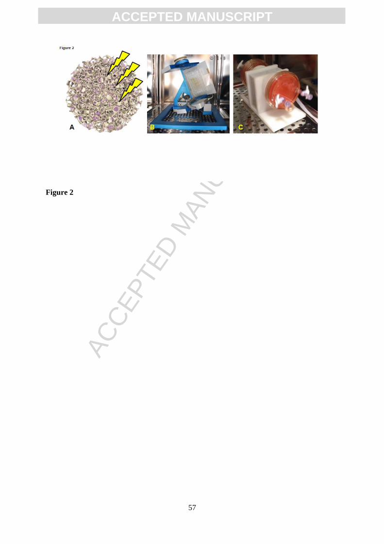
ACC
EPTE
D M
ANU
SCR
IPT
ACCEPTED MANUSCRIPT
57
Figure 2
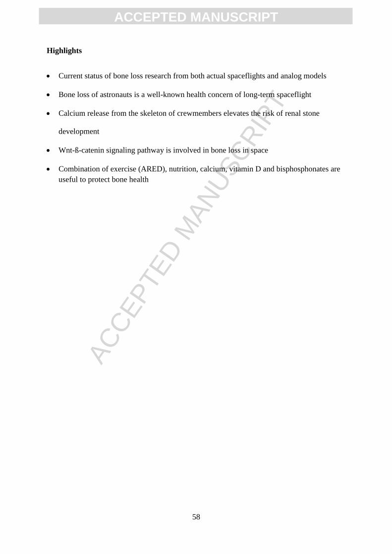
ACC
EPTE
D M
ANU
SCR
IPT
ACCEPTED MANUSCRIPT
58
Highlights
Current status of bone loss research from both actual spaceflights and analog models
Bone loss of astronauts is a well-known health concern of long-term spaceflight
Calcium release from the skeleton of crewmembers elevates the risk of renal stone
development
Wnt-ß-catenin signaling pathway is involved in bone loss in space
Combination of exercise (ARED), nutrition, calcium, vitamin D and bisphosphonates are
useful to protect bone health