the impact of depression on veterans with ptsd and...
TRANSCRIPT

Ti
LLPa
b
c
d
e
f
a
ARAA
KDPTEDV
1
aipcmS2a(op&To
h0
Biological Psychology 105 (2015) 20–28
Contents lists available at ScienceDirect
Biological Psychology
jo u r n al homep age: www.elsev ier .com/ locate /b iopsycho
he impact of depression on Veterans with PTSD and traumatic brainnjury: A diffusion tensor imaging study
inda Isaaca,b,∗, Keith L. Maina,b, Salil Somana,d, Ian H. Gotlibe, Ansgar J. Fursta,b,c,isa M. Kinoshita f, J. Kaci Fairchildb,f, Jerome A. Yesavageb,f, J. Wesson Ashforda,b,eter J. Bayleya,b, Maheen M. Adamsona,b
War Related Illness and Injury Study Center, The Veterans Affairs Palo Alto HealthCare System, Palo Alto, USADepartment of Psychiatry and Behavioral Sciences, Stanford University School of Medicine, Stanford, CA, USADepartment of Neurology and Neurological Sciences, Stanford University, Stanford, CA, USADepartment of Radiology, Stanford University, Stanford, CA, USADepartment of Psychology, Stanford University, Stanford, CA, USAThe Veterans Affairs Palo Alto HealthCare System, Palo Alto, USA
r t i c l e i n f o
rticle history:eceived 15 May 2014ccepted 23 December 2014vailable online 3 January 2015
eywords:
a b s t r a c t
A significant proportion of military personnel deployed in support of Operation Enduring Freedom andOperation Iraqi Freedom were exposed to war-zone events associated with traumatic brain injury (TBI),depression (DEP) and posttraumatic stress disorder (PTSD). The co-occurrence of TBI, PTSD and DEP inreturning Veterans has recently increased research and clinical interest. This study tested the hypothesisthat white matter abnormalities are further impacted by depression. Of particular relevance is the unci-
epressionTSDBImotionTIeterans
nate fasciculus (UF), which is a key fronto-temporal tract involved in mood regulation, and the cingulum;a tract that connects to the hippocampus involved in memory integration. Diffusion tensor imaging (DTI)was performed on 25 patients with a combination of PTSD, TBI and DEP and 20 patients with PTSD andTBI (no DEP). Microstructural changes of white matter were found in the cingulum and UF. Fractionalanisotropy (FA) was lower in Veterans with DEP compared to those without DEP.
Published by Elsevier B.V.
. Introduction
Returning Veterans from Operation Enduring Freedom (OEF)nd Operation Iraqi Freedom (OIF) have high rates of psychiatricllness (Tanielian & Jaycox, 2008). Combat elevates the risk of bothhysical and psychological injuries (Hoge et al., 2004). Compared toivilians, Veterans have a higher incidence of co-morbid posttrau-atic stress disorder (PTSD) (Richardson, Frueh, & Acierno, 2010;
utker & Allain, 1996) and depression (DEP) (Bombardier et al.,010; Brown, Fulton, Wilkeson, & Petty, 2000), present with morenger and hostility and have higher rates of completed suicidesZivin et al., 2007). Evidence indicates military-related, delayed-nset (>6 months) PTSD is associated with higher chronicity, poorerrognosis, and resistance to treatments (Brewin, Stewart, Philpott,
Hejdenberg, 2012; Horesh, Solomon, Keinan, & Ein-Dor, 2013).raumatic brain injury (TBI) is also a contributing factor. Ratesf psychiatric disturbance following TBI are higher in military
∗ Corresponding author at: 3801 Miranda Avenue, Palo Alto, CA.E-mail address: [email protected] (L. Isaac).
ttp://dx.doi.org/10.1016/j.biopsycho.2014.12.011301-0511/Published by Elsevier B.V.
personnel than in the general population. This risk remains elevatedfor several decades (Ashman et al., 2004; Benotsch et al., 2000).
The co-occurrence of TBI, PTSD and DEP in returning Veteranshas fueled both clinical concern and research interest. A betterunderstanding of the unique and shared effects of these conditionscould guide clinical best practices. For example, despite advances inour understanding of emotion (Gross, 2008; Gross, 2013; Joormann& Gotlib, 2010; Runnals et al., 2013) little is known about the effectsof war-related illnesses on emotion regulation. Research on Vet-eran psychiatric illness is grossly underrepresented compared tocivilians, especially in the neuroimaging literature.
We propose that studying DEP, PTSD, and TBI in Veteranspresents a unique opportunity to learn how combat-specific psy-chiatric illness is distinct from civilian cases (Runnals et al., 2013).The neurological interaction between PTSD, DEP and TBI remainslargely unknown. No empirical work exists that comprehensivelyevaluates the unique or shared brain impairments associated with
combat-related DEP-PTSD-TBI. Our research here aimed to studythe neurological profile of these co-morbid illnesses in Veterans.We used diffusion tensor imaging (DTI), neuropsychological data,behavioral assessments, and medical history to assess the unique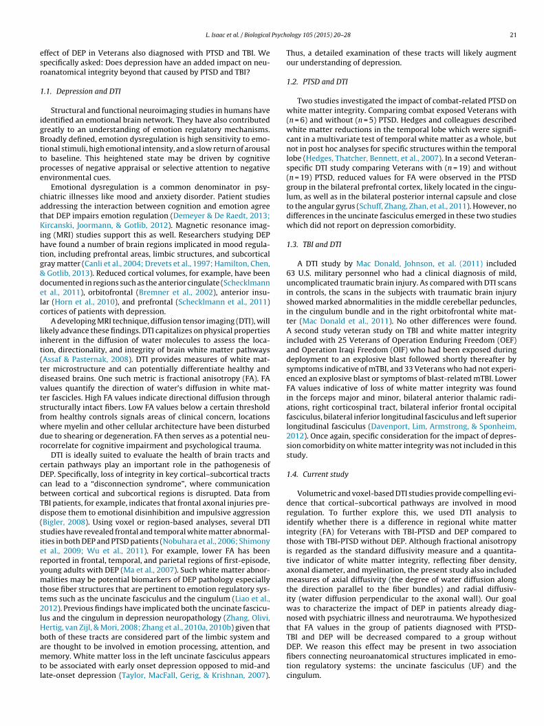
Psych
esr
1
igBttpe
catKihtg&delc
lit(tdvtsfwdr
cDcbTd(sierymtt2lHbamtl
L. Isaac et al. / Biological
ffect of DEP in Veterans also diagnosed with PTSD and TBI. Wepecifically asked: Does depression have an added impact on neu-oanatomical integrity beyond that caused by PTSD and TBI?
.1. Depression and DTI
Structural and functional neuroimaging studies in humans havedentified an emotional brain network. They have also contributedreatly to an understanding of emotion regulatory mechanisms.roadly defined, emotion dysregulation is high sensitivity to emo-ional stimuli, high emotional intensity, and a slow return of arousalo baseline. This heightened state may be driven by cognitiverocesses of negative appraisal or selective attention to negativenvironmental cues.
Emotional dysregulation is a common denominator in psy-hiatric illnesses like mood and anxiety disorder. Patient studiesddressing the interaction between cognition and emotion agreehat DEP impairs emotion regulation (Demeyer & De Raedt, 2013;ircanski, Joormann, & Gotlib, 2012). Magnetic resonance imag-
ng (MRI) studies support this as well. Researchers studying DEPave found a number of brain regions implicated in mood regula-ion, including prefrontal areas, limbic structures, and subcorticalray matter (Canli et al., 2004; Drevets et al., 1997; Hamilton, Chen,
Gotlib, 2013). Reduced cortical volumes, for example, have beenocumented in regions such as the anterior cingulate (Schecklmannt al., 2011), orbitofrontal (Bremner et al., 2002), anterior insu-ar (Horn et al., 2010), and prefrontal (Schecklmann et al., 2011)ortices of patients with depression.
A developing MRI technique, diffusion tensor imaging (DTI), willikely advance these findings. DTI capitalizes on physical propertiesnherent in the diffusion of water molecules to assess the loca-ion, directionality, and integrity of brain white matter pathwaysAssaf & Pasternak, 2008). DTI provides measures of white mat-er microstructure and can potentially differentiate healthy andiseased brains. One such metric is fractional anisotropy (FA). FAalues quantify the direction of water’s diffusion in white mat-er fascicles. High FA values indicate directional diffusion throughtructurally intact fibers. Low FA values below a certain thresholdrom healthy controls signals areas of clinical concern, locationshere myelin and other cellular architecture have been disturbedue to shearing or degeneration. FA then serves as a potential neu-ocorrelate for cognitive impairment and psychological trauma.
DTI is ideally suited to evaluate the health of brain tracts andertain pathways play an important role in the pathogenesis ofEP. Specifically, loss of integrity in key cortical–subcortical tractsan lead to a “disconnection syndrome”, where communicationetween cortical and subcortical regions is disrupted. Data fromBI patients, for example, indicates that frontal axonal injuries pre-ispose them to emotional disinhibition and impulsive aggressionBigler, 2008). Using voxel or region-based analyses, several DTItudies have revealed frontal and temporal white matter abnormal-ties in both DEP and PTSD patients (Nobuhara et al., 2006; Shimonyt al., 2009; Wu et al., 2011). For example, lower FA has beeneported in frontal, temporal, and parietal regions of first-episode,oung adults with DEP (Ma et al., 2007). Such white matter abnor-alities may be potential biomarkers of DEP pathology especially
hose fiber structures that are pertinent to emotion regulatory sys-ems such as the uncinate fasciculus and the cingulum (Liao et al.,012). Previous findings have implicated both the uncinate fascicu-
us and the cingulum in depression neuropathology (Zhang, Olivi,ertig, van Zijl, & Mori, 2008; Zhang et al., 2010a, 2010b) given thatoth of these tracts are considered part of the limbic system and
re thought to be involved in emotion processing, attention, andemory. White matter loss in the left uncinate fasciculus appearso be associated with early onset depression opposed to mid-andate-onset depression (Taylor, MacFall, Gerig, & Krishnan, 2007).
ology 105 (2015) 20–28 21
Thus, a detailed examination of these tracts will likely augmentour understanding of depression.
1.2. PTSD and DTI
Two studies investigated the impact of combat-related PTSD onwhite matter integrity. Comparing combat exposed Veterans with(n = 6) and without (n = 5) PTSD. Hedges and colleagues describedwhite matter reductions in the temporal lobe which were signifi-cant in a multivariate test of temporal white matter as a whole, butnot in post hoc analyses for specific structures within the temporallobe (Hedges, Thatcher, Bennett, et al., 2007). In a second Veteran-specific DTI study comparing Veterans with (n = 19) and without(n = 19) PTSD, reduced values for FA were observed in the PTSDgroup in the bilateral prefrontal cortex, likely located in the cingu-lum, as well as in the bilateral posterior internal capsule and closeto the angular gyrus (Schuff, Zhang, Zhan, et al., 2011). However, nodifferences in the uncinate fasciculus emerged in these two studieswhich did not report on depression comorbidity.
1.3. TBI and DTI
A DTI study by Mac Donald, Johnson, et al. (2011) included63 U.S. military personnel who had a clinical diagnosis of mild,uncomplicated traumatic brain injury. As compared with DTI scansin controls, the scans in the subjects with traumatic brain injuryshowed marked abnormalities in the middle cerebellar peduncles,in the cingulum bundle and in the right orbitofrontal white mat-ter (Mac Donald et al., 2011). No other differences were found.A second study veteran study on TBI and white matter integrityincluded with 25 Veterans of Operation Enduring Freedom (OEF)and Operation Iraqi Freedom (OIF) who had been exposed duringdeployment to an explosive blast followed shortly thereafter bysymptoms indicative of mTBI, and 33 Veterans who had not experi-enced an explosive blast or symptoms of blast-related mTBI. LowerFA values indicative of loss of white matter integrity was foundin the forceps major and minor, bilateral anterior thalamic radi-ations, right corticospinal tract, bilateral inferior frontal occipitalfasciculus, bilateral inferior longitudinal fasciculus and left superiorlongitudinal fasciculus (Davenport, Lim, Armstrong, & Sponheim,2012). Once again, specific consideration for the impact of depres-sion comorbidity on white matter integrity was not included in thisstudy.
1.4. Current study
Volumetric and voxel-based DTI studies provide compelling evi-dence that cortical–subcortical pathways are involved in moodregulation. To further explore this, we used DTI analysis toidentify whether there is a difference in regional white matterintegrity (FA) for Veterans with TBI-PTSD and DEP compared tothose with TBI-PTSD without DEP. Although fractional anisotropyis regarded as the standard diffusivity measure and a quantita-tive indicator of white matter integrity, reflecting fiber density,axonal diameter, and myelination, the present study also includedmeasures of axial diffusivity (the degree of water diffusion alongthe direction parallel to the fiber bundles) and radial diffusiv-ity (water diffusion perpendicular to the axonal wall). Our goalwas to characterize the impact of DEP in patients already diag-nosed with psychiatric illness and neurotrauma. We hypothesizedthat FA values in the group of patients diagnosed with PTSD-TBI and DEP will be decreased compared to a group without
DEP. We reason this effect may be present in two associationfibers connecting neuroanatomical structures implicated in emo-tion regulatory systems: the uncinate fasciculus (UF) and thecingulum.
2 Psychology 105 (2015) 20–28
tmategtlop
uDntta2wdtwT
2
2
Psplsosg
TpsqAsha
anAds
2
iwgigsSmioScd
FSr
Table 1Participant characteristics.
Variable DEP-PTSD-TBI (n = 25) PTSD-TBI (n = 20)
Age 46 (4.6) 44 (3.3)Gender 2 (females) 2 (females)Handedness 85% (right) 87% (right)Level of education 13.9 (2.3) 13.7 (1.8)TBI level Mild (100%) Mild (100%)PTSD diagnosis Yes (100%) Yes (100%)BDI 29.8 (6.0) 10.2 (4.0)*
MMSE (orientation) 99.6% 99.6%Learning/memory 84.5 (1.3) 85.5 (1.5)Est. verbal intelligence 102 (5.8) 104 (6.2)Digits-F 6.0 (.40) 6.0 (.93)Digits-B 4.6 (1.8) 5.8 (1.9)*
Digits-S 5.1 (1.6) 6.2 (1.3)*
Pain diagnosis 85% 82%Sleep disorder 84% 82%
Mean values and percentages, standard deviations are shown in parentheses. Lev-els of education coded in mean years completed, TBI (traumatic brain injury),PTSD (post-traumatic stress disorder), BDI = Beck Depression Inventory, MMSE (MiniMental State Examination), Est. verbal intelligence (estimated verbal intelligence),Digits-F (digit span-forward), Digits-B (digit span-backward), Digits-S (digit span-
cognition: verbal intelligence to determine estimated pre-morbid intellectual func-tioning; orientation to time and place assessed by the Mini Mental State Exam
2 L. Isaac et al. / Biological
The UF is a ventral associative bundle that connects the anterioremporal lobe, including the amygdala and hippocampus, with the
edial and lateral orbitofrontal cortex. The cingulum is a medialssociative bundle that runs within the cingulate gyrus, superioro the corpus callosum. The cingulum contains fibers of differ-nt lengths, the longest of which runs from the anterior temporalyrus to the orbitofrontal cortex. The shortest, U-fibers, connecthe medial frontal, parietal, occipital, temporal lobes, and cingu-ate cortex. Both the UF and cingulum tracts are considered partf the limbic system, and are thought to be involved in emotionrocessing, attention, and memory.
A detailed examination of these tracts has implications fornderstanding emotional regulation and treating both PTSD andEP. Co-morbid PTSD and DEP is associated with increased ill-ess burden in patients, poorer prognosis, and delayed responseo treatment. This is especially true in the Veteran popula-ion (Runnals et al., 2013). Depression is increasingly commonnd, according to the World Health Organization (WHO), by020 will be the second-most prevalent health condition world-ide. It is imperative to increase our understanding of theseisorders. To our knowledge, this is the first study with the objec-ive of investigating whether DEP has a pronounced impact onhite matter integrity above and beyond the effect of PTSD and
BI.
. Methods and materials
.1. Clinical measures and evaluation
Beck Depression Inventory – Second Edition (BDI-II) (Beck, Steer, & Brown, 1996):articipants’ depressive symptomatology was assessed using the BDI-II, a 21-itemelf-report questionnaire designed for use by both clinical and nonclinical adultopulations. Higher item scores are indicative of greater depressive symptomato-
ogy. The BDI-II’s reliability and validity has been demonstrated across numeroustudies (Beck, Steer, & Garbin, 1988). Depression is based on a self rating scale (BDI)pposed to a structured clinical or research interview. Therefore, our two-groupamples are characterized as being either in the high depressive symptomologyroup or the low depressive symptomology group.
Clinician-Administered Post-Traumatic Stress Disorder (CAPS) (Blake et al., 1995):he Clinician-Administered PTSD Scale (CAPS) is a structured interview for assessingosttraumatic stress disorder (PTSD) diagnostic status and symptom severity. Thetructure corresponds to the DSM-IV criteria with symptoms rated for both fre-uency and intensity; these two scores are summed to provide severity ratings.dditional questions assess also inquire about associated features of guilt and dis-ociation. CAPS is considered the “gold standard” for the assessment of PTSD andas very good psychometric properties across a wide variety of clinical populationsnd research settings (Weathers, Keane, & Davidson, 2001).
TBI Assessment: A multimodal approach was utilized to arrive at a TBI diagnosisnd severity classification. Clinician interviews consist of a neurological exam by aeurologist and a clinical read of neuro-images by an attending neuroradiologist.
review of records as well as military screening tools is also used to confirm aiagnosis of TBI. TBI severity was measured by the Post-Traumatic Amnesia (PTA)cale and the Loss of Consciousness (LOC) scale.
.2. Participant demographics
Written informed consent was obtained for all participants in the study. Partic-pants included 25 patients with a co-occurring PTSD, TBI and DEP and 20 patients
ith PTSD and TBI but without DEP. To minimize false positives in the depressedroup, a cut-off score of >16 was used to classify the depressed group. Partic-pants were matched for handedness (86% right), age (M = 44.6, SD = 12.0), andender distribution (90% male). TBI severity was measured by post-traumatic amne-ia (mild = <1 h) and loss of consciousness (mild = <30 min). Education (M = 13.9,D = 2.3) and deployment duration were measured in years (M = 7.9, SD = 3.5). Theale to female gender ratio in our sample is representative of Veteran samples
n previous work. Participants were not matched on BDI scores as it was used inur sample selection criteria (DEP-PTSD-TBI; M = 29.8, SD = 6.0; PTSD-TBI; M = 10.2,D = 4.0). The two groups were matched on two other prominent Veteran medicalomplaints: pain diagnosis (PTSD-TBI = 82%, DEP-PTSD-TBI = 85%), and sleep disor-
er diagnosis (PTSD-TBI = 82%, DEP-PTSD-TBI = 84%), see Table 1.Our sample represented a range of combat missions (Operation Iraqireedom = 17, Operation Enduring Freedom = 7, Gulf War (Desert Shield/Deserttorm) = 9, Vietnam = 7, Operation New Dawn = 4, and Korea = 1). Exclusion crite-ia included: schizophrenia, bipolar and psychotic disorders, current and past
sequencing).* Significant group differences at p = .05.
alcohol or substance abuse, history of anxiety disorder outside of major depres-sive episodes, history of somatoform disorders, neurological diseases, history ofdiabetes or cardiac conditions determined by self-report health measures. On thebasis of this exclusionary criteria, 24 subjects were excluded from the total numberof patients who had a DTI scan. All participants were assessed by an interdisciplinaryclinical team including a board certified psychiatrist, neurologist, neuropsychol-ogist and clinical psychologist using structured interviews (CAPS) and clinicalinstruments (Beck Depression & Anxiety Inventories). This study was approvedby the Stanford IRB committee and the VA Palo Alto Healthcare System ResearchAdministration.
2.3. Procedures
Veterans were seen at the VA Palo Alto War Related Illness and Injury Study Cen-ter site (WRIISC CA), a tertiary care clinic that provides evaluations for Veterans withdeployment-related health concerns. Veterans referred to the WRIISCs often haveone or more of the following presentations: (1) deployment-related health condi-tions, (2) complex health conditions with no known cause (medically unexplainedsymptoms), (3) chronic multi-symptom illness (CMI) and (4) little to no symptomimprovement after multiple tests/treatments (Lange et al., 2013). The goal of thefour, full-day, interdisciplinary evaluation is to provide a comprehensive assess-ment of the individual with a particular focus on post-deployment concerns thatmay be excessively complex for routine primary care. The evaluation begins withcompletion of self-report screening instruments that assess military experience,demographics, symptoms, function, deployment-related exposure, medication use,and previously diagnosed medical and mental health conditions. Clinicians withexpertise in psychiatry, radiology, clinical psychology, neuropsychology, nursing,and education evaluate the Veteran’s needs, review the screening results, and per-form standard evaluations. Team members meet at the end of all evaluations toformulate a plan and present it to the patient for further refinement. The final planis then communicated to the patient and his/her primary care provider. WRIISC CAis also engaged in establishing advanced clinical neuroimaging methodologies forimage acquisition, processing, and analysis. We perform clinically based MRI tech-niques on patients to advance our understanding of brain function in relation toVeterans health.
2.4. Neuropsychological assessment
A series of standardized neuropsychological tests were administered follow-ing the completion of all questionnaires: (1) general screening information wasobtained initially. (2) Subtests from the Repeatable Battery for the Assessmentof Neuropsychological Status (RBANS) measured major functional domains of
(MMSE); learning and memory; and attention and working memory measured bydigit span forward, backward and sequencing (Wechsler, 1939). A measure of effortwas also administered to all participants. Importantly, these measures were used toscreen out participants with global performance difficulties in the high-moderateto severe range.

Psychology 105 (2015) 20–28 23
2
Saeusaa1etsaWpw
2
bDctbfifict(edtwZ
2
wetfmfbt
2
Sratfiirgccitcowtc
3
3
es
L. Isaac et al. / Biological
.5. MRI acquisition
Neuroimaging was conducted at the Veterans Affairs Palo Alto Health Careystem (VAPAHCS). Each neuroimaging session included T1-weighted (SPGR)nd DTI sequences. All scans were run on a 3 T GE Discovery MR750 with anight channel, GE head coil. High-resolution T1-weighted images were acquiredsing a three-dimensional spoiled-gradient recalled acquisition (3D-SPGR) inteady state (272 axial slices, repetition time = 7.3 ms; echo time = 3.0 ms; flipngle = 11◦; field of view = 250 mm; slice thickness = 1.2 mm with 0.6 between slices;cquisition matrix = 256 × 256; number of excitations = 1.0; voxel dimensions:.05 mm × 1.05 mm × 0.60 mm). DTI images were acquired using a single-shot, spin-cho, echo-planar sequence in 50 axial sections (repetition time = 6600 ms; echoime = 80 ms; field of view = 240 mm; slice thickness = 2.5 mm with no gap; acqui-ition matrix = 96 × 96; number of excitations = 1). Diffusion weighting was appliedlong 30 noncollinear directions with a b-value of 1000 s/mm2 and 5 b = 0 volumes.e collected two DTI acquisitions run at NEX = 1 and combined them in post-
rocessing to improve the signal-to-noise ratio. The reconstructed voxel dimensionsere 2.5 mm × 2.5 mm × 2.5 mm.
.6. Image processing
DTI images were analyzed using mrDiffusion software, freeware developedy the Vista Laboratory, Stanford University (http://white.stanford.edu/software/).ata from the two DTI scans were averaged. The resulting file entered a prepro-essing pipeline consisting of eddy current and motion correction, alignment tohe MNI brain, resampling, and fitting of the tensor model. We performed a wholerain tractography for each patient using mrDiffusion. White matter was identi-ed and seeded using a FA mask thresholded at 0.20. This procedure yielded ∼200 Kbers per participant (depending on data). Custom MATLAB code then automaticallylassified and extracted individual fiber tracts as defined by the JHU white matterractography atlas (Wakana et al., 2007). We manually defined two reference ROIsrROIs) describing waypoints for the uncinate fasciculus and the cingulum (Huat al., 2008; Wakana et al., 2007; Zhang et al., 2008, 2010a, 2010b). The ROIs wererawn in MNI space on the ICBM-DTI-81 atlas. Fibers were retained if they passedhrough both rROIs. Extraneous fibers were subtracted from the data. Fiber tractsere then manually edited using a white matter atlas as a guide (Oishi, Faria, van
ijl, & Mori, 2011).
.7. Quantitative tract-specific measures
Additional MATLAB code quantified fiber diffusion properties. Each fiber groupas sectioned into 30 nodes. Fractional anisotropy values (FA) were calculated for
ach node by taking a weighted average of the individual fibers. A single fiber’s con-ribution to the average was determined by calculating its Mahalanobis distancerom the core tract. Thus fibers that were outliers from the core contributed mini-
ally to a node’s FA. We obtained mean FAs for a fiber group by averaging FA valuesrom all the nodes. By directly extracting and comparing tracts of interest, this DTI-ased approach provides high detection power of white matter abnormalities in thewo groups of interest.
.8. Statistical analyses
All statistical analyses were performed in SPSS software, version 18.0 (SPSS,omers, NY). In order to test our specific hypotheses, a hierarchical binary logisticegression was performed. This regression analysis is appropriate when predicting
dichotomous dependent variable (group assignment) from one or more predic-ors while controlling for one or more covariates (i.e., control variables). Ten DTIber tracts (left cingulum, right cingulum, left corticospinal, right corticospinal, left
nferior fronto-occipital, right inferior fronto-occipital, left superior longitudinal,ight superior longitudinal, left uncinate and right uncinate) were the predictors,roup (PTSD/TBI/DEP or PTSD/TBI) was the dependent variable, patient age was theovariate. Our primary fiber tracts of interest were the uncinate fasciculus and theingulum. All remaining tracts were of secondary interest. Controlling for age effectsn DTI analysis is especially critical as aging is associated with significant white mat-er deterioration (Davis et al., 2009). The model assessed whether the predictorsan correctly classify individuals who have PTSD, TBI and DEP from those who havenly PTSD and TBI. The data were screened for outliers prior to analysis. Participantsere removed when a standardized residual was greater than 3. This process failed
o reveal any outliers in the data. A Bonferroni correction was applied to all post hocomparisons with a 95% confidence interval.
. Results
.1. Neuropsychological variables
Neuropsychological results revealed that group differencesxisted only on the digit span-backward (p = .05) and digit span-equencing (p = .046) tasks, used to measure attention and working
Fig. 1. A visual depiction of the significant fiber tracts (uncinate fasciculus (purple)and. cingulum (blue) taken from a male participant in the DEP-PTSD-TBI group.
memory capacity. The group with depression scored significantlylower on both tasks (DSB: M = 4.6, SD = 1.8; DSS: M = 5.1, SD = 1.6)than the group without depression (DSB: M = 5.8, SD = 1.9; DSS:M = 6.2, SD = 1.3) suggesting that the additive impact of depressionimpairs attention and working memory. Note that while a statis-tically significant difference emerged on this measure of attentionand working memory, further work is required to assert clinicalsignificance. No other group differences were found on measuresof neuropsychological function (all p values >.05). Group differ-ences were not observed on measures of learning and memory (lowaverage; Index score range = 82–87), and estimated verbal intel-ligence (average; Index score range = 92–107). Participants wereall fully oriented to time and place as measured by the orienta-tion subtest of the Mini Mental State Exam (MMSE = 99.6% for bothgroups). No subjects were excluded on the basis of cognitive effort,see Table 1.
3.2. DTI
The model correctly classified 21 (84%) of patients who hadDEP-PTSD-TBI. However, the model was only able to correctly clas-sify 12 (60%) of the patients who had only PTSD and TBI. Theoverall percentage of correct classification was 73%. The regres-sion coefficients indicated that both the right cingulum ( ̌ = 21.04,p = .0051, Bonferroni corrected) and the left uncinate fasciculus( ̌ = 32.05, p = .0045, Bonferroni corrected) were significant pre-dictors of group assignment with a notable effect size (r2 = .41)(Fig. 1). The classification table and regression coefficients are listedin Table 2. See Figs. 2–5 for data distribution of significant findings(group differences for left uncinate, right cingulum fiber tracts). Allother DTI tracts failed to reach statistical significance (all p values>.05). The eigenvector (v1) is associated with the largest eigenvalue(AD: axial diffusivity = �1) was assumed to represent the local fiberdirection. Radial diffusivity (RD) was taken as the mean of twominor eigenvalues (�2 + �3)/2. Between group analyses of radialdiffusivity and axial diffusivity measures did not reach statisticalsignificance (RD: p = .43; AD: p = .51).
4. Discussion
Diffusion tensor imaging (DTI) provides a quantitative methodto assess the integrity and connectivity of white matter. The low FAvalues observed in the cingulum and uncinate fasciculus in our par-
ticipants suggests a loss of integrity in white matter fiber tracts thatconnect cortex (prefrontal, temporal, and occipital lobes) to subcor-tical areas, such as the amygdala and hippocampus. Lower valuesin patients with DEP suggest this condition further impairs white
24 L. Isaac et al. / Biological Psychology 105 (2015) 20–28
Table 2Regression coefficients for DTI variables by group.
Predictor Model coefficients
B S.E. Wald df Sig. Exp(B)
Right cingulum 21.04 11.24 3.50 1 .0051 1E+009*
Left cingulum −9.33 11.98 0.61 1 .436 0.00Left corticospinal −10.86 16.29 0.44 1 .505 0.00Right corticospinal 15.47 16.28 0.90 1 .342 5,205,860.32Left inferior fronto-occipital −23.71 16.26 2.13 1 .145 0.00Right inferior fronto-occipital 21.49 17.37 1.53 1 .216 2E+009Left superior longitudinal 5.86 16.00 0.13 1 .714 349.33Right superior longitudinal −11.46 16.01 0.51 1 .474 0.00Left uncinate 32.05 13.71 4.26 1 .0045 1E+005*
Right uncinate −0.42 13.91 0.00 1 .976 0.66
* A Bonferroni correction was applied to account for multiple comparisons and statistical significance was accepted at Bonferroni corrected p < 0.005.
Fig. 2. Distribution of left uncinate fractional anisotropy for DEP-PTSD-TBI Group.
Fig. 3. Distribution of left uncinate fractional anisotropy for PTSD-TBI Group.

L. Isaac et al. / Biological Psychology 105 (2015) 20–28 25
action
mt(Tadte
Fig. 4. Distribution of right cingulum fr
atter and potentially exacerbates disease. Given that the compu-ational model correctly classified 84% of the group with depressionDEP-PTSD-TBI), and only 60% the group without depression (PTSD-BI) suggests that the white matter architecture of cingulumnd uncinate fasciculus tracts may be particularly sensitive to
epression-related brain changes. It is noteworthy to mentionhat accurate classification of depression was not diminishedven in the presence of another psychiatric condition (PTSD) andFig. 5. Distribution of right cingulum fracti
al anisotropy for DEP-PTSD-TBI Group.
neurological injury (TBI). The non-significant results on measuresof axial diffusivity and radial diffusivity may be attributed to severalreasons. First, demyelination processes are most often observed inneurological conditions such as ischemic stroke (Alexander, Lee,Lazar, & Field, 2007) or multiple sclerosis (Roosendaal et al., 2009)
and less in mood disorders such as depression (Kieseppä et al.,2010; Taylor et al., 2007; Zhang et al., 2013). Second, it is possi-ble that given our complex patient sample, sensitivity in detectingonal anisotropy for PTSD-TBI Group.

2 Psych
atpfitnafteTsHtwshiihi
foi(2sa&ofes(TScmi2wpd
tSircmcftefdet
tOs(2t
6 L. Isaac et al. / Biological
xonal injury and demyelination may have been minimized due tohe co-occurring conditions of PTSD and TBI. Third, a limited sam-le size presents with potential statistical power limitations andnally we point to discrepancies in DTI methodology. Current pro-ocols for DTI acquisition, preprocessing and statistical testing areon-uniform yielding differences in results based on methodologylone. For instance, differences have been found in the same studyor statistical parametric mapping (SPM)-based methods comparedo a tract-based spatial statistics (TBSS)-based methods (Osamut al., 2010). This issue is discussed extensively in recent work byhomas and colleagues. While the utility of DTI in TBI has beenuccessfully demonstrated in several studies, a recent review byulkower, Poliak, Rosenbaum, Zimmerman, and Lipton (2013) on
he clinical utility of DTI to detect brain abnormalities in patientsith TBI, reported that only two studies included data on depres-
ion (Hulkower et al., 2013). This is somewhat unexpected given theigh co-occurrence of depression in patients with a traumatic brain
njury. The present study underscores the importance of consider-ng comorbid depression in the clinical utility of DTI for TBI as weave shown here, for the first time that depression has an added
mpact on white matter integrity in patients with TBI.The neuropsychological finding that the group with DEP was
urther impaired on a measure of attention and working mem-ry is consistent with previous work showing that a key cognitivempairment in clinical depression is attention and working memoryGotlib, Krasnoperova, Yue, & Joormann, 2004; Joormann & Gotlib,008). In fact, attentional processes are an important mediator ofeveral depressive phenomena such as attentional biases for neg-tive stimuli (Gotlib et al., 2004) and rumination (Donaldson, Lam,
Mathews, 2007). This finding is further corroborated by decadesf neuroimaging studies which consistently report reduced pre-rontal cortical blood flow and metabolism in depression (Baxtert al., 1989). We found simple verbal attention to be intact (digitpan forward) for both groups and working memory to be poorerdigit span backwards and digit span sequencing) in the DEP-PTSD-BI group compared to the PTSD-TBI group without depression.imple attention is widely believed to be a basic cognitive pro-ess necessary for higher-level cognitive processes such as decisionaking, the ability to override a prepotent response and suppress-
ng irrelevant distracters (Paelecke-Habermanna, Pohl, & Leplow,005). It is hypothesized that these differences in attention andorking memory hold explanatory value for the common com-laints of depressed patients (poor concentration, difficulty inecision making, perseveration and rumination, distractibility).
Frontal–subcortical circuits are thought to play a key role inhe regulation of motor, cognitive and motivational processes.tructural and functional abnormalities within neural systems,ncluding frontal–subcortical circuits, are implicated in the neu-obiology of depression. White matter abnormality is a potentialause of this network dysfunction. It has been hypothesized thaticrostructural changes in the white matter of frontal–subcortical
ircuits leads to a “disconnection syndrome”. Converging evidencerom postmortem and in vivo studies suggests that this disconnec-ion plays an important role in the pathophysiology of DEP (Liaot al., 2012). The present findings provide additional evidence forrontal–subcortical circuits involvement in DEP. The current studyemonstrates that white matter abnormality in DEP is pronouncednough to be detected in the presence of other psychiatric condi-ions (PTSD) and neurological insult (TBI).
The present findings provide additional evidence of a struc-ural basis for the involvement of this neurocircuitry in DEP.ur findings complement brain volumetric studies of depression
howing reduced cortical volumes in key fronto-limbic regionsBremner et al., 2002; Schecklmann et al., 2011; Shackman et al.,011) in patients with DEP. Moreover, evidence for a disconnec-ion syndrome in DEP is advanced by the present study. White
ology 105 (2015) 20–28
matter abnormality can be seen as a likely contributor for net-work dysfunction. In particular, it has been hypothesized thatmicrostructural changes in the white matter of frontal–subcorticalcircuits lead to a “disconnection syndrome” between frontal andsub-cortical regions. Converging evidence from postmortem andin vivo studies suggests that this disconnection plays an importantrole in the pathophysiology of DEP (Liao et al., 2012). The currentstudy puts forth a unique advancement insofar that white mat-ter abnormality was not only shown in DEP analogous to previousresearch (Bae et al., 2006; Colloby et al., 2011; Li et al., 2007b), butthat this finding was robust enough to be detected in the presence ofanother psychiatric condition (PTSD) and in the presence of a neu-rological insult (TBI). This finding is especially relevant to the Vet-eran patient population. PTSD, TBI and DEP co-exist because braininjuries are often sustained during traumatic experiences. Givensubsequent life changes, it is not surprising that a mood disorderlike DEP often accompanies PTSD and TBI. Importantly, alterna-tive interpretations need to be taken into consideration Perhapspatients with pre-existing white matter abnormalities are morevulnerable to depression. Equally, patients who present with whitematter abnormalities in the uncinate fasciculus and the cingulumresulting from PTSD and/or TBI are more susceptible to develop-ing depression. Such pre-existing vulnerabilities may help to clarifywhich individuals are at greatest risk for developing these disordersand how to best decrease the impact of risk factors. The chronicand relapse-prone nature of DEP suggests abnormal brain pro-cesses underlie and perpetuate this condition. Consistent with thisexpectation, a variety of neurophysiological and neurochemicalabnormalities have been discovered in DEP patients, specifically insystems that modulate emotional behavior (Mayberg et al., 2000).
However, none of these studies have isolated the specific contri-bution of DEP in patients with co-occurring diseases. This is despitedepression being the most frequent co-morbid condition in manypsychiatric and neurological illnesses. Greater understanding of theneurological characteristics of DEP will not only advance scientificknowledge but also provide the empirical basis for clinical decisionsand targeted treatment regimes.
4.1. Limitations
Summary scores for the CAPS diagnostic interview are not avail-able and therefore we cannot ascertain PTSD severity levels in oursample. It is highly recommended that future studies include PTSDseverity scores. Point of reasoning for PTSD severity scores is thatmoderate–severe PTSD may present with higher risk for developingchronic depression. Specifically in the Veteran population, higherPTSD severity scores may also present with paralleling symptomsthat may even worsen symptoms of TBI. As this is a cross-sectionalstudy, our limitations include a limited ability to infer causal rela-tionships. Although the current study did ensure that PTSD was dueto combat-related trauma by review of patients’ subjective distress,we did not specify the type of trauma (military sexual trauma (MST)vs. frontline blast exposure). Low female representation in our sam-ple limits our ability to compare subtypes of PTSD such as MSTvs. non-MST related trauma. Finally, we did not include a healthyVeteran sample as most individuals referred to WRIISC CA presentwith chronic multi-symptom illness. Thus our access to a healthyVeteran cohort is limited. Finally, because of the characteristics ofthis sample (military personnel with severe symptoms and multi-ple comorbidities), the results may or may not be generalizable tosimilar civilian samples.
4.2. Conclusion
The current study is not the first to demonstrate white mat-ter abnormality in DEP patients, but is the first to do so in the

Psych
ptcdvm
nTtct(tdtcmiiMsoihwcaa
F
A
SdsSsK
R
A
A
A
B
B
B
B
B
L. Isaac et al. / Biological
resence of other psychiatric and neurological conditions. Fromhese results we submit that microstructural abnormalities in theingulum and uncinate fasciculus may be promising biomarkers ofepression in the Veteran population. Further studies are needed toalidate these findings and explore the relationship between whiteatter degeneration, depression, and clinical outcomes.The recent wars in Afghanistan and Iraq have highlighted the
eed for increased attention to the diagnostic trio: DEP, PTSD, andBI. Although both PTSD and TBI have been commonly referredo as the “signature injuries” of the wars, DEP deserves equalonsideration given the prevalence of depressive disorders andhe increasing numbers of suicides in the Veteran populationZivin et al., 2007). Our findings suggest a broader responseo traumatic injury may be appropriate, one that acknowledgesepression in treatments for PTSD and TBI. If depression is notreated, patients with DEP-PTSD-TBI may suffer a longer diseaseourse, worsening of symptoms, and prolonged functional impair-ent. Tailoring clinical interventions to emphasize depression
n certain scenarios may lead to more successful recovery andmproved quality of life for persons with all three conditions.
oreover, clinical implications motivated by the present studyuggest utilizing DTI to aid in differential diagnosis, treatmentutcome prediction and targeting specific connectivity pathwaysn the treatment of clinical depression. For instance, if DTI mayave clinical utility for predicting which patients with depressionill respond more favorably to antidepressant medication vs. psy-
hotherapy. This effort is especially timely given that neuroimagingpproaches are currently at the forefront of MDD biomarkerdvancement.
inancial disclosures
None.
cknowledgements
The authors extend special thanks to Dr. Brian A. Wandell,tanford University, Department of Psychology, for providing theiffusion imaging analysis and visualization package (mrDiffu-ion). Special thanks are also extended to Dr. Robert F. Dougherty,tanford Center for Cognitive and Neurobiological Imaging, for con-ultation on image acquisition. Finally, we are grateful to Jenniferong for her assistance with data collection.
eferences
lexander, A. L., Lee, J. E., Lazar, M., & Field, A. S. (2007). Diffusion tensor imaging ofthe brain. Neurotherapeutics, 4, 316–329.
shman, T. A., Spielman, L. A., Hibbard, M. R., Silver, J. M., Chandna, T., & Gordon,W. A. (2004). Psychiatric challenges in the first 6 years after traumatic braininjury: Cross-sequential analyses of axis I disorders. Archives of Physical Medicine& Rehabilitation, 85(4), 36–42. http://dx.doi.org/10.1016/j.apmr.2003.08.117
ssaf, Y., & Pasternak, O. (2008). Diffusion tensor imaging (DTI)-based white mat-ter mapping in brain research: A review. Journal of Molecular Neuroscience, 34,51–61.
ae, J. N., MacFall, J. R., Krishnan, K. R., Payne, M. E., Steffens, D. C., & Tay-lor, W. D. (2006). Dorsolateral prefrontal cortex and anterior cingulate cortexwhite matter alterations in late-life depression. Biological Psychiatry, 60(12),1356–1363.
axter, L. R., Schwartz,. J. M., Phelps, M. E., Mazziotta, J. C., Guze, B. H.,& Selin, C. E. (1989). Reduction of prefrontal cortex glucose metabolismcommon to three types of depression. Archives of General Psychiatry, 46,243–250.
eck, A. T., Steer, R. A., & Brown, G. K. (1996). Manual for Beck Depression Inventory –Second Edition (BDI-II). San Antonio, TX: Psychological Corporation.
eck, A. T., Steer, R. A., & Garbin, M. G. J. (1988). Psychometric properties of theBeck Depression Inventory: Twenty-five years of evaluation. Clinical PsychologyReview, 8, 77–100.
enotsch, E. G., Brailey, K., Vasterling, J. J., Uddo, M., Constans, J. I., & Sutker, P.B. (2000). War Zone stress, personal and environmental resources, and PTSD
ology 105 (2015) 20–28 27
symptoms in Gulf War Veterans: A longitudinal perspective. Journal of AbnormalPsychology, 109(2), 205–213. http://dx.doi.org/10.1037/0021-843X.109.2.205
Bigler, E. D. (2008). Neuropsychology and clinical neuroscience of persistent post-concussive syndrome. Journal of the International Neuropsychological Society, 14,1–22.
Blake, D. D., Weathers, F. W., Nagy, L. M., Kaloupek, D. G., Gusman, F. D., Charney, D.S., et al. (1995). The development of a clinician-administered PTSD scale. Journalof Traumatic Stress, 8, 75–90.
Bombardier, C. H., Fann, J. R., Temkin, N. R., Esselman, P. C., Barber, J., & Dikmen,S. S. (2010). Rates of major depressive disorder and clinical outcomes follow-ing traumatic brain injury. Journal of the American Medical Association, 303(19),1938–1945. http://dx.doi.org/10.1001/jama.2010.599
Bremner, J. D., Vythilingam, M., Vermetten, E., Nazeer, A., Adil, J., Khan, S., et al.(2002). Reduced volume of orbitofrontal cortex in major depression. BiologicalPsychiatry, 51, 273–279.
Brewin, C. R., Stewart, L., Philpott, R., & Hejdenberg, J. (2012). Compari-son of immediate-onset and delayed-onset posttraumatic stress disorderin military veterans. Journal of Abnormal Psychology, 118(4), 767–777.http://dx.doi.org/10.1037/a0017203
Brown, E. S., Fulton, M. K., Wilkeson, A., & Petty, F. (2000). The psychiatric sequelaeof civilian trauma. Comprehensive Psychiatry, 4, 19–23.
Canli, T., Sivers, H., Thomason, M. E., Whitfield-Gabrieli, S., Gabrieli, J., & Gotlib, I. H.(2004). Brain activation to emotional words in depressed vs. healthy controls.Neuroreport, 15(17), 2585–2588.
Colloby, S. J., Firbank, M. J., Thomas, A. J., Vasudev, A., Parry, S. W., & O’Brien, J. T.(2011). White matter changes in late-life depression: A diffusion tensor imagingstudy. Journal of Affective Disorders, 135(1–3), 216–220.
Davenport, N. D., Lim, K. O., Armstrong, M. T., & Sponheim, S. R. (2012). Diffuseand spatially variable white matter disruptions are associated with blast-relatedmild traumatic brain injury. Neuroimage, 59, 2017–2024.
Davis, S. W., Dennis, N. A., Buchler, N. E. G., Madden, D. J., White, L. E., & Cabeza, R.(2009). Assessing the effects of aging on long white matter tracts using diffusiontensor imaging (DTI) tractography. Neuroimage, 46, 530–541.
Demeyer, I., & De Raedt, R. (2013). Attentional bias for emotional information inolder adults: The role of emotion and future time perspective. PLOS ONE, 8(6),e65429. http://dx.doi.org/10.1371/journal.pone.0065429
Donaldson, C., Lam, D., & Mathews, A. (2007). Rumination and attention in majordepression. Behavior Research & Therapy, 45, 2664–2678.
Drevets, W. C., Price, J. L., Simpson, J. R., Todd, R. D., Reich, T., Vannier, M., et al.(1997). Subgenual prefrontal cortex abnormalities in mood disorders. Nature,386, 824–827.
Gotlib, I. H., Krasnoperova, E., Yue, D. L., & Joormann, J. (2004). Attentional biasesfor negative interpersonal stimuli in clinical depression. Journal of AbnormalPsychology, 113(1), 127–135.
Gross, J. J. (2008). Emotion regulation. In M. Lewis, J. M. Haviland-Jones, & L. F. Barrett(Eds.), Handbook of Emotions (3rd ed., pp. 497–512). New York: Guilford.
Gross, J. J. (2013). Emotion regulation: Taking stock and moving forward. Emotion,13(3), 359–365. http://dx.doi.org/10.1037/a0032135
Hamilton, J. P., Chen, M. C., & Gotlib, I. H. (2013). Neural systems approachesto understanding major depressive disorder: An intrinsic functional orga-nization perspective. Neurobiology of Disease, 52, 4–11. http://dx.doi.org/10.1016/j.nbd.2012.01.015
Hedges, D. W., Thatcher, G. W., Bennett, P. J., et al. (2007). Brain integrity and cerebralatrophy in Vietnam combat veterans with and without posttraumatic stressdisorder. Neurocase, 13(5–6), 402–410.
Hoge, C. W., Castro, C. A., Messer, S. C., McGurk, D., Cotting, D. I., & Koffman, R.L. (2004). Combat duty in Iraq and Afghanistan, mental health problems, andbarriers to care. New England Journal of Medicine, 351(1), 13–22.
Horesh, D., Solomon, Z., Keinan, G., & Ein-Dor, T. (2013). The clinical picture oflate-onset PTSD: A 20-year longitudinal study of Israeli war veterans. PsychiatryResearch, 208(3), 265–273.
Horn, D. I., Yu, C., Steiner, J., Buchmann, J., Kaufmann, J., Osoba, A., et al. (2010).Glutamatergic and resting-state functional connectivity correlates of severity inmajor depression—The role of pregenual anterior cingulate cortex and anteriorinsula. Frontiers in Systems Neuroscience, 4, 33.
Hua, K., et al. (2008). Tract probability maps in stereotaxic spaces: Analyses ofwhite matter anatomy and tract-specific quantification. Neuroimage, 39(1),336–347.
Hulkower, M. B., Poliak, D. B., Rosenbaum, S. B., Zimmerman, M. E., & Lipton, M. L.(2013). A decade of DTI in traumatic brain injury: 10 years and 100 articles later.American Journal of Neuroradiology, 34, 2064–2074.
Joormann, J., & Gotlib, I. H. (2008). Updating the contents of working memory indepression: Interference from irrelevant negative material. Journal of AbnormalPsychology, 117(1), 182–192.
Joormann, J., & Gotlib, I. H. (2010). Emotion regulation in depression: Relation tocognitive inhibition. Cognition & Emotion, 24, 281–298.
Kieseppä, T., Eerola, M., Mäntylä, R., Neuvonen, T., Poutanen, V. P., Luoma, K., et al.(2010). Major depressive disorder and white matter abnormalities: A diffusiontensor imaging study with tract-based spatial statistics. Journal of Affective Dis-orders, 120, 240–244.
Kircanski, K., Joormann, J., & Gotlib, I. H. (2012). Cognitive aspects of depression.
Wiley Interdisciplinary Review: Cognitive Science, 3, 301–313. http://dx.doi.org/10.1002/wcs.1177Li, L., Ma, N., Li, Z., Tan, L., Liu, J., Gong, G., et al. (2007). Prefrontal white matterabnormalities in young adult with major depressive disorder: A diffusion tensorimaging study. Brain Research, 1168, 124–128.

2 Psych
L
M
M
M
N
L
O
O
P
R
R
R
S
S
8 L. Isaac et al. / Biological
iao, Y., Huang, X., Wu, Q., Yang, C., Kuang, W., Du, M., et al. (2012). Is depres-sion a disconnection syndrome? Meta-analysis of diffusion tensor imagingstudies in patients with MDD. Journal of Psychiatry & Neuroscience, 38(1), 49–56.http://dx.doi.org/10.1503/jpn.110180
a, N., Li, L., Shu, N., Liu, J., Gong, G., He, Z., et al. (2007). White matter abnormalitiesin first-episode, treatment-naive young adults with major depressive disorder.American Journal of Psychiatry, 164, 823–826.
ac Donald, C. L., Johnson, A. M., et al. (2011). Detection of blast related traumaticbrain injury in U.S. military personnel. New England Journal of Medicine, 364(22),2091–2100.
ayberg, H. S., Brannan, S. K., Tekell, J. L., Silva, J. A., Mahurin, R. K., McGinnis,S., et al. (2000). Regional metabolic effects of fluoxetine in major depression:Serial changes and relationship to clinical response. Biological Psychiatry, 48(8),830–843.
obuhara, K., Okugawa, G., Sugimoto, T., Minami, T., Tamagaki, C., Takase, K., et al.(2006). Frontal white matter anisotropy and symptom severity of late-lifedepression: A magnetic resonance diffusion tensor imaging study. Journal ofNeurology Neurosurgery & Psychiatry, 77, 120–122.
ange, G., McAndrew, L., Ashford, J. W., Reinhard, M., Peterson, M., & Helmer,D. A. (2013). War Related Illness and Injury Study Center (WRIISC): A Mul-tidisciplinary Translational Approach to the Care of Veterans With ChronicMultisymptom Illness. Military Medicine, 178(7), 705.
ishi, K., Faria, A., van Zijl, P. C. M., & Mori, S. (2011). MRI atlas of human white matter(2nd ed.). Amsterdam: Elsevier.
samu, A., Hidemasa, T., Gonoi, W., Sasaki, H., Murakami, M., Kabasawa, H., et al.(2010). Voxel-based analysis of the diffusion tensor. Neuroradiology, 52(8),699–710.
aelecke-Habermanna, Y., Pohl, J., & Leplow, B. (2005). Attention and executive func-tions in remitted major depression patients. Journal of Affective Disorders, 8(1–3),125–135.
ichardson, L. K., Frueh, B. C., & Acierno, R. (2010). Prevalence estimates of combat-related post-traumatic stress disorder: Critical review. Australian and NewZealand Journal of Psychiatry, 44, 4–19.
oosendaal, S. D., Geurts, J. J. G., Vrenken, H., Hulst, H. E., Cover, K. S., Castelijns, J. A.,et al. (2009). Regional DTI differences in multiple sclerosis patients. Neuroimage,44, 1397–1403.
unnals, J. J., Van Voorhees, E., Robbins, A. T., Brancu, M., Straits-Troster,K., Beckham, J. C., et al. (2013). Self-reported pain complaints amongAfghanistan/Iraq era men and women veterans with comorbid posttraumaticstress disorder and major depressive disorder pain medicine. Pain Medicine,http://dx.doi.org/10.1111/pme.12208
checklmann, M., Dresler, T., Beck, S., Jaye, J. T., Febres, R., Haeusler, J., et al.(2011). Reduced prefrontal oxygenation during object and spatial visual working
memory in unipolar and bipolar depression. Psychiatry Research: Neuroimaging,194(3), 378–384.chuff, N., Zhang, Y., Zhan, W., et al. (2011). Patterns of altered cortical perfusionand diminished subcortical integrity in posttraumatic stress disorder: An MRIstudy. Neuroimage, 54, 62–68.
ology 105 (2015) 20–28
Shackman, A. J., Salomons, T. V., Slagter, H. A., Fox, A. S., Winter, J. J., & Davidson,R. J. (2011). The integration of negative affect, pain, and cognitive control in thecingulate cortex. Nature Reviews Neuroscience, 12(3), 154–167. http://dx.doi.org/10.1038/nrn2994
Shimony, J. S., Sheline, Y. I., D’Angelo, G., Epstein, A. A., Benzinger, T. L., Mintun,M. A., et al. (2009). Diffuse microstructural abnormalities of normal-appearingwhite matter in late life depression: A diffusion tensor imaging study. BiologicalPsychiatry, 66, 245–252.
Sutker, P. B., & Allain, A. N. (1996). Assessment of PTSD and other mental disordersin World War II and Korean Conflict POW survivors and combat veterans. Psy-chological Assessment, 8(1), 18–25. http://dx.doi.org/10.1037/1040-3590.8.1.18
Tanielian, T. L., & Jaycox, L. H. (Eds.). (2008). Invisible wounds of war: Psychological andcognitive injuries, their consequences, and services to assist recovery. Santa Monica,CA: RAND.
Taylor, W. D., MacFall, J. R., Gerig, G., & Krishnan, R. R. (2007). Structural integrity ofthe uncinate fasciculus in geriatric depression: Relationship with age of onset.Neuropsychiatric Disease and Treatment, 3(5), 669–674.
Thomas, C., Ye, F. Q., Irfanoglu, M. O., Modi, P., Saleem, K. S., Leopold, D. A., et al. (2014).Anatomical accuracy of brain connections derived from diffusion MRI tractog-raphy is inherently limited. Proceedings of the National Academy of Sciences of theUnited States of America, 111(46), 16574–16579.
Wakana, S., et al. (2007). Reproducibility of quantitative tractography methodsapplied to cerebral white matter. Neuroimage, 36(3), 630–644.
Weathers, F. W., Keane, T. M., & Davidson, J. (2001). Clinician-administered PTSDscale: A review of the first ten years of research. Depression and Anxiety, 13,132–156.
Wechsler, D. (1939). The measurement of adult intelligence. Baltimore, MD: Williams& Wilkins.
Wu, F., Tang, Y., Xu, K., Kong, L., Sun, W., Wang, F., et al. (2011). White matter abnor-malities in medication-naive subjects with a single short-duration episode ofmajor depressive disorder. Psychiatry Research, 191(1), 80–83.
Zhang, W. H., Olivi, A., Hertig, S. J., van Zijl, P., & Mori, S. (2008). Automated fiber track-ing of human brain white matter using diffusion tensor imaging. Neuroimage,42(2), 771–777.
Zhang, Y., Zhang, J., Oishi, K., Faria, A. V., Jiang, H., Li, X., et al. (2010a). Atlas-guided tract reconstruction for automated and comprehensive examination ofthe white matter anatomy. Neuroimage, 52, 1289–1301.
Zhang, Y., Zhang, J., Oishi, K., Faria, A. V., Jiang, H., Li, X., et al. (2010b). Atlas-guided tract reconstruction for automated and comprehensive examination ofthe white matter anatomy. Neuroimage, 52(4), 1289–1301.
Zhang, A., Ajilore, O., Zhan, L., GadElkarim, J., Korthauer, L., Yang, S., et al.(2013). White matter tract integrity of anterior limb of internal capsulein major depression and type 2 diabetes. Neuropsychopharmacology, 38,
1451–1459.Zivin, K., Ilgen, M. A., Pfeiffer, P. N., Welsh, D. E., McCarthy, J., Valenstein, M.,et al. (2007). Early mortality and years of potential life lost among Vet-erans Affairs patients with depression. Psychiatric Services, 63(8), 823–826.http://dx.doi.org/10.1176/appi.ps.201100317