the immune system - pkwy.k12.mo.us bio fall 2012/chapter... · • vertebrates also develop...
TRANSCRIPT

Copyright © 2008 Pearson Education, Inc., publishing as Pearson Benjamin Cummings
PowerPoint® Lecture Presentations for
BiologyEighth Edition
Neil Campbell and Jane Reece
Lectures by Chris Romero, updated by Erin Barley with contributions from Joan Sharp
Chapter 43Chapter 43
The Immune System

Copyright © 2008 Pearson Education, Inc., publishing as Pearson Benjamin Cummings
Overview: Reconnaissance, Recognition, and Response
• Barriers help an animal to defend itself from the many dangerous pathogens it may encounter
• The immune system recognizes foreign bodies and responds with the production of immune cells and proteins
• Two major kinds of defense have evolved: innate immunity and acquired immunity
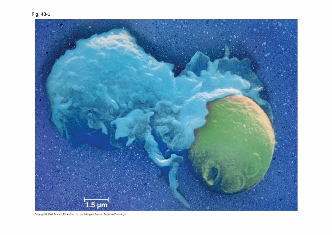
Fig. 43-1
1.5 µm

Copyright © 2008 Pearson Education, Inc., publishing as Pearson Benjamin Cummings
• Innate immunity is present before any exposure to pathogens and is effective from the time of birth
• It involves nonspecific responses to pathogens
• Innate immunity consists of external barriers plus internal cellular and chemical defenses
Copyright © 2008 Pearson Education, Inc., publishing as Pearson Benjamin Cummings
• Acquired immunity , or adaptive immunity, develops after exposure to agents such as microbes, toxins, or other foreign substances
• It involves a very specific response to pathogens

Fig. 43-2
INNATE IMMUNITY
Recognition of traitsshared by broad rangesof pathogens, using asmall set of receptors
•
•Rapid response
•Recognition of traitsspecific to particularpathogens, using a vastarray of receptors
•Slower response
ACQUIRED IMMUNITY
Pathogens(microorganisms
and viruses)
Barrier defenses:SkinMucous membranesSecretions
Internal defenses:Phagocytic cellsAntimicrobial proteinsInflammatory responseNatural killer cells
Humoral response:Antibodies defend againstinfection in body fluids.
Cell-mediated response:Cytotoxic lymphocytes defendagainst infection in body cells.

Copyright © 2008 Pearson Education, Inc., publishing as Pearson Benjamin Cummings
Concept 43.1: In innate immunity, recognition and response rely on shared traits of pathogens
• Both invertebrates and vertebrates depend on innate immunity to fight infection
• Vertebrates also develop acquired immune defenses
Copyright © 2008 Pearson Education, Inc., publishing as Pearson Benjamin Cummings
Innate Immunity of Invertebrates
• In insects, an exoskeleton made of chitin forms the first barrier to pathogens
• The digestive system is protected by low pH and lysozyme , an enzyme that digests microbial cell walls
• Hemocytes circulate within hemolymph and carry out phagocytosis , the ingestion and digestion of foreign substances including bacteria

Fig. 43-3
Microbes
PHAGOCYTIC CELL
Vacuole
Lysosomecontaining enzymes
Copyright © 2008 Pearson Education, Inc., publishing as Pearson Benjamin Cummings
• Hemocytes also secrete antimicrobial peptides that disrupt the plasma membranes of bacteria

Fig. 43-4

Copyright © 2008 Pearson Education, Inc., publishing as Pearson Benjamin Cummings
• The immune system recognizes bacteria and fungi by structures on their cell walls
• An immune response varies with the class of pathogen encountered

Fig. 43-5RESULTS
% s
urvi
val
Wild type
Fruit fly survival after infection by N. crassa fungi
100
75
50
25
00 24 7248 96 120
100
75
50
25
00 24 7248 96 120
Hours post-infection
Fruit fly survival after infection by M. luteus bacteria
Hours post-infection
% s
urvi
val
Mutant + drosomycin
Mutant + defensinMutant
Wild type
Mutant +drosomycin
Mutant +defensin
Mutant

Fig. 43-5a
RESULTS%
sur
viva
l
Wild type
Fruit fly survival after infection by N. crassa fungi
100
75
50
25
00 24 7248 96 120
Hours post-infection
Mutant + drosomycin
Mutant + defensinMutant

Fig. 43-5b
100
75
50
25
00 24 7248 96 120
Fruit fly survival after infection by M. luteus bacteria
Hours post-infection
% s
urvi
val Wild type
Mutant +drosomycin
Mutant +defensin
Mutant
RESULTS

Copyright © 2008 Pearson Education, Inc., publishing as Pearson Benjamin Cummings
Innate Immunity of Vertebrates
• The immune system of mammals is the best understood of the vertebrates
• Innate defenses include barrier defenses, phagocytosis, antimicrobial peptides
• Additional defenses are unique to vertebrates: the inflammatory response and natural killer cells

Copyright © 2008 Pearson Education, Inc., publishing as Pearson Benjamin Cummings
Barrier Defenses
• Barrier defenses include the skin and mucous membranes of the respiratory, urinary, and reproductive tracts
• Mucus traps and allows for the removal of microbes
• Many body fluids including saliva, mucus, and tears are hostile to microbes
• The low pH of skin and the digestive system prevents growth of microbes

Copyright © 2008 Pearson Education, Inc., publishing as Pearson Benjamin Cummings
Cellular Innate Defenses
• White blood cells (leukocytes) engulf pathogens in the body
• Groups of pathogens are recognized by TLR, Toll-like receptors

Fig. 43-6
EXTRACELLULARFLUID Lipopolysaccharide
FlagellinTLR4
TLR5
Helperprotein
TLR9
TLR3
WHITEBLOODCELL
VESICLE
CpG DNA
ds RNA
Inflammatoryresponses

Copyright © 2008 Pearson Education, Inc., publishing as Pearson Benjamin Cummings
• A white blood cell engulfs a microbe, then fuses with a lysosome to destroy the microbe
• There are different types of phagocytic cells:
– Neutrophils engulf and destroy microbes
– Macrophages are part of the lymphatic system and are found throughout the body
– Eosinophils discharge destructive enzymes
– Dendritic cells stimulate development of acquired immunity

Fig. 43-7
Adenoid
Tonsil
Lymphnodes
Spleen
Peyer’s patches(small intestine)
Appendix
Lymphaticvessels Lymph
nodeMasses ofdefensive cells
Bloodcapillary
Lymphaticvessel
Tissuecells
Interstitial fluid

Copyright © 2008 Pearson Education, Inc., publishing as Pearson Benjamin Cummings
Antimicrobial Peptides and Proteins
• Peptides and proteins function in innate defense by attacking microbes directly or impeding their reproduction
• Interferon proteins provide innate defense against viruses and help activate macrophages
• About 30 proteins make up the complement system , which causes lysis of invading cells and helps trigger inflammation

Copyright © 2008 Pearson Education, Inc., publishing as Pearson Benjamin Cummings
Inflammatory Responses
• Following an injury, mast cells release histamine , which promotes changes in blood vessels; this is part of the inflammatory response
• These changes increase local blood supply and allow more phagocytes and antimicrobial proteins to enter tissues
• Pus, a fluid rich in white blood cells, dead microbes, and cell debris, accumulates at the site of inflammation

Fig. 43-8-1
Pathogen Splinter
Macrophage
Mast cell
Chemicalsignals
Capillary
Phagocytic cellRed blood cells

Fig. 43-8-2
Pathogen Splinter
Macrophage
Mast cell
Chemicalsignals
Capillary
Phagocytic cellRed blood cells
Fluid

Fig. 43-8-3
Pathogen Splinter
Macrophage
Mast cell
Chemicalsignals
Capillary
Phagocytic cellRed blood cells
Fluid
Phagocytosis

Copyright © 2008 Pearson Education, Inc., publishing as Pearson Benjamin Cummings
• Inflammation can be either local or systemic (throughout the body)
• Fever is a systemic inflammatory response triggered by pyrogens released by macrophages, and toxins from pathogens
• Septic shock is a life-threatening condition caused by an overwhelming inflammatory response

Copyright © 2008 Pearson Education, Inc., publishing as Pearson Benjamin Cummings
Natural Killer Cells
• All cells in the body (except red blood cells) have a class 1 MHC protein on their surface
• Cancerous or infected cells no longer express this protein; natural killer (NK) cells attack these damaged cells

Copyright © 2008 Pearson Education, Inc., publishing as Pearson Benjamin Cummings
Innate Immune System Evasion by Pathogens
• Some pathogens avoid destruction by modifying their surface to prevent recognition or by resisting breakdown following phagocytosis
• Tuberculosis (TB) is one such disease and kills more than a million people a year

Copyright © 2008 Pearson Education, Inc., publishing as Pearson Benjamin Cummings
Concept 43.2: In acquired immunity, lymphocyte receptors provide pathogen-specific recognition
• White blood cells called lymphocytes recognize and respond to antigens, foreign molecules
• Lymphocytes that mature in the thymus above the heart are called T cells , and those that mature in bone marrow are called B cells

Copyright © 2008 Pearson Education, Inc., publishing as Pearson Benjamin Cummings
• Lymphocytes contribute to immunological memory, an enhanced response to a foreign molecule encountered previously
• Cytokines are secreted by macrophages and dendritic cells to recruit and activate lymphocytes
Copyright © 2008 Pearson Education, Inc., publishing as Pearson Benjamin Cummings
Acquired Immunity: An Overview
• B cells and T cells have receptor proteins that can bind to foreign molecules
• Each individual lymphocyte is specialized to recognize a specific type of molecule

Copyright © 2008 Pearson Education, Inc., publishing as Pearson Benjamin Cummings
Antigen Recognition by Lymphocytes
• An antigen is any foreign molecule to which a lymphocyte responds
• A single B cell or T cell has about 100,000 identical antigen receptors

Fig. 43-9
Antigen-bindingsite
Antigen-binding site
Antigen-bindingsite
Disulfidebridge
Variableregions
Constantregions
Transmembraneregion
Plasmamembrane
Lightchain
Heavy chains
T cell
αααα chain ββββ chain
Disulfide bridge
Cytoplasm of T cell
(b) T cell receptor
Cytoplasm of B cell
(a) B cell receptor
B cell
V
V
C C
V
V
C C C C
VV

Fig. 43-9a
Antigen-bindingsite
Antigen-binding site
Disulfidebridge
Variableregions
Constantregions
Transmembraneregion
Plasmamembrane
Lightchain
Heavy chains
Cytoplasm of B cell
(a) B cell receptor
B cell
V
V
C C
V
V
C C

Fig. 43-9b
Antigen-bindingsite
Variableregions
Constantregions
Transmembraneregion
Plasmamembrane
T cell
αααα chain ββββ chain
Disulfide bridge
Cytoplasm of T cell
(b) T cell receptor
C C
VV
Copyright © 2008 Pearson Education, Inc., publishing as Pearson Benjamin Cummings
• All antigen receptors on a single lymphocyte recognize the same epitope, or antigenic determinant, on an antigen
• B cells give rise to plasma cells , which secrete proteins called antibodies or immunoglobulins
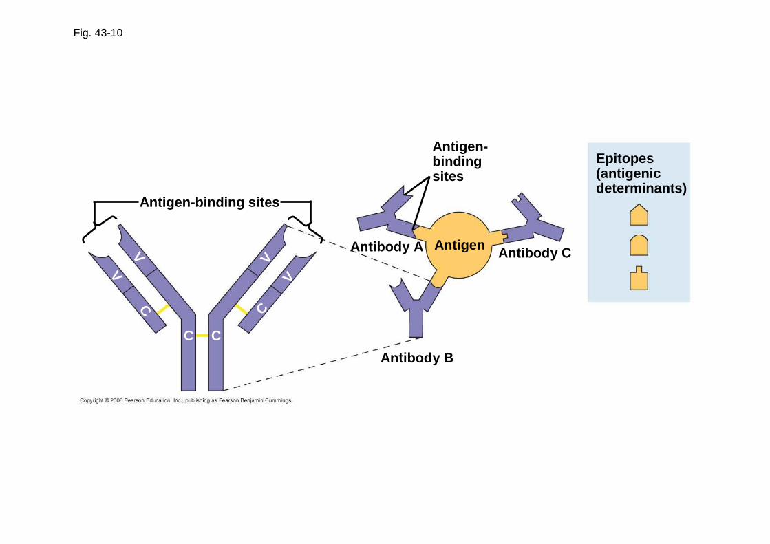
Fig. 43-10
Antigen-binding sites
Antigen-bindingsites
Epitopes(antigenicdeterminants)
Antigen
Antibody B
Antibody CAntibody A
CC
CV
V
V
V
C
Copyright © 2008 Pearson Education, Inc., publishing as Pearson Benjamin Cummings
The Antigen Receptors of B Cells and T Cells
• B cell receptors bind to specific, intact antigens
• The B cell receptor consists of two identical heavy chains and two identical light chains
• The tips of the chains form a constant (C) region, and each chain contains a variable (V) region, so named because its amino acid sequence varies extensively from one B cell to another

Copyright © 2008 Pearson Education, Inc., publishing as Pearson Benjamin Cummings
• Secreted antibodies, or immunoglobulins, are structurally similar to B cell receptors but lack transmembrane regions that anchor receptors in the plasma membrane

Copyright © 2008 Pearson Education, Inc., publishing as Pearson Benjamin Cummings
• Each T cell receptor consists of two different polypeptide chains
• The tips of the chain form a variable (V) region; the rest is a constant (C) region
• T cells can bind to an antigen that is free or on the surface of a pathogen
Video: TVideo: T Cell ReceptorsCell Receptors
Copyright © 2008 Pearson Education, Inc., publishing as Pearson Benjamin Cummings
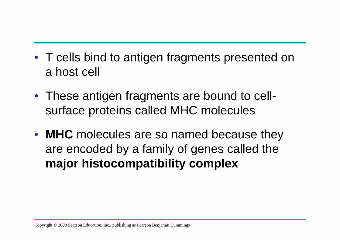
• T cells bind to antigen fragments presented on a host cell
• These antigen fragments are bound to cell-surface proteins called MHC molecules
• MHC molecules are so named because they are encoded by a family of genes called the major histocompatibility complex

Copyright © 2008 Pearson Education, Inc., publishing as Pearson Benjamin Cummings
The Role of the MHC
• In infected cells, MHC molecules bind and transport antigen fragments to the cell surface, a process called antigen presentation
• A nearby T cell can then detect the antigen fragment displayed on the cell’s surface
• Depending on their source, peptide antigens are handled by different classes of MHC molecules
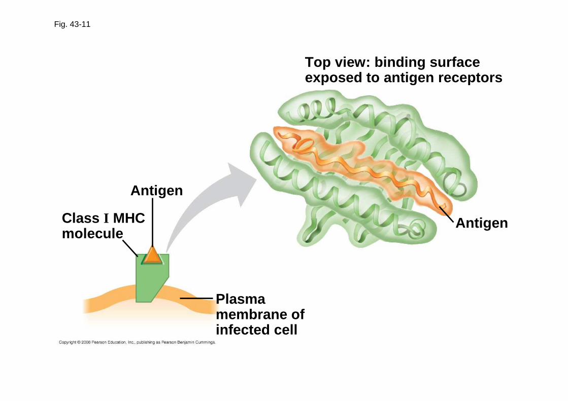
Fig. 43-11
Antigen
Top view: binding surfaceexposed to antigen receptors
Plasmamembrane ofinfected cell
AntigenClass I MHCmolecule

Copyright © 2008 Pearson Education, Inc., publishing as Pearson Benjamin Cummings
• Class I MHC molecules are found on almost all nucleated cells of the body
• They display peptide antigens to cytotoxic T cells
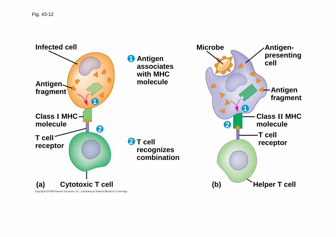
Fig. 43-12
Infected cell
Antigenfragment
Class I MHCmolecule
T cellreceptor
(a)
Antigenassociateswith MHCmolecule
T cellrecognizescombination
Cytotoxic T cell (b) Helper T cell
T cellreceptor
Class II MHCmolecule
Antigenfragment
Antigen-presentingcell
Microbe
1
11
2
22

Copyright © 2008 Pearson Education, Inc., publishing as Pearson Benjamin Cummings
• Class II MHC molecules are located mainly on dendritic cells, macrophages, and B cells
• Dendritic cells, macrophages, and B cells are antigen-presenting cells that display antigens to cytotoxic T cells and helper T cells

Copyright © 2008 Pearson Education, Inc., publishing as Pearson Benjamin Cummings
Lymphocyte Development
• The acquired immune system has three important properties:
– Receptor diversity
– A lack of reactivity against host cells
– Immunological memory

Copyright © 2008 Pearson Education, Inc., publishing as Pearson Benjamin Cummings
Generation of Lymphocyte Diversity by Gene Rearrangement
• Differences in the variable region account for specificity of antigen receptors
• The immunoglobulin (Ig) gene encodes one chain of the B cell receptor
• Many different chains can be produced from the same Ig chain gene by rearrangement of the DNA
• Rearranged DNA is transcribed and translated and the antigen receptor formed

Fig. 43-13
DNA of undifferentiated B cell
1
DNA of differentiated B cell
pre-mRNA
mRNA
Light-chain polypeptide
Variableregion
Constantregion
Translation
B cell
B cell receptor
RNA processing
Transcription
DNA deleted between randomly selected V and Jsegments
Functional gene
V37 V38 V39 V40 J1 J2 J3 J4 CJ5 Intron
V37 V38 V39 CJ5 Intron
V39 CJ5 Intron
V39 CJ5 Poly-A tailCap
CV
VV
VV
C C
C C
2
3
4

Copyright © 2008 Pearson Education, Inc., publishing as Pearson Benjamin Cummings
Origin of Self-Tolerance
• Antigen receptors are generated by random rearrangement of DNA
• As lymphocytes mature in bone marrow or the thymus, they are tested for self-reactivity
• Lymphocytes with receptors specific for the body’s own molecules are destroyed by apoptosis, or rendered nonfunctional

Copyright © 2008 Pearson Education, Inc., publishing as Pearson Benjamin Cummings
Amplifying Lymphocytes by Clonal Selection
• In the body there are few lymphocytes with antigen receptors for any particular epitope
• The binding of a mature lymphocyte to an antigen induces the lymphocyte to divide rapidly
• This proliferation of lymphocytes is called clonal selection
• Two types of clones are produced: short-lived activated effector cells and long-lived memory cells

Fig. 43-14
B cells thatdiffer inantigen specificity
Antibodymolecules
Antigenreceptor
Antigen molecules
Clone of memory cells Clone of plasma cells
Copyright © 2008 Pearson Education, Inc., publishing as Pearson Benjamin Cummings
• The first exposure to a specific antigen represents the primary immune response
• During this time, effector B cells called plasma cells are generated, and T cells are activated to their effector forms
• In the secondary immune response , memory cells facilitate a faster, more efficient response
Animation: RAnimation: R ole of B Cellsole of B Cells

Fig. 43-15
Antibodiesto A
Antibodiesto B
Secondary immune response toantigen A produces antibodies to A;primary immune response to antigenB produces antibodies to B.
Primary immune responseto antigen A producesantibodies to A.
Ant
ibod
y co
ncen
trat
ion
(arb
itrar
y un
its)
Exposureto antigen A
Exposure toantigens A and B
Time (days)
104
103
102
101
100
0 7 14 21 28 35 42 49 56
Copyright © 2008 Pearson Education, Inc., publishing as Pearson Benjamin Cummings
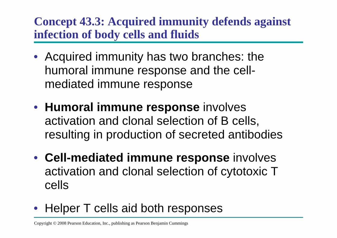
Concept 43.3: Acquired immunity defends against infection of body cells and fluids
• Acquired immunity has two branches: the humoral immune response and the cell-mediated immune response
• Humoral immune response involves activation and clonal selection of B cells, resulting in production of secreted antibodies
• Cell-mediated immune response involves activation and clonal selection of cytotoxic T cells
• Helper T cells aid both responses

Fig. 43-16
Humoral (antibody-mediated) immune response
B cell
Plasma cells
Cell-mediated immune response
Key
Stimulates
Gives rise to
+
+
++
+
+
+Memory B cells
Antigen (1st exposure)
Engulfed by
Antigen-presenting cell
MemoryHelper T cells
Helper T cell Cytotoxic T cell
MemoryCytotoxic T cells
ActiveCytotoxic T cells
Antigen (2nd exposure)
Secretedantibodies
Defend against extracellular pathogens by binding to antigens,thereby neutralizing pathogens or making them bette r targetsfor phagocytes and complement proteins.
Defend against intracellular pathogensand cancer by binding to and lysing theinfected cells or cancer cells.
+
+ +

Fig. 43-16a
KeyStimulatesGives rise to
+
MemoryHelper T cells
Antigen-presenting cell
Helper T cell
Engulfed by
Antigen (1st exposure)
+
+
+
+ +
+
Defend against extracellular pathogens
MemoryB cells
Antigen (2nd exposure)
Plasma cells
B cell
Secretedantibodies
Humoral (antibody-mediated) immune response

Fig. 43-16bCell-mediated immune response
Defend against intracellular pathogens
ActiveCytotoxic T cells
MemoryCytotoxic T cells
MemoryHelper T cells
Antigen-presenting cell
Antigen (2nd exposure)
Helper T cell
Engulfed by
Antigen (1st exposure)
Cytotoxic T cell
KeyStimulatesGives rise to
+
+
+
+
+ +
+
Copyright © 2008 Pearson Education, Inc., publishing as Pearson Benjamin Cummings
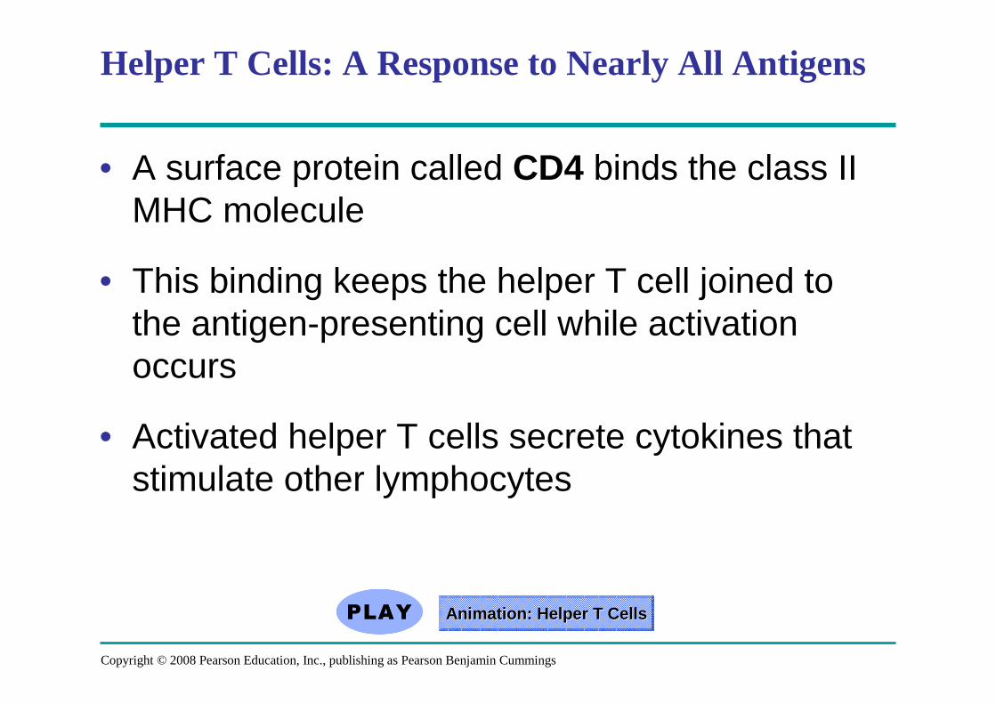
Helper T Cells: A Response to Nearly All Antigens
• A surface protein called CD4 binds the class II MHC molecule
• This binding keeps the helper T cell joined to the antigen-presenting cell while activation occurs
• Activated helper T cells secrete cytokines that stimulate other lymphocytes
Animation: HAnimation: H elper T Cellselper T Cells

Fig. 43-17
Antigen-presentingcell
Peptide antigen
Cell-mediatedimmunity (attack on
infected cells)
Class II MHC moleculeCD4TCR (T cell receptor)
Helper T cell
Humoralimmunity
(secretion ofantibodies byplasma cells) Cytotoxic T cell
Cytokines
B cell
Bacterium
+
+ +
+

Copyright © 2008 Pearson Education, Inc., publishing as Pearson Benjamin Cummings
Cytotoxic T Cells: A Response to Infected Cells
• Cytotoxic T cells are the effector cells in cell-mediated immune response
• Cytotoxic T cells make CD8, a surface protein that greatly enhances interaction between a target cell and a cytotoxic T cell
• Binding to a class I MHC complex on an infected cell activates a cytotoxic T cell and makes it an active killer
• The activated cytotoxic T cell secretes proteins that destroy the infected target cell
Animation: Animation: CCytotoxic ytotoxic T CellsT Cells
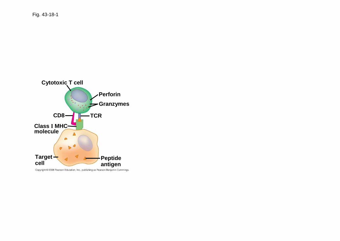
Fig. 43-18-1
Cytotoxic T cell
Perforin
Granzymes
TCRCD8
Class I MHCmolecule
Targetcell
Peptideantigen
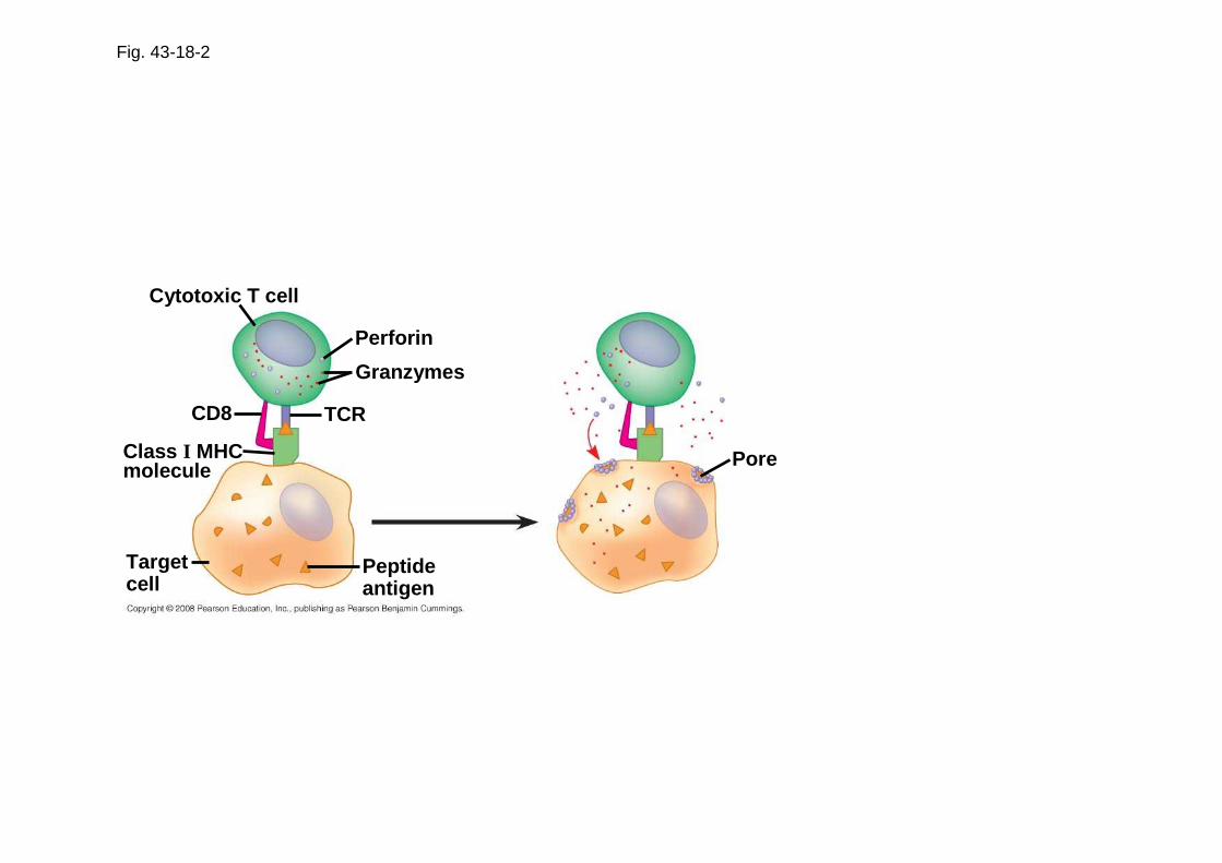
Fig. 43-18-2
Cytotoxic T cell
Perforin
Granzymes
TCRCD8
Class I MHCmolecule
Targetcell
Peptideantigen
Pore
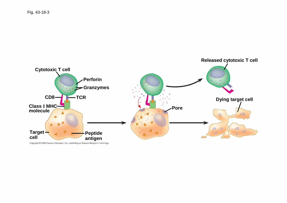
Fig. 43-18-3
Cytotoxic T cell
Perforin
Granzymes
TCRCD8
Class I MHCmolecule
Targetcell
Peptideantigen
Pore
Released cytotoxic T cell
Dying target cell

Copyright © 2008 Pearson Education, Inc., publishing as Pearson Benjamin Cummings
B Cells: A Response to Extracellular Pathogens
• The humoral response is characterized by secretion of antibodies by B cells
• Activation of B cells is aided by cytokines and antigen binding to helper T cells
• Clonal selection of B cells generates antibody-secreting plasma cells, the effector cells of humoral immunity

Fig. 43-19
Antigen-presenting cell
Endoplasmicreticulum ofplasma cell
Secretedantibodymolecules
Bacterium
B cellPeptideantigen
Class II MHCmolecule
TCR CD4
Helper T cellActivatedhelper T cell
Cytokines
Clone of memoryB cells
Clone of plasma cells
2 µm
+

Fig. 43-19-1
Antigen-presenting cell Bacterium
Peptideantigen
Class II MHCmolecule
TCR CD4
Helper T cell

Fig. 43-19-2
Antigen-presenting cell Bacterium
Peptideantigen
Class II MHCmolecule
TCR CD4
Helper T cell
B cell
Activatedhelper T cell
Cytokines
+

Fig. 43-19-3
Antigen-presenting cell Bacterium
Peptideantigen
Class II MHCmolecule
TCR CD4
Helper T cell
B cell
Activatedhelper T cell
Cytokines
+ Secretedantibodymolecules
Clone of memoryB cells
Clone of plasma cells

Fig. 43-19a
Endoplasmicreticulum ofplasma cell
2 µm

Copyright © 2008 Pearson Education, Inc., publishing as Pearson Benjamin Cummings
Antibody Classes
• The five major classes of antibodies, or immunoglobulins, differ in distribution and function
• Polyclonal antibodies are the products of many different clones of B cells following exposure to a microbial antigen
• Monoclonal antibodies are prepared from a single clone of B cells grown in culture
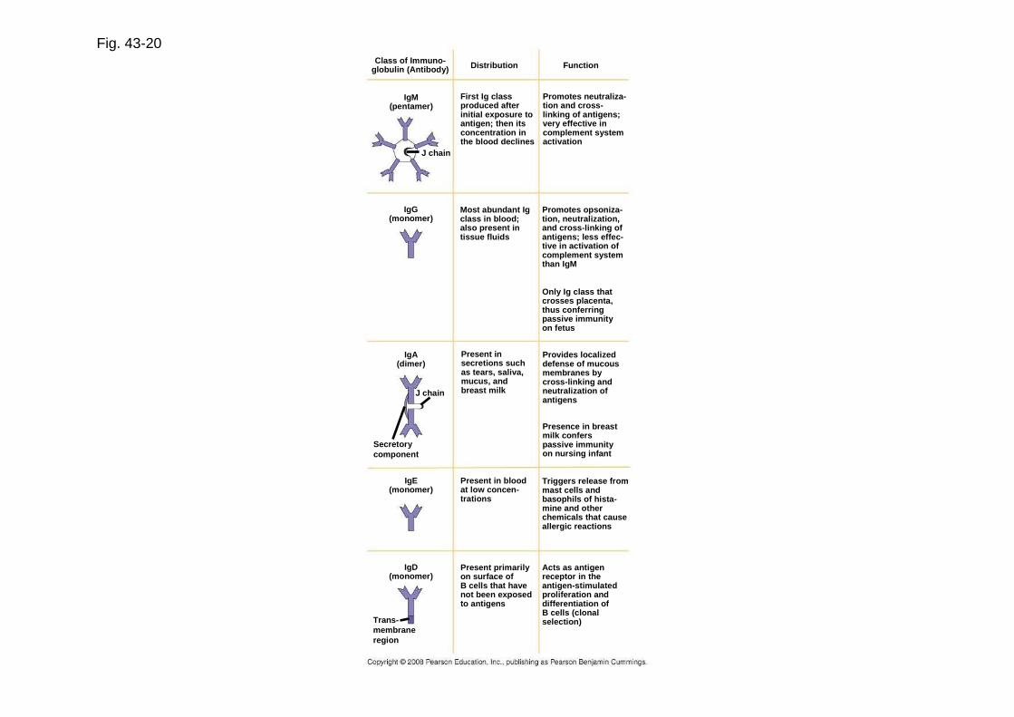
Fig. 43-20Class of Immuno-
globulin (Antibody)
IgG(monomer)
IgM(pentamer)
J chain
IgA(dimer)
IgE(monomer)
IgD(monomer)
Trans-membraneregion
J chain
Secretorycomponent
Distribution Function
First Ig classproduced afterinitial exposure toantigen; then itsconcentration inthe blood declines
Promotes neutraliza-tion and cross-linking of antigens;very effective incomplement systemactivation
Present insecretions suchas tears, saliva,mucus, andbreast milk
Only Ig class thatcrosses placenta,thus conferringpassive immunityon fetus
Triggers release frommast cells andbasophils of hista-mine and otherchemicals that causeallergic reactions
Present primarilyon surface ofB cells that havenot been exposedto antigens
Acts as antigenreceptor in theantigen-stimulatedproliferation anddifferentiation ofB cells (clonalselection)
Most abundant Igclass in blood;also present intissue fluids
Promotes opsoniza-tion, neutralization,and cross-linking ofantigens; less effec-tive in activation ofcomplement systemthan IgM
Provides localizeddefense of mucousmembranes bycross-linking andneutralization of antigens
Presence in breastmilk conferspassive immunityon nursing infant
Present in bloodat low concen-trations
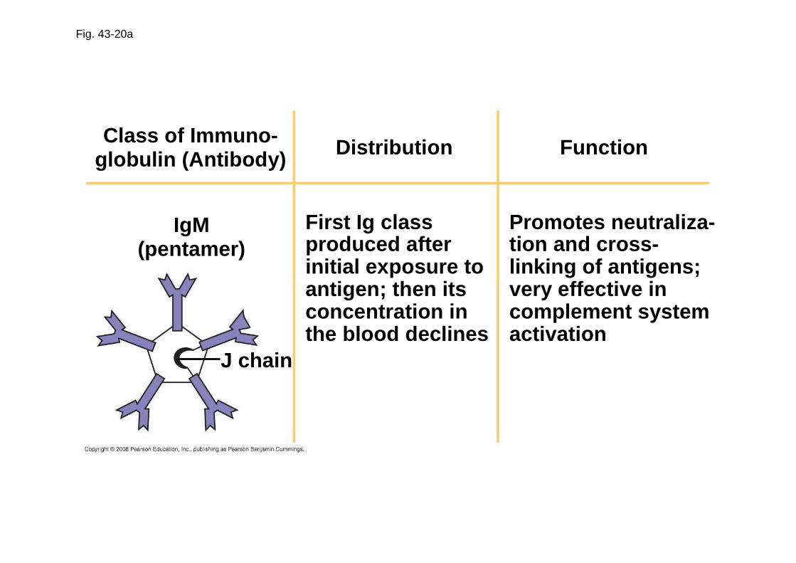
Fig. 43-20a
DistributionClass of Immuno-globulin (Antibody)
IgM(pentamer)
J chain
First Ig classproduced afterinitial exposure toantigen; then itsconcentration inthe blood declines
Promotes neutraliza-tion and cross-linking of antigens;very effective incomplement systemactivation
Function

Fig. 43-20b
Distribution FunctionClass of Immuno-globulin (Antibody)
IgG(monomer)
Most abundant Igclass in blood;also present intissue fluids
Promotes opsoniza-tion, neutralization,and cross-linking ofantigens; less effec-tive in activation ofcomplement systemthan IgM
Only Ig class thatcrosses placenta,thus conferringpassive immunityon fetus
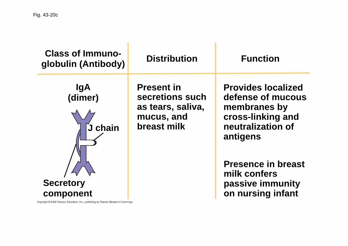
Fig. 43-20c
Distribution FunctionClass of Immuno-globulin (Antibody)
IgA(dimer)
J chain
Secretorycomponent
Present insecretions suchas tears, saliva,mucus, andbreast milk
Provides localizeddefense of mucousmembranes bycross-linking andneutralization ofantigens
Presence in breastmilk conferspassive immunityon nursing infant

Fig. 43-20d
Distribution FunctionClass of Immuno-globulin (Antibody)
IgE(monomer)
Present in bloodat low concen-trations
Triggers release frommast cells andbasophils of hista-mine and otherchemicals that causeallergic reactions
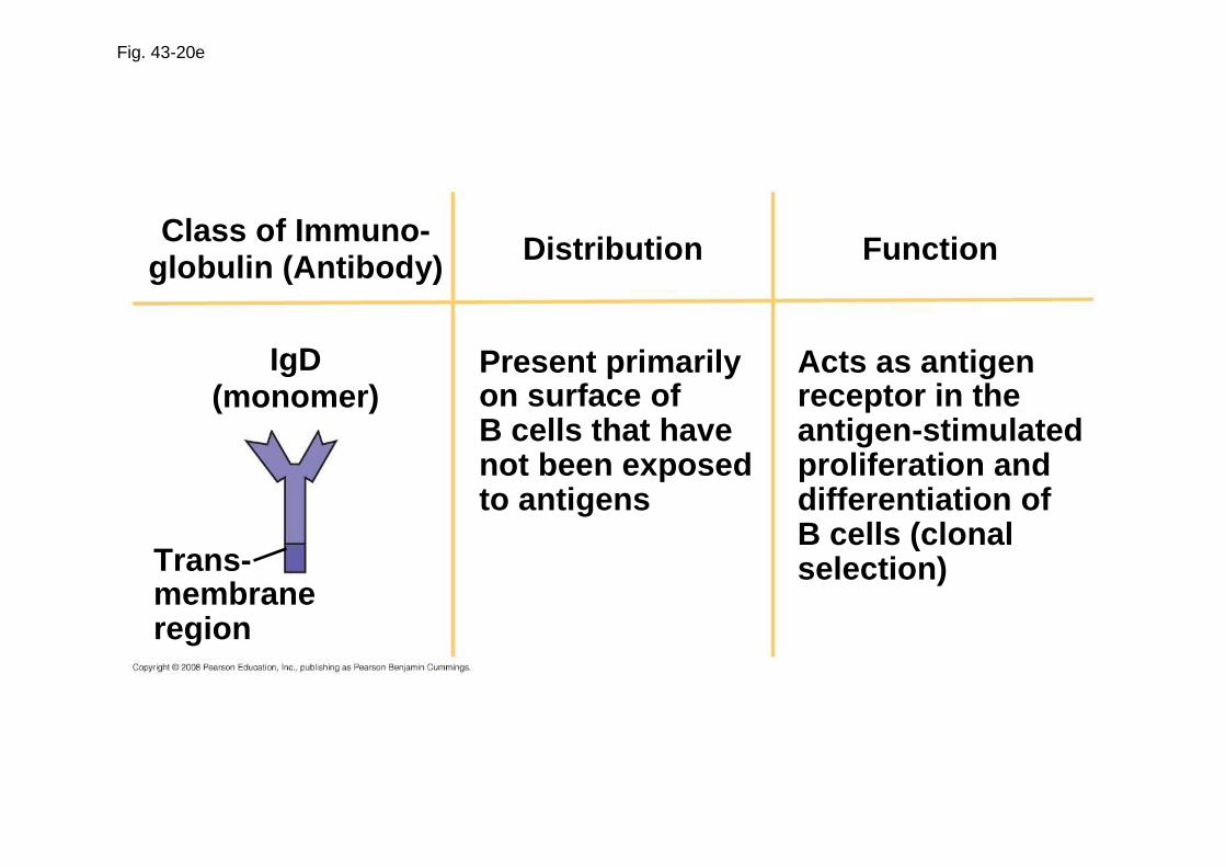
Fig. 43-20e
Distribution FunctionClass of Immuno-globulin (Antibody)
IgD(monomer)
Trans-membraneregion
Present primarilyon surface ofB cells that havenot been exposedto antigens
Acts as antigenreceptor in theantigen-stimulatedproliferation anddifferentiation ofB cells (clonalselection)
Copyright © 2008 Pearson Education, Inc., publishing as Pearson Benjamin Cummings
The Role of Antibodies in Immunity
• Neutralization occurs when a pathogen can no longer infect a host because it is bound to an antibody
• Opsonization occurs when antibodies bound to antigens increase phagocytosis
• Antibodies together with proteins of the complement system generate a membrane attack complex and cell lysis
Animation: AAnimation: A ntibodiesntibodies
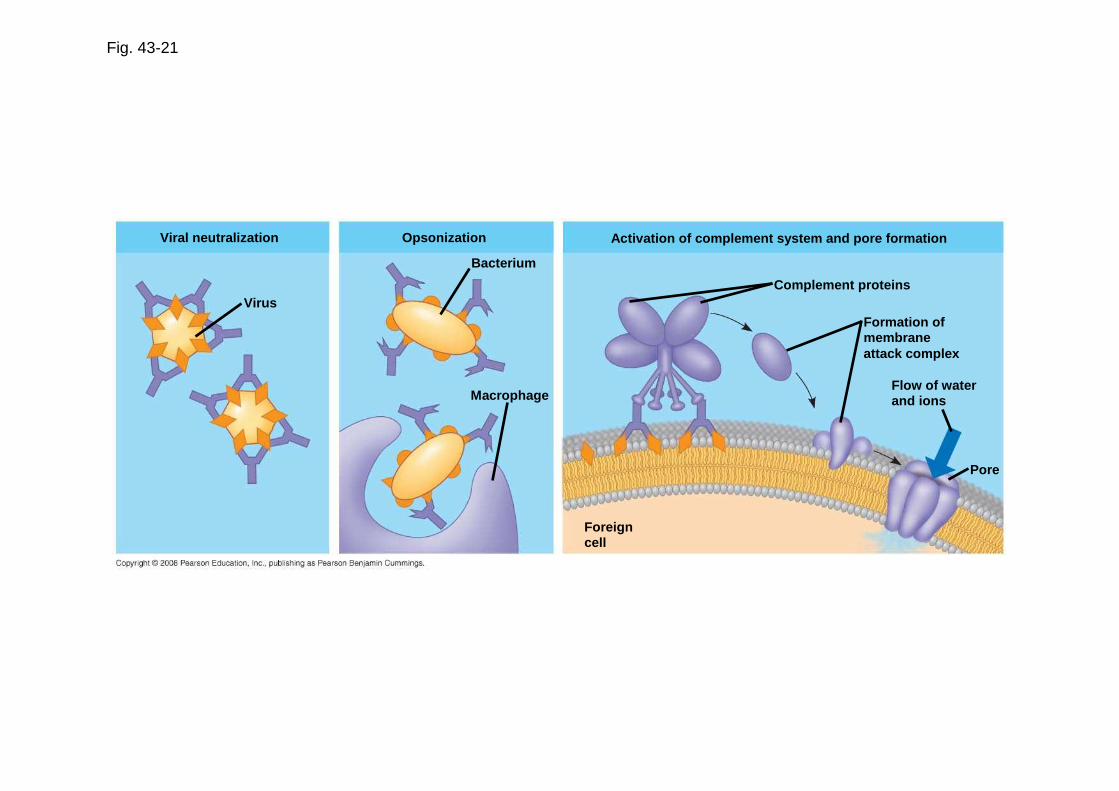
Fig. 43-21
Viral neutralization
Virus
Opsonization
Bacterium
Macrophage
Activation of complement system and pore formation
Complement proteins
Formation ofmembraneattack complex
Flow of waterand ions
Pore
Foreigncell
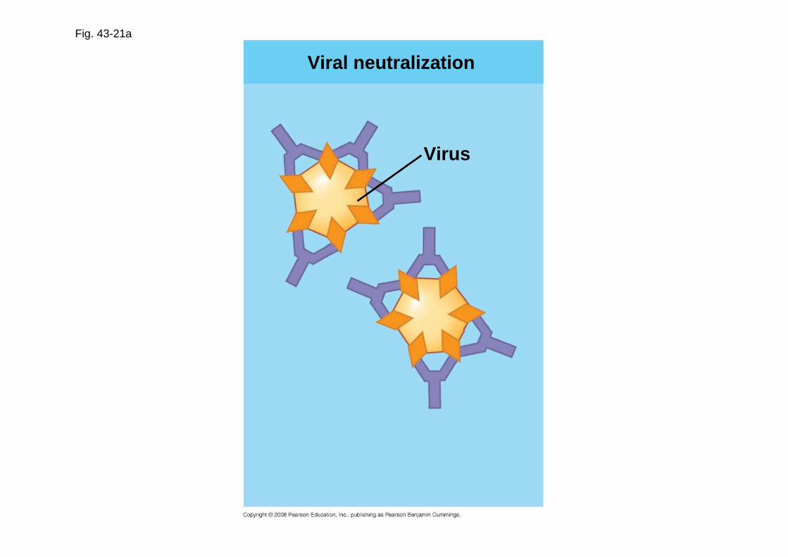
Fig. 43-21a
Viral neutralization
Virus

Fig. 43-21b
Opsonization
Bacterium
Macrophage
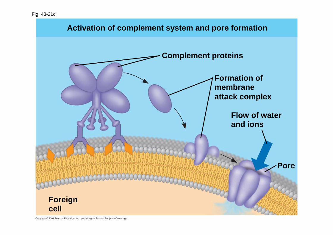
Fig. 43-21c
Activation of complement system and pore formation
Complement proteins
Formation ofmembraneattack complex
Flow of waterand ions
Pore
Foreigncell

Copyright © 2008 Pearson Education, Inc., publishing as Pearson Benjamin Cummings
Active and Passive Immunization
• Active immunity develops naturally in response to an infection
• It can also develop following immunization , also called vaccination
• In immunization, a nonpathogenic form of a microbe or part of a microbe elicits an immune response to an immunological memory
Copyright © 2008 Pearson Education, Inc., publishing as Pearson Benjamin Cummings
• Passive immunity provides immediate, short-term protection
• It is conferred naturally when IgG crosses the placenta from mother to fetus or when IgA passes from mother to infant in breast milk
• It can be conferred artificially by injecting antibodies into a nonimmune person

Fig. 43-22

Copyright © 2008 Pearson Education, Inc., publishing as Pearson Benjamin Cummings
Immune Rejection
• Cells transferred from one person to another can be attacked by immune defenses
• This complicates blood transfusions or the transplant of tissues or organs

Copyright © 2008 Pearson Education, Inc., publishing as Pearson Benjamin Cummings
Blood Groups
• Antigens on red blood cells determine whether a person has blood type A (A antigen), B (B antigen), AB (both A and B antigens), or O (neither antigen)
• Antibodies to nonself blood types exist in the body
• Transfusion with incompatible blood leads to destruction of the transfused cells
• Recipient-donor combinations can be fatal or safe

Copyright © 2008 Pearson Education, Inc., publishing as Pearson Benjamin Cummings
Tissue and Organ Transplants
• MHC molecules are different among genetically nonidentical individuals
• Differences in MHC molecules stimulate rejection of tissue grafts and organ transplants

Copyright © 2008 Pearson Education, Inc., publishing as Pearson Benjamin Cummings
• Chances of successful transplantation increase if donor and recipient MHC tissue types are well matched
• Immunosuppressive drugs facilitate transplantation
• Lymphocytes in bone marrow transplants may cause the donor tissue to reject the recipient

Copyright © 2008 Pearson Education, Inc., publishing as Pearson Benjamin Cummings
Concept 43.4: Disruption in immune system function can elicit or exacerbate disease
• Some pathogens have evolved to diminish the effectiveness of host immune responses

Copyright © 2008 Pearson Education, Inc., publishing as Pearson Benjamin Cummings
Exaggerated, Self-Directed, and Diminished Immune Responses
• If the delicate balance of the immune system is disrupted, effects range from minor to often fatal

Copyright © 2008 Pearson Education, Inc., publishing as Pearson Benjamin Cummings
Allergies
• Allergies are exaggerated (hypersensitive) responses to antigens called allergens
• In localized allergies such as hay fever, IgE antibodies produced after first exposure to an allergen attach to receptors on mast cells

Fig. 43-23
Allergen
IgE
Granule
Mast cell
Histamine
Copyright © 2008 Pearson Education, Inc., publishing as Pearson Benjamin Cummings
• The next time the allergen enters the body, it binds to mast cell–associated IgE molecules
• Mast cells release histamine and other mediators that cause vascular changes leading to typical allergy symptoms
• An acute allergic response can lead to anaphylactic shock, a life-threatening reaction that can occur within seconds of allergen exposure

Copyright © 2008 Pearson Education, Inc., publishing as Pearson Benjamin Cummings
Autoimmune Diseases
• In individuals with autoimmune diseases , the immune system loses tolerance for self and turns against certain molecules of the body
• Autoimmune diseases include systemic lupus erythematosus, rheumatoid arthritis, insulin-dependent diabetes mellitus, and multiple sclerosis

Fig. 43-24

Copyright © 2008 Pearson Education, Inc., publishing as Pearson Benjamin Cummings
Exertion, Stress, and the Immune System
• Moderate exercise improves immune system function
• Psychological stress has been shown to disrupt hormonal, nervous, and immune systems

Copyright © 2008 Pearson Education, Inc., publishing as Pearson Benjamin Cummings
Immunodeficiency Diseases
• Inborn immunodeficiency results from hereditary or developmental defects that prevent proper functioning of innate, humoral, and/or cell-mediated defenses
• Acquired immunodeficiency results from exposure to chemical and biological agents
• Acquired immunodeficiency syndrome (AIDS) is caused by a virus
Copyright © 2008 Pearson Education, Inc., publishing as Pearson Benjamin Cummings
Acquired Immune System Evasion by Pathogens
• Pathogens have evolved mechanisms to attack immune responses

Copyright © 2008 Pearson Education, Inc., publishing as Pearson Benjamin Cummings
Antigenic Variation
• Through antigenic variation, some pathogens are able to change epitope expression and prevent recognition
• The human influenza virus mutates rapidly, and new flu vaccines must be made each year
• Human viruses occasionally exchange genes with the viruses of domesticated animals
• This poses a danger as human immune systems are unable to recognize the new viral strain

Fig. 43-25
Weeks after infection
Mill
ions
of p
aras
ites
per
mL
of b
lood
Antibodies tovariant 1appear
Antibodies tovariant 2appear
Antibodies tovariant 3appear
Variant 3Variant 2Variant 1
25 26 27 280
0.5
1.0
1.5

Copyright © 2008 Pearson Education, Inc., publishing as Pearson Benjamin Cummings
Latency
• Some viruses may remain in a host in an inactive state called latency
• Herpes simplex viruses can be present in a human host without causing symptoms

Copyright © 2008 Pearson Education, Inc., publishing as Pearson Benjamin Cummings
Attack on the Immune System: HIV
• Human immunodeficiency virus (HIV) infects helper T cells
• The loss of helper T cells impairs both the humoral and cell-mediated immune responses and leads to AIDS
• HIV eludes the immune system because of antigenic variation and an ability to remain latent while integrated into host DNA
Animation: HAnimation: H IV Reproductive CycleIV Reproductive Cycle

Fig. 43-26
Latency
Relative antibodyconcentration
AIDS
Hel
per
T c
ell c
once
ntra
tion
in b
lood
(ce
lls/m
m3 )
Helper T cellconcentration
Relative HIVconcentration
Years after untreated infection0 1 2 3 4 5 6 7 8 9 10
0
200
400
600
800

Copyright © 2008 Pearson Education, Inc., publishing as Pearson Benjamin Cummings
• People with AIDS are highly susceptible to opportunistic infections and cancers that take advantage of an immune system in collapse
• The spread of HIV is a worldwide problem
• The best approach for slowing this spread is education about practices that transmit the virus

Copyright © 2008 Pearson Education, Inc., publishing as Pearson Benjamin Cummings
Cancer and Immunity
• The frequency of certain cancers increases when the immune response is impaired
• Two suggested explanations are
– Immune system normally suppresses cancerous cells
– Increased inflammation increases the risk of cancer

Fig. 43-UN1Stem cell
Cell division and gene rearrangement
Antigen
Clonal selection
Elimination ofself-reactiveB cells
Formation of activated cell populationsAntibody
Microbe
Memory cells Effector B cells
Receptors bind to antigens

Fig. 43-UN2

Copyright © 2008 Pearson Education, Inc., publishing as Pearson Benjamin Cummings
You should now be able to:
1. Distinguish between innate and acquired immunity
2. Name and describe four types of phagocytic cells
3. Describe the inflammation response

Copyright © 2008 Pearson Education, Inc., publishing as Pearson Benjamin Cummings
4. Distinguish between the following pairs of terms: antigens and antibodies; antigen and epitope; B lymphocytes and T lymphocytes; antibodies and B cell receptors; primary and secondary immune responses; humoral and cell-mediated response; active and passive immunity
5. Explain how B lymphocytes and T lymphocytes recognize specific antigens
6. Explain why the antigen receptors of lymphocytes are tested for self-reactivity

Copyright © 2008 Pearson Education, Inc., publishing as Pearson Benjamin Cummings
7. Describe clonal selection and distinguish between effector cells and memory cells
8. Describe the cellular basis for immunological memory
9. Explain how a single antigen can provoke a robust humoral response
10. Compare the processes of neutralization and opsonization

Copyright © 2008 Pearson Education, Inc., publishing as Pearson Benjamin Cummings
11. Describe the role of MHC in the rejection of tissue transplants
12. Describe an allergic reaction, including the roles of IgE, mast cells, and histamine
13. Describe some of the mechanisms that pathogens have evolved to thwart the immune response of their hosts
14. List strategies that can reduce the risk of HIV transmission