the hyperpolarization-activated cyclic nucleotide–gated
TRANSCRIPT

1521-0081/69/4/354–395$25.00 https://doi.org/10.1124/pr.117.014035PHARMACOLOGICAL REVIEWS Pharmacol Rev 69:354–395, October 2017Copyright © 2017 by The American Society for Pharmacology and Experimental Therapeutics
ASSOCIATE EDITOR: CHRISTOPHER J. GARLAND
The Hyperpolarization-Activated Cyclic Nucleotide–GatedChannels: from Biophysics to Pharmacology of a Unique
Family of Ion ChannelsLaura Sartiani, Guido Mannaioni, Alessio Masi, Maria Novella Romanelli, and Elisabetta Cerbai
Department of Neurosciences, Psychology, Drug Research, and Child Health, University of Florence, Firenze, Italy
Abstract . . . . . . . . . . . . . . . . . . . . . . . . . . . . . . . . . . . . . . . . . . . . . . . . . . . . . . . . . . . . . . . . . . . . . . . . . . . . . . . . . . . . . 355I. Introduction . . . . . . . . . . . . . . . . . . . . . . . . . . . . . . . . . . . . . . . . . . . . . . . . . . . . . . . . . . . . . . . . . . . . . . . . . . . . . . . . . 356II. Hyperpolarization-Activated Cyclic Nucleotide–Gated Channels: Basic Facts . . . . . . . . . . . . . . . . . . 356
A. Genetic and Molecular Characteristics of the HCN Family . . . . . . . . . . . . . . . . . . . . . . . . . . . . . . . . 356B. Biophysical Features of HCN Isoforms . . . . . . . . . . . . . . . . . . . . . . . . . . . . . . . . . . . . . . . . . . . . . . . . . . . 357C. Modulation by Cyclic Nucleotides . . . . . . . . . . . . . . . . . . . . . . . . . . . . . . . . . . . . . . . . . . . . . . . . . . . . . . . . 359
1. Binding of cAMP and Modification of Gating Properties . . . . . . . . . . . . . . . . . . . . . . . . . . . . . . . 3592. Other Cyclic Nucleotides. . . . . . . . . . . . . . . . . . . . . . . . . . . . . . . . . . . . . . . . . . . . . . . . . . . . . . . . . . . . . . 360
D. The Role of Ancillary Subunits and Regulatory Proteins . . . . . . . . . . . . . . . . . . . . . . . . . . . . . . . . . . 360E. From Transcriptional Control to Post-Translational Modifications. . . . . . . . . . . . . . . . . . . . . . . . . 361
1. microRNAs . . . . . . . . . . . . . . . . . . . . . . . . . . . . . . . . . . . . . . . . . . . . . . . . . . . . . . . . . . . . . . . . . . . . . . . . . . . 3612. Regulation by Membrane Phosphoinositides . . . . . . . . . . . . . . . . . . . . . . . . . . . . . . . . . . . . . . . . . . . 3623. Kinases . . . . . . . . . . . . . . . . . . . . . . . . . . . . . . . . . . . . . . . . . . . . . . . . . . . . . . . . . . . . . . . . . . . . . . . . . . . . . . 362
III. Origins, Physiology, and Pathophysiology of HCN Channels . . . . . . . . . . . . . . . . . . . . . . . . . . . . . . . . . . . 363A. HCN: Ancient and Early Channels . . . . . . . . . . . . . . . . . . . . . . . . . . . . . . . . . . . . . . . . . . . . . . . . . . . . . . . 363
1. Phylogeny . . . . . . . . . . . . . . . . . . . . . . . . . . . . . . . . . . . . . . . . . . . . . . . . . . . . . . . . . . . . . . . . . . . . . . . . . . . . 3632. HCN Channels in Stem Cells . . . . . . . . . . . . . . . . . . . . . . . . . . . . . . . . . . . . . . . . . . . . . . . . . . . . . . . . . 3633. HCN during Organogenesis . . . . . . . . . . . . . . . . . . . . . . . . . . . . . . . . . . . . . . . . . . . . . . . . . . . . . . . . . . . 363
a. Cardiogenesis . . . . . . . . . . . . . . . . . . . . . . . . . . . . . . . . . . . . . . . . . . . . . . . . . . . . . . . . . . . . . . . . . . . . . 363b. Insights from stem cell–derived cardiomyocytes . . . . . . . . . . . . . . . . . . . . . . . . . . . . . . . . . . . . 364c. HCN expression in the developing nervous system . . . . . . . . . . . . . . . . . . . . . . . . . . . . . . . . . 365
B. Distribution, Adaptive, and Maladaptive Role of HCN Channels in Mammals . . . . . . . . . . . . . 3651. HCN Current in the Heart: an Ideal Pharmacological Target . . . . . . . . . . . . . . . . . . . . . . . . . . 365
a. Expression and physiologic role of HCN isoforms in cardiac regions . . . . . . . . . . . . . . . . 366i. HCN in SAN Cells and Its Contribution to Cardiac
Pacemaking: Membrane and Calcium Clocks . . . . . . . . . . . . . . . . . . . . . . . . . . . . . . . . . . 366ii. HCN Expression and Function in Subsidiary Pacemakers . . . . . . . . . . . . . . . . . . . . . 367
b. Altered HCN function and cardiac disease . . . . . . . . . . . . . . . . . . . . . . . . . . . . . . . . . . . . . . . . . 368i. Altered HCN Properties and Dysfunction of Sinus and Atrioventricular Nodes . 368ii. Atrial Remodelling and Fibrillation . . . . . . . . . . . . . . . . . . . . . . . . . . . . . . . . . . . . . . . . . . . 368iii. Ventricular Hypertrophy and Failure . . . . . . . . . . . . . . . . . . . . . . . . . . . . . . . . . . . . . . . . . 369
2. HCN in the Central and Peripheral Nervous System . . . . . . . . . . . . . . . . . . . . . . . . . . . . . . . . . . 371a. Overview of HCN distribution at regional, cellular, and subcellular level . . . . . . . . . . . 371b. Physiologic role of HCN channels in the CNS: general aspects. . . . . . . . . . . . . . . . . . . . . . 372
i. Regulation of Resting Membrane Potential and Intrinsic Excitability . . . . . . . . . . 372
A.M. is supported by a fellowship from the Minister of Health (Bando Ricerca Finalizzata e Giovani Ricercatori 2011–2012,GR-201102346829). This work was supported by Ente Cassa di Risparmio di Firenze (Grants 2013.0683A2202.2677 to L.S. and 2013.0102to G.M.).
L.S. and G.M. are co-first authors.Address correspondence to: Prof. Elisabetta Cerbai, Department of Neurofarba, University of Florence, Viale G. Pieraccini 6,
50139 Firenze, Italy. E-mail: [email protected]://doi.org/10.1124/pr.117.014035.
354
by guest on January 16, 2022D
ownloaded from

ii. Rhythmogenesis . . . . . . . . . . . . . . . . . . . . . . . . . . . . . . . . . . . . . . . . . . . . . . . . . . . . . . . . . . . . . . 372iii. Synaptic Excitability and Plasticity . . . . . . . . . . . . . . . . . . . . . . . . . . . . . . . . . . . . . . . . . . . 373iv. Resonance Properties . . . . . . . . . . . . . . . . . . . . . . . . . . . . . . . . . . . . . . . . . . . . . . . . . . . . . . . . . 373v. Neurotransmitter Release. . . . . . . . . . . . . . . . . . . . . . . . . . . . . . . . . . . . . . . . . . . . . . . . . . . . . 374
c. HCN channels in pathologic states of the CNS . . . . . . . . . . . . . . . . . . . . . . . . . . . . . . . . . . . . . 374i. Epilepsy . . . . . . . . . . . . . . . . . . . . . . . . . . . . . . . . . . . . . . . . . . . . . . . . . . . . . . . . . . . . . . . . . . . . . . 374ii. Autism Spectrum Disorder . . . . . . . . . . . . . . . . . . . . . . . . . . . . . . . . . . . . . . . . . . . . . . . . . . . . 374iii. Schizophrenia . . . . . . . . . . . . . . . . . . . . . . . . . . . . . . . . . . . . . . . . . . . . . . . . . . . . . . . . . . . . . . . . 375iv. Substances of Abuse and Addiction. . . . . . . . . . . . . . . . . . . . . . . . . . . . . . . . . . . . . . . . . . . . 375v. Mood Disorders . . . . . . . . . . . . . . . . . . . . . . . . . . . . . . . . . . . . . . . . . . . . . . . . . . . . . . . . . . . . . . . 375vi. Fear and Anxiety . . . . . . . . . . . . . . . . . . . . . . . . . . . . . . . . . . . . . . . . . . . . . . . . . . . . . . . . . . . . . 376vii. Neurodegenerative Disorders . . . . . . . . . . . . . . . . . . . . . . . . . . . . . . . . . . . . . . . . . . . . . . . . . 376
d. Retinal cells. . . . . . . . . . . . . . . . . . . . . . . . . . . . . . . . . . . . . . . . . . . . . . . . . . . . . . . . . . . . . . . . . . . . . . . 376i. Retinitis Pigmentosa. . . . . . . . . . . . . . . . . . . . . . . . . . . . . . . . . . . . . . . . . . . . . . . . . . . . . . . . . . 377
e. Dorsal root ganglia . . . . . . . . . . . . . . . . . . . . . . . . . . . . . . . . . . . . . . . . . . . . . . . . . . . . . . . . . . . . . . . . 377i. Synaptic Transmission in Dorsal Root Ganglion Neurons . . . . . . . . . . . . . . . . . . . . . . 377ii. HCN as Pacemakers of Pain . . . . . . . . . . . . . . . . . . . . . . . . . . . . . . . . . . . . . . . . . . . . . . . . . . 377
3. HCN in Other Peripheral Districts . . . . . . . . . . . . . . . . . . . . . . . . . . . . . . . . . . . . . . . . . . . . . . . . . . . . 377IV. Pharmacology of HCN Channels . . . . . . . . . . . . . . . . . . . . . . . . . . . . . . . . . . . . . . . . . . . . . . . . . . . . . . . . . . . . . 378
A. HCN Modulation by Cyclic Nucleotides. . . . . . . . . . . . . . . . . . . . . . . . . . . . . . . . . . . . . . . . . . . . . . . . . . . 3781. Lessons from Crystallography. . . . . . . . . . . . . . . . . . . . . . . . . . . . . . . . . . . . . . . . . . . . . . . . . . . . . . . . . 3782. Structure-Activity Relationships of Cyclic Nucleotides and Analogs . . . . . . . . . . . . . . . . . . . . 379
B. Pharmacology of HCN Blockade by Exogenous Ligands . . . . . . . . . . . . . . . . . . . . . . . . . . . . . . . . . . . 3801. Ivabradine, Cilobradine, and Other Specific Bradycardic Agents . . . . . . . . . . . . . . . . . . . . . . . 3802. Isoform-Selective HCN Blockers . . . . . . . . . . . . . . . . . . . . . . . . . . . . . . . . . . . . . . . . . . . . . . . . . . . . . . 3813. HCN Blockade by Other Drugs . . . . . . . . . . . . . . . . . . . . . . . . . . . . . . . . . . . . . . . . . . . . . . . . . . . . . . . 383
a. Analgesic/Antihyperalgesic drugs . . . . . . . . . . . . . . . . . . . . . . . . . . . . . . . . . . . . . . . . . . . . . . . . . . 383b. General anesthetics . . . . . . . . . . . . . . . . . . . . . . . . . . . . . . . . . . . . . . . . . . . . . . . . . . . . . . . . . . . . . . . 383c. Local anesthetic and antiarrhythmic drugs . . . . . . . . . . . . . . . . . . . . . . . . . . . . . . . . . . . . . . . . 384
C. In Vivo Studies and Clinical Pharmacology . . . . . . . . . . . . . . . . . . . . . . . . . . . . . . . . . . . . . . . . . . . . . . . 3851. Ivabradine in Angina and Heart Failure . . . . . . . . . . . . . . . . . . . . . . . . . . . . . . . . . . . . . . . . . . . . . . 385
a. HCN blockade and arrhythmogenesis: an open issue . . . . . . . . . . . . . . . . . . . . . . . . . . . . . . . 3852. Treatment of Pain . . . . . . . . . . . . . . . . . . . . . . . . . . . . . . . . . . . . . . . . . . . . . . . . . . . . . . . . . . . . . . . . . . . . 386
a. HCN channel modulation in neuropathic pain . . . . . . . . . . . . . . . . . . . . . . . . . . . . . . . . . . . . . 386b. Participation of HCN channels in inflammatory pain behavior . . . . . . . . . . . . . . . . . . . . . 386
V. Conclusions . . . . . . . . . . . . . . . . . . . . . . . . . . . . . . . . . . . . . . . . . . . . . . . . . . . . . . . . . . . . . . . . . . . . . . . . . . . . . . . . . 387Acknowledgments . . . . . . . . . . . . . . . . . . . . . . . . . . . . . . . . . . . . . . . . . . . . . . . . . . . . . . . . . . . . . . . . . . . . . . . . . . . 387References . . . . . . . . . . . . . . . . . . . . . . . . . . . . . . . . . . . . . . . . . . . . . . . . . . . . . . . . . . . . . . . . . . . . . . . . . . . . . . . . . . 387
Abstract——Hyperpolarization-activated, cyclicnucleotide–gated (HCN) channels are important membersof thevoltage-gatedpore loopchannels family. They showunique features: they open at hyperpolarizing potential,carry a mixed Na/K current, and are regulated by cyclicnucleotides. Four different isoforms have been cloned
(HCN1–4) that can assemble to form homo- orheterotetramers, characterized by different biophysicalproperties. These proteins are widely distributedthroughout the body and involved in different physiologicprocesses, the most important being the generation ofspontaneous electrical activity in the heart and the
ABBREVIATIONS: 5-HT, 5-hydroxytryptamine (serotonin); a2-AR, a2-adrenergic receptor; AF, atrial fibrillation; ANP, atrial natriureticpeptide; AP, action potential; AVN, atrioventricular node; bpm, beats per minute; cCMP, cytidine-39,59-cyclic monophosphate; cGMP,guanosine-39,59-cyclic monophosphate; cIMP, inosine 39,59-cyclic monophosphate; CNBD, cyclic nucleotide binding domain; CNG, cyclicnucleotide gated; CNS, central nervous system; cPMP, purine 39,59-cyclic monophosphate; cUMP, uridine-39,59-cyclic monophosphate; D1,dopaminergic receptor type 1; DA, dopaminergic; DDR, diastolic depolarization rate; DRG, dorsal root ganglia; E, embryonic day; EC,entorhinal cortex; EEG, electroencephalogram; EPSP, excitatory postsynaptic potential; HCN, hyperpolarization-activated cyclic nucleotide–gated channel; HEK, human embryonic kidney; hHCN, human HCN; If, funny current; Ih, hyperpolarization-activated current; KCR1, K+
channel regulator 1; KO, knockout; mHCN, murine HCN; MiR, microRNA; MiRP1, MinK-related peptide 1; NCX, Na+/Ca2+ exchanger; NMDA,N-methyl-D-aspartate; NMR, nuclear magnetic resonance; PD, Parkinson disease; PFC, prefrontal cortex; PI(4,5)P2, phosphatidylinositol 4,5-bisphosphate; PKA, protein kinase A; PNS, peripheral nervous system; SAN, sinoatrial node; SBA, specific bradycardic agent; SNc, substantia nigra parscompacta; spHCN, sea urchin spermHCN channel; SUMO, small ubiquitin-likemodifier; TRIP8b, tetratricopeptide repeat-containing Rab8b-interactingprotein; V1/2, voltage of half-maximal activation; VTA, ventral tegmental area; WT, wild type; ZD7288, 4-(N-ethyl-N-phenylamino)-1,2 dimethyl-6-(methylamino) pyrimidinium chloride.
HCN Channels from Biophysics to Pharmacology 355

regulation of synaptic transmission in the brain. Theirrole in heart rate, neuronal pacemaking, dendriticintegration, learning and memory, and visual andpain perceptions has been extensively studied;these channels have been found also in someperipheral tissues, where their functions still needto be fully elucidated. Genetic defects and alteredexpression of HCN channels are linked to severalpathologies, which makes these proteins attractive
targets for translational research; at themoment onlyone drug (ivabradine), which specifically blocks thehyperpolarization-activated current, is clinicallyavailable. This review discusses current knowledgeabout HCN channels, starting from their biophysi-cal properties, origin, and developmental features,to (patho)physiologic role in different tissues andpharmacological modulation, ending with theirpresent and future relevance as drug targets.
I. Introduction
The family of hyperpolarization-activated, cyclicnucleotide–gated (HCN) channels has attracted in-creasing attention since the discovery of the codinggenes and corresponding proteins, in the late 1990s(Ludwig et al., 1998; Santoro et al., 1998). At that time,the announcement that the molecular fingerprint ofthe funny current (If) (Brown et al., 1979)—aliashyperpolarization-activated current (Ih) or queer current(Halliwell and Adams, 1982)—was finally sequenced andgenerated great excitement in a vast audience of scien-tists belonging to different disciplines, from cardiology toneurosciences, from biophysics to molecular biology andpharmacology. Prompted by the advancement of molec-ular, electrophysiological, and optical techniques, amass of information has been accumulating rapidlyand exponentially on the biophysical properties andregulatory features of HCN isoforms, including theirtissue distribution. Many aspects have been reviewedpreviously in excellent papers (Accili et al., 2002;DiFrancesco and Borer, 2007; Biel et al., 2009;DiFrancesco and DiFrancesco, 2015). Therefore, thisreview will recall briefly the main tracts of HCNcharacteristics, pointing to recent discoveries, andwill focus on HCN contribution to cell function anddysfunction, trying to emphasize the potential role ofthese channels as a target of existing or novelpharmacological approaches.
II. Hyperpolarization-Activated CyclicNucleotide–Gated Channels: Basic Facts
The hyperpolarization-activated cyclic nucleotide–modulated proteins are voltage-dependent ion chan-nels, conducting both Na+ andK+, blocked bymillimolarconcentrations of extracellular Cs+, and modulated bycyclic nucleotides (mainly cAMP) that contribute cru-cially to the pacemaker activity in cardiac nodal cellsand impulse generation and transmission in neurons(DiFrancesco et al., 1986; Pape and McCormick, 1989;DiFrancesco, 1993; Pape, 1996). Their molecular andfunctional expression has been also detected in humanand animal tissues not canonically classified as excit-able and in undifferentiated (e.g., stem) or immaturecell types. Altogether, these pieces of information allowspeculation that HCN channels play a role beyondpacemaking and raise the interest for HCN as targets
of therapies, including—but not limited to—ivabradine,the only available drug to date acting as a specificbradycardic agent.
Before starting with the fundamental properties ofthese channels, Brown et al. (1979) should be acknowl-edged for the first description of a current activatedupon hyperpolarization in the sinoatrial node (SAN);they termed the current funny (If) for its peculiarvoltage dependence. However, the most general termhyperpolarization-activated current (Ih) (Yanagiharaet al., 1980; Yanagihara and Irisawa, 1980) will be usedthroughout the text to indicate the current.
A. Genetic and Molecular Characteristics of theHCN Family
The HCN family consists of four isoforms (HCN1–4).The first three full-length cDNAs encoding for HCN1–3were identified in the mouse brain (Ludwig et al., 1998;Santoro et al., 1998). A fourth isoform (HCN4) wasdetected by screening a cDNA library from the humanheart, and its expression was found remarkably higherthroughout human cardiac tissue (atria and ventricles)than in brain (Ludwig et al., 1999). Afterward, HCN4was detected also in other tissues, such as thalamus andtestis (Seifert et al., 1999). A more detailed descriptionof tissue-specific distribution of HCN isoforms will begiven in the next section Distribution, Adaptive, andMaladaptive Role of HCN Channels in Mammals.
The general sequence of HCN genes resembles that ofsix-transmembrane segment, voltage-activated channelsubunits; four subunits assemble in homo- or hetero-tetramers with a stoichiometry that is not completelydefined yet (Chen et al., 2001b; Xue et al., 2002;Altomare et al., 2003; Much et al., 2003; Whitakeret al., 2007; Ye and Nerbonne, 2009). However, HCNchannels encompass a unique combination of traitstypical of different channel families (Fig. 1).
First, HCNs have a pore region between S5 and S6,highly conserved in all isoforms and very much like toK+-selective voltage-dependent channels (Kv) in theamino acid sequence (Robinson and Siegelbaum, 2003;Biel et al., 2009). Despite similarity, HCN channelsconduct both Na and K ions with a scarce selectivity(DiFrancesco, 1981b; Gauss et al., 1998; Macri et al.,2012). Before defining the structure of HCNs, theevidence that, at variance with K channels, largecations such as Ba2+ or tetraethylammonium do notblock Ih (DiFrancesco, 1981a; Ludwig et al., 1998) and
356 Sartiani et al.
that substitution of Thr/Ser residue typical of Kv
channels (Zhou and MacKinnon, 2004) with Cys pecu-liar of HCN has no effect on selectivity (Macri et al.,2012), led to hypothesize the existence of a functionallywider pore. The recent definition of HCN (Lee andMacKinnon, 2017) provides an intriguing explanation:as stated, only two filter sites are present in the HCNpore, at variance with the four sites of Kv channels;moreover, surrounding amino acids reorientate filteramino acids, namely Tyr. Selectivity depends on thelimited space allowed by two K+ ions, aligned and boundto the filters in Kv channels. In the presence of a single-bound K+ in HCN, Na+ ions can easily permeate thepore, without binding, thus conducting inward current(Lee and MacKinnon, 2017), with an exceptionally low(;1 picoSiemens) unitary channel conductance (0.5–1.7 picoSiemens for HCN1 and HCN2, respectively)(DiFrancesco, 1986, Thon et al., 2013; Liu et al., 2016).These properties are essential for the functional role ofIh. In fact, a net inward current flows during thediastolic depolarization of cardiac pacemaker cells(;260 mV) resulting from the following: 1) the relative(opposite) contribution of Na+ entry and K+ exit throughthe open channels, moving along their electrochemicalgradients, and 2) the relative permeability, approxi-mately 4:1 for K+ over Na+ (McCormick and Pape 1990b;Ho et al., 1994; Ludwig et al., 1998; Santoro et al., 1998).External K+ concentration greatly influences channelconductance (DiFrancesco, 1982; Maccaferri et al.,1993; Cerbai et al., 1994), i.e., elevation of extracellularK+ due to repetitive firing can amplify Ih and promotedepolarization.Second, HCNs possess a standard voltage sensor in
S4, very similar to depolarization-activated channels,
i.e., with a regular sequence of positive charged aminoacids (Lys and Arg) (Kaupp and Seifert, 2001). Apossible explanation of the reverse voltage dependenceof these channels comes from the recent structuralstudy by Lee and MacKinnon (2017) on HCN1. Theyhypothesize that the extraordinary size of S4 and itscloseness to the S5–S6 pore, combined with the packedconformation of the S5–S6 helices, compress the pore ina closed state when the membrane is depolarized.Eventually, the inward movement of S4 caused byhyperpolarization displaces the link between S4 andS5, pulling S5 far from S6 and opening the pore like azipper, instead of closing it as in Kv channels (Männikköet al., 2002). Such a molecular coupling mechanismbetween S4, S5, and the C-linker [the region between S6and the cyclic nucleotide binding domain (CNBD)] wasinferred previously on the basis of pioneer studies ongating properties of sea urchin sperm flagellar HCN(SpHCN) by introducing cysteine in the S4–S5 andC-linker and using Cys cross-linking agents such asCd2+ (Prole and Yellen, 2006).
Third, HCNs possess a distinctive, highly conservedregion for cyclic nucleotide binding at the C terminus(CNBD) (Kaupp and Seifert, 2001). A detailed descrip-tion of CNBD structure and structure–activity relation-ships for interaction with cyclic nucleotides is given inthe section Modulation by Cyclic Nucleotides. The firstinsight of modulation by direct cAMP binding (andunbinding) as the primary mechanism of autonomicmodulation came from the pioneer work of DarioDiFrancesco on f-channels (DiFrancesco and Tortora,1991). Binding of cAMP is not required to open HCNchannels, at variance with retinal and olfactory cyclicnucleotide–gated (CNG) channels (Kaupp and Seifert,2002; Craven and Zagotta, 2006). However, cAMPpromotes channel opening by increasing the open stateprobability (Thon et al., 2013), accelerating activation,and slowing deactivation (Wicks et al., 2011), thusultimately modifying voltage dependence and activa-tion kinetics. The CNBD acts as an autoinhibitorymechanism, with cAMP relieving inhibition (Viscomiet al., 2001; Wainger et al., 2001; Wang et al., 2002;Akimoto et al., 2014); it is also implicated, although viaa different binding site, in the inhibitory effect of theregulatory subunit tetratricopeptide repeat-containingRab8b-interacting protein (TRIP8b) of HCN in neurons(see section The Role of Ancillary Subunit and Regula-tory Proteins) (Saponaro et al., 2014). Finally, theC-terminal intracellular region of HCN4 controls—upon cAMP binding—a conformational change, leadingto the formation of a tetrameric gating ring (Zagottaet al., 2003; VanSchouwen et al., 2015).
B. Biophysical Features of HCN Isoforms
In patch-clamped cells, upon hyperpolarization belowa threshold of 240 to 260 mV, HCN channels activatein a time- and voltage-dependent manner, generating
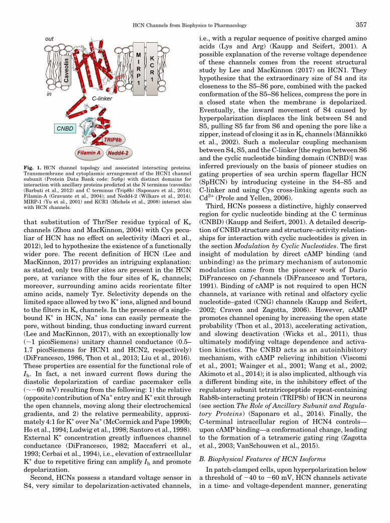
Fig. 1. HCN channel topology and associated interacting proteins.Transmembrane and cytoplasmic arrangement of the HCN1 channelsubunit (Protein Data Bank code: 5u6p) with distinct domains forinteraction with ancillary proteins predicted at the N terminus (caveolin)(Barbuti et al., 2012) and C terminus (Trip8b) (Saponaro et al., 2014);Filamin-A (Gravante et al., 2004); and Nedd4-2 (Wilkars et al., 2014).MIRP-1 (Yu et al., 2001) and KCR1 (Michels et al., 2008) interact alsowith HCN channels.
HCN Channels from Biophysics to Pharmacology 357

an inward current that does not inactivate (Fig. 2A).Driven by their respective electrochemical gradients,Na+ and K+ flow through the open channels as long asthe hyperpolarizing step is maintained, generating anet inward current. Without recapitulating all biophys-ical features, which have been extensively describedelsewhere (Biel et al., 2009), it is anyway necessary tohighlight some Ih properties to understand the mecha-nisms underlying its modulation by exogenous andendogenous substances and the consequences of gainor loss of function in inherited and acquired diseases.Heterologous re-expression of single isoforms, gener-
ating homomeric tetramers, demonstrates that HCNspossess different kinetics and voltage-dependent prop-erties. Channel opening upon hyperpolarization gener-ates a time-dependent inward current that can beinterpolated by a single- or double-exponential function(see, for example, Wicks et al., 2011; Zong et al., 2012;Kim and Holt, 2013; Nakamura et al., 2013, and, forreview of previous literature, Biel et al., 2009). The timeconstant (tau) of activation differs among isoformsubtypes. In native tissue, it depends on the coassem-bling of different isoforms in the tetrameric channel, thepresence of ancillary subunits, post-translational modifi-cations including (de)phosphorylation andN-glycosylation,the effect of ligands, and, finally, experimental conditionssuch as ionic composition of extracellular milieu (Chenet al., 2001b; Xue et al., 2002; Altomare et al., 2001; Much
et al., 2003;Whitaker et al., 2007; Ye andNerbonne, 2009).When heterologously re-expressed, HCN1homotetramericchannels exhibit the fastest kinetics of activation andHCN4 the slowest, and the two other isoforms are inbetween (Ludwig et al., 1999; Seifert et al., 1999; Stieberet al., 2003b; Stieber et al., 2005). Activation kinetics isstrongly dependent on voltage: the more negative thestep, the faster the activation. This is evident in Fig. 2A,in which a family of HCN1, HCN2, and HCN4 currenttracings are plotted as a function of time. The secondstriking difference relates to the voltage dependence:when the relative amplitude is plotted against thevoltage step, HCN1 exhibits the less negative thresholdand voltage of half-maximal activation (V1/2) and HCN2the most negative (Ludwig et al., 1999; Seifert et al.,1999; Stieber et al., 2003b, 2005). Finally, voltage de-pendence and kinetics of activation of HCN2 and HCN4are very sensitive to cAMP, at variance with HCN1(Moroni et al., 2000; Zagotta et al., 2003).
In recombinant systems or native cells, the slowexponential activation of Ih is preceded by a small initial,instantaneous current. The molecular nature of thiscurrent is still questioned because variable biophysicaland pharmacological properties (e.g., amplitude, sensi-tivity to Cs+, or organic blockers) have been reported,depending on the frequency of hyperpolarizing pulses,intracellular cAMP or Cl2 concentrations, expression ofancillary subunits, and other experimental conditions
Fig. 2. Current properties of HCN homomeric channels. (A) Representative whole-cell current recordings (lower panels) of re-expressed singlemHCN1, mHCN2, and hHCN4 isoforms. Each family of time-dependent, inward currents is generated by hyperpolarizing steps to 240/2140 mV, froma holding potential of 230 mV. Activation curves (upper panels), obtained by normalized conductance plotted as a function of test potentials and fittedby a Boltzmann function, exhibit threshold of activation and voltage of half-maximal activation less and most negative for HCN1 and HCN2 isoforms,respectively. (B) Application of cAMP to the cytoplasmic side of the membrane causes a much greater shift in the activation curve of HCN2 comparedwith HCN1 (modified from Wainger et al., 2001 with permission); (C) schematic representation of the structure of the four HCN isoforms.
358 Sartiani et al.

(Proenza et al., 2002; Mistrik et al., 2006; Proenza andYellen, 2006). Recent results obtained with mutantHCN1 and HCN2 isoforms in the so-called zipperresidues suggest that the two components (instantaneousand slow-activating current) may result from differentopen states of HCN channels (Wemhoner et al., 2012). Atvariance with the voltage- and time-dependent HCNcurrent, the physiologic significance of the instantaneouscurrent is unclear and may be dependent on cell types;indeed, a large voltage-independent inward current,blocked by Cs+ and 4-(N-ethyl-N-phenylamino)-1,2dimethyl-6-(methylamino) pyrimidinium chloride (ZD7288;see Pharmacology, section Ivabradine, Cilobradine, andOther Specific Bradycardic Agents), seems to contributeto neuronal excitability in stellate cells of ventral co-chlear nucleus (Rodrigues and Oertel, 2006).Several models have been proposed in the years to
explain HCN behavior, namely voltage dependence,kinetics, and sensitivity to cAMP. Although those basicproperties are well described by classic allosteric mod-els (DiFrancesco, 1999), others are not. A more recentfour-state model, in which HCN channels transit be-tween two modes, tried to encompass this limitationand explain hysteresis, i.e., the voltage dependence ofthe HCN channels on their prior activity. In fact, thecurrent–voltage activation curve of Ih is shifted positivelyafter long hyperpolarizing steps (persistent channelopening), and shifted negatively after long depolariza-tions (closed channels) (Männikkö et al., 2005). The sameauthors suggest that transition between the two modes,or hysteresis, has important consequences for physio-logic (and pathologic) functioning of rhythmic cells. InSAN cells, hysteresis keeps channels closed after anaction potential (AP) until complete repolarization (andcalcium current recovery), preventing a premature di-astolic depolarization; attainment of maximum hyper-polarization (between two APs) stabilizes the open stateof HCN channels, pushing the membrane potentialtoward the threshold for the next AP (Männikkö et al.,2005).
C. Modulation by Cyclic Nucleotides
1. Binding of cAMP and Modification of GatingProperties. HCN channels are primarily activated byhyperpolarization of membrane potential and are reg-ulated by cyclic nucleotides, which interact with aspecific site, the CNBD, located in the intracellularC-terminal portion (see section HCN Modulation byCyclic Nucleotides). cAMP accelerates channel openingand shifts the activation curve tomore positive potential(DiFrancesco and Tortora, 1991). The shift in V1/2
depends on the channel subtype and conditions: itvaries between 10 and 25 mV for HCN2 and HCN4(see Wahl-Schott and Biel, 2009 and references citedtherein), but only 2–6 mV for HCN1 (Wainger et al.,2001; Wang et al., 2001) (Fig. 2B). cAMP potency (EC50,i.e., the concentration of ligand that produces a half-
maximal voltage shift) is in the range 0.06–1.53 mM(Table 1).
According to a generally accepted model, the channelis normally inhibited and the binding of cAMP removesthis inhibition, inducing conformational changesand increasing the open probability of channel pore(Wainger et al., 2001). As suggested by Wang et al.(2001) from experiments involving HCN1-HCN2 chi-mera, the different sensitivity of these two isoformsdepends on differences in the sequences involved in theinteractions between CNBD and the C-linker. Studiesperformed on the isolated C-terminal domain (C-linker +CNBD) of HCN2 showed that addition of cAMP changesthe proportion between monomer and tetramer in favorof the latter (Zagotta et al., 2003). There is evidence thatthe isolated C-terminal domain of HCN1 has a higherpropensity to tetramerize than those ofHCN2andHCN4(Lolicato et al., 2011; Chow et al., 2012). Thus, a possibleexplanation to the lower sensitivity of HCN1 to thenatural agonist could be a sort of preactivation of thechannel (Chow et al., 2012), probably because it is able totrap endogenous cAMP in the CNBD (Lolicato et al.,2011). The positive effect of cAMP on tetramerization ofthe isolated intracellular region is translated, in thewhole channel, in conformational changes that propa-gate from the CNBD via the C-linker to the S6 fragment(Lee and MacKinnon, 2017).
Surprisingly, HCN3 is not activated by cAMP; rather,the activation curve is slightly shifted to more negativevoltages (Mistrík et al., 2005; Stieber et al., 2005). Thelack of sensitivity of HCN3 to cAMP, despite the pres-ence of a functional CNBD in the intracellular region,has been explained by a shorter C-terminal sequence,after the CNBD, which alters the normal autoinhibitionof the channel (Stieber et al., 2005).
In recent years, several biophysical techniques, suchas isothermal titration calorimetry, surface plasmonresonance, and fluorescence anisotropy, have allowedmeasurement of the binding affinity of cAMP for theCNBD. Lolicato et al. (2011) used surface plasmonresonance to compare the interaction of cAMP withthe C-terminal domain of HCN1, 2, and 4, finding thatthe three isoforms recognize the natural agonist withsimilar KD values (respectively, 5, 10, and 11 mM). Xuet al. (2010) used isothermal titration calorimetry andfluorescence anisotropy to measure affinity on theintracellular portion of HCN2 and 4, comparing theresults with the EC50 (Table 1); because human HCN4(hHCN4) does not express in Xenopus laevis oocytes,they used a murine HCN2 (mHCN2)-hHCN4 chimera,in which theC-linker andCNBD ofHCN2were replacedby the same portion of HCN4. cAMP potency, measuredin functional studies on the whole channel, was threetimes higher on HCN2 than on HCN4, but the samedifference was not found for affinity (measured on thepurified cytosolic domains), giving evidence that bindingand gating efficacy might have different requirements.
HCN Channels from Biophysics to Pharmacology 359

2. Other Cyclic Nucleotides. As well as cAMP,guanosine-39,59-cyclic monophosphate (cGMP) is a fullagonist of HCN channels: at saturating concentrations,the shift in V1/2 is in the same range as that of cAMP.However, cGMP displays a 10-fold lower potency forHCN channels (Table 1) (Ludwig et al., 1998). cGMPand cAMP bind to the CNBD in a similar way, onlydiffering in the orientation of the purine ring (Zagottaet al., 2003). On SpHCN, cGMP behaves as a partialagonist showing, when compared with cAMP, an in-trinsic activity of 0.5 and about 600-fold lower affinity(Kaupp and Seifert, 2001).Cytidine 39,59-cyclic monophosphate (cCMP) is also
able to modulate HCN channels, behaving as partialagonist (DiFrancesco and Tortora, 1991). On HCN2 andHCN4 channels, cCMP shifts the activation curve tomore positive potentials, speeds up current activation,and decreases current deactivation, but it has no effecton HCN1 andHCN3. The voltage shift and themaximalincrease in the current amplitude were significantlysmaller than those observed for cAMP, in agreementwith the behavior of a partial agonist (Zong et al.,2012). On HCN2, uridine 39,59-cyclic monophosphate(cUMP), purine 39,59-cyclic monophosphate (cPMP),and 2-amino-cPMP are all able to shift the activationcurve to more positive potentials and to increasechannel conductance, with efficacy similar to cAMPand cGMP; on the contrary, inosine 39,59-cyclic mono-phosphate (cIMP), as well as cCMP, behaves as weakactivators (Ng et al., 2016).In addition to cyclic mononucleotides, also cyclic
dinucleotides can modulate HCN channels. In mouseSAN myocytes, they behave as antagonists, being ableto reduce Ih (Lolicato et al., 2014). Cyclic [guanosine-(29-59)-monophosphate-adenosine-(39-59)-monophosphate],the cyclic dinucleotide that has been found inmammals,caused a shift of the activation curve toward more
negative potentials, and a ;30% reduction of firingrate. On HCN4 channel expressed in human embryonickidney (HEK)293 cells, the dose–response curve yieldedan IC50 (i.e., the concentration of ligand that produces ahalf-maximal voltage backshift) of 114 nM. Other cyclicdinucleotides could completely reverse the effect ofcAMP on the activation curve, as, for instance, cyclicdi-(39,59)-GMP,whichwas;16 times less potent than cyclic[guanosine-(29-59)-monophosphate-adenosine-(39-59)-monophosphate] (IC50 1.8 mM).
D. The Role of Ancillary Subunits andRegulatory Proteins
Different regulatory proteins form macromolecularcomplexes with HCN channel subunits and define thefeatures of HCN-mediated current in vivo, the regionalor subcellular localization of HCN proteins, as well astheir susceptibility to modulatory signals.
The number of proteins acting as HCN-bindingpartners has grown over the last decades; for someproteins, such as Mint2 (Mun18-interacting protein),synaptic scaffolding molecule, and tamalin, only a scaf-fold function for HCN2 has been identified (Kimuraet al., 2004), and a possible role in channel trafficking,distribution, and clustering has been hypothesized.
MinK-related peptide 1 (MiRP1), encoded by kcne2gene, is a single-transmembrane domain protein, whichserves as regulatory subunit of different cardiac ionchannels, including HCN channels. It enhances HCNprotein and current expression in an isoform-specificmanner (Yu et al., 2001; Decher et al., 2003; Qu et al.,2004; Brandt et al., 2009). When coexpressed with HCNchannel, current kinetics of activation is accelerated forHCN1 and HCN2, whereas it is slowed down for HCN4,which also displays a shift of the midpoint of activationto more negative voltages. High levels of MiRP1 andHCN subunits (primarily HCN4, and, depending on the
TABLE 1Potency of cyclic nucleotides on different HCN channel isoforms
EC50 is the concentration of ligand that produces a half-maximal voltage shift.
Cyclic nucleotide EC50 (mM) Isoform/Tissue References
cAMPa 0.06 mHCN1 (Xenopus laevis) Wang et al. (2001)0.10 mHCN2 (Xenopus laevis) Wang et al. (2001)0.50 mHCN2 (HEK293 cells) Ludwig et al. (1998)0.80 mHCN2 (Xenopus laevis) Zagotta et al. (2003)1.0 mHCN2 (HEK293 cells) Zong et al. (2012)0.08 mHCN2 (Xenopus laevis) Xu et al. (2010)0.24 mHCN2-h4 chimera (Xenopus laevis) Xu et al. (2010)1.53 hHCN4 (HEK293 cells) Milanesi et al. (2006)0.21 Rabbit SAN myocyte DiFrancesco and Tortora (1991)0.72 SpHCN (sea urchin sperm) Kaupp and Seifert (2001)
cGMP 6.0a mHCN2 (HEK293 cells) Ludwig et al. (1998)8.3a mHCN2 (Xenopus laevis) Zagotta et al. (2003)
13.2a hHCN4 (HEK293 cells) Lolicato et al. (2014)7.85a Rabbit SAN myocyte DiFrancesco and Tortora (1991)479b SpHCN (sea urchin sperm) Kaupp and Seifert (2001)
cCMPb 29.0 mHCN2 (HEK293 cells) Zong et al. (2012)11.85 Rabbit SAN myocyte DiFrancesco and Tortora (1991)
aFull activator.bPartial activator.
360 Sartiani et al.

species, HCN1 or HCN2) are expressed in the SAN ofsmall and large mammals, includingmouse, rabbit, andhuman (Yu et al., 2001; Accili et al., 2002; Schweizeret al., 2009). A similar interaction among MiRP1 andHCN4, 2, and 1 most likely occurs in atrial andventricular myocytes, where transcript levels of bothsubunits are lower compared with SAN (Yu et al., 2001;Decher et al., 2003; Qu et al., 2004; Stillitano et al.,2008, 2013; Sartiani et al., 2010). In these regions, themodification of HCN-mediated current—related to car-diac diseases [atrial fibrillation (AF) and ventricularhypertrophy] or to postnatal maturation—is most likelyassociated with transcriptional regulation of both HCNchannel and MiRP1 subunit.The K+ channel regulator 1 (KCR1) is a transmem-
brane protein expressed in cerebellum and heart (Hoshiet al., 1998; Michels et al., 2008). Protein expressionanalysis in rat and guinea pig revealed an extensivedistribution of the protein in the heart with largeramount in the atrioventricular node (AVN), left atrium,and ventricle, followed by right atrium and ventricle,and SAN. KCR1 is a regulatory subunit of diversenative Kv channels, as well as HCN channels. Inheterologous expression systems and native ventricularmyocytes, KCR1 interacts withHCN2, reducing currentsize and shifting Ih activation to more negative poten-tials. These modifications ultimately decrease spontane-ous rhythmicity in cultured neonatal cardiac myocytes,suggesting that KCR1 may be an important regulatorysubunit of HCN current in vivo.Caveolin-3 is a lipid raft component of myocyte
membrane that colocalizes with and affects the expres-sion and function of HCN channels, as well as theirsusceptibility to modulating signals. In SAN cells,interaction between caveolin-3 and HCN4 affects chan-nel voltage dependence by shifting V1/2 to negativevoltages; it also modifies HCN4 current kinetics byaccelerating channel deactivation (Barbuti et al., 2004,2007, 2012). Because b2-adrenoceptors, but not b1,localize to lipid rafts in the SAN, their activationgenerates a prominent signal mediating the adrenergicenhancement of HCN current and the rise of heart rate(Barbuti et al., 2007). Following investigations demon-strated that a similar colocalization between caveolin-3and HCN4 channels occurs in human atrial and embry-onic stem cell–derived cardiomyocytes (Bosman et al.,2013; Stillitano et al., 2013). In the latter, the shift ofV1/2 is directly related to the increase of caveolin-3expression during myocyte maturation.Filamin A is a cytosolic scaffolding protein that exerts
a crucial role for the trafficking of numerous ionchannels in excitable cells, including neurons andcardiac myocytes. It anchors ion channels to actincytoskeleton and clusters them in distinct locations oncell surfacemembrane. In the brain, onlyHCN1, but notHCN2, 3, or 4, associates with filamin A (Gravanteet al., 2004). In heterologous expression systems, this
interaction reduces the density of channel expression aswell as whole-cell conductance by aggregating HCN1within restricted regions of the cell membrane. Addi-tionally, filamin A promotes a reversible dynamin-dependent internalization of HCN1 channels and aredistribution of HCN1 channels on cell surface byaccumulation of channels in endosomal compartments(Noam et al., 2014). In cultured hippocampal neurons,expression of a dominant-negative filamin A increasesthe expression of native HCN1, whereas acute abroga-tion of HCN1–filamin A interaction enhances currentsize. Whether a similar interaction occurs at cardiaclevel is unknown, despite filamin A being present inmouse and human atrial myocytes (Rafizadeh et al.,2014).
TRIP8b, also termed Pex5p-related protein (PEX5Rp),and H-channel interacting protein 1 (HIP1), is a braincytoplasmic protein, member of the Rab family of smallGTPase proteins, which are important for vesicle traf-ficking (Chen et al., 2001a). In neocortical and hippo-campal pyramidal neurons, colocalization of TRIP8b toHCN1 promotes an active trafficking of the channelsfrom soma to dendrites that is important to modulatespike firing and synaptic potential (Santoro et al., 2004;Zolles et al., 2009). In TRIP8b-knockout (KO)mice, HCNsurface expression in hippocampal pyramidal neurons isdramatically reduced, as well as current size in thisregion; moreover, normal expression pattern of HCNchannels is profoundly altered in pyramidal neurondendrites (Lewis et al., 2011). Nine isoforms of TRIP8bhave been identified that differentially affect HCNchannel gating, membrane expression, and traffickingin the nervous system. The overexpression of TRIP8b incultured hippocampal pyramidal neurons or heterolo-gous expression systems exerts different effects onHCN1surface expression according to the variant type. TRIP8b(1a–4) and TRIP8b (1a), major splice variants in thehippocampus, enable a correct localization of HCN1(Lewis et al., 2009; Santoro et al., 2009). Moreover,TRIP8b (1a–4) upregulates HCN1 expression in heter-ologous systems and promotes its dendritic expression.Conversely, TRIP8b (1a) downregulates HCN1 surfaceexpression in X. laevis oocytes and inhibits the abnormalexpression of HCN1 in the axons of pyramidal neurons(Piskorowski et al., 2011). Opposed to TRIP8b, a recentpaper shows that ubiquitination by the Nedd 4-2 (neu-ronal precursor cell–expressed developmentally down-regulated four-like) protein decreases HCN1 surfaceexpression and translocation, leading to Ih loss of func-tion (Wilkars et al., 2014).
E. From Transcriptional Control toPost-Translational Modifications
1. microRNAs. Accumulating evidence on sinusnode dysfunctions has led to consider the downregula-tion of HCN channels as possible common cause ofdifferent conditions leading to bradycardia (D’Souza
HCN Channels from Biophysics to Pharmacology 361

et al., 2015). The role of microRNAs (MiR) is emergingas main transcriptional regulator of HCN channels incardiac pacemaker centers. MiR-1, one of the mainmuscle-specific microRNA (myomiR), is induced byathletic training in the heart jointly to a downregula-tion of the transcription factors Tbx3 and NRSF(D’Souza et al., 2014). These changes are consistentwith the downregulation of HCN4 and Ih found in SANcells of trained animals.On the contrary, in different cardiac pathologies,
includingmyocardial infarction (Suffredini et al., 2012;Yu et al., 2015) or age-related AF (Li et al., 2015b),expression of HCN channels is upregulated, in linewith the observed reduction of MiR-1 levels detected inthe ventricles and atria. Accordingly, in rats withmyocardial infarction, administration of ivabradine,a selective bradycardic agent (Suffredini et al., 2012),or spironolactone, an aldosterone blocker (Yu et al.,2015), counterbalances the overexpression of HCNchannels in parallel with upregulation of MiR-1.In all conditions, the mechanism responsible for the
modifications of microRNA levels in the sinus node or inthe working myocardium remains unknown.2. Regulation by Membrane Phosphoinositides.
Signaling pathways coupled to membrane phosphoino-sitide content and downstream derivatives stimulateHCN channels.Phosphatidylinositol 4,5-bisphosphate [PI(4,5)P2] is
a membrane constitutive component that increases theopening of recombinant and native HCN channels byshifting the voltage dependence of activation to morepositive potentials by 5–20 mV, depending on isoforms.It acts as intracellular allosteric activator that facili-tates channel opening (Biel et al., 2009).In the heart, basal variability of membrane PI(4,5)P2
is likely to contribute to the variations in the voltagedependence of Ih activation in cardiac cells at differentmaturation degrees (Cerbai et al., 1999b; Qu et al.,2001), derived from different regions, or following stressand pathologic conditions (Suh and Hille, 2007).In SAN cells, stimulation of bradykinin BK2 receptors
coupled to phospholipaseCenhances phosphatidylinositolkinase activity that in turn stimulates polyphosphoi-nositide synthesis, thereby enhancing HCN channelfunction (Pian et al., 2007). A similar interaction withphosphoinositides occurs in neurons, where PI(4,5)P2 acts as an allosteric modulator of HCN, causing arightward shift of voltage activation (Zolles et al., 2006;Ying et al., 2011); gating by phosphoinositides may beimportant to maintain rhythmogenesis when signalingpathways leading to phospholipid degradation reducechannel activation in nerve cells. Recent data in SpHCNsuggest that PI(4,5)P2 binds to both the transmem-brane core region and the C-linker domain of thechannel, with opposite effects (Flynn and Zagotta,2011). Whether such a dual mechanism also occursin mammalian HCN is unknown.
Other allosteric modulators of HCN channels are twomembrane phosphoinositide derivatives, phosphatidicacid and arachidonic acid, which are products ofdiacylglycerol kinase and phospholipase A2, respectively.They directly facilitate HCN channel gating by shiftingthe voltage dependence of activation to more positivevalues (Fogle et al., 2007).
3. Kinases. Phosphorylation status of HCN chan-nels is an additional regulatory mechanism control-ling HCN properties and adapting its activity to thepeculiar conditions of different types of cardiac cellsand neurons.
Despite the fact that HCN channels are consideredend effectors of cAMP, experimental evidence demon-strated a modulatory role of protein kinase A (PKA).In early cardiomyogenesis, activation of PKA hasan exclusive role in the stimulation of HCN currentfollowing b-adrenergic receptor stimulation (Abi-Gerges et al., 2000). In adult mouse SAN, PKA seemsto exert an additive positive shift of the activationcurve upon b-adrenergic receptor stimulation (Liaoet al., 2010), although the mechanism is most likelymediated by PKA-dependent effects on cAMP pro-duction or diffusion rather than phosphorylation ofthe channel (St Clair et al., 2013).
In hippocampal neurons, phospholipase C–proteinkinase C activation, phosphorylating HCN1 isoform,decreases HCN current and HCN1 surface expression(Williams et al., 2015). Similar findings were obtainedin respiratory neurons within the pre-Bötzinger com-plex (Thoby-Brisson et al., 2003).
Tyrosine kinases of the Src family have also astimulatory function in the mature cardiac cells aswell as in different types of neurons (Santoro et al.,1997; Wu and Cohen, 1997; Yu et al., 2004; Zong et al.,2005; Arinsburg et al., 2006). In these cells, thepathway contributes to regulate spontaneous elec-trogenesis by direct phosphorylation of HCN1,HCN2, and HCN4 and speeding of channel kinetics.Despite the fact that the residue (Y476) conferringsuch modulation in HCN2 channels is conserved inthe other isoforms, a stimulation of kinetics by Srctyrosine kinase has been proven only for HCN4. Inaddition, the latter isoform undergoes also a positiveshift (@+10 mV) of voltage dependence, most likelybecause of an additional phosphorylation in a differenttyrosine residue (Y531) (Li et al., 2008a). In rat ven-tricular myocytes, the receptor-like protein-tyrosinephosphatase-a controls the extent of HCN2 channelphosphorylation in this site, shifts the voltage de-pendence of activation, and decreases channel in-sertion into the membrane (Huang et al., 2008).
In neuronal cells, HCN channels are also phosphor-ylated by the serine/threonine kinase, p38 mitogen-activated protein kinase (Poolos et al., 2006), andcalcium/calmodulin-dependent protein kinase II (Shinand Chetkovich, 2007).
362 Sartiani et al.

Modulation by the small ubiquitin-like modifier(SUMO) peptide is one of the mechanisms involved inthe regulation of protein–protein interactions. HCN2SUMOylation occurs in mouse forebrain tissue (Parkeret al., 2017); in transfected HEK cells, SUMOylation ofHCN2 increased current conductance and surface ex-pression. This finding is of interest in view of HCNdysregulation in central nervous system (CNS) dis-eases, as discussed later.
III. Origins, Physiology, and Pathophysiology ofHCN Channels
A. HCN: Ancient and Early Channels
1. Phylogeny. HCN channels belong to the super-family of six-transmembrane segment channels andare related to CNG channels and voltage-dependentether-a-go-goK+ channelsKv10–Kv12 (LeeandMacKinnon,2017). Being components of the ancestral gene pattern,HCN channels are present across a wide spectrum ofinvertebrate and vertebrate species. They most likelyderive from a single ancestral gene subjected to dupli-cations and diversification events over the species thateventually generated four different isoforms prior to theorigin of the vertebrate clade (Jackson et al., 2007). Afunctional role for HCN in setting heart rhythm hasbeen postulated on the basis of pharmacological studiesalso in vertebrate ancestors (Wilson and Farrell, 2013).Based on sequence conservation analysis, HCN3,thought to be most similar to the ancestral channel,was the first to diverge as a product of the firstduplication. HCN3 was followed by the emergence ofHCN4 and then of HCN1 and HCN2, thus composing agroup of four variants that collectively shares 80%–90%sequence conservation within the core and transmem-brane regions. The latter in all vertebrate and inverte-brate contribute to common functional properties ofHCN channels. The residual variations in these regionsare responsible for subtle isoform-specific differencesrelated to inner selectivity filter, rates of channelopening, and differences in cAMP efficacy to modulateHCN isoforms.In all four vertebrate isoforms, a region of ;50
residues upstream to the start of S1 in the NH2
terminus is conserved. In mouse HCN2, this region isinvolved in intersubunit interactions of tetramer as-sembly and in the formation of functional channels(Tran et al., 2002). Analogous regions present in theother isoforms are supposed to exert similar functions.In all vertebrate HCN1, 2, and 4, but not in HCN3, a
different block in the COOH terminus is conserved. Itrepresents a PDZ-binding domain enabling channels tointeract with PDZ-containing proteins and with theTRIP8b protein (Kimura et al., 2004; Santoro et al.,2004), which regulates channel surface expression.2. HCN Channels in Stem Cells. The growing
number of studies in different types of stem cells has
led to identify the presence of several specialized ionchannels, including HCN channels (Heubach et al.,2004; Wang et al., 2005; Sartiani et al., 2007). Assuggested for other bioelectric signals, mainly K+ orCa2+ channels, HCN channels also may act as regula-tors of a wide range of stem cell functions, includingproliferation, migration, differentiation, and tissueregeneration of nonexcitable cells (Blackiston et al.,2009; Sundelacruz et al., 2009, 2015; Levin, 2014).Indeed, HCN-mediated mechanisms are most likelyinvolved in the development of a mature phenotype ofolfactory sensory neurons, where HCN channels areexpressed precociously and drive axon organization(Mobley et al., 2010).
Expression pattern of HCN channels in stem cells isspecies- and/or origin-dependent, despite the relativelyhomogenous phenotype of potency markers (Li andDeng, 2011). HCN1 channels are highly expressed inhuman pluripotent stem cells, but not in mouse cells,where HCN3 largely predominates, suggesting thatcurrent phenotype and regulation might diverge amongspecies, as observed in differentiated cells. Differently,human bone marrow–derived stem cells express HCN2channels. Despite the presence of HCN transcripts andproteins in stem cells, only one study has been per-formed in mouse pluripotent stem cells, where thechannels were found involved in cell proliferation, inparticular in cell cycle progression from G0 to G1 phase(Lau et al., 2011).
3. HCN during Organogenesis.a. Cardiogenesis. Occurrence of spontaneous elec-
trical activity is an early event in cardiac morphogen-esis. In the mouse, the whole process has beenthoroughly dissected, identifying that this pacemakeractivity is detectable since embryonic day (E) 7.5 inprecardiac mesoderm (cardiac crescent) of the firstheart field (Liang et al., 2013; Später et al., 2013;Barbuti and Robinson, 2015). Cells comprised in thisearly pacemaker region provide efficient peristalticcontractions necessary in the primitive heart tube andexpress two distinct markers, the transcription factorNkx2-5 and HCN4 channels (Christoffels et al., 2010).Lineage-tracing experiments show that the Nkx2-5+/HCN4+ cells do not give rise to the cardiac conductionsystem in the developing heart, but generate atrial andventricular precursors (Moorman and Christoffels,2003). At E8, posterior heart field precursors express-ing the transcription factor Tbx18 start to proliferateand will form the sinus venosus, a symmetric structureof the heart tube. Following expression of two addi-tional markers (Shox2 and CD166), these cells willbecome the leading pacemaker (SAN precursors),when at E9.5 a subgroup of cells further expressesthe second heart field marker Isl-1 and the transcrip-tion repressor Tbx3.
Subsequent increase of Tbx3 and HCN4 and decreaseof CD166 expression ultimately form the sinus atrial
HCN Channels from Biophysics to Pharmacology 363

node in the right atrium, whose typical panel ofmarkerswillmaintain Isl-1, Tbx18, Shox2, Tbx3, and a high levelof HCN4 throughout life. In the left atria, activity of thehomeobox factor Pitx2c specifically suppresses the SANgene program, allowing the correct asymmetric devel-opment of the conduction system (Mommersteeg et al.,2007). Despite the fact that HCN4 is functionallyexpressed ever since the appearance of the pacemakeractivity in the precardiac mesoderm, HCN4 starts toplay a fundamental function during the formation of theSAN between E9.5 and E11.5, since global and cardiac-specific HCN4-KO mice die in this developmental stage(Stieber et al., 2003a). However, studies in HCN4-deficient embryos show significantly reduced contrac-tion rates and immature AP in deficient embryoscompared with wild type (WT), indicating that evenbefore SAN formation HCN4 is critical for normalcardiac development.Studies with specific deletion of HCN1 and HCN2
confirmed the role of HCN4, because HCN1 (Nolanet al., 2003) or HCN2 (Ludwig et al., 2003; Cao andOertel, 2011) KO animals do not evidence major cardiacalterations in developing embryos. This also suggeststhat expression of HCN1 and HCN2 channels in theheart is delayed during cardiogenesis. Indeed, tran-scription profiling throughout mouse cardiac develop-ment indicates that embryonic heart before E9.5displays abundant HCN4 transcript, whereas otherHCN transcripts are almost absent. Toward birth,HCN transcription profile in the atrium and ventriclechanges remarkably, because HCN4 is strongly down-regulated, whereasHCN1 andHCN2 transcripts slowlyemerge. HCN3 isoform shows highest levels at earlyembryonic stages and then fades to very low levels(Schweizer et al., 2009).Little information is available on the developmental
changes of HCN channels occurring in humans. In alimited time window, i.e., from 11 to 14 weeks of fetallife, a strong protein expression of HCN4 is present inthe whole heart that contributes to a remarkable HCN-mediated current in ventricular myocytes (Bosmanet al., 2013). At this stage, current density is largerthan that retrieved in human healthy adult ventricularcardiomyocytes and close to the values described inventricular myocytes from ischemic patients (Fig. 3),corroborating the concept of HCN channel expression asa marker of fetal gene reprogramming in cardiacdisease (see Altered HCN function and cardiac disease).b. Insights from stem cell–derived cardiomyocytes.
A different approach to address the developmentalchanges during cardiogenesis consists in the use ofpluripotent stem cells, whose cardiomyogenic potentialin vitro is well established and widely used in the lastdecades. Following established protocol of differentia-tion, the in vitro model recapitulates many of thedevelopmental stages described for in vivo cardiogene-sis, thus representing a highly valuable tool to
investigate the embryonic/fetal modifications of cardiaccells difficult to address in other models. It is also wellknown that the cardiomyocyte population arising fromthis model is heterogeneous and composed of atrial-,ventricular-, and SAN-like cells that have been thor-oughly characterized for their electrical and structuralproperties more than 20 years ago (Maltsev et al., 1993).Molecular insights have been reviewed recently(Barbuti and Robinson, 2015). An interesting findingemerged from mouse pluripotent cells is the variablepropensity to develop SAN-like or atrial/ventricular-like lineage. The former is characterized by a prominentexpression of HCN4/HCN1 channels that are colocal-ized with caveolin-3 and present together with T-typecalcium channels CaV3.1/3.2, thus recapitulating thepattern of channels typically expressed in native SAN(van Kempen et al., 2003; Marionneau et al., 2005;Yanagi et al., 2007; Barbuti et al., 2009). Differently,atrial/ventricular-like lineage is characterized byhigher levels of HCN2 and HCN3 compared withHCN4 and HCN1 (White and Claycomb, 2005; Quet al., 2008).
Phenotype heterogeneity and lineage preference alsofeature human pluripotent stem cells (He et al., 2003;Mummery et al., 2003), where atrial/ventricular pheno-types associate with HCN2 preponderance over HCN4(Satin et al., 2004). Consistency of findings amongdifferent mammalian species warrants further investi-gation to better understand whether distinct, still un-identified pluripotency states drive a preferentiallineage specification. This hypothesis applies to recentstudies on pacemaking activity in early (days 11–21 ofdifferentiation) cardiomyocytes from pluripotent cells(Weisbrod et al., 2013), where HCN current, detectablein most of beating cells, is determined by HCN4 andHCN2 isoforms. The concomitant association withCaV1.3, Na+/Ca2+ exchanger (NCX)-1, and Tbx3 in thesecells defines a panel of genes typical of human SAN andperinodal areas (Chandler et al., 2009), suggesting thatthese cells are early pacemaker or SAN-like cells ratherthan immature atrial or ventricular cardiomyocytes.Accordingly, cell lines prone to develop atrial/ventricularphenotype exhibit properties resembling those presentin adult atrial/ventricular cells, such as expressionof NaV1.5, CaV1.2, and HCN2, whereas CaV1.3 andHCN4 are absent (Satin et al., 2004). Differently,studies from our group, performed in a different cellline maintained in long-term culture, evidence the earlyemergence of immature SAN-like cells, representingthe most frequent cardiomyocyte population after 15–30 days of differentiation. This single phenotype di-verges into distinct atrial/ventricular myocytes at latermaturation stages (55–110 days) (Sartiani et al., 2007;Paci et al., 2012; Bosman et al., 2013). During thisdevelopmental period in vitro, HCN channel expressionshifts from a SAN-like pattern, mainly composed ofHCN1 and HCN4, to an atrial/ventricular pattern,
364 Sartiani et al.

where HCN2 isoform predominates and remains con-stant. The functional counterpart in the early stage isdefined by a robust HCN current, which throughoutmaturation declines in amplitude and activates muchslower, in accordance to a lower expression of HCN1,approaching values like those encountered in na-tive human fetal and adult hypertrophic ventricularcardiomyocytes (Fig. 3). The findings further strengthenthe similarities between in vitro and in vivo cardiacdifferentiation. The advantageous property has beenfurther exploited to investigate the subcellular compart-mentation of HCN4 channel in the human setting.During development, HCN4 protein signal shifts from awidespread localization in a-actinin–positive immaturecells to restricted sites inmature cardiomyocytes. At thisdevelopmental stage, HCN4 increasingly colocalizeswith caveolin-3, thus providing a possible explanationfor the negative shift ofHCNcurrent threshold. A similarmodification of caveolin-3/HCN4 interaction occurs innative human cardiomyocytes, as suggested by a similarnegative shift of HCN activation threshold characteriz-ing the transition from fetal to adult cardiomyocytes(Bosman et al., 2013).c. HCN expression in the developing nervous system.
At variance with cardiogenesis and stem cell–derivedcardiomyocytes, systematic studies investigating isoform-and age-dependent changes in HCN expression levelsduring pre- and postnatal development of the CNS aremissing, with available data referring to selectedregions. In CA1 hippocampal pyramidal cell layer ofembryonic rats, a robust increase of HCN1 transcriptand protein expression occurs during development,with concomitant reduction of HCN4 and relativelystable HCN2 levels. By birth, the contribution of HCN1to the total HCN channel pool has risen from 30%
to 60%. At subcellular level, the proximal-to-distaldendritic gradient of HCN1 is already present atpostnatal day 2 (Bender et al., 2001; Surges et al.,2006; Brewster et al., 2007). Similarly, overall Ihdensity increases nearly sixfold in rat thalamocorticalrelay neurons during the first 3 months of postnatallife, accompanied by a progressive decrease in cAMPsensitivity. In keeping, quantitative analyses of HCNchannel isoforms revealed a steady increase of tran-script and protein expression levels of HCN1 andHCN2, with reduced relative abundance of HCN4(Kanyshkova et al., 2009; Yoshimoto et al., 2015). Aninteresting observation comes from studies in thebrainstem auditory neurons, where HCN channelsare crucially involved in the location of sounds (Leaoet al., 2006). This function is poorly present at birth ingerbils (as in humans) and undergoes intense post-natal adaption; at this stage, the onset of maturehearing with precise temporal resolution was associ-ated with marked developmental changes in Ih. Inparticular, the kinetics of activation-deactivation be-came faster and conductance larger, and activationwas shifted rightward. Overall, these changes wereattributable in part to an increasing role of HCN1isoforms, and largely (V1/2 shift) to the maturation ofsignaling pathways, in particular cAMP- and PI(4,5)P2-dependent modulation (Khurana et al., 2012).
B. Distribution, Adaptive, and Maladaptive Role ofHCN Channels in Mammals
1. HCN Current in the Heart: an Ideal Pharmaco-logical Target. Excellent reviews have described thekey role of cardiac Ih in automaticity and its interplaywith other ion currents in the SAN (Accili et al., 2002;DiFrancesco, 2006, 2010; Biel et al., 2009), as well as in
Fig. 3. Ih current expression in cardiomyopathies and in cardiac development. (A) Data points represent the ratio between current density measuredin ventricular myocytes from diseased hearts and respective control. In neonatal rat cardiomyocytes (Neo), values measured at 2 weeks are comparedwith those at 2 days after birth. po-HF, pmi-HF: relative increase of Ih in rats with overt heart failure, resulting from pressure overload or followingmyocardial infarction, respectively. mLVH, sLVH: relative increase of Ih in rats with mild or severe left ventricular hypertrophy caused by aorticbanding or long-lasting pressure overload, respectively. DCM, ICM: relative increase of Ih in patients undergoing cardiac transplantation for terminaldilated or ischemic cardiomyopathy, respectively. For all conditions, the relative increase of current density is statistically significant versus controls,that is, normotensive rats, sham-operated rats, or undiseased donor hearts not transplanted for technical reasons, with the exception of DCM patients.fCM, ESC-laCM, and ESC-erCM: relative increase of Ih in human fetal (11–14 weeks) cardiomyocytes and in cardiomyocytes differentiated from humanembryonic stem cells in late and early developmental stages, respectively. (B) Representative recordings of Ih in a control (CTR), a hypertrophic (ICM),a fetal (fCM), and a late ESC-derived cardiomyocyte (ESC-laCM).
HCN Channels from Biophysics to Pharmacology 365

the conduction system (Mangoni and Nargeot, 2008).We refer the reader to these papers for a systematicdiscussion of physiologic aspects and to recent opticalmapping (Torrente et al., 2015) and computationalapproaches (Fabbri et al., 2017). Overall, HCN channelscontribute to two essential features of primary andsubsidiary pacemakers: the appearance of a diastolicdepolarization and the modulation of its steepness bythe sympatho-vagal balance or other endogenous fac-tors (Fig. 4). Indeed, a relevant fraction of the antiar-rhythmic and antianginal effect of old and new drugs(from beta-blockers to digoxin and ivabradine) residesin the indirect (antiadrenergic or vagomimetic) or direct(HCN blockade) effect on Ih in pacemaker cells. Accord-ing to recent computational approach modeling thehuman SAN AP, HCN current exerts its modulatoryrole mainly by changing the rate of the diastolic de-polarization phase (DDR) over the first 100ms followingthe maximum diastolic potential (Fabbri et al., 2017).This might be more relevant in species with low heartrate (e.g., humans versus rodents) because, as elegantlydiscussed by Zaza (2016), a nonlinear relationshipexists between DDR and time: the longer the cyclelength, the greater the bradycardic effect of reducingDDR (and vice versa).a. Expression and physiologic role of HCN isoforms
in cardiac regions. Ih may exhibit different electro-physiological properties (such as activation thresholdand amplitude), also depending on the relative expres-sion of different isoforms. Indeed, in the heart, HCNchannels exhibit a regional specific distribution(Herrmann et al., 2012; Deng et al., 2015; Li et al.,2015a). A remarkable variation in the total amounts ofHCN channels differentiates pacemaker centers fromworking myocardium, being HCN transcripts andproteins expressed at highest levels in the SAN andin the conduction system (AVN and Purkinje fibers).Additionally, isoform multiplicity differs according tospecies, except for HCN3, which displays a weakexpression, regardless of regions and species. In theSAN of humans, rabbits, mice, and dogs, HCN4 isthe main protein isoform compared with the others, theremaining fraction being composed byHCN2 andHCN1in humans andHCN1 inmouse and rabbits. Differently,rat SAN expresses similar amount of HCN2 and HCN4(Huang et al., 2016).In mouse AVN, almost all cells express HCN1 and
HCN4, whereas HCN2 is limited to some regions. Thebundle of His is particularly enriched of HCN4, whereasbundle branches also display HCN1 and HCN2(Herrmann et al., 2012). Human, rabbit, and rat AVNlargely express HCN4 protein isoform, with HCN1present in smaller amount (Dobrzynski et al., 2013).The expression pattern of HCN protein in atria and
ventricles also displays isoform variability; however,most mammalians, including humans, exhibit a preva-lence of HCN4 and HCN2, followed by smaller amount
of HCN1 and negligible levels of HCN3 (Lezoualc’het al., 2007; Stillitano et al., 2008).
i. HCN in SAN Cells and Its Contribution to CardiacPacemaking: Membrane and Calcium Clocks. HCNchannels have long been recognized for their primaryrole in pacemaker impulse generation and regulation.However, the specific contribution of HCN has consider-ably evolved in recent years. Two main mechanisms, thevoltage clock and the Ca2+ clock, mainly sustained byHCN current and Ca2+ release from sarcoplasmic re-ticulum, respectively, are supposed to contribute to acoordinated system that jointly drives spontaneouselectrical activity in SAN. The relative contributionof the two mechanisms to basal rhythm and itsadaption to autonomic balance have been the objectof an intense debate; to have a taste of different—sometimes conflicting—views, the reader is referred toa lively point–counterpoint exercise by the leadingscientists in the field (DiFrancesco and Noble, 2012;Maltsev and Lakatta, 2012) and the enlighteningaccompanying comment (Rosen et al., 2012).
In brief, in the SAN during the diastolic phase, thefraction of open HCN channels provides a steady-stateinward current drivingmembrane potential (270/240mV,depending on cells) to depolarize toward the thresholdrequired to generate a spontaneous AP (DiFrancescoet al., 1986). A key role of HCN channels, particularly theHCN4 isoform, is suggested—besides other arguments—by bradycardia (or severe bradycardia) consequent to thefollowing: 1) loss of HCN4 function in KO mouse modelsor patients carrying HCN4 mutations (see later in thissection), and 2) the effect of selective HCN blockers (seesection Pharmacology of HCN Channels). It is wellrecognized that the steepness of the diastolic depolariza-tion in pacemaker cells also results from the concerted(simultaneous or sequential) work of other membraneionic conductances: NCX, L- and T-type Ca2+ channels,Na+/K+ ATPase, voltage-dependent K+ and backgroundNa+ currents—just to mention some relevant ones. Thepoint by DiFrancesco and Noble (2012) is that theseconductances concert to set the pacing rate—the mem-brane clock—with Ih being the conductor, also in force ofits exquisite sensitivity to autonomic balance (via in-tracellular cAMP). Lakatta’s group (Vinogradova et al.,2010) named calcium clock the periodic, spontaneoussubmembrane calcium release from sarcoplasmicreticulum, triggering Ca2+ extrusion via NCX current,which in turns depolarizes the membrane, activates T-and L-type calcium current, and finally triggers APs. Theregular sequence of APs and its adaption to autonomicinput might be the result not of a main conductor such asIh, rather of the interplay between the rate of spontaneousCa2+ release—roughly periodic—and the fine adjustmentoperated by the balance of inward/outward membraneconductances, including Ih. At variance with ivabradine,there are not blockers of calcium-clock players specific forSAN cells; in fact, their loss of function impairs atrial and
366 Sartiani et al.

ventricular contractility in vivo. The scientific debate hasbeen also fueled by discrepant results obtained in mousemodels undergoing genetic deletion of sinoatrial HCN4,ranging from lethal bradycardia (Baruscotti et al., 2011)to minor rhythm alterations (Herrmann et al., 2007).Although experimental strategies of genetic manipula-tion as well as the contribution of different HCN isoformsmay help explain different results in HCN4-KOmice, it isworth mentioning the following: 1) KO of STIM1, a keyregulator of calcium dynamics, also causes severe brady-cardia in mice (Zhang et al., 2015), and 2) completeablation of If by a dominant-negative HCN4 mutantchannel expressed in SAN cells alters membrane excit-ability and calcium cycling (i.e., both clocks), pacemakingand conduction impairment being partially rescued byadditional KO of the muscarinic G protein–activated(GIRK4) channels (Mesirca et al., 2014). Interestingly,the latter model confirmed the prominent role of Ih inSAN as sensor of the autonomic nervous system input.ii. HCN Expression and Function in Subsidiary
Pacemakers. In the AVN the function of HCN chan-nels does not diverge substantially; in fact, although thespecific role played by HCN channels in this region isless investigated, robust experimental evidence indi-cates that HCN channels are implicated in AVNpacemaking and conduction (Liu et al., 2008; Margeret al., 2011; Verrier et al., 2014, 2015). Of note, abolition
of HCN4 sensitivity through conditional expressionof dominant-negative HCN4 channels lacking cAMPsensitivity reduces the spontaneous activity of AVNcells under basal conditions, but does not impair themaximal response to b-adrenergic stimulation, suggest-ing that HCN4 channels influence AVN basal activity,but are not obligatory for b-adrenergic regulation(Marger et al., 2011).
Transgenic mouse models have further consolidatedthe functions of HCN channels in SANandAVN cells. Infact, inducible cardiac ablation of HCN4 in mice leadsto progressive severe bradycardia, followed by AVNblock, eventually resulting in cardiac arrest and death(Baruscotti et al., 2011).
The physiologic role of HCN channels in the healthyworking myocardium remains an issue for which aconclusive function is still unclear. Since the earlyevidence obtained in human atrial appendage fibers,Ih has been hypothesized to support the spontaneouselectrical activity observed in the atrial tissue. Sub-sequently, a series of studies investigated the specificproperties of HCN current in atrial myocytes, showingthat voltage dependence, activation kinetics, and ionicselectivity are similar to those retrieved in SAN cells(Carmeliet, 1984). The role of HCN channel in the atriadiffers from that in SAN because most healthyatrial cardiomyocytes have a stable resting membrane
Fig. 4. Role of HCN channels in cardiac electrophysiological abnormalities. Pathologic implications of dysfunctional HCN channels in cardiac regionsarising from transcriptional or post-transcriptional alterations of Ih due to mutations or modulation by endogenous factors. Schematic traces representthe consequences of HCN loss of function (LOF) or gain of function (GOF).
HCN Channels from Biophysics to Pharmacology 367

potential and infrequently display spontaneous electro-genesis, in line with a low contribution of HCN currentto resting membrane potentials (280/270 mV) andabsence of spontaneous automaticity. However, whenthe integrity of the intracellular milieu is preserved,some human atrial myocytes display a clear diastolicdepolarization phase, suggesting that HCN current inthe atria may overtly influence electrogenesis, in par-ticular when favoring conditions are present, such asreduced repolarizing currents or increased adrenergictone (Cerbai and Mugelli, 2006).Similar observations are drawn for HCN channels in
the healthy ventricle, where HCN current is readilydetectable in most cells displaying a stable restingmembrane potential (Cerbai and Mugelli, 2006;Sartiani et al., 2015). Interestingly, recent evidencehas uncovered a different function for HCN in themouse ventricle, where the channel appears involvedin the prolongation of AP repolarization. This led topropose that HCN channels, and particularly HCN3,might mediate a depolarizing background current thatregulates ventricular resting potential and counter-acts the action of hyperpolarizing potassium currentsin late repolarization (Fenske et al., 2011). Thesefindings partially agree with a different study in themouse, where HCN2 and HCN4 isoforms appear topredominate in controlling the late phase of repolari-zation (Hofmann et al., 2012).b. Altered HCN function and cardiac disease.i. Altered HCN Properties and Dysfunction of Sinus
and Atrioventricular Nodes. The understanding ofpacemaker alterations leading to cardiac arrhythmias israpidly enlarging the genetic basis; currently, the com-bined efforts of clinical practice and appropriate trans-genic models have helped to identify some modificationsof HCN channel functions and regulatory proteins associ-ated with dysfunctional cardiac pacemaking and/orconduction (Verkerk and Wilders, 2014).Screening analysis performed in patients with idio-
pathic bradycardia has uncovered different loss-of-function mutations of HCN4 gene leading to impairedimpulse generation capacity of SAN cells associated ornot with conduction dysfunctions (AVN block andaltered chronotropic response) (Fig. 4). Deep brady-cardia and AVN block are also found in adult trans-genic mice with inducible cardiac ablation of HCN4(Baruscotti et al., 2011), further corroborating thepathophysiological implications of HCN4 reductionin cardiac pacemaker and conduction. Interestingly,HCN4 mutations have been identified in families withbradycardia and left ventricular noncompaction cardio-myopathy, a complex clinical phenotype that associatesHCN alterations to cardiac structural abnormalities(Milano et al., 2014). Recently, a novel loss-of-functionmutation of HCN4 channel has been identified during ascreening in patients with sick sinus and Brugadasyndromes (Biel et al., 2016), a finding that further
complicates the understanding of proarrhythmic role ofHCN channel dysfunctions.
In the healthy heart, a proper sensitivity of HCN4 tocAMP is also important to set basal HCN currentmagnitude and the contribution of HCN to restingpacemaker automaticity. In fact, transgenic miceexpressing human mutated HCN4 gene lacking thecAMP binding site (CNBD) results in basal bradycardiaand reduced heart rate sensitivity to adrenergic stim-ulation (Alig et al., 2009). However, the same modelproved that rate adaption during physical activity ispreserved, thus indicating that different mechanisms,alone or in combination with HCN channels, areenrolled to increase heart rate following adrenergicstimulation (Alig et al., 2009; Rosen et al., 2012). Ofnote, adrenergic regulation of heart rate is also pre-served, at least partially, in condition of HCN channelblockade, because the state amplifies the effects of anyautonomic stimuli on cycle length (Zaza and Lombardi,2001).
As opposed to the above reports, a recent study inpatients with inappropriate sinus tachycardia hasidentified a gain-of-function mutation (R524Q) in car-diac HCN4 channel. Mutant channels display a highersensitivity to cAMP and mediate a larger than normalcurrent during the diastolic depolarization, a finding inline with the enhanced cardiac rate detected in thesepatients (Baruscotti et al., 2016). As a confirmation ofthe role of Ih in human SAN pacemaking, a recentcomputational approach shows that all HCN4 muta-tions associated with a loss of function slow thepacemaker rate down in simulated APs, with negligibleeffects on other AP parameters, whereas the only gain-of-function mutation described to date has oppositeconsequences (Fabbri et al., 2017).
Regulatory proteins also have a role in HCN channeldysfunction in pacemaker centers. Currently, a singlemutation in the MiRP1/KCNE2 gene has been associ-ated with symptomatic severe sinus bradycardia andsuppression of HCN current in vitro, an effect conceiv-able with an altered interaction between the regulatorysubunit and HCN channels (Nawathe et al., 2013).
Sinus bradycardia may also be attributed to non-genetic causes, such as those occurring physiologicallywith endurance training (Dobrzynski et al., 2013;D’Souza et al., 2014) or ageing (Monfredi and Boyett,2015). Both conditions are associated with complexand still incompletely defined modifications of SANand atria that may predispose to develop atrialtachyarrhythmias. On the other side, pathologic condi-tions such as AF (Jackson et al., 2017) and heart failure(Wang and Hill, 2010) may also lead to sinus nodedysfunctions associated with anatomic and electricalchanges.
ii. Atrial Remodelling and Fibrillation. HCN cur-rent constitutively present in the human atria has sincelong been proposed to sustain atrial arrhythmias
368 Sartiani et al.

associated with different cardiac pathologies or trig-gered by various modulatory signals (Opthof, 1998)(Fig. 4).In diseased conditions, such as cardiac hypertrophy
and failure, atrial dysfunction and arrhythmias mayoccur because of adverse remodeling triggered bychronic impairment of ventricular function. In thecontext of chronic heart failure, experimental datahave documented an altered expression of HCN chan-nels in right atrial tissue, where HCN4 transcript wasfound significantly increased (Zicha et al., 2005). Inline with this study, in atrial tissue from failing humanhearts, HCN2 and HCN4 transcripts and proteinswere also increased and most likely related to theenhanced HCN current and the positive voltage shift ofchannel activation (Stillitano et al., 2008). Interest-ingly, in a different study (Lai et al., 1999), the over-expression of HCN2 in the left atria is positivelyrelated with left atrial filling pressure, an indicatorof congestive heart failure, further corroborating thelink between left ventricular dysfunction and atrialelectrical remodeling.A limited number of studies have investigated the
issue in a defined arrhythmic state, such as chronic AF.In this setting, a significant increase of HCN currenthas been reported in atrial cardiomyocytes from dis-eased patients compared with control (Stillitano et al.,2013). In particular, at voltage values around myocyteresting membrane potential (@270 mV), HCN frac-tional activation is 10% larger (from 20% to 30%)because of a positive voltage shift in channel activation,suggesting that chronic AF modifies atrial electrogen-esis also by enhancing the contribution of HCN current.The molecular counterpart of this modification is notobvious, consisting of unchanged transcript levels forHCN1/HCN2 and reduction of HCN4 and MiR-1;however, protein amounts are preserved, most likelybecause of post-transcriptional processing. The modi-fications of HCN transcripts are in agreement withthose reported in a previous study performed onpatients in chronic AF (Lezoualc’h et al., 2007). Themechanism underlying the increase of HCN current inchronic AF is not clear; it is possible to hypothesize theoccurrence of modifications of HCN regulatory sub-units that affect channel function. In this regard, thelack of quantitative modifications of caveolin-3 possiblyescludes changes of the interaction between caveolin-3and HCN channels in the diseased atria.At variance with the above reports, recent studies in a
canine model and in humans (Li et al., 2014b, 2015b)have identified an age-related increased expression ofHCN2 and HCN4 transcripts and proteins in the atria.These modifications are associated with proarrhythmicalterations in the canine atria and propensity to AF. Inhumans, similar modifications were found to be associ-ated with reduction of MiR-1 and MiR-133A that weregreater in patients with AF (Li et al., 2015b).
The arrhythmogenic potential of adverse cardiacremodeling can be amplified or reduced by differenttriggers present at local or systemic levels. Some ofthose having important implications for AF, such ascatecholamines, serotonin, atrial natriuretic peptide(ANP), and adenosine, also modulate the function ofHCN channels via activation of different types of Gprotein–coupled receptors (Table 2). In the humanatrium, serotonin and catecholamines interact withand activate Gs-coupled receptors, namely subtype-4serotonin receptor and b1- and b2-adrenoceptors (Pinoet al., 1998; Lonardo et al., 2005). Following increase ofintracellular cAMP, both serotonin and catecholaminesexert a stimulatory effect on HCN amplitude, shiftingthe activation voltage to positive values. Differently,ANP is synthesized and stored as a prohormone in theatria, and, upon distension, it is released, allowingactivation of two receptors (peptide receptors A andB). Both of them stimulate a guanylyl-cyclase activity,thereby increasing intracellular concentration of cGMP;in this case, intracellular cAMP level rises because ofcGMP-mediated inhibition of cAMP phosphodiesterases(Lonardo et al., 2004). It has been hypothesized that, inconditions of HCN channel gain of function, as thosedetermined by adverse remodeling, thesemediators maypromote the function of HCN channels at physiologicpotentials, enhancing the propensity to arrhythmias.Among the observations corroborating the hypothesisare the persistence of allmediator activity and associatedsignaling during AF and the values of affinity constantsof serotonin and ANP for the corresponding receptors,which are closer to the physiologic concentrations ofserotonin or ANP derived by local release during plateletaggregation or cell stretching. Further investigations areneeded to better clarify the pathophysiological implica-tions of these pathways in atrial arrhythmias and toevaluate the interplay of concomitant excitatory effectsexerted on HCN channels expressed in atrial myocytesand in SAN cells.
iii. Ventricular Hypertrophy and Failure. Over thelast decades, much attention has focused on HCNcurrent in nonpacemaker cells and its potential role intriggering ventricular arrhythmias. The first evidenceswere obtained in the spontaneous hypertensive rat,which develops age-related cardiac hypertrophy associ-ated with several electrophysiological alterations atventricular level, including an unusual diastolic depolar-ization phase (Barbieri et al., 1994). Subsequently, thispeculiar trait was associated with the occurrence of anatypical Ih that resulted to linearly relate to the severityof cardiac hypertrophy (Cerbai et al., 1994). Ventricular Ihexhibits electrophysiological properties and sensitivityto pharmacological blockade similar to those describedin atrial and SAN cells. However, as described for theatrial current, voltage dependence is shifted to morenegative potentials compared with SAN cells, beingactivation threshold at approximately 270 mV. The
HCN Channels from Biophysics to Pharmacology 369

TABLE
2Exa
mples
ofI h
mod
ulationby
endo
geno
usag
onists
andinvo
lved
receptors/sign
alingpa
thway
s
Ago
nist
Tissu
e/Cell
Recep
torSubtyp
e(H
CN
Isoform)
SignalingPathway
HCN-M
ediatedEffect
Referen
ces
5-HT
SAN/atrium
(human
)5-HT4
Gs/cA
MP
(↑)
Hea
rtrate
(↑)
Pinoet
al.(199
8)an
dLon
ardo
etal.(200
5)MSO
(brainstem
)5-HT1A
cAMP
(↑)
Aud
itorysystem
Koet
al.(201
6)RTN
(brainstem
)5-HT7
cAMP
(↑)
Respiratory
rhythm
Haw
kins
etal.(201
5)DRG
5-HT7
cAMP
(↑)
Mecha
nicalpa
inhy
persen
sitivity
(↓)
San
telloan
dNev
ian(201
5)
Tha
lamocorticalneu
rons
5-HT1/2(H
CN2)
cAMP
(↑)
Trans
mission
ofas
cend
ing
inpu
tsto
cortex
McC
ormickan
dPap
e(199
0a,b)
ACh
SAN,atrium,A
VN
M2(H
CN4,
HCN2)
Gi/cAMP
(↓)
Hea
rtrate,cond
uction
(↓)
DiFranc
esco
etal.(198
9)ADO
SAN,atrium,A
VN
A1
Gi/cAMP
(↓)
Hea
rtrate,cond
uction
(↓)
Zaz
aet
al.(199
6)an
dPorciatti
etal.(199
7)ANP
Atrium
(human
)NP-A
cGMP→cA
MP(↑)
Arrhy
thmias(?)
Lon
ardo
etal.(200
4)DA
PFC
laye
rIIIpy
ramidal
neu
rons
D1(H
CN?)
cAMP
(↑)
Spa
tial
working
mem
ory
Wan
get
al.(200
7)an
dArn
sten
andJin(201
4)NA
Hea
rtb1-A
RG
s/cA
MP
(↑)
Hea
rtrate,cond
uction
,arrh
ythm
ias(↑)
DiFranc
esco
andTortora
(199
1)b2-A
RG
s-G
i/cAMP
(↑)
Hea
rtrate
(↑)
(arrhy
thmias?)
Cerba
iet
al.(199
9a)an
dBarbu
tiet
al.(200
7)b3-A
RG
i/NO
(?)
Not
define
d(↓I h)
Sartian
iet
al.(200
6)PMC
laye
rV
pyramidal
neu
rons
a2-A
R(H
CN?)
Gi/cAMP
(↓)
Volun
tary
mov
emen
tShe
etset
al.(201
1)
Tha
lamocorticalneu
rons
a1-A
R(H
CN2)
cAMP
(↑)
Trans
mission
ofas
cend
ing
inpu
tsto
cortex
McC
ormickan
dPap
e(199
0a,b)
PFC
laye
rIIIpy
ramidal
neu
rons
a2-A
R(H
CN?)
cAMP
(↓)
Spa
tial
working
mem
ory
Wan
get
al.(200
7)an
dArn
sten
andJin(201
4)PGE2Subs
tance
PDRG
ER
Gs/cA
MP
(↑)
Inflam
matorypa
inJa
frian
dWeinr
eich
(199
8),
Mom
inet
al.(200
8),an
dResta
etal.(201
6)Various
(e.g.,brad
ykinin,
M1ag
onist)
SAN
BK
2,M
1(heterolog
ously
expr
essedHCN)
PLC/PI(4,5)P2
(↑I h)
Pianet
al.(200
7)IL
N(thalam
us)
Circadian
rhythm
Zolles
etal.(20
06)a
ndYing
etal.(201
1)Pain
Pianet
al.(200
6,20
07)
—Celllines,DRG,SAN
(HCN1,
HCN2,
HCN4)
Src
TK
HCN
phosph
orylation(↑I h)
orde
phosph
orylation
(↓I h)
Zong
etal.(200
5)RPTPs
Arinsb
urg
etal.(20
06)
Huan
get
al.(200
8)
ACh,
acetylch
oline;
ADO,ad
enosine;
A1,type
-1ad
enosinereceptor;b1-,
b2-,
b3-A
R,b-adr
energicreceptor
subtyp
es;BK
2,type
-2brad
ykin
receptor;D
1,type
-1do
paminergicreceptor;ER,pr
ostglandin
E2receptor;IL
N,
intergen
iculate
nucleu
s;M
1,typ
e1mus
carinicreceptor;M
2,typ
e2muscarinic
receptor;M
SO,m
edials
upe
rior
olive;
NA,n
orad
renaline;
NP-A
,typ
e-1natriureticpe
ptidereceptor;P
FC,p
refron
talc
ortex;
PGE2,
prostaglan
dinE2;
PLC,p
hosph
olipas
eC;P
MC,p
rimarymotor
cortex
;RPTP,r
ecep
tor-like
protein-tyrosineph
osph
atas
e;RTN,retrotrape
zoid
nucleu
s;Src
TK,S
rctyrosinekinas
e.
370 Sartiani et al.

value is conceivable with a definite contribution of HCNcurrent to ventricular resting membrane potential, rang-ing from 280 to 290 mV. The atypical voltage depen-dence of HCN current in the ventricle most likely arisesfrom specific isoform assembling into functional chan-nels, aswell as themultiplicity of secondary subunits thatinfluence HCN current properties, analogously to whatwas reported for nodal and immature cardiomyocytes(Cerbai et al., 1999b; Barbuti et al., 2004, 2007, 2012;Bosman et al., 2013).HCN current in the ventricle exhibits a typical
sensitivity to autonomic transmitters, mimicking thatdescribed in the SAN cells (Cerbai et al., 1999a). Uponadrenergic stimulation, the marked positive shift ofHCN voltage dependence most likely reflects a majorcontribution of HCN2 and HCN4 to tetramer assembly,being these isoforms most sensitive to cAMP-mediatedeffects (Stillitano et al., 2008; Wahl-Schott et al., 2014).The occurrence and density of HCN current in humancardiac ventricular myocytes isolated from explantedhearts are larger compared with control ventricle andrelated to disease etiology, being more prominent inischemic than in dilated cardiomyopathy (Cerbai et al.,1997, 2001; Hoppe and Beuckelmann, 1998). In thissetting, the gain of function of HCN current relates toincreased levels of HCN4 and HCN2 proteins andtranscripts (Fernández-Velasco et al., 2003; Stillitanoet al., 2008; Suffredini et al., 2012) (Figs. 3 and 4).Following on these pieces of evidence, a number of
studies have extensively documented that increase inHCN channel expression is a common trait of differentmodels of cardiac hypertrophy (Stilli et al., 2001;Fernández-Velasco et al., 2003) and myocardial infarc-tion (Sartiani et al., 2006; Suffredini et al., 2012). Hence,HCN channels most likely have their place in thefetal/embryonic gene pattern re-expressed duringfunctional remodeling of the diseased ventricle(Swynghedauw, 1999). Interestingly, the expressionof the HCN channels appears more pronounced inventricular regions with the greatest overload, suggest-ing the possibility of mechano‐sensitive mechanism ofchannel expression (Fernández-Velasco et al., 2003).Despite the emerging appraisal of HCN channel
as representative marker of cardiac remodeling, itsarrhythmogenic role in the ventricle has long remainedunproven both in experimental and clinical studies. Arecent study in a bradycardic model of heart failure hasprovided insight into the role of HCN channels in theinitiation of triggered activity. The model, expressing adominant-negative form of the transcriptional repres-sor neuron‐restrictive silencing factor, a regulator of thefetal cardiac gene program, develops heart failure anddilated cardiomyopathy. Along this process, HCN2 andHCN4 channels are upregulated, leading to enhancedHCN current density associated with ventricular tachy-cardia, premature ventricular contractions, and suddencardiac death (Kuwabara et al., 2013; Yamada et al.,
2014). Accordingly, b‐adrenergic stimulation increasedthe susceptibility ofmyocytes to early after‐depolarizationsand spontaneous APs. These findings are in agreementwith another study (Hofmann et al., 2012) showing thatHCN activity increases the arrhythmogenic potential inthe failing myocytes through prolongation of the repolar-ization phase in ventricular APs.
The consolidated knowledge on the pathways in-volved in cardiac remodeling has led to investigatewhether abnormal neurohumoral signals, known totrigger the onset and progression of adverse cardiacmodifications, are also involved in HCN channel over-expression. Among the most important signals, adren-ergic and renin-angiotensin-aldosterone systems areresponsible for increased levels of hormones, whichare released systemically and locally (Swynghedauw,1999; De Mello, 2004) and have a well-established rolein the proarrhythmic alterations of the ventricle, suchas the reduction of transient outward potassium chan-nel and the prolongation of AP duration. Indeed, insimilar experimental context, chronic administration ofangiotensin II receptor antagonists, losartan andirbesartan, is effective in restoring normal electrogenesisin the ventricle, including the expression and function ofHCN channels (Cerbai et al., 2000, 2003).
More recently, a different pharmacological strategyhas emerged from clinical trials demonstrating theefficacy of pacing to modify positively the progressionand worsening of cardiac failure (Rao et al., 2007; Foxet al., 2008). That high heart rate was an independentrisk factor for cardiovascular mortality and morbiditywas demonstrated since the early Framingham studies(Kannel et al., 1987). Accordingly, growing clinicalevidence also prompted studies aimed at testingheart-rate–reducing drugs to reverse and/or amelioratethe functional remodeling of the hypertrophic ventriclein animal models (Dedkov et al., 2007). In line withthese observations, experimental findings obtained inthe postinfarcted rat demonstrate that chronic treat-ment of ivabradine, the only bradycardic agent inclinical use, is able to ameliorate cardiac function andto counteract the global electrophysiological remodelingof the ventricle, including the overexpression of HCNchannels (Ceconi et al., 2011; Suffredini et al., 2012).The mechanism(s) responsible for the benefit of heart-rate–lowering drugs in heart hypertrophy and failurewill be further discussed later (section Ivabradine inAngina and Heart Failure).
2. HCN in the Central and Peripheral NervousSystem.
a. Overview of HCN distribution at regional, cellular,and subcellular level. Data on HCN channel expres-sion at regional level come from in situ hybridizationand immunohistochemistry performed in brain-widesections from both adult and developing rodent brains(Moosmang et al., 1999; Monteggia et al., 2000; Santoroet al., 2000; Bender et al., 2001; Notomi and Shigemoto,
HCN Channels from Biophysics to Pharmacology 371

2004). In addition, single-cell reverse-transcriptionpolymerase chain reaction and electron microscopyanalysis have providedmore detailed information aboutthe expression pattern of HCN genes with cellular andsubcellular resolution in selected CNS areas in animalmodels (Franz et al., 2000; Luján et al., 2005; Benderet al., 2007; Brewster et al., 2007; Huang et al., 2011;Ramakrishnan et al., 2012; Paspalas et al., 2013;Dufour et al., 2014). All four HCN isoforms areexpressed in the brain. The relative abundance of eachsubunit depends on the area, nerve cell type, or sub-cellular compartment. Despite the different methodol-ogies and animal species, all reports addressing HCNdistribution show good agreement. Overall, HCN sub-units are strongly expressed in the CNS and peripheralnervous system (PNS), with subunit-specific pattern. Inthe CNS, HCN1 is highly expressed in the neocortex,hippocampus, cerebellar cortex, brainstem, and spinalcord. HCN2 is nearly ubiquitous across the CNS, butespecially abundant in thalamic and brainstem nuclei.Conversely, a limited number of areas exhibits a strongHCN4 expression, such as the olfactory bulb and thethalamus, with a distribution pattern that appearscomplementary to that of HCN1. The expression ofHCN3 is scattered throughout the brain andmodest. Allisoforms, but HCN3, are present in the retina (Fyk-Kolodziej and Pourcho, 2007). In the PNS, all HCNsubunits are expressed. HCN1 is the most abundant indorsal root ganglia (DRG) (Chaplan et al., 2003),although a prominent function of HCN2 in the trans-mission of painful stimuli has also been reported(Emery et al., 2011) (see later). The expression of HCNchannels has been found also in enteric neurons(Galligan et al., 1990) and in the spiral (Chen, 1997)and trigeminal ganglion neurons (Janigro et al., 1997).b. Physiologic role of HCN channels in the CNS:
general aspects. A large body of literature has accu-mulated over the past two decades on the role of HCNchannels in nervous cells. The basic aspects of HCNchannel physiology in the CNS have been the topic ofrecent reviews (He et al., 2014; Shah, 2016). We willprovide a brief overview of HCN channel function inneuronal physiology and then focus on selected contextsof specific relevance for the purpose of this review. HCNcurrent is a major determinant of neuronal excitabilityin many neuronal types. The action of HCN channels onneuronal excitability arises from four fundamentalproperties: 1) selectivity for sodium and potassium; 2)activation curve at potentials close to firing threshold;3) reversal potential at depolarized values (;235 mV)with respect to resting membrane potential; and 4) slowgating kinetics. Because of these properties, HCNchannels generate a tonic depolarizing current, whichdrives the membrane potential toward firing threshold.Furthermore, HCN current offsets both negative andpositive perturbations inmembrane potential by respond-ingwith increased activation or deactivation, respectively.
Due to the slowkinetics, this filtering action is effective forlow-frequency oscillatory inputs (high-pass filtering). Fi-nally, modulation by the action of cAMP, PKA, PI(4,5)P2,or ancillary subunits adds to the complexity and diversityof functions of HCN channels in the CNS.
i. Regulation of Resting Membrane Potential andIntrinsic Excitability. The influence exerted by HCNcurrent on the intrinsic excitability of the neuron has acomplex nature. At265mV, a fraction of HCN channelsis tonically open (Kase and Imoto, 2012). This results inpersistent sodium inflow, which depolarizes the mem-brane and counteracts hyperpolarization. Depolariza-tion causes channel deactivation, and the consequentpause in current flow has a hyperpolarizing effect.Hence, by opposing negative and positive perturbations,tonic HCN current stabilizes membrane potential in thesubthreshold range. Therefore, HCN loss of functioncauses membrane hyperpolarization and reduction ofintrinsic excitability. At the same time, however, inputresistance at subthreshold potentials increases and theneuron becomes more responsive toward synaptic stim-ulation. This dual action exerted by HCN current onneuronal function has been clearly established in manydiverse brain areas, including the thalamus, the hippo-campus, and the cerebellum, and has important reper-cussions on higher-order brain functions such aslearning and memory, control of circadian rhythm,and sleep/wakefulness state (McCormick and Pape,1990b; Maccaferri et al., 1993; Gasparini andDiFrancesco, 1997; Nolan et al., 2003). Very recently,a novel role for HCN1 in the control of the neuron’sintrinsic excitability has been proposed. In the axoninitial segment of medial superior olive principal neu-rons, HCN1-mediated current reduces spike probabilityby elevating the firing threshold. The 5-HT1A receptorsignaling shifts HCN channel activation curve in thehyperpolarizing direction, thereby relieving the brakeon spike probability (Ko et al., 2016). Serotonin-dependent modulation of HCN current is also involvedin the control of respiratory rhythm in the retrotrapezoidnucleus. Activation of the 5-HT7 receptor causes a cAMP-dependent depolarizing shift in the activation curve ofHCN current, increasing the firing rate of retrotrapezoidnucleus neurons in vitro and respiratory rhythm in vivo(Hawkins et al., 2015).
ii. Rhythmogenesis. In a functional interplay withother conductances, the activation/deactivation cycle ofHCN channels sets the pace of subthreshold membraneoscillations, which may eventually generate AP firing.HCN current cooperateswith a subthreshold, persistentNa+ current to determine a 4–10 Hz (u-like) rhythm inlayer II grid cells of the entorhinal cortex (EC) (Alonsoand Llinas, 1989). Entorhinal grid cells show periodic,hexagonally-patterned firing locations that scale up pro-gressively along the dorsal–ventral axis of medial EC,which are critically involved in spatial navigation (Moseret al., 2015). In this structure, relative HCN1/HCN2
372 Sartiani et al.

expression ratio goes down, along with oscillation fre-quency, following a dorsal–ventral gradient (Notomi andShigemoto, 2004). This frequency gradient is consistentwith the distinct time constants of HCN1 and HCN2isoforms. InHCN1-KOmice, the dorsal–ventral gradientof the grid pattern is preserved, but the size andspacing of the grid fields, as well as the period of theaccompanying umodulation, are expanded (Giocomo andHasselmo, 2009; Giocomo et al., 2011). In thalamocorticalneurons, HCN current mainly results from HCN2expression. Adrenergic and serotonergic stimula-tion causes a cAMP-dependent modulation of HCN2-mediated current, which in turn determines the firingmode of thalamocortical neurons. During wakefulnessand rapid eye movement sleep, thalamocortical neu-rons fire in a single-spike, or transmission mode. Inthis state, information is effectively transmitted to thecortex. During non-rapid eye movement sleep andabsence seizures, thalamocortical neurons fire in aburst mode, and transfer of signals to the cortex isbelieved to be not as effective (McCormick and Pape,1990a, b). In agreement, global HCN2 deletion causesabsence epilepsy with typical spike-and-wave dis-charges in electroencephalogram (EEG) recordings(Ludwig et al., 2003). In the intergeniculate leafletneurons, a retino-recipient thalamic structure impli-cated in orchestrating circadian rhythm, a role forHCN3 subunit has been described. In this work,HCN3-mediated current drives low-threshold burstfiring and spontaneous oscillations and is bidirection-ally modulated by PI(4,5)P2. Depletion of PI(4,5)P2 orpharmacologic block of HCN current results in a pro-found inhibition of excitability (Ying et al., 2011).iii. Synaptic Excitability and Plasticity. Functional
HCN channels are strongly expressed along the den-dritic arborizations of neocortical and hippocampalneurons with a soma-to-dendrites expression gradient(Bender et al., 2001; Lörincz et al., 2002; Harnett et al.,2015). In these areas, HCN current constitutes a shuntconductance accelerating the membrane’s time con-stant and the decay phase of excitatory postsynapticpotentials (EPSPs). As a corollary, temporal summa-tion during an EPSP sequence is minimized. Thisfunction, discovered and characterized in pyramidalneurons of the hippocampal CA1 region and somato-sensory cortex (Magee, 1998, 1999; Williams andStuart, 2000; Berger et al., 2003), has then beendescribed in subcortical structures (Ying et al., 2007;Engel and Seutin, 2015; Masi et al., 2015). In thedendritic compartment of CA1 pyramidal neurons,the shunting effect exerted by HCN current limitsthe activation of voltage-dependent calcium entry,with important consequences on synaptic excitability(Tsay et al., 2007). The nonuniform distribution of HCNcurrent is also responsible for a phenomenon termed“site independence” of synaptic potentials, whereby thedecay time constant of distally-generated EPSPs is
similar to that of proximally-generated EPSPs (Williamsand Stuart, 2000). As a rule, HCN current accelerates thekinetics of both forward- and backward-propagating de-polarization waves, thus increasing the precision withwhich the dendrite detects the concurrence of salientevents, including EPSP–AP coincidence, an event leadingto plasticity (Pavlov et al., 2011). In several brain areas,long-term potentiation and associated cognitive functionsmay be constrained by normal HCN function. Indeed,HCN1-KO mice show improved hippocampal-dependentlearning and memory performance. In these mice, prox-imal synapses (CA3–CA1 contacts) function normally,whereas they are potentiated at distal locations (EC layerIII–CA1 contacts), as predicted by stronger expression ofHCN channels in this compartment (Nolan et al., 2004).HCN current affects cortical functions such as cognition,movement planning, and execution by setting the connec-tivity strength within local cortical microcircuits. HCNcurrent is abundantly expressed in dorsal–lateral pre-frontal cortex (PFC) layer III neurons, a populationcrucially involved in spatial working memory. In theseneurons, HCN channels are modulated by the oppositeaction of a2-adrenergic receptor (a2-AR) and dopaminer-gic receptor type 1 (D1) stimulation, via cAMP signaling.When a2-AR stimulation inhibits cAMP–HCN channelsignaling, neurons aremore tightly connected to recurringmicrocircuit activity and performance is optimal. Incontrast, exposure to stress causes a D1-mediated en-hancement of cAMP–HCNactivity, leading to a functionaldisconnection from the local network and impairment ofspatial working memory (Wang et al., 2007; Arnsten andJin, 2014). In the mouse primary motor cortex, HCNexpression is specifically elevated in corticospinal neuronsof layer V. HCN current confers these neurons a 4 Hz–resonance preference and gates synaptic inputs fromlayer II/III pyramidal neurons, determining the efficacyof signal transmission between the two layers. In thiscontext as well, a2-AR stimulation modulates Ih function(Sheets et al., 2011).
iv. Resonance Properties. HCN current in the den-dritic compartment also confers the neuron-specificresonance properties, i.e., the ability to respond prefer-entially to inputs at a certain frequency, and thusdetermines to what extent the neuron responds tosynchronous network activity. Due to its slow gatingkinetics, HCN current has high-pass filtering propertiesthat, combined with the low-pass filtering actionexerted by membrane time constant (Bedard et al.,2006), result in a resonance preference of 1–10 Hz(Hutcheon et al., 1996). The increased power of uoscillation due to HCN1 deletion and potentiation ofperforant path–CA1 pyramidal neurons synapse hasbeen proposed as the physiologic background ofhippocampal-dependent learning and memory storage(Nolan et al., 2004). The ability to resonate in a HCN-dependent manner has been reported for other areas,but the functional significance at higher-order level has
HCN Channels from Biophysics to Pharmacology 373

yet to be established (Ulrich, 2002; Wang et al., 2006;Xue et al., 2012b; Borel et al., 2013).v. Neurotransmitter Release. HCN current at syn-
aptic terminals has been reported to control efficacy ofvesicle release. HCN1 andHCN2 isoforms are present atGABAergic terminals of pallidal axon collaterals (Boyeset al., 2007) as well as glutamatergic terminals onto EClayer III pyramidal neurons (Huang et al., 2011). In bothsettings, pharmacological or genetic inactivation leads toelevation of spontaneous synaptic release. Huang et al.(2011) have suggested that HCN current inhibitoryaction is exerted by setting the potential to values whereCav3.2N-type channels are less active. A follow-up studyfrom the same authors shows that HCN1 channelsrestrict the rate of exocytosis from a subset of corticalsynaptic terminals within the EC and constrain non-AP–dependent and AP-dependent spontaneous release, aswell as synchronous, evoked release (Huang et al., 2017).Despite a large mass of information on the cellular
effects of HCN channels, our knowledge of their role inmore integrate CNS functions and behavior is ratherindirect and derived from pathophysiological studies, asdescribed below. An interesting insight comes fromrecent evidence in Aplysia californica, whose neuro-anatomy and physiology are by far less complex thanthe mammalian brain (Yang et al., 2015). A. californicamotor neurons possess only one HCN isoform, acHCN;Ih appears to be involved in classic conditioning uponstimulation by nitric oxide/cGMP signaling and sub-sequent enhancement of a N-methyl-D-aspartate(NMDA)-like current pathway, similarly to the mam-malian hippocampal neurons (Neitz et al., 2014).c. HCN channels in pathologic states of the CNS.
As discussed earlier, HCN current affects the excitabilityof many neuronal types in a complex manner. Figure 5exemplifies the major pathophysiological implications ofdysfunctional HCN channels in CNS and PNS describedto date.i. Epilepsy. The majority of studies have reported
that increased neuronal excitability, eventually leadingto epileptic state, is associated with loss-of-functionmutations in HCN1 and HCN2 isoforms (Nava et al.,2014; DiFrancesco and DiFrancesco, 2015). HCN1-nullmice are more susceptible to kainic acid–induced sei-zures, and, in spite of the hyperpolarization in restingmembrane potential, synaptic excitability of corticaland hippocampal neurons is increased in these mice(Huang et al., 2009). Global HCN2 deletion results inthe appearance of absence seizures (Ludwig et al.,2003). Mice carrying a C terminus–truncated HCN2protein manifest generalized spike–wave absence sei-zures. Next-generation sequencing analysis on patients’cohorts has revealed association with single-point mu-tations leading to loss-of-function phenotype in HCN2isoform and idiopathic generalized epilepsy (Tang et al.,2008; DiFrancesco et al., 2011). Another study, how-ever, reported a single-point mutation in HCN2 gene
leading to a gain of function, showing a significantassociation with febrile seizures (Dibbens et al., 2010).In agreement with the hypothesis that epilepsy corre-lates with a negative functional modulation of HCNcurrent is the evidence that the established anticonvul-sants such as lamotrigine (Poolos et al., 2002), gabapentin(Surges et al., 2003), and acetazolamide (Munsch andPape, 1999) activate HCN current in vitro. However, itis not clear whether HCN current upregulation con-tributes to the antiepileptic efficacy of these com-pounds. HCN current loss of function may also resultfrom alterations in the expression of auxiliary sub-units, such as TRIP8b. TRIP8b-KO mice featurespontaneous spike–wave discharges on EEG that arethe electrographic hallmark of absence seizures, resem-bling the scenario of a global HCN2 deletion. Comparedwith global HCN2 KOs, TRIP8b KOs have no cardiacphenotype and less severe seizure phenotype. Mechanis-tically, TRIP8b-KOmice have significantly reducedHCNchannel expression and function in thalamic-projectingcortical layer 5b neurons and thalamic relay neurons, butpreserved function in inhibitory neurons of the reticularthalamic nucleus (Heuermann et al., 2016). Finally, anumber of studies report a remodelling of HCN1/2expression following experimental seizure induction,suggesting a reciprocal cause–effect relation betweenHCN channels and epilepsy (Brewster et al., 2002;Bender et al., 2003). HCN channels are upregulated inrat CA1 pyramidal neurons following hyperthermia-induced seizures. Interestingly, an adaptive increase inGABAergic activity has been reported in this paradigm.GABAergic inhibitory postsynaptic potentials at highfrequency (;50 Hz) tend to summate and to activateHCN channels enough to cause rebound firing at the endof the inhibitory train. This mechanism has beenproposed as an explanation for the paradoxical in-crease in GABAergic inhibition in experimentalmodels of epilepsy (Chen et al., 2001; Dyhrfjeld-Johnsen et al., 2009). In summary, a relatively largenumber of preclinical and human genetic studiessupports the evidence linking HCN channelopathyto epilepsy. These studies do not always hint at aunivocal cause–effect relation between HCN alter-ation and disease. In fact, both up- and downregula-tion have been reported in association to epilepticstates, and HCN expression remodelling is some-times a consequence rather than the cause of sei-zures. For these reasons, the hypothesis that HCNchannels may serve as new-generation targets for thedevelopment of antiepileptic drugs, yet promising,deserves further experimental testing.
ii. Autism Spectrum Disorder. It has been recentlyestablished that a reduction in Ih density is one of thealterations determined by Shank3 haploinsufficiency inreprogrammed human neurons (Yi et al., 2016). Shank3is a ubiquitous scaffolding protein enriched at post-synaptic densities associated with autism spectrum
374 Sartiani et al.

disorder and other neuropsychiatric disorders. Similarreduction in Ih density was found in hippocampalneurons from transgenic mice carrying a similar Shank3mutation. Finally, chronic treatment of developingmouse neurons with ZD7288 reproduces some of themorphologic abnormalities caused by mutant Shank3.iii. Schizophrenia. Schizophrenia is characterized
by deficits in PFC function and alteration in cAMPsignaling pathway. In layer II/III of the PFC, HCN1 andcAMP/phosphodiesterase signalingmachinery colocalizesto the neck of thin spines to regulate gating of afferentinputs during the execution of working memory tasks.As discussed earlier, unbalance of cAMP–HCN signal-ing results in network disconnection and reducedperformance in these tasks (Arnsten, 2011). Consis-tently, D1 stimulation in rat or monkey PFC impairsworking memory performance, and HCN channel block-ade in PFC prevents this impairment in rats exposed toeither stress or D1 receptor stimulation. These findingssuggest that D1 stimulation or stress weakens PFCfunction via opening of HCN channels at networksynapses. This effect can be counterbalanced by
elevation of cAMP levels with type-4 phosphodiesteraseinhibitors (Paspalas et al., 2013; Gamo et al., 2015).Rescue of synaptic connectivity in the PFC seems to bethe rationale at the basis of the clinical efficacy of thea2A agonist guanfacine in PFC-related disorders(Arnsten and Jin, 2014).
iv. Substances of Abuse and Addiction. As a con-ductance involved in the rhythmic activity of the DAreward system, HCN current has been implicated inpathophysiology of addiction. Modulation of HCN cur-rent has been associated with exposure to ethanol(Okamoto et al., 2006), cocaine (Arencibia-Albite et al.,2012, 2017; Goertz et al., 2015), and methamphetamine(Gonzalez et al., 2016). It has recently been proposedthat the reported impairment of long-term potentiationand spatial memory formation caused by cannabinoidsrequires activation of cannabinoid receptor type 1 andcGMP-dependent upregulation of HCN current in su-perficial CA1 pyramidal cells (Maroso et al., 2016).
v. Mood Disorders. Recent studies have suggested arole for HCN channels in animal models of depression.Chetkovich and coworkers (Lewis et al., 2011) have
Fig. 5. HCN channels in nervous system diseases. Pathologic implications of dysfunctional HCN channels in CNS and PNS arising from preclinicaland clinical studies. Alterations in HCN function have been implicated in animal models of major neurologic (gray) and neuropsychiatric disorders(lavender). Neurologic disorders are linked to HCN current dysfunction in specific brain areas. Schematic traces represent the three principal effects ofHCN loss of function (LOF) on intrinsic firing (i), stimulated firing (ii), and synaptic input integration (iii).
HCN Channels from Biophysics to Pharmacology 375

generated a KO mouse lacking the HCN channelauxiliary subunit TRIP8b that significantly regulatesthe voltage gating and kinetics of Ih. TRIP8b deletiondramatically reduces Ih expression, disrupting subcel-lular distribution, in hippocampal pyramidal neurons.Behaviorally, mice lacking TRIP8b demonstrate motor-learning deficits and enhanced resistance to multipletasks of despair with high predictive validity forantidepressant efficacy. Of note, similar resistance tobehavioral despair was observed in distinct mutantmice lacking HCN1 or HCN2 (Lewis et al., 2011).Similarly, another study has shown that HCNcurrent–mediated depressant effects are localized inthe dorsal hippocampus, as focal infusion of lentiviralparticles carrying HCN1 short hairpin RNA hasanxiolytic- and antidepressant-like effects. Indeed, knock-ing down HCN1 channels increases cellular excitabilityand results in physiologic changes consistent with areduction of HCN current. Moreover, treated animalsdisplay antidepressant- and anxiolytic-like behaviorsassociatedwithwidespread enhancement of hippocampalactivity and upregulation of brain-derived neurotrophicfactor–mammalian target ofrapamycin signaling path-ways (Kim et al., 2012). HCN-dependent hyperactivity ofventral tegmental area (VTA) DA neurons has beencausally associated with susceptibility in social defeatstress paradigms in rats and mice. In the rat study,chronic treatment with the selective serotonin reuptakeinhibitor and antidepressant fluoxetine, besides exertingthe expected effects, normalizes both HCN current ampli-tude and VTA firing rate, suggesting a link between theactions at behavioral and cellular levels. The effects offluoxetine are in part mimicked by intracerebral infusionof ZD7288, thus suggesting a mechanistic involvement ofHCN current in the development of depressive stateassociated with social defeat stress (Cao et al., 2010). Inthe mouse, upregulated HCN current causes hyperactiv-ity of VTA DA neurons and susceptibility to depression.Paradoxically, further enhancement of HCN functionwith lamotrigine or optogenetic hyperactivation of VTAdischarge rate is able to rescue susceptibility to socialdefeat stress (Friedman et al., 2014).vi. Fear and Anxiety. HCN currents control the
excitability of principal neurons in the basolateralamygdala by reducing synaptic excitability (Park et al.,2007). In a recent study, HCN1 was identified in one ofthree loci associated with conditioned fear acquisitionand expression in a mouse genetic reference panel. Thisdiscovery was validated using behavioral pharmacologyand revealed that HCN current in the basolateralamygdala is required for fear acquisition and expression(Knoll et al., 2016). Collectively, these data indicate thatHCN channels support negative mood states in animalmodels in area- and subunit-specific fashion. Direct orindirect negativemodulation ofHCNcurrent in the brainareas subtending anxiety level, fear, and depressionmayhave a potential therapeutic efficacy.
vii. Neurodegenerative Disorders. Parkinson dis-ease (PD) is caused by the progressive degeneration ofnigrostriatal DA neurons and resulting fall of dopaminelevels. In midbrain DA neurons, HCN current resultsfromHCN2 andHCN4 expression, and substantia nigrapars compacta (SNc) DA neurons show the highestexpression levels (Neuhoff et al., 2002; Dufour et al.,2014). Interestingly, HCN current density is reduced inSNc DA neurons of MitoPark mice, a genetic mousemodel in which a DA-targeted mitochondrial defectleads to selective nigrostriatal degeneration andPD-like phenotype (Good et al., 2011; Branch et al.,2016). Of note, this defect marks early, asymptomaticdisease stages and is not due to HCN transcript down-regulation. In addition, it has been reported that1-methyl-4-phenylpyridinium, a toxin capable of induc-ing PD-like selective nigrostriatal degeneration inprimates and rodents, is a voltage-dependent inhibitorof HCN channels in SNc DA neurons. The 1-methyl-4-phenylpyridinium–induced HCN block results in in-creased dendritic excitability and overall responsivenessto excitatory synaptic inputs (Masi et al., 2013). Inagreement with relative HCN expression levels, theeffect of HCN block on synaptic excitability is signifi-cantly greater in SNc compared with VTA DA neurons(Masi et al., 2015). Although preliminary, these findingssuggest that alteredHCN channel expression or functionin experimental PDmodels deserves consideration in thecontext of differential vulnerability. A single study hasinvestigated the electrophysiological actions of riluzole,a medication used to retard the onset of respiratoryimpairment in the treatment of amyotrophic lateralsclerosis, on the firing activity of hypoglossal motorneurons in rat brain slices, and reported that HCNcurrent undergoes a negative shift in the activationcurve following drug administration. HCN inactivationcontributes to the riluzole-induced reduction in firingrate of hypoglossal motor neurons, the cellular mecha-nism regarded as the presumed basis of its therapeuticaction (Bellingham, 2013). One study has described thatHCN1 levels diminished in the temporal lobe of ma-caques during aging and Alzheimer’s disease patients.The study also suggests a causal role of HCN1 down-regulation in the increased susceptibility of Alzheimer’sdisease patients to epileptic seizures (Saito et al., 2012).
d. Retinal cells. In situ hybridization studies haveshown high levels of HCN1 in retinal photoreceptors,whereas HCN2, 3, and 4 were not initially found inthese cells (Moosmang et al., 2001). However, laterstudies have demonstrated that all four HCN channelisoforms (HCN1–4) were differentially expressed indifferent classes of retinal bipolar cells particularlyclustered at synaptic terminals between bipolar cellsand photoreceptors, thus suggesting a HCN possiblerole in the control of transmitter release (Muller et al.,2003). In particular, it has been suggested that Ih istonically active in rod bipolar cells in darkness, where it
376 Sartiani et al.
quickens rod bipolar cell responses to dim light stimuli,thus partly explaining the visual side effects of HCNinhibitors (Cangiano et al., 2007).i. Retinitis Pigmentosa. Although HCN are
expressed in the retina, HCN dysfunction was notassociated with any retinal disorder until recently. Ithas been known that patients during prolongedtreatment with Ih inhibitor ivabradine experiencedvisual symptoms such as phosphenes (described assensations of enhanced brightness in a fully main-tained visual field) (Cervetto et al., 2007). However,these symptoms were dose dependent and reverted tonormal 1 week after discontinuation of ivabradinewithout any alteration of retinal morphology andmodification of HCN distribution in healthy animals(Della Santina et al., 2010). Moreover, HCN inhibitionin dystrophic mice had no effect on either extent orprogression of retinal degeneration. However, morerecent observations pointed out the importance ofHCN1 in a mouse model of CNGB1-linked retinitispigmentosa in the progression of the disease. Indeed,the absence of HCN1 in CNGB1-KO mice exacerbatedphotoreceptor degeneration, thus identifying HCN1as a major modifier of photoreceptor degeneration.This observation suggested that pharmacological in-hibition of HCN channels may enhance disease pro-gression in retinitis pigmentosa and achromatopsia(Schön et al., 2016).e. Dorsal root ganglia.i. Synaptic Transmission in Dorsal Root Ganglion
Neurons. Shortly after the first report of ahyperpolarization-activated queer current in hippo-campal neurons (Halliwell and Adams, 1982), Ih wasdescribed also in neuronal cells of the embryonicmouseDRG (Mayer and Westbrook, 1983). In these neurons,it was noticed the presence of a time- and voltage-dependent conductance activated by membrane hy-perpolarization owning similar characteristics of the“pacemaker current,” If, identified in SAN cells a few yearsbefore (Brown et al., 1979; Yanagihara and Irisawa, 1980).Its expression was shown to be most prominent in large-and medium-sized neurons, whereas only half of smallsized DRG neurons displayed a functional Ih (Scroggset al., 1994). Of all the four HCN isoforms, HCN1 andHCN2 are predominantly expressed in DRGs, whereasexpression of HCN3 and HCN4 is low or undetectable(Acosta et al., 2012). In particular, HCN2 is mainlyabundant in small-sized neurons, whereas HCN1 is thepredominant subunit expressed in large neurons (Schnorret al., 2014). Consistently, Ih in large- and medium-sizedDRG neurons has a faster activation kinetic and is lesscAMP-sensitive as compared with the characteristics ofthe current in small-diameter neurons (Gao et al., 2012).HCN1- and HCN2-KO studies largely confirm HCN
distribution in different DRG neuronal subtypes. Spe-cifically, HCN1 deletion erased Ih in large DRG neu-rons, whereas slowly activating cAMP-sensitive Ih was
still present in smaller DRG neurons (Momin et al.,2008). In contrast, genetic deletion of HCN2 removedthe cAMP-sensitive component of Ih and abolished APfiring caused by an elevation of cAMP in nociceptors(Emery et al., 2011).
ii. HCN as Pacemakers of Pain. The precise role ofIh in DRG both in physiology and in pathology is notperfectly understood. Indeed, healthy sensory neuronsseem largely unaffected by HCN blockade. Moreover,clinical studies with Ih blocker ivabradine did not showany adverse drug reaction such as dysesthesias andparesthesias (Herrmann et al., 2015). Therefore, Ihfunction seems to be mainly affected following patho-logic states, such as in neuronal damage or in experi-mental model of inflammation (Papp et al., 2010; Acostaet al., 2012; Weng et al., 2012; Schnorr et al., 2014).Indeed, in these scenarios, HCN channels have beenreported to increase their expression and/or function(Chaplan et al., 2003; Yao et al., 2003; Jiang et al.,2008). These observations suggested an associationbetween HCN overexpression/overfunction and patho-logic conditions, such as allodynia and hyperalgesia(Herrmann et al., 2015). Multiple mechanisms mostlikely underlie the phenomenon, including the follow-ing: 1) increased and modified gene expression (Pappet al., 2010; Descoeur et al., 2011; Schnorr et al., 2014);2) PI(4,5)P2-mediated pathway interaction (Pian et al.,2006, 2007); 3) PKA overactivation (Cheng and Zhou,2013); and 4) elevated intracellular levels of cAMPtriggeredby inflammatorysubstances suchasprostaglandinE2, serotonin, and substance P (Jafri and Weinreich,1998; Momin et al., 2008; Resta et al., 2016).
3. HCN in Other Peripheral Districts. In rat kidneymedulla, HCN1, HCN2, andHCN4 isoforms are presentat transcript and protein levels. Theypartially colocalizedwith the Na+/K+-ATPase at the basolateral membrane,suggesting a potential link to Na+ and K+ homeostasis.However, channels in this tissue mediate a HCN-likecationic current with different properties compared withHCN current described in neuronal and cardiac cells,including lack of blockade by Cs+ and ZD7288 (Bolívaret al., 2008), atypical properties that require furtherinvestigation. A recent study has identified the expres-sion of HCN1 and HCN3 channels in rat nephronproximal tubule and that of HCN3 in the thick ascendinglimb ofHenle. In this region, the channelmost likely playsa role in the modulation of Na+/K+-ATPase activity and inacid-base homeostasis (Lopez-Gonzalez et al., 2016).
In the upper urinary tract of mice, optical mappingand video microscopy analysis have shown that HCNchannel is involved in the pacemaker depolarizationthat initiates spontaneous peristalsis. Pacemaker po-tentials appear to originate in cells expressing HCN3channels located in the pelvis–kidney junction; here,they couple to smooth muscle via gap junctions andtrigger ureter peristalsis. Furthermore, HCN3-positivecells coexpress T-type calcium channels that jointly
HCN Channels from Biophysics to Pharmacology 377

regulate the origin and rate of peristaltic activity(Hurtado et al., 2014; Hashitani et al., 2017).In rat urinary bladder, all HCN isoforms are present.
HCN1, the most prominent isoform, displays an immu-nolocalization restricted to interstitial cells of Cajal.Based on channel blockade by ZD7288, the function ofHCN channels in these cells seems related to musclerelaxation (He et al., 2012). Following studies haveconfirmed the presence of all HCN isoforms in intersti-tial cells of Cajal of human bladder (Xue et al., 2012a).Recently, in a model of bladder hyperactivity, a higherexpression of all HCN channel isoforms has beenreported in association with increased current conduc-tance; this finding suggests that Ih might play animportant role in bladder dysfunctions (Deng et al.,2015). Based on the inhibitory effect of ZD7288, HCNchannels might be also involved in uterine contractionsin term-pregnant rats (Alotaibi et al., 2017).Similarly to the urinary tract, a functional role has
been postulated for HCN channels in smooth musclecells from various vascular districts, such as the portalvein (Greenwood and Prestwich, 2002) and lymphaticvasa (McCloskey et al., 1999). HCN4 expression hasbeen detected in endothelial populations in the aorta,pulmonary artery, and coronary arteries at specificdevelopmental stages (Liang et al., 2013); however,the role of Ih in endothelial function remains elusive.More defined is the (dys)function of HCN in barorecep-tor neurons; in the aortic ganglion, HCN downregula-tion appears to contribute to blunted excitability indiabetic rats (Li et al., 2008b; Li and Zheng, 2011).The functional expression of HCN channels in pan-
creatic b-cells (El-Kholy et al., 2007; Zhang et al., 2009)and a-cells (Zhang et al., 2008) has been demonstratedseveral years ago by using electrophysiological andpharmacological approaches. In both cell types, channelshave been postulated to modulate hormone secretion,although the relevance of HCN blockade in preventinginsulin secretion appears only at low extracellular potas-sium concentration. This observation might be relevantto tone down the risk of adverse effect of Ih blockers. Ahyperpolarization-activated current has been describedin interstitial cells of Cajal, which act as pacemaker cellsin the gastrointestinal tract and transduce mechanicaland neurohumoral signals to smooth muscle cells. How-ever, the role of HCN channels is strictly dependent ontheir regional distribution: Ih blockers inhibit and cAMP-mediated signaling enhance pacemaking in colonic, butnot intestinal, Cajal cells (Shahi et al., 2014). Thepharmacological relevance of Ih modulation in gut motil-ity abnormalities has not been investigated.
IV. Pharmacology of HCN Channels
A. HCN Modulation by Cyclic Nucleotides
1. Lessons from Crystallography. Up to December2016, 22 different structures of the intracellular
C-terminal domain have been deposited in the ProteinData Bank; of these, 20 were obtained from X-raydiffraction and two from solution nuclear magneticresonance (NMR) (Table 3). The first crystal structuresof the HCN2 C-terminal domain, in complex with cAMPand cGMP, were solved in 2003, allowing a detaileddescription of this portion (Zagotta et al., 2003). Thecytosolic region contains two domains, the C-linker andthe CNBD; the former connects the latter to the S6transmembrane fragment. The C-linker consists of sixa-helices (A9-F9) of variable length. The CNBD is formedby four helices (A, P, B, C), with eight b-strandsarranged in a jelly roll-like topology between helix Aand B; a phosphate-binding cassette is located betweenb-strands 6 and 7, and include the short P helix. TheC-terminal domains assemble as tetramers, with alarge subunit–subunit interface almost completelyformed by the C-linker region: the first two helices (A9, B9)form a helix-turn-helix motif that interacts withthe neighboring subunits (C9 and D9 helices). Cyclicnucleotides establish a series of interactions within theCNBD, involving residues either in b-strands or inhelices. The negatively charged cyclic phosphate isengaged in an ionic interaction with R591, whosemutation largely decreased cAMP affinity (Chen et al.,2001b); the two oxygens of the phosphate form H-bondwith the NH of C584 (Oeq) and of T592 (Oax). The 29-OHis H-bonded to E582. This pattern of interactions iscommon to all cyclic nucleotides crystallized to date inthe CNBD of HCN channels. Interestingly, cAMP andcGMP in the binding site assume different conforma-tions, characterized by an orientation anti and syn,respectively, of the purine ring. The NH2 group ofadenosine forms an H-bond with the carbonyl oxygenof R632, and some hydrophobic interactions are formedbetween the aromatic ring and residues in the C-helixand the b-roll. The NH2 group of guanosine is engagedin H-bond with T592, whereas the N1 nitrogen isconnected to R632 (C-helix) through a water molecule.cGMP is the only nucleotide that binds in a synorientation: the purine nucleotides cIMP, cPMP, and2-NH2-cPMP, as well as the pyrimidine cCMP andcUMP, all bind in an anti-arrangement (Ng et al., 2016).
As stated before, HCN subunits display differentsensitivity to cAMP; however, the crystal structures ofthe intracellular region of HCN1, HCN2, and HNC4with bound cAMP are almost superimposable. Themajor structural variation was observed in the loopbetween the b4 and b5 strands, a region that is part ofthe binding site of cyclic nucleotides: some residuesestablish contacts with the purine ring of cAMP, and inthis region there are localized some sequence differ-ences (Xu et al., 2010, 2012; Lolicato et al., 2011). Xuet al. (2010, 2012) studied the different sensitivity ofHCN2 and HCN4 to cAMP (Table 1), suggesting theimportance of two substitutions in the b4–b5 loop,M572 and S575 in HCN2, which become Thr and Ala
378 Sartiani et al.
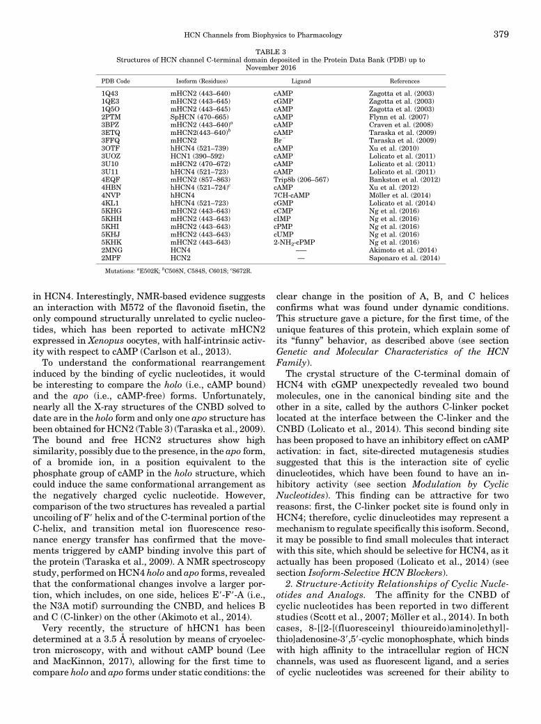
in HCN4. Interestingly, NMR-based evidence suggestsan interaction with M572 of the flavonoid fisetin, theonly compound structurally unrelated to cyclic nucleo-tides, which has been reported to activate mHCN2expressed in Xenopus oocytes, with half-intrinsic activ-ity with respect to cAMP (Carlson et al., 2013).To understand the conformational rearrangement
induced by the binding of cyclic nucleotides, it wouldbe interesting to compare the holo (i.e., cAMP bound)and the apo (i.e., cAMP-free) forms. Unfortunately,nearly all the X-ray structures of the CNBD solved todate are in the holo form and only one apo structure hasbeen obtained for HCN2 (Table 3) (Taraska et al., 2009).The bound and free HCN2 structures show highsimilarity, possibly due to the presence, in the apo form,of a bromide ion, in a position equivalent to thephosphate group of cAMP in the holo structure, whichcould induce the same conformational arrangement asthe negatively charged cyclic nucleotide. However,comparison of the two structures has revealed a partialuncoiling of F9 helix and of the C-terminal portion of theC-helix, and transition metal ion fluorescence reso-nance energy transfer has confirmed that the move-ments triggered by cAMP binding involve this part ofthe protein (Taraska et al., 2009). A NMR spectroscopystudy, performed onHCN4 holo and apo forms, revealedthat the conformational changes involve a larger por-tion, which includes, on one side, helices E9-F9-A (i.e.,the N3A motif) surrounding the CNBD, and helices Band C (C-linker) on the other (Akimoto et al., 2014).Very recently, the structure of hHCN1 has been
determined at a 3.5 Å resolution by means of cryoelec-tron microscopy, with and without cAMP bound (Leeand MacKinnon, 2017), allowing for the first time tocompare holo and apo forms under static conditions: the
clear change in the position of A, B, and C helicesconfirms what was found under dynamic conditions.This structure gave a picture, for the first time, of theunique features of this protein, which explain some ofits “funny” behavior, as described above (see sectionGenetic and Molecular Characteristics of the HCNFamily).
The crystal structure of the C-terminal domain ofHCN4 with cGMP unexpectedly revealed two boundmolecules, one in the canonical binding site and theother in a site, called by the authors C-linker pocketlocated at the interface between the C-linker and theCNBD (Lolicato et al., 2014). This second binding sitehas been proposed to have an inhibitory effect on cAMPactivation: in fact, site-directed mutagenesis studiessuggested that this is the interaction site of cyclicdinucleotides, which have been found to have an in-hibitory activity (see section Modulation by CyclicNucleotides). This finding can be attractive for tworeasons: first, the C-linker pocket site is found only inHCN4; therefore, cyclic dinucleotides may represent amechanism to regulate specifically this isoform. Second,it may be possible to find small molecules that interactwith this site, which should be selective for HCN4, as itactually has been proposed (Lolicato et al., 2014) (seesection Isoform-Selective HCN Blockers).
2. Structure-Activity Relationships of Cyclic Nucle-otides and Analogs. The affinity for the CNBD ofcyclic nucleotides has been reported in two differentstudies (Scott et al., 2007; Möller et al., 2014). In bothcases, 8-[[2-[(fluoresceinyl thioureido)amino]ethyl]-thio]adenosine-39,59-cyclic monophosphate, which bindswith high affinity to the intracellular region of HCNchannels, was used as fluorescent ligand, and a seriesof cyclic nucleotides was screened for their ability to
TABLE 3Structures of HCN channel C-terminal domain deposited in the Protein Data Bank (PDB) up to
November 2016
PDB Code Isoform (Residues) Ligand References
1Q43 mHCN2 (443–640) cAMP Zagotta et al. (2003)1QE3 mHCN2 (443–645) cGMP Zagotta et al. (2003)1Q5O mHCN2 (443–645) cAMP Zagotta et al. (2003)2PTM SpHCN (470–665) cAMP Flynn et al. (2007)3BPZ mHCN2 (443–640)a cAMP Craven et al. (2008)3ETQ mHCN2(443–640)b cAMP Taraska et al. (2009)3FFQ mHCN2 Br2 Taraska et al. (2009)3OTF hHCN4 (521–739) cAMP Xu et al. (2010)3UOZ HCN1 (390–592) cAMP Lolicato et al. (2011)3U10 mHCN2 (470–672) cAMP Lolicato et al. (2011)3U11 hHCN4 (521–723) cAMP Lolicato et al. (2011)4EQF mHCN2 (857–863) Trip8b (206–567) Bankston et al. (2012)4HBN hHCN4 (521–724)c cAMP Xu et al. (2012)4NVP hHCN4 7CH-cAMP Möller et al. (2014)4KL1 hHCN4 (521–723) cGMP Lolicato et al. (2014)5KHG mHCN2 (443–643) cCMP Ng et al. (2016)5KHH mHCN2 (443–643) cIMP Ng et al. (2016)5KHI mHCN2 (443–643) cPMP Ng et al. (2016)5KHJ mHCN2 (443–643) cUMP Ng et al. (2016)5KHK mHCN2 (443–643) 2-NH2-cPMP Ng et al. (2016)2MNG HCN4 –— Akimoto et al. (2014)2MPF HCN2 — Saponaro et al. (2014)
Mutations: aE502K; bC508N, C584S, C601S; cS672R.
HCN Channels from Biophysics to Pharmacology 379

displace it from the CNBD. The two studies useddifferent proteins and number of compounds, butreached similar conclusions. In the first one, theaffinity of 20 ligands was determined on a dimericconstruct, made with the rat HCN2 CNBD and theDNA binding domain of the cAMP receptor protein(Scott et al., 2007). In the more recent one, 47 analogswere screened on the C-terminal domain (C-linker andCNBD) of mHCN1, mHCN2, and hHCN4 channels,kept as monomers by means of a maltose-bindingprotein tag (Möller et al., 2014).The 47 tested compounds did not display selectivity
for the three isoforms, but structural modificationsgreatly affected affinity. Among the naturally occurringcyclic nucleotides, cAMP was, as expected, the mostpotent, whereas others (cGMP, cCMP, cIMP, cPMP,cUMP) interacted with lower affinity. The pivotal role ofthe ionic bond with R591 has been confirmed by thedrop-in affinity found for the cAMP and cGMP analogshaving the equatorial phosphate oxygen atom substitutedwith sulfur; a similar replacement on the axial oxygenatom, involved in H-bond with Thr, reduced affinitytoo, although to a lesser extent. The 29-hydroxyl groupforms anH-bond as donor with a key glutamate residue(Zhou and Siegelbaum, 2007); this interaction isnecessary because methylation strongly reduced orabolished binding. Substitution in position 8 with Br,fluorescein, or other groups gave compounds withimproved affinity, indicating some space available forbulky substituents. On the contrary, modifications onthe NH2 groups of cAMP and cGMP were detrimental,suggesting that these groups too are involved in binding.Unexpectedly, the replacement of the nitrogen atom inposition 7 with CH on cAMP, cGMP, and other analogsimproved affinity: the EC50 for 7CH-cAMP were 100-,64-, and 108-fold lower than those of cAMP on, re-spectively, HCN1, HCN2, and HCN4 C-terminal do-mains. The reason for this increase in affinity was clearfrom the X-ray structure of the complex of 7CH-cAMPwith the HCN4 intracellular region; although cAMP andits CH analog interact in the same way, the hydrophobicenvironment surrounding the seven position of thepurine ring stabilizes a lipophilic CH group better thana hydrophilic N atom. On human HCN4 channel,expressed in HEK293 cells, 7CH-cAMP behaves as anactivator with potency four times higher than cAMP;moreover, it displayed some selectivity forHCNchannelswith respect to PKA and to exchange protein directlyactivated by cAMP.
B. Pharmacology of HCN Blockade byExogenous Ligands
1. Ivabradine, Cilobradine, and Other SpecificBradycardic Agents. The definition “specific bradycardicagent” (SBA) refers to a group of structurally diversesubstances, discovered in the second half of 1900. Thesecompounds belong to two different chemical classes:
N-alkyl-aminoimidazolines, exemplified by alinidine(2-[N-allyl-N-(2,6-dichlorophenyl)amino]imidazoline) andphenylalkylamines structurally related to falipamil(AQ-A-39) (Fig. 6). The word “specific”meant that thesemolecules, and their congeners, were able to reduceheart rate at concentrations at which other hemody-namic parameters were not affected; it was proposedthat these compounds exerted their activity directly onthe SAN, blocking the funny current, just discovered atthat time (Kobinger and Lillie, 1987). Later on, it wasfound that the activity of SBAs was not confined tocardiac tissue because these compounds could block thehyperpolarization-activated current also in neurons(Pape, 1994).
The development of alinidine and analogs was stop-ped due to the concern about the formation of clonidineas metabolite, and by the observation of visual sideeffects in phase I clinical trials (Kobinger, 1987). On thecontrary, the research on phenylalkylamines yieldedsome interesting compounds. Increasing the size of thefive-membered pyrrolidone ring of falipamil into aseven-membered benzazepinone moiety gave a morepotent and safer compound, zatebradine (UL-FS49),which entered clinical trials; the development of thismolecule too was halted due to adverse effects. Thebenzazepinone moiety is found also on other twophenylalkylamines: cilobradine (DH-AK269) and ivab-radine (S16257). Both compounds are reduced-flexibilityanalogs of zatebradine: in cilobradine, the three-methylene chain became part of a six-membered piperi-dine ring, whereas in ivabradine the phenethyl group wasincorporated into a bicyclo[4.2.0]octa-1(6),2,4-trien-7-ylmethyl moiety. In both cases, a chiral center was in-troduced into the molecules, and the eutomer is theS enantiomer (Figure 6).
The three drugs have a similar mode of action: theyreduce spontaneous AP firing in SAN preparation,mainly through a reduction in the rate of diastolicdepolarization (Thollon et al., 1994). On sheep Purkinjefibers, these drugs reduced Ih amplitude, withoutmodifying the voltage dependence (Van Bogaert andPittoors, 2003). Ih blockade is use-dependent (Goethalset al., 1993) and occurs on the open channel (Goethalset al., 1993; DiFrancesco, 1994; Bois et al., 2007). Noeffect on contractility is produced (Lillie and Kobinger,1986; Bois et al., 1996). Cilobradine is reported to bemore potent than zatebradine in sheep Purkinjie fibersandmouse DRG (Raes et al., 1998). At variance with theother two drugs, zatebradine prolonged APs in somecardiac preparations (Thollon et al., 1994; Perez et al.,1995b), showing class III antiarrhythmic properties(Valenzuela et al., 1996). This could be one of thereasons for which the development of zatebradine hasstopped; another was, again, the observation of visualside effects, coming from zatebradine blockade of Ihinvolved in the recovery of photoresponse in retinal rods(Satoh and Yamada, 2002). Site-directed mutagenesis,
380 Sartiani et al.

electrophysiology, and molecular modeling studies sug-gest that cilobradine and ivabradine share a commonbinding site, located in the internal mouth of thechannel pore (Cheng et al., 2007; Bucchi et al., 2013);the proposed mechanism is that these drugs enter thebinding site from the intracellular side of the channel(Bucchi et al., 2006).ZD7288 (Fig. 6) is another blocker of thehyperpolarization-
activated current, which is structurally unrelated to theother SBAs. Its pharmacological characterization showedthat it behaves as specific bradycardic agent acting on theSAN without having any other direct effects on the heart(BoSmith et al., 1993).Different fromphenylalkylamines,the Ih blockade byZD7288 isnotuse-dependent, but thereis evidence from site-directed mutagenesis that thebinding site is the same of ivabradine (Bucchi et al.,2013). ZD7288 has not been developed as drug, but it iswidely used as a tool to study Ih.The word “specific” does not imply that SBAs are
completely selective for HCN channels; all compoundsreported in this section are able to interact also withother targets. For instance, alinidine is a K+ channelblocker (McPherson and Angus, 1989; Jonas et al.,1992); ZD7288 interacts with Na+ and Ca2+ channels(Sanchez-Alonso et al., 2008; Wu et al., 2012). Severalpapers have reported that the interaction of ivabradinewith other ion channels occurs at concentration higherthan those active on HCN channels (Perez et al., 1995a;Bois et al., 1996; Delpon et al., 1996; Koncz et al., 2011);however,more recently, two independent studies showedthat ivabradine blocks human ether a-go-go (hERG) K+
channels and HCN4 channels with similar potency(Lees-Miller et al., 2015; Melgari et al., 2015), raisingconcern on the safety of this drug (Hancox et al., 2015).
The SBAs discussed in this section were disclosedbefore discovering the heterogeneity of HCN channelfamily; their ability to discriminate among HCN chan-nel isoforms has been checked only for zatebradine,cilobradine, and ivabradine (see next section).
2. Isoform-Selective HCN Blockers. The ability ofivabradine, cilobradine, and zatebradine to discrimi-nate among HCN channel isoforms has been tested onhuman homomeric HCN1–4 channels expressed inHEK293 cells (Stieber et al., 2006). The three com-pounds did not display selectivity or preference for oneisoform over the others; the IC50 were in all cases in thelow micromolar range. The lack of selectivity may notsurprise, if one considers that the four isoforms share ahigh degree (80%–90%) of sequence identity in theregion comprising the six transmembrane segments(S1–S6) and the CNBD (Kaupp and Seifert, 2001); thisregion harbors the proposed binding site for ivabradine,cilobradine, and ZD7288. However, sequence alignmentof the S5-P-S6 region of HCN1, HCN2, and HCN4isoforms—where site-directed mutagenesis studies lo-cate the residues important for binding (Shin et al.,2001; Cheng et al., 2007; Chan et al., 2009; Bucchi et al.,2013)—reveals some differences in the sequence, that,in principle, could pave the way to selectivity forcompounds’ interaction at this site.
Indeed, structural modifications of the nonselectiveblocker zatebradine gave compounds (Fig. 7) showing
Fig. 6. Chemical structure of specific bradycardic agents.
HCN Channels from Biophysics to Pharmacology 381

different activity on homomeric HCN1, HCN2, andHCN4 channels expressed in HEK293 cells. The in-clusion of the three-methylene chain of zatebradineinto a cyclohexane ring gave EC18, showing a sixfoldpreference for HCN4 over HCN1 and HCN2 channels(Del Lungo et al., 2012). The replacement of the three-methylene chain with a cis-butene moiety, combinedwith the introduction of a chiral center on thephenethyl group, gaveMEL55A [labeled in the originalpaper as (R)-5], which was fourfold and 11-fold morepotent on HCN2 than on HCN1 and HCN4, respec-tively (Melchiorre et al., 2010). MEL57A, serendipi-tously discoveredwhile synthesizingMEL55A, displayed30- and 170-fold selectivity for HCN1 over HCN2 andHCN4, respectively. Notably, the selectivity found inrecombinant systemswasmaintained in tissues express-ing different HCN isoforms. On guinea pig SAN cells, thereduction of Ih produced by EC18 was threefold higherthan that induced by MEL57A; both compounds weretested at concentrations close to the IC50 values. Incontrast, in mouse DRG neurons, MEL57A, but notEC18, significantly reduced Ih. In dog cardiac Purkinjefibers, EC18 reduced the amplitude and slowed the slopeof the spontaneous diastolic depolarization, whereasMEL57A was ineffective (Del Lungo et al., 2012). Incultured DRG, MEL55A reduced Ih amplitude and cellexcitability, its effect being quantitatively similar to that
of ivabradine (unpublished data). The utility of isoform-selective and HCN-channel–specific compounds appearsevident in studying Ih from tissues expressing differentisoforms: for instance, the application of the HCN4-preferring blocker EC18 suggested a functional role ofthis isoform in mediating Ih in GABAergic interneuronsof the dorsal part of the lateral geniculate nucleus (Leistet al., 2016).
Researchers at Johnson & Johnson (San Diego, CA)disclosed a HCN1-selective compound. Starting from thehigh throughput screening of a proprietary library, andafter optimization of the initial lead, piperazine 1 wasobtained (Fig. 7); this compound was 12-, 7-, and 10-foldmorepotent onhomomericHCN1channels than onHCN2,HCN3, and HCN4, respectively (McClure et al., 2011).Differently from the nonselective compound ZD7288, 1 dis-played antihyperalgesic activity in a spared nerve injurymodel at doses at which heart rate was not affected.
Among the compounds described in the next section,amiodarone shows some isoform selectivity: its IC50
value for blockade of homomeric channels expressed inXenopus oocytes is 4- and 22-fold lower on HCN4 thanon HCN1 and HCN2, respectively (Fan et al., 2011).However, multi-ion channels blocking properties mayobscure the selectivity of this compound.
Although it is reasonable to hypothesize that EC18,MEL55A, and MEL57A, being structural analogs of
Fig. 7. Chemical structure of isoform-selective HCN blockers.
382 Sartiani et al.

ivabradine and cilobradine, bind to the same interactionsite, nothing is known about the binding site of com-pound 1 and amiodarone. As reported in the previoussection (Lessons from Crystallography), the X-ray struc-ture of the complex cGMP-HCN4 revealed the existenceof a pocket in the C-linker, proposed to be the bindingsite for cyclic dinucleotides, but available also forsynthetic modulators (Lolicato et al., 2014). Moleculesbinding at this site should display selectivity becausethis pocket is not present in other isoforms. Indeed, bymeans of virtual screening, a compound has been dis-closed (N9-biphenyl-2-yl-N-[1-(3-cyanobenzyl)piperidin-4-yl]-N-(pyridin-3-ylmethyl)urea, 2; Fig. 7), which wasfound able to antagonize cAMP activation on HCN4,but not on HCN2 channels. This pocket may repre-sent a new opportunity for delivering HCN4-selectivecompounds.3. HCN Blockade by Other Drugs. Several drugs
have been shown to block HCN channel, a property thatcan either contribute to their pharmacological activityor, alternatively, produce side effects. Some of thecompoundsmentioned in this sectionareGprotein–coupledreceptor modulators, controlling cAMP concentrationthrough adenylyl cyclase–coupled Ga proteins; therefore,they could be endowedwithmixed direct–indirect activity.In the following discussion, only compounds used in theclinic are included; for a more comprehensive list, seeRomanelli et al. (2016). The compounds have been dividedinto subgroups according to their pharmacological activity.a. Analgesic/Antihyperalgesic drugs. Dexmedetomidine
and clonidine are a2-AR agonists commonly used forseveral indications, including analgesia. They sharesome structural similarities, carrying a five-memberedimidazole or imidazoline ring. In addition, as discussedin section Ivabradine, Cilobradine, and Other SpecificBradycardic Agents, clonidine is a metabolite ofthe SBA alinidine. Both compounds have been shownto block recombinant HCN channels expressed inHEK293 cells (Knaus et al., 2007; Yang et al., 2014),and in vivo studies suggested that the analgesia bydexmedetomidine can be produced by ana2-independentmechanism (Brummett et al., 2011; Yang et al., 2014).For both drugs, bradycardia is a common side effect(Anger, 2013; Isbister et al., 2017), although onlyclonidine has been tested on SAN (Knaus et al., 2007).The analgesic activity of both compounds can also bedue to a receptor-mediated mechanism: a2-AR activa-tion by these drugs is reported to decrease Ih in severalneuronal preparations (Parkis and Berger, 1997; Carret al., 2007; Shirasaka et al., 2007; Inyushin et al.,2010), including some involved in pain perception (Yagiand Sumino, 1998; Takeda et al., 2002). In someinstances, it appears that the receptor-mediated in-hibition of HCN channels does not occur through theclassic Ga,i-linked reduction of cAMP concentration,rather through activation of protein kinase C via Gbg
modulation (Carr et al., 2007; Inyushin et al., 2010).
Loperamide and tramadol are opioid receptor ago-nists, which have been reported to block Ih by opioidreceptor–independent mechanisms. Tramadol wastested on GH3 cells, where it inhibited Ih with micro-molar potency; the effect was not affected by preincu-bation with naloxone (Liu et al., 2009). Loperamide ismainly used as antidiarrheal drug, but it showedanalgesic activity and antihyperalgesic properties inanimal models of neuropathic pain; in some instances,this activity was not dependent of opioid receptors(Ringkamp et al., 2012). Loperamide was able to blockHCN1 and HCN4 channels expressed in HEK293 cells(Vasilyev et al., 2009), and to inhibit Ih in rat DRG,acting from the extracellular side of the channel(Vasilyev et al., 2007); the authors suggested that thiseffect could contribute to the analgesic activity of thedrug.
The antihyperalgesic activity of minocycline has beentested in several models of neuropathic pain, and it hasbeen associated with inhibition of microglia activation(Mika, 2008; Möller et al., 2016); the molecular mech-anism is unknown, but could be related also to the ionchannel–blocking properties of this drug. On substan-tia gelatinosa neurons of rat spinal cord, minocyclinereduced Ih and decreased the rate of AP firing; themaximal inhibition was 40% and was produced by adirect interaction with the channel from the extracel-lular side (Liu et al., 2015). Potency was in themicromolar range, but IC50 value was two orders ofmagnitude higher than that for Na+ channel blockadein DRG (Kim et al., 2011).
Eugenol, a dental analgesic obtained from clove oil,was found able to block hyperpolarization-activatedcurrent and decrease firing of APs in trigeminal gan-glion neurons. After rat infraorbital nerve chronicconstriction injury, it reversed mechanical allodynia atlower doses with respect to those active in thermalhyperalgesia. The Ih-blocking properties may contrib-ute to the analgesic and antihyperalgesic activity of thismolecule (Yeon et al., 2011).
b. General anesthetics. Propofol is a general anes-thetic, with additional antiemetic and antiepilepticproperties (Kotani et al., 2008; Lundström et al., 2010).Propofol is a small lipophilic molecule, which interactswith multiple targets, including several ion channels(Kojima et al., 2015). Ih blockade by propofol has beenstudied in several tissues: hippocampal neurons(Funahashi et al., 2001; Higuchi et al., 2003), areapostrema (Funahashi et al., 2004), thalamocorticalneurons (Ying et al., 2006; Chen et al., 2009a), and EC(Li et al., 2016). Overall, these studies suggest that Ihblockade can contribute substantially to the pharmaco-logical actions of the drug, which seems to involvemainly HCN1 and HCN2 channel isoforms (Yinget al., 2006; Chen et al., 2009a). In X. laevis oocytesexpressing homomeric HCN1, HCN2, and HCN4, andheteromeric HCN1-HCN2 channels, HCN1 was the
HCN Channels from Biophysics to Pharmacology 383

most sensitive isoform (Cacheaux et al., 2005; Chenet al., 2005). Consistent with HCN channel blockade arethe findings that propofol reduces mechanical andthermal hyperalgesia in a peripheral nerve ligationmodel of neuropathic pain (Tibbs et al., 2013), and thatbradycardia is a common side effect after administra-tion of this drug (Lundström et al., 2010).Ketamine is another general anesthetic withmultiple
action sites, among which HCN channels: in HEK293cells the drug strongly inhibited homomeric HCN1 andheteromeric HCN1–HCN2 channels, but not homo-meric HCN2 channels. This activity is likely to contrib-ute to the hypnotic action of ketamine, as demonstratedin HCN1-KO mice (Chen et al., 2009a). InterestinglyHCN1 inhibition alone can theoretically explain thechanges in EEG produced by ketamine or propofol,without taking into account interaction with NMDA orGABA receptors (Bojak et al., 2013).Other volatile general anesthetics have been shown
to reduce Ih; for instance, halotane and isofluraneinteract with HCN1 and HCN2 channels, producing ahyperpolarizing shift in voltage dependence of activa-tion and a decrease in maximal available current atclinically relevant concentrations (Chen et al., 2005,2009b). Studies using global and forebrain-selectiveHCN1-KO mice showed that interaction with this iso-form in the forebrain contributes to hypnotic andamnestic effects of isoflurane and sevoflurane, but notto their immobilizing action (Zhou et al., 2015b). Inthalamocortical neurons, xenon reduced signal propa-gation by interacting with HCN2 channels, the pre-dominant isoform expressed in this tissue (Ludwiget al., 1998); this effect was not seen in HCN2-KOanimals. Accordingly, xenon produced a sedative effectin WT mice, but not in mice lacking HCN2 channels(Mattusch et al., 2015).c. Local anesthetic and antiarrhythmic drugs.
The Ih-blocking activity of the local anesthetic andantiarrhythmic drug lidocaine has been measured inrecombinant systems and on neuronal and cardiactissues. Lidocaine was tested on homomeric mHCN1,mHCN2, and mHCN4, and heteromeric mHCN1–HCN2 channels expressed in X. laevis oocytes andHEK293 cells (Meng et al., 2011): on all isoforms thedrug caused a decrease in both tonic and maximalcurrent and slowed current activation kinetics; onlyfor HCN1-containing channels it produced a hyper-polarizing shift in voltage dependence of activation.On rat DRG (Bischoff et al., 2003; Putrenko andSchwarz, 2011) and in substantia gelatinosa neurons(Hu et al., 2016), the blockade was reversible; the dose–response curves yielded similar IC50 values (;80–99 mM). In a mice model of sciatic nerve block, lidocainewas more potent inWTmice that in HCN12/2mice, andthe duration of lidocaine anesthesia was longer in WTthan inHCN12/2mice (Zhou et al., 2015a). Itwas suggestedthat HCN channel blockade might substantially contribute
to the drug action during epidural and spinal anesthesia.When tested on rat ventrobasal thalamocortical relayneurons, lidocaine blocked Ih with potency similar tothat found in DRG (Putrenko and Schwarz, 2011); theauthors suggest that this activity could contribute tolidocaine’s systemic analgesic actions and may par-tially explain neurotoxicity.
In addition to lidocaine, bupivacaine and mepivacainealso have been tested on rat DRG neurons (Bischoffet al., 2003). The mechanism of Ih blockade was similarto that of lidocaine; bupivacaine was about twice andfour times as potent as lidocaine and mepivacaine,respectively.
In cardiac tissues, the Ih-blocking properties of lido-caine have been demonstrated more than 30 years agoin sheep cardiac Purkinje fibers (Carmeliet and Saikawa,1982), and more recently on rabbit SAN myocytes(Rocchetti et al., 1999). In the latter study, the IC50 value(38 mM, twofold lower than in neuronal tissues) sug-gested some concern on a possible interference of thedrug on normal cardiac automaticity. Indeed, whentested on recombinant mHCN4 channel, Ih blockadewas found significant also at physiologic potentials(270 mV) (Meng et al., 2011).
However, under different experimental conditions,the effect produced by lidocaine on recombinant rabbitHCN4 channels, at 270 mV, was considered clinicallynot relevant (Tamura et al., 2009). This study wasperformed on a large series of antiarrhythmic drugs, togain information about their effects on the pacemakercurrent for a more rational clinical use of these com-pounds. To simulate physiologic conditions, i.e., thetonic stimulation by the sympathetic nerve and thepresence in the cytosol of cyclic nucleotides, the blockingactivity was measured in presence of 0.3 mM intracel-lular cAMP, and the calculated IC50 values werecompared with therapeutic concentrations. For severalcompounds (quinidine, disopyramide, cibenzoline,mexiletine, aprindine, propafenone, flecainide, propranolol,verapamil, sotalol, as well as lidocaine), the effect wasconsidered negligible; on the contrary, the IC50 values ofamiodarone and bepridil were close to their therapeuticconcentrations.
The effect of amiodarone on Ih was further character-ized on human HCN channel isoforms (HCN1, HCN2,and HCN4) expressed in X. laevis oocytes (Fan et al.,2011): on all isoforms the blockade was concentration-and use-dependent, but the activation curves were notmodified. Interestingly, amiodarone was found to be4 and 20 times more potent on HCN4 that on HCN2 andHCN1, respectively. Amiodarone was found to block Ihalso in rabbit SAN preparations (Satoh, 1991), ventric-ular myocytes from spontaneously hypertensive andnormal rats (Li et al., 2014a), but not in isolated humanatrial myocytes (Hoppe and Beuckelmann, 1998). Inventricular myocytes from spontaneously hypertensiverats, amiodarone significantly suppressed Ih density
384 Sartiani et al.

and downregulated HCN2 and HCN4 expression (Liet al., 2014a).Dronedarone is a structural analog of amiodarone,
used in the treatment of AF. On hHCN4 channelexpressed in Chinese hamster ovary cells, dronedaronewas equipotent with amiodarone and ivabradine inblocking Ih (Bogdan et al., 2011). Clinical trials haveevidenced a mild bradycardic effect of this drug (Singhet al., 2007), which has been shown to be mainly due toHCN channel inhibition (Sobrado et al., 2013). In aporcine model, dronedarone induced a significant re-duction in ventricular rate during AF, owing to Ihinhibition in AVN, which slowed conduction (Verrieret al., 2014).
C. In Vivo Studies and Clinical Pharmacology
1. Ivabradine in Angina and Heart Failure. Thecurrent therapeutic indication for ivabradine stemsfrom its primary development as selective HCN blocker:ivabradine is the only commercially available specificbradycardic agent. To understand why bradycardiarepresents an attractive target in angina, it is worthto recall briefly the mechanisms underlying myocardialischemia. Ischemic cardiomyopathy arises from animbalance between oxygen supply to and demand bythe myocardium. Several factors contribute to thisdisequilibrium, namely flow reduction through epicardialcoronary arteries, due to plaques, vasospasm, or—asmorerecently acquired—microcirculatory dysfunction (for areview, see Camici et al., 2016). Myocardial perfusionand oxygen extraction occur mainly during the diastolicphase, in either healthy or diseased heart, but allowinga sufficient rest time between two consecutive beats ismore crucial for the latter condition, due to the unevenblood flow. At the same time, heart rate is the primarydeterminant of oxygen demand. Therefore, decreasingheart rate is a primary therapeutic goal in ischemiccardiomyopathy, traditionally achieved by using beta-blockers, which attenuate angina symptoms. Neverthe-less, a significant percentage of patients do not achievethe optimal beta-blocker dosage because of adverseeffects or contraindications. Thus, recent EuropeanSociety of Cardiology guidelines recommend ivabradineas a second-line therapy (class IIa, level B) in patientswith ischemic cardiomyopathy intolerant to or poorlycontrolled by beta-blockers (Montalescot et al., 2013).This recommendation is based on clinical evidence: inthe BEAUTIFUL trial, ivabradine reduced severe car-diovascular events in patients with angina and a heartrate$70 beats perminute (bpm) (Fox et al., 2008). Aloneor combinedwith beta-blockers, ivabradine reduces SANrhythm, both at rest and during exercise, without(further) depressing contractility.However, recent work suggests that the interplay
between heart rate and cardiac protection might bemore complex than a plain cardiac cycle/oxygen balancescheme. In animal models of ischemic cardiomyopathy,
ivabradine protects against risk factors, such asatherosclerotic plaque formation (Custodis et al.,2008), energetic imbalance, and electrophysiologicalremodeling (Ceconi et al., 2011). These results led tonew hypothesis on the relationship between heart ratereduction and improved coronary perfusion, because ofcollateral artery growth and/or recovery of endothelialcell function during bradycardia (Schirmer et al., 2012).Thus, the distinction between classic and pleiotropic(ancillary) mechanisms of action is tenue, because—based on present knowledge—all can be largelyascribed to HCN blockade in SAN cells. Indeed, atvariance with beta-blockers, ivabradine does notpossess intrinsic vasoconstrictive activity, whichmay explain a gain of function during exercise, i.e.,by preserving coronary dilation mediated by catechol-amines (Camici et al., 2016). In the same line, thereduction of heart rate, but not the dosage, wassignificantly related to the survival in patients withheart failure treated with beta-blockers (McAlisteret al., 2009).
In the SHIFT Trial (Bohm et al., 2010), addingivabradine to current standard therapy in patients withheart failure safely lowered heart rate below 70 bpm,which is considered a desirable target to improve out-come in patients with ventricular dysfunction not oftenachieved with beta-blockers due to side effects. Morerecently, the SIGNIFY study in patients with preservedejection fraction and coronary artery disease, in whichivabradine was added to standard background therapy,showed a significant increase in cardiovascular eventsconcomitant to, or despite, heart rate reduction (about60 bpm versus 70 bpm) (Fox et al., 2014). Based on theresults of these three large studies and their post hocanalysis, ivabradine should be used as a second-linetherapy to reduce the risk of hospitalization in patientswith symptomatic chronic heart failure and restingheart rate.70 bpm despite maximally tolerated dosageof beta-blockers (or contraindications to them) (Ponikowskiet al., 2016). Adverse effects reported in these large trialsalso reflect ivabradine cardiac and extracardiac actions:themost commonare bradycardia and visual disturbances(phosphenes) due to blockade of—respectively—cardiacand retinal HCN channels.
a. HCN blockade and arrhythmogenesis: an openissue. Far less defined and consequently exploitableare the potential antiarrhythmic profile of ivabradineand in general HCN blockade. Verrier et al. (2013)showed that the capability to control ventricular rate bydronedarone, the de-iodinated analogous of amiodar-one, resides in its HCN-blocking effect on AVN conduc-tion in a pig model of AF. This is not surprising because,as described above, HCN4 channels are functionallyrelevant in nodal conduction (Liu et al., 2008). Sub-sequent work by the same group reported a similareffect in pigs treated with ivabradine alone (Verrieret al., 2014) or with ranolazine (Verrier et al., 2015): the
HCN Channels from Biophysics to Pharmacology 385

association reduced AVN conduction and AF dominant,leaving ventricular contractility and duration of the QTinterval (an ECG marker of repolarization velocity)unaltered. Finally, recent reports in AF patientsshowed improved rate control with ivabradine addedto beta-blockade (Caminiti et al., 2016; Kosiuk et al.,2016), although these sporadic observations are obvi-ously too weak for drawing conclusions.As mentioned above, a subgroup analysis of the
SIGNIFY study unraveled increased cardiac events inpatients with preserved contractile function treatedwith ivabradine, but neither bradycardia nor emergentAF appear to have an impact (Fox et al., 2015). Aproarrhythmic potential of HCN blockade has beeninferred based on telemetric recording of ECGs in micetreated with different specific bradycardic agents(Stieber et al., 2004). Periodic fluctuations of the R-Rinterval appeared in the ECG, depending on dosage anddrug type, the order of potency being cilobradine .zatebradine .. ivabradine, without significant alter-ations in AVN conduction. These fluctuations resemblea well-known phenomenon termed Wenckebach’s peri-odicity, typical of the AVN. In SAN cells, themechanismunderlying the sinoatrialWenckebach’s periodicitymayreside in the different HCN expression and sensitivityto bradycardic agents in the central versus peripheralsinus node (Stieber et al., 2004; Mangoni and Nargeot,2008). Finally, the bradycardic effect of ivabradinemight stimulate the sympathetic drive, which is per sea proarrhythmic stimulus (Dias da Silva et al., 2015).However, extrapolation of data obtained in rodents to
the clinical settingmay be unwarranted, and indeed theusefulness of ivabradine has been postulated in AF,although limited clinical evidence exists at present(Turley et al., 2016). Of note, Ih seems to undergosignificant remodeling in chronic AF, as described above(Stillitano et al., 2013). Such a gain of function in theworking atrial myocardium parallels that observed byseveral groups, including some of us, in hypertrophiedand failing ventricular cardiomyocytes [reviewed inSartiani et al. (2015)]. It is interesting to observe thatchronic treatment with ivabradine exerts a reverseremodeling action in animal models of heart failure(Ceconi et al., 2011), by counteracting electrophysiolog-ical abnormalities, including HCN overexpression andfunction in atrial and ventricular cardiomyocytes(Suffredini et al., 2012). Finally, off-label uses ofivabradine have been reported in other settings(Oliphant et al., 2016), although evidence from clinicaltrials is lacking.2. Treatment of Pain.a. HCN channel modulation in neuropathic pain.
The analgesic action of the HCN channel blockerZD7288 in different models of neuropathic pain hasbeen largely demonstrated (Dalle and Eisenach, 2005;Jiang et al., 2008; Takasu et al., 2010; Nava et al., 2014).In particular, tactile allodynia and thermal hyperalgesia
were largely decreased by ZD7288 application. This isnot surprising due to the wide expression of HCNchannels along the peripheral pain neuraxis (Pappet al., 2006; Papp et al., 2010). Interesting results derivedfrom genetically engineered HCN mutants underlinedthe important role played by HCN2 in neuropathic painmaintenance (Schnorr et al., 2014). Emery et al. (2011,2012) demonstrated that conditional deletion of HCN2abolished pain behavior in the chronic constriction injurymodel. These results were largely confirmed in a differ-ent model of neuropathy using a virtually identicalHCN2 mutant (snsHCN2KO) (Herrmann et al., 2015),thus pointing out HCN2 as the most important subunitmodulating neuropathic pain.
However, in chemotherapy-induced periphery neuro-pathic models, HCN1 modification seems to play aprominent role compared with HCN2 in modulatingpain sensitization. Recently, Zhang and Dougherty(2014) demonstrated an increased expression of HCN1in DRG neurons in a rat model of paclitaxel-inducedperipheral neuropathy. They also observed an in-creased frequency of spontaneous discharging neuronsin both myelinated and unmyelinated neurons. Thehyperpolarization-activated current mediated byHCN1 may facilitate the rise of spontaneous activityin these neurons (Zhang and Dougherty, 2014). Fur-thermore, Descoeur et al. (2011) observed a significantincrease in HCN1 mRNA levels in DRG fromoxaliplatin-treated mice. Moreover, they observed thativabradine exerted analgesic effect on cold allodynia,but not on mechanical hyperalgesia. These two exam-ples provide evidence of a prominent role for HCN1channel in chemotherapy-induced neuropathy.
Recently, the role of HCN channels in CNS forneuropathic pain has also been investigated. Interest-ingly, sciatic nerve injury caused an activity-dependentdysfunction of HCN channels in the dendrites of layer5 pyramidal neurons, resulting in an increased neuro-nal activity in cortical areas, which has been postulatedto be one of the key manifestations of chronic pain(Kuner, 2010; Saab, 2012). It was demonstrated thatspecific activation of 5-HT7 receptor increased HCNchannel function, thus restoring normal dendritic in-tegration and reducing mechanical pain hypersensitiv-ity in nerve-injured animals in vivo (Santello andNevian, 2015).
b. Participation of HCN channels in inflammatorypain behavior. HCN appears to play a role also inmodulation of inflammatory pain. Both nonselective Ihblockers and genetic deletion of HCN display a signif-icant analgesic effect in different animal models of in-flammation (Dunlop et al., 2009; Emery et al., 2011). ThecAMP-sensitive isoform HCN2 seems to be particularlyinvolved in heat hyperalgesia, as inferred from experi-ments using conditional HCN2-KO mice challenged withcarrageenan or prostaglandin E2–mediated inflammatorystimuli (Emery et al., 2011). The inflammatory processes,
386 Sartiani et al.
which activate signaling transduction pathways, cause anincrease of cAMP in pain-sensing neurons responsible forperipheral HCN2 channel activation, which in turn con-trols the acute phase of neuronal hypersensitivity. Indeed,cAMP shifts the resting membrane potential to depolar-ized values and increases AP firing in isolated DRGneurons, whereas these modifications do not occur inHCN2KO or by using nonselective HCN channel blockers(Emery et al., 2011; Schnorr et al., 2014; Young et al.,2014). On the contrary, models of inflammation notimplying Gs activation are insensitive to HCN2 deletionor HCN channel blockade (Schnorr et al., 2014), thusdemonstrating the absence of HCN influence on cAMP-independent nociceptive behavior.Similarly, chronic inflammatory states, in which
transcriptional regulatory processes are more impor-tant compared with intracellular cAMP increase forHCN-mediated sensitization, have been observed to bepartially HCN-independent. Indeed, it appears thatHCN channels only modulate tactile hypersensitivity,being heat hyperalgesia not affected by either HCN2deletion or HCN channel block in chronic inflammatorymodels (Luo et al., 2007; Weng et al., 2012; Schnorret al., 2014). These results suggest that distinct neuro-nal pathways convey mechanical and thermal sensiti-zation in chronic inflammatory conditions (Abrahamsenet al., 2008; Cavanaugh et al., 2009; Herrmann et al.,2015). Nonetheless, pharmacological targets modulat-ing Gi-coupled receptors have been reported to interactvia HCN modulation to a decreased nociceptive re-sponse (Resta et al., 2016).
V. Conclusions
Despite the mass of information that has beenaccumulating on the biophysical and molecular proper-ties of HCN channels and their (patho)physiologic rolein excitable (and not-excitable) cells, we are now seeingthe beginning of pharmacological and therapeutic ex-ploitation. Indeed, the unique properties of this channelfamily required a long-standing effort for clarifying itsstructural features and modulatory pathways. It is notsurprising that the first (and single for the moment)HCN-related drug in the medical armamentarium wasa bradycardic agent, targeting the earliest physiologicfunction ascribed to Ih.Still, clues emerge for opportunities of future in-
vestigation aimed at identifying pharmacological strat-egies: the prevalence of different HCN subtypes inorgans and tissues, the possibility to target HCN gainor loss of function associated with disease, the feasibil-ity of novel isoform-selective drugs, as well as thediscovery of HCN-mediated effects for old medicines.The modulation of neuropathic pain could be likely thenext in line precisely because of a combination of thesefavorable factors. However, the impressive evidenceemerging from genetic and molecular studies involving
HCN channels in the CNS could pave the way to un-foreseen therapeutic innovation.
Acknowledgments
We thank Alessandro Mugelli for invaluable support and insightfulcontribution to authors’ research in this field.
Authorship Contributions
Wrote or contributed to the writing of the manuscript: Sartiani,Masi, Mannaioni, Romanelli, Cerbai.
ReferencesAbi-Gerges N, Ji GJ, Lu ZJ, Fischmeister R, Hescheler J, and Fleischmann BK (2000)Functional expression and regulation of the hyperpolarization activated non-selective cation current in embryonic stem cell-derived cardiomyocytes. J Physiol523:377–389.
Abrahamsen B, Zhao J, Asante CO, Cendan CM, Marsh S, Martinez-Barbera JP,Nassar MA, Dickenson AH, and Wood JN (2008) The cell and molecular basis ofmechanical, cold, and inflammatory pain. Science 321:702–705.
Accili EA, Proenza C, Baruscotti M, and DiFrancesco D (2002) From funny current toHCN channels: 20 years of excitation. News Physiol Sci 17:32–37.
Acosta C, McMullan S, Djouhri L, Gao L, Watkins R, Berry C, Dempsey K,and Lawson SN (2012) HCN1 and HCN2 in Rat DRG neurons: levels in nociceptorsand non-nociceptors, NT3-dependence and influence of CFA-induced skin in-flammation on HCN2 and NT3 expression. PLoS One 7:e50442.
Akimoto M, Zhang Z, Boulton S, Selvaratnam R, VanSchouwen B, Gloyd M, AcciliEA, Lange OF, and Melacini G (2014) A mechanism for the auto-inhibition ofhyperpolarization-activated cyclic nucleotide-gated (HCN) channel opening and itsrelief by cAMP. J Biol Chem 289:22205–22220.
Alig J, Marger L, Mesirca P, Ehmke H, Mangoni ME, and Isbrandt D (2009) Controlof heart rate by cAMP sensitivity of HCN channels. Proc Natl Acad Sci USA 106:12189–12194.
Alonso A and Llinás RR (1989) Subthreshold Na+-dependent theta-like rhythmicityin stellate cells of entorhinal cortex layer II. Nature 342:175–177.
Alotaibi M, Kahlat K, Nedjadi T, and Djouhri L (2017) Effects of ZD7288, ahyperpolarization-activated cyclic nucleotide-gated (HCN) channel blocker, onterm-pregnant rat uterine contractility in vitro. Theriogenology 90:141–146.
Altomare C, Bucchi A, Camatini E, Baruscotti M, Viscomi C, Moroni A,and DiFrancesco D (2001) Integrated allosteric model of voltage gating of HCNchannels. J Gen Physiol 117:519–532.
Altomare C, Terragni B, Brioschi C, Milanesi R, Pagliuca C, Viscomi C, Moroni A,Baruscotti M, and DiFrancesco D (2003) Heteromeric HCN1-HCN4 channels: acomparison with native pacemaker channels from the rabbit sinoatrial node. JPhysiol 549:347–359.
Anger KE (2013) Dexmedetomidine: a review of its use for the management of pain,agitation, and delirium in the intensive care unit. Curr Pharm Des 19:4003–4013.
Arencibia-Albite F, Vázquez R, Velásquez-Martinez MC, and Jiménez-Rivera CA(2012) Cocaine sensitization inhibits the hyperpolarization-activated cation cur-rent Ih and reduces cell size in dopamine neurons of the ventral tegmental area. JNeurophysiol 107:2271–2282.
Arencibia-Albite F, Vázquez-Torres R, and Jiménez-Rivera CA (2017) Cocaine sen-sitization increases subthreshold activity in dopamine neurons from the ventraltegmental area. J Neurophysiol 117:612–623.
Arinsburg SS, Cohen IS, and Yu HG (2006) Constitutively active Src tyrosine kinasechanges gating of HCN4 channels through direct binding to the channel proteins. JCardiovasc Pharmacol 47:578–586.
Arnsten AF (2011) Prefrontal cortical network connections: key site of vulnerabilityin stress and schizophrenia. Int J Dev Neurosci 29:215–223.
Arnsten AF and Jin LE (2014) Molecular influences on working memory circuits indorsolateral prefrontal cortex. Prog Mol Biol Transl Sci 122:211–231.
Bankston JR, Camp SS, DiMaio F, Lewis AS, Chetkovich DM, and Zagotta WN(2012) Structure and stoichiometry of an accessory subunit TRIP8b interactionwith hyperpolarization-activated cyclic nucleotide-gated channels. Proc Natl AcadSci USA 109:7899–7904.
Barbieri M, Varani K, Cerbai E, Guerra L, Li Q, Borea PA, and Mugelli A (1994)Electrophysiological basis for the enhanced cardiac arrhythmogenic effect of iso-prenaline in aged spontaneously hypertensive rats. J Mol Cell Cardiol 26:849–860.
Barbuti A, Crespi A, Capilupo D, Mazzocchi N, Baruscotti M, and DiFrancesco D(2009) Molecular composition and functional properties of f-channels in murineembryonic stem cell-derived pacemaker cells. J Mol Cell Cardiol 46:343–351.
Barbuti A, Gravante B, Riolfo M, Milanesi R, Terragni B, and DiFrancesco D (2004)Localization of pacemaker channels in lipid rafts regulates channel kinetics. CircRes 94:1325–1331.
Barbuti A and Robinson RB (2015) Stem cell-derived nodal-like cardiomyocytes as anovel pharmacologic tool: insights from sinoatrial node development and function.Pharmacol Rev 67:368–388.
Barbuti A, Scavone A, Mazzocchi N, Terragni B, Baruscotti M, and Difrancesco D(2012) A caveolin-binding domain in the HCN4 channels mediates functional in-teraction with caveolin proteins. J Mol Cell Cardiol 53:187–195.
Barbuti A, Terragni B, Brioschi C, and DiFrancesco D (2007) Localization off-channels to caveolae mediates specific beta2-adrenergic receptor modulation ofrate in sinoatrial myocytes. J Mol Cell Cardiol 42:71–78.
Baruscotti M, Bianco E, Bucchi A, and DiFrancesco D (2016) Current understandingof the pathophysiological mechanisms responsible for inappropriate sinus tachy-cardia: role of the If “funny” current. J Interv Card Electrophysiol 46:19–28.
HCN Channels from Biophysics to Pharmacology 387

Baruscotti M, Bucchi A, Viscomi C, Mandelli G, Consalez G, Gnecchi-Rusconi T,Montano N, Casali KR, Micheloni S, Barbuti A, et al. (2011) Deep bradycardia andheart block caused by inducible cardiac-specific knockout of the pacemaker channelgene Hcn4. Proc Natl Acad Sci USA 108:1705–1710.
Bédard C, Kröger H, and Destexhe A (2006) Model of low-pass filtering of local fieldpotentials in brain tissue. Phys Rev E Stat Nonlin Soft Matter Phys 73:051911.
Bellingham MC (2013) Pre- and postsynaptic mechanisms underlying inhibition ofhypoglossal motor neuron excitability by riluzole. J Neurophysiol 110:1047–1061.
Bender RA, Brewster A, Santoro B, Ludwig A, Hofmann F, Biel M, and Baram TZ(2001) Differential and age-dependent expression of hyperpolarization-activated,cyclic nucleotide-gated cation channel isoforms 1-4 suggests evolving roles in thedeveloping rat hippocampus. Neuroscience 106:689–698.
Bender RA, Kirschstein T, Kretz O, Brewster AL, Richichi C, Rüschenschmidt C,Shigemoto R, Beck H, Frotscher M, and Baram TZ (2007) Localization of HCN1channels to presynaptic compartments: novel plasticity that may contribute tohippocampal maturation. J Neurosci 27:4697–4706.
Bender RA, Soleymani SV, Brewster AL, Nguyen ST, Beck H, Mathern GW,and Baram TZ (2003) Enhanced expression of a specific hyperpolarization-activated cyclic nucleotide-gated cation channel (HCN) in surviving dentate gyrusgranule cells of human and experimental epileptic hippocampus. J Neurosci 23:6826–6836.
Berger T, Senn W, and Lüscher HR (2003) Hyperpolarization-activated current Ihdisconnects somatic and dendritic spike initiation zones in layer V pyramidalneurons. J Neurophysiol 90:2428–2437.
Biel M, Wahl-Schott C, Michalakis S, and Zong X (2009) Hyperpolarization-activatedcation channels: from genes to function. Physiol Rev 89:847–885.
Biel S, Aquila M, Hertel B, Berthold A, Neumann T, DiFrancesco D, Moroni A, ThielG, and Kauferstein S (2016) Mutation in S6 domain of HCN4 channel in patientwith suspected Brugada syndrome modifies channel function. Pflugers Arch 468:1663–1671.
Bischoff U, Bräu ME, Vogel W, Hempelmann G, and Olschewski A (2003) Localanaesthetics block hyperpolarization-activated inward current in rat small dorsalroot ganglion neurones. Br J Pharmacol 139:1273–1280.
Blackiston DJ, McLaughlin KA, and Levin M (2009) Bioelectric controls of cell pro-liferation: ion channels, membrane voltage and the cell cycle. Cell Cycle 8:3527–3536.
Bogdan R, Goegelein H, and Ruetten H (2011) Effect of dronedarone on Na+, Ca2+ andHCN channels. Naunyn Schmiedebergs Arch Pharmacol 383:347–356.
Böhm M, Swedberg K, Komajda M, Borer JS, Ford I, Dubost-Brama A, Lerebours G,and Tavazzi L; SHIFT Investigators (2010) Heart rate as a risk factor in chronicheart failure (SHIFT): the association between heart rate and outcomes in arandomised placebo-controlled trial. Lancet 376:886–894.
Bois P, Bescond J, Renaudon B, and Lenfant J (1996) Mode of action of bradycardicagent, S 16257, on ionic currents of rabbit sinoatrial node cells. Br J Pharmacol118:1051–1057.
Bois P, Guinamard R, Chemaly AE, Faivre J-F, and Bescond J (2007) Molecularregulation and pharmacology of pacemaker channels. Curr Pharm Des 13:2338–2349.
Bojak I, Day HC, and Liley DT (2013) Ketamine, propofol, and the EEG: a neuralfield analysis of HCN1-mediated interactions. Front Comput Neurosci 7:22.
Bolívar JJ, Tapia D, Arenas G, Castañón-Arreola M, Torres H, and Galarraga E(2008) A hyperpolarization-activated, cyclic nucleotide-gated, (Ih-like) cationiccurrent and HCN gene expression in renal inner medullary collecting duct cells.Am J Physiol Cell Physiol 294:C893–C906.
Borel M, Guadagna S, Jang HJ, Kwag J, and Paulsen O (2013) Frequency de-pendence of CA3 spike phase response arising from h-current properties. Front CellNeurosci 7:263.
Bosman A, Sartiani L, Spinelli V, Del Lungo M, Stillitano F, Nosi D, Mugelli A,Cerbai E, and Jaconi M (2013) Molecular and functional evidence of HCN4 andcaveolin-3 interaction during cardiomyocyte differentiation from human embryonicstem cells. Stem Cells Dev 22:1717–1727.
BoSmith RE, Briggs I, and Sturgess NC (1993) Inhibitory action of ZENECA ZD7288on whole-cell hyperpolarization activated inward current (If) in guinea-pig disso-ciated sinoatrial node cells. Br J Pharmacol 110:313–349.
Boyes J, Bolam JP, Shigemoto R, and Stanford IM (2007) Functional presynapticHCN channels in the rat globus pallidus. Eur J Neurosci 25:2081–2092.
Branch SY, Chen C, Sharma R, Lechleiter JD, Li S, and Beckstead MJ (2016) Do-paminergic neurons exhibit an age-dependent decline in electrophysiological pa-rameters in the MitoPark mouse model of Parkinson’s disease. J Neurosci 36:4026–4037.
Brandt MC, Endres-Becker J, Zagidullin N, Motloch LJ, Er F, Rottlaender D, MichelsG, Herzig S, and Hoppe UC (2009) Effects of KCNE2 on HCN isoforms: distinctmodulation of membrane expression and single channel properties. Am J PhysiolHeart Circ Physiol 297:H355–H363.
Brewster A, Bender RA, Chen Y, Dube C, Eghbal-Ahmadi M, and Baram TZ (2002)Developmental febrile seizures modulate hippocampal gene expression ofhyperpolarization-activated channels in an isoform- and cell-specific manner.J Neurosci 22:4591–4599.
Brewster AL, Chen Y, Bender RA, Yeh A, Shigemoto R, and Baram TZ (2007)Quantitative analysis and subcellular distribution of mRNA and protein expres-sion of the hyperpolarization-activated cyclic nucleotide-gated channels throughoutdevelopment in rat hippocampus. Cereb Cortex 17:702–712.
Brown HF, DiFrancesco D, and Noble SJ (1979) How does adrenaline accelerate theheart? Nature 280:235–236.
Brummett CM, Hong EK, Janda AM, Amodeo FS, and Lydic R (2011) Perineuraldexmedetomidine added to ropivacaine for sciatic nerve block in rats prolongs theduration of analgesia by blocking the hyperpolarization-activated cation current.Anesthesiology 115:836–843.
Bucchi A, Baruscotti M, Nardini M, Barbuti A, Micheloni S, Bolognesi M,and DiFrancesco D (2013) Identification of the molecular site of ivabradine bindingto HCN4 channels. PLoS One 8:e53132.
Bucchi A, Tognati A, Milanesi R, Baruscotti M, and DiFrancesco D (2006) Propertiesof ivabradine-induced block of HCN1 and HCN4 pacemaker channels. J Physiol572:335–346.
Cacheaux LP, Topf N, Tibbs GR, Schaefer UR, Levi R, Harrison NL, Abbott GW,and Goldstein PA (2005) Impairment of hyperpolarization-activated, cyclicnucleotide-gated channel function by the intravenous general anesthetic propofol.J Pharmacol Exp Ther 315:517–525.
Camici PG, Gloekler S, Levy BI, Skalidis E, Tagliamonte E, Vardas P, and Heusch G(2016) Ivabradine in chronic stable angina: effects by and beyond heart rate re-duction. Int J Cardiol 215:1–6.
Caminiti G, Fossati C, Rosano G, and Volterrani M (2016) Addition of ivabradine tobetablockers in patients with atrial fibrillation: Effects on heart rate and exercisetolerance. Int J Cardiol 202:73–74.
Cangiano L, Gargini C, Della Santina L, Demontis GC, and Cervetto L (2007) High-pass filtering of input signals by the Ih current in a non-spiking neuron, the retinalrod bipolar cell. PLoS One 2:e1327.
Cao JL, Covington IIIHE, Friedman AK, Wilkinson MB, Walsh JJ, Cooper DC,Nestler EJ, and Han MH (2010) Mesolimbic dopamine neurons in the brain rewardcircuit mediate susceptibility to social defeat and antidepressant action. J Neurosci30:16453–16458.
Cao XJ and Oertel D (2011) The magnitudes of hyperpolarization-activated and low-voltage-activated potassium currents co-vary in neurons of the ventral cochlearnucleus. J Neurophysiol 106:630–640.
Carlson AE, Rosenbaum JC, Brelidze TI, Klevit RE, and Zagotta WN (2013) Flavo-noid regulation of HCN2 channels. J Biol Chem 288:33136–33145.
Carmeliet E (1984) Existence of pacemaker current Ifin human atrial appendagefibres. J Physiol 357:125P.
Carmeliet E and Saikawa T (1982) Shortening of the action potential and reduction ofpacemaker activity by lidocaine, quinidine, and procainamide in sheep cardiacpurkinje fibers: an effect on Na or K currents? Circ Res 50:257–272.
Carr DB, Andrews GD, Glen WB, and Lavin A (2007) a2-Noradrenergic receptorsactivation enhances excitability and synaptic integration in rat prefrontal cortexpyramidal neurons via inhibition of HCN currents. J Physiol 584:437–450.
Cavanaugh DJ, Lee H, Lo L, Shields SD, Zylka MJ, Basbaum AI, and Anderson DJ(2009) Distinct subsets of unmyelinated primary sensory fibers mediate behavioralresponses to noxious thermal and mechanical stimuli. Proc Natl Acad Sci USA 106:9075–9080.
Ceconi C, Comini L, Suffredini S, Stillitano F, Bouly M, Cerbai E, Mugelli A,and Ferrari R (2011) Heart rate reduction with ivabradine prevents the globalphenotype of left ventricular remodeling. Am J Physiol Heart Circ Physiol 300:H366–H373.
Cerbai E, Barbieri M, and Mugelli A (1994) Characterization of thehyperpolarization-activated current, I(f), in ventricular myocytes isolated fromhypertensive rats. J Physiol 481:585–591.
Cerbai E, Crucitti A, Sartiani L, De Paoli P, Pino R, Rodriguez ML, Gensini G,and Mugelli A (2000) Long-term treatment of spontaneously hypertensive ratswith losartan and electrophysiological remodeling of cardiac myocytes. CardiovascRes 45:388–396.
Cerbai E, De Paoli P, Sartiani L, Lonardo G, and Mugelli A (2003) Treatment withirbesartan counteracts the functional remodeling of ventricular myocytes fromhypertensive rats. J Cardiovasc Pharmacol 41:804–812.
Cerbai E and Mugelli A (2006) I(f) in non-pacemaker cells: role and pharmacologicalimplications. Pharmacol Res 53:416–423.
Cerbai E, Pino R, Porciatti F, Sani G, Toscano M, Maccherini M, Giunti G,and Mugelli A (1997) Characterization of the hyperpolarization-activated current, I(f), in ventricular myocytes from human failing heart. Circulation 95:568–571.
Cerbai E, Pino R, Rodriguez ML, and Mugelli A (1999a) Modulation of the pacemakercurrent If by beta-adrenoceptor subtypes in ventricular myocytes isolated fromhypertensive and normotensive rats. Cardiovasc Res 42:121–129.
Cerbai E, Pino R, Sartiani L, and Mugelli A (1999b) Influence of postnatal-development on I(f) occurrence and properties in neonatal rat ventricular myo-cytes. Cardiovasc Res 42:416–423.
Cerbai E, Sartiani L, DePaoli P, Pino R, Maccherini M, Bizzarri F, DiCiolla F, DavoliG, Sani G, and Mugelli A (2001) The properties of the pacemaker current I(F)inhuman ventricular myocytes are modulated by cardiac disease. J Mol Cell Cardiol33:441–448.
Cervetto L, Demontis GC, and Gargini C (2007) Cellular mechanisms underlying thepharmacological induction of phosphenes. Br J Pharmacol 150:383–390.
Chan Y-C, Wang K, Au KW, Lau C-P, Tse H-F, and Li RA (2009) Probing the bra-dycardic drug binding receptor of HCN-encoded pacemaker channels [publishedcorrection appears in Pflugers Arch (2010) 459:519]. Pflugers Arch 459:25–38.
Chandler NJ, Greener ID, Tellez JO, Inada S, Musa H, Molenaar P, Difrancesco D,Baruscotti M, Longhi R, Anderson RH, et al. (2009) Molecular architecture of thehuman sinus node: insights into the function of the cardiac pacemaker. Circulation119:1562–1575.
Chaplan SR, Guo HQ, Lee DH, Luo L, Liu C, Kuei C, Velumian AA, Butler MP,Brown SM, and Dubin AE (2003) Neuronal hyperpolarization-activated pacemakerchannels drive neuropathic pain. J Neurosci 23:1169–1178.
Chen C (1997) Hyperpolarization-activated current (Ih) in primary auditory neurons.Hear Res 110:179–190.
Chen K, Aradi I, Thon N, Eghbal-Ahmadi M, Baram TZ, and Soltesz I (2001) Persis-tently modified h-channels after complex febrile seizures convert the seizure-inducedenhancement of inhibition to hyperexcitability. Nat Med 7:331–337.
Chen S, Liang MC, Chia JN, Ngsee JK, and Ting AE (2001a) Rab8b and its inter-acting partner TRIP8b are involved in regulated secretion in AtT20 cells. J BiolChem 276:13209–13216.
Chen S, Wang J, and Siegelbaum SA (2001b) Properties of hyperpolarization-activated pacemaker current defined by coassembly of HCN1 and HCN2 subunitsand basal modulation by cyclic nucleotide. J Gen Physiol 117:491–504.
Chen X, Shu S, and Bayliss DA (2009a) HCN1 channel subunits are a molecularsubstrate for hypnotic actions of ketamine. J Neurosci 29:600–609.
388 Sartiani et al.

Chen X, Shu S, Kennedy DP, Willcox SC, and Bayliss DA (2009b) Subunit-specificeffects of isoflurane on neuronal Ih in HCN1 knockout mice. J Neurophysiol 101:129–140.
Chen X, Sirois JE, Lei Q, Talley EM, Lynch IIIC, and Bayliss DA (2005) HCNsubunit-specific and cAMP-modulated effects of anesthetics on neuronal pace-maker currents. J Neurosci 25:5803–5814.
Cheng L, Kinard K, Rajamani R, and Sanguinetti MC (2007) Molecular mapping ofthe binding site for a blocker of hyperpolarization-activated, cyclic nucleotide-modulated pacemaker channels. J Pharmacol Exp Ther 322:931–939.
Cheng Q and Zhou Y (2013) Novel role of KT5720 on regulating hyperpolarization-activated cyclic nucleotide-gated channel activity and dorsal root ganglion neuronexcitability. DNA Cell Biol 32:320–328.
Chow SS, Van Petegem F, and Accili EA (2012) Energetics of cyclic AMP binding toHCN channel C terminus reveal negative cooperativity. J Biol Chem 287:600–606.
Christoffels VM, Smits GJ, Kispert A, and Moorman AF (2010) Development of thepacemaker tissues of the heart. Circ Res 106:240–254.
Craven KB, Olivier NB, and Zagotta WN (2008) C-terminal movement during gatingin cyclic nucleotide-modulated channels. J Biol Chem 283:14728–14738.
Craven KB and Zagotta WN (2006) CNG and HCN channels: two peas, one pod. AnnuRev Physiol 68:375–401.
Custodis F, Baumhäkel M, Schlimmer N, List F, Gensch C, Böhm M, and Laufs U(2008) Heart rate reduction by ivabradine reduces oxidative stress, improves en-dothelial function, and prevents atherosclerosis in apolipoprotein E-deficient mice.Circulation 117:2377–2387.
Dalle C and Eisenach JC (2005) Peripheral block of the hyperpolarization-activatedcation current (Ih) reduces mechanical allodynia in animal models of postoperativeand neuropathic pain. Reg Anesth Pain Med 30:243–248.
Decher N, Bundis F, Vajna R, and Steinmeyer K (2003) KCNE2 modulates currentamplitudes and activation kinetics of HCN4: influence of KCNE family memberson HCN4 currents. Pflugers Arch 446:633–640.
Dedkov EI, Zheng W, Christensen LP, Weiss RM, Mahlberg-Gaudin F, and TomanekRJ (2007) Preservation of coronary reserve by ivabradine-induced reduction inheart rate in infarcted rats is associated with decrease in perivascular collagen. AmJ Physiol Heart Circ Physiol 293:H590–H598.
Della Santina L, Bouly M, Asta A, Demontis GC, Cervetto L, and Gargini C (2010)Effect of HCN channel inhibition on retinal morphology and function in normal anddystrophic rodents. Invest Ophthalmol Vis Sci 51:1016–1023.
Del Lungo M, Melchiorre M, Guandalini L, Sartiani L, Mugelli A, Koncz I, Szel T,Varro A, Romanelli MN, and Cerbai E (2012) Novel blockers of hyperpolarization-activated current with isoform selectivity in recombinant cells and native tissue. BrJ Pharmacol 166:602–616.
Delpón E, Valenzuela C, Pérez O, Franqueza L, Gay P, Snyders DJ, and Tamargo J(1996) Mechanisms of block of a human cloned potassium channel by theenantiomers of a new bradycardic agent: S-16257-2 and S-16260-2. Br J Pharmacol117:1293–1301.
De Mello WC (2004) Heart failure: how important is cellular sequestration? The roleof the renin-angiotensin-aldosterone system. J Mol Cell Cardiol 37:431–438.
Deng T, Zhang Q, Wang Q, Zhong X, and Li L (2015) Changes in hyperpolarization-activated cyclic nucleotide-gated channel expression and activity in bladder in-terstitial cells of Cajal from rats with detrusor overactivity. Int Urogynecol J PelvicFloor Dysfunct 26:1139–1145.
Descoeur J, Pereira V, Pizzoccaro A, Francois A, Ling B, Maffre V, Couette B, BusserollesJ, Courteix C, Noel J, et al. (2011) Oxaliplatin-induced cold hypersensitivity isdue to remodelling of ion channel expression in nociceptors. EMBO Mol Med 3:266–278.
Dias da Silva VJ, Tobaldini E, Rocchetti M, Wu MA, Malfatto G, Montano N,and Zaza A (2015) Modulation of sympathetic activity and heart rate variability byivabradine. Cardiovasc Res 108:31–38.
Dibbens LM, Reid CA, Hodgson B, Thomas EA, Phillips AM, Gazina E, Cromer BA,Clarke AL, Baram TZ, Scheffer IE, et al. (2010) Augmented currents of an HCN2variant in patients with febrile seizure syndromes. Ann Neurol 67:542–546.
DiFrancesco D (1981a) A new interpretation of the pace-maker current in calf Pur-kinje fibres. J Physiol 314:359–376.
DiFrancesco D (1981b) A study of the ionic nature of the pace-maker current in calfPurkinje fibres. J Physiol 314:377–393.
DiFrancesco D (1982) Block and activation of the pace-maker channel in calf purkinjefibres: effects of potassium, caesium and rubidium. J Physiol 329:485–507.
DiFrancesco D (1986) Characterization of single pacemaker channels in cardiac sino-atrial node cells. Nature 324:470–473.
DiFrancesco D (1993) Pacemaker mechanisms in cardiac tissue. Annu Rev Physiol55:455–472.
DiFrancesco D (1994) Some properties of the UL-FS 49 block of thehyperpolarization-activated current (i(f)) in sino-atrial node myocytes. PflugersArch 427:64–70.
DiFrancesco D (1999) Dual allosteric modulation of pacemaker (f) channels by cAMPand voltage in rabbit SA node. J Physiol 515:367–376.
DiFrancesco D (2006) Serious workings of the funny current. Prog Biophys Mol Biol90:13–25.
DiFrancesco D (2010) The role of the funny current in pacemaker activity. Circ Res106:434–446.
DiFrancesco D and Borer JS (2007) The funny current: cellular basis for the control ofheart rate. Drugs 67 (Suppl 2):15–24.
DiFrancesco D, Ducouret P, and Robinson RB (1989) Muscarinic modulation of car-diac rate at low acetylcholine concentrations. Science 243:669–671.
DiFrancesco D, Ferroni A, Mazzanti M, and Tromba C (1986) Properties of thehyperpolarizing-activated current (if) in cells isolated from the rabbit sino-atrialnode. J Physiol 377:61–88.
DiFrancesco D and Noble D (2012) The funny current has a major pacemaking role inthe sinus node. Heart Rhythm 9:299–301.
DiFrancesco D and Tortora P (1991) Direct activation of cardiac pacemaker channelsby intracellular cyclic AMP. Nature 351:145–147.
DiFrancesco JC, Barbuti A, Milanesi R, Coco S, Bucchi A, Bottelli G, Ferrarese C,Franceschetti S, Terragni B, Baruscotti M, et al. (2011) Recessive loss-of-functionmutation in the pacemaker HCN2 channel causing increased neuronal excitabilityin a patient with idiopathic generalized epilepsy. J Neurosci 31:17327–17337.
DiFrancesco JC and DiFrancesco D (2015) Dysfunctional HCN ion channels in neu-rological diseases. Front Cell Neurosci 6:174.
Dobrzynski H, Anderson RH, Atkinson A, Borbas Z, D’Souza A, Fraser JF, Inada S,Logantha SJ, Monfredi O, Morris GM, et al. (2013) Structure, function and clinicalrelevance of the cardiac conduction system, including the atrioventricular ring andoutflow tract tissues. Pharmacol Ther 139:260–288.
D’Souza A, Bucchi A, Johnsen AB, Logantha SJ, Monfredi O, Yanni J, Prehar S, HartG, Cartwright E, Wisloff U, et al. (2014) Exercise training reduces resting heartrate via downregulation of the funny channel HCN4. Nat Commun 5:3775.
D’Souza A, Sharma S, and Boyett MR (2015) CrossTalk opposing view: bradycardiain the trained athlete is attributable to a downregulation of a pacemaker channelin the sinus node. J Physiol 593:1749–1751.
Dufour MA, Woodhouse A, and Goaillard JM (2014) Somatodendritic ion channelexpression in substantia nigra pars compacta dopaminergic neurons across post-natal development. J Neurosci Res 92:981–999.
Dunlop J, Vasilyev D, Lu P, Cummons T, and Bowlby MR (2009) Hyperpolarization-activated cyclic nucleotide-gated (HCN) channels and pain. Curr Pharm Des 15:1767–1772.
Dyhrfjeld-Johnsen J, Morgan RJ, and Soltesz I (2009) Double Trouble? Potential forhyperexcitability following both channelopathic up- and downregulation of I(h) inepilepsy. Front Neurosci 3:25–33.
El-Kholy W, MacDonald PE, Fox JM, Bhattacharjee A, Xue T, Gao X, Zhang Y,Stieber J, Li RA, Tsushima RG, et al. (2007) Hyperpolarization-activated cyclicnucleotide-gated channels in pancreatic beta-cells. Mol Endocrinol 21:753–764.
Emery EC, Young GT, Berrocoso EM, Chen L, and McNaughton PA (2011) HCN2 ionchannels play a central role in inflammatory and neuropathic pain. Science 333:1462–1466.
Emery EC, Young GT, and McNaughton PA (2012) HCN2 ion channels: an emergingrole as the pacemakers of pain. Trends Pharmacol Sci 33:456–463.
Engel D and Seutin V (2015) High dendritic expression of Ih in the proximity of theaxon origin controls the integrative properties of nigral dopamine neurons. JPhysiol 593:4905–4922.
Fabbri A, Fantini M, Wilders R, and Severi S (2017) Computational analysis of thehuman sinus node action potential: model development and effects of mutations. JPhysiol 595:2365–2396.
Fan X, Chen Y, Wu P, Xing J, Chen H, Song T, Yang J, Zhang J, and Huang C (2011)Novel electropharmacological activity of amiodarone on human HCN channelsheterologously expressed in the Xenopus oocytes. Eur J Pharmacol 669:15–23.
Fenske S, Mader R, Scharr A, Paparizos C, Cao-Ehlker X, Michalakis S, Shaltiel L,Weidinger M, Stieber J, Feil S, et al. (2011) HCN3 contributes to the ventricularaction potential waveform in the murine heart. Circ Res 109:1015–1023.
Fernández-Velasco M, Goren N, Benito G, Blanco-Rivero J, Boscá L, and Delgado C(2003) Regional distribution of hyperpolarization-activated current (If) andhyperpolarization-activated cyclic nucleotide-gated channel mRNA expression inventricular cells from control and hypertrophied rat hearts. J Physiol 553:395–405.
Flynn GE, Black KD, Islas LD, Sankaran B, and Zagotta WN (2007) Structure andrearrangements in the carboxy-terminal region of SpIH channels. Structure 15:671–682.
Flynn GE and Zagotta WN (2011) Molecular mechanism underlying phosphatidylinositol4,5-bisphosphate-induced inhibition of SpIH channels. J Biol Chem 286:15535–15542.
Fogle KJ, Lyashchenko AK, Turbendian HK, and Tibbs GR (2007) HCN pacemakerchannel activation is controlled by acidic lipids downstream of diacylglycerol ki-nase and phospholipase A2. J Neurosci 27:2802–2814.
Fox K, Ford I, Steg PG, Tardif JC, Tendera M, and Ferrari R; SIGNIFY Investigators(2014) Ivabradine in stable coronary artery disease without clinical heart failure.NEngl J Med 371:1091–1099.
Fox K, Ford I, Steg PG, Tardif JC, Tendera M, and Ferrari R; SIGNIFY Investigators(2015) Bradycardia and atrial fibrillation in patients with stable coronary arterydisease treated with ivabradine: an analysis from the SIGNIFY study. Eur Heart J36:3291–3296.
Fox K, Ford I, Steg PG, Tendera M, Robertson M, and Ferrari R; BEAUTIFULinvestigators (2008) Heart rate as a prognostic risk factor in patients with coronaryartery disease and left-ventricular systolic dysfunction (BEAUTIFUL): a subgroupanalysis of a randomised controlled trial. Lancet 372:817–821.
Franz O, Liss B, Neu A, and Roeper J (2000) Single-cell mRNA expression of HCN1correlates with a fast gating phenotype of hyperpolarization-activated cyclicnucleotide-gated ion channels (Ih) in central neurons. Eur J Neurosci 12:2685–2693.
Friedman AK, Walsh JJ, Juarez B, Ku SM, Chaudhury D, Wang J, Li X, Dietz DM,Pan N, Vialou VF, et al. (2014) Enhancing depression mechanisms in midbraindopamine neurons achieves homeostatic resilience. Science 344:313–319.
Funahashi M, Higuchi H, Miyawaki T, Shimada M, and Matsuo R (2001) Propofolsuppresses a hyperpolarization-activated inward current in rat hippocampal CA1neurons. Neurosci Lett 311:177–180.
Funahashi M, Mitoh Y, and Matsuo R (2004) The sensitivity of hyperpolarization-activated cation current (Ih) to propofol in rat area postrema neurons. Brain Res1015:198–201.
Fyk-Kolodziej B and Pourcho RG (2007) Differential distribution of hyperpolarization-activated and cyclic nucleotide-gated channels in cone bipolar cells of the rat ret-ina. J Comp Neurol 501:891–903.
Galligan JJ, Tatsumi H, Shen KZ, Surprenant A, and North RA (1990) Cation cur-rent activated by hyperpolarization (IH) in guinea pig enteric neurons. Am JPhysiol 259:G966–G972.
Gamo NJ, Lur G, Higley MJ, Wang M, Paspalas CD, Vijayraghavan S, Yang Y,Ramos BP, Peng K, Kata A, et al. (2015) Stress impairs prefrontal cortical functionvia D1 dopamine receptor interactions with hyperpolarization-activated cyclicnucleotide-gated channels. Biol Psychiatry 78:860–870.
HCN Channels from Biophysics to Pharmacology 389
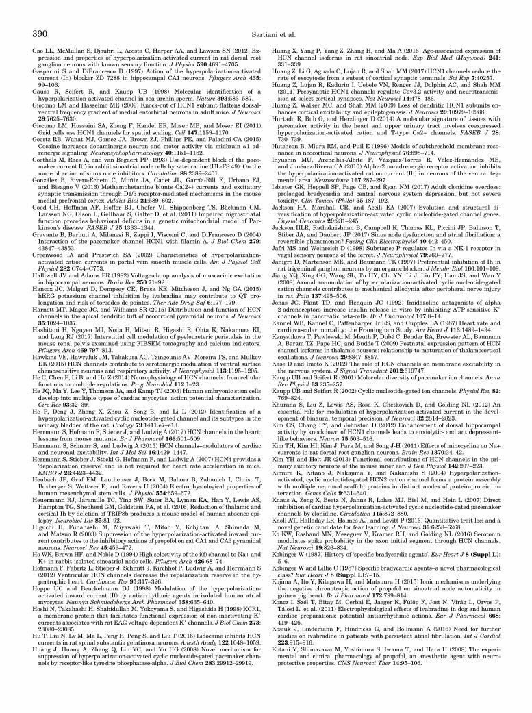
Gao LL, McMullan S, Djouhri L, Acosta C, Harper AA, and Lawson SN (2012) Ex-pression and properties of hyperpolarization-activated current in rat dorsal rootganglion neurons with known sensory function. J Physiol 590:4691–4705.
Gasparini S and DiFrancesco D (1997) Action of the hyperpolarization-activatedcurrent (Ih) blocker ZD 7288 in hippocampal CA1 neurons. Pflugers Arch 435:99–106.
Gauss R, Seifert R, and Kaupp UB (1998) Molecular identification of ahyperpolarization-activated channel in sea urchin sperm. Nature 393:583–587.
Giocomo LM and Hasselmo ME (2009) Knock-out of HCN1 subunit flattens dorsal-ventral frequency gradient of medial entorhinal neurons in adult mice. J Neurosci29:7625–7630.
Giocomo LM, Hussaini SA, Zheng F, Kandel ER, Moser MB, and Moser EI (2011)Grid cells use HCN1 channels for spatial scaling. Cell 147:1159–1170.
Goertz RB, Wanat MJ, Gomez JA, Brown ZJ, Phillips PE, and Paladini CA (2015)Cocaine increases dopaminergic neuron and motor activity via midbrain a1 ad-renergic signaling. Neuropsychopharmacology 40:1151–1162.
Goethals M, Raes A, and van Bogaert PP (1993) Use-dependent block of the pace-maker current I(f) in rabbit sinoatrial node cells by zatebradine (UL-FS 49). On themode of action of sinus node inhibitors. Circulation 88:2389–2401.
González B, Rivero-Echeto C, Muñiz JA, Cadet JL, García-Rill E, Urbano FJ,and Bisagno V (2016) Methamphetamine blunts Ca(2+) currents and excitatorysynaptic transmission through D1/5 receptor-mediated mechanisms in the mousemedial prefrontal cortex. Addict Biol 21:589–602.
Good CH, Hoffman AF, Hoffer BJ, Chefer VI, Shippenberg TS, Bäckman CM,Larsson NG, Olson L, Gellhaar S, Galter D, et al. (2011) Impaired nigrostriatalfunction precedes behavioral deficits in a genetic mitochondrial model of Par-kinson’s disease. FASEB J 25:1333–1344.
Gravante B, Barbuti A, Milanesi R, Zappi I, Viscomi C, and DiFrancesco D (2004)Interaction of the pacemaker channel HCN1 with filamin A. J Biol Chem 279:43847–43853.
Greenwood IA and Prestwich SA (2002) Characteristics of hyperpolarization-activated cation currents in portal vein smooth muscle cells. Am J Physiol CellPhysiol 282:C744–C753.
Halliwell JV and Adams PR (1982) Voltage-clamp analysis of muscarinic excitationin hippocampal neurons. Brain Res 250:71–92.
Hancox JC, Melgari D, Dempsey CE, Brack KE, Mitcheson J, and Ng GA (2015)hERG potassium channel inhibition by ivabradine may contribute to QT pro-longation and risk of torsades de pointes. Ther Adv Drug Saf 6:177–179.
Harnett MT, Magee JC, and Williams SR (2015) Distribution and function of HCNchannels in the apical dendritic tuft of neocortical pyramidal neurons. J Neurosci35:1024–1037.
Hashitani H, Nguyen MJ, Noda H, Mitsui R, Higashi R, Ohta K, Nakamura KI,and Lang RJ (2017) Interstitial cell modulation of pyeloureteric peristalsis in themouse renal pelvis examined using FIBSEM tomography and calcium indicators.Pflugers Arch 469:797–813.
Hawkins VE, Hawryluk JM, Takakura AC, Tzingounis AV, Moreira TS, and MulkeyDK (2015) HCN channels contribute to serotonergic modulation of ventral surfacechemosensitive neurons and respiratory activity. J Neurophysiol 113:1195–1205.
He C, Chen F, Li B, and Hu Z (2014) Neurophysiology of HCN channels: from cellularfunctions to multiple regulations. Prog Neurobiol 112:1–23.
He JQ, Ma Y, Lee Y, Thomson JA, and Kamp TJ (2003) Human embryonic stem cellsdevelop into multiple types of cardiac myocytes: action potential characterization.Circ Res 93:32–39.
He P, Deng J, Zhong X, Zhou Z, Song B, and Li L (2012) Identification of ahyperpolarization-activated cyclic nucleotide-gated channel and its subtypes in theurinary bladder of the rat. Urology 79:1411.e7–e13.
Herrmann S, Hofmann F, Stieber J, and Ludwig A (2012) HCN channels in the heart:lessons from mouse mutants. Br J Pharmacol 166:501–509.
Herrmann S, Schnorr S, and Ludwig A (2015) HCN channels–modulators of cardiacand neuronal excitability. Int J Mol Sci 16:1429–1447.
Herrmann S, Stieber J, Stöckl G, Hofmann F, and Ludwig A (2007) HCN4 provides a‘depolarization reserve’ and is not required for heart rate acceleration in mice.EMBO J 26:4423–4432.
Heubach JF, Graf EM, Leutheuser J, Bock M, Balana B, Zahanich I, Christ T,Boxberger S, Wettwer E, and Ravens U (2004) Electrophysiological properties ofhuman mesenchymal stem cells. J Physiol 554:659–672.
Heuermann RJ, Jaramillo TC, Ying SW, Suter BA, Lyman KA, Han Y, Lewis AS,Hampton TG, Shepherd GM, Goldstein PA, et al. (2016) Reduction of thalamic andcortical Ih by deletion of TRIP8b produces a mouse model of human absence epi-lepsy. Neurobiol Dis 85:81–92.
Higuchi H, Funahashi M, Miyawaki T, Mitoh Y, Kohjitani A, Shimada M,and Matsuo R (2003) Suppression of the hyperpolarization-activated inward cur-rent contributes to the inhibitory actions of propofol on rat CA1 and CA3 pyramidalneurons. Neurosci Res 45:459–472.
Ho WK, Brown HF, and Noble D (1994) High selectivity of the i(f) channel to Na+ andK+ in rabbit isolated sinoatrial node cells. Pflugers Arch 426:68–74.
Hofmann F, Fabritz L, Stieber J, Schmitt J, Kirchhof P, Ludwig A, and Herrmann S(2012) Ventricular HCN channels decrease the repolarization reserve in the hy-pertrophic heart. Cardiovasc Res 95:317–326.
Hoppe UC and Beuckelmann DJ (1998) Modulation of the hyperpolarization-activated inward current (If) by antiarrhythmic agents in isolated human atrialmyocytes. Naunyn Schmiedebergs Arch Pharmacol 358:635–640.
Hoshi N, Takahashi H, Shahidullah M, Yokoyama S, and Higashida H (1998) KCR1,a membrane protein that facilitates functional expression of non-inactivating K+
currents associates with rat EAG voltage-dependent K+ channels. J Biol Chem 273:23080–23085.
Hu T, Liu N, Lv M, Ma L, Peng H, Peng S, and Liu T (2016) Lidocaine inhibits HCNcurrents in rat spinal substantia gelatinosa neurons. Anesth Analg 122:1048–1059.
Huang J, Huang A, Zhang Q, Lin YC, and Yu HG (2008) Novel mechanism forsuppression of hyperpolarization-activated cyclic nucleotide-gated pacemaker chan-nels by receptor-like tyrosine phosphatase-alpha. J Biol Chem 283:29912–29919.
Huang X, Yang P, Yang Z, Zhang H, and Ma A (2016) Age-associated expression ofHCN channel isoforms in rat sinoatrial node. Exp Biol Med (Maywood) 241:331–339.
Huang Z, Li G, Aguado C, Lujan R, and Shah MM (2017) HCN1 channels reduce therate of exocytosis from a subset of cortical synaptic terminals. Sci Rep 7:40257.
Huang Z, Lujan R, Kadurin I, Uebele VN, Renger JJ, Dolphin AC, and Shah MM(2011) Presynaptic HCN1 channels regulate Cav3.2 activity and neurotransmis-sion at select cortical synapses. Nat Neurosci 14:478–486.
Huang Z, Walker MC, and Shah MM (2009) Loss of dendritic HCN1 subunits en-hances cortical excitability and epileptogenesis. J Neurosci 29:10979–10988.
Hurtado R, Bub G, and Herzlinger D (2014) A molecular signature of tissues withpacemaker activity in the heart and upper urinary tract involves coexpressedhyperpolarization-activated cation and T-type Ca2+ channels. FASEB J 28:730–739.
Hutcheon B, Miura RM, and Puil E (1996) Models of subthreshold membrane reso-nance in neocortical neurons. J Neurophysiol 76:698–714.
Inyushin MU, Arencibia-Albite F, Vázquez-Torres R, Vélez-Hernández ME,and Jiménez-Rivera CA (2010) Alpha-2 noradrenergic receptor activation inhibitsthe hyperpolarization-activated cation current (Ih) in neurons of the ventral teg-mental area. Neuroscience 167:287–297.
Isbister GK, Heppell SP, Page CB, and Ryan NM (2017) Adult clonidine overdose:prolonged bradycardia and central nervous system depression, but not severetoxicity. Clin Toxicol (Phila) 55:187–192.
Jackson HA, Marshall CR, and Accili EA (2007) Evolution and structural di-versification of hyperpolarization-activated cyclic nucleotide-gated channel genes.Physiol Genomics 29:231–245.
Jackson IILR, Rathakrishnan B, Campbell K, Thomas KL, Piccini JP, Bahnson T,Stiber JA, and Daubert JP (2017) Sinus node dysfunction and atrial fibrillation: areversible phenomenon? Pacing Clin Electrophysiol 40:442–450.
Jafri MS and Weinreich D (1998) Substance P regulates Ih via a NK-1 receptor invagal sensory neurons of the ferret. J Neurophysiol 79:769–777.
Janigro D, Martenson ME, and Baumann TK (1997) Preferential inhibition of Ih inrat trigeminal ganglion neurons by an organic blocker. J Membr Biol 160:101–109.
Jiang YQ, Xing GG, Wang SL, Tu HY, Chi YN, Li J, Liu FY, Han JS, and Wan Y(2008) Axonal accumulation of hyperpolarization-activated cyclic nucleotide-gatedcation channels contributes to mechanical allodynia after peripheral nerve injuryin rat. Pain 137:495–506.
Jonas JC, Plant TD, and Henquin JC (1992) Imidazoline antagonists of alpha2-adrenoceptors increase insulin release in vitro by inhibiting ATP-sensitive K+
channels in pancreatic beta-cells. Br J Pharmacol 107:8–14.Kannel WB, Kannel C, Paffenbarger Jr.RS, and Cupples LA (1987) Heart rate andcardiovascular mortality: the Framingham Study. Am Heart J 113:1489–1494.
Kanyshkova T, Pawlowski M, Meuth P, Dubé C, Bender RA, Brewster AL, BaumannA, Baram TZ, Pape HC, and Budde T (2009) Postnatal expression pattern of HCNchannel isoforms in thalamic neurons: relationship to maturation of thalamocorticaloscillations. J Neurosci 29:8847–8857.
Kase D and Imoto K (2012) The role of HCN channels on membrane excitability inthe nervous system. J Signal Transduct 2012:619747.
Kaupp UB and Seifert R (2001) Molecular diversity of pacemaker ion channels. AnnuRev Physiol 63:235–257.
Kaupp UB and Seifert R (2002) Cyclic nucleotide-gated ion channels. Physiol Rev 82:769–824.
Khurana S, Liu Z, Lewis AS, Rosa K, Chetkovich D, and Golding NL (2012) Anessential role for modulation of hyperpolarization-activated current in the devel-opment of binaural temporal precision. J Neurosci 32:2814–2823.
Kim CS, Chang PY, and Johnston D (2012) Enhancement of dorsal hippocampalactivity by knockdown of HCN1 channels leads to anxiolytic- and antidepressant-like behaviors. Neuron 75:503–516.
Kim TH, Kim HI, Kim J, Park M, and Song J-H (2011) Effects of minocycline on Na+currents in rat dorsal root ganglion neurons. Brain Res 1370:34–42.
Kim YH and Holt JR (2013) Functional contributions of HCN channels in the pri-mary auditory neurons of the mouse inner ear. J Gen Physiol 142:207–223.
Kimura K, Kitano J, Nakajima Y, and Nakanishi S (2004) Hyperpolarization-activated, cyclic nucleotide-gated HCN2 cation channel forms a protein assemblywith multiple neuronal scaffold proteins in distinct modes of protein-protein in-teraction. Genes Cells 9:631–640.
Knaus A, Zong X, Beetz N, Jahns R, Lohse MJ, Biel M, and Hein L (2007) Directinhibition of cardiac hyperpolarization-activated cyclic nucleotide-gated pacemakerchannels by clonidine. Circulation 115:872–880.
Knoll AT, Halladay LR, Holmes AJ, and Levitt P (2016) Quantitative trait loci and anovel genetic candidate for fear learning. J Neurosci 36:6258–6268.
Ko KW, Rasband MN, Meseguer V, Kramer RH, and Golding NL (2016) Serotoninmodulates spike probability in the axon initial segment through HCN channels.Nat Neurosci 19:826–834.
Kobinger W (1987) History of ‘specific bradycardic agents’. Eur Heart J 8 (Suppl L):5–6.
Kobinger W and Lillie C (1987) Specific bradycardic agents–a novel pharmacologicalclass? Eur Heart J 8 (Suppl L):7–15.
Kojima A, Ito Y, Kitagawa H, and Matsuura H (2015) Ionic mechanisms underlyingthe negative chronotropic action of propofol on sinoatrial node automaticity inguinea pig heart. Br J Pharmacol 172:799–814.
Koncz I, Szél T, Bitay M, Cerbai E, Jaeger K, Fülöp F, Jost N, Virág L, Orvos P,Tálosi L, et al. (2011) Electrophysiological effects of ivabradine in dog and humancardiac preparations: potential antiarrhythmic actions. Eur J Pharmacol 668:419–426.
Kosiuk J, Lindemann F, Hindricks G, and Bollmann A (2016) Need for furtherstudies on ivabradine in patients with persistent atrial fibrillation. Int J Cardiol223:915–916.
Kotani Y, Shimazawa M, Yoshimura S, Iwama T, and Hara H (2008) The experi-mental and clinical pharmacology of propofol, an anesthetic agent with neuro-protective properties. CNS Neurosci Ther 14:95–106.
390 Sartiani et al.

Kuner R (2010) Central mechanisms of pathological pain. Nat Med 16:1258–1266.Kuwabara Y, Kuwahara K, Takano M, Kinoshita H, Arai Y, Yasuno S, Nakagawa Y,Igata S, Usami S, Minami T, et al. (2013) Increased expression of HCN channels inthe ventricular myocardium contributes to enhanced arrhythmicity in mousefailing hearts. J Am Heart Assoc 2:e000150.
Lai LP, Su MJ, Lin JL, Tsai CH, Lin FY, Chen YS, Hwang JJ, Huang SK, Tseng YZ,and Lien WP (1999) Measurement of funny current (I(f)) channel mRNA in humanatrial tissue: correlation with left atrial filling pressure and atrial fibrillation. JCardiovasc Electrophysiol 10:947–953.
Lau YT, Wong CK, Luo J, Leung LH, Tsang PF, Bian ZX, and Tsang SY (2011)Effects of hyperpolarization-activated cyclic nucleotide-gated (HCN) channelblockers on the proliferation and cell cycle progression of embryonic stem cells.Pflugers Arch 461:191–202.
Leao RN, Sun H, Svahn K, Berntson A, Youssoufian M, Paolini AG, Fyffe RE,and Walmsley B (2006) Topographic organization in the auditory brainstem ofjuvenile mice is disrupted in congenital deafness. J Physiol 571:563–578.
Lee CH and MacKinnon R (2017) Structures of the human HCN1 hyperpolarization-activated channel. Cell 168:111–120.e11.
Lees-Miller JP, Guo J, Wang Y, Perissinotti LL, Noskov SY, and Duff HJ (2015)Ivabradine prolongs phase 3 of cardiac repolarization and blocks the hERG1(KCNH2) current over a concentration-range overlapping with that required toblock HCN4. J Mol Cell Cardiol 85:71–78.
Leist M, Datunashvilli M, Kanyshkova T, Zobeiri M, Aissaoui A, Cerina M, RomanelliMN, Pape HC, and Budde T (2016) Two types of interneurons in the mouse lateralgeniculate nucleus are characterized by different h-current density. Sci Rep 6:24904.
Levin M (2014) Molecular bioelectricity: how endogenous voltage potentials controlcell behavior and instruct pattern regulation in vivo. Mol Biol Cell 25:3835–3850.
Lewis AS, Schwartz E, Chan CS, Noam Y, Shin M, Wadman WJ, Surmeier DJ,Baram TZ, Macdonald RL, and Chetkovich DM (2009) Alternatively spliced iso-forms of TRIP8b differentially control h channel trafficking and function. J Neu-rosci 29:6250–6265.
Lewis AS, Vaidya SP, Blaiss CA, Liu Z, Stoub TR, Brager DH, Chen X, Bender RA,Estep CM, Popov AB, et al. (2011) Deletion of the hyperpolarization-activated cy-clic nucleotide-gated channel auxiliary subunit TRIP8b impairs hippocampal Ihlocalization and function and promotes antidepressant behavior in mice. J Neu-rosci 31:7424–7440.
Lezoualc’h F, Steplewski K, Sartiani L, Mugelli A, Fischmeister R, and Bril A (2007)Quantitative mRNA analysis of serotonin 5-HT4 receptor isoforms, calcium han-dling proteins and ion channels in human atrial fibrillation. Biochem Biophys ResCommun 357:218–224.
Li CH, Zhang Q, Teng B, Mustafa SJ, Huang JY, and Yu HG (2008a) Src tyrosinekinase alters gating of hyperpolarization-activated HCN4 pacemaker channelthrough Tyr531. Am J Physiol Cell Physiol 294:C355–C362.
Li GR and Deng XL (2011) Functional ion channels in stem cells. World J Stem Cells3:19–24.
Li H, Zhou Y, Jiang B, Zhao X, Li X, Yang X, and Jiang W (2014a) Dual effects ofamiodarone on pacemaker currents in hypertrophied ventricular myocytes isolatedfrom spontaneously hypertensive rats. Clin Exp Pharmacol Physiol 41:698–707.
Li N, Csepe TA, Hansen BJ, Dobrzynski H, Higgins RS, Kilic A, Mohler PJ, JanssenPM, Rosen MR, Biesiadecki BJ, et al. (2015a) Molecular mapping of sinoatrial nodeHCN channel expression in the human heart. Circ Arrhythm Electrophysiol 8:1219–1227.
Li X, Pan K, Zhu D, Li Y, and Tao G (2016) Propofol postsynaptically suppressesstellate neuron excitability in the entorhinal cortex by influencing the HCN andTREK-2 channels. Neurosci Lett 619:54–59.
Li YD, Hong YF, Yusufuaji Y, Tang BP, Zhou XH, Xu GJ, Li JX, Sun L, Zhang JH,Xin Q, et al. (2015b) Altered expression of hyperpolarization-activated cyclicnucleotide-gated channels and microRNA-1 and -133 in patients with age-associated atrial fibrillation. Mol Med Rep 12:3243–3248.
Li YD, Hong YF, Zhang Y, Zhou XH, Ji YT, Li HL, Hu GJ, Li JX, Sun L, Zhang JH,et al. (2014b) Association between reversal in the expression of hyperpolarization-activated cyclic nucleotide-gated (HCN) channel and age-related atrial fibrillation.Med Sci Monit 20:2292–2297.
Li YL, Tran TP, Muelleman R, and Schultz HD (2008b) Blunted excitability of aorticbaroreceptor neurons in diabetic rats: involvement of hyperpolarization-activatedchannel. Cardiovasc Res 79:715–721.
Li YL and Zheng H (2011) Angiotensin II-NADPH oxidase-derived superoxide me-diates diabetes-attenuated cell excitability of aortic baroreceptor neurons. Am JPhysiol Cell Physiol 301:C1368–C1377.
Liang X, Wang G, Lin L, Lowe J, Zhang Q, Bu L, Chen Y, Chen J, Sun Y, and EvansSM (2013) HCN4 dynamically marks the first heart field and conduction systemprecursors. Circ Res 113:399–407.
Liao Z, Lockhead D, Larson ED, and Proenza C (2010) Phosphorylation and modu-lation of hyperpolarization-activated HCN4 channels by protein kinase A in themouse sinoatrial node. J Gen Physiol 136:247–258.
Lillie C and Kobinger W (1986) Investigations into the bradycardic effects of UL-FS49 (1,3,4,5-tetrahydro-7,8-dimethoxy-3-[3-[[2-(3,4-dimethoxyphenyl)ethyl] methylimino]-propyl]-2H-3-benzazepin-2-on-hydrochloride) in isolated guinea pig atria. J CardiovascPharmacol 8:791–797.
Liu C, Xie C, Grant K, Su Z, Gao W, Liu Q, and Zhou L (2016) Patch-clampfluorometry-based channel counting to determine HCN channel conductance. JGen Physiol 148:65–76.
Liu J, Noble PJ, Xiao G, Abdelrahman M, Dobrzynski H, Boyett MR, Lei M,and Noble D (2008) Role of pacemaking current in cardiac nodes: insights from acomparative study of sinoatrial node and atrioventricular node. Prog Biophys MolBiol 96:294–304.
Liu N, Zhang D, Zhu M, Luo S, and Liu T (2015) Minocycline inhibitshyperpolarization-activated currents in rat substantia gelatinosa neurons. Neu-ropharmacology 95:110–120.
Liu Y-C, Wang Y-J, Wu P-Y, and Wu S-N (2009) Tramadol-induced block ofhyperpolarization-activated cation current in rat pituitary lactotrophs. NaunynSchmiedebergs Arch Pharmacol 379:127–135.
Lolicato M, Bucchi A, Arrigoni C, Zucca S, Nardini M, Schroeder I, Simmons K,Aquila M, DiFrancesco D, Bolognesi M, et al. (2014) Cyclic dinucleotides bind theC-linker of HCN4 to control channel cAMP responsiveness. Nat Chem Biol 10:457–462.
Lolicato M, Nardini M, Gazzarrini S, Möller S, Bertinetti D, Herberg FW, BolognesiM, Martin H, Fasolini M, Bertrand JA, et al. (2011) Tetramerization dynamics ofC-terminal domain underlies isoform-specific cAMP gating in hyperpolarization-activated cyclic nucleotide-gated channels. J Biol Chem 286:44811–44820.
Lonardo G, Cerbai E, Casini S, Giunti G, Bonacchi M, Battaglia F, Fiorani B,Stefano PL, Sani G, and Mugelli A (2004) Atrial natriuretic peptide modulatesthe hyperpolarization-activated current (If) in human atrial myocytes. CardiovascRes 63:528–536.
Lonardo G, Cerbai E, Casini S, Giunti G, Bonacchi M, Battaglia F, Fiorani B,Stefano PL, Sani G, and Mugelli A (2005) Pharmacological modulation of thehyperpolarization-activated current (If) in human atrial myocytes: focus on Gprotein-coupled receptors. J Mol Cell Cardiol 38:453–460.
López-González Z, Ayala-Aguilera C, Martinez-Morales F, Galicia-Cruz O, Salvador-Hernández C, Pedraza-Chaverri J, Medeiros M, Hernández AM, and Escobar LI(2016) Immunolocalization of hyperpolarization-activated cationic HCN1 andHCN3 channels in the rat nephron: regulation of HCN3 by potassium diets. His-tochem Cell Biol 145:25–40.
Lörincz A, Notomi T, Tamás G, Shigemoto R, and Nusser Z (2002) Polarized andcompartment-dependent distribution of HCN1 in pyramidal cell dendrites. NatNeurosci 5:1185–1193.
Ludwig A, Budde T, Stieber J, Moosmang S, Wahl C, Holthoff K, Langebartels A,Wotjak C, Munsch T, Zong X, et al. (2003) Absence epilepsy and sinus dysrhythmiain mice lacking the pacemaker channel HCN2. EMBO J 22:216–224.
Ludwig A, Zong X, Jeglitsch M, Hofmann F, and Biel M (1998) A family ofhyperpolarization-activated mammalian cation channels. Nature 393:587–591.
Ludwig A, Zong X, Stieber J, Hullin R, Hofmann F, and Biel M (1999) Two pacemakerchannels from human heart with profoundly different activation kinetics. EMBO J18:2323–2329.
Luján R, Albasanz JL, Shigemoto R, and Juiz JM (2005) Preferential localization ofthe hyperpolarization-activated cyclic nucleotide-gated cation channel subunitHCN1 in basket cell terminals of the rat cerebellum. Eur J Neurosci 21:2073–2082.
Lundström S, Twycross R, Mihalyo M, and Wilcock A (2010) Propofol. J PainSymptom Manage 40:466–470.
Luo L, Chang L, Brown SM, Ao H, Lee DH, Higuera ES, Dubin AE, and Chaplan SR(2007) Role of peripheral hyperpolarization-activated cyclic nucleotide-modulatedchannel pacemaker channels in acute and chronic pain models in the rat. Neuro-science 144:1477–1485.
Maccaferri G, Mangoni M, Lazzari A, and DiFrancesco D (1993) Properties of thehyperpolarization-activated current in rat hippocampal CA1 pyramidal cells. JNeurophysiol 69:2129–2136.
Macri V, Angoli D, and Accili EA (2012) Architecture of the HCN selectivity filter andcontrol of cation permeation. Sci Rep 2:894.
Magee JC (1998) Dendritic hyperpolarization-activated currents modify theintegrative properties of hippocampal CA1 pyramidal neurons. J Neurosci 18:7613–7624.
Magee JC (1999) Dendritic Ih normalizes temporal summation in hippocampal CA1neurons. Nat Neurosci 2:848.
Maltsev VA and Lakatta EG (2012) The funny current in the context of the coupled-clock pacemaker cell system. Heart Rhythm 9:302–307.
Maltsev VA, Rohwedel J, Hescheler J, and Wobus AM (1993) Embryonic stem cellsdifferentiate in vitro into cardiomyocytes representing sinusnodal, atrial andventricular cell types. Mech Dev 44:41–50.
Mangoni ME and Nargeot J (2008) Genesis and regulation of the heart automaticity.Physiol Rev 88:919–982.
Männikkö R, Elinder F, and Larsson HP (2002) Voltage-sensing mechanism is con-served among ion channels gated by opposite voltages. Nature 419:837–841.
Männikkö R, Pandey S, Larsson HP, and Elinder F (2005) Hysteresis in the voltagedependence of HCN channels: conversion between two modes affects pacemakerproperties. J Gen Physiol 125:305–326.
Marger L, Mesirca P, Alig J, Torrente A, Dubel S, Engeland B, Kanani S, FontanaudP, Striessnig J, Shin HS, et al. (2011) Functional roles of Ca(v)1.3, Ca(v)3.1 andHCN channels in automaticity of mouse atrioventricular cells: insights into theatrioventricular pacemaker mechanism. Channels (Austin) 5:251–261.
Marionneau C, Couette B, Liu J, Li H, Mangoni ME, Nargeot J, Lei M, Escande D,and Demolombe S (2005) Specific pattern of ionic channel gene expression asso-ciated with pacemaker activity in the mouse heart. J Physiol 562:223–234.
Maroso M, Szabo GG, Kim HK, Alexander A, Bui AD, Lee SH, Lutz B, and Soltesz I(2016) Cannabinoid control of learning and memory through HCN channels.Neuron 89:1059–1073.
Masi A, Narducci R, Landucci E, Moroni F, and Mannaioni G (2013) MPP(+)-dependent inhibition of Ih reduces spontaneous activity and enhances EPSPsummation in nigral dopamine neurons. Br J Pharmacol 169:130–142.
Masi A, Narducci R, Resta F, Carbone C, Kobayashi K, and Mannaioni G (2015)Differential contribution of Ih to the integration of excitatory synaptic inputs insubstantia nigra pars compacta and ventral tegmental area dopaminergic neurons.Eur J Neurosci 42:2699–2706.
Mattusch C, Kratzer S, Buerge M, Kreuzer M, Engel T, Kopp C, Biel M, Hammelmann V,Ying S-W, Goldstein PA, et al. (2015) Impact of hyperpolarization-activated,cyclic nucleotide-gated cation channel type 2 for the xenon-mediated anestheticeffect: evidence from in vitro and in vivo experiments. Anesthesiology 122:1047–1059.
Mayer ML and Westbrook GL (1983) A voltage-clamp analysis of inward (anomalous)rectification in mouse spinal sensory ganglion neurones. J Physiol 340:19–45.
HCN Channels from Biophysics to Pharmacology 391

McAlister FA, Wiebe N, Ezekowitz JA, Leung AA, and Armstrong PW (2009) Meta-analysis: b-blocker dose, heart rate reduction, and death in patients with heartfailure. Ann Intern Med 150:784–794.
McCloskey KD, Toland HM, Hollywood MA, Thornbury KD, and McHale NG (1999)Hyperpolarisation-activated inward current in isolated sheep mesenteric lym-phatic smooth muscle. J Physiol 521:201–211.
McClure KJ, Maher M, Wu N, Chaplan SR, Eckert IIIWA, Lee DH, Wickenden AD,Hermann M, Allison B, Hawryluk N, et al. (2011) Discovery of a novel series ofselective HCN1 blockers. Bioorg Med Chem Lett 21:5197–5201.
McCormick DA and Pape HC (1990a) Noradrenergic and serotonergic modulation ofa hyperpolarization-activated cation current in thalamic relay neurones. J Physiol431:319–342.
McCormick DA and Pape HC (1990b) Properties of a hyperpolarization-activatedcation current and its role in rhythmic oscillation in thalamic relay neurones. JPhysiol 431:291–318.
McPherson GA and Angus JA (1989) Phentolamine and structurally related com-pounds selectively antagonize the vascular actions of the K+ channel opener,cromromakalim. Br J Pharmacol 97:941–949.
Melchiorre M, Del Lungo M, Guandalini L, Martini E, Dei S, Manetti D, Scapecchi S,Teodori E, Sartiani L, Mugelli A, et al. (2010) Design, synthesis, and preliminarybiological evaluation of new isoform-selective f-current blockers. J Med Chem 53:6773–6777.
Melgari D, Brack KE, Zhang C, Zhang Y, El Harchi A, Mitcheson JS, Dempsey CE,Ng GA, and Hancox JC (2015) hERG potassium channel blockade by the HCNchannel inhibitor bradycardic agent ivabradine. J Am Heart Assoc 4:e001813.
Meng Q-T, Xia Z-Y, Liu J, Bayliss DA, and Chen X (2011) Local anesthetic inhibitshyperpolarization-activated cationic currents. Mol Pharmacol 79:866–873.
Mesirca P, Alig J, Torrente AG, Müller JC, Marger L, Rollin A, Marquilly C,Vincent A, Dubel S, Bidaud I, et al. (2014) Cardiac arrhythmia induced by ge-netic silencing of ‘funny’ (f) channels is rescued by GIRK4 inactivation. NatCommun 5:4664.
Michels G, Er F, Khan IF, Endres-Becker J, Brandt MC, Gassanov N, Johns DC,and Hoppe UC (2008) K+ channel regulator KCR1 suppresses heart rhythm bymodulating the pacemaker current If. PLoS One 3:e1511.
Mika J (2008) Modulation of microglia can attenuate neuropathic pain symptoms andenhance morphine effectiveness. Pharmacol Rep 60:297–307.
Milanesi R, Baruscotti M, Gnecchi-Ruscone T, and DiFrancesco D (2006) Familialsinus bradycardia associated with a mutation in the cardiac pacemaker channel. NEngl J Med 354:151–157.
Milano A, Vermeer AM, Lodder EM, Barc J, Verkerk AO, Postma AV, van der Bilt IA,Baars MJ, van Haelst PL, Caliskan K, et al. (2014) HCN4 mutations in multiplefamilies with bradycardia and left ventricular noncompaction cardiomyopathy. JAm Coll Cardiol 64:745–756.
Mistrík P, Mader R, Michalakis S, Weidinger M, Pfeifer A, and Biel M (2005) Themurine HCN3 gene encodes a hyperpolarization-activated cation channel withslow kinetics and unique response to cyclic nucleotides. J Biol Chem 280:27056–27061.
Mistrík P, Pfeifer A, and Biel M (2006) The enhancement of HCN channel in-stantaneous current facilitated by slow deactivation is regulated by intracellularchloride concentration. Pflugers Arch 452:718–727.
Mobley AS, Miller AM, Araneda RC, Maurer LR, Müller F, and Greer CA (2010)Hyperpolarization-activated cyclic nucleotide-gated channels in olfactory sensoryneurons regulate axon extension and glomerular formation. J Neurosci 30:16498–16508.
Möller S, Alfieri A, Bertinetti D, Aquila M, Schwede F, Lolicato M, Rehmann H,Moroni A, and Herberg FW (2014) Cyclic nucleotide mapping of hyperpolarization-activated cyclic nucleotide-gated (HCN) channels. ACS Chem Biol 9:1128–1137.
Möller T, Bard F, Bhattacharya A, Biber K, Campbell B, Dale E, Eder C, Gan L,Garden GA, Hughes ZA, et al. (2016) Critical data-based re-evaluation of mino-cycline as a putative specific microglia inhibitor. Glia 64:1788–1794.
Momin A, Cadiou H, Mason A, and McNaughton PA (2008) Role of thehyperpolarization-activated current Ih in somatosensory neurons. J Physiol 586:5911–5929.
Mommersteeg MT, Hoogaars WM, Prall OW, de Gier-de Vries C, Wiese C, Clout DE,Papaioannou VE, Brown NA, Harvey RP, Moorman AF, et al. (2007) Molecularpathway for the localized formation of the sinoatrial node. Circ Res 100:354–362.
Monfredi O and Boyett MR (2015) Sick sinus syndrome and atrial fibrillation in olderpersons - A view from the sinoatrial nodal myocyte. J Mol Cell Cardiol 83:88–100.
Montalescot G, Sechtem U, Achenbach S, Andreotti F, Arden C, Budaj A, BugiardiniR, Crea F, Cuisset T, Di Mario C, et al.; Task Force Members; ESC Committee forPractice Guidelines; Document Reviewers (2013) 2013 ESC guidelines on themanagement of stable coronary artery disease: the Task Force on the managementof stable coronary artery disease of the European Society of Cardiology. Eur HeartJ 34:2949–3003.
Monteggia LM, Eisch AJ, Tang MD, Kaczmarek LK, and Nestler EJ (2000) Cloningand localization of the hyperpolarization-activated cyclic nucleotide-gated channelfamily in rat brain. Brain Res Mol Brain Res 81:129–139.
Moorman AF and Christoffels VM (2003) Cardiac chamber formation: development,genes, and evolution. Physiol Rev 83:1223–1267.
Moosmang S, Biel M, Hofmann F, and Ludwig A (1999) Differential distribution offour hyperpolarization-activated cation channels in mouse brain. Biol Chem 380:975–980.
Moosmang S, Stieber J, Zong X, Biel M, Hofmann F, and Ludwig A (2001) Cellularexpression and functional characterization of four hyperpolarization-activatedpacemaker channels in cardiac and neuronal tissues. Eur J Biochem 268:1646–1652.
Moroni A, Barbuti A, Altomare C, Viscomi C, Morgan J, Baruscotti M,and DiFrancesco D (2000) Kinetic and ionic properties of the human HCN2 pace-maker channel. Pflugers Arch 439:618–626.
Moser MB, Rowland DC, and Moser EI (2015) Place cells, grid cells, and memory.Cold Spring Harb Perspect Biol 7:a021808.
Much B, Wahl-Schott C, Zong X, Schneider A, Baumann L, Moosmang S, Ludwig A,and Biel M (2003) Role of subunit heteromerization and N-linked glycosylation inthe formation of functional hyperpolarization-activated cyclic nucleotide-gatedchannels. J Biol Chem 278:43781–43786.
Müller F, Scholten A, Ivanova E, Haverkamp S, Kremmer E, and Kaupp UB (2003)HCN channels are expressed differentially in retinal bipolar cells and concentratedat synaptic terminals. Eur J Neurosci 17:2084–2096.
Mummery C, Ward-van Oostwaard D, Doevendans P, Spijker R, van den Brink S,Hassink R, van der Heyden M, Opthof T, Pera M, de la Riviere AB, et al. (2003)Differentiation of human embryonic stem cells to cardiomyocytes: role of coculturewith visceral endoderm-like cells. Circulation 107:2733–2740.
Munsch T and Pape HC (1999) Upregulation of the hyperpolarization-activatedcation current in rat thalamic relay neurones by acetazolamide. J Physiol 519:505–514.
Nakamura Y, Shi X, Numata T, Mori Y, Inoue R, Lossin C, Baram TZ, and Hirose S(2013) Novel HCN2 mutation contributes to febrile seizures by shifting the chan-nel’s kinetics in a temperature-dependent manner. PLoS One 8:e80376.
Nava C, Dalle C, Rastetter A, Striano P, de Kovel CG, Nabbout R, Cancès C, Ville D,Brilstra EH, Gobbi G, et al.; EuroEPINOMICS RES Consortium (2014) De novomutations in HCN1 cause early infantile epileptic encephalopathy. Nat Genet 46:640–645.
Nawathe PA, Kryukova Y, Oren RV, Milanesi R, Clancy CE, Lu JT, Moss AJ,Difrancesco D, and Robinson RB (2013) An LQTS6 MiRP1 mutation suppressespacemaker current and is associated with sinus bradycardia. J Cardiovasc Elec-trophysiol 24:1021–1027.
Neitz A, Mergia E, Imbrosci B, Petrasch-Parwez E, Eysel UT, Koesling D,and Mittmann T (2014) Postsynaptic NO/cGMP increases NMDA receptor currentsvia hyperpolarization-activated cyclic nucleotide-gated channels in the hippocam-pus. Cereb Cortex 24:1923–1936.
Neuhoff H, Neu A, Liss B, and Roeper J (2002) I(h) channels contribute to the dif-ferent functional properties of identified dopaminergic subpopulations in themidbrain. J Neurosci 22:1290–1302.
Ng LC, Putrenko I, Baronas V, Van Petegem F, and Accili EA (2016) Cyclicpurine and pyrimidine nucleotides bind to the HCN2 ion channel and var-iably promote C-terminal domain interactions and opening. Structure 24:1629–1642.
Noam Y, Ehrengruber MU, Koh A, Feyen P, Manders EM, Abbott GW, Wadman WJ,and Baram TZ (2014) Filamin A promotes dynamin-dependent internalization ofhyperpolarization-activated cyclic nucleotide-gated type 1 (HCN1) channels andrestricts Ih in hippocampal neurons. J Biol Chem 289:5889–5903.
Nolan MF, Malleret G, Dudman JT, Buhl DL, Santoro B, Gibbs E, Vronskaya S,Buzsáki G, Siegelbaum SA, Kandel ER, et al. (2004) A behavioral role for dendriticintegration: HCN1 channels constrain spatial memory and plasticity at inputs todistal dendrites of CA1 pyramidal neurons. Cell 119:719–732.
Nolan MF, Malleret G, Lee KH, Gibbs E, Dudman JT, Santoro B, Yin D, ThompsonRF, Siegelbaum SA, Kandel ER, et al. (2003) The hyperpolarization-activatedHCN1 channel is important for motor learning and neuronal integration by cere-bellar Purkinje cells. Cell 115:551–564.
Notomi T and Shigemoto R (2004) Immunohistochemical localization of Ih channelsubunits, HCN1-4, in the rat brain. J Comp Neurol 471:241–276.
Okamoto T, Harnett MT, and Morikawa H (2006) Hyperpolarization-activated cationcurrent (Ih) is an ethanol target in midbrain dopamine neurons of mice. J Neu-rophysiol 95:619–626.
Oliphant CS, Owens RE, Bolorunduro OB, and Jha SK (2016) Ivabradine: a review oflabeled and off-label uses. Am J Cardiovasc Drugs 16:337–347.
Opthof T (1998) The membrane current (I(f)) in human atrial cells: implications foratrial arrhythmias. Cardiovasc Res 38:537–540.
Paci M, Sartiani L, Del Lungo M, Jaconi M, Mugelli A, Cerbai E, and Severi S (2012)Mathematical modelling of the action potential of human embryonic stem cellderived cardiomyocytes. Biomed Eng Online 11:61.
Pape HC (1994) Specific bradycardic agents block the hyperpolarization-activatedcation current in central neurons. Neuroscience 59:363–373.
Pape HC (1996) Queer current and pacemaker: the hyperpolarization-activated cat-ion current in neurons. Annu Rev Physiol 58:299–327.
Pape HC and McCormick DA (1989) Noradrenaline and serotonin selectively mod-ulate thalamic burst firing by enhancing a hyperpolarization-activated cationcurrent. Nature 340:715–718.
Papp I, Holló K, and Antal M (2010) Plasticity of hyperpolarization-activated andcyclic nucleotid-gated cation channel subunit 2 expression in the spinal dorsal hornin inflammatory pain. Eur J Neurosci 32:1193–1201.
Papp I, Szucs P, Holló K, Erdélyi F, Szabó G, and Antal M (2006) Hyperpolarization-activated and cyclic nucleotide-gated cation channel subunit 2 ion channelsmodulate synaptic transmission from nociceptive primary afferents containingsubstance P to secondary sensory neurons in laminae I-IIo of the rodent spinaldorsal horn. Eur J Neurosci 24:1341–1352.
Park K, Lee S, Kang SJ, Choi S, and Shin KS (2007) Hyperpolarization-activatedcurrents control the excitability of principal neurons in the basolateral amygdala.Biochem Biophys Res Commun 361:718–724.
Parker AR, Welch MA, Forster LA, Tasneem SM, Dubhashi JA, and Baro DJ(2017) SUMOylation of the hyperpolarization-activated cyclic nucleotide-gatedchannel 2 increases surface expression and the maximal conductance of thehyperpolarization-activated current. Front Mol Neurosci 9:168.
Parkis MA and Berger AJ (1997) Clonidine reduces hyperpolarization-activatedinward current (Ih) in rat hypoglossal motoneurons. Brain Res 769:108–118.
Paspalas CD, Wang M, and Arnsten AF (2013) Constellation of HCN channels andcAMP regulating proteins in dendritic spines of the primate prefrontal cortex:potential substrate for working memory deficits in schizophrenia. Cereb Cortex 23:1643–1654.
Pavlov I, Scimemi A, Savtchenko L, Kullmann DM, and Walker MC (2011) I(h)-mediated depolarization enhances the temporal precision of neuronal integration.Nat Commun 2:199.
392 Sartiani et al.
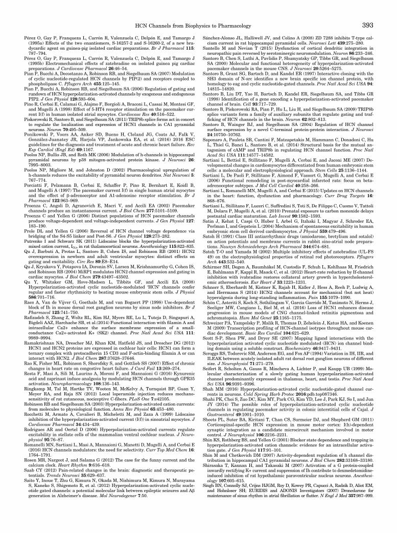
Pérez O, Gay P, Franqueza L, Carrón R, Valenzuela C, Delpón E, and Tamargo J(1995a) Effects of the two enantiomers, S-16257-2 and S-16260-2, of a new bra-dycardic agent on guinea-pig isolated cardiac preparations. Br J Pharmacol 115:787–794.
Pérez O, Gay P, Franqueza L, Carrón R, Valenzuela C, Delpón E, and Tamargo J(1995b) Electromechanical effects of zatebradine on isolated guinea pig cardiacpreparations. J Cardiovasc Pharmacol 26:46–54.
Pian P, Bucchi A, Decostanzo A, Robinson RB, and Siegelbaum SA (2007) Modulationof cyclic nucleotide-regulated HCN channels by PIP(2) and receptors coupled tophospholipase C. Pflugers Arch 455:125–145.
Pian P, Bucchi A, Robinson RB, and Siegelbaum SA (2006) Regulation of gating andrundown of HCN hyperpolarization-activated channels by exogenous and endogenousPIP2. J Gen Physiol 128:593–604.
Pino R, Cerbai E, Calamai G, Alajmo F, Borgioli A, Braconi L, Cassai M, Montesi GF,and Mugelli A (1998) Effect of 5-HT4 receptor stimulation on the pacemaker cur-rent I(f) in human isolated atrial myocytes. Cardiovasc Res 40:516–522.
Piskorowski R, Santoro B, and Siegelbaum SA (2011) TRIP8b splice forms act in concertto regulate the localization and expression of HCN1 channels in CA1 pyramidalneurons. Neuron 70:495–509.
Ponikowski P, Voors AA, Anker SD, Bueno H, Cleland JG, Coats AJ, Falk V,González-Juanatey JR, Harjola VP, Jankowska EA, et al. (2016) 2016 ESCguidelines for the diagnosis and treatment of acute and chronic heart failure. RevEsp Cardiol (Engl Ed) 69:1167.
Poolos NP, Bullis JB, and Roth MK (2006) Modulation of h-channels in hippocampalpyramidal neurons by p38 mitogen-activated protein kinase. J Neurosci 26:7995–8003.
Poolos NP, Migliore M, and Johnston D (2002) Pharmacological upregulation ofh-channels reduces the excitability of pyramidal neuron dendrites. Nat Neurosci 5:767–774.
Porciatti F, Pelzmann B, Cerbai E, Schaffer P, Pino R, Bernhart E, Koidl B,and Mugelli A (1997) The pacemaker current I(f) in single human atrial myocytesand the effect of b-adrenoceptor and A1-adenosine receptor stimulation. Br JPharmacol 122:963–969.
Proenza C, Angoli D, Agranovich E, Macri V, and Accili EA (2002) Pacemakerchannels produce an instantaneous current. J Biol Chem 277:5101–5109.
Proenza C and Yellen G (2006) Distinct populations of HCN pacemaker channelsproduce voltage-dependent and voltage-independent currents. J Gen Physiol 127:183–190.
Prole DL and Yellen G (2006) Reversal of HCN channel voltage dependence viabridging of the S4-S5 linker and Post-S6. J Gen Physiol 128:273–282.
Putrenko I and Schwarz SK (2011) Lidocaine blocks the hyperpolarization-activatedmixed cation current, I(h), in rat thalamocortical neurons. Anesthesiology 115:822–835.
Qu J, Barbuti A, Protas L, Santoro B, Cohen IS, and Robinson RB (2001) HCN2overexpression in newborn and adult ventricular myocytes: distinct effects ongating and excitability. Circ Res 89:E8–E14.
Qu J, Kryukova Y, Potapova IA, Doronin SV, Larsen M, Krishnamurthy G, Cohen IS,and Robinson RB (2004) MiRP1 modulates HCN2 channel expression and gating incardiac myocytes. J Biol Chem 279:43497–43502.
Qu Y, Whitaker GM, Hove-Madsen L, Tibbits GF, and Accili EA (2008)Hyperpolarization-activated cyclic nucleotide-modulated ‘HCN’ channels conferregular and faster rhythmicity to beating mouse embryonic stem cells. J Physiol586:701–716.
Raes A, Van de Vijver G, Goethals M, and van Bogaert PP (1998) Use-dependentblock of Ih in mouse dorsal root ganglion neurons by sinus node inhibitors. Br JPharmacol 125:741–750.
Rafizadeh S, Zhang Z, Woltz RL, Kim HJ, Myers RE, Lu L, Tuteja D, Singapuri A,Bigdeli AAZ, Harchache SB, et al.(2014) Functional interaction with filamin A andintracellular Ca2+ enhance the surface membrane expression of a small-conductance Ca2+-activated K+ (SK2) channel. Proc Natl Acad Sci USA 111:9989–9994.
Ramakrishnan NA, Drescher MJ, Khan KM, Hatfield JS, and Drescher DG (2012)HCN1 and HCN2 proteins are expressed in cochlear hair cells: HCN1 can form aternary complex with protocadherin 15 CD3 and F-actin-binding filamin A or caninteract with HCN2. J Biol Chem 287:37628–37646.
Rao K, Fisher ML, Robinson S, Shorofsky S, and Gottlieb SS (2007) Effect of chronicchanges in heart rate on congestive heart failure. J Card Fail 13:269–274.
Resta F, Masi A, Sili M, Laurino A, Moroni F, and Mannaioni G (2016) Kynurenicacid and zaprinast induce analgesia by modulating HCN channels through GPR35activation. Neuropharmacology 108:136–143.
Ringkamp M, Tal M, Hartke TV, Wooten M, McKelvy A, Turnquist BP, Guan Y,Meyer RA, and Raja SN (2012) Local loperamide injection reduces mechano-sensitivity of rat cutaneous, nociceptive C-fibers. PLoS One 7:e42105.
Robinson RB and Siegelbaum SA (2003) Hyperpolarization-activated cation currents:from molecules to physiological function. Annu Rev Physiol 65:453–480.
Rocchetti M, Armato A, Cavalieri B, Micheletti M, and Zaza A (1999) Lidocaineinhibition of the hyperpolarization-activated current (I(f)) in sinoatrial myocytes. JCardiovasc Pharmacol 34:434–439.
Rodrigues AR and Oertel D (2006) Hyperpolarization-activated currents regulateexcitability in stellate cells of the mammalian ventral cochlear nucleus. J Neuro-physiol 95:76–87.
Romanelli MN, Sartiani L, Masi A, Mannaioni G, Manetti D, Mugelli A, and Cerbai E(2016) HCN channels modulators: the need for selectivity. Curr Top Med Chem 16:1764–1791.
Rosen MR, Nargeot J, and Salama G (2012) The case for the funny current and thecalcium clock. Heart Rhythm 9:616–618.
Saab CY (2012) Pain-related changes in the brain: diagnostic and therapeutic po-tentials. Trends Neurosci 35:629–637.
Saito Y, Inoue T, Zhu G, Kimura N, Okada M, Nishimura M, Kimura N, MurayamaS, Kaneko S, Shigemoto R, et al. (2012) Hyperpolarization-activated cyclic nucle-otide gated channels: a potential molecular link between epileptic seizures and Abgeneration in Alzheimer’s disease. Mol Neurodegener 7:50.
Sánchez-Alonso JL, Halliwell JV, and Colino A (2008) ZD 7288 inhibits T-type cal-cium current in rat hippocampal pyramidal cells. Neurosci Lett 439:275–280.
Santello M and Nevian T (2015) Dysfunction of cortical dendritic integration inneuropathic pain reversed by serotoninergic neuromodulation.Neuron 86:233–246.
Santoro B, Chen S, Luthi A, Pavlidis P, Shumyatsky GP, Tibbs GR, and SiegelbaumSA (2000) Molecular and functional heterogeneity of hyperpolarization-activatedpacemaker channels in the mouse CNS. J Neurosci 20:5264–5275.
Santoro B, Grant SG, Bartsch D, and Kandel ER (1997) Interactive cloning with theSH3 domain of N-src identifies a new brain specific ion channel protein, withhomology to eag and cyclic nucleotide-gated channels. Proc Natl Acad Sci USA 94:14815–14820.
Santoro B, Liu DT, Yao H, Bartsch D, Kandel ER, Siegelbaum SA, and Tibbs GR(1998) Identification of a gene encoding a hyperpolarization-activated pacemakerchannel of brain. Cell 93:717–729.
Santoro B, Piskorowski RA, Pian P, Hu L, Liu H, and Siegelbaum SA (2009) TRIP8bsplice variants form a family of auxiliary subunits that regulate gating and traf-ficking of HCN channels in the brain. Neuron 62:802–813.
Santoro B, Wainger BJ, and Siegelbaum SA (2004) Regulation of HCN channelsurface expression by a novel C-terminal protein-protein interaction. J Neurosci24:10750–10762.
Saponaro A, Pauleta SR, Cantini F, Matzapetakis M, Hammann C, Donadoni C, HuL, Thiel G, Banci L, Santoro B, et al. (2014) Structural basis for the mutual an-tagonism of cAMP and TRIP8b in regulating HCN channel function. Proc NatlAcad Sci USA 111:14577–14582.
Sartiani L, Bettiol E, Stillitano F, Mugelli A, Cerbai E, and Jaconi ME (2007) De-velopmental changes in cardiomyocytes differentiated from human embryonic stemcells: a molecular and electrophysiological approach. Stem Cells 25:1136–1144.
Sartiani L, De Paoli P, Stillitano F, Aimond F, Vassort G, Mugelli A, and Cerbai E(2006) Functional remodeling in post-myocardial infarcted rats: focus on beta-adrenoceptor subtypes. J Mol Cell Cardiol 40:258–266.
Sartiani L, Romanelli MN, Mugelli A, and Cerbai E (2015) Updates on HCN channelsin the heart: function, dysfunction and pharmacology. Curr Drug Targets 16:868–876.
Sartiani L, Stillitano F, Luceri C, Suffredini S, Toti S, De Filippo C, Cuomo V, TattoliM, Dolara P, Mugelli A, et al. (2010) Prenatal exposure to carbon monoxide delayspostnatal cardiac maturation. Lab Invest 90:1582–1593.
Satin J, Kehat I, Caspi O, Huber I, Arbel G, Itzhaki I, Magyar J, Schroder EA,Perlman I, and Gepstein L (2004) Mechanism of spontaneous excitability in humanembryonic stem cell derived cardiomyocytes. J Physiol 559:479–496.
Satoh H (1991) Class III antiarrhythmic drugs (amiodarone, bretylium and sotalol)on action potentials and membrane currents in rabbit sino-atrial node prepara-tions. Naunyn Schmiedebergs Arch Pharmacol 344:674–681.
Satoh T-O and Yamada M (2002) Multiple inhibitory effects of zatebradine (UL-FS49) on the electrophysiological properties of retinal rod photoreceptors. PflugersArch 443:532–540.
Schirmer SH, Degen A, Baumhäkel M, Custodis F, Schuh L, Kohlhaas M, FriedrichE, Bahlmann F, Kappl R, Maack C, et al. (2012) Heart-rate reduction by If-channelinhibition with ivabradine restores collateral artery growth in hypercholesterol-emic atherosclerosis. Eur Heart J 33:1223–1231.
Schnorr S, Eberhardt M, Kistner K, Rajab H, Käßer J, Hess A, Reeh P, Ludwig A,and Herrmann S (2014) HCN2 channels account for mechanical (but not heat)hyperalgesia during long-standing inflammation. Pain 155:1079–1090.
Schön C, Asteriti S, Koch S, Sothilingam V, Garcia Garrido M, Tanimoto N, Herms J,Seeliger MW, Cangiano L, Biel M, et al. (2016) Loss of HCN1 enhances diseaseprogression in mouse models of CNG channel-linked retinitis pigmentosa andachromatopsia. Hum Mol Genet 25:1165–1175.
Schweizer PA, Yampolsky P, Malik R, Thomas D, Zehelein J, Katus HA, and KoenenM (2009) Transcription profiling of HCN-channel isotypes throughout mouse car-diac development. Basic Res Cardiol 104:621–629.
Scott S-P, Shea PW, and Dryer SE (2007) Mapping ligand interactions with thehyperpolarization activated cyclic nucleotide modulated (HCN) ion channel bind-ing domain using a soluble construct. Biochemistry 46:9417–9431.
Scroggs RS, Todorovic SM, Anderson EG, and Fox AP (1994) Variation in IH, IIR, andILEAK between acutely isolated adult rat dorsal root ganglion neurons of differentsize. J Neurophysiol 71:271–279.
Seifert R, Scholten A, Gauss R, Mincheva A, Lichter P, and Kaupp UB (1999) Mo-lecular characterization of a slowly gating human hyperpolarization-activatedchannel predominantly expressed in thalamus, heart, and testis. Proc Natl AcadSci USA 96:9391–9396.
Shah MM (2016) Hyperpolarization-activated cyclic nucleotide-gated channel cur-rents in neurons. Cold Spring Harb Protoc 2016:pdb.top087346.
Shahi PK, Choi S, Zuo DC, Kim MY, Park CG, Kim YD, Lee J, Park KJ, So I, and JunJY (2014) The possible roles of hyperpolarization-activated cyclic nucleotidechannels in regulating pacemaker activity in colonic interstitial cells of Cajal. JGastroenterol 49:1001–1010.
Sheets PL, Suter BA, Kiritani T, Chan CS, Surmeier DJ, and Shepherd GM (2011)Corticospinal-specific HCN expression in mouse motor cortex: I(h)-dependentsynaptic integration as a candidate microcircuit mechanism involved in motorcontrol. J Neurophysiol 106:2216–2231.
Shin KS, Rothberg BS, and Yellen G (2001) Blocker state dependence and trapping inhyperpolarization-activated cation channels: evidence for an intracellular activa-tion gate. J Gen Physiol 117:91–101.
Shin M and Chetkovich DM (2007) Activity-dependent regulation of h channel dis-tribution in hippocampal CA1 pyramidal neurons. J Biol Chem 282:33168–33180.
Shirasaka T, Kannan H, and Takasaki M (2007) Activation of a G protein-coupledinwardly rectifying K+ current and suppression of Ih contribute to dexmedetomidine-induced inhibition of rat hypothalamic paraventricular nucleus neurons. Anesthesi-ology 107:605–615.
Singh BN, Connolly SJ, Crijns HJGM, Roy D, Kowey PR, Capucci A, Radzik D, Aliot EM,and Hohnloser SH; EURIDIS and ADONIS Investigators (2007) Dronedarone formaintenance of sinus rhythm in atrial fibrillation or flutter. N Engl J Med 357:987–999.
HCN Channels from Biophysics to Pharmacology 393

Sobrado LF, Varone BB, Machado AD, Nearing BD, Zeng D, Belardinelli L,and Verrier RL (2013) Dronedarone’s inhibition of If current is the primarymechanism responsible for its bradycardic effect. J Cardiovasc Electrophysiol 24:914–918.
Später D, Abramczuk MK, Buac K, Zangi L, Stachel MW, Clarke J, Sahara M,Ludwig A, and Chien KR (2013) A HCN4+ cardiomyogenic progenitor derived fromthe first heart field and human pluripotent stem cells. Nat Cell Biol 15:1098–1106.
St Clair JR, Liao Z, Larson ED, and Proenza C (2013) PKA-independent activation ofI(f) by cAMP in mouse sinoatrial myocytes. Channels (Austin) 7:318–321.
Stieber J, Herrmann S, Feil S, Löster J, Feil R, Biel M, Hofmann F, and Ludwig A(2003a) The hyperpolarization-activated channel HCN4 is required for the gener-ation of pacemaker action potentials in the embryonic heart. Proc Natl Acad SciUSA 100:15235–15240.
Stieber J, Hofmann F, and Ludwig A (2004) Pacemaker channels and sinus nodearrhythmia. Trends Cardiovasc Med 14:23–28.
Stieber J, Stöckl G, Herrmann S, Hassfurth B, and Hofmann F (2005) Functionalexpression of the human HCN3 channel. J Biol Chem 280:34635–34643.
Stieber J, Thomer A, Much B, Schneider A, Biel M, and Hofmann F (2003b) Molec-ular basis for the different activation kinetics of the pacemaker channels HCN2and HCN4. J Biol Chem 278:33672–33680.
Stieber J, Wieland K, Stöckl G, Ludwig A, and Hofmann F (2006) Bradycardic andproarrhythmic properties of sinus node inhibitors. Mol Pharmacol 69:1328–1337.
Stilli D, Sgoifo A, Macchi E, Zaniboni M, De Iasio S, Cerbai E, Mugelli A, Lagrasta C,Olivetti G, and Musso E (2001) Myocardial remodeling and arrhythmogenesis inmoderate cardiac hypertrophy in rats. Am J Physiol Heart Circ Physiol 280:H142–H150.
Stillitano F, Lonardo G, Giunti G, Del Lungo M, Coppini R, Spinelli V, Sartiani L,Poggesi C, Mugelli A, and Cerbai E (2013) Chronic atrial fibrillation alters thefunctional properties of If in the human atrium. J Cardiovasc Electrophysiol 24:1391–1400.
Stillitano F, Lonardo G, Zicha S, Varro A, Cerbai E, Mugelli A, and Nattel S (2008)Molecular basis of funny current (If) in normal and failing human heart. J Mol CellCardiol 45:289–299.
Suffredini S, Stillitano F, Comini L, Bouly M, Brogioni S, Ceconi C, Ferrari R,Mugelli A, and Cerbai E (2012) Long-term treatment with ivabradine in post-myocardial infarcted rats counteracts f-channel overexpression. Br J Pharmacol165:1457–1466.
Suh BC and Hille B (2007) Regulation of KCNQ channels by manipulation of phos-phoinositides. J Physiol 582:911–916.
Sundelacruz S, Levin M, and Kaplan DL (2009) Role of membrane potential in theregulation of cell proliferation and differentiation. Stem Cell Rev 5:231–246.
Sundelacruz S, Levin M, and Kaplan DL (2015) Comparison of the depolarizationresponse of human mesenchymal stem cells from different donors. Sci Rep 5:18279.
Surges R, Brewster AL, Bender RA, Beck H, Feuerstein TJ, and Baram TZ (2006)Regulated expression of HCN channels and cAMP levels shape the properties ofthe h current in developing rat hippocampus. Eur J Neurosci 24:94–104.
Surges R, Freiman TM, and Feuerstein TJ (2003) Gabapentin increases thehyperpolarization-activated cation current Ih in rat CA1 pyramidal cells. Epilepsia44:150–156.
Swynghedauw B (1999) Molecular mechanisms of myocardial remodeling. PhysiolRev 79:215–262.
Takasu K, Ono H, and Tanabe M (2010) Spinal hyperpolarization-activated cyclicnucleotide-gated cation channels at primary afferent terminals contribute tochronic pain. Pain 151:87–96.
Takeda M, Ikeda M, Tanimoto T, Lipski J, and Matsumoto S (2002) Changes of theexcitability of rat trigeminal root ganglion neurons evoked by a(2)-adrenoreceptors.Neuroscience 115:731–741.
Tamura A, Ogura T, Uemura H, Reien Y, Kishimoto T, Nagai T, Komuro I, MiyazakiM, and Nakaya H (2009) Effects of antiarrhythmic drugs on the hyperpolarization-activated cyclic nucleotide-gated channel current. J Pharmacol Sci 110:150–159.
Tang B, Sander T, Craven KB, Hempelmann A, and Escayg A (2008) Mutationanalysis of the hyperpolarization-activated cyclic nucleotide-gated channels HCN1and HCN2 in idiopathic generalized epilepsy. Neurobiol Dis 29:59–70.
Taraska JW, Puljung MC, Olivier NB, Flynn GE, and Zagotta WN (2009) Mappingthe structure and conformational movements of proteins with transition metal ionFRET. Nat Methods 6:532–537.
Thoby-Brisson M, Cauli B, Champagnat J, Fortin G, and Katz DM (2003) Expressionof functional tyrosine kinase B receptors by rhythmically active respiratory neu-rons in the pre-Bötzinger complex of neonatal mice. J Neurosci 23:7685–7689.
Thollon C, Cambarrat C, Vian J, Prost J-F, Peglion JL, and Vilaine JP (1994) Elec-trophysiological effects of S 16257, a novel sino-atrial node modulator, on rabbitand guinea-pig cardiac preparations: comparison with UL-FS 49. Br J Pharmacol112:37–42.
Thon S, Schmauder R, and Benndorf K (2013) Elementary functional properties ofsingle HCN2 channels. Biophys J 105:1581–1589.
Tibbs GR, Rowley TJ, Sanford RL, Herold KF, Proekt A, Hemmings Jr.HC, AndersenOS, Goldstein PA, and Flood PD (2013) HCN1 channels as targets for anestheticand nonanesthetic propofol analogs in the amelioration of mechanical and thermalhyperalgesia in a mouse model of neuropathic pain. J Pharmacol Exp Ther 345:363–373.
Torrente AG, Zhang R, Zaini A, Giani JF, Kang J, Lamp ST, Philipson KD,and Goldhaber JI (2015) Burst pacemaker activity of the sinoatrial node in sodium-calcium exchanger knockout mice. Proc Natl Acad Sci USA 112:9769–9774.
Tran N, Proenza C, Macri V, Petigara F, Sloan E, Samler S, and Accili EA (2002) Aconserved domain in the NH2 terminus important for assembly and functionalexpression of pacemaker channels. J Biol Chem 277:43588–43592.
Tsay D, Dudman JT, and Siegelbaum SA (2007) HCN1 channels constrain synapti-cally evoked Ca2+ spikes in distal dendrites of CA1 pyramidal neurons.Neuron 56:1076–1089.
Turley SL, Francis KE, Lowe DK, and Cahoon Jr.WD (2016) Emerging role of ivabra-dine for rate control in atrial fibrillation. Ther Adv Cardiovasc Dis 10:348–352.
Ulrich D (2002) Dendritic resonance in rat neocortical pyramidal cells. J Neuro-physiol 87:2753–2759.
Valenzuela C, Delpón E, Franqueza L, Gay P, Pérez O, Tamargo J, and Snyders DJ(1996) Class III antiarrhythmic effects of zatebradine: time-, state-, use-, andvoltage-dependent block of hKv1.5 channels. Circulation 94:562–570.
Van Bogaert PP and Pittoors F (2003) Use-dependent blockade of cardiac pacemakercurrent (If) by cilobradine and zatebradine. Eur J Pharmacol 478:161–171.
van KempenM, van Ginneken A, de Grijs I, Mutsaers N, Opthof T, Jongsma H, and vander Heyden M (2003) Expression of the electrophysiological system during murineembryonic stem cell cardiac differentiation. Cell Physiol Biochem 13:263–270.
VanSchouwen B, Akimoto M, Sayadi M, Fogolari F, and Melacini G (2015) Role ofdynamics in the autoinhibition and activation of the hyperpolarization-activatedcyclic nucleotide-modulated (HCN) ion channels. J Biol Chem 290:17642–17654.
Vasilyev DV, Shan Q, Lee Y, Mayer SC, Bowlby MR, Strassle BW, Kaftan EJ, RogersKE, and Dunlop J (2007) Direct inhibition of Ih by analgesic loperamide in rat DRGneurons. J Neurophysiol 97:3713–3721.
Vasilyev DV, Shan QJ, Lee YT, Soloveva V, Nawoschik SP, Kaftan EJ, Dunlop J,Mayer SC, and Bowlby MR (2009) A novel high-throughput screening assay forHCN channel blocker using membrane potential-sensitive dye and FLIPR. J Bio-mol Screen 14:1119–1128.
Verkerk AO and Wilders R (2014) Pacemaker activity of the human sinoatrial node:effects of HCN4 mutations on the hyperpolarization-activated current. Europace16:384–395.
Verrier RL, Bonatti R, Silva AF, Batatinha JA, Nearing BD, Liu G, Rajamani S, ZengD, and Belardinelli L (2014) If inhibition in the atrioventricular node by ivabradinecauses rate-dependent slowing of conduction and reduces ventricular rate duringatrial fibrillation. Heart Rhythm 11:2288–2296.
Verrier RL, Silva AF, Bonatti R, Batatinha JA, Nearing BD, Liu G, Rajamani S, ZengD, and Belardinelli L (2015) Combined actions of ivabradine and ranolazine reduceventricular rate during atrial fibrillation. J Cardiovasc Electrophysiol 26:329–335.
Verrier RL, Sobrado MF, Pagotto VP, Kanas AF, Machado AD, Varone BB, SobradoLF, Nearing BD, Zeng D, and Belardinelli L (2013) Inhibition of I(f) in the atrio-ventricular node as a mechanism for dronedarone’s reduction in ventricular rateduring atrial fibrillation. Heart Rhythm 10:1692–1697.
Vinogradova TM, Brochet DX, Sirenko S, Li Y, Spurgeon H, and Lakatta EG (2010)Sarcoplasmic reticulum Ca2+ pumping kinetics regulates timing of local Ca2+releases and spontaneous beating rate of rabbit sinoatrial node pacemaker cells.Circ Res 107:767–775.
Viscomi C, Altomare C, Bucchi A, Camatini E, Baruscotti M, Moroni A,and DiFrancesco D (2001) C terminus-mediated control of voltage and cAMPgating of hyperpolarization-activated cyclic nucleotide-gated channels. J BiolChem 276:29930–29934.
Wahl-Schott C and Biel M (2009) HCN channels: structure, cellular regulation andphysiological function. Cell Mol Life Sci 66:470–494.
Wahl-Schott C, Fenske S, and Biel M (2014) HCN channels: new roles in sinoatrialnode function. Curr Opin Pharmacol 15:83–90.
Wainger BJ, DeGennaro M, Santoro B, Siegelbaum SA, and Tibbs GR (2001) Mo-lecular mechanism of cAMP modulation of HCN pacemaker channels. Nature 411:805–810.
Wang J, Chen S, Nolan MF, and Siegelbaum SA (2002) Activity-dependent regulationof HCN pacemaker channels by cyclic AMP: signaling through dynamic allostericcoupling. Neuron 36:451–461.
Wang J, Chen S, and Siegelbaum SA (2001) Regulation of hyperpolarization-activated HCN channel gating and cAMP modulation due to interactions of COOHterminus and core transmembrane regions. J Gen Physiol 118:237–250.
Wang K, Xue T, Tsang SY, Van Huizen R, Wong CW, Lai KW, Ye Z, Cheng L, Au KW,Zhang J, et al. (2005) Electrophysiological properties of pluripotent human andmouse embryonic stem cells. Stem Cells 23:1526–1534.
Wang M, Ramos BP, Paspalas CD, Shu Y, Simen A, Duque A, Vijayraghavan S,Brennan A, Dudley A, Nou E, et al. (2007) Alpha2A-adrenoceptors strengthenworking memory networks by inhibiting cAMP-HCN channel signaling in pre-frontal cortex. Cell 129:397–410.
Wang WT, Wan YH, Zhu JL, Lei GS, Wang YY, Zhang P, and Hu SJ (2006) Theta-frequency membrane resonance and its ionic mechanisms in rat subicular pyra-midal neurons. Neuroscience 140:45–55.
Wang Y and Hill JA (2010) Electrophysiological remodeling in heart failure. J MolCell Cardiol 48:619–632.
Weisbrod D, Peretz A, Ziskind A, Menaker N, Oz S, Barad L, Eliyahu S, Itskovitz-Eldor J, Dascal N, Khananshvili D, et al. (2013) SK4 Ca2+ activated K+ channel isa critical player in cardiac pacemaker derived from human embryonic stem cells.Proc Natl Acad Sci USA 110:E1685–E1694.
Wemhöner K, Silbernagel N, Marzian S, Netter MF, Rinné S, Stansfeld PJ,and Decher N (2012) A leucine zipper motif essential for gating ofhyperpolarization-activated channels. J Biol Chem 287:40150–40160.
Weng X, Smith T, Sathish J, and Djouhri L (2012) Chronic inflammatory pain isassociated with increased excitability and hyperpolarization-activated current (Ih)in C- but not Ad-nociceptors. Pain 153:900–914.
Whitaker GM, Angoli D, Nazzari H, Shigemoto R, and Accili EA (2007) HCN2 andHCN4 isoforms self-assemble and co-assemble with equal preference to formfunctional pacemaker channels. J Biol Chem 282:22900–22909.
White SM and Claycomb WC (2005) Embryonic stem cells form an organized, func-tional cardiac conduction system in vitro. Am J Physiol Heart Circ Physiol 288:H670–H679.
Wicks NL, Wong T, Sun J, Madden Z, and Young EC (2011) Cytoplasmic cAMP-sensing domain of hyperpolarization-activated cation (HCN) channels uses twostructurally distinct mechanisms to regulate voltage gating. Proc Natl Acad SciUSA 108:609–614.
Wilkars W, Wollberg J, Mohr E, Han M, Chetkovich DM, Bähring R, and Bender RA(2014) Nedd4-2 regulates surface expression and may affect N-glycosylation ofhyperpolarization-activated cyclic nucleotide-gated (HCN)-1 channels. FASEB J28:2177–2190.
394 Sartiani et al.

Williams AD, Jung S, and Poolos NP (2015) Protein kinase C bidirectionally modu-lates Ih and hyperpolarization-activated cyclic nucleotide-gated (HCN) channelsurface expression in hippocampal pyramidal neurons. J Physiol 593:2779–2792.
Williams SR and Stuart GJ (2000) Site independence of EPSP time course is medi-ated by dendritic I(h) in neocortical pyramidal neurons. J Neurophysiol 83:3177–3182.
Wilson CM and Farrell AP (2013) Pharmacological characterization of the heartbeatin an extant vertebrate ancestor, the Pacific hagfish, Eptatretus stoutii. CompBiochem Physiol A Mol Integr Physiol 164:258–263.
Wu JY and Cohen IS (1997) Tyrosine kinase inhibition reduces i(f) in rabbit sino-atrial node myocytes. Pflugers Arch 434:509–514.
Wu X, Liao L, Liu X, Luo F, Yang T, and Li C (2012) Is ZD7288 a selective blocker ofhyperpolarization-activated cyclic nucleotide-gated channel currents? Channels(Austin) 6:438–442.
Xu X, Marni F, Wu S, Su Z, Musayev F, Shrestha S, Xie C, Gao W, Liu Q, and Zhou L(2012) Local and global interpretations of a disease-causing mutation near theligand entry path in hyperpolarization-activated cAMP-gated channel. Structure20:2116–2123.
Xu X, Vysotskaya ZV, Liu Q, and Zhou L (2010) Structural basis for the cAMP-dependent gating in the human HCN4 channel. J Biol Chem 285:37082–37091.
Xue L, Li Y, Han X, Yao L, Yuan J, Qin W, Liu F, and Wang H (2012a) Investigationof hyperpolarization-activated cyclic nucleotide-gated channels in interstitial cellsof Cajal of human bladder. Urology 80:224.e13–224.e18.
Xue T, Marbán E, and Li RA (2002) Dominant-negative suppression of HCN1- andHCN2-encoded pacemaker currents by an engineered HCN1 construct: insightsinto structure-function relationships and multimerization. Circ Res 90:1267–1273.
Xue WN, Wang Y, He SM, Wang XL, Zhu JL, and Gao GD (2012b) SK- and h-currentcontribute to the generation of theta-like resonance of rat substantia nigra parscompacta dopaminergic neurons at hyperpolarized membrane potentials. BrainStruct Funct 217:379–394.
Yagi J and Sumino R (1998) Inhibition of a hyperpolarization-activated current byclonidine in rat dorsal root ganglion neurons. J Neurophysiol 80:1094–1104.
Yamada Y, Kinoshita H, Kuwahara K, Nakagawa Y, Kuwabara Y, Minami T,Yamada C, Shibata J, Nakao K, Cho K, et al. (2014) Inhibition of N-type Ca2+channels ameliorates an imbalance in cardiac autonomic nerve activity and pre-vents lethal arrhythmias in mice with heart failure. Cardiovasc Res 104:183–193.
Yanagi K, Takano M, Narazaki G, Uosaki H, Hoshino T, Ishii T, Misaki T,and Yamashita JK (2007) Hyperpolarization-activated cyclic nucleotide-gatedchannels and T-type calcium channels confer automaticity of embryonic stem cell-derived cardiomyocytes. Stem Cells 25:2712–2719.
Yanagihara K and Irisawa H (1980) Inward current activated during hyperpolar-ization in the rabbit sinoatrial node cell. Pflugers Arch 385:11–19.
Yanagihara K, Noma A, and Irisawa H (1980) Reconstruction of sino-atrial nodepacemaker potential based on the voltage clamp experiments. Jpn J Physiol 30:841–857.
Yang Q, Kuzyk P, Antonov I, Bostwick CJ, Kohn AB, Moroz LL, and Hawkins RD(2015) Hyperpolarization-activated, cyclic nucleotide-gated cation channels inAplysia: contribution to classical conditioning. Proc Natl Acad Sci USA 112:16030–16035.
Yang Y-C, Meng Q-T, Pan X, Xia Z-Y, and Chen X-D (2014) Dexmedetomidine pro-duced analgesic effect via inhibition of HCN currents. Eur J Pharmacol 740:560–564.
Yao H, Donnelly DF, Ma C, and LaMotte RH (2003) Upregulation of thehyperpolarization-activated cation current after chronic compression of the dorsalroot ganglion. J Neurosci 23:2069–2074.
Ye B and Nerbonne JM (2009) Proteolytic processing of HCN2 and co-assembly withHCN4 in the generation of cardiac pacemaker channels. J Biol Chem 284:25553–25559.
Yeon K-Y, Chung G, Kim YH, Hwang JH, Davies AJ, Park M-K, Ahn DK, Kim JS,Jung SJ, and Oh SB (2011) Eugenol reverses mechanical allodynia after peripheralnerve injury by inhibiting hyperpolarization-activated cyclic nucleotide-gated(HCN) channels. Pain 152:2108–2116.
Yi F, Danko T, Botelho SC, Patzke C, Pak C, Wernig M, and Südhof TC (2016)Autism-associated SHANK3 haploinsufficiency causes Ih channelopathy in humanneurons. Science 352:aaf2669.
Ying S-W, Abbas SY, Harrison NL, and Goldstein PA (2006) Propofol block of I(h)contributes to the suppression of neuronal excitability and rhythmic burst firing inthalamocortical neurons. Eur J Neurosci 23:465–480.
Ying SW, Jia F, Abbas SY, Hofmann F, Ludwig A, and Goldstein PA (2007) DendriticHCN2 channels constrain glutamate-driven excitability in reticular thalamicneurons. J Neurosci 27:8719–8732.
Ying SW, Tibbs GR, Picollo A, Abbas SY, Sanford RL, Accardi A, Hofmann F, LudwigA, and Goldstein PA (2011) PIP2-mediated HCN3 channel gating is crucial forrhythmic burst firing in thalamic intergeniculate leaflet neurons. J Neurosci 31:10412–10423.
Yoshimoto R, Iwasaki S, Takago H, Nakajima T, Sahara Y, and Kitamura K (2015)Developmental increase in hyperpolarization-activated current regulates intrinsicfiring properties in rat vestibular ganglion cells. Neuroscience 284:632–642.
Young GT, Emery EC, Mooney ER, Tsantoulas C, and McNaughton PA (2014) In-flammatory and neuropathic pain are rapidly suppressed by peripheral block ofhyperpolarisation-activated cyclic nucleotide-gated ion channels. Pain 155:1708–1719.
Yu H, Wu J, Potapova I, Wymore RT, Holmes B, Zuckerman J, Pan Z, Wang H, ShiW, Robinson RB, et al. (2001) MinK-related peptide 1: a beta subunit for the HCNion channel subunit family enhances expression and speeds activation. Circ Res 88:E84–E87.
Yu HD, Xia S, Zha CQ, Deng SB, Du JL, and She Q (2015) Spironolactone regulatesHCN protein expression through micro-RNA-1 in rats with myocardial infarction. JCardiovasc Pharmacol 65:587–592.
Yu HG, Lu Z, Pan Z, and Cohen IS (2004) Tyrosine kinase inhibition differentiallyregulates heterologously expressed HCN channels. Pflugers Arch 447:392–400.
Zagotta WN, Olivier NB, Black KD, Young EC, Olson R, and Gouaux E (2003)Structural basis for modulation and agonist specificity of HCN pacemaker chan-nels. Nature 425:200–205.
Zaza A (2016) Electrophysiology meets geometry. Europace 18:317.Zaza A and Lombardi F (2001) Autonomic indexes based on the analysis of heart ratevariability: a view from the sinus node. Cardiovasc Res 50:434–442.
Zaza A, Rocchetti M, and DiFrancesco D (1996) Modulation of the hyperpolarization-activated current (I(f)) by adenosine in rabbit sinoatrial myocytes. Circulation 94:734–741.
Zhang H and Dougherty PM (2014) Enhanced excitability of primary sensory neuronsand altered gene expression of neuronal ion channels in dorsal root ganglion inpaclitaxel-induced peripheral neuropathy. Anesthesiology 120:1463–1475.
Zhang H, Sun AY, Kim JJ, Graham V, Finch EA, Nepliouev I, Zhao G, Li T, LedererWJ, Stiber JA, et al. (2015) STIM1-Ca2+ signaling modulates automaticity of themouse sinoatrial node. Proc Natl Acad Sci USA 112:E5618–E5627.
Zhang Y, Liu Y, Qu J, Hardy A, Zhang N, Diao J, Strijbos PJ, Tsushima R, RobinsonRB, Gaisano HY, et al. (2009) Functional characterization of hyperpolarization-activated cyclic nucleotide-gated channels in rat pancreatic beta cells. J Endocrinol203:45–53.
Zhang Y, Zhang N, Gyulkhandanyan AV, Xu E, Gaisano HY, Wheeler MB, and WangQ (2008) Presence of functional hyperpolarisation-activated cyclic nucleotide-gatedchannels in clonal alpha cell lines and rat islet alpha cells. Diabetologia 51:2290–2298.
Zhou C, Ke B, Zhao Y, Liang P, Liao D, Li T, Liu J, and Chen X (2015a)Hyperpolarization-activated cyclic nucleotide-gated channels may contribute toregional anesthetic effects of lidocaine. Anesthesiology 122:606–618.
Zhou C, Liang P, Liu J, Ke B, Wang X, Li F, Li T, Bayliss DA, and Chen X (2015b)HCN1 channels contribute to the effects of amnesia and hypnosis but not immo-bility of volatile anesthetics. Anesth Analg 121:661–666.
Zhou L and Siegelbaum SA (2007) Gating of HCN channels by cyclic nucleotides:residue contacts that underlie ligand binding, selectivity, and efficacy. Structure15:655–670.
Zhou M and MacKinnon R (2004) A mutant KcsA K(+) channel with altered con-duction properties and selectivity filter ion distribution. J Mol Biol 338:839–846.
Zicha S, Fernández-Velasco M, Lonardo G, L’Heureux N, and Nattel S (2005) Sinusnode dysfunction and hyperpolarization-activated (HCN) channel subunit remod-eling in a canine heart failure model. Cardiovasc Res 66:472–481.
Zolles G, Klöcker N, Wenzel D, Weisser-Thomas J, Fleischmann BK, Roeper J,and Fakler B (2006) Pacemaking by HCN channels requires interaction withphosphoinositides. Neuron 52:1027–1036.
Zolles G, Wenzel D, Bildl W, Schulte U, Hofmann A, Müller CS, Thumfart JO, VlachosA, Deller T, Pfeifer A, et al. (2009) Association with the auxiliary subunit PEX5R/Trip8b controls responsiveness of HCN channels to cAMP and adrenergic stimula-tion. Neuron 62:814–825.
Zong X, Eckert C, Yuan H, Wahl-Schott C, Abicht H, Fang L, Li R, Mistrik P,Gerstner A, Much B, et al. (2005) A novel mechanism of modulation ofhyperpolarization-activated cyclic nucleotide-gated channels by Src kinase. J BiolChem 280:34224–34232.
Zong X, Krause S, Chen C-C, Krüger J, Gruner C, Cao-Ehlker X, Fenske S, Wahl-Schott C, and Biel M (2012) Regulation of hyperpolarization-activated cyclicnucleotide-gated (HCN) channel activity by cCMP. J Biol Chem 287:26506–26512.
HCN Channels from Biophysics to Pharmacology 395