the hydraulic mechanism of the spider legjeb.biologists.org/content/jexbio/36/2/423.full.pdf · the...
TRANSCRIPT

[423
THE HYDRAULIC MECHANISM OFTHE SPIDER LEG
BY D. A. PARRY AND R. H. J. BROWN
Department of Zoology, University of Cambridge
(Received ia February 1959)
INTRODUCTION
Hinge joints occur in the legs of various arthropods, notably Limuhis (Snodgrass,1952); arachnids; diplopods, chilopods and pauropods (Manton, 19586). At ahinge joint flexion is muscular, but the anatomical form of the joint rarely permitsthe presence of antagonistic extensors (see Manton, 19580,%. 151 for an exception).In the recent account of the leg muscles of the house spider Tegenaria sp., Parry(1957) suggested that extension might be due to hydraulic forces in the leg. Manton(19584) attributed to the same cause the full extension of the arachnid leg when thelimb tip is off the ground but considered that extension during a propulsive strokewas passive and due to the ground reaction on the limb and so, indirectly, todepressor muscles.
In the present paper support is given to the idea that active extension occurs at thehinge joints of the Tegenaria leg and that the mechanism is a hydraulic one. First,we measure the internal pressure in the leg of intact spiders and find it to be sur-prisingly high. Secondly, we establish an empirical relation between the internalpressure of the leg and the torque at the hinge joints. Thirdly, we measure thetorques actually developed at the hinge joints when a spider accelerates a massattached to the leg, and use the relation between torque and pressure to show thatthe measured torques can be accounted for by the pressures which occur in the leg.
MEASUREMENT OF INTERNAL PRESSURE
Methods
The pressure inside the leg of an intact living spider can be measured by takingadvantage of the thin flexible articular membranes at the two hinge joints. Thisprinciple is employed in two ways:
(a) Direct observation of the membrane (Fig. 1). A sleeve is sealed over the legand the membrane observed through a binocular microscope. To measure themaintained resting pressure, the pressure in the sleeve is slowly raised until themembrane just collapses. To measure transient pressures developed when the spideris struggling, the sleeve pressure is raised to a predetermined value above the restingpressure so that the membrane collapses, and the spider is then stimulated. Thereinflation of the membrane indicates that the internal pressure equals or exceedsthe sleeve pressure.

424 D. A. PARRY AND R. H. J. BROWN
(b) Use of a high impedance transducer. The Cambridge Instrument Companyvery kindly lent us one of their photo-electric transducers and associated equip-ment. The principle of this instrument is illustrated in a purely formal way inFig. 2. The stiffness of the diaphragm is such that the instrument is almost iso-metric, a pressure change of 1 cm. Hg producing a volume change of 5 x io~3
mm.8 (cf. the joint volume of the spider which is about 0-5 mm.3). One leg of aliving spider is sealed into the cavity as shown, the stop-cock, S, is opened and azero recorded. The pressure is then raised to a pressure P, above the internalresting pressure of the spider, so that the articular membrane collapses. The stop-cock <S is then closed and the recorder started. The spider is stimulated with apaint brush or a puff of air and the excess of internal pressure above P is thusrecorded against time.
Compressed air
Fig. 1. Method of mounting a spicier for observation of the articularmembrane during the application of external pressure.
RESULTS
Resting pressures. When a spider is immobilized with one leg in a sleeve, it main-tains a positive pressure of approximately 5 cm. Hg which we call the restingpressure. The following figures (each a mean of four successive determinations)indicate the sort of variation encountered in different spiders: 4, 5, 5, 8, 9, n ,11 cm. Hg. In the absence of other evidence we presume that these pressures aretypical of the resting spider when supporting its own weight in normal life.
Transient pressures. When a spider, mounted as indicated in Fig. 2, is stimulatedby a puff of air or by a paint brush the internal pressure rises to several times theresting value and then falls again in a time which rarely exceeds 1 sec. and is usually

Hydraulic mechanism of the spider leg 425
about \ sec. A transducer record showing an unusually high transient pressure isshown in Fig. 3. The peak pressures which have been recorded in this way are asfollows:
Spider A u , 15, 17, 17, 19, 20, 22, 23, 24 cm. HgSpider BSpider B (next day)Spider C
16, 24, 27, 39, 39+ cm.Hg16, 20, 23, 24, 25, 25, 45 cm. Hg25. as. 3°. 3°. 4° cm. Hg
It will be seen that pressures of up to 45 cm. Hg have been recorded.Comparable results have been obtained by the simpler method (a) described
above. The maximum sleeve pressures against which a spider was observed to re-inflate its articular membrane was 40 cm. Hg. This was repeated on the same spideron two successive days.
Compressed air
Diaphragm / \ Mirror
\
Lamp
Fig. 2. Measurement of transient pressures in the leg of a spiderusing a high impedance photo-electric transducer.
Recorder
4 0 - C(T>. Hg
2 0 - :
10 -
0 -
1 sec.
bff
Fig. 3

426 D. A. PARRY AND R. H. J. BROWN
RELATION BETWEEN PRESSURE AND TORQUE AT A JOINT
Direct determination
A leg is removed at the coxa-trochanter joint where normal autotomy occurs (Parry,1957) and waxed into the end of a Perspex tube with a 1:1 beeswax-resin mixtureKrogh & Weis-Foch, 1951). The tip is cut off and the leg perfused with 1*3%magnesium chloride to relax the muscles. The tip is then sealed with wax and thePerspex tube mounted in the way shown in Fig. 4, thus enabling the relationshipbetween internal pressure and torque at the joint to be determined under staticconditions for different joint angles.
Compreued air
Free to route about pivotcoincident with hinge joint
Arm of torsionbalance
Fig. 4. Measurement of the relation between the internal pressure of the leg, the joint angleand the torque developed at the hinge joint.
Fig. 5 shows the relationship between pressure and torsion balance reading forthe femur-patella joint ('upper joint') and the tibia-basitarsus joint ('lower joint')of a particular leg whose size is indicated by the fact that the distance between thetwo joints was 11 mm. The distance between the upper joint and the torsion balancepivot was 10 mm.; that between the lower joint and the pivot was 8 mm. For a givenjoint angle (6) the relation between pressure (P) and torque (C) is linear, that isC = kd P, where kg decreases as 6 increases. If C is expressed in dyne-cm, and Pin dynes/cm.2, ke has the dimension cm.3 and its value for the selected values of 6is shown in the figure.
Indirect determination
It can be shown that if the torque C is due to the internal fluid pressure, kg isnumerically equal to A Vj&6, where A V is the small change in the volume of thejoint due to a small change of angle A#. The relation between AFand A0 is measured,

200
mg. 100 -
Joint angle Slope
80° 0-169x10"'cm.'
100° 0-147
120° 0-134140° 0-126160° 0-119
Lower Joint: torque arm 8 mm.I I i_
10
3001-
20 30 40cm. Hg
Upper joint: torque arm 10 mmI I I
50
Joint angle Slope
40° 0-440x10-' cm.»
0-368
0-3200-313
\ 0-295
mg. 150 -
160° 0-199
30cm. Hg
Fig. 5. Relation between the internal pressure and the torsion balance reading for different jointangles. The 'torque arm' is the distance between the joint and the arm of the balance (seeFig. 4)-

428 D. A. PARRY AND R. H. J. BROWN
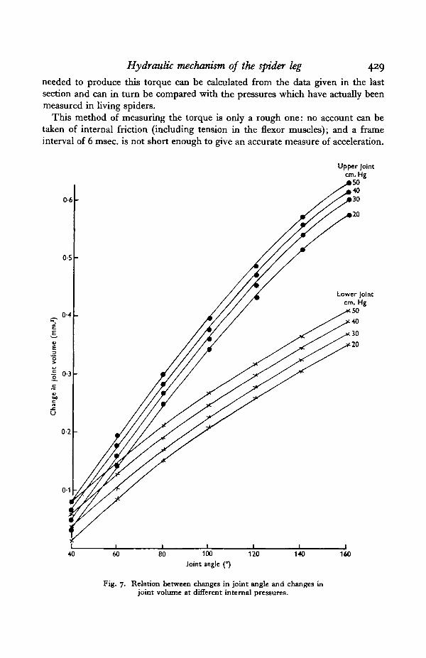
using the apparatus shown in Fig. 6. At a given pressure the joint angle is increasedin 200 stages from 400 to 16o° and the movement of the bubble is measured with atravelling micrometer. A second series of readings is taken as the angle is de-creased, and a mean for each angle is determined. In the event, the differencebetween the ascending and descending readings was negligible.
Air bubble
Fig. 6. Measurement of the relation between a change of joint angleand the consequent change in joint volume.
The results shown in Fig. 7 were obtained from the same leg as those of Fig. 5and so ke ( = CjP) can be directly compared with AF/A0:
Joint angle
Upper joint
Lower joint
4O° 6o°O'44 C37— 0-31
_ O . 2 Q
8o°0-320-29
0-310-27
i20 1400-30 0-290-26 —
1600
0-20 mm.1
— mm.'/radian
0-12 mm.1
— mm^/radian
The agreement between the direct and indirect determinations of kg is good,although there is a small consistent discrepancy in the measurements of the upperjoint. In the next section the values for kg obtained by direct measurement areused.
FORCES DEVELOPED DURING EXTENSION
The extension torques developed at the hinge joints in normal walking are thoseproducing a force at the foot (likely to be greatest in the fourth pair of legs whichpush forwards) together with those associated with accelerating and raising thedistributed mass of the leg (likely to be greatest in the first pair of legs whichstraighten forwards). No attempt has been made to measure these torques.Instead, the leg of an intact living spider was loaded with additional mass in theform of a brass ring of 45 mg. and photographed by high-speed cinematography(Brown & Popple, 1955) while it was extending in a horizontal plane about theupper hinge joint. Analysis of the film provides the angular acceleration of the leg,from which the torque developed at the hinge joint can be determined. The pressure
Hydraulic mechanism, of the spider leg 429
needed to produce this torque can be calculated from the data given in the lastsection and can in turn be compared with the pressures which have actually beenmeasured in living spiders.
This method of measuring the torque is only a rough one: no account can betaken of internal friction (including tension in the flexor muscles); and a frameinterval of 6 msec, is not short enough to give an accurate measure of acceleration.
40 60 80 100
Joint ingle (°)120 140 160
Fig. 7. Relation between changes in joint angle and changes injoint volume at different internal pressures.

43° D. A. PARRY AND R. H. J. BROWN
From four sequences the following example is selected as involving the greatestacceleration which has been encountered:
tAngular velocity (<u)Angular acceleration («i)Torque (Ici>)Pressure ( *
i 6 ia 182O-0 47-s so-4
3-3 4-6 o-47125 174 I7'83i 45 5
radian /sec.x ioJ radian/sec.1
dyne-cm.cm. Hg
In the other three sequences obtained (two different spiders) the maximumtorques developed during a 6-msec. period, and the corresponding pressures, were:130 dyne-cm. (35 cm. Hg); 77 dyne-cm. (18 cm. Hg); and 54 dyne-cm. (13 cm.Hg). It will be seen that the pressures required to produce these torques all liewithin the range of pressures which the spiders have been shown to develop. Thefigures furthermore establish directly that the torques developed at the hinge jointscan be considerably greater than are required merely to extend the leg when it isoff the ground (cf. Manton, 1958ft).
DISCUSSION
The hydraulic mechanism
Anatomy. In the spider, prosoma and abdomen are separated by a narrowpedicel (Fig. 8). The prosomal tergum and sternum are relatively rigid plates—the one convex, the other flatter—connected together by an unsclerotized lateralregion. The legs are inserted into this lateral region and are without articulations
Fig. 8. Tegenaria atrica showing the flexible lateral region whichintervenes between prosomal tergum and sternum.
with either tergum or sternum, which are therefore free to move apart or cometogether (see Fig. 8). According to Brown (1939) there are no tergo-sternal muscles,but dorso-ventral muscles run from the tergum to the endo-skeleton, the stomachand the proximal ends of the coxae of the legs.
The abdomen is relatively flexible and becomes wrinkled and apparently shrunkenwhen an animal is short of food or water. Millot (1949) describes a thin subcuticularmuscle layer; otherwise the abdominal musculature is scanty (Brown, 1939). Theheart lies in the abdomen and a single large aorta runs through the pedicel into theprosoma where arteries distribute the blood peripherally.

Hydraulic mechanism of the spider leg 431
Resting pressure. Under the conditions of our experiments the haemocoelicpressure in the legs of unstimulated spiders is about 5 cm. Hg and we suggest thatthe normal resting pressure is of approximately this value. Regarding the pro-duction and distribution of this pressure there are three possibilities:
(1) The pressure might be uniformly distributed throughout the prosoma andabdomen and due to muscle tension. This seems unlikely because the isolatedabdomen is found to become grossly distended at such a pressure and yet its muscu-lature appears inadequate to maintain the characteristic shape in life. It should benoted that if the prosoma and abdomen were at the same pressure no load would beimposed on the heart, whose systolic pressure need only be sufficient to overcomethe peripheral resistance.
(2) The pressure might be limited to the prosoma, the abdomen being at atmos-pheric pressure. This accords with the observed lack of turgidity in the abdomenbut demands that the systolic pressure of the heart should just exceed the prosomalpressure, thus developing considerably more pressure than is usual among arthro-pods (compare data given in Prosser (1950), Table 67; but note also Harris &Crofton (1957) on the nematode Ascaris in which the mean internal pressure is7 cm. Hg).
There is a little evidence that the heart may in fact produce the required highpressure. The blood flow in a spider's leg can be seen by transmitted light if the legis sealed into a flat-sided cell filled with a physiological solution (1 1. of spiderRinger is made up with: 144 c.c. M/15 KH2PO4; 144 c.c. M/15 NajHPO^; 347 c.c.O-54M NaCl; 5-56 c.c. C-36M CaCl2.6HaO; 5-56 c.c. O-36M MgCLj^HiO).
If the prosoma is now compressed dorso-ventrally the blood flow in the legeventually stops. The haemocoelic pressure at which this occurs can be measuredin the usual way by determining the external pressure which has to be applied tothe leg in order just to collapse the articular membrane. The pressures thusmeasured will presumably represent the maximum which the heart can produce.Readings were as follows: 10-4, io-6, ivz and n-6 cm. Hg.
Clearly this needs further investigation, but so far as the available evidence goes,it suggests that the ' maintained pressure' of 5 cm. Hg occurs in the prosoma, butnot the abdomen. This being the case it might be expected that some form of valveor sphincter would occur in the pedicel and would play an important part inregulating prosomal pressure.
(3) A third possibility should be mentioned: that the pressure is limited to thelegs alone. This would still demand a high heart pressure, and would involve asphincter at the base of the leg and suitable muscles in the leg, of which there areno signs.
Transient pressure. Pressures of up to 45 cm. Hg, maintained for a fraction of asecond, have been measured on a stimulated spider. If two legs are separatelysleeved and subjected to the same external pressure, the articular membranes arefound to inflate and deflate simultaneously when the spider is stimulated. Thissuggests that the transient pressure arises in the prosoma rather than in the indi-vidual legs (where, as noted above, no muscles seem suitably disposed to produce

432 D. A. PARRY AND R. H. J. BROWN
it) and it may well be due to the sudden activation of the prosomal muscles andslight dorso-ventral compression. This sudden rise of pressure would, of course,temporarily stop the prosomal circulation, and in this connexion it is interestingto note the well developed aortic valve in the pedicel (see, for example, Millot,1949, fig. 419, based on the beautiful work of Schneider, 1892).
Dynamic considerations. By adding mass to the leg of a living spider and findingthe acceleration during an extensor movement we have measured the torques whichcan be developed at a hinge joint We have then calculated the internal pressureneeded to develop these torques, using values for kg empirically determined. How-ever, kg was measured under static conditions and it remains to be seen whetherit can be applied under dynamic ones. Two considerations arise:
(1) When a hinge joint is extending fluid must flow into the joint thus settingup a pressure gradient in the leg so that the pressure in the joint will not be thesame as the pressure in the prosoma. A rough application of Poisseuille's equationshows that this gradient may be insignificant. The maximum observed rate ofextension of the upper hinge joint was about 50 radian/sec, at a joint angle of about900. This is equivalent to a volume change of 15 mm.3/sec. Taking the viscosity ofthe blood to be 0-05 dyne-sec./cm.2 (as in human blood—no direct measurementsare available), and the length of the upper part of the leg to be 1 cm., we get:
r4 Ap = 0-002 dyne-cm.2,
where r = radius of duct and A/> = pressure difference between prosoma and upperhinge joint. Now as the channel is of indeterminate shape no precise value can beassigned to r; but it can be seen that A/> is negligible provided that the equivalentduct is not less than 0-02 cm. radius, or two-fifths of the actual radius of thefemur, which is not unreasonable. However, the matter needs further investigation.
(2) If the prosoma were very indistensible then the withdrawal of a smallquantity of fluid would produce a significant fall in pressure. But in the section onanatomy we have pointed out that the prosoma is built in such a way that changes involume can readily be produced.
Bleeding. It might be thought that, owing to the high pressures occurring in thespider, bleeding would be a serious hazard, and it is the case that spiders readily diefrom this cause. Perhaps the most frequent form of damage is the loss of a leg andhere a special protective mechanism has been evolved (Parry, 1957). But minordamage to the prosoma need not be fatal; the withdrawal of blood together with therelaxation of the prosomal muscles would produce an immediate fall in pressureand the wound might heal in a few days. During this time the spider is not im-mobilized, but we have no reason to think that ordinary walking necessitates thehigh pressures which we have measured in uninjured animals, especially if thehind legs (the only ones whose propulsive stroke is an extension) contribute lessforce than usual.

Hydraulic mechanism of the spider leg 433
SUMMARY
1. The blood pressure inside the leg of the house spider Tegenaria atrica has beenmeasured. Maintained pressures of about 5 cm. Hg and transient pressures of upto 40 cm. Hg have been found.
2. The relation between the blood pressure in the leg and the extension torqueat the hinge joints has been established.
3. Considerable torques can be developed at the hinge joints during extension,for example, when accelerating a mass fixed to the leg. The transient pressuresfound to arise in the leg are adequate to account for these torques.
4. The hydraulic mechanism is discussed. The available evidence suggests thatthe pressure found in the legs occurs also in the prosoma but not in the abdomen,in which case the maintained pressure must be due to the heart. This, however,requires further investigation.
We are greatly indebted to the Cambridge Instrument Company for the loan ofa photo-electric transducer; and also to many friends who have supplied us withspiders. The spider Ringer was based on determinations of freezing-point depres-sion and sodium and potassium concentration of Tegenaria blood made for one ofus some years ago by Dr P. C. Croghan and not hitherto published.
REFERENCESBROWN, R. B. (1939). The musculature of Agelena naevia. J. Morph. 64, 115-66.BROWN, R. H. J. & POPPLE, J. A. (1955). Multiple flash photography. Med. Biol. IUuitr. 5, 23-8.HARRIS, J. E. & CROFTON, H. D. (1957). Structure and function in the Nematodes. J. Exp. Biol.
34. 116-30.KROGH, A. & WEIS-FOCH, T. (1951). Respiratory exchange of the desert locust. J. Exp. Biol. 38,
344-57-MANTON, S. M. (1958a). Evolution of arthropodan locomotory mechanisms, Pt. 6. J. Unn. Soc.
(Zool.), 43, 488-556.MANTON, S. M. (19586). Hydrostatic pressure and leg extension in arthropods. Ann. Mag. Nat.
Hist. Series 13, 1, 161-82.MILLOT, J. (1949). Ordre des Araneides, in Grasse, P.-P. (directeur): Traiti de Zoologie, 6. Paris:
Masson.PARRY, D. A. (1957). Spider leg muscles and the autotomy mechanism. Quart. J. Micr. Set. 98.
331-4°-PROSSER, C. L. (ed.) (1950). Comparative Animal Physiology. Philadelphia: Saunders.SCHNEIDER, A. (1892). Melanges arachnologiques. Tablettes Zoologiques, 2, 135—98.SNODGRASS, R. E. (1952). A Textbook of Arthropod Anatomy. New York: Comstock.