the history of near-field optics1 - uni-jena.deto+nanooptics... · the history of near-field...
TRANSCRIPT

The History of Near-field Optics1
Lukas Novotny
The Institute of Optics, University of Rochester, Rochester, NY, 14627.
Abstract
This article provides a review of early work and developments in the field of near-field
optics. The roots trace back to the letters exchanged between Edward Hutchinson Synge
and Albert Einstein in 1928 and, because of the analogy to antenna theory and light-
ning rods, the origins project back to the time of Benjamin Franklin who discovered the
wonderful Effect of Points both in drawing off and throwing off the Electrical Fire. The
modern interest was mainly inspired by the invention of scanning probe microscopy and
by the first optical near-field measurements by Dieter W. Pohl and co-workers at the IBM
Research Laboratory in Ruschlikon, Switzerland, and also by parallel developments of
other groups. Near-field optics received inspiration from the fields of surface enhanced
spectroscopy and from studies of energy transfer. While optical near-fields were exten-
sively exploited for overcoming the diffraction limit in optical imaging the study of their
physical aspects revealed unique properties which cannot be imitated by free propagating
radiation.
1Adapted from L. Novotny, ”The History of Near-field Optics,” Progress in Optics 50, E. Wolf (ed.),chapter 5, p.137- 184 (Elsevier, Amsterdam, The Netherlands, 2007).
137

1 INTRODUCTION 138
1 Introduction
Near-field Optics is the study of non-propagating inhomogeneous fields and their inter-
action with matter. Optical near-fields are localized to the source region of optical radi-
ation or to the surfaces of materials interacting with free radiation (secondary sources).
In many situations, optical near-fields are explored for their ability to localize optical
energy to length scales smaller than the diffraction limit of roughly λ/2, with λ be-
ing the wavelength of light. This localization is being explored for ultrasensitive detec-
tion (Fischer, 1985, 1986; Levene, 2003) and for high-resolution optical microscopy and
spectroscopy (Novotny and Stranick, 2006, e.g.). Optical near-fields can have physical
properties that are drastically different from their free-propagating counterparts. Ex-
amples are spatial and temporal coherence (Carminati and Greffet, 1999; Apostol and
Dogariu, 2003; Roychowdhury and Wolf, 2003), the polarization state (Setala, 2002; Ellis,
2005), thermal energy density (Shchegrov, 2000), and the very nature of the light-matter
interaction (Carniglia and Mandel, 1971; Agarwal, 1975; Keller, 2002; Zurita-Sanchez,
2004; Henkel, 2005).
An angular spectrum representation of electromagnetic fields yields a decomposition
into homogeneous plane waves and inhomogeneous evanescent waves. This decomposition
depends on the particular choice of a reference axis (optical axis). The near-field region
is generally characterized by the region in space where the evanescent waves cannot be
neglected. The most elementary near-field is the one associated with a single evanescent
wave as generated, for example, by total internal reflection at the surface of a dielectric
material. Mathematically, the evanescent wave is a solution of the Helmholtz equation in
free-space. However, because of its exponential distance dependence the evanescent wave
would yield an infinite energy at a distance far away from its mathematical origin. There-
fore, on physical grounds, the evanescent wave cannot exist in free-space and is restricted
to material boundaries making it impossible to decouple the evanescent wave from its
source. Consequently, an evanescent wave cannot exist in the absence of other waves in
space. This property makes a theoretical understanding of light-matter interactions in
the optical near-field challenging. For example, evanescent waves cannot be quantized
because they do not form an orthonormal set of solutions. Only when the evanescent
waves are complemented by other solutions (e.g. exciting and reflected planes waves) a
quantization can be accomplished (Carniglia and Mandel, 1971). Unlike free propagating
radiation, evanescent waves are not purely transverse (∇·E is not zero everywhere) and
hence longitudinal fields enter the light-matter interaction in the near-field. For example,
already in 1891 Paul Drude and Walther Nernst investigated experimentally the excita-
tion of fluorescence by a standing evanescent wave (Drude and Nernst, 1891). The study

1 INTRODUCTION 139
confirmed that maxima of the field coincide with the maxima of the fluorescence yield,
i.e. with the light-matter interaction strength. While today this study might seem trivial
other fundamental aspects of optical near-fields still pose a great theoretical challenge.
For example, because an evanescent field is coupled to its source the light-matter inter-
action in the near-field influences the very nature of the source. When two atoms A and
B interact over a short distance their physical properties become coupled and causality
makes it impossible to define either of them as the source of the interaction (Power and
Thirunamachandran, 1997; Keller, 2002).
Near-field optics deals with optical interactions on a subwavelength scale. In this
sense, nonradiative interactions are of key interest. However, nonradiative interactions
are found in so many different fields of study that it is rather difficult to incorporate them
consistently into the field of near-field optics. For example, optical near-fields are key
ingredients in Van der Waals attraction and in Forster energy transfer. The importance
of near-fields was also realized in Arnold Sommerfeld’s analysis of dipole radiation over
lossy ground (Sommerfeld, 1909) and in Jonathan A. W. Zenneck’s and Demetrius Hon-
dros’ study of guided electromagnetic waves on metal surfaces (Zenneck, 1907; Hondros,
1909). It would be a long haul to describe these developments in detail. We will touch on
them only marginally in order to bring near-field optics into the proper historical context.
A short account of the history of near-field optics can be found in the 1993 proceedings
of the first conference on near-field optics held in Arc-et-Senans (Pohl, 1993). Another
historical review written by Dieter W. Pohl summarizes the developments of the decade
1984–1994 (Pohl, 2004).
Research in the field of near-field optics was vitally important for the advance of the
more general field of nano-optics (Novotny and Hecht, 2006) and the now independent
fields of single molecule spectroscopy (Xie and Trautman, 1998) and nanoplasmonics (Xia
and Halas, 2005). Over the past ten years, developments in near-field optics and near-field
optical microscopy have been summarized in several review articles and books (Girard and
Dereux, 1996; Paesler and Moyer, 1996; Fillard, 1996; Fischer, 1998; Dunn, 1999; Ohtsu
and Hori, 1999; Courjon, 2003; Kawata, 2002; Richards and Zayats, 2004; Wiederrecht,
2004; Hong, 2004; Prasad, 2004; Keller, 2005; Novotny and Stranick, 2006; Novotny and
Hecht, 2006; Bouhelier, 2006). As with all review articles and historical perspectives, it
is not possible to account for all the individual contributions in the field. The purpose of
this article is to provide a rough chronological outline of developments that lead to the
field of near-field optics as it is known today. More recent developments are touched upon
only peripherially.
This article is organized as follows: Following this short introduction I shortly review
the classical diffraction limit and its consequences, and I discuss the basic ideas behind
near-field optical microscopy. Section 3 then outlines the very first ideas behind near-

2 THE DIFFRACTION LIMIT 140
field microscopy which date back to the prophetic letters of Edward Hutchinson Synge
sent in 1928 to Albert Einstein. Over the years, Synge’s ideas were reinvented several
times and his papers resurfaced only in the mid of the 1980s. After reviewing these very
early proposals, Section 4 concentrates on the first experimental developments starting
with acoustical experiments performed in 1956 by Albert V. Baez, followed by the mi-
crowave experiments of Ash and Nicholls in 1972. A section on surface plasmons and
surface enhanced Raman scattering (SERS) reviews research in the 1970s and the early
1980s related to the development of a theoretical understanding of the SERS effect. The
theoretical models developed in the SERS community brought important inspiration to
the field of near-field optics as evidenced by the 1985 paper by John Wessel. With a
similar purpose, Section 6 touches upon studies of energy transfer processes and reviews
the ideas of Hans Kuhn of making use of optical near-fields for contact printing. First
experimental developments of near-field optical microscopy are the subject of Section 7.
Starting with the 1982 IBM patent by Dieter W. Pohl and the first measurements by his
group in the same year we review independent parallel developments such as the one by
Ulrich Ch. Fischer and Hans Kuhn at the Max-Planck-Institute in Gottingen and the one
by the group of Aaron Lewis at Cornell University. The following years are characterized
by technical improvements and first applications of near-field optical microscopy. New
modalities, such as the photon scanning tunneling microscope (PSTM), were developed
and it was realized that the interpretation of the contrast in the recorded images is not
an easy task. Section 8 shortly reviews early theoretical studies, outlines the challenges
associated with light-matter interactions in the near-field, and discusses the problem of
inverse scattering. Scattering-based methods as well as tip-enhancement methods are
then reviewed in Section 9. For metal tips and scattering particles, the similarity between
near-field optics and antenna theory is particularly evident and this analogy is discussed
in Section 10. In the same section, I also shortly reflect on Benjamin Franklin’s invention
of the lightning rod and its ability to localize static fields. The article is concluded with
some final remarks in Section 11.
2 The Diffraction Limit
Near-field optics has its origin in the effort of overcoming the diffraction limit of optical
imaging. At the end of the nineteenth century, Abbe and Rayleigh derived a criterion
for the minimum distance ∆x between two point sources at which they can still be un-
ambiguously distinguished as two separate sources (Abbe, 1873; Rayleigh, 1896). Abbe’s

2 THE DIFFRACTION LIMIT 141
criterion is illustrated in Fig. 1a and reads as2
∆x = 0.61λ/NA . (1)
Here, NA = n sin Θmax is the numerical aperture, a property of the optical system. n is
the index of refraction of the surrounding medium and Θmax is the maximum collection
angle of the optical system. The best possible NA is NA = n which, for optical glasses,
is NA ≈ 1.5 and hence ∆x ≈ λ/3. The resolution can be increased further by the use
of two opposing objectives as demonstrated by Hell and co-workers (Schrader and Hell,
1996).
The diffraction limit is often associated with Heisenberg’s uncertainty principle (Vigoureux
and Courjon, 1992). From Fig. 1a we recognize the maximum transverse wavenum-
ber as Max[
k‖
]
= ±k sin Θmax which defines the bandwidth of spatial frequencies as
∆k = 2 Max[
k‖
]
= 4πNA/λ. Inserting into Eq. 1 we obtain ∆x ∆k = 0.61 4π. In agree-
ment with the uncertainty principle this product is larger than 1/2. At first sight, the
uncertainty product associated with the diffraction limit could be increased by a factor of
≈ 4π. To accomplish this, the Airy function defining Eq. 1 would need to be replaced by
the minimum uncertainty function, i.e. a Gaussian function. However, a Gaussian func-
tion has a Gaussian spectrum and hence involves evanescent waves which do not propagate
through the optical system. It is the hard cut-off of spatial frequencies which gives rise to
the Airy function and to a weaker uncertainty product. It is important to note that Abbe’s
and Rayleigh’s criteria make no use of any information that is available on the properties
of the two emitters. Furthermore, these criteria assume that the light-matter interaction
is linear. However, by taking into account knowledge about the excitation properties or
the spectral properties of the sample it is possible to stretch the resolution limit beyond
the diffraction limit (Toraldo di Francia, 1955). For example, the distance between a
red emitting molecule and a green emitting molecule can be measured with nanometer
accuracy by using spectral filters to only pass the radiation from one molecule at a given
time. In this case, resolution is reduced to the problem of position accuracy (Novotny
and Hecht, 2006). Recent work has demonstrated that resolution can be extended beyond
Abbe’s limit by driving the light-matter interaction into saturation (Klar, 2000; Willig,
2006) or by recording higher harmonics of the sample’s spatial frequencies (Gustafsson,
2005).
In near-field optics, the bandwidth of spatial frequencies is no longer limited by
∆k = 4πNA/λ. Instead, the dependence on the wavelength λ is replaced by a dependence
on a characteristic length d (e.g. aperture diameter or tip diameter) of a local probe. As
illustrated in Fig. 1b,c, in near-field optical microscopy the local probe ensures a confined
2This result is based on a scalar paraxial approximation (sinΘmax ≈ Θmax) but it provides satisfactoryaccuracy even for high NA systems (Novotny and Hecht, 2006).

3 SYNGE AND EINSTEIN 142
photon flux between the probe and the sample surface. The probe is raster-scanned over
the sample surface and for predefined positions (x,y) of the probe a remote detector ac-
quires the optical response. In this way an optical contrast image can be recorded. A
reproduction of an early near-field optical scan trace is shown in Fig. 2. It was recorded
on 22 October 1982 and is an excerpt from the laboratory book of Winfried Denk then
working with Dieter Pohl at the IBM Research Laboratory.
3 Synge and Einstein
It is well known that the original idea of using a tiny aperture in an opaque screen for
performing near-field optical imaging has been published by Edward Hutchinson Synge
in 1928 (Synge, 1928), an Irishman living in Dublin. Synge’s contributions and his later
life are summarized in a beautiful article by McMullan (McMullan, 1990). Here, we shall
image
sample
a)
kΘmax
b)
scanR » λ
R « λ
d « λ
image
sample
∆ x
∆ x « λ
c)
scan
R « λ
d « λ
image
sample
∆ x « λ
Figure 1: Schematic comparison of (a) diffraction-limited optical microscopy and (b,c)near-field optical microscopy. In (a) the point-spread function is defined by the wavelengthλ and the numerical aperture NA = n sin Θmax whereas in (b,c) it depends on the sizeand proximity of a local probe (e.g. aperture or particle). Near-field optical imaging is asampling technique, i.e. the sample is probed point-by-point by raster-scanning the localprobe over the sample surface and recording for every image pixel a corresponding opticalsignature. The local probe functions as an optical antenna, converting localized energyinto radiation, and vice versa.

3 SYNGE AND EINSTEIN 143
revisit Synge’s key ideas and outline their relationship to the developments in the field
of near-field optics. In his 1928 publication, Synge writes ”The idea of the method is
exceedingly simple, and it has been suggested to me by a distinguished physicist that it
would be of advantage to give it publicity, even though I was unable to develop it in more
than an abstract way.” Today, we know that the distinguished physicist to whom Synge
was referring to is Albert Einstein.
In a letter dated 22 April 1928 Synge describes to Einstein a microscopic method in
which not the field penetrating through a tiny aperture is used as a light source but the
scattered field from a tiny particle. Synge states that ”By means of the method the present
theoretical limitation of the resolving power in microscopy seems to be completely removed
and everything comes to depend upon technical perfection.” In the same letter, Synge pro-
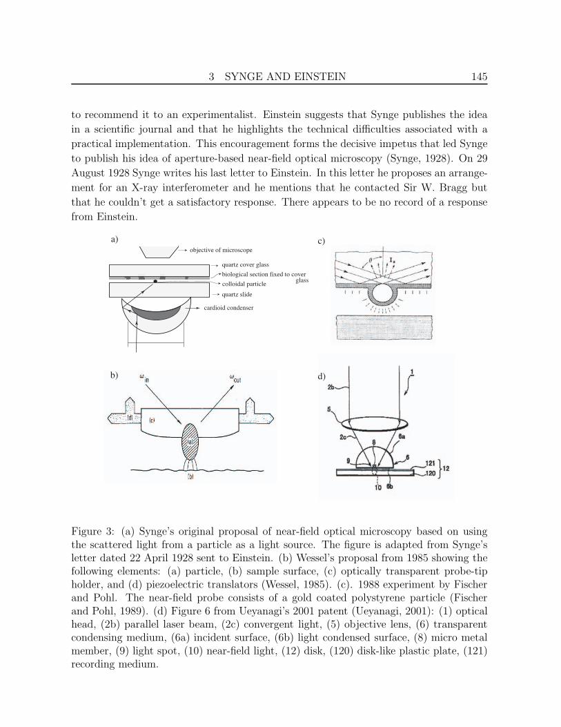
vides a sketch of the apparatus. Fig. 3a shows an adaptation of this sketch. Synge writes
”If a small colloidal particle, e.g. of gold, be deposited upon a quartz slide placed above a
Zeiss cardioid condenser of NA 1.05, then, all rays of light from the condenser which reach
the surface of the slide will be totally reflected by the surface, except those which strike
the surface at the base of the particle. These will be scattered in all directions and if the
objective of a microscope is suitably arranged above the slide, a proportion of the rays so
scattered will come to a focus in the eye of an observer, or upon a photographic plate, or a
photo-electrical cell suitably placed.” Synge describes here what is called today dark-field
microscopy, a technique invented at the turn of the twentieth century by the Austrian
chemist Richard Adolf Zsigmondy. In his letter, Synge then proposes to place a very thin
stained biological section onto a quartz cover glass and to raster scan it in close distance
Figure 2: Early near-field optical scan trace recorded with an aperture-type probe. Thedata is from the laboratory book of Winfried Denk then working with Dieter Pohl at theIBM Research Laboratory in Switzerland.

3 SYNGE AND EINSTEIN 144
over the irradiated particle. He argues that the amount of light received from the particle
and collected by the objective will depend upon the relative opacity of the different parts
of the section. In the remainder of the letter, Synge addresses different technical difficul-
ties, describes the scanning process, and also proposes to embed the particle into the end
face of the quartz slide. He also realizes a potential difficulty: ”I am not sure how near
the biological section could be brought to the surface of the quartz plate without impairing
the totality of the reflection.” He thinks this is the only potential theoretical limitation
and that everything else depends only on perfecting the technique. Synge also writes that
there is no institution in his country that could carry out the necessary experiments and
he suggests that the experiments could be undertaken by the Institute of Physics in Berlin.
On 3 May 1928 Einstein replies with a short letter written in German and sent from
Berlin. He states that he believes that Synge’s basic idea is correct but that his particu-
lar implementation seems to be of no use (”prinzipiell unbrauchbar”). He argues that if
the distance between the sample surface and the surface supporting the particle becomes
small then there will be considerable light leakage (frustrated total internal reflection) and
the total image field will become bright. Instead, Einstein suggests of using the light that
penetrates through a tiny hole in an opaque layer as a light source. He also states that he
couldn’t read many parts of Synge’s letter. This could be related to Synge’s challenging
handwriting (which improved in Synge’s second letter).
Only five days later, on 9 May 1928 (imagine the efficiency of the postal service at
that time), Synge replies to Einstein’s letter and states ”It was my original idea to have
a very small hole in an opaque plate, as you suggest, and it was in this form that I had
mentioned it to several people.” In the same letter Synge suggests what later became the
most standard way of fabricating aperture probes used in near-field optical microscopy:
”A better way could be, if one could construct a little cone or pyramid of quartz glass
having its point P brought to a sharpness of order 10−6 cm. One could then coat the sides
and point with some suitable metal (e.g. in a vacuum tube) and then remove the metal
from the point, until P was just exposed. I do not think such a thing would be beyond the
capacities of a clever experimentalist.” In a following paragraph Synge states that he is
”sure that some idea of the kind will be made use of ultimately, but it obviously requires
to drop into the brain of an experimental genius.” A couple of decades later this prophecy
was indeed verified.
Einstein’s reply from 14 May 1928 does not address the new thoughts of Synge. In-
stead, Einstein refers to the original idea of using the scattered light from a tiny particle
as a light source and he reiterates the problem of total internal reflection. Einstein states
that he believes that it is practically impossible to generate a tiny light source through
total internal reflection and scattering. He adds that he neither believes in the promise
of the other proposals for generating a tiny light source and that it makes him hesitant
3 SYNGE AND EINSTEIN 145
to recommend it to an experimentalist. Einstein suggests that Synge publishes the idea
in a scientific journal and that he highlights the technical difficulties associated with a
practical implementation. This encouragement forms the decisive impetus that led Synge
to publish his idea of aperture-based near-field optical microscopy (Synge, 1928). On 29
August 1928 Synge writes his last letter to Einstein. In this letter he proposes an arrange-
ment for an X-ray interferometer and he mentions that he contacted Sir W. Bragg but
that he couldn’t get a satisfactory response. There appears to be no record of a response
from Einstein.
cardioid condenser
quartz cover glass
objective of microscope
quartz slide
biological section fixed to cover glasscolloidal particle
a)
b)
c)
d)
Figure 3: (a) Synge’s original proposal of near-field optical microscopy based on usingthe scattered light from a particle as a light source. The figure is adapted from Synge’sletter dated 22 April 1928 sent to Einstein. (b) Wessel’s proposal from 1985 showing thefollowing elements: (a) particle, (b) sample surface, (c) optically transparent probe-tipholder, and (d) piezoelectric translators (Wessel, 1985). (c). 1988 experiment by Fischerand Pohl. The near-field probe consists of a gold coated polystyrene particle (Fischerand Pohl, 1989). (d) Figure 6 from Ueyanagi’s 2001 patent (Ueyanagi, 2001): (1) opticalhead, (2b) parallel laser beam, (2c) convergent light, (5) objective lens, (6) transparentcondensing medium, (6a) incident surface, (6b) light condensed surface, (8) micro metalmember, (9) light spot, (10) near-field light, (12) disk, (120) disk-like plastic plate, (121)recording medium.

3 SYNGE AND EINSTEIN 146
Synge’s original proposals to overcome the diffraction limit of light microscopy were
reinvented over the years and form the basis for both ’aperture’ and ’apertureless’ scan-
ning near-field optical microscopy. Synge was also the first to propose the principle of
scanning which forms today a key ingredient in a wide range of technologies ranging from
television to scanning electron microscopy (SEM). Before Synge’s time the idea of manip-
ulating the position of a source relative to a target in an imaging apparatus and thereby
improving its imaging capabilities was not known (McMullan, 1990). In a follow-up paper
in 1932, Synge suggested the use of piezo-electric quartz crystals for rapidly and accu-
rately scanning the specimen (Synge, 1932). He estimated that a translation of 5µm can
be accomplished with a voltage of 250 V. This estimate matches perfectly the sensitivity
of present-day piezo-electric transducers used in scanning probe microscopy. The idea
of using piezo-electric transducers to control the gap between the probe (e.g. aperture)
and the sample surface did not occur to him. This concept had to wait fifty years and
was a key ingredient in the development of scanning tunneling microscopy (STM) (Bin-
nig, 1982). In the same 1932 paper, Synge also suggested for the first time to use image
processing to highlight certain features of an image before displaying it. According to
McMullen (McMullan, 1990), Synge had many other visionary ideas, some of which be-
came of importance in other scientific fields. In 1936 Synge had a mental breakdown and
had to spend the rest of his life in a Dublin nursing home.
Although Einstein didn’t believe in the practical realization of Synge’s original idea
the concept of using the scattered light from a tiny particle as a light source is today well-
established and experimentally verified (Fischer and Pohl, 1989; Malmqvist and Hertz,
1994; Kawata, 1994; Anger, 2006; Kuhn, 2006). In 1985 John Wessel proposed a method
very similar to Synge’s original idea (Wessel, 1985). His proposed arrangement is depicted
in Fig. 3b. Wessel suggests to excite the particle resonantly thereby creating a locally
enhanced field which serves as a light source. He is the first to mention the analogy to
classical antenna theory. He writes: ”The particle serves as an antenna that receives an
incoming electromagnetic field.” Wessel did not know of Synge’s ideas and his proposal
was likely inspired by the invention of STM (Binnig, 1982) and the discovery of surface en-
hanced Raman scattering (SERS) (Fleischmann, 1974; Jeanmaire and Van Duyne, 1977;
Albrecht and Creighton, 1977). The quest for an understanding of SERS gave rise to
many theoretical studies aimed at predicting the electromagnetic field enhancement near
laser-irradiated metal particles and clusters thereof (Gersten and Nitzan, 1980; Wokaun,
1981; Boardman, 1982; Metiu, 1984; Meier, 1985). This era can be considered as the first
phase of what is called today nanoplasmonics (Xia and Halas, 2005). In 1988 Ulrich Ch.
Fischer and Dieter W. Pohl have carried out an experiment that is very similar to Synge’s
and Wessel’s proposal (Fischer and Pohl, 1989). Instead of using a laser-irradiated solid
metal particle as a local light source, they used a gold covered polystyrene particle (c.f.
4 FIRST DEVELOPMENTS 147
Fig. 3c), a structure that was later extensively developed and named gold nanoshell (Jack-
son, 2003). Fischer and Pohl imaged a thin metal film with 320nm holes and demonstrated
a spatial resolution of ≈ 50 nm. Their results provide the first experimental evidence that
near-field scanning optical microscopy as originally proposed by Synge is feasible.
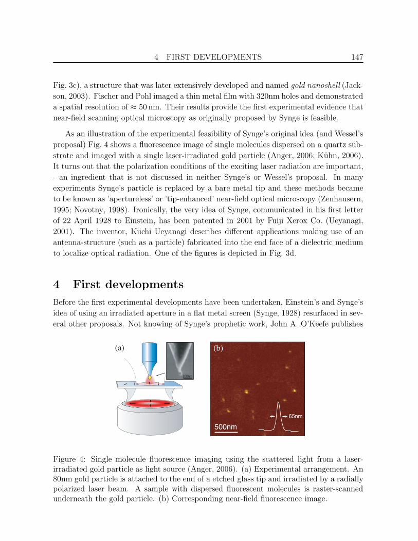
As an illustration of the experimental feasibility of Synge’s original idea (and Wessel’s
proposal) Fig. 4 shows a fluorescence image of single molecules dispersed on a quartz sub-
strate and imaged with a single laser-irradiated gold particle (Anger, 2006; Kuhn, 2006).
It turns out that the polarization conditions of the exciting laser radiation are important,
- an ingredient that is not discussed in neither Synge’s or Wessel’s proposal. In many
experiments Synge’s particle is replaced by a bare metal tip and these methods became
to be known as ’apertureless’ or ’tip-enhanced’ near-field optical microscopy (Zenhausern,
1995; Novotny, 1998). Ironically, the very idea of Synge, communicated in his first letter
of 22 April 1928 to Einstein, has been patented in 2001 by Fuiji Xerox Co. (Ueyanagi,
2001). The inventor, Kiichi Ueyanagi describes different applications making use of an
antenna-structure (such as a particle) fabricated into the end face of a dielectric medium
to localize optical radiation. One of the figures is depicted in Fig. 3d.
4 First developments
Before the first experimental developments have been undertaken, Einstein’s and Synge’s
idea of using an irradiated aperture in a flat metal screen (Synge, 1928) resurfaced in sev-
eral other proposals. Not knowing of Synge’s prophetic work, John A. O’Keefe publishes
500nm
(b)(a)
65nm
(a)
200nm
Figure 4: Single molecule fluorescence imaging using the scattered light from a laser-irradiated gold particle as light source (Anger, 2006). (a) Experimental arrangement. An80nm gold particle is attached to the end of a etched glass tip and irradiated by a radiallypolarized laser beam. A sample with dispersed fluorescent molecules is raster-scannedunderneath the gold particle. (b) Corresponding near-field fluorescence image.

4 FIRST DEVELOPMENTS 148
in 1956 a short proposal starting with ”The following is presented as a concept illustrat-
ing a method by which it might conceivably be possible to go beyond the resolving power
of visible light” (O’Keefe, 1956). Similar to Synge, he proposes to employ an irradiated
small hole in an aluminum coating as a light source. He writes ”By scanning, an image
can be built up whose detail will correspond to the size of the hole, even if it is smaller
than the wavelength of the light used.” O’Keefe indicates that he believes the realization
of his proposal is remote because of the difficulty of providing for relative motion between
the pinhole and the object. Only a few months later, Albert V. Baez publishes an article
in which he discusses the results of a near-field acoustical experiment (Baez, 1956). For
his demonstration, Baez used acoustic waves with a wavelength of 14cm (2.4 kHz) and
confirmed a resolution not limited by diffraction. Baez used his own fingers as test objects
and his results are mentioned only very qualitatively in the article.
More than ten years later Charles W. McCutchen discusses the possibility of overcom-
ing the diffraction limit of optical imaging by convolving the spatial frequencies of the
sample with the spatial frequencies of a probe object (McCutchen, 1967). His consider-
ation is based on the assumption that the plane containing the probe object constitutes
a spatial filter function for the field emanating from the sample plane. The probe object
shifts the spatial spectrum of the sample field thereby making high spatial frequencies
propagate. This principle is analogous to a radio receiver where a local oscillator (probe
particle) shifts the signal frequencies (sample field) into the pass band (propagating radi-
ation). According to McCutchen, it doesn’t matter whether a particle or an aperture is
scanned over the surface of a sample. What matters is that the probe object is very small
so it provides the necessary high spatial frequencies. Of course, the application of simple
linear filter theory is an oversimplification and it cannot account for strong interactions
between sample and probe, e.g. for field enhancement effects and resonant interactions.
Nevertheless, it provides an intuitive picture which is important for the understanding of
image formation in near-field optical microsopy. McCutchen believes that it isn’t practical
to place the probe object right next to the sample and he considers the effect of moving
the probe object behind the condenser lens. He argues that the bandwidth of spatial
frequencies can be doubled, - a principle employed today in structured illumination mi-
croscopy (Gustafsson, 2005). McCutchen writes ”In an extreme form, the technique would
consist of scanning the specimen past a minute aperture, much smaller than the Abbe limit.
... Obviously, it would be hard to use this method on any but the flattest of surfaces, and
I wonder if there are many jobs it could do that reflection electron microscopy would not
do better.” McCutchen’s scepticism is justified, but it is the scientific curiosity and the
potential benefit of combining the chemical specificity of optical spectroscopy with high-
resolution optical microscopy which drives the progress in near-field optics forward. In
1984, Gail A. Massey uses Fourier optics to formulate the image forming process described
by McCutchen (Massey, 1984), however without knowing of McCutchen’s work. Massey

5 SURFACE PLASMONS AND SURFACE ENHANCED RAMAN SCATTERING149
concludes that for an aperture of size d a lateral resolution of ≈ d can be achieved with
a depth of focus of ≈ d/3 (Massey, 1984).
In 1970, H. Nassenstein proposes to illuminate a sample with evanescent waves thereby
generating scattered waves which contain information on spatial frequencies of the sample
spectrum beyond the classical resolution limit (Nassenstein, 1970). Similar experiments
were reported before in the context of holography Stetson (1967). Since evanescent waves
are bound to their source (primary or secondary), Nassenstein’s proposal implies that a
secondary object (the probe) be brought close to the sample. Today, total internal re-
flection fluorescence (TIRF) microscopy is a powerful tool in biological research because
it effectively reduces background fluorescence. There are commercially available TIRF
microscope objectives with NA’s of 1.5 − 1.65.
The first experimental validation of near-field microscopy using electromagnetic radi-
ation has been undertaken by Eric A. Ash and G. Nicholls at University College, London.
In a paper published in 1972 they use 10 GHz microwaves (λ=3 cm) and an aperture of
1.5 mm to image an aluminum test pattern deposited on a glass slide (Ash and Nicholls,
1972). At a separation between aperture and sample plane of 0.5 mm they are able to
achieve a resolution of better than λ/60, clearly beyond the diffraction limit of standard
microscopy. Ash and Nicholls state ”.. one might hope to build a super-resolution optical
microscope,” and they add ”Although the optical microscope may not be beyond reach, in
our view a more immediately hopeful application is the construction of an infrared mi-
croscope with a resolution comparable with that normally attainable in the visible optics
spectrum.” However, the history of near-field optics proceeded in the other sequence. In
1982 the first optical scan images were recorded by Dieter W. Pohl and co-workers at the
IBM Research Laboratories (c.f. Fig. 2) and the infrared analogue has been demonstrated
later in 1986 by Gail A. Massey using 100µm radiation (Massey, 1985).
5 Surface Plasmons and Surface Enhanced Raman
Scattering
The field of near-field optics has been greatly established by the quest for diffraction-
unlimited microscopy and spectroscopy. But there are at least two other important de-
velopments which had a significant influence on the shaping of the field: 1) research
in the field of metal optics and 2) the discovery of surface enhanced Raman scattering
(SERS). In 1968 two different experiments, one by Andreas Otto and the other by Erwin
Kretschmann and Heinz Rather, have demonstrated that plasma oscillations on thin metal
films can be excited with an optical beam undergoing total internal reflection (Otto, 1968;
Kretschmann and Rather, 1968). It was shown that these plasma oscillations are polari-

5 SURFACE PLASMONS AND SURFACE ENHANCED RAMAN SCATTERING150
tons, i.e. their existence is coupled to an electromagnetic field, and today these excitations
are referred to as surface plasmon polaritons. It is interesting to note that already in 1959
T. Turbadar used the Kretschmann configuration for reflectance measurements on alu-
minum films and he noticed a characteristic reflection dip beyond the critical angle of total
internal reflection. The reflection dip was shown to be consistent with thin film theory and
it was noted that it must be associated with an evanescent wave because there is no trans-
mitted light (Turbadar, 1959). The existence of optical surface waves on metal surfaces
was also noticed in 1941 by Ugo Fano in his theoretical analysis of anomalous diffraction
gratings (Fano, 1941). The later experiments of Otto, Kretschmann and Raether gave
rise to various dedicated experiments aimed at understanding the confinement of optical
fields near the surface of metals. On planar metal surfaces, plasmons are nonradiative.
They propagate along the surface and their wavelength at a given frequency ω is shorter
than the one in free space. At the surface plasmon frequency, and in the ideal case of no
damping, the wavelength of the plasmon polariton goes to zero and the associated field
becomes localized to the very surface of the metal, i.e. the decay length of the evanescent
wave goes to zero. Besides the interest in surface plasmons propagating along extended
interfaces, experimental and theoretical work had also been undertaken to understand
the optical response of finite sized metal particles. The work of Uwe Kreibig provided
an understanding for the transition of macroscopic electromagnetic theories applicable to
particles larger than the mean-free path of electrons in the metal to quantum theories
applicable to clusters of a few metal atoms (Kreibig, 1974). Surface plasmons associated
with finite metal particles and clusters thereof gained a lot of importance for the under-
standing of the SERS effect which was discovered in 1974 (Fleischmann, 1974). Shortly
after, it was realized that the phenomenon is due to the increased surface of roughened
metal surfaces and the associated electromagnetic field enhancement (Jeanmaire and Van
Duyne, 1977; Albrecht and Creighton, 1977). In the early 1980s much theoretical work
was produced with the aim of understanding the SERS effect (Chang and Furtak, 1981).
The near-fields of various metal nanoparticle arrangements were calculated and it was con-
cluded that the effect is mediated by so-called ’hot spots’, i.e. localized regions between
metal particle aggregates that exhibit particularly strong electromagnetic field enhance-
ment. A recent retrospective has been written by Martin Moskovits (Moskovits, 2005).
The enhanced near fields around metal nanoparticles has also been shown to lead to sev-
eral interesting photochemical phenomena, including near-field driven plasmon-resonant
particle growth (Chen and Osgood, 1983). The quest for an understanding of the SERS
effect marked the beginning of the first wave of nanoplasmonics and gave rise to new
schemes of near-field optical microscopy, such as Wessel’s proposal (Wessel, 1985). The
electromagnetic theories and calculations developed in the early phase of SERS proved
very beneficial for the understanding of light localization and near-field interactions. On
the other hand, the development of near field optical microscopy provided for the first

6 STUDIES AND APPLICATIONS OF ENERGY TRANSFER 151
time direct access to the fields associated with surface plasmon polaritons (Specht, 1992;
Marti, 1993; Dawson, 1994) and gave rise to the recent revival of nanoplasmonics.
6 Studies and Applications of Energy Transfer
Independent of the activities in SERS, plasmonics, or near-field microscopy, short-range
optical interactions were also investigated in the context of energy transfer between
molecules (Forster, 1946) and fluorescence quenching near metal surfaces (Kuhn, 1970;
Drexhage, 1970; Zingsheim, 1976). Already in 1970 Hans Kuhn suggested to make use
of optical near-fields for contact imaging (Kuhn, 1970). He envisioned to use short-range
energy transfer from electronically excited dye molecules as a method for the duplication
of nanostructures. The scheme was later implemented by Hans P. Zingsheim and Ulrich
Ch. Fischer (Fischer and Zingsheim, 1982). In this experiment a monomolecular film of
(a) (b)
(d)(c)
Figure 5: Near-field optical contact printing originally proposed by Hans Kuhn. Ananoscale pattern is transfered to a dye layer by placing a laser-irradiated nanostruc-tured metal film on top of the dye layer. Short-range bleaching the fluorescence of thedye layer gives rise to spatial patterning of the fluorescence intensity. (a) Fluorescenceimage of the dye layer during contact with a metal pattern consisting of platinum disks(diameter 8µm). Close to the disks the fluorescence is diminished because of fluorescencequenching. (b) After release of the metal mask regions of the dye layer which were closeto individual metal disks exhibit stronger fluorescence (quenching lowers the bleachingrate). (c,d) show the corresponding arrangements of layer and mask. From (Fischer andZingsheim, 1982).
7 FIRST DEVELOPMENTS OF NEAR-FIELD OPTICAL MICROSCOPY 152
dye molecules serves as a light sensitive film, and a very thin, only partially absorbing,
planar metal pattern which is embedded into the surface of a pliable polymer film, serves
as a conformal mask (c.f Fig. 5). After the film is brought into contact with the mask
and after it has been irradiated with light, the structure is transferred from the mask to
the monomolecular film as a pattern of areas where the dye is bleached and where it is
not bleached. The resolution of this pattern transfer is not limited by the wavelength
of light but by the range of the energy transfer mechanism and the distance between
the mask and the dye layer (Fischer, 1998). In principle, this process of pattern transfer
makes use of the short range of the near-field of single electronically exited molecules
in order to obtain diffraction-unlimited resolution down to the molecular scale. Studies
of energy-transfer processes established that many types of near-field interactions can
be understood on a classical phenomenological basis (Chance, 1978; Kerker, 1980). The
phenomenological theory treats the quantum mechanical transition probability between
two states as a classical dipole oscillating at the transition frequency. The theory turns
out to be equivalent to a rigorous quantum-electromagnetic theory in the weak coupling
regime (Novotny and Hecht, 2006) but it is much easier to deal with. An extensive re-
view of surface enhanced spectroscopy and energy transfer processes between molecules
and surfaces of various shapes has been provided in 1984 by Horia Metiu (Metiu, 1984)
and also by George W. Ford and Willes H. Weber (Ford, 1984). The latter review also
discusses the nonlocal dielectric response near material boundaries (spatial dispersion).
In later work, Forster energy transfer has been proposed as an interaction mechanism
for near-field optical imaging (Kopelman and Tan, 1994; Fujihira, 1996; Sekatskii, 1996;
Vickery and Dunn, 1999). In 1999 the group of Robert C. Dunn provided the first ex-
perimental demonstration of near-field energy transfer microscopy with sub-wavelength
resolution (Vickery and Dunn, 1999) and the following year the emission from a single
molecule was used for the first time as a local light source (Michaelis, 2000).
7 First developments of Near-field optical Microscopy
First dedicated efforts of demonstrating near-field microscopy at optical frequencies started
in the early 1980s. The first developments proceeded without the knowledge of previous
proposals and the prophetic papers by Synge. On 27 December 1982 Dieter W. Pohl,
then working at the IBM Research Laboratory in Switzerland, filed a patent titled optical
near-field scanning microscope (Pohl, 1984b) which describes an aperture-based method
very similar as conceived by Synge more than fifty years earlier. The patent states ”The
term ’near-field’ is intended to express the fact that the aperture is located near the object
at a distance smaller than the wavelength. The term ’aperture’ is used here to describe
the pointed end of a light waveguide which forms an entrance pupil with a diameter of less
than 1µm.” In the same year, Pohl and co-workers managed to overcome the remaining

7 FIRST DEVELOPMENTS OF NEAR-FIELD OPTICAL MICROSCOPY 153
experimental hurdles and recorded the first optical scan trace shown in Fig. 2. Their first
scientific publication appeared with some delay in 1984 (Pohl, 1984). In this publication
near-field optical microscopy is referred to as optical stethoscopy in analogy to the acoustic
stethoscope used in medical diagnosis. Pohl, Denk, and Lanz write ”The familiar medical
doctor’s stethoscope, for instance, allows localization of the position of the heart to within
less than 10 cm by moving the stethoscope over the patient’s chest, and listening to the
sound of the heart beat. Assuming a sound frequency of 30-100 Hz, corresponding to a
wavelength of almost 100m, the stethoscope provides a resolving power of roughly λ/1000
! ” Their aperture light-source was formed by pressing an aluminum coated, electrochem-
ically etched quartz crystal towards a transparent sample. Laser light is coupled into the
crystal and as soon as light is transmitted through the sample a tiny aperture has been
formed. This procedure for forming apertures has been later named pounding or punching.
During the same time other groups have started similar efforts. Results by Ulrich
Ch. Fischer were presented in a talk by Hans Kuhn at the second Meeting of Molecular
Electronic Devices in 1983. The proceedings of this meeting were published much later,
in 1987, because the editor passed away in the mean time (Kuhn, 1987). One of the scan
traces was also reproduced in the 1984 yearbook of the Max-Planck-Society (Kuhn, 1984)
(summary of research activities of 1983) and is shown in Fig. 6 along with the outline of
the experimental setup. In their experiments, Fischer and Kuhn used a glass hemisphere
coated with a Tantalum/Tungsten layer in which a 100nm hole was fabricated. A similar
metal layer (20nm thickness) with ten times larger holes (1µm) was fabricated onto a
glass slide and served as a test sample. The hemisphere was brought close to the sample
and then scanned (without feedback control) along the surface. In Fig. 6b two different
(b)(a)
Figure 6: Experiment by Ulrich Ch. Fischer performed in 1983 while working with HansKuhn. a) Schematic of the experiment. A metal-coated glass hemisphere with a 100nmhole is used to irradiate a test sample made of a metal layer with 1µm holes depositedonto a glass slide. From (Kuhn, 1987). b) Optical scan traces recorded along the solidline (top trace) and along the dashed line (bottom trace). From (Kuhn, 1984).

7 FIRST DEVELOPMENTS OF NEAR-FIELD OPTICAL MICROSCOPY 154
scan traces are shown, one through the center of the sample hole (top) and one closer
to its edge (bottom). The slope of the upper trace indicates a resolution better than
the diffraction limit. During the same time, Fischer and Zingsheim pioneered what is
called today nanosphere lithography (Fischer and Zingsheim, 1982). In this technique, a
suspension of colloidal particles with a diameter of > 100 nm is deposited onto a plane
surface. Subsequent evaporation of the solvent arranges the particles in a hexagonal two-
dimensional lattice. Vacuum deposition of metals and other materials through the voids
between neighboring spheres leads to arrays of triangular structures on the surface. The
size and the periodicity of these patterns can be varied by the size of the original par-
ticles. Double exposure from different angles of incidence allows more complex pattern
to be created (Haynes and Van Duyne, 2001). The resulting Fischer patterns are used
as test samples for microscopy or for Raman active substrates for the detection of target
agents.
At the time of the experiments of Fischer and the experiments performed at IBM by
Pohl and co-workers another development was underway at Cornell University. Aaron
Lewis and co-workers studied light transmission through arrays of holes in planar metal
films and were working towards a ”scanning nanometer optical spectral microscope.” The
first document mentioning their effort is an abstract from the 1983 Biophysics Meet-
ing (Lewis, 1983). One year later they published an article presenting light transmission
through 30 nm apertures in metal films (Lewis, 1984). In the same article they discuss
”the possibility of constructing a scanning optical microscope based on near field imaging
which could potentially have spatial resolutions as small as one-tenth (of) the wavelength
of the incident light” (Lewis, 1984). Their first experimental results made use of ther-
mally pulled glass capillaries, as used in the patch-clamp technique, and were published in
1986 (Harootunian, 1986). These experiments used near-field excited fluorescence as con-
trast mechanism and demonstrated a resolution on the order of the aperture diameter of
100 nm. In the same publication, Lewis and co-workers introduced the acronym NSOM,
standing for ”near-field scanning optical microscopy”. Earlier, in the same year, Urs
Durig, Dieter W. Pohl and Flavio Rohner publish transmission near-field images recorded
with aperture probes and by using active distance control via electron tunneling between
probe and sample (Durig, 1986). They refer to the technique as NFOS microscopy. The
acronym SNOM was created later in 1988 to emphasize the analogy to SEM, STM, and
other scanning microscopies. The optical near-fields near irradiated apertures were not
only considered for microscopy but also for sensing applications. In 1986 Ulrich Ch. Fis-
cher publishes a study on the transmission of tiny apertures in metal films (Fischer, 1985).
The following year he demonstrates that the aperture acts as probe of its microenviron-
ment through enhanced light scattering and fluorescence (Fischer, 1986). This approach
has been later revisited and applied to studies of single-molecule dynamics (Levene, 2003).

7 FIRST DEVELOPMENTS OF NEAR-FIELD OPTICAL MICROSCOPY 155
A significant breakthrough in the application of near-field optical microscopy came
in 1991 when Eric Betzig and co-workers introduced aperture probes formed at the end
of metal coated, thermally pulled quartz fibers (Betzig, 1991). This method became
widely used and was also adopted by the first companies that pursued a commercializa-
tion of near-field optical microscopes. Another important technical improvement came
in the following year, in 1992, when two independent groups introduced the shear-force
technique to control the distance between probe and sample (Toledo-Crow, 1992; Bet-
zig, 1992). In 1995, the shear-force method was combined with the sensing capability of
a tuning fork crystal (Karrai and Grober, 1995) which, today, is the most widely used
method for probe-sample distance control. The group of Betzig at Bell Laboratories pi-
oneered many applications of near-field optical microscopy, published many highlights,
and contributed to the general acceptance of the method (Betzig and Trautman, 1992).
In 1994, near-field microscopy was applied for the very first imaging of single fluorescent
molecules (Trautman, 1994). While single molecules have been detected before by use of
spectral identification at cryogenic temperatures (Moerner and Kador, 1989; Orrit and
Bernard, 1990), it was the spatial mapping which triggered the birth of single molecule
spectroscopy (Xie and Trautman, 1998). As an illustration, Fig. 7 shows a fluorescence
image of a sample with single dye molecules recorded with an aperture probe that was
created by focused ion beam milling (Veerman, 1998).
Over the years, many variations of near-field optical microscopy have been explored
and developed. Probably the most notable one is photon scanning tunneling microscopy
(PSTM), also called scanning tunneling optical microscopy (STOM), developed in 1989
by three different groups (Courjon, 1989; Reddik, 1989; deFornel, 1989). This method
1 mμ
(a) (b)
Figure 7: (a) SEM image of smooth aperture probe fabricated by slicing away the endof a metal coated, tapered optical fiber with a focused ion beam (FIB). From (Veerman,1998). (b) Single molecule fluorescence image acquired with a FIB milled aperture probe.From (Moerland, 2005).

7 FIRST DEVELOPMENTS OF NEAR-FIELD OPTICAL MICROSCOPY 156
is the optical analog of STM as it probes the tunneling photons between a surface and
a local probe. Typically, an evanescent field is created by total internal reflection at a
dielectric-air interface and the tip of a pointed optical fiber is used to locally convert the
evanescent field into a propagating waveguide mode, similar to frustrated total internal
reflection (Novotny and Hecht, 2006). PSTM is a very attractive method because of its
simple physical picture, but the recorded images were not easy to interpret, mainly be-
cause of multiple scattering between separated objects on the sample. The reconstruction
of the sample features based on the measured optical information, the inverse scatter-
ing problem, can be facilitated by tuning the angle of incidence of the total internally
reflected wave (Garcia and Nieto-Vesperinas, 1995). Several groups have theoretically
established that PSTM measurements are inherently holographic and that they provide
enough information to determine the two dimensional structure of a thin sample (Gr-
effet, 1995; Bozhevolnyi and Vohnsen, 1996; Carney, 2004). A series of related studies
on near-field phase conjugation have been performed by Sergey I. Bozhevolnyi and co-
workers (Bozhevolnyi, 1994, 1995).
In modified form, the PSTM found interesting applications in optoelectronics and op-
tical waveguides. Here, the evanescent tail of a waveguide mode is directly measured with
a near-field probe rendering spatial maps of the electromagnetic field distribution. In
a landmark experiment, Niek van Hulst’s group combined this method with heterodyne
detection and generated phase and amplitude maps of femtosecond pulses propagating
along ridge waveguides (Balistreri, 2001). These experiments, shown in Fig. 8, directly
visualized the electromagnetic field associated with an optical pulse and demonstrated
Figure 8: Measurement of the electromagnetic field of a light pulse propagating along aridge waveguide. Left: Schematic of a heterodyne PSTM with variable time delay in thereference arm. The instrument probes the evanescent tail of a waveguide mode and rendersa temporally and spatially resolved map of the phase and amplitude distribution of theelectromagnetic field associated with the pulse. Right: Map of the measured amplitudemultiplied with the cosine of the measured phase for a single tracked pulse. The areadepicted is an enlargement of a small part of the actual scan. From (Balistreri, 2001).

7 FIRST DEVELOPMENTS OF NEAR-FIELD OPTICAL MICROSCOPY 157
that optical processes and phenomena can be probed by sampling their evanescent fields.
Recently it was demonstrated that waveguide modes can also be probed by light scattering
from a metal tip (Stefanon, 2005). PSTM was also applied to various other phenomena,
such as light localization in random media (Gresillon, 1999; Bozhevolnyi, 2002) or prop-
agation of surface plasmon polaritons (Marti, 1993; Dawson, 1994; Krenn and Weeber,
2004). Fig. 9 shows an example of a recent measurement by the group of Joachim R.
Krenn. The figure depicts a map of the measured light intensity of a surface plasmon
propagating along a silver nanowire.
In the 1990s, near-field optical microscopy has been applied to various problems and
the progress is best summarized by referring to more detailed review papers (Girard and
Dereux, 1996; Fischer, 1998; Dunn, 1999; Pohl, 2004). Very high spatial resolutions were
demonstrated, but the nature of the optical contrast in the recorded images was often
not understood. In fact, some optical images exhibited suspiciously close resemblance
with the simultaneously recorded shear-force images. It was soon realized that two dis-
tinct properties contribute to the recorded optical signal: 1) the local material-specific
response due to the probe-sample interaction, and 2) the vertical motion of the probe
due to probe-sample distance control. While the former is the true optical contrast, the
latter is an artifact and is due to the fact that the strength of the near-field interaction
depends on the proximity of the probe. A variation of the optical signal is generated even
if the probe is located at a fixed lateral position and its vertical position over the sample
surface is varied. In 1997, Bert Hecht and collaborators have published an article titled
Facts and Artifacts in Near-field Optical Microscopy in which they concluded that many
published images represent the path of the probe rather than the true optical properties
of the sample (Hecht, 1997). Following this paper, previous results had to be re-examined
and new results had to be critically tested before being published.
Figure 9: Cylindrical surface plasmon propagating towards the end of a silver nanowire.The figure shows a map of the light intensity measured with a PSTM. From (Ditlbacher,2005).
8 THEORETICAL NEAR-FIELD OPTICS 158
8 Theoretical Near-field Optics
Initial theoretical work in the field of near-field optics aimed at developing an understand-
ing of image formation in near-field optical imaging. Approximate methods such as scalar
diffraction theory break down in the near-field making it necessary to solve the full vecto-
rial wave equation for a given problem. In a 1931 publication, Synge writes ”This theory
(diffraction theory) is, in most cases, a very good approximation, and forms the basis of
the theory of resolution of optical instruments as usually presented, but it is by no means
an absolute theory, such a theory requiring the solution of the electromagnetic equations,
subject to boundary conditions which have been a bar to their solution except in one very
simple case. In general we may say that when we come down to magnitudes of the order
of a wavelength the approximate theory ceases to be a good approximation” (Synge, 1931).
The theory for understanding the field distributions near tiny apertures in metal screens
has been developed long before the interest in near-field optics started. It was Hans Bethe
in 1944 who provided the first rigorous description (Bethe, 1944). His theory was later
corrected and generalized by C. J. Bouwkamp in 1950 (Bouwkamp, 1950,b). For real met-
als and for screens of finite thickness the BetheBouwkamp theory still agrees qualitatively
with the true field distribution (Novotny and Hecht, 2006). First theoretical studies of
near-field optical microscopy made use of the formulas of Bethe and Bouwkamp and used
Fourier optics to propagate the fields (Massey, 1984; Betzig, 1986; Roberts, 1987; Leviatan,
1986). Also, intuitive models were developed in which the near-field probe was treated as
an elementary dipole (Van Labeke and Barchiesi, 1993; Labani, 1990). In the 1990s differ-
ent methods were introduced to solve the full vectorial wave equation for a given near-field
(a) (b)
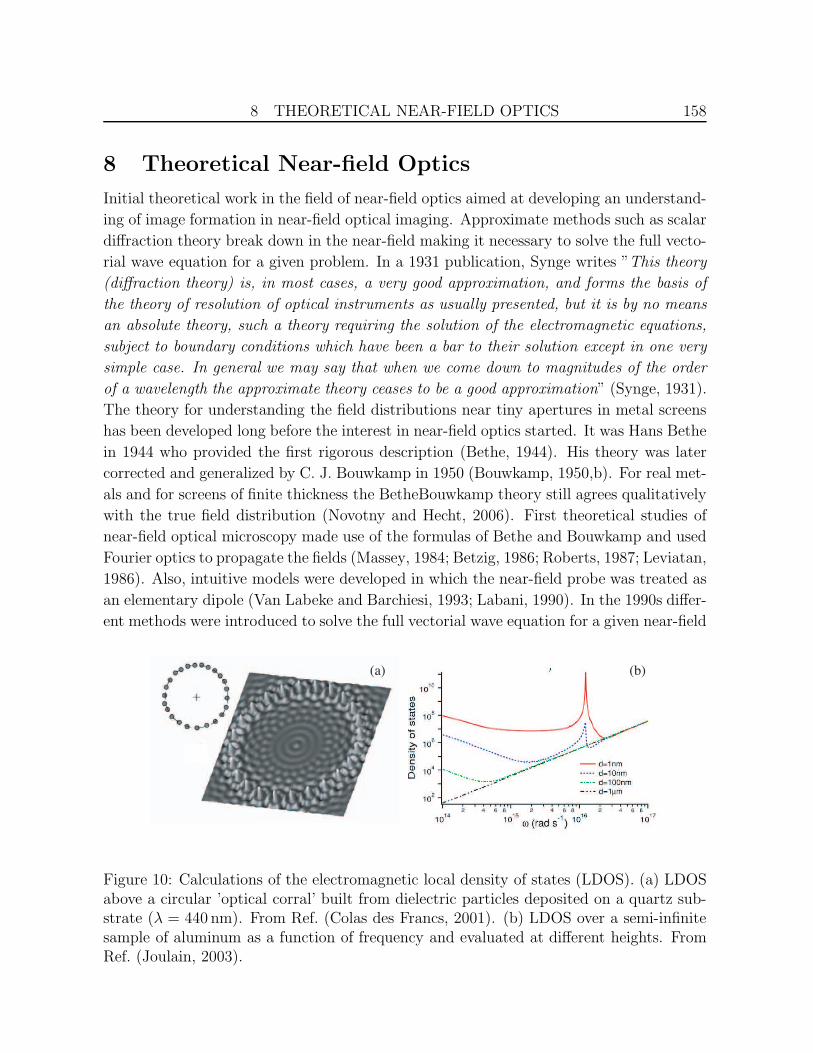
Figure 10: Calculations of the electromagnetic local density of states (LDOS). (a) LDOSabove a circular ’optical corral’ built from dielectric particles deposited on a quartz sub-strate (λ = 440 nm). From Ref. (Colas des Francs, 2001). (b) LDOS over a semi-infinitesample of aluminum as a function of frequency and evaluated at different heights. FromRef. (Joulain, 2003).
8 THEORETICAL NEAR-FIELD OPTICS 159
configuration. Among the most widely used methods were plane wave expansion tech-
niques (Van Labeke and Barchiesi, 1992), Green’s function techniques (Dereux and Pohl,
1993; Girard and Dereux, 1994; Martin, 1994), and the multiple multipole (MMP) tech-
nique (Novotny, 1994,b, 1995). These methods have an analytical foundation and are
summarized in Ref. (Novotny and Hecht, 2006). A review of early theoretical activities
in near-field optics has been written by Christian Girard and Alain Dereux (Girard and
Dereux, 1996). The goal of these early studies was to understand how interactions in
the near-field are mapped to the farfield. Most commonly, the inverse scattering problem
cannot be solved in a unique way and calculations of field-distributions are needed to
provide prior knowledge about source and scattering objects and to restrict the set of
possible solutions. For example, experimental near-field images revealed contrast reversal
if the collection angle of the near-field scattered light was modified (Hecht, 1995). It was
shown that this contrast reversal originates from evanescent wave scattering giving rise
to supercritical light propagation (forbidden light) in a dielectric sample (Novotny, 1997).
In 1995 it was shown that in some cases it is possible to assume that the interaction be-
tween probe and object can be neglected (weak coupling) which results in a simple linear
detection process (Carminati and Greffet, 1995). In this regime, the reciprocity theorem
requires that near-field optical images recorded in the PSTM mode and images recorded
with aperture-based microscopy are equivalent (Mendez, 1997). The theory underlying
image formation in near-field optics has been reviewed in 1997 by Jean-Jacques Greffet
and Remi Carminati (Greffet and Carminati, 1997). Theoretical studies gave input to
improved instrument design and predicted new detection strategies for optimizing the
signal-to-noise ratio (SNR) in a given measurement (Girard, 1994; Greffet and Carminati,
1997; Novotny, 1997b; Hecht, 1998). Ultimately, it is the SNR which determines the best
achievable resolution because, according to the principle of analytical continuation, a sig-
nal with finite support can be exactly reconstructed from a noise-free measurement in
an arbitrarily small spatial domain (Devaney and Wolf, 1973; Wolf and Nieto-Vesperinas,
1985).
More recent theoretical studies aimed at understanding the physical properties of op-
tical near-fields. Among the topics studied were coherence properties (Carminati and
Greffet, 1999; Roychowdhury and Wolf, 2003), the polarization state of optical near-
fields (Setala, 2002; Ellis, 2005), spontaneous emission (Girard, 1995; Novotny, 1996)
and local density of states near nanoscale structures c.f. Fig. 10) (Colas des Francs,
2001; Joulain, 2003; Novotny and Hecht, 2006), reciprocity relations in the optical near-
field (Carminati, 1998), and fluctuation-induced friction (Zurita-Sanchez, 2004). The
fluctuational properties of optical near-fields have been discussed in two recent review pa-
pers (Henkel, 2005; Joulain, 2005). Studies of near-field inverse scattering have been pur-
sued by different groups, mainly using the PSTM geometry (Garcia and Nieto-Vesperinas,
1995; Greffet, 1995; Bozhevolnyi and Vohnsen, 1996; Greffet and Carminati, 1997; Car-
8 THEORETICAL NEAR-FIELD OPTICS 160
ney and Schotland, 2003). It was shown that samples can be uniquely reconstructed from
near-field optical measurements by use of near-field tomography (Carney, 2004).
While first studies on the quantum nature of evanescent fields have been performed
already in the 1970s by Girish S.. Agarwal (Agarwal, 1975) and Chuck Carniglia and
Leonard Mandel (Carniglia and Mandel, 1971) a quantized theory of spatially confined
light has been put forth by Ole Keller in 1998 (Keller, 1998, 2000b, 2005). Keller also
described the birth process of the photon wavefunction and pointed out that in a near-
field interaction the photon is destroyed before it is fully born (Keller, 2002). Another
problem is the fact that it is not strictly possible to separate the source of radiation from
the sink of radiation. Instead, source and sink appear as a coupled object or, more for-
mally, it is not possible to independently define the state of source and detector (Power
and Thirunamachandran, 1997). A remarkable result of Keller’s work is the finding that
only after an infinite time after its birth, the energy of a photon is ~ωo, ωo being the
transition frequency. At shorter times, the photon energy is larger than ~ωo. Part of the
problem of defining a near-field photon is associated with the fact that the near-field is not
purely transverse, which can be easily verified for an evanescent wave and its excitation.
Standard quantum electrodynamics (QED) proceeds by invoking the Coulomb gauge and
quantizing the retarded transverse field. It gives little attention to the ’attached’ field.
However, from single molecule experiments it is known that a molecule close to an inter-
face interacts with the total field and not only with its transverse part (Drexhage, 1974).
Future theories and experiments will shed more light on the existence of near-field photons.
Because of their localized nature, optical near-fields can vary substantially over the
length scale defined by a quantum system, such as a quantum dot or a molecule. Hence,
the light-matter interaction can no longer be restricted to the dipole selection rules and
higher order multipolar transitions need to be considered (Zurita-Sanchez and Novotny,
2002). In the extreme case, the multipolar expansion does not converge and the optical
near-field becomes a probe for the local orbital overlap between the system’s ground state
and excited state. It is also interesting to note that in the light-matter interaction the
momentum of a photon (p = 2π~/λ) is typically neglected because it is much smaller
than the electron momentum in matter (p =√
2meffE). Consequently, photoinduced
band-to-band transitions in an electronic dispersion diagram happen vertically. However,
the momentum of a photon associated with the optical near-field is no longer defined
by the wavelength but by a characteristic length d associated with the optical confine-
ment (p = 2π~/d). This makes the near-field momentum comparable with the electron
momentum and hence intraband transitions become possible (Beversluis, 2003). The
high momenta associated with the optical near-field as well as the possibility of accessing
dipole-forbidden transitions will enrich optical spectroscopy and open up new and exciting
frontiers.

9 NEAR-FIELD SCATTERING AND FIELD ENHANCEMENT 161
9 Near-field Scattering and Field Enhancement
Let us now go back in time to revisit Synge’s original proposal of using the light scattered
by a small particle as a light source. When brought close to a sample surface, the particle
not only scatters the incident field but also the field that is scattered from the surface.
In fact, there is an infinite number of scattering iterations between particle and sample.
Depending on the properties of particle and sample and on the excitation conditions only
one or a few terms in this series are relevant. For example, in Wessel’s proposal (Wessel,
1985), the particle’s response is resonant with the incident field and hence the enhanced
field generated by the particle can be regarded as an independent light source exciting
the sample at short distance. On the other hand, one could consider a situation in which
the interaction between the exciting field and the sample is more dominant than the in-
teraction of the external field and the particle. In this case, the particle acts as a passive
probe that scatters away the near-field of the irradiated sample. Both approaches have
been implemented in near-field optics using pointed probes such as dielectric or metal tips
as local scatterers. In weak scattering the probe acts as a local perturbation (Zenhausern,
1994; Bachelot, 1994; Inouye and Kawata, 1994) whereas in strong scattering the interac-
tion between the probe and the exciting field dominates and the probe acts as an optical
Figure 11: ’Apertureless’ near-field optical microscope using heterodyne detection.Original drawing from the patent of H. Kumar Wickramasinghe and Clayton C.Williams (Wickramasinghe and Williams, 1990). (12) end of a tip, (14) tip, (40) opticalsource, (42) acousto-optic modulator, (44,46) lens, (48) beam splitter, (50) pin photodi-ode.

9 NEAR-FIELD SCATTERING AND FIELD ENHANCEMENT 162
antenna (c.f. section 10), a device that efficiently converts the energy of free propagating
radiation into localized energy, and vice versa (Keilmann, 1995; Novotny, 1998). Whether
a probe acts as a local perturbation or as an optical antenna depends on the particular
experimental implementation. A recent review of tip-based near-field optical microscopy
can be found in Ref. (Novotny and Stranick, 2006).
As mentioned before, the first experimental demonstration of Synge’s particle-based
idea has been presented by the pioneers of near-field optical microscopy, Ulrich Ch. Fis-
cher and Dieter W. Pohl (Fischer and Pohl, 1989). In 1992, scattering from a metal
tip was applied for the detection and imaging of surface plasmon polaritons (Specht,
1992; Hollander, 1995) and in 1994 experiments by the groups of Satoshi Kawata (In-
ouye and Kawata, 1994), A. Claude Boccara (Bachelot, 1994), and H. Kumar Wickra-
masinghe (Zenhausern, 1994) established that scattering-based near-field microcopy is a
viable alternative to standard aperture based approaches. While the original role of the
aperture was to confine an optical field beyond the limits of diffraction, the role of the
tip was to establish a local interaction and to scatter away the local field. To express
this different viewpoint, scattering-based approaches are also referred to as apertureless
near-field optical microscopy. Wickramasinghe’s experiments were already proposed in a
patent filed in 1989 in which he named the method ”apertureless near-field optical mi-
croscopy” (Wickramasinghe and Williams, 1990). Fig. 11 depicts the first figure from this
patent. They write: ”For example, an ideal conical tip having a single atom or group of
atoms at the very end which is illuminated by a focused light source, will result in optical
evanescent fields diverging from the tip. The divergent fields will interact with the sample
surface on a local scale. These fields will be scattered by the surface and a portion will
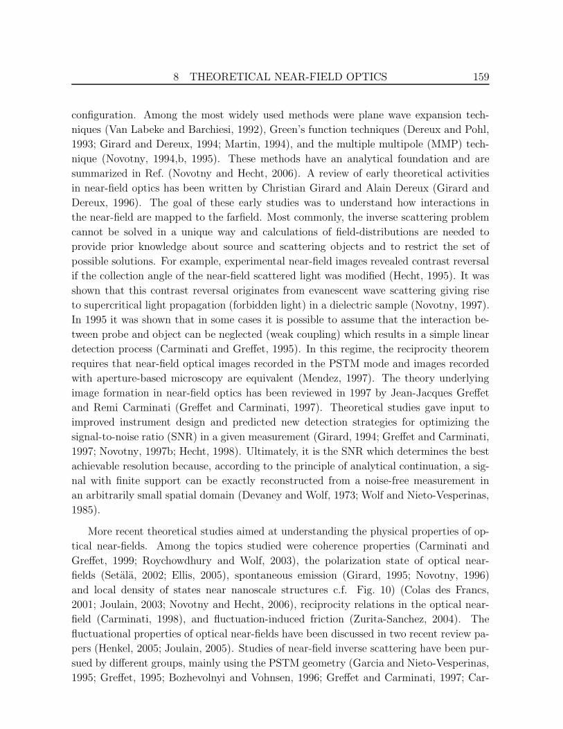
(a) (b) (c)
Figure 12: Scattering-based near-field optical microscopy of a gold Fischer pattern withpolystyrene residues deposited on a silicon surface. The method detects the backscatteredoptical signal at a higher harmonic of the modulation frequency Ω. (a) Schematic ofthe experiment, (b) topographical map, (c) optical map. Based on the different opticalcontrast, gold and polystyrene can be distinguished with a spatial resolution of ≈ 10 nm.From (Hillenbrand and Keilmann, 2002).

9 NEAR-FIELD SCATTERING AND FIELD ENHANCEMENT 163
propagate into the far field, where the fields may be detected, providing a useful signal
for measuring the local optical and topographical properties of the surface with high res-
olution.” This sentence assumes that the interaction between the tip and the excitation
field is stronger than the interaction between the sample and the excitation field, similar
to Wessel’s proposal. In their patent, Wickramasinghe and Williams propose to use a
double modulation technique combined with heterodyne detection in order to extract the
near-field signal from the detected scattered light. In their later experiments, Wickramas-
inghe and co-workers implemented a slightly different interferometric detection scheme
and demonstrated extremely high spatial resolutions (Zenhausern, 1995). However, as
discussed before, the origin of the contrast in the recorded images has been debated. A
Japanese patent with very similar ideas was filed in 1992 by Satoshi Kawata and co-
workers (Kawata, 1992). The patent describes a near-field optical microscope using a
combination of total-internal reflection illumination and light scattering from a vertically
modulated tip. A parallel effort in scattering-type near-field microscopy was also pursued
by Fritz Keilmann with experiments performed at radio and microwave frequencies and
later in the infrared (Keilmann, 1996; Knoll, 1997; Knoll and Keilmann, 1998). In 1989,
a few months after Wickramasinghe’s patent submission, Keilmann filed a patent titled
scanning tip for optical radiation in which he proposes the fabrication of a coaxial tip,
i.e. an aperture with a center conductor (Keilmann, 1991), an endeavor first undertaken
by Fee, Chu, and Hansch at microwave frequencies (Fee, 1989) and later by Fischer and
Zapletal in the optical frequency regime (Fischer and Zapletal, 1992). Coaxial waveguides
have no cut-off and therefore provide near-unity power transmission. Later, Fritz Keil-
mann and Bernhard Knoll implemented a method to extract the near-field signal from
the scattered light (Wurtz, 1998; Knoll and Keilmann, 2000). The method makes use of
vertical probe modulation with frequency Ω and demodulation of the scattered light at
higher harmonics nΩ. A combination with heterodyne detection allowed Fritz Keilmann
and Rainer Hillenbrand to separately measure amplitude and phase of the scattered sig-
nal and to extract material specific optical parameters (Hillenbrand and Keilmann, 2000,
2002; Keilmann and Hillenbrand, 2004). As an example, Fig. 12 shows an image of a gold
Fischer pattern with polystyrene residues deposited on a silicon surface. Polystyrene and
gold yield clearly different optical contrast.
In 1997 it was proposed to use the enhanced field at a laser-irradiated metal tip as an
excitation source, similar to Wessel’s idea (Novotny, 1998). The interaction of the locally
enhanced field with the sample surface generates an optical response which is then coupled
out by the same tip and detected in the farfield. This strong-scattering scheme makes it
possible to detect a spectroscopic response at frequencies different from the excitation fre-
quency thereby making it possible to explore the full range of linear and nonlinear optical
spectroscopy. The first experimental demonstration employed two-photon excited fluo-
rescence and was published in 1999 (Sanchez, 1999). Similar experiments have also been

9 NEAR-FIELD SCATTERING AND FIELD ENHANCEMENT 164
performed by other groups (Hamann, 2000) and today, the method is most widely referred
to as tip-enhanced near-field optical microscopy (Novotny and Stranick, 2006; Bouhelier,
2006). Following these experiments, the method was extended to other spectroscopic
interactions such as Raman scattering (Stockle, 2000; Anderson, 2002; Hayazawa, 2002;
Hartschuh, 2003) and coherent anti-Stokes Raman scattering (Ichimura, 2005). As an
illustration, Fig. 13 shows a near-field Raman scattering image of a single-walled carbon
nanotube sample along with a Raman scattering spectrum recorded when the tip is placed
on top of the nanotube.
The field enhancement at a laser-irradiated metal tip has been the subject of many
theoretical studies. Winfried Denk and Dieter W. Pohl demonstrated that the quasi-static
fields in the gap between a tip and a substrate can be extremely strong and that this effect
can be instrumental for inelastic tunneling and light emission during scanning tunneling
microscopy (STM) (Denk and Pohl, 1991). Later, it was theoretically established that
the field enhancement effect must be driven by an external field polarized along the axis
of the pointed probe (Novotny, 1997; Martin and Girard, 1997; Furukawa and Kawata,
1998). Interestingly, the field enhancement was also studied in the context of STM as
it was believed to mediate the transfer of atoms from the tip to the sample (Jersch and
Dickman, 1996; Gorbunov and Pompe, 1994; Bragas, 1998).
(a) (b)
200 nm
1200 1600 2000 2400 2800
(c)
ν = 2615 cm-1
ν (cm-1)
Figure 13: Near-field Raman imaging of a single-walled carbon nanotube sample. (a)Topography showing a network of carbon nanotubes overgrown with water droplets. (b)Raman scattering image of the same sample area recorded by integrating, for each imagepixel, the photon counts that fall into a narrow spectral bandwidth centered aroundν = 2615 cm2 (indicated by the yellow stripe in c). (c) Raman scattering spectrumrecorded on top of the nanotube. Adapted from (Hartschuh, 2003).

10 NEAR-FIELD OPTICS AND ANTENNA THEORY 165
10 Near-field Optics and Antenna Theory
In this last section I intend to outline similarities between near-field optics and antenna
theory (Pohl, 2000). These similarities project the roots of near-field optics more than 100
years back to the time of Guglielmo Marconi’s experiments on radiowave transmission.
The quality of an antenna is characterized by quantities such as radiation efficiency and
antenna gain, and it is plausible that similar quantities are applicable to optical near-field
probes. Therefore, established antenna-concepts can provide inspiration for novel near-
field probes.
The primary function of a near-field probe is the concentration of electromagnetic en-
ergy on a sample surface, similar to a standard electromagnetic antenna that concentrates
propagating radiation into a confined zone called the feedgap. In the feedgap, electric cir-
cuitry either releases or receives the power associated with the electromagnetic field. The
challenge in the design of an antenna is to efficiently couple the power flow between the
near-zone and the far-zone of the source (or receiver). This criterion holds also for a
quantum source, such as a single molecule, which emits a single photon at a time. The
most efficient antenna designs that have been implemented at optical frequencies are the
half-wave antenna (Muhlschlegel, 2005) and the bow-tie antenna (Grober, 1997; Crozier,
2003; Schuck, 2005; Farahani, 2005). However, by making use of electromagnetic reso-
nances associated with surface plasmons and phonons any nanostructure can be viewed
as an optical antenna. Of course, the efficiency depends on the material properties and
the geometry of the nanostructure.
As mentioned earlier, the association of near-field optical microscopy with antenna
theory has already been made by Wessel (Wessel, 1985). However, the notion of ’optical
antennas’ can be traced back even earlier. For example, an antenna attached to a metal-
to-metal point contact was used in 1968 by Ali Javan and co-workers for frequency mixing
of infrared radiation (Hocker, 1968). The rectification efficiency of these whisker diodes
could be increased by suitably kinking or bending the wire antenna (Matarrese and Even-
son, 1970). The length L (tip to kink) had to be adjusted in relation to the wavelength
and angle of incidence, and the strongest response was obtained for the fundamental res-
onance of L ≈ λ/2.7 (Rothammel, 2001). These experiments were performed at infrared
wavelengths of λ = 10 .. 337µm where metals are good conductors. Whisker wires are rou-
tinely employed for delivering electromagnetic energy to miniature semiconductor circuits
such as diodes or field effect transistors. Similar frequency mixing experiments have later
been performed by Wolfgang Krieger et al. at the tunnel junction of an STM (Krieger,
1990).
In essence, an antenna efficiently converts the energy of free-propagating radiation to
localized energy, and vice versa. For example, the antenna of a cell phone is used to
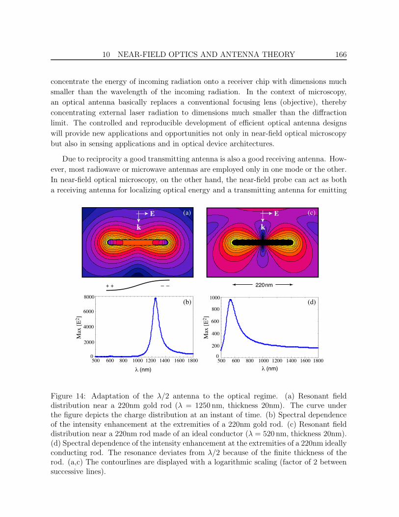
10 NEAR-FIELD OPTICS AND ANTENNA THEORY 166
concentrate the energy of incoming radiation onto a receiver chip with dimensions much
smaller than the wavelength of the incoming radiation. In the context of microscopy,
an optical antenna basically replaces a conventional focusing lens (objective), thereby
concentrating external laser radiation to dimensions much smaller than the diffraction
limit. The controlled and reproducible development of efficient optical antenna designs
will provide new applications and opportunities not only in near-field optical microscopy
but also in sensing applications and in optical device architectures.
Due to reciprocity a good transmitting antenna is also a good receiving antenna. How-
ever, most radiowave or microwave antennas are employed only in one mode or the other.
In near-field optical microscopy, on the other hand, the near-field probe can act as both
a receiving antenna for localizing optical energy and a transmitting antenna for emitting
λ (nm)
600 800 1000 1200 1400 1600
200
400
600
800
1000
5000
1800600 800 1000 1200 1400 1600
2000
4000
6000
500 1800
8000
0
Max
[E
2]
Max
[E
2]
λ (nm)
+ + − − 220 nm
k
E
k
E(a)
(b)
(c)
(d)
Figure 14: Adaptation of the λ/2 antenna to the optical regime. (a) Resonant fielddistribution near a 220nm gold rod (λ = 1250 nm, thickness 20nm). The curve underthe figure depicts the charge distribution at an instant of time. (b) Spectral dependenceof the intensity enhancement at the extremities of a 220nm gold rod. (c) Resonant fielddistribution near a 220nm rod made of an ideal conductor (λ = 520 nm, thickness 20nm).(d) Spectral dependence of the intensity enhancement at the extremities of a 220nm ideallyconducting rod. The resonance deviates from λ/2 because of the finite thickness of therod. (a,c) The contourlines are displayed with a logarithmic scaling (factor of 2 betweensuccessive lines).

10 NEAR-FIELD OPTICS AND ANTENNA THEORY 167
the optical response. Classical antenna design assumes that there is no time lag between
the conduction electrons and the exterior field. Consequently, electromagnetic penetra-
tion into the metal (skin effect) is negligible. At optical frequencies, on the other hand,
the ’inertia’ of conduction electrons causes electromagnetic fields to penetrate into the
metals and antenna design is no longer a pure function of the the ’exterior’ wavelength of
the fields. Therefore, antenna designs cannot be directly downscaled from the microwave
to the optical regime (cf. Fig. 14) and a scaled (effective) wavelength has to be invoked
instead (Novotny, 2007). Existing antenna concepts can provide the necessary inspiration
for their optical counterparts. For example, the self-similar antenna proposed by Mark
Stockman and co-workers (Li, 2003) has similarities with the well-established Yagi-Uda
antenna developed in the 1920s in Japan and today widely used as receiving antenna in
the UHF/VHF region. As an illustration, Fig. 14 shows a calculation of the resonance of
a λ/2 antenna. Because of surface plasmon resonances, the response at optical frequencies
is very strong but the resonance is no longer at λ/2. Instead, the resonance wavelength
is determined both by the external irradiation and the dielectric properties of the material.
Pointed metal structures were used for the localization of electromagnetic energy al-
ready in the 18th century, long before the era of antenna design began. The most promi-
nent invention making use of this property is Benjamin Franklin’s lightning rod (Krider,
2006). It can be viewed as a sharply pointed electrode with a strongly enhanced static
field at its apex. This field originates from the potential difference between the tip’s
support (ground) and a counter electrode (atmosphere). For strong enough fields the tip
initiates a plasma channel (discharge) and conducts electrons into ground thereby low-
Figure 15: Sketch from Benjamin Franklin’s letter sent to Peter Collinson on 29 July1750. The contour defined by A, B, C, D, E represents an electrified body and the dashedlines around it indicate an ”Atmosphere of Electrical Particles”. See text for details.From (Labaree, 1961b).

10 NEAR-FIELD OPTICS AND ANTENNA THEORY 168
ering the initial potential difference. In a letter sent to Peter Collinson, a fellow of the
Royal Society in London, on 25 May 1747, Benjamin Franklin writes ”.. In pursuing our
Electrical Enquiries, we had observ’d some particular Phenomena, ..” and then adds ”The
first is the wonderful Effect of Points both in drawing off and throwing off the Electrical
Fire” (Labaree, 1961). If the ’Fire’ to which Franklin is referring to were associated with
’Radiation’ we could accept his phrase as the definition of an antenna, namely a device that
attracts or sends off electrical radiation. The sketch shown in Fig. 15 has been depicted
by Franklin in a letter to Collinson, dated 29 July 1750 (Labaree, 1961b). The contour
defined by A, B, C, D, E represents an electrified body and the dashed lines around it
indicate an ”Atmosphere of Electrical Particles”, i.e. the electric field. Franklin argues
that the Atmosphere to the right of L,C,M has the least surface to rest on and hence the
attraction at the point C is weakest. As a consequence, the particles expand easiest at
point C. He concludes ”On these Accounts, we suppose Electrified Bodies discharge their
Atmospheres upon unelectrified Bodies more easily, and at greater Distance, from their
Angles and Points, than from their smoothe Sides.” Franklin adds ”These Points will also
discharge into the Air, when the Body has too great an electrical Atmosphere ... ” In the
same letter, Franklin speculates that the Fires of Electricity and Lightning are the same
and he proposes the construction of grounded lightning rods to be fixed on buildings for
the protection against lightning. He also writes ”To determine the Question, Whether
the Clouds that contain Lightning are electrified or not, I would propose an Experiment to
be try’d where it may be done conveniently.” His following proposal inspired the famous
’sentry-box’ experiment performed at Mary-la-Ville, France, in 1752 by Dalibard and De-
lors (Krider, 2006).
It might appear to be a far stretch from Franklin’s lightning rod to near-field optical
microscopy but, similar to an antenna or a near-field probe, the efficiency of a lightning
rod is characterized by its ability to concentrate and localize electric fields (Denk and
Pohl, 1991). However, despite this apparent similarity a lightning rod is not an antenna.
The lightning rod is a static device whose properties are dictated by the Laplace equation.
Consequently, the surface of the lightning rod is an equipotential surface and the geomet-
rical singularity at the tip apex gives rise to a singular field, irrespective of the shape of
the shaft or wire on which the tip is mounted. On the other hand, an antenna interacts
with electromagnetic radiation and hence its behavior is dictated by the wave equation.
Because of the finite penetration of optical radiation into metals the quasi-static approx-
imation is only valid for structures smaller than the skin depth but such small structures
are not the most efficient antennas. Therefore, inspiration for efficient optical near-field
probes has to be drawn from antenna theory despite the fact that a direct downscaling of
traditional antenna designs to optical wavelengths is not possible.
11 CONCLUDING REMARKS 169
11 Concluding Remarks
This article provides an overview of discoveries and achievements which were influential
to the shaping of near-field optics. Important achievements cannot always be accredited
to a single person or a single group because different developments progressed in parallel.
I would like to emphasize that the history presented in this article cannot be complete
because all resources are finite. I hope no important details have been left out. Any
omissions are solely due to my personal ignorance.
As is evident from this article the roots of near-field optics are not well defined and the
shaping of the field depended on many developments. The modern interest has its origins
in the 1982 experiments by Pohl and co-workers and in parallel developments by other
groups. Near-field optics received important inspiration from the early work in nanoplas-
monics which was mainly aimed at answering open questions in SERS, and from studies
of energy transfer. The prophetic papers and letters of Synge emerged at a later stage.
From todays perspective, near-field optics has a lot in common with classical antenna the-
ory extended into the optical regime and this is where it connects to the current second
wave of nanoplasmonics. Interestingly, the first experimental developments in near-field
optical microscopy were concentrated on the idea of using an aperture as a means to
further confine a diffraction-limited focus and this mindset delayed experimental work on
antenna-inspired concepts. Today, Synge’s original particle (tip) idea finds applications
in various studies and, despite Einstein’s initial skepticism, it is likely to be adopted in
future commercial instruments.
Acknowledgments
This article is the result of the valuable input and help of many friends and colleagues. I
am very grateful to Barbara Schirmer who patiently helped to research and translate the
resources forming the basis of this article, and I like to thank my parents in law, Grazia and
Hans Schicht, for ’full board’ during the writing period in Switzerland. I appreciate the
input and advice from various friends and colleagues, particularly Niek F. van Hulst, Fritz
Keilmann, Ulrich Ch. Fischer, Jean-Jacques Greffet, Alexandre Bouhelier, Ole Keller, and
Bert Hecht. I would like to acknowledge the Einstein Archives in Jerusalem, Israel, for
making the Einstein-Synge letters available and for the permission of citing them. I am
grateful for financial support by the Department of Energy (grant DE-FG02-01ER15204)
and the Air Force Office of Scientific Research (grant F-49620-03-1-0379).

REFERENCES 170
References
E. Abbe, “Beitrage zur Theorie des Mikroskops und der mikroskopischen Wahrnehmung,” Archivf. Miroskop. Anat., vol. 9, p. 413, 1873.
G. S. Agarwal, “Quantum electrodynamics in the presence of dielectrics and conductors. I.Electromagnetic-field response functions and black-body fluctuations in finite geometries,”Phys. Rev. A 11, 230-242 (1975).
M. G. Albrecht and J. A. Creighton, ”Anomalously intense Raman spectra of pyridine at a silverelectrode,” J. Am. Chem. Soc. 99, p. 5215-5217 (1977).
M. S. Anderson, Locally enhanced Raman spectroscopy with an atomic force microscope, Appl.Phys. Lett., vol. 76, pp. 3130-3132, 2002.
P. Anger, P. Bharadwaj, and L. Novotny, “Enhancement and quenching of single moleculefluorescence,” Phys. Rev. Lett., vol. 96, 113002, 2006.
A. Apostol and A. Dogariu, “Spatial correlations in the near field of random media,” Phys. Rev.Lett. 91, 093901 (2003).
E. A. Ash and G. Nicholls, “Super-resolution aperture scanning microscope,” Nature, vol. 237,pp. 510–513, 1972.
R. Bachelot, P. Gleyzes, and A. C. Boccara, ”Near-field optical microscopy by local perturbationof a diffraction spot,” Microscopy Microanalysis Microstructures, vol. 5(4-6), p. 389-397, 1994.
A. V. Baez, ”Is resolving power independent of wavelength possible? An experiment with asonic ’macroscope’,” J. Opt. Soc. Am., vol. 46, p. 901, 1956.
M.L.M. Balistreri, H. Gersen, J.P. Korterik, L. Kuipers, and N.F. van Hulst. ”Tracking fem-tosecond laser pulses in space and time.” Science, 294:1080–1082, 2001.
H. A. Bethe, “Theory of diffraction by small holes,” Phys. Rev. vol. 66, pp. 163–182, 1944.
E Betzig, A Harootunian, A Lewis, and M Isaacson, ”Near-field diffraction by a slit: implicationsfor superresolution microscopy,” Appl. Opt. 25, pp. 1890-900, 1986.
E. Betzig, J. K. Trautman, T. D. Harris, J. S. Weiner, and R. L. Kostelar, “Breaking thediffraction barrier: optical microscopy on a nanometric scale,” Science, vol. 251, pp. 1468–1470, 1991.
E. Betzig, P. L. Finn, and S. J. Weiner, “Combined shear force and near-field scanning opticalmicroscopy,” Appl. Phys. Lett., vol. 60, p. 2484–2486, 1992.
E.Betzig and J.K. Trautman, “Near-field optics: Microscopy, spectroscopy, and surface modifi-cation beyond the diffraction limit,” Science, vol.257, p.189-195, 1992.

REFERENCES 171
M. R. Beversluis, A. Bouhelier, and L. Novotny, “Continuum generation from single gold nanos-tructures through near-field mediated intraband transitions,” Phys. Rev. B., vol. 68, 115433,2003.
G. Binnig, H. Rohrer, C. Gerber, and E. Weibel, “Tunneling through a controllable vacuumgap,” Appl. Phys. Lett., vol. 40, pp. 178–180, 1982.
A. D. Boardman (ed.), Electromagnetic Surface Modes, Wiley, New York (1982).
A. Bouhelier, ”Field-enhanced scanning near-field optical microscopy,” Micr. Res. and Techn.69, p. 563-579, 2006.
C. J. Bouwkamp, “On Bethe’s theory of diffraction by small holes,” Philips Res. Rep., vol. 5,pp. 321–332, 1950.
C. J. Bouwkamp, “On the diffraction of electromagnetic waves by small circular disks and holes,”Philips Res. Rep., vol. 5, pp. 401–422, 1950.
S. I. Bozhevolnyi, O. Keller, and I. I. Smolyaninov, Phase conjugation of an optical near field,Opt. Lett. 19, p. 1601- 1603 (1994).
S. I. Bozhevolnyi, O. Keller, and I. I. Smolyaninov, Scattered light enhancement near a phaseconjugating mirror, Opt. Commun. 115, p. 115-120 (1995).
S. I. Bozhevolnyi and B. Vohnsen, Near-field optical holography, Phys. Rev. Lett. 77, p. 3351-3354 (1996).
S. I. Bozhevolnyi, V. S. Volkov, and K. Leosson, Localization and waveguiding of surface plasmonpolaritons in random nanostructures, Phys. Rev. Lett. 89, 186801, 2002.
A.V. Bragas, S.M. Landi, and O. E. Martinez, Laser field enhancement at the scanning tunnelingmicroscope junction measured by optical rectification, Appl. Phys. Lett., vol. 72, pp. 2075-2077-1926, 1998.
R. Carminati and J.-J. Greffet, ”Two-dimensional numerical simulation of the photon scanningtunneling microscope. Concept of transfer function,” Opt. Commun. 116, p. 316-321, (1995).
R. Carminati, M. Nieto-Vesperinas, and J.-J. Greffet, “Reciprocity of evanescent electromagneticwaves,” J. Opt. Soc. Am. A., vol. 15, pp. 706-712, 1998.
R. Carminati, and J.-J. Greffet, “Near-field effects in spatial coherence of thermal sources,”Phys. Rev. Lett. 82, 1660-1663 (1999).
P. S. Carney and J. C. Schotland, “Near-field Tomography,” in Inside Out: Inverse Problems(G. Uhlman, ed.), Cambridge: Cambridge University Press (2003).
P. S. Carney et al., ”Computational Lens for the Near Field,” Phys. Rev. Lett. 92, 163903 (2004)
C. K. Carniglia and L. Mandel, ”Quantization of Evanescent Electromagnetic Waves,” Phys.Rev. D 3, p. 280-296, (1971).

REFERENCES 172
R. R. Chance, A. Prock, and R. Silbey, “Molecular fluorescence and energy transfer near inter-faces,” in Advances in Chemical Physics (I. Prigogine and S. A. Rice, eds.) 37, 1–65, NewYork: Wiley (1978).
Surface-Enhanced Raman Scattering, R. K. Chang and T. E. Furtak (eds.), Plenum, New York,1981.
C. J. Chen and R. Osgood, ”Direct observation of the local-field-enhanced surface photochemicalreactions,” Phys. Rev. Lett. vol. 50, p. 1705-1708, 1983.
G. Colas des Francs, C. Girard, J.-C. Weeber, C. Chicanne, T. David, A. Dereux, and D.Peyrade, ”Optical analogy to electronic quantum corrals,” Phys. Rev. Lett. 86, 4950-4953(2001)
D. Courjon, K. Sarayeddine, and M. Spajer, “Scanning tunneling optical microscopy,” OpticsComm., vol. 71, pp. 23–28, 1989.
D. Courjon, Near Field Microscopy and Near Field Optics, Imperial College Press, 2003.
K. B. Crozier, A. Sundaramurthy, G. S. Kino, and C. F. Quate, ”Optical antennas: Resonatorsfor local field enhancement,” J. Appl. Phys. 94, 4632-4642 (2003).
P. Dawson, F. de Fornel, and J-P. Goudonnet , ”Imaging of surface plasmon propagation andedge interaction using a photon scanning tunneling microscope,” Phys. Rev. Lett. vol. 72, p.2927-2930, 1994.
F. deFornel, J. P. Goudonnet, L. Salomon, and E. Lesniewska, ”An evanescent field opticalmicroscope,” Proc. SPIE 1139, p. 77-84, SPIE, Washington, 1989.
W. Denk and D. W. Pohl, ”Near-field optics: Microscopy with nanometer-size fields, J. Vac.Sci. Technol., vol. 9, pp. 510-513, 1991.
A. Dereux and D. W. Pohl, “The 90 prism edge as a model SNOM probe: Near-field, photontunneling, and far-field properties,” in Near-field Optics, D. W. Pohl and D. Courjon eds.,NATO ASI Ser. E 242, p. 189-198, Dordrecht: Kluwer (1993).
A. J. Devaney and E. Wolf, “Multipole expansions and plane wave representations of the elec-tromagnetic field,” J. Math. Phys. 15, p. 234-244 (1974).
H. Ditlbacher et al., ”Silver nanowires as surface plasmon resonators,” Phys. Rev. Lett., vol. 95,257403 (2005).
K. Drexhage, “Influence of a dielectric interface on fluorescent decay time,” J. Lumin. 1,2,693–701 (1970).
K. H. Drexhage, ”Interaction of Light with monomolecular dye layers,” in Progress in Optics,E. Wolf (ed.), vol. 12, pp. 163-232, North-Holland Publishing Co., Amsterdam, 1974.
P. Drude and W. Nernst, ”Uber die Fluorescenzwirkungen stehender Lichtwellen,” Nachr. Kgl.Ges. Wiss. Gott., 189(10), p. 346-358, 1891.

REFERENCES 173
U. Durig, D.W. Pohl, F. Rohner, ”Near-field optical-scanning microscopy,” J. Appl. Phys.,vol. 59, p. 3318-3327 (1986)
B. Dunn, “Near-field scanning optical microscopy,” Chemical Reviews, vol.99, p.2891-2928, 1999.
J. Ellis, A. Dogariu, S. Ponomarenko, and E. Wolf, ”Degree of polarization of statisticallystationary electromagnetic fields,” Opt. Commun. 248, pp. 333-337 (2005).
Fano U., 1941, The theory of anomalous diffraction gratings and of quasi-stationary waves onmetallic surfaces (Sommerfeld’s waves), J. Opt. Soc. Am. 31, 213-222.
J. N. Farahani, D. W. Pohl, H.-J. Eisler, and B. Hecht, ”Single quantum dot coupled to ascanning optical antenna: A tunable superemitter,” Phys. Rev. Lett. 95, 017402 (2005).
M. Fee, S. Chu, and T. D. Hansch, ”Scanning electromagnetic transmission line microscope withsub-wavelength resolution,” Opt. Comm. vol. 69, p. 219–224 (1989).
J. P. Fillard, Near-field Optics and Nanoscopy, World Scientific Publishing Company, 1996.
U. Ch. Fischer and H.P. Zingsheim, ”Submicroscopic contact imaging with visible light by energytransfer,” Appl. Phys. Lett. 40, p. 195-197, (1982)
U. Ch. Fischer, ”Optical characteristics of 0.1 µm circular apertures in a metal film as lightsources for scanning ultramicroscopy,” J. Vac. Sci. Technol. B 3, p. 386 (1985).
U. Ch. Fischer, ”Submicrometer aperture in a thin metal film as a probe of its microenvironmentthrough enhanced light scattering and fluorescence,” J. Opt. Soc. Am. B 3, p. 1239-1244,(1986).
U.Ch. Fischer and D.W. Pohl, ”Observation on single-particle plasmons by near-field opticalmicroscopy,” Phys. Rev. Lett. 62, 458 (1989).
U. Ch. Fischer and M. Zapletal, ”The concept of a coaxial tip as a probe for scanning near fieldoptical microscopy and steps towards a realisation,” Ultramicroscopy vol. 42-44, p. 393-398(1992).
U. Ch. Fischer, ”Scanning near-field optical microscopy,” in Scanning Probe Microscopy. Ana-lytical Methods, R. Wiesendanger (ed.), pp. 161-210, Springer (1998).
M. Fleischmann, P. J. Hendra, and A. J. McQuillan, “Raman spectra of pyridine adsorbed at asilver electrode,” Chem. Phys. Lett. 26, 163 (1974).
G. W. Ford and W. H. Weber, Electromagnetic interactions of molecules with metal surfaces,Phys. Rep. 113, p. 195-287 (1984).
Th. Forster, “Energiewanderung und Fluoreszenz,” Naturwissenschaften 33, 166–175 (1946);Th. Forster, “Zwischenmolekulare Energiewanderung und Fluoreszenz,” Ann. Phys. (Leipzig)2, 55–75 (1948). An English translation of Forster’s original work is provided by R. S. Knox,“Intermolecular energy migration and fluorescence,” in Biological Physics (E. Mielczarek, R. S.Knox, and E. Greenbaum, eds.), 148–160, New York: American Institute of Physics (1993).

REFERENCES 174
M. Fujihira, ”Fluorescence microscopy and spectroscopy by scanning nearfield optical /atomicforce microscope (SNOM-AFM),” in Optics at the Nanometer Scale: Imaging and Storingwith Photonic Near Fields, NATO Science Series E-319, M. Nieto-Vesperinas and N. Garcia(eds.), p. 205221, Kluwer, The Netherlands, (1996).
H. Furukawa and S.Kawata, ”Local field enhancement with an apertureless near-field-microscopeprobe,” Opt. Comm., vol.148, pp. 221-224, 1998.
N. Garcia and M. Nieto-Vesperinas, ”Direct solution to the inverse scattering problem for sur-faces from near-field intensities without phase retrieval,” Opt. Lett. 20, 949 (1995).
J. Gersten and A. Nitzan, ”Electromagnetic theory of enhanced Raman scattering of moleculesadsorbed on rough surfaces,” J. Chem . Phys. 73, p. 3023-3037 (1980).
J.-J. Greffet, A. Sentenac, and R. Carminati, ”Surface profile reconstruction using near-fielddata,” Opt. Comm. 116, p. 20-24, 1995.
J.-J. Greffet and R. Carminati, “Image formation in near-field optics,” Prog. Surf. Sci. 56,133–237 (1997).
S. Gresillon, L. Aigouy, A. C. Boccara, J. C. Rivoal, X. Quelin, C. Desmarest, P. Gadenne, V.A. Shubin, A. K. Sarychev, and V. M. Shalaev, ”Experimental observation of localized opticalexcitations in random metal-dielectric films,” Phys. Rev. Lett. 82, p. 4520-4523 (1999)
Ch. Girard, A. Dereux, O. J. F. Martin, and M. Devel, “Importance of confined fields in near-field optical imaging of subwavelength objects,” Phys. Rev. B 50, p. 14467-14473 (1994).
C. Girard and A. Dereux, ”Optical spectroscopy of a surface at the nanometer scale: A theo-retical study in real space,” Phys. Rev. B 49, p. 11344-11351, 1994.
C. Girard, O. J. F. Martin, and A. Dereux, “Molecular lifetime changes induced by nanometerscale optical fields,” Phys. Rev. Lett. 75, 3098-3101 (1995)
C. Girard and A. Dereux, ”Near-field Optics Theories,” Rep. Prog. Phys. 59, p.657-699, (1996).
A. A. Gorbunov and W. Pompe, Thin film nanoprocessing by laser/STM combination, Phys.Stat. Sol. A, vol. 145, pp. 333-338, 1994.
R. D. Grober, R. J. Schoelkopf, and D. E. Prober, “Optical antenna: towards a unity efficiencynear-field optical probe,” Appl. Phys. Lett., vol. 70, pp. 1354–1356, 1997.
M. G. L. Gustafsson, ”Nonlinear structured-illumination microscopy: Wide-field fluorescenceimaging with theoretically unlimited resolution,” Proc. Nat. Acad. Sci. 102, p. 13081-13086(2005).
H. F. Hamann, A. Gallagher, and D. J. Nesbitt, Near-field fluorescence imaging by localizedfield enhancement near a sharp probe tip, Appl. Phys. Lett. 76, pp. 1953-1959, 2000.
A. Harootunian, E. Betzig, M. Isaacson, A. Lewis, ”Super-resolution fluorescence near-fieldscanning optical microscopy,” Appl. Phys. Lett., vol. 49, p. 674-676 (1986)

REFERENCES 175
A. Hartschuh, E.J. Sanchez, X.S. Xie, and L. Novotny. ”High-resolution near-field raman mi-croscopy of single-walled carbon nanotubes.” Phys. Rev. Lett., 90:095503, 2003.
N. Hayazawa, Y. Inouye, Z. Sekkat, and S. Kawata, ”Near-field Raman imaging of organicmolecules by an apertureless metallic probe scanning optical microscope,” J. Chem. Phys.vol. 117, pp.1296-1301, 2002.
C. L. Haynes and R. P. Van Duyne, ”Nanosphere lithography: A versatile nanofabrication toolfor studies of size-dependent nanoparticle optics,” J. Phys. Chem. B 105, p. 5599-5611, 2001.
B. Hecht, D. W. Pohl, H. Heinzelmann, and L. Novotny, Tunnel near-field optical microscopy:TNOM-2, ’ in Photons and Local Probes (O. Marti and R. Moller, eds.), NATO AdvancedStudy Institute, Series E, pp. 93-107, Dordrecht: Kluwer Academic Publishers, 1995.
B. Hecht, H. Bielefeldt, L. Novotny, Y. Inouye, and D.W. Pohl, “Facts and Artifacts in Near–Field Optical Microscopy,” J. Appl. Phys. 81, 2492‘–2498 (1997).
B. Hecht, H. Bielefeld, D. W. Pohl, L. Novotny, and H. Heinzelmann, “Influence of detectionconditions on near-field optical imaging,” J. Appl. Phys. 84, 5873–5882 (1998).
C. Henkel, ”Nanometer scale electromagnetic field fluctuations,” in Handbook of Computationaland Theoretical Nanotechnology, M. Rieth and W. Schommers (eds.), Chapter 25, AmericanScientific Publishers, Stevenson Ranch, California, (2005).
R. Hillenbrand and F. Keilmann, ”Complex optical constants on a subwavelength scale,” Phys.Rev. Lett. 85, p. 3029-3032, 2000.
R. Hillenbrand and F. Keilmann, ”Material-specific mapping of metal/semiconductor/dielectricnanosystems at 10 nm resolution by backscattering near-field optical microscopy,” Appl. Phys.Lett. vol. 80, 25 (2002)
L. O. Hocker, D. R. Sokoloff, V. Daneu, A. Szoke, And A. Javan, ”Frequency mixing in theinfrared and far-infrared using metal-to-metal point contact diode,” Appl. Phys. Lett. 12,401-402, 1968.
R. B. G. de Hollander, N. F. van Hulst, and R. P. H. Kooyman, ”Near-field plasmon and forcemicroscopy,” Ultramicroscopy 57, p. 263–269 (1995).
D. Hondros. Uber elektromagnetische Drahtwellen. Ann. d. Physik 30, 905–950 (1909).
M. K. Hong, A. Swan, and S. Erramilli, ”Evanescent wave vibrational microscopy,” Optics &Photonics News, p. 30-35, July 2004.
T. Ichimura, N. Hayazawa, M. Hashimoto, Y. Inouye, and S. Kawata. ”Tip-enhanced coherentanti-stokes raman scattering for vibrational nanoimaging.” Phys. Rev. Lett., 92:220801, 2004.
Y. Inouye and S. Kawata, ”Near-field scanning optical microscope with a metallic probe tip,”Opt. Lett. 19, p. 159-161, 1994.

REFERENCES 176
J.B. Jackson, S.L. Westcott, L.R. Hirsch, J.L. West, and N.J. Halas. Controlling the surfaceenhanced raman effect via the nanoshell geometry. Appl. Phys. Lett. 82, 257–259 (2003).
D. L. Jeanmaire and R. P. Van Duyne, ”Surface Raman spectroelectrochemistry: Part I. Hete-rocyclic, aromatic, and aliphatic amines adsorbed on the anodized silver electrode,” J. Elec-troanal. Chem. 84, 1-20 (1977).
]J. Jersch and K. Dickman, Nanostructure fabrication using laser field enhancement in the nearfield of a scanning tunneling microscope tip, em Appl. Phys. Lett. vol. 68, pp. 868-870, 1996.
K. Joulain, R. Carminati, J.-P. Mulet, and J.-J. Greffet, “Definition and measurement of thelocal density of electromagnetic states close to an interface,” Phys. Rev. B 68, 245405 (2003)
K. Joulain, J.P. Mulet, F. Marquier, R. Carminati, J.J. Greffet, ”Surface electromagnetic wavesthermally excited : radiative heat transfer, coherence properties and Casimir forces revisitedin the near field”, Surf. Sci. Rep. 57, p. 59-112 (2005).
K. Karrai and R. D. Grober, “Piezoelectric tip-sample distance control for near field opticalmicroscopes,” Appl. Phys. Lett., vol. 66, p. 1842–1844, 1995.
S. Kawata, Y. Inouye, and T. Sugiura, ”Scanning optical microscope system,” Japanese Patent3,196,945, 23 October 1992.
S. Kawata, Y. Inouye and T. Sugiura, ”Near-field scanning optical microscope with a lasertrapped probe,” Jpn. J. Appl. Phys. 33, L1725-L1727, 1994.
S. Kawata, M. Ohtsu, and M. Irie (eds.), Nano-Optics, Springer Verlag, 2002.
F. Keilmann, ”Scanning tip for optical radiation,” U.S. Patent 4,994,118, 19 February 1991.
F. Keilmann, ”FIR microscopy,” Infrared Phys. Technol. 36, pp. 217-224 (1995).
F. Keilmann, D. W. van der Weide, T. Eickelkamp, R. Merz, and D. Stockle, Extreme sub-wavelength resolution with a scanning radio-frequency transmission microscope, Opt. Comm.vol. 129, pp. 15-18, 1996.
F. Keilmann, ”Towards SNIM: Scanning near-field microscopy in the infrared,” in Optics at theNanometer Scale: Imaging and Storing with Photonic Near Fields, NATO Science Series E-319, M. Nieto-Vesperinas and N. Garcia (eds.), p. 235-245, Kluwer, The Netherlands, (1996).
F. Keilmann and R. Hillenbrand, Near-field microscopy by elastic light scattering from a tip,Phil. Trans. R. Soc. Lond. A, vol. 362, pp. 787-797, 2004.
O. Keller, ”Propagator picture of the spatial confinement of quantized light emitted from anatom,” Phys. Rev. A 58, pp. 3407-3425, 1998.
O. Keller, ”Space-time description of photon emission from an atom,” Phys. Rev. A 62, 022111,2000.
O. Keller, ”Near-field optics: The nightmare of the photon,” J. Chem. Phys. 112, p. 7856-7863(2000).

REFERENCES 177
O. Keller, ”On the theory of spatial localization of photons,” Phys. Rep. 411, p. 1-232, 2005.
M. Kerker, D.-S. Wang, and H. Chew, ”Surface enhanced Raman scattering (SERS) by moleculesadsorbed at spherical particles: errata,” Appl. Opt., vol. 19, p. 4159-4174, 1980.
T. A. Klar, S. Jakobs, M. Dyba, A. Egner, and S. W. Hell, “Fluorescence microscopy withdiffraction resolution barrier broken by stimulated emission,” Proc. Nat. Acad. Sci. 97, 8206–8210 (2000).
B. Knoll, F. Keilmann, A. Kramer, and R. Guckenberger, ”Contrast of microwave near-fieldmicroscopy,” Appl. Phys. Lett., vol. 70, pp.2667–2669, 1997.
B. Knoll and F. Keilmann, Scanning microscopy by mid-infrared near-field scattering, Appl.Phys. A, vol. 66, pp.477-481, 1998.
B. Knoll and F. Keilmann, ”Enhanced dielectric contrast in scattering-type scanning near-fieldoptical microscopy,” Opt. Com. 182, p. 321-328 (2000)
R. Kopelman and W. H. Tan, ”Near-field optical microscopy, spectroscopy, and chemical sen-sors,” Appl. Spectrosc. Rev. 29, p. 39–66 (1994).
U. Kreibig, ”Electronic properties of small silver particles: the optical constants and their tem-perature dependence,” Phys. F: Metal Phys., vol. 4, July, 1974.
J. R. Krenn and J.-C. Weeber, ”Surface plasmon polaritons in metal stripes and wires,” Phil.Trans. R. Soc. Lond. A, vol. 362, 739-756 (2004).
E. Kretschmann and H. Rather, ”Radiative decay of nonradiative surface plasmon excited bylight,” Z. Naturf., vol. 23A, p. 2135-2136, (1968)
P. Krider, ”Benjamin Franklin and lightning rods,” Physics Today, p. 42-48, January 2006.
W. Krieger, T. Suzuki, M. Volcker, and H. Walther, ”Generation of microwave radiation in thetunneling junction of a scanning tunneling microscope,” Phys. Rev. B 41, p. 10229-10232,1990.
S. Kuhn, U. Hakanson, L. Rogobete, V. Sandoghdar , ”Enhancement of single-molecule fluo-rescence using a gold nanoparticle as an optical nanoantenna, Phys. Rev. Lett. 97, 017402(2006).
H. Kuhn, ”On Possible Ways of Assembling Simple Organised Systems of Molecules”, In: ”Struc-tural Chemistry and Molecular Biology”, A. Rich and N. Davidson (eds.), Freeman, SanFrancisco, p. 566-571 (1968)
H. Kuhn, ”Molekularer Systemaufbau,” Max-Planck-Gesellschaft Jahrbuch 1984, p. 506-508,1984.
H. Kuhn, ”Self-organizing molecular electronic devices ?,” in Molecular Electronic Devices II,F.L. Carter (ed.), p. 411-426, Dekker, N.Y. (1987)

REFERENCES 178
B. Labani, C. Girard, D. Courjon, D. Van Labeke, ”Optical interaction between a dielectric tipand a nanometric lattice: implications for near-field microscopy,” J. Opt. Soc. Am. B 7, pp.936-943, 1990, C. Girard and D. Courjon, ”Model for scanning tunneling optical microscopy:A microscopic self-consistent approach,” Phys. Rev. B 42, p. 9340-9349, 1990.
The Papers of Benjamin Franklin, L. W. Labaree et. al. (eds.), vol. 3, p. 156-164, Yale UniversityPress, New Haven, CT, (1961).
The Papers of Benjamin Franklin, L. W. Labaree et. al. (eds.), vol. 4, p. 9-34, Yale UniversityPress, New Haven, CT, (1961).
M. J. Levene, J. Korlach, S. W. Turner, M. Foquet, H. G. Craighead, W. W. Webb, ”Zero-ModeWaveguides for Single-Molecule Analysis at High Concentrations,” Science 299, pp. 682 - 686,2003.
Y. Leviatan, ”Study of near-zone fields of a small aperture,” J. Appl. Phys. 60, pp. 1577-1583,(1986); Y. Leviatan, ”Electromagnetic coupling between two half-space regions separatedby two slot-perforated parallel conducting screens,” IEEE Trans. on Microwave Theory andTechn. 36, pp. 44-52, (1988).
A. Lewis, M. Isaacson, A. Muray, and A. Harootunian, ”Scanning optical spectral microscopywith 500A spatial resolution,” abstract W-AM-Pos25, Biophys. J., vol. 41, p. 405a (1983)
A.Lewis, M.Isaacson, A.Harootunian, and A.Muray, “Development of a 500 A resolution lightmicroscope,” Ultramicroscopy, vol.13, pp.227-231, 1984.
K. Li, M. I. Stockman, and D. J. Bergman, ”Self-similar chain of metal nanospheres as anefficient nanolens,” Phys. Rev. Lett. 91, 227402 (2003).
L. Malmqvist and H. M. Hertz:, ”Two-color trapped-particle optical microscopy,” Opt. Lett. 19,853-855, (1994).
O. Marti, H. Bielefeldt, B. Hecht, S. Herminghaus, P. Leiderer, and J. Mlynek, ”INear-fieldoptical measurement of the surface plasmon field,” Opt. Comm. vol. 96, p. 225-228, 1993.
O. J. F. Martin, A. Dereux, and C. Girard, “Iterative scheme for computing exactly the total fieldpropagating in dielectric structures of arbitrary shape,” J. Opt. Soc. Am. A 11, 1073–1080(1994).
O. J. F. Martin and C. Girard, Controlling and tuning strong optical field gradients at a localprobe microscope tip apex, App. Phys. Lett., vol.70, pp.705-707, 1997.
G. A. Massey, “Microscopy and pattern generation with scanned evanescent waves,” Appl. Opt.,vol. 23, pp. 658–660, 1984.
Gail A. Massey, Jeffrey A. Davis, S. M. Katnik, and E. Omon, “Subwavelength resolution far-infrared microscopy,” Appl. Opt., vol. 24, pp. 1498–1501, 1985.
L. M. Matarrese and K. M. Evenson, Improved coupling to infrared whisker diodes by use ofantenna theory, Appl. Phys. Lett. 17, p. 8-10, 1970.

REFERENCES 179
E. R. Mendez, J. J. Greffet and R. Carminati, ”On the equivalence between the illumination andcollection modes of the scanning near-field optical microscope” Opt. Commun. 142, p. 7-13(1997).
C. W. McCutchen, “Superresolution in microscopy and the Abbe resolution limit,” J. Opt. Soc.Am., vol. 57, pp. 1190-1192, 1967.
D. McMullan, ”The prehistory of scanned image microscopy. Part I: Scanned optical micro-scopes,” Royal Microscopical Society Proceedings, vol. 25(2), pp. 127-131, 1990.
M. Meier, A. Wokaun, and P. F. Liao, ”Enhanced fields on rough surfaces: dipolar interactionsamong particles of sizes exceeding the Rayleigh limit,” J. Opt. Soc. Am. B 2, p. 931-949,(1985).
H. Metiu, “Surface enhanced spectroscopy,” in Progress in Surface Science (I. Prigogine andS. A. Rice, eds.) 17, 153–320, New York: Pergamon Press (1984).
J. Michaelis, C. Hettich, J. Mlynek and V. Sandoghdar, ”Optical microscopy with a single-molecule light source,” Nature 405, p. 325–328 (2000).
R. J. Moerland, N. F. van Hulst, H. Gersen, and L. Kuipers ”Probing the negative permittivityperfect lens at optical frequencies using near-field optics and single molecule detection,” Opt.Express 13, 1604-1614, 2005.
W. E. Moerner and L. Kador, “Optical detection and spectroscopy of single molecules in asolid,” Phys. Rev. Lett., vol. 62, p. 2535-2538, 1989.
M. Moskovits, ”Surface-enhanced Raman spectroscopy: a brief retrospective,” J. Raman Spec-trosc. vol. 36, p. 485-496, 2005.
P. Muhlschlegel, H.-J. Eisler, O. J. F. Martin, B. Hecht, and D. W. Pohl, ”Resonant opticalantennas,” Science 308, p. 1607-11609 (2005).
H. Nassenstein, “Superresolution by diffraction of subwaves,” Opt. Comm., vol. 2, pp. 231-234,1970.
L. Novotny, D. W. Pohl, and P. Regli, “Light propagation through nanometer-sized structures:the two-dimensional-aperture scanning near-field optical microscope,” J. Opt. Soc. Am. A 11,1768–1779 (1994).
L. Novotny and C. Hafner, “Light propagation in a cylindrical waveguide with a complex,metallic, dielectric function,” Phys. Rev. E 50, 4094–4106 (1994).
L. Novotny and D. W. Pohl, “Light propagation in scanning near-field optical microscopy,” inPhotons and Local Probes (O. Marti and R. Moller, eds.), NATO Advanced Study Institute,Series E, pp. 21–33, Dordrecht: Kluwer Academic Publishers, 1995.
L. Novotny, “Single molecule fluorescence in inhomogeneous environments,” App. Phys. Lett.,vol. 69, pp. 3806–3808, 1996.
REFERENCES 180
L. Novotny, R.X. Bian, and X.S. Xie, “Theory of nanometric optical tweezers,” Phys. Rev. Lett.,vol.79, pp.645-648, 1997.
L. Novotny, “Allowed and forbidden light in near-field optics. I. A single dipolar light source,”J. Opt. Soc. Am. A 14, p. 91–104 (1997).
L. Novotny, “Allowed and forbidden light in near-field optics. II. Interacting dipolar particles,”J. Opt. Soc. Am. A 14, p. 105–113 (1997).
L. Novotny, E. J. Sanchez, and X. S. Xie, “Near-field optical imaging using metal tips illuminatedby higher-order Hermite-Gaussian beams,” Ultramicroscopy, vol. 71, pp. 21–29, 1998.
L. Novotny and B. Hecht, Principles of Nano-Optics, Cambridge University Press, Cambridge,2006.
L. Novotny and S. J. Stranick, “Near-field optical microscopy and spectroscopy with pointedprobes,” Ann.Rev.Phys.Chem., vol. 57, p. 303-331, 2006.
L. Novotny, 2007, ”Effective wavelength scaling for optical antennas,” Phys. Rev. Lett. 98,266802 (2007).
M. Ohtsu and M. Hori, Near-field Nano-optics New York: Kluwer Academic, Plenum (1999).
J. A. O’Keefe, “Resolving power of visible light,” J. Opt. Soc. Am., vol. 46, p. 359, 1956.
M. Orrit and J. Bernard, “Single pentacene molecules detected by fluorescence excitation in ap-terphenyl crystal,” Phys. Rev. Lett., vol. 65, p. 2716-2719, 1990.
A. Otto, Excitation of nonradiative surface plasma waves in silver by the method of frustratedtotal reflection. Z. Phys. 216, 398–410 (1968); A. Otto, ”A new method for exciting non-radioactive surface plasma oscillations,” Phys. Stat. Sol. vol. 26, p. K99-K101, (1968)
M. A. Paesler and P. J. Moyer, Near Field Optics: Theory, Instrumentation and Applications,Wiley Interscience, New York, 1996.
D.W. Pohl, W.Denk, and M.Lanz, “Optical stethoscopy: Image recording with resolutionlambda/20,” Appl. Phys. Lett., vol.44, p.651-653, 1984.
D. W. Pohl, ”Optical near field scanning microscope,” European Patent 0112401 A1, 4 July1984.
D. W. Pohl, ”Some remarks on the history of near-field optics,” in Near-field Optics, D. W. Pohland D. Courjon eds., NATO ASI Ser. E 242, p. 1-5, Dordrecht: Kluwer (1993).
D. W. Pohl, ”Optics at the nanometre scale,” Phil. Trans. R. Soc. Lond. A, vol. 362, 701-717(2004).
D. W. Pohl, Near-field optics seen as an antenna problem, in Near-field Optics, Principles andApplications, X. Zhu and M. Ohtsu (eds.), p. 9-21, World Scientific, Singapore (2000).
REFERENCES 181
E. A. Power and T. Thirunamachandran, ”Analysis of the causal behavior in energy transferbetween atoms,” Phys. Rev. A 56, p. 3395-408, 1997.
P. N. Prasad, Nanophotonics, Wiley Interscience, 2004.
Lord Rayleigh, “On the theory of optical images with special reference to the optical microscope,”Phil. Mag., vol. 5(42), pp. 167-195, (1896).
R. C. Reddik, R. J. Warmack, and T. L. Ferrell, “New form of scanning optical microscopy,”Phys. Rev. B, vol. 39, pp. 767–770, 1989.
Special Issue on Nano-Optics and Near Field Microscopy, D. Richards and A. Zayats (eds.),Phil. Trans. R. Soc. Lond. A, vol. 362, No. 1817, pp. 699-919, 2004.
A. Roberts, ”Electromagnetic theory of diffraction by a circular aperture in a thick, perfectlyconducting screen,” J. Opt. Soc. Am. A 4, pp. 1970-1983, (1987); A. Roberts, ”Near-zonefields behind circular apertures in thick, perfectly conducting screens,” J. Appl. Phys. 65,2896-2899, (1989); A. Roberts, ”Small-hole coupling of radiation into a near-field probe,” J.Appl. Phys. 70, pp. 4045-4049, (1991).
See, for example, A. Kirschke and K. Rothammel, Rothammels Antennenbuch (12th ed.), DARCVerlag, Baunatal, 2001.
H. Roychowdhury and E. Wolf, “Effects of spatial coherence on near-field spectra,” Opt. Lett.28, pp. 170-172 (2003).
E. J. Sanchez, L. Novotny, and X. S. Xie, “Near-field fluorescence microscopy based on two-photon excitation with metal tips,” Phys. Rev. Lett., vol. 82, pp. 4014–4017, 1999.
M. Schrader and S. W. Hell, ”4Pi-confocal images with axial superresolution,” J. Microsc. 183,p. 110, 1996.
P.J. Schuck, D. P. Fromm, A. Sundaramurthy, G. S. Kino, and W. E. Moerner, Improving themismatch between light and nanoscale objects with gold bowtie nanoantennas, Phys. Rev.Lett. 94, 017402, 2005.
S. K. Sekatskii and V. S. Letokhov, ”Single fluorescence centers on the tips of crystal needles:first observation and prospects for application in scanning one-atom fluorescence microscopy,”Appl. Phys. B 63, p. 525–530 (1996).
T. Setala, M. Kaivola, and A. T. Friberg, ”Degree of polarization in near fields of thermalsources: Effects of surface waves,” Phys. Rev. Lett. 88, 123902 (2002).
A. V. Shchegrov, K. Joulain, R. Carminati, and J.-J. Greffet, “Near-field spectral effects due toelectromagnetic surface exciations,” Phys. Rev. Lett. 85, 1548-1551 (2000).
A. Sommerfeld, “Uber die Ausbreitung der Wellen in der drahtlosen Telegraphie,” Ann. d.Physik 28, 665–736 (1909).

REFERENCES 182
M. Specht, J. D. Pedarning, W. M. Heckl, and T. W. Hansch, Scanning plasmon near-fieldmicroscope, Phys. Rev. Lett., vol. 68, pp.476–479, 1992.
I. Stefanon, S. Blaize, A. Bruyant, S. Aubert, G. Lerondel, R. Bachelot, and P. Royer, ”Hetero-dyne detection of guided waves using a scattering-type scanning near-field optical microscope,”Opt. Expr. vol. 13, p. 5553-5564, 2005.
Karl A. Stetson, “Holography with total internally reflected light,” Appl. Phys. Lett., vol. 11,pp. 225-227, 1967.
R. M. Stockle, Y. D. Suh, V. Deckert, and R. Zenobi, Nanoscale chemical analysis by tip-enhanced Raman spectroscopy, Chem. Phys. Lett., vol. 318, pp.131-136 (2000).
E. H. Synge, “A suggested model for extending microscopic resolution into the ultra-microscopicregion,” Phil. Mag., vol. 6, pp. 356–362, 1928.
E. H. Synge, “III. A microscopic method,” Phil. Mag.S.7, vol. 7, pp. 65-80, 1931.
E. H. Synge, “An application of piezo-electricity to microscopy,” Phil. Mag.S.7, vol. 13, pp. 297-300, 1932.
R. Toledo-Crow, P. C. Yang, Y. Chen, and M. Vaez-Iravani, “Near-field differential scanningoptical microscope with atomic force regulation,” Appl. Phys. Lett., vol. 60, p. 2957–2959,1992.
G. Toraldo di Francia, “Resolving power and information,” J. Opt. Soc. Am., vol. 45, pp. 497-501, 1955.
J. K. Trautman, J. J. Macklin, L. E. Brus, and E. Betzig, “Near-field spectroscopy of singlemolecules at room temperature,” Nature, vol. 369, p. 40–42, 1994.
Turbadar T., 1959, Complete absorption of light by thin metal films, Proc. Phys. Soc. Lond. 73,40-44.
Kiichi Ueyanagi, ”Optical head, magneto-optical head, disk apparatus and manufacturingmethod of optical head,” U.S. Patent 2001/0009541 A1, 26 July 2001.
D. Van Labeke and D. Barchiesi, ” Scanning-tunneling optical microscopy: a theoretical macro-scopic approach,” J. Opt. Soc. Am. A 9, p. 732-739, 1992.
D. Van Labeke and D. Barchiesi, ”Probes for scanning tunneling optical microscopy: a theoret-ical comparison,” J. Opt. Soc. Am. A 10, p. 2193-2201, 1993.
S. A. Vickery and R. C. Dunn, ”Scanning Near-field fluorescence resonance energy transfermicroscopy,” Biophys J. 76, p. 1812–1818 (1999).
J. M. Vigou-reux and D. Courjon, ”Detection of nonradiative fields in light of the Heisenberguncertainty principle and the Rayleigh criterion,” Appl. Opt. 31, p. 3170-3177, 1992.

REFERENCES 183
J. A. Veerman, A. M. Otter, L. Kuipers, and N. F. van Hulst, ”High definition aperture probesfor near-field optical microscopy fabricated by focused ion beam milling,” App. Phys. Lett.72, 3115-3117, 1998.
J. Wessel, “Surface-enhanced optical microscopy,” J. Opt. Soc. Am. B, vol. 2, pp. 1538–1540,1985.
H. K. Wickramasinghe and C. C. Williams, ”Apertureless near field optical microscope,” U.S.Patent 4,947,034, 7 August 1990.
G. P. Wiederrecht , ”Near-field optical imaging of noble metal nanoparticles,” Eur. Phys. J.Appl. Phys. 28, p. 3-18, 2004.
K. I. Willig, S. O. Rizzoli, V. Westphal, R. Jahn, and S. W. Hell, “STED microscopy revealsthat synaptotagmin remains clustered after synaptic vesicle exocytosis,” Nature 440, 935-939(2006).
A. Wokaun, J. G. Bergman, J. P. Heritage, A. M. Glass, P. F. Liao, and D. H. Olson, ”Surfacesecond-harmonic generation from metal island films and microlithographic structures,” Phys.Rev. B 24, p. 849-856, 1981.
E. Wolf and M. Nieto-Vesperinas, “Analyticity of the angular spectrum amplitude of scatteredfields and some of its consequences,” J. Opt. Soc. Am. A 2, 886–889 (1985).
G. Wurtz, R. Bachelot, and P. Royer, ”A reflection-mode apertureless scanning near-field opticalmicroscope developed from a commercial scanning probe microscope,” Rev. Scient. Instrum.69, p. 1735-1743, 1998.
Y. Xia and N. J. Halas (eds.), Synthesis and Plasmonic Properties of Nanostructures, MRSBulletin, May Issue, 338 (2005).
X. S. Xie and J. K. Trautman, “Optical studies of single molecules at room temperature,” Annu.Rev. Phys. Chem. 49, 441–480 (1998).
F. Zenhausern, M. P. O’Boyle and H. K. Wickramasinghe, “Apertureless near-field optical mi-croscope,” Appl. Phys. Lett., vol. 65, pp. 1623–1625, 1994.
F. Zenhausern, Y. Martin, and H.K. Wickramasinghe. ”Scanning interferometric aperturelessmicroscopy: Optical imaging at 10 angstrom resolution.” Science, 269:1083, 1995.
J. Zenneck, “Fortpflanzung ebener elektromagnetischer Wellen langs einer ebenen Leiterflache,”Ann. d. Physik 23, 846–866 (1907).
H. P. Zingsheim, ”The physical deposition of information on a molecular scale,” Ber. d. Bun-senges. 11, p. 1185-1188, 1976.
J. R. Zurita-Sanchez and L. Novotny, “Multipolar interband absorption in a semiconductorquantum dot. I. Electric quadrupole enhancement,” J. Opt. Soc. Am. B, vol. 19, pp. 1355–1362, 2002.
J. R. Zurita-Sanchez, J.-J. Greffet, and L. Novotny, “Near-field friction due to fluctuating fields,”Phys. Rev. A, vol. 69, 022902, 2004.