the handkerchief guide: a simple and practical method to
TRANSCRIPT

55:311
Introduction
Cerebellar ataxic gait is characterized by widened stance,
prolonged double support period, variable foot placement, irregular
foot trajectories, and a resulting unstable, stumbling path with
veering to the more severely affected side1)–4).
Contact of the finger with a stationary surface can greatly
attenuate postural sway during standing and walking, even when
the touch is so light that it does not provide mechanical
support5)–7). Improvements in postural stability with light touch
have been reported in older adults8), in individuals with vestibular
impairments9), in congenital blindness10), and in patients with
peripheral neuropathy11). It has been reported that touching a
non-rigid surface such as a cloth curtain suspended from the
Original Article
The handkerchief guide: a simple and practical method
to improve ataxic gait in cerebellar subjects
Kiyomi Nagumo, M.D.1)*, Yumiko Kunimi2), Susumu Nomura2),
Masatosi Beppu2) and Keizo Hirayama, M.D.3)
AbstractObjective: Ataxic gait can be remarkably improved by a simple method called the “handkerchief guide” involving the patient and caregiver holding opposite ends of a handkerchief and walking together. Our objective was to assess the effect of the handkerchief guide on gait in patients with cerebellar ataxia.Methods: Gait analysis was carried out on seven patients with degenerative cerebellar disease (DCD), seven patients with unilateral cerebellar vascular disease (CVD), and seven healthy control (HC) subjects. All subjects performed two walking tasks: free walking (FW) and handkerchief-guided walking (HGW) on a 10 m pathway. In the HGW condition, each subject walked with the caregiver while maintaining slight tension on the handkerchief. The HCs and patients with DCD held the handkerchief with their right hand, while the patients with unilateral limb ataxia due to CVD grasped it with their affected and unaffected hands in different trials. We measured 10 gait parameters.Results: The HGW attenuated body-sway, lengthened step, and increased gait velocity in patients with cerebellar ataxia. In DCD, the HGW significantly improved seven parameters. In CVD, HGW with the affected hand improved five parameters, and HGW with the unaffected hand improved seven parameters.Conclusions: The HGW stabilized upright posture in patients with cerebellar ataxia during level-ground walking, probably by enabling subconscious postural adjustments to minimize changes in the arm and hand position relative to trunk, and in arm configuration. This led to improvement of gait performance. The handkerchief guide may be useful for walk training in patients with cerebellar ataxia.Abbreviations: COM, center of mass; COG, center of gravity (projection of the COM onto the ground plane); COP, center of pressure; CVD, cerebellar vascular disease; DCD, degenerative cerebellar disease; FW, free walking; HAT, head, arms, and trunk segment; HC, healthy control; HGW, handkerchief-guided walking.(Rinsho Shinkeigaku (Clin Neurol) 2015;55:311-319)Key words: stroke, gait ataxia, spinocerebellar ataxia, posture, rehabilitation
*Corresponding author: Department of Neurology, Ushioda General Hospital〔1-6-20 Yako, Tsurumiku Yokohama City, Kanagawa Prefecture 230-0001〕1)Department of Neurology, Ushioda General Hospital2)Department of Rehabilitation Engineering, Kanagawa Rehabilitation Hospital3)Department of Neurology, Chiba University School of Medicine(Received: 17 June 2014)

臨床神経学 55 巻 5 号(2015:5)55:312
ceiling reduced postural sway in normal subjects12).
We have found that ataxic gait in patients with cerebellar
diseases is remarkably improved by just holding a handkerchief
with one hand while a caregiver holds the other end and walks
along with the patient13). The aim of the present study was to
assess by gait analysis the effect of the handkerchief guide on
gait in patients with cerebellar ataxia.
Methods
SubjectsWe examined 14 patients with cerebellar ataxia, comprising 7
patients with degenerative cerebellar diseases (DCD) and 7
patients with unilateral cerebellar vascular disease (CVD) (Table 1).
All patients were able to walk alone or with assistance for 10 m.
All patients had mild to moderate cerebellar ataxia, but did not
have pyramidal sign, extrapyramidal sign, sensory disturbance,
or muscle weakness. All patients with DCD showed symmetrical
cerebellar ataxia, while those with CVD had unilateral cerebellar
ataxia. The severity of cerebellar ataxia was evaluated using the
Hirayama and Kita ataxic scale14), which ranged from 0 (no
ataxia) to V (extreme ataxia) (Table 2), and the International
Cooperating Ataxia Rating Scale (ICARS)15), which consisted of
four items: 1) Posture and gait disturbance (34 points), 2) Limb
ataxia (52 points), 3) Dysarthria (8 points), and 4) Oculomotor
disorders (6 points). Higher point totals corresponded to more
severe motor ataxia. All patients underwent head MRI and CT
examinations. Genetic screening was also done, revealing that
one of the patients with DCD had dentato-rubro-pallido-luysian
atrophy (DRPLA).
Two healthy females and five healthy males with a mean age of
61.7 ± 3.0 (range: 59–66) y, mean height 166.9 ± 11.1 cm, and
Table 1 Clinical data for patients with cerebellar disease.
PatientAge (y)
Gender Diagnosis LOIHeight (cm)
Weight (kg)
Upper limb score
Lower limb score
Rating (/100)
D1 64 M MSA-C 2 y 172 70 I I 18
D2 70 F MSA-C 2 y 150 61 I I 20
D3 69 M ILOCA 2 y 171 67 I I 24
D4 71 M ILOCA 2 y 175 72 I II 28
D5 73 F ADCD 6 y 150 50 II I 29
D6 27 M SAOA 3 y 176 59 III III 37
D7 66 M DRPLA 19 y 169 58 III III 44
Mean ± SD 62.9 ± 16.1 5.1 ± 6.3 y 166.1 ± 11.3 62.4 ± 7.7 28.6 ± 9.3
C1 72 MInfarction left
SCA3 mo 163 74 I II 12
C2 66 MHemorrhage left
SCA0.5 mo 167 74 I II 14
C3 67 FHemorrhage right
SCA2 mo 150 57 I II 21
C4 69 MHemorrhage left
SCA3 mo 167 61 II II 34
C5 62 MHemorrhage right
SCA3 mo 165 52 II II 37
C6 56 MHemorrhage left
SCA5 mo 169 72 II III 38
C7 56 FHemorrhage left
SCA5 mo 154 55 II III 40
Mean ± SD 64.0 ± 6.2 3.1 ± 1.6 mo 162.1 ± 7.4 63.6 ± 9.4 28.0 ± 12.0
Ataxia was clinically assessed on Hirayama and Kitaʼs ataxia scale14) and on the International Cooperating Ataxia Rating Scale (ICARS)15). In the
patient column, “D” indicates a patient with degenerative cerebellar disease, while “C” indicates a patient with unilateral cerebellar vascular
disease. ADCD = autosomal dominant spinocerebellar degeneration; DRPLA = dentato-rubro-pallido-luysian atrophy; ILOCA = idiopathic late
onset cerebellar ataxia; LOI = length of illness; MSA-C = multiple system atrophy-cerebellar dysfunction subtype; SAOA = sporadic adult-
onset ataxia of unknown etiology; SCA = superior cerebellar artery.

The handkerchief guide 55:313
mean weight 60.8 ± 12.9 kg served as healthy controls (HC).
This study was approved by the local ethics committee, and
all patients gave written informed consent.
Study protocolA. Task
All subjects performed two tasks, free walking (FW) and
handkerchief-guided walking (HGW), in that order. In FW, each
subject was instructed to walk at a self-determined speed on a
10 m pathway. The caregiver walked along with the subjects. In
HGW, a 47 cm cotton handkerchief was folded along a diagonal
line, and was then folded again at the midline to form a triangular
shape. The subject and the caregiver held opposite ends of the
handkerchief (Fig. 1). Each subject walked together with the
caregiver, while maintaining slight tension on the handkerchief
by pulling it lightly towards the subject. Apart from this general
guideline, the subjects received no further instruction as to the
amount of pulling force to be exerted, and no attempt was made
to regulate pulling forces during the experiments. The caregiver
was required to check whether the subject was holding the
handkerchief, not to pull the handkerchief intentionally, and to
prevent patient falls. In HC subjects and patients with DCD,
we analyzed the gait with the dominant right hand holding
the handkerchief, while in patients with CVD, we analyzed the
gait when each hand held the handkerchief to determine the
influence of unilateral lesions on the ipsilateral hand (ataxic
hand) and on the contralateral hand (normal hand).
B. Gait analysisTwelve infrared-reflecting markers 20 mm in diameter were
bilaterally attached to the leg and trunk at the following
positions: 1) foot, head of fifth metatarsal bone; 2) ankle, lateral
malleolus; 3) knee, lateral knee joint space; 4) hip, the straight
line from the greater trochanter of the hip joint to the anterior
superior iliac spine 1/3 from the greater trochanter; 5) shoulder,
Fig. 1 Handkerchief-guided walking.
A subject and a caregiver grip opposite ends of a handkerchief folded
into a triangular shape. The subject walks along with the caregiver
while maintaining light tension on the handkerchief by pulling lightly
toward the subject. Refer to Table 1 for details on patient D7 with
cerebellar disease.
Table 2 Scaling of cerebellar ataxia by Hirayama and Kita14).
A. Severity of lower extremity (gait) involvement is graded as follows.
Grade I (slight degree) : walks independently.
Grade II (mild degree) : walks with occasional assistance.
Grade III (moderate degree): assistance from others is always needed to walk.
Grade IV (severe degree) : wheelchair-bound.
Grade V (extreme degree) : bedridden.
B. Severity of upper extremity involvement is graded as follows.
Grade I (slight degree) : hand is mildly unskillful.
Grade II (mild degree) : hand is unskillful, but there is no need for mechanical aids for eating. Writing a letter is possible but the letters are poor.
Grade III (moderate degree): hand is very unskillful and mechanical aids are required for eating. Writing is possible but the letters are difficult to read.
Grade IV (severe degree) : hand is extremely unskillful and assistance from others is required for eating. Writing is not possible.
Grade V (extreme degree) : not only the hand, but also the arm is unskillful and useless. Assistance from others is required continually for everyday tasks.
臨床神経学 55 巻 5 号(2015:5)55:314
the center of acromion; 6) vertex, a hat with one marker; and
7) one dummy marker, right posterior superior iliac spine. The
subjects had the full marker set applied and were then asked to
walk on a 10 m walkway in the laboratory 8 to 10 times. The
positions of the markers were captured with a 6-camera Vicon
370 system (Oxford Metrics, Oxford, UK). Forces were
measured with force plates instrumented with strain gauges
(2.4 m long and 1.2 m wide; G-3100S, Anima, Tokyo, Japan).
A 6-camera video-based kinematic data acquisition system
synchronously collected the unprocessed kinematic and force
plate data at 60 Hz following the method of Kunimi et al.16). The
marker trajectories were preprocessed using commercial
software provided by Vicon. This software fitted a clinically
evaluated kinematic model to the marker trajectories, and
extracted velocities and the path of the center of mass. It also
generated animated stick figures that were used to identify the
heel-strike and toe-off times during walking. Kinematic data
were sampled within a stationary orthogonal laboratory
coordinate system defined by a vertically oriented z-axis and a
y-axis parallel to the path of progression.
Assessment of motor performance on two independent levelsA. Qualitative analysis of body sway and forward progression
Qualitative analysis of the sway of the head, arms and trunk
(HAT), and of the center of mass (COM) was performed in the
frontal and lateral planes of the stick figures (Fig. 2). Horizontal
trajectories of the center of gravity (COG) and the center of
pressure (COP: geometric mean of all pressure applied to the
sole of the foot) in relation to foot placements provided other
qualitative data to be analyzed (Fig. 3).
B. Quantitative assessment of gait parametersThe 10 gait parameters were measured, or calculated over
15–20 gait cycles, as follows. We quantified walking performances
with 10 gait parameters specially selected to capture known
features of cerebellar gait ataxia (Fig. 4). We measured (a) lateral
body sway of the head and of the COM, calculating the mean
amplitude (the mean unsigned deviation from the mean position
in the walking cycle) of medial-lateral (ML) body sway at the
head and COM; (b) temporal parameters: the duration of the
stance phase and the double limb support time. These two
temporal parameters are increased when balance is compromised
due to gait instability17); (c) spatial parameters: gait velocity,
step length, cadence, step width, step-length variability, and
step-length ratio. Variability measures were calculated using the
coefficient of variation CV. The step length ratio is useful as a
measure of step symmetry, the ratio rising closer to 100% as the
gait improves17).
C. Statistical analysisWe used a non-parametric test because of the relatively small
sample size in each of the cerebellar disease groups. First, we
compared gait performance between the HC and cerebellar
disease groups during FW using the Kruskal-Wallis test. When
the test yielded a significant effect, post-hoc analysis was done
using the Mann-Whitney U test. Second, we compared walking
performance between FW and HGW using the Wilcoxon signed-
rank test in HC and patients with DCD (*, P < 0.05). Finally,
we compared walking performance among FW, HGW with ataxic
hand, and HGW with normal hand using the Friedman test in
patients with CVD. When the Friedman test yielded a significant
effect, post-hoc analysis was done using the Wilcoxon signed-
rank test for pairwise comparisons between assessments. For
the two post-hoc analyses, we report two significance levels:
uncorrected (*, P < 0.05) and Bonferroni-corrected for multiple
comparisons (**, P < 0.05/3).
Results
Free walking in patients with cerebellar disease compared with healthy controls
The Kruskal-Wallis test showed significant differences between
the two groups in 9 of 10 gait parameters: lateral sway of the
head and COM were larger, the duration of the stance phase
and double limb support time were longer, step width was wider,
step length was shorter, step length variability was larger, step
length ratio was smaller, and gait velocity was lower in patients
with cerebellar diseases than in HCs. We did not find a
significant difference in cadence. Post-hoc Mann-Whitney U tests
revealed that DCD and CVD were significantly different from
HCs on the 9 measures (Table 3).
Comparison between FW and HGW1. Healthy controls1.1 Qualitative assessment of body sway and forward progression (Fig. 2, 3)1.1.1 Frontal image
In FW, the trajectories of the head and COM showed small V
shapes (Fig. 2). The lower extremity and the trunk sidewall
formed a straight line. In HGW, the arm holding the handkerchief
was flexed at the elbow, and this arm and hand maintained a fixed
position in relation to the trunk. The COM sway and the posture
of the HAT segment were the same as those in FW.
1.1.2 Lateral image
In FW, the head and shoulder described a smooth, sinusoidal
vertical displacement reflecting that of the trunk as the body
moved forward, and the HAT segment made regular and rapid
progress while maintaining an upright posture (Fig. 2). Similar
results were seen in HGW.

The handkerchief guide 55:315
1.1.3 Horizontal image
In FW, the COG trajectory described an approximately
sinusoidal waveform in the plane of progression, passing outside
or slightly within the medial border of the supporting foot (Fig.
3). The COP traveled from heel to toe almost in parallel with the
COG trajectory. The COG and COP trajectories in HGW were
similar to those in FW.
1.2 Quantitative assessment of gait parametersWe did not find significant differences in any gait parameters
between FW and HGW in HCs (Fig. 4).
Fig. 2 Stick figures showing gait pattern viewed from the frontal and lateral plane.
A typical case of HC (A), DCD (B), and CVD (C) are demonstrated. The frontal images give the view from the rear of the subjects. Note
that in a control subject (A), the head and the COM show small V-shapes in the frontal plane during both FW and HGW; the HAT segment is
maintained in an upright posture and moves rapidly and regularly in the lateral plane during both FW and HGW. In patients with cerebellar
disease (B, C), the head and the COM move erratically during FW and show great horizontal sway in the frontal plane; the HAT segment
sways back and forth and progresses slowly and irregularly in the lateral plane. During HGW, HGW-AH, and -NH, the position of the
arm and hand gripping the handkerchief is held constant, and the arm is immobilized with respect to the trunk, resulting in a decreased
sway of the whole body and the COM in the frontal plane; HGW reduces the sway of the HAT segment, imparts a HAT posture that
is closer to upright, and makes progression rapid and regular in the lateral plane. Refer to Table 1 for details on patient D7 and C6
with cerebellar disease. COM = center of mass, FW = free walking, HAT = head, arms, and trunk, HGW = handkerchief-guided walking,
HGW-AH = handkerchief-guided walking with ataxic hand, HGW-NH = handkerchief-guided walking with normal hand.
臨床神経学 55 巻 5 号(2015:5)55:316
2. Degenerative cerebellar disease2.1 Qualitative assessment of body sway and forward progression (Fig. 2, 3)2.1.1 Frontal image
In FW, the head and the COM showed large and irregular
horizontal sway (Fig. 2). In HGW, the arm holding the
handkerchief kept a fixed angle to the trunk, and as a result, the
head and COM sway were attenuated markedly.
2.1.2 Lateral image
In FW, the head and shoulder showed vertical and irregular
sway, and the HAT segment showed irregular and slow progress
with moderate antero-posterior sway (Fig. 2). In HGW, the
vertical movement of the head and shoulder became smooth, and
the HAT segment took regular and large steps forward in an
approximately upright posture.
2.1.3 Horizontal image
In FW, the COG trajectory was irregularly sinusoidal with
large side-to-side amplitude, approaching the base of support at
every step (Fig. 3). The COP trajectories had shapes of various
forms and irregular lengths, different at every step. In HGW, the
COG trajectory weaved less and tended to be straight. The COP
tended to describe a straight line from heel to toe, in contrast
with FW.
2.2 Quantitative assessment of gait parametersHGW showed significant improvement compared with FW in 7
out of 10 gait parameters including lateral head sway, lateral
COM sway, duration of stance phase, duration of double limb
support time, gait velocity, step length, and cadence (Fig. 4). We
did not find significant differences in three gait parameters: the
step width, the step length variability, and the step length ratio,
although the three parameters tended to be improved in HGW
compared with FW.
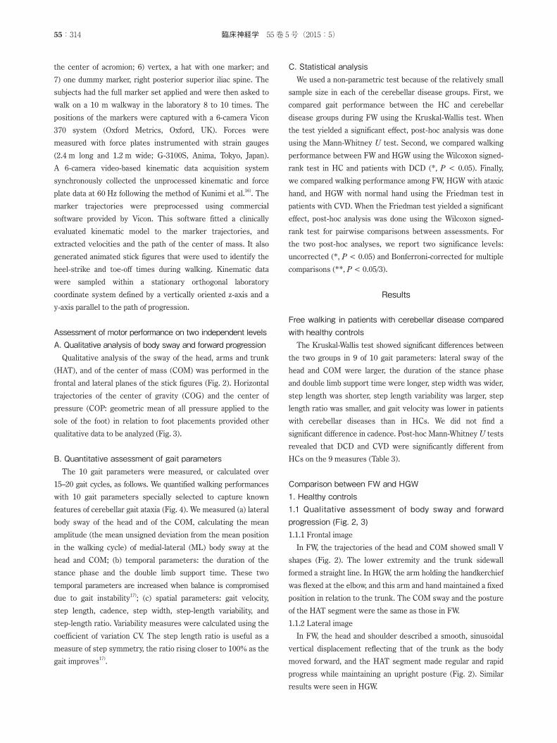
Fig. 3 Horizontal trajectories of the COG and COP relative to foot placements over one trial.
Representative of a typical HC (A), DCD (B), and CVD (C), the same cases as in Fig. 2. In a control subject (A), the COG passes outside or
slightly within the medial border of the supporting foot and passes the midline of the plane of progression at the point of mid-double-
support in walking. In a patient with DCD (B), the COG weaves tortuously and grossly, approaching the inside of the foot with every step
during FW. The COG straightens out to become only mildly weaving during HGW. In a patient with CVD (C), the COG travels moderately
tortuously during FW, mainly following the foot sole on the unaffected side, and away from the sole of the affected side. On HGW-AH
and -NH, the COG advances mildly tortuously, contacting the heels of the soles on both sides. COG = center of gravity, COP = center of
pressure, FW = free walking, HGW = handkerchief-guided walking, HGW-AH = handkerchief-guided walking with ataxic hand. HGW-NH
= handkerchief-guided walking with normal hand.

The handkerchief guide 55:317
3. Unilateral cerebellar vascular disease3.1 Qualitative assessments of body sway and forward progression (Fig. 2, 3)3.1.1 Frontal image
In FW, horizontal displacements of the head and the COM
were large and irregular (Fig. 2). In HGW with the ataxic hand,
the arm and hand grasping the handkerchief maintained an
almost steady position in relation to the trunk, and the head and
COM sway were attenuated. In HGW with the normal hand
compared to HGW with the ataxic hand, the arm and hand
Fig. 4 Comparison of the gait parameters important for patients with cerebellar diseases with healthy controls.
The links between different bars represent significant differences. In both cerebellar groups (DCD and CVD), a considerable improvement
in gait is seen in handkerchief-guided walking. In DCD, the HGW significantly improves seven parameters. In CVD, HGW with the
affected hand improves five parameters, and HGW with the unaffected hand improves seven parameters. COM = center of mass: CVD =
unilateral cerebellar vascular disease: DCD = degenerative cerebellar disease: DLS = double limb support time: HC = healthy controls:
stance = stance phase. DCD; *, P < 0.05, CVD; **, P < 0.05/3; *, P < 0.05. NS = not significant.
Table 3 Free walking in patients with cerebellar disease compared with healthy controls.
Gait parameters
Lateral head sway
Lateral COM sway
Stance DLSGait
velocityStep length Cadence Step width
Step-length variability
Step-length ratio
DCD 0.002** 0.001** 0.038* 0.038* 0.004** 0.017* 0.165 0.001** 0.001** 0.011**
CVD 0.001** 0.001** 0.041* 0.001** 0.001** 0.001** 0.097 0.001** 0.001** 0.001**
For each combination of patient group and gait parameter, P values are given (Mann-Whitney U test). The parameters of the cerebellar
groups show impairment relative to HCs during free walking. These include lateral body sway of head and COM, gait velocity, step length, step
width, step-length variability, step-length ratio, stance phase, and double limb support time. We do not find a significant difference in cadence.
COM = center of mass, CVD = unilateral cerebellar vascular disease, DCD = degenerative cerebellar disease, DLS = double limb support
time, HC = healthy controls, stance = stance phase. Asterisks indicate significance. *, P < 0.05; **, P < 0.05/3.

臨床神経学 55 巻 5 号(2015:5)55:318
maintained a fixed position in relation to the trunk, and,
accordingly, the head and COM sway were particularly
attenuated, resulting in a small V-shaped trajectory.
3.1.2 Lateral image
In FW, the head and shoulder moved up and down slightly, and
the HAT segment made irregular and slow progress with an
approximately upright posture (Fig. 2). In HGW with the ataxic
hand, vertical movement of the head and shoulder improved and
became smooth, and the HAT segment took large and regular
steps forward with a fixed ante-flexed posture. In HGW with the
normal hand, vertical displacement of the head and shoulder
were similarly improved as in HGW with the ataxic hand, but the
HAT segment made forward progress more regularly and with a
more nearly upright posture than in HGW with the ataxic hand.
3.1.3 Horizontal image
In FW, the COG trajectory weaved moderately, mainly
following the foot sole of the unaffected side away from the
affected side (Fig. 3). The COP traveled along either the middle
or the inside of the foot sole on the affected side. In HGW with
the ataxic hand, the COG trajectory weaved less than in FW, and
was in contact with the foot sole (heel) bilaterally. The COP
passed along a straight line in about the middle of the foot sole.
In HGW with the normal hand, the COG and COP trajectories
were almost the same as those in HGW with the ataxic hand.
3.2 Quantitative assessment of gait parametersThe Friedman test showed that there were significant
differences among the three types of walking in 7 of 10 gait
parameters: lateral head sway, lateral COM sway, double limb
support time, gait velocity, step length, step length variability,
and step length ratio (Fig. 4). Post-hoc Wilcoxon signed-rank
tests revealed that HGW with the ataxic hand and with the
normal hand were significantly different from FW for the first
five of the seven measures: a decrease in lateral head sway, a
reduction in lateral COM sway, an increase in gait velocity, an
increase in step length, and shorter double limb support time
(Fig. 4). These results led to a posture closer to upright during
HGW. In addition, HGW with the normal hand as opposed to the
ataxic hand revealed significant improvements in two measures
compared with FW: a decrease in step-length variability, and an
increase in the step-length ratio (Fig. 4). We did not find
significant differences among the three walking types for the
other three measures of gait: cadence, stance duration, and step
width.
Discussion
Gait analysis revealed a larger lateral sway of the head and the
COM, a longer duration of the stance phase and double-limb
support time, shorter step length, greater step width, larger step
length variability, smaller step length ratio, and a slower gait
velocity in patients with cerebellar diseases as compared with
the HCs. These results are compatible with previous studies1)–4).
This was the first study to show that ataxic gait is improved
considerably by the handkerchief guide, a simple method.
Quantitative gait analysis revealed an increased velocity, longer
step length, decreased lateral sway of the head and COM, and
shorter double limb support time. Qualitative analysis showed
that the COG trajectory became more regular, smooth, and
linear, staying within the medial borders of the supporting feet
during level walking. These results indicated that upright
posture had been stabilized in cases of cerebellar ataxic gait18).
From qualitative analysis of stick figures drawn from our data in
the frontal plane, control of upright posture might be explained
as the result of stabilization at a subconscious level of the arm
and hand position in relation to the trunk and of the arm
configuration.
A light touch on a rigid surface using the index finger has been
reported to be useful for postural adjustments in normal subjects
during standing and walking5)–7). Two control mechanisms are
involved in reducing postural sway: one is the tactile and
proprioceptive afferent information from the arm and hand19)20),
and the other is the constraint of the supra-postural task of
holding the arm in constant light contact12). Both mechanisms are
likely to work to improve gait during use of the handkerchief
guide. Nevertheless, two differences exist between the
handkerchief guide and a light touch on a stationary surface.
First, since the handkerchief is a stable point with an added
predictable movement, a grip is preferred by most subjects to
prevent the handkerchief from slipping from between the fingers
during locomotion21)22). Second, body sway could be reduced
based on the information on the modulation of the force on
the handkerchief induced by actual sway23). In addition, two
mechanisms may operate in HGW: one would be the handgrip
facilitated mechanisms of inter-limb coordination subserving
locomotor synergies24)25), and the other would be the interpersonal
synchronization that occurs during side-by-side walking of the
patient and caregiver26).
Using the HGW with the unaffected hand improved gait more
than with the affected hand in patients with CVD. We interpreted
this finding as resulting from more efficient maintenance of a
fixed posture of the arm and hand in relation to the trunk with
the unaffected hand than with the affected one. The cerebellum
is supposed to play a role in the stabilization of the kinematic
chain connecting the arm to the trunk. HC subjects did not show
improvement of gait with the handkerchief guide. This could be
explained by postulating a destabilization of upright posture by
movements of the contact point due to the caregiverʼs sway20).
We use a handkerchief to improve ataxic gait. The folded
handkerchief is a useful coupler for transmitting the pulling force
from the patient to a caregiver during walking, while allowing

The handkerchief guide 55:319
both vertical and back and forth movement. Holding hands can
have the same effect as the handkerchief guide while the subjectʼs arm is directly restricted by the caregiver. From the above,
the handkerchief guide appears to be a simple and easy in way to
assist a patient with cerebellar ataxia. This may be useful in gait
training of cerebellar disease patients, because gait with the HGW
is closer to FW than to direct-contact caregiver-assisted gait.Acknowledgments: We would like to express our gratitude to
Dr. Hitoshi Shinoto for comments on the manuscript, and to
Dr. Yoshikazu Kyuma, Dr. Masao Murai, and Machiko Takahashi of the
Nanasawa Rehabilitation Cerebrovascular Center, and to Drs Takamiti
Kubokura, Yumi Miyazwa, and Kenji Ishihara of the Ushioda General
Hospital for their support throughout the present study.
※ The authors declare there is no conflict of interest relevant to this
article.
References
1) Holmes G. Clinical symptoms of cerebellar disease and their
interpretation. Lancet 1922;203:59-65.
2) Mitoma H, Hayashi R, Yanagisawa N, et al. Characteristics
of parkinsonian and ataxic gaits: a study using surface
electromyograms, angular displacements, and floor reaction
forces. J Neurol Sci 2000;174:22-39.
3) Hudson CC, Krebs DE. Frontal plane dynamic stability and
coordination in subjects with cerebellar degeneration. Exp
Brain Res 2000;132:103-113.
4) Ilg W, Golla H, Thier P, et al. Specific influences of cerebellar
dysfunctions on gait. Brain 2007;130:786-798.
5) Holden M, Ventura J, Lackner JR. Stabilization of posture by
precision contact of the index finger. J Vestib Res 1994;4:285-
301.
6) Jeka JJ, Lackner JR. Fingertip contact influences human
postural control. Exp Brain Res 1994;100:495-502.
7) Dickstein R, Laufer Y. Light touch and center of mass stability
during treadmill locomotion. Gait Posture 2004;20:41-47.
8) Tremblay F, Mireault AC, Dessureault L, et al. Postural
stabilization from fingertip contact: I. Variations in sway
attenuation, perceived stability and contact forces with aging.
Exp Brain Res 2004;157:275-285.
9) Lackner JR, DiZio P, Jeka J, et al. Precision contact of the
fingertip reduces postural sway of individuals with bilateral
vestibular loss. Exp Brain Res 1999;126:459-466.
10) Dickstein R, Peterka RJ, Horak FB. Effects of light fingertip
touch on postural responses in subjects with diabetic
neuropathy. J Neurol Neurosurg Psychiatry 2003;74:620-626.
11) Jeka JJ, Easton RD, Bentzen BL, et al. Haptic cues for
orientation and postural control in sighted and blind individuals.
Percept Psychophys 1996;58:409-423.
12) Riley MA, Stoffregen TA, Grocki MJ, et al. Postural stabilization
for the control of touching. Hum Mov Sci 1999;18:795-817.
13) Nagumo K, Hirayama K, Kunimi Y, et al. Cerebellar ataxia
gait on handkerchief-guided task. Correlation between the
handkerchief tension and the center of mass acceleration.
Rinsho Shinkeigaku 2010;50:1121. Abstract.
14) Hirayama K, Kita K. Spinocerebellar degeneration. Japanese
collection of clinical statistics, the first volume. Nippon Rinsho
1992;50:123-133.
15) Trouillas P, Takayanagi T, Hallett M, et al. International
Cooperative Ataxia Rating Scale for pharmacological assessment
of the cerebellar syndrome. The Ataxia Neuropharmacology
Committee of the World Federation of Neurology. J Neurol Sci
1997;145:205-211.
16) Kunimi Y, Nomura S, Beppu M. The influence that the center
of gravity position of the rucksack gives on a gait locomotion.
Jap J Mountain Medicine 2007;27:111-114.
17) Kirtley C. Clinical gait analysis-theory and practice. Oxford:
Elsevier Churchill Livingstone; 2006.
18) Massion J. Postural control systems in developmental per-
spective. Neurosci Biobehav Rev 1998;22:465-472.
19) Kouzaki M, Masani K. Reduced postural sway during quiet
standing by light touch is due to finger tactile feedback but
not mechanical support. Exp Brain Res 2008;188:153-158.
20) Jeka JJ, Schöner G, Dijkstra T, et al. Coupling of fingertip
somatosensory information to head and body sway. Exp Brain
Res 1997;113:475-483.
21) Johansson RS, Häger C, Riso R. Somatosensory control of
precision grip during unpredictable pulling loads. II. Changes
in load force rate. Exp Brain Res 1992;89:192-203.
22) Gysin P, Kaminski TR, Gordon AM. Coordination of fingertip
forces in object transport during locomotion. Exp Brain Res
2003;149:371-379.
23) Krishnamoorthy V, Slijper H, Latash ML. Effects of different
types of light touch on postural sway. Exp Brain Res 2002;
147:71-79.
24) Zehr EP, Duysens J. Regulation of arm and leg movement
during human locomotion. Neuroscientist 2004;10:347-361.
25) Dietz V, Michel J. Human bipeds use quadrupedal coordination
during locomotion. Ann N Y Acad Sci 2009;1164:97-103.
26) Nessler JA, Gilliland SJ. Kinematic analysis of side-by-side
stepping with intentional and unintentional synchronization.
Gait Posture 2010;31:527-529.