the haemangioblast generates haematopoietic cells through a haemogenic endothelium stage
TRANSCRIPT
LETTERS
The haemangioblast generates haematopoietic cellsthrough a haemogenic endothelium stageChristophe Lancrin1, Patrycja Sroczynska1, Catherine Stephenson1, Terry Allen2, Valerie Kouskoff3
& Georges Lacaud1
It has been proposed that during embryonic development haema-topoietic cells arise from a mesodermal progenitor with bothendothelial and haematopoietic potential called the haemangio-blast1,2. A conflicting theory instead associates the first haemato-poietic cells with a phenotypically differentiated endothelial cellthat has haematopoietic potential (that is, a haemogenic endo-thelium)3–5. Support for the haemangioblast concept was initiallyprovided by the identification during mouse embryonic stem celldifferentiation of a clonal precursor, the blast colony-forming cell(BL-CFC), which gives rise to blast colonies with both endothelialand haematopoietic components6,7. Although recent studies havenow provided evidence for the presence of this bipotentialprecursor in vivo8,9, the precise mechanism for generation of hae-matopoietic cells from the haemangioblast still remains completelyunknown. Here we demonstrate that the haemangioblast generateshaematopoietic cells through the formation of a haemogenic endo-thelium intermediate, providing the first direct link between thesetwo precursor populations. The cell population containing the hae-mogenic endothelium is transiently generated during BL-CFCdevelopment. This cell population is also present in gastrulatingmouse embryos and generates haematopoietic cells on furtherculture. At the molecular level, we demonstrate that the transcrip-tion factor Tal1 (also known as Scl; ref. 10) is indispensable for theestablishment of this haemogenic endothelium population whereasthe core binding factor Runx1 (also known as AML1; ref. 11) iscritical for generation of definitive haematopoietic cells from hae-mogenic endothelium. Together our results merge the two a prioriconflicting theories on the origin of haematopoietic developmentinto a single linear developmental process.
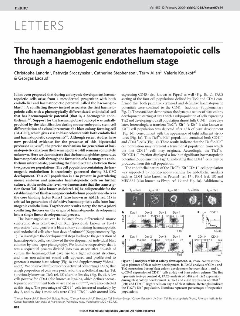
The haemangioblast can be isolated from differentiated mouseembryonic stem cells based on Kdr (previously known as Flk-1)expression12 and generates a blast colony containing haematopoieticand endothelial cells after four days of culture6,7 (Supplementary Fig.1). To investigate the developmental steps leading to the generation ofhaematopoietic cells, we followed the development of individual blastcolonies by time-lapse photography. We found retrospectively that itwas a sequential process divided into two stages: after 36–48 h ofculture the haemangioblast gave rise to a tight adherent structure,and then non-adherent round cells appeared and proliferated togenerate a mature blast colony (Fig. 1a and Supplementary Videos 1and 2). We observed by fluorescence-activated cell sorting (FACS) thata high proportion of cells were positive for the endothelial marker Tek(previously known as Tie2; ref. 13) after the first day (Fig. 1b, d). A fewcells positive for CD41 (also known as Itga2b), which defines haema-topoietic commitment both in vivo and in vitro14–16, were also detectedat this stage. The percentage of CD411 cells increased markedly byday 2, and by day 4 most cells were CD411Tie22, with around 30%
expressing CD45 (also known as Ptprc) as well (Fig. 1b, c). FACSsorting of the four cell populations defined by Tie2 and CD41 con-firmed that both primitive erythroid and definitive haematopoieticpotentials were confined to the CD411 fractions (SupplementaryFig. 2). These analyses demonstrate the dynamic nature of blast colonydevelopment starting at day 1 with a subpopulation of cells expressingTie2 and developing to a cell population almost fully CD411 three dayslater. Interestingly, a transient Tie2hic-Kit1 (c-Kit1 is also known asKit1) cell population was detected after 48 h of blast development(Fig. 1d), concomitant with the appearance of tight adherent struc-tures (Fig. 1a). This Tie2hic-Kit1 population contained both CD412
and CD411 cells (Fig. 1e). These results indicate that the Tie2hic-Kit1
cell population may represent a transitional population from whichthe first CD411 cells may originate. Accordingly, the Tie2hic-Kit1CD412 fraction displayed a low but significant haematopoieticpotential (Supplementary Fig. 3), indicating that CD411 cells may beproduced from this cell population.
The endothelial nature of the Tie2hic-Kit1CD412 cell populationwas supported by homogeneous staining for endothelial markerssuch as CD31 (also known as Pecam1; ref. 17), Flk-1 (ref. 18) andMECA32 (also known as Plvap; ref. 19 and Fig. 2a). Additionally,
1Cancer Research UK Stem Cell Biology Group, 2Cancer Research UK Structural Cell Biology Group, 3Cancer Research UK Stem Cell Haematopoiesis Group, Paterson Institute forCancer Research, University of Manchester, Wilmslow road, Manchester M20 4BX, UK.
T0 + 24 h T0 + 36 h T0 + 48 h T0 + 60 h T0 + 65 h
1.7 3.4
47.747.2
89.9 5.5
1.43.2
43.2 11.2
24.521
70.8 6.1
10.412.7
Day 1 Day 2 Day 3 Day 4
Tie2 CD45
CD41+
46.9 2.9
446.2
28.2 21.8
13.736.3
1.7 11.6
38.947.7
45.9 12.3
437.8
Tie2
Day 1 Day 2 Day 3 Day 4 CD41+
Tie2
62.8 11.5
1.424.3
5.6 20.4
20.153.9
CD41–
30CD
41c-
Kit
c-K
it
Cou
nts
a
b c
d e
Figure 1 | Analysis of blast colony development. a, Phase-contrast time-lapse pictures of blast colony development. b, FACS analysis of CD41 andTie2 expression during blast colony development between days 1 and 4.c, CD45 expression of CD411 cells at day 4 of blast colony culture. The linerepresents isotype control. d, FACS analysis of c-Kit and Tie2 expressionduring blast colony development. e, Tie2 and c-Kit expression of CD411
(left) and CD412 (right) cells on day 2 of blast culture. Rectangles indicatethe Tie2hic-Kit1 population. Numbers represent percentages of respectivepopulations.
Vol 457 | 12 February 2009 | doi:10.1038/nature07679
892 Macmillan Publishers Limited. All rights reserved©2009

these cells expressed other endothelial genes, such as Cd34 (ref. 20),endoglin (Eng; ref. 21), Flt1 (ref. 22) and VE-cadherin (Cdh5; ref. 23and Supplementary Fig. 4a), and generated endothelial networks onculture in matrigel plugs (Fig. 2b). Immunostaining for CD31,expressed specifically by Tie2hic-Kit1 cells at day 3 of blast develop-ment (Supplementary Fig. 4b), and for CD41 demonstrated the pres-ence of CD311CD412 endothelial cells corresponding to the core oftightly associated cells of blast colonies (Supplementary Fig. 4c).
In addition to its clear endothelial signature, the Tie2hic-Kit1CD412
subpopulation expressed transcription factors associated with theonset of haematopoiesis (Supplementary Fig. 5a). To test whether thispopulation contained endothelial cells with haematopoietic potential(that is, a haemogenic endothelium), Tie2hic-Kit1CD412 cells werecultured in conditions supporting the generation of haematopoieticcells from the aorta–gonad–mesonephros (AGM) region of mouseembryo24. After two days, non-adherent round cells were observed(Supplementary Fig. 5b), and FACS analysis demonstrated that around70% of the cells expressed CD41 and that some of them had down-regulated Tie2 expression (Fig. 2c). Isolation of these newly generatedCD411 cells further confirmed that haematopoietic potential waspresent and restricted to this fraction (Supplementary Fig. 5c). Toevaluate the proportion of Tie2hic-Kit1CD412 cells becomingCD411, we labelled them with carboxy-fluorescein succinimidyl ester(CFSE), a fluorescent dye equally distributed after each cell division.After 24 h of culture, about 40% of cells were CD411 cells, with a CFSEmean fluorescence 1.5 times lower than in CD412 cells (SupplementaryFig. 5d). This result indicates that CD411 cells were produced fromaround 25% (that is, 40% divided by 1.5) of the Tie2hic-Kit1CD412
cells. By limiting dilution analysis, the frequency of Tie2hic-Kit1CD412 cells generating more differentiated haematopoietic cellswas found to be around 1.2% (Supplementary Fig. 5e). This number,lower than in the CFSE analysis, reflects the fact that only a fraction ofCD411 cells are able to respond to haematopoietic growth factors. Theclonality of the haemogenic endothelium was demonstrated by
generation of haematopoietic cells from single Tie2hic-Kit1CD412
cells (Supplementary Fig. 5f). Finally, Tie2hic-Kit1CD412 cells didnot express the mesodermal marker brachyury25 (Supplementary Fig.6a) and were unable to generate blast colonies (Supplementary Fig. 6b)indicating that the precursors present in this cell population wereclearly distinct and downstream from haemangioblasts. Overall, thesedata demonstrate the presence of clonal haemogenic endothelial cellprecursors in the Tie2hic-Kit1CD412 cell population.
A prediction from these results is that a Tie21c-Kit1CD412 haemo-genic population should be present in vivo at the onset of blood develop-ment. We were indeed able to identify Tie21c-Kit1CD412 cells inembryos at the neural plate stage of gastrulation and their frequenciesincreased in subsequent stages (Fig. 2d). Immunostainings demon-strated that they were localized in developing blood islands of early head-fold embryos (Fig. 2e, f). FACS-isolated Tie21c-Kit1CD412 cells platedon OP9 stromal cells generated round non-adherent cells giving rise toCD451 cells and primitive and definitive haematopoietic colonies afterreplating (data not shown and Fig. 2g). Tie21c-Kit1CD412 cells werealso detected within the AGM region of E10.5 embryos in the dorsal aorta(Supplementary Fig. 7a–c) and these cells also had the capacity togenerate haematopoietic cells (Supplementary Fig. 7d, e), but the originof these Tie21c-Kit1CD412 cells in the AGM and their precisedevelopmental potential remain to be characterized. Together these dataindicate that the Tie21c-Kit1CD412 progenitor population detected ingastrulating embryos may represent an intermediate between the hae-mangioblast, predominantly found in the primitive streak8, and thehaematopoietic precursors found in the yolk sac26.
To investigate further the molecular mechanisms implicated in thegeneration of the Tie21c-Kit1CD412 population, we analysed theeffects of knockdown of two critical genes for early haematopoiesis,Runx1 and Scl, on this cell population. The transcription factorRunx1 is required in vivo for the generation of definitive haemato-poietic cells11 and its absence results in vitro in the generation of 20times fewer blast colonies, with residual blast colonies restricted to aprimitive haematopoietic fate27. We performed a time-lapse analysisof blast colony development from isolated Runx12/2 Flk-11 cells.Clusters of tightly associated adherent cells were observed (Fig. 3a),but only a few of these clusters later generated blast colonies (data notshown). Cells were observed to emerge from most clusters but diedinstead of proliferating (Fig. 3a and Supplementary Video 3).Immunofluorescence analyses confirmed that cells within these clus-ters expressed CD31 but that no CD411 cells were present(Supplementary Fig. 8). The defect in haematopoietic developmentwas further observed by FACS analysis that showed a marked reduc-tion in the frequency of CD411 cells (Fig. 3b) and an increasedfrequency in the Tie2hic-Kit1 cell population (Fig. 3c).
To examine further the requirement for Runx1, we generated aRunx12/2 embryonic stem cell line containing a doxycycline-inducible Runx1 complementary DNA (iRunx1). When doxycyclinewas added at day 2 of blast development, at least a tenfold increase inCD411 cell frequency was observed after 24 h, associated with down-regulation of Tie2 expression (Supplementary Fig. 9a) and inductionof expression of genes involved in myeloid cell development(Supplementary Fig. 9b). To establish whether the developmentalblock observed in the absence of Runx1 was at the level of theTie2hic-Kit1CD412 cell population, we isolated these cells andcultured them in the presence of doxycycline. After 48 h, around60% of the cells expressed CD41 and one-third of these cells down-regulated Tie2 expression (Fig. 3d). Additionally, the CD411 cellsincorporated acetylated low-density lipoprotein (LDL), which is takenup by endothelial cells28, further supporting their endothelial origin(Fig. 3e). Finally, definitive haematopoietic precursors were exclusivelydetected in colony assays in cultures induced with doxycycline (Fig. 3f).Together, these results demonstrate that the Tie2hic-Kit1CD412 hae-mogenic endothelial cell population is generated in absence of Runx1but that Runx1 is indispensable for the generation of definitive hae-matopoietic cells from this population.
b99.4
0.6
88.6
11.4
88.5
11.5
Tie2
CD41–
CD41
a
T0 T0 + 2 daysc
98
10.03
Tie2
30.1
30.738
d E7.0Mid-streak
E7.5Neural plate
E8.0Headfold
E8.25Early somite
0.1 3.1 5.42
Tie2
c-Kit ×5
c-Kit ×40
Tie2 ×40
c-Kit ×40
Tie2 ×40
am
ac
ecc al
ch
bi
bi
am
c-Kit CD41 ×10
c-Kit CD41
×20bi
bi
ecc
dede
c-Kit CD41
×20
e f14.5 5.8
16.763
*
g
c-K
it
c-K
it
c-Kit
CD
31
CD
41
CD
45
ME
CA
32
Flk-
1
Figure 2 | Tie2hic-Kit1CD412 cells can generate haematopoieticprogenitors. a, FACS analyses of CD31, Flk-1 and MECA32 expression byTie2hic-Kit1CD412 cells. b, Generation of endothelial networks in matrigelby isolated day-2 Tie2hic-Kit1CD412 cells. c, Tie2hic-Kit1CD412 cells atday 2 of blast development were sorted and cultured. FACS analysis of CD41and Tie2 expression at T0 and T0 1 2 days. d, FACS analysis for the presenceof Tie21c-Kit1CD412cells in gastrulating embryos. e, f, Immunostaining ofgastrulating embryos for Tie2 and c-Kit (e) and c-Kit and CD41 (f). ac,amniotic cavity; al allantois; am, amnion; bi, yolk sac blood islands; ch,chorion; de, decidua; ecc, exocoelomic cavity. Original magnifications areindicated. g, FACS analysis of CD45 and c-Kit expression (top) andMay–Grunwald Giemsa staining (bottom) of cells generated by isolatedE7.75 mouse embryo Tie21c-Kit1CD412 cells co-cultured with OP9 cells.Macrophage (arrow) and mast cells (asterisk) are indicated. Numbersindicate percentages of respective populations.
NATURE | Vol 457 | 12 February 2009 LETTERS
893 Macmillan Publishers Limited. All rights reserved©2009

Accordingly, Runx12/2 Flk-11 cells generated significantly morecolonies of tightly associated cells than wild-type cells in BL-CFC clo-nogenic assays (Supplementary Fig. 10). Their number was inverselycorrelated with the number of blast colonies observed with wild-typecells (Supplementary Fig. 11a). The presence of haemogenic endothe-lium in these tight structures was supported by detection of a largeproportion of CD311 or Tie21c-Kit1 cells in individual colonies byimmunostaining (Supplementary Figs 8 and 11b, respectively) and ofTie2hic-Kit1CD412 cells by FACS analysis on pooled colonies(Supplementary Fig. 11c). Additionally, around 75% of individualcolonies (26 out of 34) were able to give rise to haematopoietic cellson Runx1 re-expression (Supplementary Fig. 11d). These data supportthe notion that these tight colonies generated from the haemangioblastcontain cells with haemogenic endothelium potential that are unableto initiate haematopoiesis in the absence of Runx1.
Scl is another critical regulator of haematopoiesis as Scl2/2 cells areunable to generate either primitive or definitive haematopoiesis10.Plating of Scl2/2 Flk-11 cells resulted, as shown previously29, in theabsence of blast colonies but also in a complete lack of clusters oftightly associated adherent cells (Fig. 4a). We were unable to detectany CD411 cells (Fig. 4b). Although Tie2 was expressed at levelssimilar to those in Scl1/1 cells, only a very small fraction of cellsco-expressed Tie2 and c-Kit (Fig. 4c) and none of these cellsexpressed CD31, Flk-1 or MECA32 (data not shown). These dataindicate that Scl is critical for the generation of Tie2hic-Kit1CD412
haemogenic endothelium population and place its role in haemato-poietic specification before Runx1 requirement.
In summary, our data provide the first evidence for a direct linkbetween the haemangioblast and a downstream haemogenic endothe-lium. The generation of this cell population is characterized by theupregulation of c-Kit in a Tie2hi population and this process is Scl-dependent (Fig. 4d). The consecutive generation of definitive haema-topoietic cells, characterized by CD41 expression and Tie2 downre-gulation, requires the transcription factor Runx1, whereas primitive
haematopoiesis is Runx1-independent. Identification of these discretedevelopmental steps will provide the opportunity to explore furtherthe molecular regulation of haematopoietic development.
METHODS SUMMARYTime-lapse analyses were performed in a Solent Scientific environmental cham-
ber kept at 37 uC for the duration of the analysis and observed under a Zeiss
Axiovert 200M microscope. Brachyury–green fluorescent protein (GFP)25,
Scl2/2 (ref. 10), Ainv18 (ref. 30), Ainv18 Runx11/2, Runx12/2 and iRunx1
embryonic stem cell lines were used. Flow cytometry and cell sorting were per-
formed on a FACSCalibur (Becton Dickinson) and FACSVantage or FACSAria
cell sorters (Becton Dickinson). For immunostainings, frozen sections were
fixed, blocked, incubated with primary Tie2, c-Kit and CD41 antibodies and
then with secondary antibodies, mounted and observed under a Zeiss Axiovert
200M microscope.
Full Methods and any associated references are available in the online version ofthe paper at www.nature.com/nature.
Received 8 August; accepted 1 December 2008.Published online 28 January 2009.
1. Sabin, F. R. Studies on the origin of blood vessels and of red corpuscules as seen inthe living blastoderm of the chick during the second day of incubation:contributions to embryology. Contrib. Embryol. 9, 213–262 (1920).
2. Murray, P. D. F. The development in vitro of the blood of the early chick embryo.Proc. R. Soc. Lond. 11, 497–521 (1932).
3. Jaffredo, T., Gautier, R., Eichmann, A. & Dieterlen-Lievre, F. Intraaortichemopoietic cells are derived from endothelial cells during ontogeny.Development 125, 4575–4583 (1998).
4. Nishikawa, S. I. et al. In vitro generation of lymphohematopoietic cells fromendothelial cells purified from murine embryos. Immunity 8, 761–769 (1998).
5. North, T. E. et al. Runx1 expression marks long-term repopulating hematopoieticstem cells in the midgestation mouse embryo. Immunity 16, 661–672 (2002).
6. Kennedy, M. et al. A common precursor for primitive erythropoiesis and definitivehaematopoiesis. Nature 386, 488–493 (1997).
7. Choi, K., Kennedy, M., Kazarov, A., Papadimitriou, J. C. & Keller, G. A commonprecursor for hematopoietic and endothelial cells. Development 125, 725–732 (1998).
8. Huber, T. L., Kouskoff, V., Fehling, H. J., Palis, J. & Keller, G. Haemangioblastcommitment is initiated in the primitive streak of the mouse embryo. Nature 432,625–630 (2004).
9. Vogeli, K. M., Jin, S. W., Martin, G. R. & Stainier, D. Y. A common progenitor forhaematopoietic and endothelial lineages in the zebrafish gastrula. Nature 443,337–339 (2006).
10. Porcher, C. et al. The T cell leukemia oncoprotein SCL/tal-1 is essential fordevelopment of all hematopoietic lineages. Cell 86, 47–57 (1996).
11. Okuda, T., van Deursen, J., Hiebert, S. W., Grosveld, G. & Downing, J. R. AML1, thetarget of multiple chromosomal translocations in human leukemia, is essential fornormal fetal liver hematopoiesis. Cell 84, 321–330 (1996).
12. Faloon, P. et al. Basic fibroblast growth factor positively regulates hematopoieticdevelopment. Development 127, 1931–1941 (2000).
1.59 0.02
0.0298.37
1 0.07
0.3498.58
1.06 0.05
0.5298.37
1.69 0.03
0.2798
Day 1 Day 2 Day 3 Day 4
0.43 1.04
55.2843.25
0 1.01
95.733.26
0 1.03
95.983
0.03 1.64
93.334.99
CD41
Tie2
b
c Day 1 Day 2 Day 3 Day 4
Scl–/–Scl+/+
aHaemangioblast
Flk1+ brachyury+
Haemogenic endothelium
Tie2hic-Kit+CD41–
Definitivehaematopoiesis
Primitivehaematopoiesis
Scl
Runx1
d
c-K
itc-
Kit
CD41+CD45– CD41+CD45+
Figure 4 | Scl requirement during blast colony development. a, Phase-contrast pictures of day-3 blast colony culture from Scl1/1 and Scl2/2
embryonic stem cells. b, FACS analysis of CD41 and c-Kit expression duringScl2/2 blast colony development. Numbers indicate the percentage of therespective populations. c, FACS analysis of Tie2 and c-Kit expression duringScl2/2 blast colony development. d, Model of haemangioblastdifferentiation towards haematopoiesis in the yolk sac.
Day 1 Day 2 Day 3
2.44 0.04
0.0397.5
37.3 0.3
0.0462.36
34.4 0.8
0.464.4
10.3 2.3
2.185.3
Day 1 Day 2 Day 3 Day 4
CD41
b
a
c
7.1 7.3
12.972.7
3.8 28.6
28.938.7
7.4 24.2
19.149.3
4.7 6.8
10.278.3
Tie2
7.6 10.6
14.667.2
30.3 19.7
10.739.3
44.5 10.3
2.542.7
52.5 2.1
3.242.2
Day 1 Day 2 Day 3 Day 4
Tie2
NoDox
Dox
d
96.5
0.050
70.2
22.62.2
T0 + 2 daysT0
40.123.5
26.6
41.8 3.9
1.9
No Dox Dox
18.3 22.2
23.8
CD41
No Dox Dox
e
f
c-K
itc-
Kit
Col
onie
s p
er30
,000
cel
lsC
D41
Dil-
Ac-
LDL
0
400
800
1,200
Figure 3 | Runx1 requirement in blast colony development. a, Phase-contrast time-lapse photographs of Runx12/2 blast colony development.b, FACS analysis of CD41 and c-Kit expression during Runx12/2 blastcolony development between days 1 and 4. c, FACS analysis of Tie2 and c-Kitexpression during Runx11/1 (top) and Runx12/2 (bottom) blast colonydevelopment. d, FACS analysis of Tie2 and CD41 expression after 2 days ofculture of iRunx1 Tie2hic-Kit1CD412 cells in the absence or presence ofdoxycycline (Dox; 0.1 mg ml21). e, Dil-Ac-LDL and CD41 expressionanalysis of iRunx1 Tie2hic-Kit1CD412 cells were evaluated in the absence(left) and presence (right) of doxycycline. Numbers indicate the percentageof the respective populations. f, Numbers of definitive haematopoieticcolonies generated in methylcellulose by iRunx1 cells collected after twodays of culture with or without doxycycline. Error bars indicate standarddeviation of the mean (n 5 3).
LETTERS NATURE | Vol 457 | 12 February 2009
894 Macmillan Publishers Limited. All rights reserved©2009

13. Dumont, D. J., Yamaguchi, T. P., Conlon, R. A., Rossant, J. & Breitman, M. L. tek, anovel tyrosine kinase gene located on mouse chromosome 4, is expressed inendothelial cells and their presumptive precursors. Oncogene 7, 1471–1480(1992).
14. Ferkowicz, M. J. et al. CD41 expression defines the onset of primitive and definitivehematopoiesis in the murine embryo. Development 130, 4393–4403 (2003).
15. Mikkola, H. K., Fujiwara, Y., Schlaeger, T. M., Traver, D. & Orkin, S. H. Expression ofCD41 marks the initiation of definitive hematopoiesis in the mouse embryo. Blood101, 508–516 (2003).
16. Li, W., Ferkowicz, M. J., Johnson, S. A., Shelley, W. C. & Yoder, M. C. Endothelialcells in the early murine yolk sac give rise to CD41-expressing hematopoietic cells.Stem Cells Dev. 14, 44–54 (2005).
17. Newman, P. J. et al. PECAM-1 (CD31) cloning and relation to adhesion moleculesof the immunoglobulin gene superfamily. Science 247, 1219–1222 (1990).
18. Hirai, H. et al. Hemogenic and nonhemogenic endothelium can be distinguishedby the activity of fetal liver kinase (Flk)-1 promoter/enhancer during mouseembryogenesis. Blood 101, 886–893 (2003).
19. Hallmann, R., Mayer, D. N., Berg, E. L., Broermann, R. & Butcher, E. C. Novel mouseendothelial cell surface marker is suppressed during differentiation of the bloodbrain barrier. Dev. Dyn. 202, 325–332 (1995).
20. Young, P. E., Baumhueter, S. & Lasky, L. A. The sialomucin CD34 is expressed onhematopoietic cells and blood vessels during murine development. Blood 85,96–105 (1995).
21. Li, D. Y. et al. Defective angiogenesis in mice lacking endoglin. Science 284,1534–1537 (1999).
22. Fong, G. H., Rossant, J., Gertsenstein, M. & Breitman, M. L. Role of the Flt-1receptor tyrosine kinase in regulating the assembly of vascular endothelium.Nature 376, 66–70 (1995).
23. Breier, G. et al. Molecular cloning and expression of murine vascular endothelial-cadherin in early stage development of cardiovascular system. Blood 87, 630–641(1996).
24. Mukouyama, Y. et al. The AML1 transcription factor functions to develop andmaintain hematogenic precursor cells in the embryonicaorta–gonad–mesonephros region. Dev. Biol. 220, 27–36 (2000).
25. Fehling, H. J. et al. Tracking mesoderm induction and its specification to thehemangioblast during embryonic stem cell differentiation. Development 130,4217–4227 (2003).
26. Palis, J., Robertson, S., Kennedy, M., Wall, C. & Keller, G. Development of erythroidand myeloid progenitors in the yolk sac and embryo proper of the mouse.Development 126, 5073–5084 (1999).
27. Lacaud, G. et al. Runx1 is essential for hematopoietic commitment at thehemangioblast stage of development in vitro. Blood 100, 458–466 (2002).
28. Voyta, J. C., Via, D. P., Butterfield, C. E. & Zetter, B. R. Identification and isolation ofendothelial cells based on their increased uptake of acetylated-low densitylipoprotein. J. Cell Biol. 99, 2034–2040 (1984).
29. D’Souza, S. L., Elefanty, A. G. & Keller, G. SCL/Tal-1 is essential for hematopoieticcommitment of the hemangioblast but not for its development. Blood 105,3862–3870 (2005).
30. Kyba, M., Perlingeiro, R. C. & Daley, G. Q. HoxB4 confers definitivelymphoid–myeloid engraftment potential on embryonic stem cell and yolk sachematopoietic progenitors. Cell 109, 29–37 (2002).
Supplementary Information is linked to the online version of the paper atwww.nature.com/nature.
Acknowledgements We thank K. Labib, C. Miller and T. Somervaille for criticalreading of the manuscript, J. Barry and M. Hughes for cell sorting, S. Bagley for helpwith the time-lapse photography, G. Ashton for help with preparation of sections,and L. Gautreau for advice with the OP9 cultures. Cancer Research UK supportedthis work.
Author Contributions P.S. designed and performed experiments, and analysed thedata. C.S. performed experiments. T.A. designed research. C.L., V.K. and G.L.designed the research, performed experiments, analysed the data and wrote thepaper.
Author Information Reprints and permissions information is available atwww.nature.com/reprints. Correspondence and requests for materials should beaddressed to G.L. ([email protected]).
NATURE | Vol 457 | 12 February 2009 LETTERS
895 Macmillan Publishers Limited. All rights reserved©2009

METHODSEmbryonic stem cell growth and differentiation. Brachyury–GFP25, Scl2/2
(ref. 10), Ainv18 (ref. 30), Ainv18 Runx11/2, Runx12/2 and iRunx1 embryonic
stem cell lines (data not shown) were used. Growth and differentiation of embry-
onic stem cells were performed as described previously25.
Embryo generation. Timed matings of ICR mice were set up and the morning of
vaginal plug detection was considered day 0.5. Gastrulating embryos were staged
by morphological landmarks. All animal work was performed under regulation
in accordance with the UK Animal Scientific Procedures Act (ASPA) 1986.
Adherent BL-CFC and haemogenic endothelium cultures. Flk-11 embryoidbody cells were plated on gelatin in the BL-CFC media described previously25.
For haemogenic endothelium culture, Tie21c-Kit1CD412 cells were cultured
either on gelatin (cells isolated from day-2 or day-3 BL-CFC cultures) or on OP9
stromal cells (cells isolated from embryos) in conditions described previously24.
Cultures were maintained in a humidified chamber in a normal or low-O2 (5%)
5% CO2/air mixture at 37 uC.
Matrigel plug assays. Tie21c-Kit1CD412 cells isolated from day-2 BL-CFC
liquid culture were transferred to 50-ml Matrigel plugs in 96-well plates.
Matrigel (BD Biosciences) was diluted 1:1 with IMDM supplemented with
10% FCS, 50 ng ml21 vascular endothelial growth factor and 5 ng ml21 basic
fibroblast growth factor and allowed to solidify at 37 uC before the addition of
cells. Cultures were maintained at 37 uC, 5% CO2 and 5% O2 for five days.
Flow cytometry and cell sorting. Staining was done as described previously25
and analyses were performed with a FACSCalibur (Becton Dickinson). Sorts
were performed with a FACSVantage or a FACSAria (Becton Dickinson).
Monoclonal antibodies and streptavidin used were Flk-1–biotin25, CD31–biotin
(MEC13.3, BD Bioscience), MECA32–biotin (BD Bioscience), Tie2–biotin
(TEK4, eBioscience), Tie2–phycoerythrin (TEK4, eBioscience), c-Kit–APC(2B8, eBioscience), CD41–FITC (MWReg30, BD Bioscience), streptavidin–phy-
coerythrin-Cy5 (BD Bioscience) and streptavidin–phycoerythrin-Cy7
(eBioscience). Tie21c-Kit1CD412 cells from iRunx1 day-3 blast culture were
incubated with Dil-Ac-LDL (10 mg ml21; Invitrogen Molecular Probes) for 3 h,
washed twice to remove all Dil-Ac-LDL, and Runx1 expression was induced with
doxycycline. FACS analysis was performed two days later.
CFSE labelling. Tie21c-Kit1CD412 cells were resuspended in PBS 5% FCS at
0.5 3 106 cells per ml and CFSE was added to 5 mM final concentration. Cells
were incubated for 5 min at room temperature (20 uC), washed three times and
put back in culture.
Limiting dilution and single-cell analyses. Defined numbers (400, 200, 100 and
50 cells) of Tie2hic-Kit1CD412 cells were plated on OP9 stromal cells using a
FACSAria equipped with an automatic cell deposition unit (Becton Dickinson).
For each cell number a minimum of 24 wells was seeded. The cells were first
cultured for two days in conditions supporting the transition of haemogenic
endothelial cells to haematopoietic progenitors24 and subsequently in conditions
supporting the differentiation to mature haematopoietic cells. The wells were
scored for the presence of haematopoietic cells two days later. The haematopoie-
tic nature of the cells was confirmed by May–Grunwald Giemsa staining. The
fraction of wells negative for haematopoietic cells was plotted against each dilu-
tion and the linear regression was calculated with the Origin software
(OriginLab). To obtain the frequency of Tie2hic-Kit1CD412 cells able to gen-
erate haematopoietic cells we used the limdil function of the statistical package R
(http://bioinf.wehi.edu.au/software/limdil/index.html). For single-cell analysis,
single Tie2hic-Kit1CD412 cells were sorted on 96-well plates coated with gelatin.
The culture conditions were as described previously and the wells were scored for
the presence of haematopoietic cells after eight days. The haematopoietic nature
of the cells was confirmed by May–Grunwald Giemsa staining.
Immuno-histochemistry. 7–10-mm sections were cut with a Leica CM3050S
cryostat from dissected frozen embryos embedded in OCT (Tissue Tek).
Sections were fixed with acetone, incubated with 3% H2O2, and blocked with
10% goat serum (DAKO) and avidin/biotin blocking kit (Vector). Primary
antibodies, Tie2–biotin (TEK4, eBioscience), c-Kit (ACK2, eBioscience) and
CD41–biotin (MWReg30, eBioscience) were incubated overnight at 4 uC.
Incubations with secondary streptavidin–AlexaFluor555 (Invitrogen) and goat
anti-rat IgG2b–HRP (ABD Serotec) were followed by AlexaFluor488 tyramide
signal amplification (Invitrogen). The sections were mounted with ProLong
Gold antifade reagent with DAPI (4,6-diamidino-2-phenylindole, Invitrogen),
analysed using a Zeiss Axiovert 200M microscope and processed with
Metamorph software (Universal Imaging).
Time-lapse photography. An Axiovert 200M microscope equipped with a Zeiss
310 objective lens, a Roper Coolsnap HQ Camera and Solent Scientific envir-
onmental chamber kept at 37 uC for the duration of the experiment was used for
time-lapse analysis. Phase-contrast images were taken every minute
(Supplementary Videos 1 and 3) or every 5 min (Supplementary Video 2) with
the image-capture and processing software MetaMorph (Universal Imaging).
Video animations were made using the software Imaris (Bitplane). The movies
were edited and compressed using Final Cut Studio 2 (Apple).
Gene expression analysis. For gene-specific PCR, total RNA was extracted from
each sample with an RNeasy mini kit and treated with RNase-free DNase (Qiagen).
500–1,000 ng of total RNA were reverse-transcribed into complementary DNA with
random hexamer using an Omniscript RT kit (Qiagen). The PCR reactions were
performed using GoTaq (Promega) and 0.2mM of each primer. Cycling conditions
were as follows: 94 uC for 5 min followed by 30–35 cycles of amplification (94 uCdenaturation for 30 s, 60 uC annealing for 30 s, 72 uC elongation for 60 s) with a final
incubation at 72 uC for 10 min. The list of primers is available on request.
doi:10.1038/nature07679
Macmillan Publishers Limited. All rights reserved©2009