the gut microbiota, bacterial metabolites and colorectal...
TRANSCRIPT
Recent advances in our understanding of the compo-sition and metabolism of the human microbiota have established that it exerts an important influence on human health. Importantly, accumulating data sug-gest that the microbiota has a role in the aetiology of several types of cancer by influencing inflammation, DNA damag e and apoptosis. As our greatest exposure to microorganisms occurs in the gut, particularly the large intestine, the involvement of the gut microbiota in colorectal cancer (CRC) is an active area of research.
CRC is the third most common cause of cancer mortality in the world1. The disease typically develops over many years via a sequence of genetic changes, which is known as the adenoma–carcinoma sequence2 (BOX 1). Tumours are more frequent in the distal large intestine (which includes the descending colon and rectum) compared with the more proximal regions of the large intestine3, which might reflect differences in the lumenal environment of these gut compartments4. Although some forms of CRC tend to be heritable5, most CRC cases show an association with diet and lifestyle1,6. In addition, individuals with inflammatory bowel dis-ease (IBD) show an increased incidence of CRC (this is known as colitis-associated cancer (CAC))7,8.
The potential roles of particular pathogenic bacteria in promoting CRC via pro-inflammatory interactions with host cells have recently been reviewed9–13. However, it has become increasingly clear that the collective activi-ties of the resident gut microbiota (and not just specific pathogens) — particularly their metabolic products — strongly influence protection against, and predisposition to, the development of CRC4,9. In this Review, we discuss
recent studies that illustrate the complex relationship between diet and microbial metabolism in the context of CRC, with an emphasis on the effects of protective and detrimental bacterial metabolites on inflammation and cancer. Rather than single causative organisms, we propose that the net metabolic output of the microbiota and the inflammatory signals that this elicits are major contributors to CRC and should be investigated to improve our understanding of this disease.
Diet and the composition of the gut microbiota
The different regions of the gastrointestinal tract vary widely in terms of transit time, pH, exposure to oxygen, nutrient availability, host secretions (such as bile and digestive enzymes), mucosal surfaces and interactions with the immune system, all of which affect microbial colonization14. The large intestine contains the most dense and metabolically active microbial community (>1011 cells per g contents) in healthy adults, which is dominated by anaerobic bacteria that belong to two phyla — the Firmicutes and Bacteroidetes — in addition to Actinobacteria, Proteobacteria and Verrucomicrobia15. Despite substantial inter-individual variation in the composition of the microbial community, human studies have shown that dietary composition has an important effect on the gut microbiota, such that changes in the faecal microbiota are detectable as early as a few days after switching between carefully controlled diets16,17. Many of the species that respond to changes in carbo-hydrate intake seem to belong to the Firmicutes and Actinobacteria, which are nutritionally-specialized14,18. Among the Firmicutes, Ruminococcaceae (particularly
The gut microbiota, bacterial metabolites and colorectal cancerPetra Louis1, Georgina L. Hold2 and Harry J. Flint1
Abstract | Accumulating evidence suggests that the human intestinal microbiota contributes to the aetiology of colorectal cancer (CRC), not only via the pro-carcinogenic activities of specific pathogens but also via the influence of the wider microbial community, particularly its metabolome. Recent data have shown that the short-chain fatty acids acetate, propionate and butyrate function in the suppression of inflammation and cancer, whereas other microbial metabolites, such as secondary bile acids, promote carcinogenesis. In this Review, we discuss the relationship between diet, microbial metabolism and CRC and argue that the cumulative effects of microbial metabolites should be considered in order to better predict and prevent cancer progression.
1Microbiology Group, Rowett Institute of Nutrition and Health, University of Aberdeen, Greenburn Road, Bucksburn, Aberdeen AB21 9SB, UK.2Gastrointestinal Research Group, Division of Applied Medicine, University of Aberdeen, Foresterhill, Aberdeen, AB25 2ZD, UK.Correspondence to H.J.F. e-mail: [email protected]:10.1038/nrmicro3344Published online 8 September 2014
Nature Reviews Microbiology | AOP, published online 8 September 2014; doi:10.1038/nrmicro3344 REVIEWS
NATURE REVIEWS | MICROBIOLOGY ADVANCE ONLINE PUBLICATION | 1
© 2014 Macmillan Publishers Limited. All rights reserved
Resistant starch The fraction of dietary starch that is not digested in the small intestine by host enzymes and reaches the colon.
Non-digestible carbohydratesDietary carbohydrates that are not digested by mammalian enzymes in the small intestine and reach the colon, where they may be used as substrates for the resident microbiota.
MucinA high molecular weight glycoprotein that is produced by the gut epithelium and forms the mucus layer that lines the gut wall; it can be used as an energy source by some gut bacteria.
Non-starch polysaccharides Non-digestible carbohydrates other than resistant starch, including cellulose, arabinoxylans, xyloglucans, pectins and gums.
Ruminococcus bromii) increase in response to diets that are enriched in resistant starch, whereas Lachnospiraceae increase in response to a wheat bran-enriched diet16,19. Substantial changes in the composition of faecal microbi-ota, such as a decrease in butyrate-producing Firmicutes (mainly Roseburia spp. and Eubacterium rectale) and Actinobacteria (such as Bifidobacterium spp. and Col-linsella aerofaciens), have also been observed in response to low-carbohydrate, weight-loss diets16,20. More wide-ranging compositional changes were recently reported in response to a switch between extreme plant-based diets (that contained high levels of fibre and low levels of fat and protein, comprising 32% and 10% of caloric intake, respectively) and animal-based diets (that contained no fibre and had high levels of fat and protein, comprising 70% and 30% of caloric intake, respectively). In response to the animal-based diet, the abundance of Bacteroidetes (such as Bacteroides spp. and Alistipes spp.) and Bilophila wadsworthia increased, whereas the number of several members of the Firmicutes decreased17. There is also evidence that variations in habitual dietary intake are responsible for differences in gut microbiota profiles; for example, individuals with a high proportion of Prevotella spp. in their faecal microbiota tend to consume more fibre, whereas Bacteroides spp. are enriched in individu-als who consume high levels of protein and fat21,22. This suggests an ecological division within the Bacteroidetes, in which Prevotella spp. are better equipped than Bac-teroides spp. for the degradation of fibre. Interestingly, metagenomic analysis has recently revealed that the faecal microbiota has a bimodal distribution in the gen-eral population, such that some individuals show less diversity (known as low gene count (LGC)) than others (known as high gene count (HGC))23. The LGC commu-nities tend to be dominated by Bacteroides spp. and show a decrease in butyrate-producing Firmicutes, and indi-viduals who have this profile have a higher incidence of obesity and metabolic syndrome23,24. Furthermore, obese volunteers with an LGC microbiota who switched to a controlled weight-loss diet showed an increase in the diversity of the microbiota, which approached that of a HGC community24.Thus, diet clearly has a major impact
on the composition of the gut microbiota, so dietary interventions are likely to influence susceptibility to dis-eases that have a microbial component, such as CRC.
Although the link between fibre intake and cancer risk has been debated, recent meta-analysis studies indicate that a high-fibre intake, particularly of cereals and whole grains, is associated with a decreased risk of CRC25, and patients with advanced colorectal adeno-mas (which are CRC precursor lesions) are reported to have lower dietary fibre intake compared with healthy controls26. By contrast, diets that are rich in red and processed meat, fat and alcohol are associated with an increased risk of CRC6. The lower incidence of CRC in rural native Africans compared with African Americans corresponds to higher dietary intake of non-digestible carbohydrates relative to protein and fat, as well as major differences in the fermentation capacity of the gut microbiota27.
Microbial metabolism in the gut
Undigested dietary components that reach the large intestine and host products (mainly mucin) are fermented by the anaerobic microbial community to produce an extraordinarily wide range of metabolites, which reflects both the chemical diversity of the available substrates and the remarkable biochemical capacity of the micro-biota14,28. The major fermentation products in healthy adults are gases and organic acids (FIG. 1), particularly the three short-chain fatty acids (SCFAs) acetate, propionate and butyrate (typically in a 3/1/1 ratio), which have a combined concentration of 50–150 mM in the colon29. Non-digestible carbohydrates are usually the primary substrates for microbial fermentation and include the structural polysaccharides of plant cell walls (non-starch polysaccharides), resistant starch and certain soluble oli-gosaccharides (for example, fructo-oligosaccharides)18. The availability of non-digestible carbohydrates in the colon varies widely with diet and with meal times, in contrast to the almost constant supply of endogenously derived products, such as mucin.
Bacterial metabolism in the colon is not solely fer-mentative but can also include anaerobic respiration, in
Box 1 | Genetic basis of CRC development: the adenoma–carcinoma sequence
More than 1.2 million new cases of colorectal cancer (CRC) are reported each year, most of which occur sporadically as a result of the accumulation of mutations and epigenetic modifications in several genes. The sequential accumulation of genetic alterations that is thought to drive malignant progression involves the transition from normal mucosa to pre-malignant lesions, with progression to colorectal adenomas and fulminant CRC occurring over several years13. This pathogenetic framework is known as the adenoma–carcinoma sequence132. The initial mutations most often occur in the adenomatous polyposis coli (APC) tumour-suppressor gene, which encodes a multifunctional protein that has important roles in the WNT signalling pathway, intercellular adhesion, cytoskeleton stabilization, cell cycle regulation and apoptosis. Mutations in APC confer a selective growth advantage and thereby potentiate the growth of the mutated cell. Further mutations in another gene, such as KRAS, which is a gene that is usually involved in G protein signal transduction and the modulation of cellular proliferation and differentiation, promotes rapid clonal growth and an increase in cell numbers2. This process of mutation followed by clonal expansion continues, and mutations in genes such as PIK3CA, SMAD4, TP53, CTNNB1 and BRAF eventually result in malignancy. Not all adenomas progress to invasive cancer, although all adenomas have the capacity for malignant transformation. The pathological features of adenomas (such as size, type, histological grade and presence of dysplastic foci) are all predictive of their malignant potential; however, it is still unclear why some adenomas progress to malignancy, whereas others stabilize or even regress. Notably, adenomas harbour increased numbers of inflammatory cells, which are much higher than those expected in healthy colonic tissue133.
REVIEWS
2 | ADVANCE ONLINE PUBLICATION www.nature.com/reviews/micro
© 2014 Macmillan Publishers Limited. All rights reserved
AcetogenicA term used to describe bacteria that produce acetate from carbon dioxide and hydrogen (or formate) via the Wood–Ljungdahl pathway.
MethanogenesisThe process by which methane is formed from carbon dioxide and hydrogen (or formate) by methanogenic archaea.
which nitrate, sulphate and various organic compounds function as electron acceptors30. Facultative anaerobes, including Proteobacteria, are able to use available oxy-gen as an electron acceptor, which increases their energy recovery from substrates, compared with most obligate anaerobes, except Bacteroides spp.31 and Faecalibacteriu m prausnitzii32, which can also use oxygen. Bacteroides spp. have cytochromes, whereas F. prausnitzii seems to depend on extracellular electron transfer by flavins and thiols. Importantly, owing to the oxygen sensitivity of these anaerobes, this form of metabolism can occur only at low oxygen concentrations, and its consequences for the microbial ecology of the gut mucosa require fur-ther investigation33. Microorganisms that use hydrogen and formate (FIG. 1), including methanogenic archaea (such as Methanobrevibacter smithii), acetogenic bacteria
(such as Blautia hydrogenotrophica) and sulphate-reduc-ing bacteria (such as Desulfovibrio spp.)34, have a par-ticularly important role in anaerobic metabolism via interspecies cross-feeding interactions35. The abundance of methanogenic archaea in the adult gut varies, and vari-ations in gut transit (which is influenced by diet) might have an important role in determining the relative con-tributions of methanogenesis, acetogenesis and sulphate reduction36.
Protective metabolites
For simplicity, bacterial metabolites and enzymatic activities are divided in the following sections accord-ing to whether they are predicted to have mostly pro-tective or mostly detrimental effects on gut health and carcinogenesis. However, it should be emphasized
Nature Reviews | Microbiology
Methane
Acetate
Butyrate
Butyryl-P
Acetyl-CoA
Acetoacetyl-CoA
Butyryl-CoA
Acetyl-CoA
Methanobrevibacter
smithii
Desulfovibrio spp.
Blautia
hydrogenotrophica
Coprococcus catus
Megasphaera
elsdenii
Ruminococcus
bromii
Coprococcus eutactus
Coprococcus comes
Veillonella spp.
Phascolarcto-
bacterium
succinatutens
Roseburia inulinivorans
Ruminococcus obeum
Eubacterium hallii
Anaerostipes spp.
Eubacterium rectale
Roseburia spp.Eubacterium hallii
Anaerostipes spp.Coprococcus catus
Hydrogen sulphide
Sulphate
H2 + CO2or
Formate
Sulphatereduction
Acetaldehyde
Ethanol
Pyruvate
PEP DHAP + L-lactaldehyde
Hexoses and pentoses Fucose and rhamnose
Faecalibacterium
prausnitzii
Veillonella spp.
Lactate
Lactoyl-CoA
Acrylatepathway
Succinatepathway
Succinate
Propionyl-CoA Propionyl-CoA
Oxaloacetate
Succinyl-CoA
Propionyl-CoA
Propane-1,2-diol
Bacteroidetes
Propionate
Salmonella enterica
Propanediolpathway
Glycolysis and pentose conversionsMethanogenesis
Wood– Ljungdahlpathway
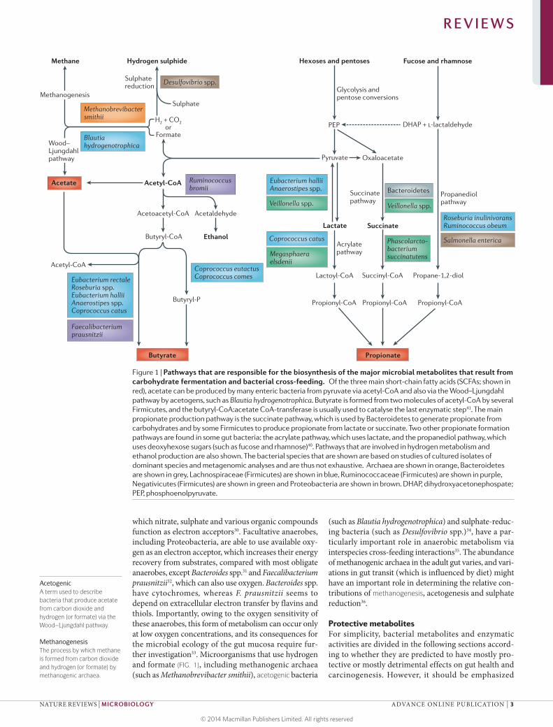
Figure 1 | Pathways that are responsible for the biosynthesis of the major microbial metabolites that result from carbohydrate fermentation and bacterial cross-feeding. Of the three main short-chain fatty acids (SCFAs; shown in red), acetate can be produced by many enteric bacteria from pyruvate via acetyl-CoA and also via the Wood–Ljungdahl pathway by acetogens, such as Blautia hydrogenotrophica. Butyrate is formed from two molecules of acetyl-CoA by several Firmicutes, and the butyryl-CoA:acetate CoA-transferase is usually used to catalyse the last enzymatic step41. The main propionate production pathway is the succinate pathway, which is used by Bacteroidetes to generate propionate from carbohydrates and by some Firmicutes to produce propionate from lactate or succinate. Two other propionate formation pathways are found in some gut bacteria: the acrylate pathway, which uses lactate, and the propanediol pathway, which uses deoxyhexose sugars (such as fucose and rhamnose)40. Pathways that are involved in hydrogen metabolism and ethanol production are also shown. The bacterial species that are shown are based on studies of cultured isolates of dominant species and metagenomic analyses and are thus not exhaustive. Archaea are shown in orange, Bacteroidetes are shown in grey, Lachnospiraceae (Firmicutes) are shown in blue, Ruminococcaceae (Firmicutes) are shown in purple, Negativicutes (Firmicutes) are shown in green and Proteobacteria are shown in brown. DHAP, dihydroxyacetonephospate; PEP, phosphoenolpyruvate.
REVIEWS
NATURE REVIEWS | MICROBIOLOGY ADVANCE ONLINE PUBLICATION | 3
© 2014 Macmillan Publishers Limited. All rights reserved
Wood–Ljungdahl pathway The collection of sequential biochemical reactions that lead to the formation of acetate from carbon dioxide and hydrogen. Several bacteria use this pathway for the generation of energy, and it is also used by certain bacteria and archaea for the assimilation of carbon dioxide into biomass.
Acrylate pathwayThe collection of sequential biochemical reactions that lead to the conversion of lactate to propionate by certain Firmicutes bacteria.
Propanediol pathwayThe collection of sequential biochemical reactions that lead to the conversion of deoxy-sugars (such as rhamnose and fucose) to propionate by certain gut bacteria.
Histone deacetylases(HDACs). Enzymes that remove acetyl groups from histones, which are structural proteins that package DNA into structural units. Deacetylation leads to more condensed chromatin, which alters gene expression.
Colonic regulatory T cells(cTReg cells). A subset of T lymphocytes that are found in the colon and are crucial for the maintenance of immune tolerance.
that the effects of some metabolites probably change as a function of their concentration (for example, the polyamines) or environmental conditions (for example, bacterial β-glucuronidases), as detailed below.
Microbial production of SCFAs. The production of micro-bial SCFAs is influenced by diet (BOX 2). Acetate, which is the most abundant SCFA, is produced by most enteric bacteria as a fermentation product, but it is also formed by acetogenic bacteria, such as B. hydrogenotrophic a, from H2 and CO2 or from formate via the Wood–Ljungdahl pathway37 (FIG. 1). Acetogenic bacteria can produce three molecules of acetate from one molecule of glucose, but non-acetogenic anaerobes, which comprise most of the microbiota, must dispose of reducing equivalents by forming other products in addition to (or instead of) acetate, including succinate, propionate, butyrate, for-mate, d-lactate, l-lactate and ethanol (FIG. 1). The relative synthesis of the different fermentation products varies according to the composition of the microbiota and environmental conditions, including pH, hydrogen par-tial pressure and available substrates29,36,38,39. Recent work on cultured isolates and metagenomics data indicate that propionate and butyrate are mainly produced by the action of different groups of enteric bacteria on carbohy-drates40 (FIG. 1). Propionate is mostly formed via the suc-cinate pathway by Bacteroidetes and by some Firmicutes that belong to the Negativicutes class (such as Phascolarc-tobacterium succinatutens, Dialister spp. and Veillonella spp.). Succinate is a metabolic end-product for some bac-teria under some environmental conditions29, but special-ist succinate utilizers, such as P. succinatutens, convert most of the succinate that is produced into propionate. Two other pathways also contribute to the formation of propionate: the acrylate pathway and the propanediol
pathway (FIG. 1). The acrylate pathway, in which lactate is a substrate, shows a limited distribution, whereas the propanediol pathway, in which deoxyhexose sugars (such as fucose and rhamnose) are substrates, is present in some Firmicutes (including Roseburia inulinivorans and Ruminococcus obeum) and in Proteobacteria40. The pro-portion of propionate that is present in total faecal SCFA correlates with the relative abundance of Bacteroidetes19, which confirms that the succinate pathway is the dominant source of propionate.
Butyrate is produced by some Firmicutes using either the butyryl-CoA:acetate CoA-transferase enzyme or, less commonly, phosphotransbutyrylase and butyrate kinase to catalyse the final steps of the pathway41 (FIG. 1). Species that use the butyryl-CoA:acetate CoA-transferase route include several of the most abundant species in the healthy gut microbiota (including Faecalibacteriu m prausnitzii, Roseburia spp., Eubacterium rectale, Eubacteriu m hallii and Anaerostipes spp.)14,16,42, which are generally net users of acetate43,44, such that the concentration of acetate in the gut lumen is determined by the balance of production, use and mucosal uptake. A subset of Lachnospiraceae, includ-ing E. halli i and Anaerostipes spp., can use lactate and acetate to produce butyrate43. Thus, these organisms may have an important role in stabilizing the microbial eco-system by preventing the accumulation of lactate38. Only a few anaerobes are known to produce both propionate and butyrate, and they do so from different substrates: R. inulinivorans produces butyrate from glucose and pro-duces propionate from fucose, whereas Coprococcus catus produces butyrate from fructose and produces propionate from lactate (via the acrylate pathway)40.
Impact of SCFAs on host cells. Acetate, propionate and butyrate are rapidly adsorbed from the gut lumen, but their subsequent distribution, fate and effects on host cell metabolism differ. Butyrate is preferentially used as an energy source by gut epithelial cells, and its con-centration in the systemic circulation is low. Propionate is mostly metabolized in the liver, and only acetate achieves relatively high concentrations (0.10–0.15 mM) in peripheral blood45.
Intracellular butyrate and propionate (but not ace-tate) inhibit the activity of histone deacetylases (HDACs) in colonocytes and immune cells, which promotes the hyperacetylation of histones, in addition to some tran-scription factors and proteins that are involved in signal transduction46,47. This has multiple consequences for gene expression and cellular differentiation48, includ-ing the downregulation of pro-inflammatory cytokines, such as interleukin-6 (IL-6) and IL-12, in colonic macro-phages49 (FIG. 2). SCFAs exert potentially important anti-inflammatory effects and have been shown to regulate colonic regulatory T cells (cTReg cells) in mice50–56 (FIG. 2). Recent evidence shows that butyrate and propionate induce the differentiation of regulatory T cells that express the transcription factor FOXP3, which have a crucial role in controlling intestinal inflammation50,51,57. It is proposed that butyrate causes increased acetylation of histone H3 in the promoter and enhancer regions of the FOXP3 locus, which results in increased expression
Box 2 | Impact of diet on the production of SCFAs
In general, ingestion of non-digestible carbohydrates (such as fibre and resistant starch) leads to increased colonic fermentation, increased gut transit and stool output and a decrease in the pH of the intestinal lumen. Gut transit has an important impact on substrate fermentation and the absorption of short-chain fatty acids (SCFAs)134,135 and is itself partly regulated by SCFA concentrations136. In overweight human volunteers, low-carbohydrate weight-loss diets result in a decrease in the concentrations of all three faecal SCFAs (that is, acetate, butyrate and propionate; the greatest decrease is in butyrate levels) compared with volunteers on a control diet with normal carbohydrate intake. It was suggested that this effect is mainly caused by a decrease in the abundance of one major group of butyrate-producing Firmicutes in the colon (Eubacterium rectale and Roseburia spp.), which reflects a diet-mediated change in the composition of the gut microbiota20,83. In another recent study in healthy human adults, supplementation with resistant starch and non-starch polysaccharides led to an increase in the faecal concentrations of acetate and butyrate137, and a significant positive correlation was observed between butyrate levels and ammonia excretion. This could be caused by the decrease in colonic pH that results from more active fermentation. An inverse relationship between faecal pH and butyrate formation has been shown in vivo134 and also in an in vitro continuous culture model, which involved the replacement of a microbial community that contained abundant butyrate-producing Firmicutes at pH 5.5 by a Bacteroides-dominated community that produced more propionate and acetate at pH 6.5 (REF. 39). More acidic conditions also favour the excretion of ammonia as a result of protonation, which produces the poorly absorbed ammonium ion137. In conclusion, the actual concentrations of SCFAs in the gut, as well as total SCFA production, are influenced by diet composition and intake.
REVIEWS
4 | ADVANCE ONLINE PUBLICATION www.nature.com/reviews/micro
© 2014 Macmillan Publishers Limited. All rights reserved
AP-1 signalling pathwayA pathway that regulates gene expression via the transcription factor activator protein 1 (AP-1).
of FOXP3 (REF. 51). It is possible that propionate func-tions by the same mechanism, but this requires further study49,51,57. Lactate has also been reported to inhibit HDACs, although the high concentrations that are required are probably not physiological58. Interestingly, the transporter SLC5A8, which is a tumour suppres-sor protein, is involved in the transport of propionat e, butyrate and lactate59,60. Although Slc5a8−/− mice do not show an increase in carcinogen-induced tumour
formation60, alternative transport mechanisms for SCFAs (which include passive diffusion) might occur when their concentrations are sufficiently high59.
Extracellular SCFAs are involved in several potentially important interactions with surface-exposed receptors of host cells (FIG. 2). G protein-coupled receptor 41 (GPR41; also known as FFA3), GPR43 (also known as FFA2) and GPR109A are expressed on various host cells, including colonocytes. GPR43 recognizes all three major SCFAs61, but the affinities for GPR41 are in the order propion-ate > butyrate >> acetate45, whereas GPR109A interacts only with butyrate62. Butyrate-driven signalling interac-tions that involve GPR109A might be involved in the anti-inflammatory action of butyrate by promoting the differentiation of regulatory T cells (TReg cells) and IL-10-producing T cells62, as well as by blocking acti-vation of nuclear factor-κB (NF-κB) and induction of apoptosis by a mechanism that is independent of HDAC inhibition63. Interactions of acetate and propionate with GPR43 are proposed to have an important role in induc-ing their anti-inflammatory effects via the modulation of TReg cells50,64. GPR43 and GPR109A are tumour-sup-pressor genes and might mediate some of the cancer-protective effects of propionate and butyrate that are associated with high fibre intake59. Other important antitumorigenic effects of butyrate include inhibiting proliferation and selectively inducing apoptosis of CRC cells46,48,65,66. The mechanisms that are involved remain to be fully elucidated, but the promotion of apoptosis involves changes in transcriptional regulation owing to HDAC inhibition47 and possibly G protein-coupled receptor interactions, as discussed above46. As a likely consequence of HDAC inhibition, butyrate and, to a lesser extent, propionate have been shown to activate the AP-1 signalling pathway in epithelial cell lines, which has an important role in the control of cell proliferation and apoptosis67. By contrast, a recent study using a mouse line that is genetically susceptible to CRC, suggests that low concentrations of butyrate might promote CRC by stimulating the proliferation of colonic epithelial cells. However, the influence of concurrent changes in the composition of the microbiota could not be excluded68.
The anti-inflammatory effects of SCFAs (FIG. 2) are not only important because of their influence on host cells but it is also possible that they contribute to homeostasis of the gut microbiota. An intriguing hypothesis proposes that the anti-inflammatory effect of high butyrate levels tends to limit immune responses towards the gut micro-biota, whereas low butyrate concentrations trigger a pro-inflammatory state that results in remodelling of the gut microbiota via the suppression of potential pathogens and restoration of butyrate-producing species49.
Biotransformation of phytochemicals and xenobiotics. Many different dietary compounds that are present in fruit, vegetables, cereals, seeds, nuts, spices and beverages have been suggested to protect against different types of cancer5,69. Most studies have investigated polyphenol s, but non-phenolic compounds, such as glucosinolate s (which are derived from brassica vegetables), are also protective70. Phytochemicals have several effects, including antioxidant
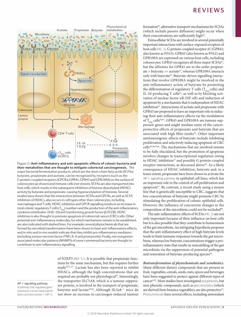
Figure 2 | Anti-inflammatory and anti-apoptotic effects of colonic bacteria and their metabolites that are thought to mitigate colorectal carcinogenesis. The major bacterial fermentation products, which are the short-chain fatty acids (SCFAs) butyrate, propionate and acetate, can be recognized by receptors (such as the
(GPCRs) GPR41, GPR43 and GPR109A) on the surface of colonocytes (as shown) and immune cells (not shown). SCFAs are also transported into host cells, which results in the subsequent inhibition of histone deacetylase (HDAC) activity by butyrate and propionate, causing hyperacetylation of histones. Several studies have shown that the interactions between SCFAs and GPCRs, as well as SCFA inhibition of HDACs, also occurs in cell types other than colonocytes, including
total colonic regulatory T cell (cTReg
) numbers and the production of the anti-inflammatory cytokines interleukin-10 (IL-10) and transforming growth factor-β (TGFβ). HDAC inhibition is also thought to promote apoptosis of colorectal cancer (CRC) cells. Other potential anti-inflammatory molecules, for which mechanisms remain to be established, are also indicated with dashed lines. For example, several phytochemicals that are formed by microbial transformation have been shown to have anti-inflammatory effects, and in vitro and in vivo models indicate that they inhibit pro-inflammatory mediators (including tumour necrosis factor (TNF), IL-6 and prostanoids). Finally, microorganism-associated molecular patterns (MAMPS) of some commensal bacteria are thought to contribute to anti-inflammatory signalling.
Nature Reviews | Microbiology
HDACs
Apoptosis
Signal transduction
Phytochemicalmetabolites
MAMPs
Colonocyte
Acetate Propionate Butyrate
Macrophage
Laminapropria
cTReg cells(FOXP3)
CD4+ T cells
IL-10TGFβ
Hyperacetylation
Histones
TNFIL-6Prostanoids
mediators
GPR41 GPR43 GPR109A
Microbiota
REVIEWS
NATURE REVIEWS | MICROBIOLOGY ADVANCE ONLINE PUBLICATION | 5
© 2014 Macmillan Publishers Limited. All rights reserved
PolyphenolsOrganic compounds that contain phenol moieties; they are produced by plants and are involved in many biological processes, including defence against herbivores.
GlucosinolatesSulphur-containing natural compounds that are present in Brassica vegetables. They are converted to secondary compounds (including isothiocyanates) that are thought to have health-promoting effects.
PhytochemicalsNatural compounds that are produced by plants and belong to a range of biochemical classes.
XenobioticA chemical compound that is not normally found in an organism, such as a drug or environmental pollutant.
GlycosidesChemical compounds that are bound to a sugar residue via a glycosidic linkage.
GlucuronidesChemical compounds that are bound to a glucuronic acid residue via a glycosidic linkage.
AglyconesThe compounds that are produced following the removal of the sugar or sugar acid (such as glucuronic acid) residue from a glycoside or glucuronide.
NitrosationThe process by which compounds are converted to nitroso group (-N=O)- containing derivatives.
effects, modulation of xenobiotic detoxification path-ways and modulation of cell proliferation, apoptosis and inflammation69 (FIG. 2). Antioxidants neutralize reactive oxygen species (ROS), which are by-products of energy metabolism and can damage cellular struc-tures, including DNA, which results in the generation of mutations that predispose the cell to the develop-ment of cancer. It is currently unclear whether direct antioxidant effects have an important role in cancer prevention in vivo, as it has been questioned whether sufficient systemic concentrations can be achieved71. Furthermore, some studies have reported an increase in the occurrence of certain cancers in response to supplementation with some antioxidants72.
Dietary phytochemicals are usually present as glycoside s or are bound to fibre, and only limited uptake takes place in the small intestine. Up to 95% of dietary phytochemicals reach the large intestine and are released and transformed to other metabolites by the gut micro-biota73–75. The conversions that occur include hydro-genation, dehydroxylation and demethylation, which can change metabolite bioactivity. Several phenolic metabolites that are formed by microbial transformation have been shown to inhibit pro-inflammatory media-tors (including tumour necrosis factor (TNF), NF-κB and prostanoids)75–77 (FIG. 2). Many phenolic compounds have been shown to exert antimicrobial effects that can alter the composition of the gut microbiota, as differ-ent bacterial species show varying levels of sensitiv-ity78. The resultant changes in microbial composition could have knock-on effects — for example, via changes in SCFA profiles or the suppression of pathogenic microorganisms.
After absorption in the gut, phytochemicals are con-jugated to methyl, sulphate and glucuronic acid groups in the liver, which facilitates their excretion into the gut via bile. In the gut, bacterial β-glucuronidases convert glucuronides back to the respective aglycones, which can then be re-absorbed. Thus, bacterial β-glucuronidase activity and enterohepatic circulation extend the reten-tion time of phytochemicals in the body. However, microbial β-glucuronidase activity also interferes with the detoxification and excretion of toxic xenobiotics, such as drugs and environmental pollutants, and it has been reported that high β-glucuronidase activity is associated with an increased risk of cancer79. Further-more, protein-rich diets deliver toxic compounds, such as heterocyclic amines, which are also subject to glucu-ronidation80 and protein-rich diets are associated with high faecal β-glucuronidase activity81. Gut microbial β-glucuronidase activity can also interfere with cancer treatment; for example, the toxicity of irinotecan (which is a drug that is commonly used to treat colon cancer) was alleviated in a mouse model by co-administration of a β-glucuronidase inhibitor that specifically targets bacterial β-glucuronidases82. A recent study used phy-logenetic analysis of β-glucuronidase genes to identify the bacterial species that express β-glucuronidase, which revealed that a major β-glucuronidase gene (gus, also known as uidA, which was initially characterized in Escherichia coli) seems to be present in several species
that belong to the Firmicutes81. The functional role of a second β-glucuronidase gene, which is more prevalent in Bacteroidetes, remains to be established81.
Detrimental metabolites
Products of protein fermentation. High protein intake results in an increase in the fermentation of diet-derived protein in the colon, as indicated by the increase in amino acid-derived products, such as branched-chain fatty acids and phenylacetic acid27,83,84. A subset of bacteria, including several Bacteroides spp. and some Firmicutes, ferment aromatic amino acids to produce potentially bioactive products, including phenylacetic acid, phenols, indoles and p-cresol85. Some nitrogenous products, particularly N-nitroso compounds (NOCs), have the potential to promote cancer and exert carcino-genic effects via DNA alkylation, which can cause muta-tions4 (FIG. 3). The intake of preformed (that is, dietary) NOCs is positively correlated with CRC in European populations86, but these compounds can also be formed endogenously via acid-driven nitrosation in the stom-ach and by the nitrosation of amines that are derived from the microbial fermentation of protein in the large intestine87. An increase in faecal NOCs has been found in individuals on high-protein diets in controlled die-tary intervention studies83. Nitroreductases and nitrate reductases that are encoded by Proteobacteria probably contribute to nitrosation reactions. Ammonia, which is another product of protein fermentation, is also a poten-tial carcinogenic agent at relatively low concentrations, as shown by the increase in mucosal damage and colonic adenocarcinoma in a rat model84,87.
Polyamines are involved in a range of essential physi-ological functions, such as the maintenance of the struc-tural integrity of membranes and nucleic acids, gene regulation and translation88,89. The major polyamines putrescine, spermidine and spermine are produced from arginine in host tissues, but polyamine synthesis also occurs in gut bacteria90. High levels of polyamines are toxic and are associated with various diseases, including cancer, and oxidative stress that results from polyamine catabolism is thought to be the underlying mechanism of toxicity89. In addition to contributing directly to the polyamine pool by synthesizing these compounds, cer-tain gut bacteria (such as enterotoxigenic Bacteroides fra-gilis) upregulate polyamine production by host cells89. Indeed, several pathogens, including Shigella flexneri, Streptococcus pneumoniae, Salmonella enterica subsp. enterica serovar Typhimurium and Helicobacter pylori, exploit polyamines to increase their virulence88.
There is evidence from both humans and animal models that dietary supplementation with non-digestibl e carbohydrates can decrease protein fermentation in the large intestine, which coincides with a decrease in the genotoxicity of faecal water84. Diets that include resistant starch lead to a reduction in DNA damage and tumour formation in a rat model66 and may attenuate the detrimental effects of high levels of dietary pro-tein91. However, the effects of a high-protein diet on the risk of CRC in humans remains a complex question, as it is strongly influenced by the specific source of protein
REVIEWS
6 | ADVANCE ONLINE PUBLICATION www.nature.com/reviews/micro
© 2014 Macmillan Publishers Limited. All rights reserved

Secondary bile acidsMetabolites that are produced by enteric bacteria from primary bile acids.
Primary bile acidsSteroid acids that are synthesized in the liver and secreted into the gut to promote fat digestion.
and changes in the intake of other dietary components92. Thus, the contribution of red and processed meats to cancer risk may be partly due to factors other than pro-tein metabolites — for example, haem, which promotes nitrosation86,93.
Hydrogen sulphide. Hydrogen sulphide is produced in the gut via the reduction of diet-derived sulphate and the metabolism of other compounds, including sulphur amino acids94 and taurine (see below). Spe-cialist sulphate-reducing bacteria that are related to Desulfovibri o spp. are detectable in low numbers in most individuals and are able to use lactate as a co-substrate for growth and sulphide formation95. Sulphide is toxic to colonocytes and inhibits butyrate oxidation, which results in the breakdown of the colonocyte barrier96. It is also genotoxic to non-transformed human cell lines
at concentrations (0.25–2 mM) that are detectable in the colonic lumen97, and the mechanism of DNA dam-age is proposed to involve ROS98 (FIG. 3). Higher stool sulphide levels have been detected in patients with CRC compared with healthy controls, but an increase in Desulfovibri o spp. has not been found in faecal samples from patients with CRC in the few studies that have been conducted so far35. Thus, hydrogen sulphide levels might be primarily driven by changes in bacterial activity rather than by bacterial abundance.
Bile acid metabolism. The interplay between diet, bile acids and the gut microbiota is complex. High-fat diets, which are positively correlated with the incidence of CRC, lead to an increase in bile secretion, and increased faecal bile acid concentrations have been reported in patients with CRC99–101. In addition, higher fat intake correlates with higher faecal concentrations of second-ary bile acids in African Americans compared with rural Africans27, and recent evidence suggests that the second-ary bile acid deoxycholic acid promotes liver cancer102. Bile acids exert strong antimicrobial activities, as they damage bacterial cell membranes owing to their amphi-pathic properties and are therefore likely to modify the composition of the gut microbiota. Rats that are fed a diet that is supplemented with deoxycholic acid show a decrease in the production of SCFAs and major changes in the composition of the microbiota, with a relative increase in Gammaproteobacteria and certain Firmi-cutes at the expense of Bacteroidetes, which is similar to what is observed in response to high-fat diets in mice103.
The primary bile acids cholic acid and chenodeoxy-cholic acid are produced in the liver from cholesterol, are conjugated to glycine or taurine (which render the bile acids more hydrophilic and facilitate their action as emulsifiers) and are excreted into the duodenum to facilitate fat digestion. Limited biotransformation of pri-mary bile acids by the gut microbiota occurs in the small intestine before they are re-absorbed in the distal ileum for enterohepatic circulation104. However, the fraction of bile acid that escapes re-absorption in the small intestine (approximately 5% of the total pool) undergoes exten-sive transformation by the microbiota in the large intes-tine104. Bile salt hydrolases (which are found in all the major bacterial divisions and the methanogenic archaea) cleave glycine and taurine residues from the primary bile acids104,105, which converts them into several different secondary bile acids by dehydrogenation and dehydrox-ylation reactions. The main secondary bile acids that are produced are deoxycholic acid and lithocholic acid, both of which are produced by 7α-dehydroxylation104. The genes that are responsible for this conversion (which are encoded in the bai operon) have been investigated in detail in Clostridium scindens106, but they seem to be less widespread than bile salt hydrolases in the human gut microbiota.
Bile acids have been implicated in carcinogenesis in different regions of the intestinal tract and associated organs, owing to the generation of ROS and reactive nitrogen species (RNS), both of which cause DNA dam-age100. In addition, animal studies have shown that the
Figure 3 | Pro-inflammatory and DNA-damaging effects of colonic bacteria and their metabolites that are thought to contribute to colorectal carcinogenesis. Several bacterial metabolites, including hydrogen sulphide, secondary bile acids, polyamines and reactive oxygen species (ROS) have the potential to cause direct DNA damage or to provoke inflammation (via interleukin 6 (IL-6) and tumour necrosis factor (TNF) production), which thus promotes carcinogenesis. N-nitroso compounds (NOCs) can promote cancer by generating mutations owing to DNA alkylation. Pathogenic bacteria, in particular, also exert pro-inflammatory effects via the recognition of microor-ganism-associated molecular patterns (MAMPs) by Toll-like receptors (TLRs), which leads to detection by dendritic cells and the activation of T helper 17 (T
H H17 cells
promote the expression of the pro-inflammatory mediator IL-23 and block expression of the anti-inflammatory mediator IL-10. Tumour-associated loss of barrier function, which is mediated by MAMPs, can also result in increased bacterial translocation, and this further drives pro-inflammatory pathways, thereby increasing tumorigenesis.
Nature Reviews | Microbiology
NOCs
DNA alkylation
MAMPs
Pathogen Microbiota
TH17 cell
MAMPdetection
Dendritic cell
Bacterialtranslocation
IL-23
IL-10
IL-6TNF
Colonocyte
TLR
Loss of barrierfunction
ToxinsH2S
Secondary bile acidsROS
Bacterial
REVIEWS
NATURE REVIEWS | MICROBIOLOGY ADVANCE ONLINE PUBLICATION | 7
© 2014 Macmillan Publishers Limited. All rights reserved

Pattern recognition receptors(PRRs). Innate immune proteins that are essential for recognizing and responding to microorganisms. The most common PRRs include Toll-like receptors, NOD-like receptors, RIGI-like receptors and DNA receptors (that is, cytosolic sensors for DNA).
Microorganism-associated molecular patterns(MAMPS). Conserved microbial components, such as lipopolysaccharide, peptidoglycan, flagellin and nucleic acids, that are detected by the host innate immune system via pattern recognition receptors.
ColitisInflammation of the colon or large intestine.
administration of bile acids results in a higher incidence of tumours in the gut100. The molecular mechanisms that mediate the cytotoxic effects of bile acids are com-plex99. Secondary bile acids are more hydrophobic and thus more potent at disrupting cell membranes, which is likely to lead to the generation of ROS via the activa-tion of membrane-associated proteins such as NAD(P)H oxidases and phospholipase A2, but other mechanisms may also be involved107. For example, bile acids function as hormones that interact with nuclear receptors and acti-vate cellular signalling pathways that promote apoptosis; however, constant exposure to high levels of bile acids can lead to resistance to apoptosis99. Furthermore, some bile acids seem to counteract the cytotoxic effects of others; for example, ursodeoxycholic acid, which is produced by Ruminococcus gnavus, seems to inhibit the production of ROS and to protect cells from the cytotoxic effects of deoxycholic acid99,108. Taurine conjugation increases in individuals who are on meat-rich diets, and the sulphonic acid moiety of taurine is reduced to hydrogen sulphide after deconjugation of the bile acid104. Diets that promote high levels of taurine conjugation lead to a bloom in the sulphite-reducing bacterium Bilophila wadsworthia, which is associated with pathological gut conditions17,109.
Ethanol. Excessive consumption of ethanol is consid-ered to be an important risk factor for several cancers6, and microbial metabolism may contribute to its toxicity, particularly in the upper gastrointestinal tract. Ethanol is produced by many anaerobic bacteria that inhabit the colon when they are grown in pure culture, but the level of endogenous ethanol production by the colonic microbiota in vivo is unknown. Although ethanol itself is not regarded as a carcinogen, its immediate oxidation product acetaldehyde is highly toxic and carcinogenic, and causes effects that range from DNA damage to the degradation of the vitamin folate110. Interestingly, studies of the oral microbiota have shown that microorganisms contribute to the production of acetaldehyde from etha-nol, which suggests that the gut microbiota might also contribute to this process110,111.
The microbiota and inflammation
It has become increasingly clear that the microbiota has a major influence on immune responses, and chronic inflammation is a well-established risk factor for CRC. As the colonic mucosa is constantly exposed to the gut microbiota and its metabolites (FIGS 2,3), bacterial stimulation of immune responses has the potential to cause continuous low-grade inflammation. The tumour microenvironment contains several different immune cell types, including tissue-associated macrophages (TAMs) and other innate immune cells, as well as T cells and B cells, which communicate with each other and the other cells in the tumour microenvironment via direct contact or via cytokine and/or chemokine signalling to control tumour growth112. TAMs primarily promote tumour growth, and high numbers of TAMs gener-ally correlate with cancer progression112. After TAMs, T cells are the most numerically abundant immune cells in the tumour microenvironment and can exert both
tumour-promoting and tumour-suppressive effects. Increased numbers of CD4+ T helper 1 (TH1) cells and CD8+ cytotoxic T cells are associated with the direct lysis of cancer cells and the production of cytotoxic cytokines113 that limit the progression of CRC. How-ever, other T cell subsets, such as interferon-γ (IFNγ)-producing TH1 cells, promote tumorigenesis via cytokine production and cytotoxic mechanisms112. Interestingly, inflammation in the absence of the gut microbiota or microbial products is insufficient to induce CRC114. There is also clear evidence that the gut microbiota of mice influences adenoma formation. Adenomatous polyposis coli (ApcMin) mice that have a mutation in one copy of the tumour suppressor gene Apc spontaneously form many benign adenomas in the small intestine, and the number of colon tumours that are formed in ApcMin mice is greater than that in wild-type C57BL/6 mice. However, germ-free ApcMin mice exhibit a twofold reduc-tion in small intestinal adenomas compared with ApcMin mice that have a conventional microbiota115. In addi-tion, disruption of microbial sensing by innate immune receptor signalling also results in reduced tumorigen-esis116,117. Host recognition of the microbiota occurs via various pattern recognition receptors (PRRs; such as Toll-like receptors (TLRs)) that control the inflammatory response to microorganism-associated molecular patterns (MAMPs; such as lipopolysaccharide (LPS), flagellin and nucleic acids)9 (FIG. 3). PRRs have a key role in maintain-ing mucosal homeostasis and controlling inflammation in the colonic environment. In particular, alterations in signalling of TLR4, which is the major receptor for LPS, have been linked to the progression of CRC118.
The role of specific pathogens in CRC. In the previous sections, we have described the role of the microbiota (as an integrated community) and its metabolites in the aetiology of CRC. That said, several bacterial patho-gens seem to be directly and specifically involved in promoting CRC; for example, enterotoxigenic B. fra-gilis and adherent-invasive E. coli strain NC101 have been shown to promote CAC in genetically susceptible mice114,119,120. The pro-inflammatory effects of entero-toxigenic B. fragilis result in the induction of spermine oxidase in colonocytes, which leads to the production of ROS and consequent DNA damage121. However, most cases of CRC do not arise from a background of colitis, which suggests that other microbial species are associ-ated with the pathogenesis of CRC. Recent studies indi-cate that Fusobacteriu m spp. and Campylobacte r spp. are over-represented in CRC tissue10,122,123; thus, these genera may form part of a metagenomic CRC signa-ture123. Invasive strains of Fusobacterium nucleatum accelerate the onset of colonic tumours and drive the transition to a pro-inflammatory microenvironment that is conducive to colorectal tumorigenesis10. Bind-ing of the F. nucleatum adhesin FadA to E-cadherin induces its tumour-suppressor activity and activates β-catenin, which further promotes the growth of tumour cells124. However, despite these associations, it remains difficult to establish whether there is a causal link to specific pathogens (BOX 3). However, it is clear that the
REVIEWS
8 | ADVANCE ONLINE PUBLICATION www.nature.com/reviews/micro
© 2014 Macmillan Publishers Limited. All rights reserved
mechanisms by which these individual pathogens pro-mote carcino genesis, including the induction of inflam-matory cascades, the generation of ROS, DNA damage and the disruption of DNA repair processes are similar to the mechanisms that are associated with the activities of microbial metabolites13,102.
Several lines of evidence indicate that inflamma-tion results in the enrichment of certain bacterial groups that have pro-carcinogenic traits, including Fusobacterium spp., Streptococcus gallolyticus subsp. gallolyticus (formerly known as Streptococcus bovis bio-type 1), enterotoxic B. fragilis and adherent-invasive
E. coli112,114,119,125,126. Inflammation creates an opportu-nity for certain bacteria, such as E. coli, to adhere to the colonic mucosa by decreasing the production of protective mucins and antimicrobial peptides127,128. The reduced barrier function enables bacteria to more read-ily interact with the epithelium, resulting in increased delivery of mutagenic metabolites, including colibactin, which is a putative hybrid peptide–polyketide that is produced by the Enterobacteriaceae and causes DNA damage114,129. Colibactin-producing E. coli strains can induce DNA double-strand breaks in the host cell and thereby activate DNA damage signalling cascades, which leads to chronic mitotic and chromosomal aberrations as well as an increased frequency of gene mutation and anchorage-independent growth125. Increased levels of Proteobacteria, particularly the Enterobacteriaceae (including E. coli) are found in the gut microbiota of patients with IBD, which is a known risk factor for CRC130. Early genetic events in the pathogenesis of CRC, such as the activation of β-catenin and mutation of the APC gene, seem to result in a loss of barrier function in the colonic epithelium, which leads to the translocation of microbial products into the tumour microenviron-ment131.This process results in the downstream produc-tion of tumour-promoting cytokines via the activation of IL-23-producing myeloid cells, which promote tumour growth131. Although defective barrier func-tion enables the translocation of microbial products, it also enables the colonization of invasive-adherent bacteria at neoplastic sites.
Outlook
The studies that are discussed in this Review highlight that progression to CRC is influenced not only by the presence of specific pathogens but also by the metabolic output of the entire microbiota (FIG. 4). In addition to high microbial diversity and low pathogen abundance, microbial SCFAs have a major role in maintaining intestinal homeostasis. They suppress the growth of Gram-negative pathogens, function as energy sources and anti-inflammatory agents and promote apoptosis of cancer cells. Thus, prominent butyrate-producing species are not only indicators of a diverse, healthy
Box 3 | Microbiota changes associated with CRC
There has been a recent surge in studies that compare the composition of the microbial community in patients with colorectal cancer (CRC) with that of healthy subjects10,122,123,138,139 in an effort to determine whether changes in the gut microbiota are a cause of the disease. However, as CRC develops over many years, it is a challenge to determine whether the associated changes in the gut microbiota are a consequence of diet alterations or physiology or whether they are causative. In one article, a ‘driver–passenger’ model for CRC was proposed in an attempt to distinguish between causative organisms and those that respond to disease progression11. The analysis of faecal samples from patients with CRC using 16S ribosomal RNA gene sequencing has shown that Bacteroides fragilis and several enterobacterial operational taxonomic units (OTUs) are enriched compared with healthy controls, whereas levels of five OTUs that correspond to butyrate-producing Lachnospiraceae were reduced140. Changes also occur in the tumour-associated microbiota, in which an increase in Fusobacterium spp. seems to be consistent between studies11,122. Deep metatranscriptomic sequencing has more recently shown that Leptotrichia spp. and Campylobacter spp. co-occur with Fusobacterium spp.123. Clear differences in the composition of the gut microbiota have been reported in mice following tumour induction using carcinogenic agents: compared with untreated mice, Bacteroides spp., Akkermansia spp. and Odoribacter spp. increase, whereas Prevotella spp. and Porphyromonas spp. decrease141. The increasing list of potentially carcinogenic bacteria provides support for the hypothesis that tumorigenesis is driven by mechanisms and/or pathways that are common to many bacterial groups rather than a single organism.
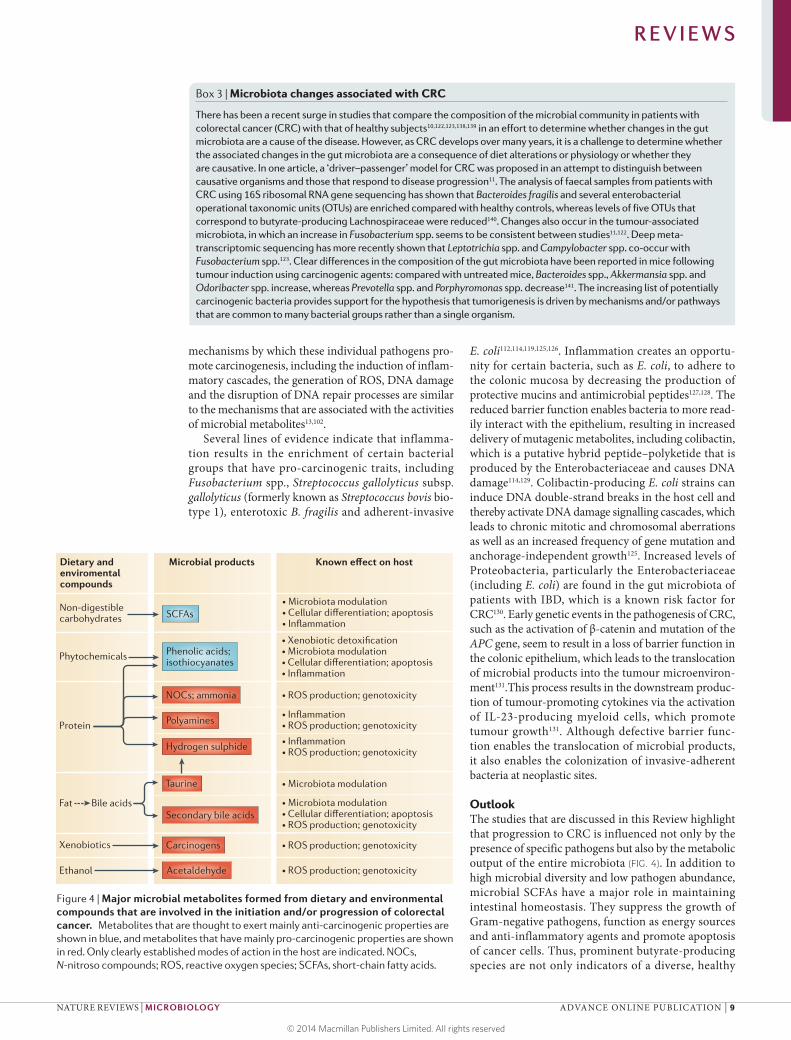
Figure 4 | Major microbial metabolites formed from dietary and environmental compounds that are involved in the initiation and/or progression of colorectal cancer. Metabolites that are thought to exert mainly anti-carcinogenic properties are shown in blue, and metabolites that have mainly pro-carcinogenic properties are shown in red. Only clearly established modes of action in the host are indicated. NOCs, N-nitroso compounds; ROS, reactive oxygen species; SCFAs, short-chain fatty acids.
Nature Reviews | Microbiology
Dietary and enviromental compounds
Microbial products Known effect on host
Non-digestible carbohydrates
Phytochemicals
Protein
Bile acidsFat
Xenobiotics
Ethanol
SCFAs Microbiota modulation
Microbiota modulation
Microbiota modulation
Microbiota modulation
Polyamines
Taurine
Secondary bile acids
Carcinogens
Acetaldehyde
isothiocyanates
REVIEWS
NATURE REVIEWS | MICROBIOLOGY ADVANCE ONLINE PUBLICATION | 9
© 2014 Macmillan Publishers Limited. All rights reserved
microbiota — as suggested by recent studies — but also seem to be actively involved in maintaining a stable and healthy gut community. By contrast, dysbiosis is characterized by a reduction in microbial diversity and an increase in pro-inflammatory, pathogenic species, which can be caused by an unhealthy diet, antimicrobial therapy or genetic predisposition (which is exemplified by Crohn’s disease).
There is increasing evidence that diets that are low in fibre and high in fat and sugar result in a less diverse gut microbiota, which, combined with the detrimental effects of these diets that are mediated by dietary com-ponents and microbial metabolites, is likely to increase the risk of CRC. Metabolomics is yielding new infor-mation on microbial metabolite profiles and respon-siveness to controlled dietary manipulations in patient groups that show differing risks of CRC. However, we need a better fundamental understanding of, and an ability to predict, the effects of diet on the microbial
metabolome of individuals. Analysis of cultured isolates and metagenomic data are helping to define which bac-teria are responsible for different metabolic activities, but the use of defined consortia to investigate metabolite cross-feeding and to reconstruct pathways that involve multiple organisms is mostly unexplored. Ultimately, such work can explain the effects of inter-individual and diet-induced variation on the composition of the microbiota and on metabolite profiles. However, it is also crucial that we address the quantitative contribu-tion of different metabolites, not only by considering metabolite fluxes but also by taking account of gut tran-sit, absorption and tissue distribution within the body. Therefore, theoretical modelling, together with studies of microbial ecology and physiology in the gut, has a vital contribution to make in the future in order to inte-grate the complex interactions that occur both within the microbial community and between the microbiota and the host.
1. Jemal, A. et al. Global cancer statistics. CA Cancer J. Clin. 61, 69–90 (2011).
2. Fearon, E. R. & Vogelstein, B. A genetic model for colorectal tumorigenesis. Cell 61, 759–767 (1990).
3. Rabeneck, L., Davila, J. A. & El-Serag, H. B. Is there a true ‘shift’ to the right colon in the incidence of colorectal cancer? Am. J. Gastroenterol. 98, 1400–1409 (2003).
4. Gill, C. I. R. & Rowland, I. R. Diet and cancer: assessing the risk. Br. J. Nutr. 88, S73–S87 (2002).
5. Burn, J. et al. Long-term effect of aspirin on cancer risk in carriers of hereditary colorectal cancer: an analysis from the CAPP2 randomised controlled trial. Lancet 378, 2081–2087 (2011).
6. World Cancer Research Fund and American Institute for Cancer Research. Food, Nutrition, Physical Activity, and the Prevention of Cancer: A Global Perspective. (AICR, 2007).
7. Jess, T., Gamborg, M., Matzen, P., Munkholm, P. & Sørensen, T. I. A. Increased risk of intestinal cancer in Crohn’s disease: a meta-analysis of population-based cohort studies. Am. J. Gastroenterol. 100, 2724–2729 (2005).
8. Danese, S., Malesci, A. & Vetrano, S. Colitis-associated cancer: the dark side of inflammatory bowel disease. Gut 60, 1609–1610 (2011).
9. Schwabe, R. F. & Jobin, C. The microbiome and cancer. Nature Rev. Cancer 13, 800–812 (2013).
10. Kostic, A. D. et al. Fusobacterium nucleatum potentiates intestinal tumorigenesis and modulates the tumor–immune microenvironment. Cell Host Microbe 14, 207–215 (2013).
11. Tjalsma, H., Boleij, A., Marchesi, J. R. & Dutilh, B. E. A bacterial driver–passenger model for colorectal cancer: beyond the usual suspects. Nature Rev. Microbiol. 10, 575–582 (2012).
12. Elinav, E. et al. Inflammation-induced cancer: crosstalk between tumours, immune cells and microorganisms. Nature Rev. Cancer 13, 759–771 (2013).
13. Sears, C. L. & Garrett, W. S. Microbes, microbiota and colon cancer. Cell Host Microbe 15, 317–328 (2014).
14. Flint, H. J., Scott, K. P., Louis, P. & Duncan, S. H. The role of the gut microbiota in nutrition and health. Nature Rev. Gastroenterol. Hepatol. 9, 577–589 (2012).
15. Eckburg, P. B. et al. Microbiology: diversity of the human intestinal microbial flora. Science 308, 1635–1638 (2005).
16. Walker, A. W. et al. Dominant and diet-responsive groups of bacteria within the human colonic microbiota. ISME J. 5, 220–230 (2011).This study is a carefully controlled human dietary trial that demonstrates rapid and reversible changes in the relative abundance of specific bacterial groups in response to carbohydrate intake.
17. David, L. A. et al. Diet rapidly and reproducibly alters the human gut microbiome. Nature 505, 559–563 (2014).
18. Flint, H. J., Scott, K. P., Duncan, S. H., Louis, P. & Forano, E. Microbial degradation of complex
carbohydrates in the gut. Gut Microbes 3, 289–306 (2012).
19. Salonen, A. et al. Impact of diet and individual variation on intestinal microbiota composition and fermentation products in obese men. ISME J. http://dx.doi.org/10.1038/ismej.2014.63 (2014).
20. Duncan, S. H. et al. Reduced dietary intake of carbohydrates by obese subjects results in decreased concentrations of butyrate and butyrate-producing bacteria in feces. Appl. Environ. Microbiol. 73, 1073–1078 (2007).
21. Wu, G. D. et al. Linking long-term dietary patterns with gut microbial enterotypes. Science 334, 105–108 (2011).
22. De Filippo, C. et al. Impact of diet in shaping gut microbiota revealed by a comparative study in children from Europe and rural Africa. Proc. Natl Acad. Sci. USA 107, 14691–14696 (2010).
23. Le Chatelier, E. et al. Richness of human gut microbiome correlates with metabolic markers. Nature 500, 541–546 (2013).
24. Cotillard, A. et al. Dietary intervention impact on gut microbial gene richness. Nature 500, 585–588 (2013).
25. Aune, D. et al. Dietary fibre, whole grains, and risk of colorectal cancer: systematic review and dose-response meta-analysis of prospective studies. BMJ 343, 1082 (2011).
26. Chen, H. -M. et al. Decreased dietary fiber intake and structural alteration of gut microbiota in patients with advanced colorectal adenoma. Am. J. Clin. Nutr. 97, 1044–1052 (2013).
27. Ou, J. et al. Diet, microbiota, and microbial metabolites in colon cancer risk in rural Africans and African Americans. Am. J. Clin. Nutr. 98, 111–120 (2013).
28. Marcobal, A. et al. A metabolomic view of how the human gut microbiota impacts the host metabolome using humanized and gnotobiotic mice. ISME J. 7, 1933–1943 (2013).
29. Macfarlane, G. T. & Gibson, G. R. in Gastrointestinal Microbiology Vol. 1 (eds Mackie, R. I. & White, B. A.) 269–318 (Chapman and Hall, 1997).
30. Sieber, J. R., McInerney, M. J. & Gunsalus, R. P. Genomic insights into syntrophy: the paradigm for anaerobic metabolic cooperation. Annu. Rev. Microbiol. 66, 429–452 (2012).
31. Baughn, A. D. & Malamy, M. H. The strict anaerobe Bacteroides fragilis grows in and benefits from nanomolar concentrations of oxygen. Nature 427, 441–444 (2004).
32. Khan, M. T. et al. The gut anaerobe Faecalibacterium prausnitzii uses an extracellular electron shuttle to grow at oxic–anoxic interphases. ISME J. 6, 1578–1585 (2012).
33. Circu, M. L. & Aw, T. Y. Intestinal redox biology and oxidative stress. Semin. Cell Dev. Biol. 23, 729–737 (2012).
34. Nava, G. M., Carbonero, F., Croix, J. A., Greenberg, E. & Gaskins, H. R. Abundance and diversity of mucosa-
associated hydrogenotrophic microbes in the healthy human colon. ISME J. 6, 57–70 (2012).
35. Carbonero, F., Benefiel, A. C. & Gaskins, H. R. Contributions of the microbial hydrogen economy to colonic homeostasis. Nature Rev. Gastroenterol. Hepatol. 9, 504–518 (2012).
36. Lewis, S. & Cochrane, S. Alteration of sulfate and hydrogen metabolism in the human colon by changing intestinal transit rate. Am. J. Gastroenterol. 102, 624–633 (2007).
37. Miller, T. L. & Wolin, M. J. Pathways of acetate, propionate, and butyrate formation by the human fecal microbial flora. Appl. Environ. Microbiol. 62, 1589–1592 (1996).
38. Belenguer, A. et al. Impact of pH on lactate formation and utilization by human fecal microbial communities. Appl. Environ. Microbiol. 73, 6526–6533 (2007).
39. Walker, A. W., Duncan, S. H., McWilliam Leitch, E. C., Child, M. W. & Flint, H. J. pH and peptide supply can radically alter bacterial populations and short-chain fatty acid ratios within microbial communities from the human colon. Appl. Environ. Microbiol. 71, 3692–3700 (2005).
40. Reichardt, N. et al. Phylogenetic distribution of three pathways for propionate production within the human gut microbiota. ISME J. 8, 1323–1335 (2014).
41. Louis, P. et al. Restricted distribution of the butyrate kinase pathway among butyrate-producing bacteria from the human colon. J. Bacteriol. 186, 2099–2106 (2004).
42. Louis, P., Young, P., Holtrop, G. & Flint, H. J. Diversity of human colonic butyrate-producing bacteria revealed by analysis of the butyryl-CoA:acetate CoA-transferase gene. Environ. Microbiol. 12, 304–314 (2010).
43. Louis, P. & Flint, H. J. Diversity, metabolism and microbial ecology of butyrate-producing bacteria from the human large intestine. FEMS Microbiol. Lett. 294, 1–8 (2009).
44. Barcenilla, A. et al. Phylogenetic relationships of butyrate-producing bacteria from the human gut. Appl. Environ. Microbiol. 66, 1654–1661 (2000).
45. Sleeth, M. L., Thompson, E. L., Ford, H. E., Zac-Varghese, S. E. K. & Frost, G. Free fatty acid receptor 2 and nutrient sensing: a proposed role for fibre, fermentable carbohydrates and short-chain fatty acids in appetite regulation. Nutr. Res. Rev. 23, 135–145 (2010).
46. Fung, K. Y. C., Cosgrove, L., Lockett, T., Head, R. & Topping, D. L. A review of the potential mechanisms for the lowering of colorectal oncogenesis by butyrate. Br. J. Nutr. 108, 820–831 (2012).
47. Wilson, A. J. et al. Apoptotic sensitivity of colon cancer cells to histone deacetylase inhibitors is mediated by an Sp1/Sp3-activated transcriptional program involving immediate-early gene induction. Cancer Res. 70, 609–620 (2010).
48. Hamer, H. M. et al. Review article: the role of butyrate on colonic function. Aliment. Pharmacol. Ther. 27, 104–119 (2008).
REVIEWS
10 | ADVANCE ONLINE PUBLICATION www.nature.com/reviews/micro
© 2014 Macmillan Publishers Limited. All rights reserved
49. Chang, P. V., Hao, L., Offermanns, S. & Medzhitov, R. The microbial metabolite butyrate regulates intestinal macrophage function via histone deacetylase inhibition. Proc. Natl Acad. Sci. USA 111, 2247–2252 (2014).This study provides evidence that the inhibition of HDACs by butyrate is responsible for anti-inflammatory effects in colonic macrophages.
50. Smith, P. M. et al. The microbial metabolites, short-chain fatty acids, regulate colonic T reg cell homeostasis. Science 341, 569–573 (2013).This study shows that propionate has an anti-inflammatory effect via the modulation of cTReg cells.
51. Furusawa, Y. et al. Commensal microbe-derived butyrate induces the differentiation of colonic regulatory T cells. Nature 504, 446–450 (2013).
52. Atarashi, K. et al. Induction of colonic regulatory T cells by indigenous Clostridium species. Science 331, 337–341 (2011).
53. Geuking, M. et al. Intestinal bacterial colonization induces mutualistic regulatory T cell responses. Immunity 34, 794–806 (2011).
54. Round, J. L. & Mazmanian, S. K. Inducible Foxp3+ regulatory T-cell development by a commensal bacterium of the intestinal microbiota. Proc. Natl Acad. Sci. USA 107, 12204–12209 (2010).
55. O’Mahony, C. et al. Commensal-induced regulatory T cells mediate protection against pathogen-stimulated NF-κB activation. PLoS Pathog. 4, e1000112 (2008).
56. Chung, H. et al. Gut immune maturation depends on colonization with a host-specific microbiota. Cell 149, 1578–1593 (2012).
57. Arpaia, N. et al. Metabolites produced by commensal bacteria promote peripheral regulatory T-cell generation. Nature 504, 451–455 (2013).This study shows that propionate and butyrate, but not acetate, promote the generation of anti-inflammatory TReg cells via the inhibition of HDACs.
58. Latham, T. et al. Lactate, a product of glycolytic metabolism, inhibits histone deacetylase activity and promotes changes in gene expression. Nucleic Acids Res. 40, 4794–4803 (2012).
59. Ganapathy, V., Thangaraju, M., Prasad, P. D., Martin, P. M. & Singh, N. Transporters and receptors for short-chain fatty acids as the molecular link between colonic bacteria and the host. Curr. Opin. Pharmacol. 13, 869–874 (2013).
60. Frank, H. et al. Lactaturia and loss of sodium-dependent lactate uptake in the colon of SLC5A8-deficient mice. J. Biol. Chem. 283, 24729–24737 (2008).
61. Brown, A. J. et al. The orphan G protein-coupled receptors GPR41 and GPR43 are activated by propionate and other short chain carboxylic acids. J. Biol. Chem. 278, 11312–11319 (2003).
62. Singh, N. et al. Activation of Gpr109a, receptor for niacin and the commensal metabolite butyrate, suppresses colonic inflammation and carcinogenesis. Immunity 40, 128–139 (2014).
63. Thangaraju, M. et al. GPFM 09A is a G-protein-coupled receptor for the bacterial fermentation product butyrate and functions as a tumor suppressor in colon. Cancer Res. 69, 2826–2832 (2009).
64. Maslowski, K. M. et al. Regulation of inflammatory responses by gut microbiota and chemoattractant receptor GPR43. Nature 461, 1282–1286 (2009).
65. Buda, A. et al. Butyrate downregulates a2ß1 integrin: a possible role in the induction of apoptosis in colorectal cancer cell lines. Gut 52, 729–734 (2003).
66. Clarke, J. M., Topping, D. L., Bird, A. R., Young, G. P. & Cobiac, L. Effects of high-amylose maize starch and butyrylated high-amylose maize starch on azoxymethane-induced intestinal cancer in rats. Carcinogenesis 29, 2190–2194 (2008).
67. Nepelska, M. et al. Butyrate produced by commensal bacteria potentiates phorbol esters induced AP-1 response in human intestinal epithelial cells. PLoS ONE 7, e52869 (2012).
68. Belcheva, A. et al. Gut microbial metabolism drives transformation of Msh2-deficient colon epithelial cells. Cell 158, 288–299 (2014).
69. Ramos, S. Cancer chemoprevention and chemotherapy: dietary polyphenols and signalling pathways. Mol. Nutr. Food Res. 52, 507–526 (2008).
70. Herr, I. & Büchler, M. W. Dietary constituents of broccoli and other cruciferous vegetables: implications for prevention and therapy of cancer. Cancer Treat. Rev. 36, 377–383 (2010).
71. Chiva-Blanch, G. & Visioli, F. Polyphenols and health: moving beyond antioxidants. J. Berry Res. 2, 63–71 (2012).
72. Bennett, L. L., Rojas, S. & Seefeldt, T. Role of antioxidants in the prevention of cancer. J. Exp. Clin. Med. 4, 215–222 (2012).
73. Russell, W. & Duthie, G. Plant secondary metabolites and gut health: the case for phenolic acids. Proc. Nutr. Soc. 70, 389–396 (2011).
74. Russell, W. R., Hoyles, L., Flint, H. J. & Dumas, M. -E. Colonic bacterial metabolites and human health. Curr. Opin. Microbiol. 16, 246–254 (2013).
75. Cardona, F., Andrés-Lacueva, C., Tulipani, S., Tinahones, F. J. & Queipo-Ortuño, M. I. Benefits of polyphenols on gut microbiota and implications in human health. J. Nutr. Biochem. 24, 1415–1422 (2013).
76. Russell, W. R., Labat, A., Scobbie, L. & Duncan, S. H. Availability of blueberry phenolics for microbial metabolism in the colon and the potential inflammatory implications. Mol. Nutr. Food Res. 51, 726–731 (2007).
77. Larrosa, M. et al. Polyphenol metabolites from colonic microbiota exert anti-inflammatory activity on different inflammation models. Mol. Nutr. Food Res. 53, 1044–1054 (2009).
78. Etxeberria, U. et al. Impact of polyphenols and polyphenol-rich dietary sources on gut microbiota composition. J. Agr. Food Chem. 61, 9517–9533 (2013).
79. Kim, D. -H. & Jin, Y. -H. Intestinal bacterial ß-glucuronidase activity of patients with colon cancer. Arch. Pharm. Res. 24, 564–567 (2001).
80. Humblot, C. et al. ß-glucuronidase in human intestinal microbiota is necessary for the colonic genotoxicity of the food-borne carcinogen 2-amino-3-methylimidazo[4,5-f]quinoline in rats. Carcinogenesis 28, 2419–2425 (2007).
81. McIntosh, F. M. et al. Phylogenetic distribution of genes encoding β-glucuronidase activity in human colonic bacteria and the impact of diet on faecal glycosidase activities. Environ. Microbiol. 14, 1876–1887 (2012).
82. Wallace, B. D. et al. Alleviating cancer drug toxicity by inhibiting a bacterial enzyme. Science 330, 831–835 (2010).This paper describes the design of an inhibitor that specifically targets bacterial β-glucuronidases, which protects against chemotherapy-associated toxicity in a mouse model.
83. Russell, W. R. et al. High-protein, reduced-carbohydrate weight-loss diets promote metabolite profiles likely to be detrimental to colonic health. Am. J. Clin. Nutr. 93, 1062–1072 (2011).This human intervention study shows that a diet that is low in carbohydrates and high in protein decreases faecal cancer-protective metabolites and butyrate, whereas levels of potentially harmful N-nitroso compounds increase.
84. Windey, K., de Preter, V. & Verbeke, K. Relevance of protein fermentation to gut health. Mol. Nutr. Food Res. 56, 184–196 (2012).
85. Russell, W. R. et al. Major phenylpropanoid-derived metabolites in the human gut can arise from microbial fermentation of protein. Mol. Nutr. Food Res. 57, 523–535 (2013).
86. Loh, Y. H. et al. N-nitroso compounds and cancer incidence: The European Prospective Investigation into Cancer and Nutrition (EPIC)-Norfolk Study. Am. J. Clin. Nutr. 93, 1053–1061 (2011).
87. Hughes, R. & Rowland, I. R. Metabolic activities of the gut microflora in relation to cancer. Microb. Ecol. Health Dis. 12, 179–185 (2000).
88. Di Martino, M. L. et al. Polyamines: emerging players in bacteria–host interactions. Int. J. Med. Microbiol. 303, 484–491 (2013).
89. Pegg, A. E. Toxicity of polyamines and their metabolic products. Chem. Res. Toxicol. 26, 1782–1800 (2013).
90. Hanfrey, C. C. et al. Alternative spermidine biosynthetic route is critical for growth of Campylobacter jejuni and is the dominant polyamine pathway in human gut microbiota. J. Biol. Chem. 286, 43301–43312 (2011).
91. Toden, S., Bird, A. R., Topping, D. L. & Conlon, M. A. Resistant starch prevents colonic DNA damage induced by high dietary cooked red meat or casein in rats. Cancer Biol. Ther. 5, 267–272 (2006).
92. Windey, K. et al. Modulation of protein fermentation does not affect fecal water toxicity: a randomized cross-over study in healthy subjects. PLoS ONE 7, e52387 (2012).
93. Kuhnle, G. G. C. et al. Diet-induced endogenous formation of nitroso compounds in the GI tract. Free Radic. Bio. Med. 43, 1040–1047 (2007).
94. Magee, E. A., Richardson, C. J., Hughes, R. & Cummings, J. H. Contribution of dietary protein to sulfide production in the large intestine: an in vitro and a controlled feeding study in humans. Am. J. Clin. Nutr. 72, 1488–1494 (2000).
95. Marquet, P., Duncan, S. H., Chassard, C., Bernalier-Donadille, A. & Flint, H. J. Lactate has the potential to promote hydrogen sulphide formation in the human colon. FEMS Microbiol. Lett. 299, 128–134 (2009).
96. Roediger, W. E. W., Moore, J. & Babidge, W. Colonic sulfide in pathogenesis and treatment of ulcerative colitis. Dig. Dis. Sci. 42, 1571–1579 (1997).
97. Attene-Ramos, M. S. et al. DNA damage and toxicogenomic analyses of hydrogen sulfide in human intestinal epithelial FHs 74 int cells. Environ. Mol. Mutag. 51, 304–314 (2010).
98. Attene-Ramos, M. S., Wagner, E. D., Gaskins, H. R. & Plewa, M. J. Hydrogen sulfide induces direct radical-associated DNA damage. Mol. Cancer Res. 5, 455–459 (2007).
99. Barrasa, J. I., Olmo, N., Lizarbe, M. A. & Turnay, J. Bile acids in the colon, from healthy to cytotoxic molecules. Toxicol. In Vitro 27, 964–977 (2013).
100. Bernstein, H., Bernstein, C., Payne, C. M. & Dvorak, K. Bile acids as endogenous etiologic agents in gastrointestinal cancer. World J. Gastroentero. 15, 3329–3340 (2009).
101. Ou, J., DeLany, J. P., Zhang, M., Sharma, S. & O’Keefe, S. J. D. Association between low colonic short-chain fatty acids and high bile acids in high colon cancer risk populations. Nutr. Cancer 64, 34–40 (2012).
102. Yoshimoto, S. et al. Obesity-induced gut microbial metabolite promotes liver cancer through senescence secretome. Nature 499, 97–101 (2013).
103. Islam, K. B. M. S. et al. Bile acid is a host factor that regulates the composition of the cecal microbiota in rats. Gastroenterology 141, 1773–1781 (2011).This study shows that bile acids have a strong modulatory effect on the gut microbiota, which suggests that they contribute to changes in the microbiota in response to high-fat diets.
104. Ridlon, J. M., Kang, D. -J. & Hylemon, P. B. Bile salt biotransformations by human intestinal bacteria. J. Lipid Res. 47, 241–259 (2006).
105. Jones, B. V., Begley, M., Hill, C., Gahan, C. G. M. & Marchesi, J. R. Functional and comparative metagenomic analysis of bile salt hydrolase activity in the human gut microbiome. Proc. Natl Acad. Sci. USA 105, 13580–13585 (2008).
106. Ridlon, J. M. & Hylemon, P. B. Identification and characterization of two bile acid coenzyme A transferases from Clostridium scindens, a bile acid 7a-dehydroxylating intestinal bacterium. J. Lipid Res. 53, 66–76 (2012).
107. Májer, F. et al. New highly toxic bile acids derived from deoxycholic acid, chenodeoxycholic acid and lithocholic acid. Bioorgan. Med. Chem. 22, 256–268 (2014).
108. Lee, J. Y. et al. Contribution of the 7β-hydroxysteroid dehydrogenase from Ruminococcus gnavus N53 to ursodeoxycholic acid formation in the human colon. J. Lipid Res. 54, 3062–3069 (2013).
109. Devkota, S. et al. Dietary-fat-induced taurocholic acid promotes pathobiont expansion and colitis in Il10 –/–
mice. Nature 486, 104–108 (2012).110. Homann, N. Alcohol and upper gastrointestinal tract
cancer: the role of local acetaldehyde production. Addict. Biol. 6, 309–323 (2001).
111. Hooper, S. J., Wilson, M. J. & Crean, S. J. Exploring the link between microorganisms and oral cancer: a systematic review of the literature. Head Neck 31, 1228–1239 (2009).
112. Grivennikov, S. I., Greten, F. R. & Karin, M. Immunity, inflammation, and cancer. Cell 140, 883–899 (2010).
113. Schreiber, R. D., Old, L. J. & Smyth, M. J. Cancer immunoediting: integrating immunity’s roles in cancer suppression and promotion. Science 331, 1565–1570 (2011).
114. Arthur, J. C. et al. Intestinal inflammation targets cancer-inducing activity of the microbiota. Science 338, 120–123 (2012).
115. Dove, W. F. et al. Intestinal neoplasia in the ApcMin mouse: independence from the microbial and natural killer (beige locus) status. Cancer Res. 57, 812–814 (1997).
116. Fukata, M. et al. Toll-like receptor-4 promotes the development of colitis-associated colorectal tumors. Gastroenterology 133, 1869–1881 (2007).
REVIEWS
NATURE REVIEWS | MICROBIOLOGY ADVANCE ONLINE PUBLICATION | 11
© 2014 Macmillan Publishers Limited. All rights reserved
117. Kobayashi, K. S. et al. Nod2-dependent regulation of innate and adaptive immunity in the intestinal tract. Science 307, 731–734 (2005).
118. Neal, M. D. et al. Toll-like receptor 4 is expressed on intestinal stem cells and regulates their proliferation and apoptosis via the p53 up-regulated modulator of apoptosis. J. Biol. Chem. 287, 37296–37308 (2012).
119. Wu, S. et al. A human colonic commensal promotes colon tumorigenesis via activation of T helper type 17 T cell responses. Nature Med. 15, 1016–1022 (2009).
120. Sears, C. L. Enterotoxigenic Bacteroides fragilis: a rogue among symbiotes. Clin. Microbiol. Rev. 22, 349–369 (2009).
121. Goodwin, A. C. et al. Polyamine catabolism contributes to enterotoxigenic Bacteroides fragilis-induced colon tumorigenesis. Proc. Natl Acad. Sci. USA 108, 15354–15359 (2011).
122. Kostic, A. D. et al. Genomic analysis identifies association of Fusobacterium with colorectal carcinoma. Genome Res. 22, 292–298 (2012).
123. Warren, R. L. et al. Co-occurrence of anaerobic bacteria in colorectal carcinomas. Microbiome 1, 16 (2013).This study identifies a potential polymicrobial signature of Gram-negative anaerobic bacteria that is associated with CRC tissue.
124. Rubinstein, M. R. et al. Fusobacterium nucleatum promotes colorectal carcinogenesis by modulating E-cadherin/ß-catenin signaling via its FadA adhesin. Cell Host Microbe 14, 195–206 (2013).
125. Cuevas-Ramos, G. et al. Escherichia coli induces DNA damage in vivo and triggers genomic instability in mammalian cells. Proc. Natl Acad. Sci. USA 107, 11537–11542 (2010).
126. Nougayrède, J. -P. et al. Escherichia coli induces DNA double-strand breaks in eukaryotic cells. Science 313, 848–851 (2006).
127. Inaba, Y. et al. Expression of the antimicrobial peptide α-defensin/cryptdins in intestinal crypts decreases at the initial phase of intestinal inflammation in a model of inflammatory bowel disease, IL-10-deficient mice. Inflamm. Bowel Dis. 16, 1488–1495 (2010).
128. Schwerbrock, N. M. J. et al. Interleukin 10-deficient mice exhibit defective colonic Muc2 synthesis before and after induction of colitis by commensal bacteria. Inflamm. Bowel Dis. 10, 811–823 (2004).
129. Secher, T., Samba-Louaka, A., Oswald, E. & Nougayrède, J. -P. Escherichia coli producing colibactin triggers premature and transmissible senescence in mammalian cells. PLoS ONE 8, e77157 (2013).
130. Mukhopadhya, I., Hansen, R., El-Omar, E. M. & Hold, G. L. IBD — what role do Proteobacteria play? Nature Rev. Gastroenterol. Hepatol. 9, 219–230 (2012).
131. Grivennikov, S. I. et al. Adenoma-linked barrier defects and microbial products drive IL-23/IL-17-mediated tumour growth. Nature 491, 254–258 (2012).This study demonstrates that defective intestinal barrier function at tumour sites facilitates bacterial translocation, which leads to the activation of myeloid cell-derived cytokine networks (involving IL-17 and IL-23) and the promotion of tumour growth.
132. Vogelstein, B. et al. Cancer genome landscapes. Science 340, 1546–1558 (2013).
133. McLean, M. H. et al. The inflammatory microenvironment in colorectal neoplasia. PLoS ONE 6, e15366 (2011).
134. Lewis, S. J. & Heaton, K. W. Increasing butyrate concentration in the distal colon by accelerating intestinal transit. Gut 41, 245–251 (1997).
135. Stephen, A. M., Wiggins, H. S. & Cummings, J. H. Effect of changing transit time on colonic microbial metabolism in man. Gut 28, 601–609 (1987).
136. Wichmann, A. et al. Microbial modulation of energy availability in the colon regulates intestinal transit. Cell Host Microbe 14, 582–590 (2013).
137. McOrist, A. L. et al. Fecal butyrate levels vary widely among individuals but are usually increased by a diet high in resistant starch. J. Nutr. 141, 883–889 (2011).
138. Shen, X. J. et al. Molecular characterization of mucosal adherent bacteria and associations with colorectal adenomas. Gut Microbes 1, 138–147 (2010).
139. Sanapareddy, N. et al. Increased rectal microbial richness is associated with the presence of colorectal adenomas in humans. ISME J. 6, 1858–1868 (2012).
140. Wang, T. et al. Structural segregation of gut microbiota between colorectal cancer patients and healthy volunteers. ISME J. 6, 320–329 (2012).This study finds that the faecal microbiota of patients with CRC and healthy controls differs.
141. Zackular, J. P. et al. The gut microbiome modulates colon tumorigenesis. mBio 4, e00692-13 (2013).This study provides evidence that the composition of the gut microbiota influences the development of carcinogen-induced tumours in the intestines of mice.
AcknowledgementsP.L. and H.F. acknowledge support from the Scottish Government Food Land and People programme. The authors thank A. Walker and R. Barker for critical reading of the manuscript.
Competing interests statement The authors declare no competing interests.
REVIEWS
12 | ADVANCE ONLINE PUBLICATION www.nature.com/reviews/micro
© 2014 Macmillan Publishers Limited. All rights reserved