the guanosine nucleotide (p)ppgpp initiates development and a
TRANSCRIPT

The guanosine nucleotide (p)ppGppinitiates development and A-factorproduction in Myxococcus xanthusBaruch Z. Harris,1 Dale Kaiser,2 and Mitchell Singer1,3
1Section of Microbiology, Division of Biological Sciences, University of California at Davis, Davis, California 95616 USA;2Departments of Biochemistry and Developmental Biology, Beckman Center for Molecular and Genetic Medicine, StanfordSchool of Medicine, Stanford University, Stanford, California 94305-5307 USA
Guanosine 3*-di-5*-(tri)di-phosphate nucleotides [(p)ppGpp], synthesized in response to amino acid limitation,induce early gene expression leading to multicellular fruiting body formation in Myxococcus xanthus. Amutant (DK527) that fails to accumulate (p)ppGpp in response to starvation was found to be blocked indevelopment prior to aggregation. By use of a series of developmentally regulated Tn5lac transcriptionalfusion reporters, the time of developmental arrest in DK527 was narrowed to within the few hours ofdevelopment, the period of starvation recognition. The mutant is also defective in the production of A-factor,an early extracellular cell-density signal. The relA gene from Escherichia coli, which encodes aribosome-dependent (p)ppGpp synthetase, rescues this mutant. We also demonstrate that inactivation of theM. xanthus relA homolog blocks development and the accumulation of (p)ppGpp. Moreover, the wild-typeallele of Myxococcus relA rescues DK527. These observations support a model in which accumulation of(p)ppGpp, in response to starvation, initiates the program of fruiting body development, including theproduction of A-factor.
[Key Words: Guanosine tetraphosphate; Myxococcus xanthus development; RelA; stringent response; A-factor]
Received November 21, 1997; revised version accepted February 10, 1998.
When Myxobacteria are deprived of essential nutrients,they undergo a developmental program in which∼100,000 cells aggregate to form a multicellular fruitingbody. Individual cells within the fruiting bodies differ-entiate into environmentally resistant and metabolicallyquiescent myxospores. Previous work has demonstratedthat carbon, nitrogen, or phosphate deprivation (Dwor-kin 1962; Wireman et al. 1977; Manoil and Kaiser 1980a;Manoil 1982; Shimkets 1984), but not purine or pyrimi-dine starvation (Kimsey and Kaiser 1991; Singer and Kai-ser 1995) initiates fruiting body development. M. xan-thus is bacteriolytic and feeds on the proteins, peptides,and amino acids of prey bacteria but is unable to utilizethe sugars liberated because of the absence of certainglycolytic enzymes such as pyruvate kinase (Watson andDworkin 1968). Studies probing the nutritional require-ments of M. xanthus have demonstrated this organism’spreference for amino acids and small molecular weightcarbon compounds such as pyruvate, acetate (Bretscherand Kaiser 1978), and to a lesser extent certain Kreb’scycle intermediates (Watson and Dworkin 1968).
M. xanthus is unable to synthesize leucine, isoleucine,or valine. Starvation for any of these three essential
amino acids, or of an auxotroph for its newly essentialamino acid, or addition of a tRNA charging inhibitor(such as serine hydroxamate or tyrosinol) elicits a devel-opmental response (Manoil and Kaiser 1980a,b). All ofthese conditions lead to a decrease in the correspondingcharged tRNAs, suggesting a connection between theinitiation of fruiting body development and a stringentresponse. The stringent response couples the availabilityof aminoacylated (charged) tRNA molecules to the rateof protein synthesis through the signaling moleculesguanosine-58-(tri)di-38-diphosphate [(p)ppGpp] (for review,see Cashel et al. 1996). In Escherichia coli, for example,ribosomal and tRNA synthesis is immediately inhibitedin response to amino acid limitation (Cashel 1969;Cashel and Gallant 1969; Stent and Brenner 1961). Ac-cumulation of (p)ppGpp arrests synthesis of stable RNA,activates amino acid biosynthetic operons, increases pro-teolysis, and inhibits the synthesis of DNA, membranes,and cell walls (Cashel et al. 1996). In addition, recentwork by Chakraburtty and Bibb (1997) has implicated(p)ppGpp as a positive regulator of antibiotic, pigment,and aerial mycelium formation in Streptomyces coeli-color A3(2).
Two independent lines of evidence support the hy-pothesis that (p)ppGpp is a general starvation signal forM. xanthus. First, Manoil and Kaiser (1980a,b) found that
3Corresponding author.E-MAIL [email protected]; FAX (530) 752-9014.
1022 GENES & DEVELOPMENT 12:1022–1035 © 1998 by Cold Spring Harbor Laboratory Press ISSN 0890-9369/98 $5.00; www.genesdev.org
Cold Spring Harbor Laboratory Press on April 4, 2019 - Published by genesdev.cshlp.orgDownloaded from

(p)ppGpp accumulates rapidly after transfer of growingcells to nutrient-limited conditions. Furthermore, allknown conditions that initiate fruiting body develop-ment have been found to elicit an increase in intracellu-lar (p)ppGpp concentration (Manoil and Kaiser 1980a,b).Second, we recently demonstrated that expression of theE. coli relA+ gene in M. xanthus, even in the presence ofample nutrients, resulted in a simultaneous increase in(p)ppGpp accumulation and activation of early develop-mentally specific gene expression (Singer and Kaiser1995). The finding that ectopic expression of E. coli relAand subsequent (p)ppGpp accumulation induces devel-opment implies that this nucleotide is sufficient to ini-tiate the developmental program.
Before this work, the question remained whether(p)ppGpp is also necessary for development. We havetaken two approaches to address this question. First, wehave further examined the developmental block associ-ated with a known mutant, DK527, which has lost theability to accumulate (p)ppGpp in response to starvation(Manoil and Kaiser 1980a,b). Second, we initiated asearch for the M. xanthus homolog of relA, which isresponsible in E. coli and Salmonella typhimurium forribosome-dependent (p)ppGpp synthetase activity, andhave characterized the phenotype of a disruption in thisgene with respect to development.
Results
Mutant DK527, which fails to develop, uncouplesRNA synthesis from amino acid availability
DK527 was derived from DK101 (Table 1), which iscapable of responding to starvation by accumulating(p)ppGpp, and subsequently forming fruiting bodies withspores (Manoil and Kaiser 1980b). The failure of DK527to accumulate (p)ppGpp after starvation parallels a simi-lar failure in the relA− (relaxed) mutants of E. coli (Boreket al. 1956; Stent and Brenner 1961; Fiil and Friesen 1968;Cashel and Gallant 1969). If DK527 is the Myxococcusversion of a relaxed mutant, its stable RNA synthesisshould be uncoupled from amino acid availability. Totest this deduction, the synthesis of RNA by M. xanthusDK101 and the DK527 mutant strain were comparedwith that of wild-type E. coli and an E. coli relA− mutant.As shown in Figure 1A, when wild-type M. xanthus cellsare abruptly deprived of seryl-tRNA by the addition ofserine hydroxamate, a competitive inhibitor of seryl-tRNA charging in E. coli (Tosa and Pizer 1971), within 30min total RNA synthesis decreases to ∼50% of the pre-treatment rate. Apparently, wild-type M. xanthus, likewild-type E. coli (Fig. 1B), interrupts stable RNA synthe-sis when seryl-tRNA is no longer available for proteinsynthesis. The response of M. xanthus is more gradualthan that of E. coli, consistent with the ∼10-fold lowerrate of cell growth and RNA synthesis in M. xanthus ascompared with E. coli. Serine hydroxamate induces anincrease in the intracellular level of (p)ppGpp in M. xan-thus (Manoil and Kaiser 1980a; Singer and Kaiser 1995).Thus, it appears that wild-type M. xanthus, like E. coli,
couples stable RNA synthesis to aminoacyl tRNA avail-ability. In contrast, when the DK527 mutant was ex-posed to serine hydroxamate, total RNA synthesis con-tinued at the pretreatment rate (Fig. 1A), like the E. colirelA mutant (Fig. 1B).
The E. coli relA gene rescues the developmental defectof DK527
The failure of DK527 to develop fruiting bodies in re-sponse to starvation accompanies its failure to accumu-late (p)ppGpp. To determine whether the failure to de-velop results from the inability to accumulate (p)ppGpp,the E. coli relA gene was tested for its ability to restoredevelopment in DK527. M. xanthus DK101 and DK527strains were transformed with plasmid pMS132, whichcarries the E. coli relA gene under the transcriptionalcontrol of the light-inducible M. xanthus carQRS pro-moter (Singer and Kaiser 1995). To provide negative con-trols, these same strains were transformed with plasmidpMS131, which is identical to pMS132 but lacks the E.coli relA gene (Singer and Kaiser 1995). Both plasmids(pMS131 and pMS132) have the myxophage Mx8 attach-ment site Mx8attP, which provides high-efficiency, site-specific integration at the chromosomal Mx8 attB site(Orndorff et al. 1984), and the nptII gene encoding kana-mycin resistance. Following transformation, the pres-ence of a single integrated copy of pMS131 or pMS132 inthe M. xanthus genome was confirmed by Southern blot(data not shown). Inducible expression of E. coli RelAprotein was demonstrated by Western blotting cultureswith or without exposure to light (Fig. 2).
Plasmid-carrying derivatives of DK101 and DK527were also examined for the initiation of fruiting bodydevelopment when bacteria were starved and expressionof E. coli RelA was induced by exposure to light. The M.xanthus DK527 strain containing the E. coli relA+ generegained the ability to form mounds when placed on star-vation medium in the presence of light; plasmid-contain-ing DK527 cells lacking E. coli relA+ were, like DK527itself, unable to form mounds. DK101 strains, with orwithout the E. coli relA+ gene, were unaffected with re-spect to mound formation. It should be mentioned thatunder these conditions, development is inhibited at themound stage (Li et al. 1992; Singer and Kaiser 1995).
Western Blot analysis (Fig. 2) shows that cultures be-fore exposure to light (0 hr) were expressing a smallamount of E. coli RelA protein; this lower level of E. coliRelA protein was sufficient to rescue fruiting body de-velopment of DK527 under starvation conditions. Cul-tures carrying the E. coli relA+ gene were starved in thedark; they too had regained the ability to form fruitingbodies and to sporulate. DK527 strains carrying the con-trol plasmid pMS131 (no E. coli relA+) remained unableto form fruiting bodies. In addition to regaining the abil-ity to develop in response to starvation, DK527 strainscarrying E. coli relA+ regain the ability to accumulate(p)ppGpp in response to starvation. This experiment im-plies that a relatively small amount of E. coli RelA pro-
Relaxed mutant of M. xanthus
GENES & DEVELOPMENT 1023
Cold Spring Harbor Laboratory Press on April 4, 2019 - Published by genesdev.cshlp.orgDownloaded from

tein is sufficient to produce the (p)ppGpp needed to res-cue DK527.
The DK527 mutant grows slowly, even in completemedium. Long term cultures accumulate strains—pre-
sumably with suppressor mutations—that can growfaster than DK527. Therefore, it was necessary to testwhether the recovery of development in the strains thatcarried the E. coli relA+ gene might be the result of a
Table 1. Strains and plasmids
Plasmid Construction or relevant markers Source
G4 M. xanthus cosmid R. Gill (University of Colorado, Health Sciences, Denver)D10 M. xanthus cosmid R. GillpBGS18 cloning vector carrying Kmr Spratt et al. (1986)pBluescript SK+ cloning vector carrying Apr StratagenepTTQ18 Ptac expression vector AmershampPLH323 cloning vector carrying Kmr and Mx8attP site P Hartzell (University of Idaho, Moscow)pSWU29 cloning vector carrying Tcr and Mx8attP site S. Wu (Stanford University, CA)pPCR-A 311-bp PCR fragment cloned into pBGS18 this studypMS131 control for pMS131, lacks E. coli relA Singer and Kaiser (1995)pMS132 E. coli relA gene under Pcar control Singer and Kaiser (1995)pMS300 1.2-kb SacI fragment from pMS302 in pBGS18 this studypMS302 4.8-kb PstI fragment from G4 in pBGS18 this studypMS320 4-kb PstI–NcoI of pMS302 this studypMS321 4-kb PstI–HindIII fragment from pMS320 this study
cloned into pSWU29 (Tcr)pMS323 1-kb SacI–NcoI fragment from pMS320 into
pBGS18this study
pMS325 480-bp EagI (filled in)–XhoI fragment clonedinto HindIII (filled in)–XhoI sites of pMS321(Tcr)
this study
pMS350 4.8-kb PstI fragment from G4 into pPLH343 this studypMS379 3.2-kb XhoI–PstI fragment into pBGS18 this studypMS380 267-bp BamHI–XhoI PCR fragment this study
cloned into pMS379pMS381 3.4-kb BamHI–PstI fragment from pMS380 this study
cloned into pTTQ18–RelA under Ptac control.
Strains Relevant genotype
M. xanthusDK101 sglA1 D. Kaiser (Stanford University, CA)DK527 DK101 relA527 Manoil and Kaiser (1980a)DK10528 DK527 pMS132 this studyDK10529 DK101 pMS132 this studyDK480 DK101asgB480 Kuspa et al. (1986)DK4324 DK101asgB480 V4521 Kuspa et al. (1986)DK4521 V4521 Kroos et al. (1986)DK4400 V4400 Kroos et al. (1986)DK4408 V4408 Kroos et al. (1986)DK4455 V4455 Kroos et al. (1986)MS8 DK101 relA<pMS302 (tandom duplication of
relA region)this study
MS9 DK527relA<pMS302 (tandom duplication ofrelA region)
this study
MS10 DK101 relA<pMS300 this studyMS11 DK101 pMS321 this studyMS12 MS10 pMS321 this studyMS13 DK527 pMS321 this studyMS15 DK527 pMS325 this studyMS16 DK101 pMS325 this study
E. coliMG1655 wild type B. Bachman (Yale University, New Haven, CT)CF1651 relAD251<kan M. Cashel (National Institutes of Health, Bethesda, MD)MS-Ec12 MG1655 pMS381 this studyMS-Ec51 CF1651 pMS381 this study
Harris et al.
1024 GENES & DEVELOPMENT
Cold Spring Harbor Laboratory Press on April 4, 2019 - Published by genesdev.cshlp.orgDownloaded from

second-site suppressor. To check this possibility, theplasmid pMS132 in strain DK10528 (DK527 pMS132)was replaced with pMS133, which lacks the E. coli relA+
gene but encodes tetracycline resistance (Tcr) (Singerand Kaiser 1995). Replacement was achieved by trans-duction and homologous recombination. Ten Tcr trans-ductants of DK10528 were then screened with an E. colirelA+ gene-specific probe by Southern blot (data notshown). All ten transductants had lost the E. coli relAgene. Seven of the ten transductants had lost the abilityof DK10528 to develop, returning to the phenotype ofDK527. Because seven of the ten transductants ofDK10528 lost the ability to develop following exchangeof pMS133 for pMS132, it is clear that the developmentalcompetence of DK10528 was associated with the relA+
gene and not with a second-site suppressor elsewhere inthe genome. The three Tcr transductants that retainedthe Agg+ Fb+ phenotype may be caused by suppressor(s)that had arisen while those transductants grew on theirTc selection plate. These experiments confirm that res-
cue of the DK527 mutation by the E. coli relA+ gene isthe result of complementation by the gene and its pro-tein product.
DK527 fails to express early developmental markers
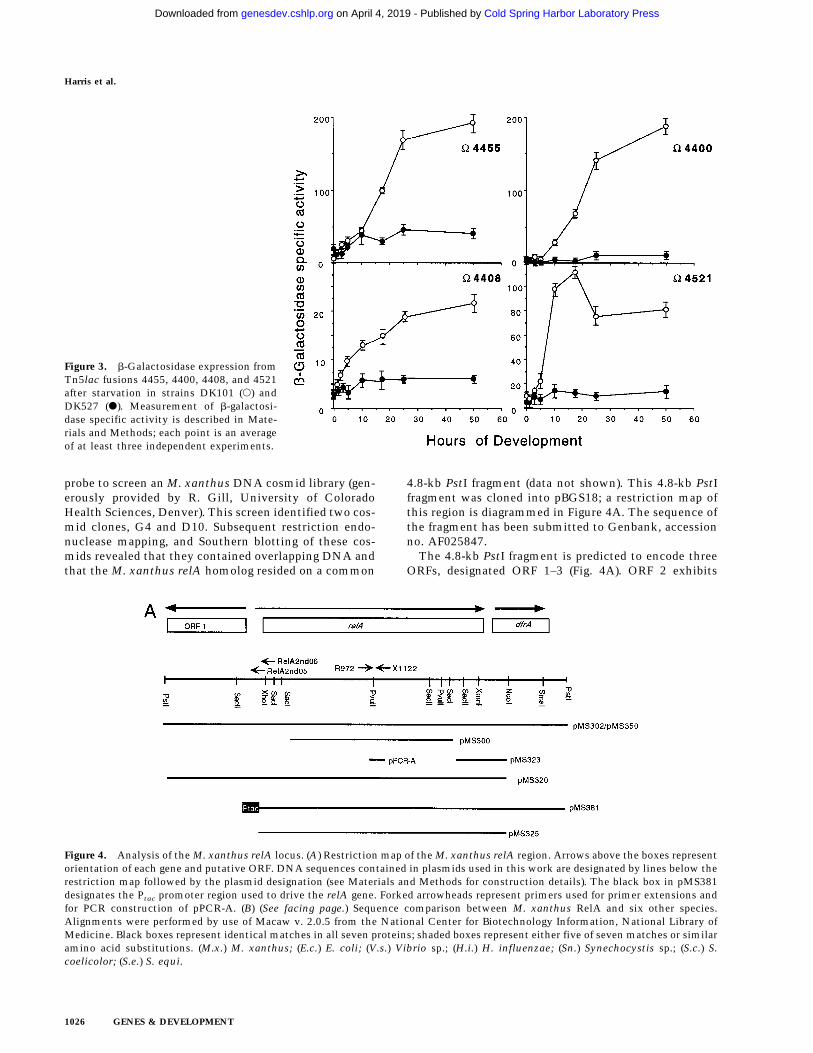
DK527 cannot undergo aggregation (Manoil and Kaiser1980b), but wild-type cells (DK101) show their first mor-phological signs of aggregation at ∼6 hr postinitiation. Todefine more precisely the stage at which the mutation inDK527 causes it to deviate from the normal developmen-tal program, four well-characterized Tn5lac transcrip-tional fusion reporters, V4408, V4455, V4521, andV4400, representative of the early stages of developmentwere examined in the DK527 genetic background. Thesereporters begin their b-galactosidase expression at differ-ent fusion-specific times in the 0–6 hr interval. No in-crease in b-galactosidase expression from any of thesefour reporter fusions was observed in DK527, as com-pared with DK101 (Fig. 3). Thus, the mutation in DK527prevents cells from initiating early gene expression asdefined by these Tn5lac reporters. These experimentsconfine the temporal block to within the first few hoursof the normal developmental program. Because (p)ppGppis synthesized then, this is expected to be the time whenstarvation is recognized and the decision to developfruiting bodies is made.
Cloning the M. xanthus RelA homolog
On the basis of the phenotype of DK527 and its comple-mentation by the E. coli relA gene, a search for the M.xanthus relA homolog was initiated by use of a PCR-based approach. Taking advantage of regions of aminoacid identity between the known amino acid sequencesof RelA proteins from several different Eubacteria(Metzger et al. 1988; Sarubbi et al. 1989; Chakraburtty etal. 1996), PCR primers were designed to amplify DNAsequences encoding the M. xanthus relA homolog. Asingle 311-bp PCR product was detected by use of prim-ers R-1750 and X-1125 (see Materials and Methods).DNA sequence analysis of this fragment predicted a pro-tein coding sequence that was 49% identical and 75%similar to both the E. coli RelA and SpoT proteins, whichare very similar to each other (Chakraburtty et al 1996).
The 311-bp PCR-generated fragment was used as a
Figure 2. Expression of E. coli RelA in M. xanthus strainsDK101 and DK527 under starvation in the dark (time 0) andafter 5 hr of exposure to light. Western blotting is described inMaterials and Methods. Arrows indicate the expected mobilityof E. coli RelA protein.
Figure 1. Effects of serine hydroxamate-induced serine starva-tion on RNA synthesis in M. xanthus, DK101 (s) or DK527 (d)(A), and E. coli, wild-type (s) or relA− (d) (B). Strains were grownat 30°C in either M9–glucose (E. coli) to an OD450 of 0.2 to 0.3or in M1 medium (M. xanthus) at 33°C to a density of 60–100Klett units, as described in Materials and Methods. Sampleswere pulse-labeled at the indicated times (in min) with [3H]uri-dine, and RNA synthesis rates were determined as described inMaterials and Methods. Each time point records an average ofthree independent experiments.
Relaxed mutant of M. xanthus
GENES & DEVELOPMENT 1025
Cold Spring Harbor Laboratory Press on April 4, 2019 - Published by genesdev.cshlp.orgDownloaded from
probe to screen an M. xanthus DNA cosmid library (gen-erously provided by R. Gill, University of ColoradoHealth Sciences, Denver). This screen identified two cos-mid clones, G4 and D10. Subsequent restriction endo-nuclease mapping, and Southern blotting of these cos-mids revealed that they contained overlapping DNA andthat the M. xanthus relA homolog resided on a common
4.8-kb PstI fragment (data not shown). This 4.8-kb PstIfragment was cloned into pBGS18; a restriction map ofthis region is diagrammed in Figure 4A. The sequence ofthe fragment has been submitted to Genbank, accessionno. AF025847.
The 4.8-kb PstI fragment is predicted to encode threeORFs, designated ORF 1–3 (Fig. 4A). ORF 2 exhibits
Figure 4. Analysis of the M. xanthus relA locus. (A) Restriction map of the M. xanthus relA region. Arrows above the boxes representorientation of each gene and putative ORF. DNA sequences contained in plasmids used in this work are designated by lines below therestriction map followed by the plasmid designation (see Materials and Methods for construction details). The black box in pMS381designates the Ptac promoter region used to drive the relA gene. Forked arrowheads represent primers used for primer extensions andfor PCR construction of pPCR-A. (B) (See facing page.) Sequence comparison between M. xanthus RelA and six other species.Alignments were performed by use of Macaw v. 2.0.5 from the National Center for Biotechnology Information, National Library ofMedicine. Black boxes represent identical matches in all seven proteins; shaded boxes represent either five of seven matches or similaramino acid substitutions. (M.x.) M. xanthus; (E.c.) E. coli; (V.s.) Vibrio sp.; (H.i.) H. influenzae; (Sn.) Synechocystis sp.; (S.c.) S.coelicolor; (S.e.) S. equi.
Figure 3. b-Galactosidase expression fromTn5lac fusions 4455, 4400, 4408, and 4521after starvation in strains DK101 (s) andDK527 (d). Measurement of b-galactosi-dase specific activity is described in Mate-rials and Methods; each point is an averageof at least three independent experiments.
Harris et al.
1026 GENES & DEVELOPMENT
Cold Spring Harbor Laboratory Press on April 4, 2019 - Published by genesdev.cshlp.orgDownloaded from

Figure 4. (Continued; See facing page for legend.)
Relaxed mutant of M. xanthus
GENES & DEVELOPMENT 1027
Cold Spring Harbor Laboratory Press on April 4, 2019 - Published by genesdev.cshlp.orgDownloaded from

strong similarity to RelA and SpoT; Figure 4B shows se-quence similarity between ORF 2 and several bacterialRelA proteins. ORF 3 shows similarity to a group ofsmall acid-soluble proteins implicated in translation in-hibition, the YER057c/YJGF family (Oka et al. 1995; Ce-ciliani et al. 1996; Schmiedeknecht et al. 1996). Bothputative ORFs would be transcribed in the same direc-tion. Sequence analysis of DNA 58 to the putative relA/spoT gene (which includes the putative ORF 1) shows nosignificant similarity to anything in the database whentranslated in all six potential reading frames.
Primer extension analysis was used to identify thetranscriptional start site for the relA/spoT gene; a single58 end was identified (Fig. 5A). On the basis of the resultsof these experiments, on sequence similarity, and on theavailability of a promising Shine–Dalgarno site nearby,we have placed the putative translational start at a GTGlocated 106 bp downstream from the transcriptionalstart site (Fig. 5A); this relA gene would encode a proteinof 757 amino acids with a formal molecular mass of 85kD. Sequence analysis also revealed a potential 8-bpDNA stem and 3-bp loop structure located between relAand dfrA (DG = −18.3 kcal), followed by a run of three Tresidues (Fig. 5B). This putative stem-loop structure islocated 15 bp from the presumed translational start ofdfrA and could represent a transcriptional terminator forthe relA gene.
Locating the relA gene on the M. xanthus physicalmap
By use of the 311-bp PCR-generated fragment describedabove as a probe for Southern blot analysis of M. xanthuschromosomal DNA, two unique restriction fragmentswere identified. This implies that M. xanthus, like E.coli, may have one relA gene and one spoT gene. In E.coli, the relA and spoT genes are physically separatedand map at 60 and 82 min, respectively. To determinewhether the two putative M. xanthus genes were physi-cally separated, their locations were mapped to thephysical M. xanthus genome map (Shimkets 1993). ThePCR-generated fragment was used as a probe to deter-mine the position of each putative gene by use of pulsed-field electrophoresis (Chen et al. 1990). This analysisplaced one of the genes at position 1–1.3Mb (AseI frag-ment N and SpeI fragment U8) and the other at position4.8–5Mb (AseI fragment P and SpeI fragment S) on the9.5-Mb M. xanthus physical map (Fig. 6). To determinethe precise location of the cloned gene, the entire 4.8-kbPstI fragment was used to probe DNA isolated from anordered M. xanthus YAC library (Kuspa et al. 1989). Hy-bridization results indicated that the cloned gene resideson YACs 1372 and 1334, placing it between tgl and 442on the physical map (Fig. 6).
The cloned gene has RelA function
Because ORF 2 displays sequence similarity to both RelAand SpoT proteins, a functional assay was required to
indicate its role. For this purpose, we tested the M. xan-thus clone for its capacity to complement an E. coli relAdeletion strain to produce (p)ppGpp. Plasmid pMS381,which contains the M. xanthus gene under the transcrip-tional control of the IPTG inducible tac promoter, wasintroduced into a DrelA E. coli strain, CF165 (Xiao et al.1991). Cells carrying either pMS381 or pTTQ18 wereassayed for their ability to produce (p)ppGpp in responseto IPTG. Cells carrying pMS381 were able to produce(p)ppGpp in an IPTG-dependent fashion (Fig. 7), demon-strating that expression of the M. xanthus gene corre-lates with an increase in the production of this nucleo-tide. Strains carrying the control plasmid pTTQ18showed no increase in (p)ppGpp levels (Fig. 7). Previ-ously, the overproduction of E. coli relA in both E. coli(Schreiber et al. 1991) and M. xanthus (Singer and Kaiser1995) have been shown to increase the level of (p)ppGpp.
Disruption of relA prevents the accumulationof (p)ppGpp in response to amino acid starvationand causes an early developmental arrest
To assess the role of relA in (p)ppGpp synthesis and de-velopment in M. xanthus, the gene was disrupted by in-sertional mutagenesis. An internal 1.2-kb SacI fragment(Fig. 4A) cloned to the Kmr plasmid pBGS18, designatedpMS300, was integrated at the chromosomal locus byhomologous recombination. Integration of the plasmidat the chromosomal relA locus would result in a tandemduplication of the 1.2-kb region and generate two trun-cated genes. The structure of the expected tandem du-plication was confirmed by Southern blot analysis (datanot shown) and the resulting strain was designatedMS10.
This relA insertion mutant, MS10, along with itsisogenic parent, DK101, were assayed for their abilityto synthesize (p)ppGpp in response to amino aciddeprivation. Under these conditions DK101 accumulates(p)ppGpp within the first 15 min of starvation and thelevels slowly decay (Fig. 8), consistent with previousstudies (Manoil and Kaiser 1980a; Singer and Kaiser1995). In contrast, strain MS10 does not accumulate(p)ppGpp even after 60 min (Fig. 9).
Previously, we have proposed that for the purpose ofinitiating fruiting body development, M. xanthus sensesits nutritional status by measuring its intracellular lev-els of (p)ppGpp and thereby monitoring its capacity forprotein synthesis. It follows that cells unable to produce(p)ppGpp will be unable to initiate development. DK101and MS10 strains were examined for the ability to formfruiting bodies and spores. No fruiting bodies, mounds,or aggregates were obtained from the MS10 relA inser-tion strain, even after 5 days. Wild-type cells form fruit-ing bodies within 24 hr. Furthermore, when spore assayswere performed after 24, 72, and 120 hr, the relA inser-tion strain produced <0.01% of the wild-type viablespore level.
Because dfrA lies 72-bp downstream to relA, it was
Harris et al.
1028 GENES & DEVELOPMENT
Cold Spring Harbor Laboratory Press on April 4, 2019 - Published by genesdev.cshlp.orgDownloaded from

possible that these two genes constituted a single operonand that the insertion into the upstream relA gene alsoinactivated dfrA. Two approaches were taken to demon-strate that the developmental phenotype associated with
MS10 is caused by a disruption of relA and not the resultof a polar effect on the downstream dfrA gene. In the firstapproach, we disrupted the intercistronic region betweendfrA and relA genes by inserting plasmid pMS323 intoDK101. This plasmid contains a 1-kb SacI–NcoI frag-ment carrying the 38-end of relA and 58-end of the dfrAgene (Fig. 4A). Integration of this plasmid would dupli-cate this region, producing one complete relA gene andone complete dfrA gene that are separated by vector se-quences. If dfrA is expressed from the relA promoter, theonly intact dfrA gene would now be separated from itspromoter. Alternatively, if dfrA is being expressed fromits own promoter or is not essential for development, nodevelopmental phenotype should be observed. The re-sulting strain, designated MS11, was assayed for devel-opment along with strains MS10 and DK101. MS11 de-veloped and sporulated normally (unlike MS10), suggest-ing that either dfrA is not required for development ordfrA expression is independent of relA expression.
The second approach was to demonstrate that the M.xanthus relA gene was sufficient to rescue the develop-mental defect of strain MS10 when expressed in trans.For this purpose, we introduced a second copy of the relAgene into strains MS10 and DK101 and designated thesestrains MS12 and MS13, respectively. Plasmid pMS321contains the intact M. xanthus relA gene and the first130 bases of the dfrA gene cloned into the tetracyclineresistance Mx8att vector, pSWU29. When assayed fordevelopment, strain MS13 regained the ability to formfruiting bodies and could sporulate to 80% of wild-typelevels, demonstrating that the defect of MS10 is causedby the disruption of relA and not caused by polar effectson dfrA.
The M. xanthus relA gene rescues the DK527 mutant
Previously, we demonstrated that the E. coli relA genecould rescue the developmental defect of DK527. To de-termine if the developmental block of strain DK527could be rescued by the M. xanthus relA gene, two plas-mids were constructed containing the M. xanthus relAgene. Plasmid pMS325 also contains Mx8attP, allowingus to test complementation when the M. xanthus relA+
Figure 6. Physical mapping of the relAgene. (A) Slot blots using total YAC DNAfrom an ordered YAC library, probed withDNA fragment harboring the M. xanthusrelA gene. (B) Schematic of the 3.6- to 4.8-Mb region of the physical map of M. xan-thus. YACs covering this region are indi-cated and labeled at the bottom. The posi-tion of relA is designated by the shadedbox. The top line represents AseI and SpeIfragments as designated by Chen et al.(1990); the second line represents the posi-tion of previously mapped markers(Shimkets 1993).
Figure 5. (A) Nucleotide sequence of the 58-region of relA.Transcriptional start site for relA is indicated by +1 and boldcaps. The −10 and −35 regions are underlined, and the putativeribosome binding site is indicated by bold caps. (PE) Primerextension product; (T, G, C, A) DNA sequence reactions withthe same primer used in the primer extension analysis as indi-cated in Materials and Methods; (Control) tRNA control primerextension reaction. (B) Nucleotide sequence of the intergenicregion between relA and dfrA. A putative 9-bp stem 3-bp loopstructure, which may act as a transcriptional terminator, isshown.
Relaxed mutant of M. xanthus
GENES & DEVELOPMENT 1029
Cold Spring Harbor Laboratory Press on April 4, 2019 - Published by genesdev.cshlp.orgDownloaded from

gene is introduced at the Mx8attB site. Plasmid pMS302lacks an Mx8attP site, but can integrate by homologousrecombination in the relA gene. This would result in atandem duplication of the relA region. When these plas-mids were introduced into DK527, both plasmid-carry-ing strains were found to regain the ability to producefruiting bodies (the pMS325-carrying strain is shown inFig. 9) and sporulate based on direct spore count (data notshown) and viable spore assay (Table 2). Control plas-mids lacking the M. xanthus relA gene were unable torescue the developmental defect of DK527. These datastrongly suggest that DK527 has a mutant allele of relA.
(p)ppGpp production is required forA-factor production
One of the earliest responses to starvation is the produc-tion of an extracellular population density signal, Myxo-coccus A-factor (Kuspa et al. 1986). Because the DK527mutant fails to express the A-factor-dependent fusionsV4521 or V4400 (Fig. 3), we suspected that DK527 mightbe deficient in A-factor production. To examine the re-lationship between (p)ppGpp accumulation and A-factorproduction, we measured the amount of A-factor pro-duced by DK527 and the relA insertion mutant. Twomethods were used to collect A-factor: extracellularcomplementation (an in situ method in which wholecells are the A-factor producers), and collection of con-ditioned medium that serves as the source of A-factor.
The bioassay for A-factor employs an A-signaling de-fective tester strain (DK4324, asgB V4521, Kuspa et al.1986). Six different A-factor donor strains—DK101 (wild-type, positive control), DK527, DK10528 (E. coli relA+),DK10529, MS10 (the relA insertion null), and asgB mu-tant DK480 (negative control) were each mixed withDK4324. In accordance with earlier experiments (Kuspaand Kaiser 1989; Kuspa et al. 1986), DK101 (asgB+) wasable to rescue the A-signaling defect of strain DK4324,whereas the asgB480 (A-factor defective) mutant straincould not (Table 3). Strains DK527 and MS10 had signifi-
cantly less ability to rescue the defect of the tester strain(Table 3), estimated as 16% and 7% of wild-type A-factoractivity, respectively. The E. coli relA+ plasmid restoredto DK527 the ability to produce A-factor to 78% of thewild-type level. DK10529 produced the same level of A-factor as DK10528 (within experimental error).
A-factor production was also tested by bioassay of theconditioned medium: MC7 suspension buffer after cellshad been agitated and starved therein. MC7 buffer fromeach of the strains described above—DK101, DK527,DK10528, MS10, and DK480—was assayed for A-factoractivity at 3, 6, and 9 hr postinitiation (Table 3). Consis-tent with the in situ assays, DK101 produced and re-leased A-factor, whereas DK480, DK527, and MS10 pro-
Figure 7. Expression of M. xanthus relA in E. coli leads to(p)ppGpp production. One-dimensional thin layer chromatogra-phy autoradiographs of extracts from E. coli strains carryingeither pMS381 (M. xanthus relA) or pTTQ18 (vector), withequal radioactivity loaded at the origin in each lane.
Figure 8. Disruption of the M. xanthus relA gene prevents theaccumulation of (p)ppGpp. One dimensional thin layer chroma-tography autoradiograph of extracts from M. xanthus strainsharboring either pMS300 (MS10) or no insertion (wild type,DK101), with equal radioactivity loaded at the origin.
Figure 9. Rescue of M. xanthus DK527 fruiting body formationby wild-type M. xanthus relA. Strains DK101 and DK527 withno plasmid (A,B) or with pMS325 (M. xanthus relA integrated atthe Mx8att site) (C,D) were spotted on TPM agar as described inMaterials and Methods and photographed at 72 hr poststarva-tion.
Harris et al.
1030 GENES & DEVELOPMENT
Cold Spring Harbor Laboratory Press on April 4, 2019 - Published by genesdev.cshlp.orgDownloaded from

duced only 2%, 8%, and 3% of wild-type A-factor activ-ity after 3 hr. Not only were DK527 and MS10 deficientfor A-factor production at 3 hr, normally the time of peaklevel of A-factor production (Kuspa et al. 1992), but therewas no evidence for delayed production of A-factor at 6or 9 hr postinitiation (Table 3). Finally, the DK10528strain recovered the ability to produce A-factor and torelease it into the medium. The data of Table 3 showsthat DK527 is defective in the production of A-factor andthat the defect can be rescued by expression of the E. colirelA+ gene in DK527.
Discussion
When faced with starvation, M. xanthus has a choicebetween two responses to that stress: It can either slow
its rate of growth to one that can be sustained at thenutrient level of its surroundings, or it can initiate fruit-ing body development. What role might (p)ppGpp havein that decision? As shown previously, expression of theE. coli relA+ gene in M. xanthus leads to accumulation of(p)ppGpp and activation of early developmental gene ex-pression, even in the presence of a nutrient level suffi-cient to support growth (Singer and Kaiser 1995). Thus,accumulation of (p)ppGpp appears to be sufficient to ini-tiate the developmental process and represents nutrientlimitation.
Previously, we showed that ectopic production of(p)ppGpp in M. xanthus is sufficient to activate the A-factor-dependent V4521 fusion in the presence of nutri-ents (Singer and Kaiser 1995). However, (p)ppGpp cannotactivate the asgB480 mutant. Thus both (p)ppGpp and
Table 2. Sporulation in DK527 rescue strains
Percent DK101 viable spores (± S.D.)
Strain72 hr
(3 days)120 hr(5 days)
168 hr(7 days)
DK101 100 (±14.0) 100 (±2.7) 100 (±1.7)(wild type)
DK527 0.0007 (±0.0002) 0.0002 (±0.00009) 0.0002 (±0.0001)(relA527 mutant)
MS16 84.8 (±10.4) 71.9 (±5.6) 78.6 (±2.5)(DK101 pMS325)
MS17 109.4 (±15.3) 107.1 (±5.7) 102.1 (±7.0)(DK527 pMS325)
Table 3. A-factor production
Donor strain
In situ productiona Production in suspensiona
U/ml %W.T. timeb U/ml %W.T.c
DK101 480 ± 86 100 3 (hr) 36 ± 3.8 100(wild type) 6 (hr) 29 ± 3.2 100
9 (hr) 22 ± 3.1 100DK480 28 ± 12 6 3 (hr) 0.8 ± 0.3 2
(DK101 asgB) 6 (hr) 1.1 ± 0.4 49 (hr) 1.2 ± 0.4 5
DK527 76 ± 22 16 3 (hr) 2.2 ± 0.8 8(DK101relA527) 6 (hr) 2.3 ± 0.9 7
9 (hr) 2.2 ± 0.8 10MS10 35 ± 08 7 3 (hr) 1.1 ± 0.2 3
(DK101relA<pMS300) 6 (hr) 0.9 ± 0.4 39 (hr) 0.8 ± 0.4 4
DK10529d 425 ± 73 88 3 (hr) 39 ± 4.8 108(DK101 pMS132) 6 (hr) 32 ± 4.2 110
9 (hr) 21 ± 3.1 95DK10528d 375 ± 76 78 3 (hr) 30 ± 4.6 83
(DK527 pMS132) 6 (hr) 26 ± 4.8 899 (hr) 23 ± 4.8 104
aValues are shown as units per milliliter of cells (or per milliliter of supernatant recovered from shaken suspensions) at a cell densityof 1000 Klett units, prepared as described in Materials and Methods. The values given are an average of three independent experiments.bTime values indicate that the duration cells were cultured in MC7 buffer prior to bioassay of A-factor.c%W.T. activity was calculated by comparing the A-factor activity found in DK480 and DK527 to the amount found in the wild-typestrain. Wild-type activity was considered 100% for each time point.dBecause levels of (p)ppGpp adequate for fruiting body development are produced by these strains, experiments were performed in thedark, under conditions that lead to fruiting body formation for these strains.
Relaxed mutant of M. xanthus
GENES & DEVELOPMENT 1031
Cold Spring Harbor Laboratory Press on April 4, 2019 - Published by genesdev.cshlp.orgDownloaded from

asgB+ are required for activation of V4521. Because nei-ther DK527 nor the relA insertion strain is able to pro-duce (p)ppGpp, (p)ppGpp may work through asgB (and/orasgA and asgC) to initiate A-factor production. Werethere a second or alternative starvation sensor as well as(p)ppGpp, then this hypothetical sensor might allowstarvation-induced A-factor production even in the ab-sence of (p)ppGpp accumulation. However, little A-fac-tor was produced by either strain unable to produce(p)ppGpp, whereas restoration of (p)ppGpp production bythe E. coli relA+ largely restored A-factor production.These data are consistent with a model in which indi-vidual cells monitor their nutritional status by means ofchanges in the intracellular concentration of (p)ppGpp,such that accumulation of this nucleotide initiates thedevelopmental program. One output of the early parts ofthe program would be the activation of A-factor produc-tion through the asgA, asgB, and asgC gene products.
Consistent with the suggestion that (p)ppGpp worksthrough asg gene products is the recent work of Plamannand colleagues, showing that asgC is the M. xanthushomolog of rpoD, the E. coli gene that encodes the s70
subunit of its RNA polymerase (Davis et al. 1995). TheasgC767 mutation, which almost completely eliminatesA-factor production (Kuspa and Kaiser 1989), resultsfrom a glutamic acid to lysine change at amino acid po-sition 598 in the M. xanthus s70 homolog (Davis et al.1995). This homolog is called SigA in M. xanthus (Inouye1990). Glutamate 598 is adjacent to an equivalent aminoacid position in the E. coli s70 protein, at which suppres-sors of a DrelA DspoT double-null mutation arise (Hern-andez and Cashel 1995). Experiments with E. coli mu-tants carrying either the rpoD(P504L) allele or therpoD(S506) mutation suggest that region 3 of the E. colis70 subunit may be directly involved in (p)ppGpp-medi-ated gene regulation (Hernandez and Cashel 1995).
Experiments reported here combined with those ofSinger and Kaiser (1995) imply that the accumulation of(p)ppGpp in response to nutrient deprivation is both nec-essary and sufficient to activate the developmental pro-gram of M. xanthus. Moreover, intracellular productionof (p)ppGpp precedes, and is necessary for, extracellularproduction of A-factor. The next challenge is to under-stand how (p)ppGpp and A-factor jointly regulate V4521and other genes that are both starvation and A-factordependent, and how the critical choice between slowgrowth and initiation of fruiting body development ismade.
Materials and methods
Bacterial strains, phage, and plasmids
All strains and plasmids used are listed in Table 1. The referencestrain used for these studies was DK101 (Manoil and Kaiser1980a), a developmentally competent M. xanthus strain thatcontains an sglA1 allele, allowing cells to grow well dispersed inliquid culture (Hodgkin and Kaiser 1979). For simplicity, DK101has been referred to as wild-type throughout the paper. Theisolation of DK527 from DK101 has been described (Manoil andKaiser 1980a). The transducing mxyophages Mx4ts18ts27hrm
(Campos et al. 1978) and Mx8clp2 (Martin et al. 1978) have beendescribed, as well as the E. coli strain MG1655 and the isogenicDrelA+ strain (Singer et al. 1991). The Tn5lac transcriptionalfusions V4408, V4455, V4400, and V4521 have also been de-scribed previously (Kroos et al. 1986). DK101 and DK527 deriva-tives harboring each fusion were constructed by Mx8- or Mx4-mediated transduction. The presence of a single Tn5 element inthe M. xanthus chromosome was confirmed by Southern blot-ting.
Plasmid construction
The 4.8-kb PstI fragment encoding the M. xanthus relA wascloned from cosmid G4 into the PstI sites of pBGS18, giving riseto pMS302, and into the Mx8attP containing vector pPLH343,giving rise to pMS350. Subclones containing small fragments ofpMS302 were cloned into either pBGS18 or pBluescriptSK(Stratagene, La Jolla, CA) by use of standard cloning protocols(Sambrook et al. 1989) and are listed in Table 1.
Plasmid pMS321 was constructed by digesting pMS302 withNcoI and blunting the end with T4 DNA polymerase, then di-gesting with BamHI. This fragment was then cloned into theBamHI–SmaI sites of pSWU29, a plasmid that carries theMx8attP region and tetracycline resistance. This plasmid re-moves the downstream dfrA ORF from the original insert.
Plasmid pMS325 was constructed by digesting pMS321 withHindIII and blunting the end with DNA polymerase Klenowfragment, then by cutting with XhoI. This procedure removedall M. xanthus sequences upstream to the internal XhoI site ofrelA. Next, a 480-bp fragment containing 58 sequences from theupstream EagI site (blunted by DNA polymerase Klenow frag-ment) to the internal XhoI site was inserted. The final con-struct, which removes upstream and downstream ORFs, wasverified by sequencing with an internal primer.
Plasmid pMS381, containing the M. xanthus relA gene underthe control of the E. coli tac Promoter (Ptac), was constructed ina two-step procedure. First, a 477-bp PCR fragment was isolatedwith primers RELA2nd06 (58-AGTTCTCCGCCTGCTTCTCC-38) and RelABam-1 (58-TAGGATCCGGGTCAACGAAAGCG-AACGCA-38), placing a BamHI restriction site 20 bp from theGTG translational start. This fragment contains the 58 end ofthe relA gene without its promoter. The resulting 477-bp PCRproduct was then digested with XhoI and BamHI, giving rise toa 267-bp fragment encoding the 58 end of the relA gene. The267-bp XhoI–BamHI fragment was then cloned into pMS302and designated pMS380. The construct was verified by sequenc-ing with an internal primer. To construct pMS381, the 3.3-kbBamHI–HindIII fragment from pMS380 was cloned into theBamHI–HindIII site of pTTQ18 (Amersham, Arlington Heights,IL), placing M. xanthus relA under the control of Ptac. Thejunction fragment was confirmed by DNA sequencing.
Growth and development
M. xanthus cells were grown in either CTT liquid [1% casitone(Difco Laboratories), 10 mM (Tris-H)ydrochloride (pH 7.6), 1 mM
KHPO4 (pH 7.8), 8 mM MgSO4], M1 liquid (Zusman et al. 1971),or A1 liquid (Bretscher and Kaiser 1978) at 33°C with vigorousagitation. For solid support, agar to a final concentration of 1.5%(wt/vol) (CTT and M1), or agarose to a final concentration of0.8% (wt/vol) (A1) was added as described (Bretscher and Kaiser1978). Where indicated, medium was supplemented with oxy-tetracycline (12 µg/ml) or kanamycin (40 µg/ml). Cell growth inliquid cultures was monitored with a Klett–Summerson photo-electric colorimeter (model 800-3) equipped with a red filter;100 Klett units corresponds to 5 × 10⁸ cells/ml. E. coli cellswere grown in M9 complete medium (Singer et al. 1991). For
Harris et al.
1032 GENES & DEVELOPMENT
Cold Spring Harbor Laboratory Press on April 4, 2019 - Published by genesdev.cshlp.orgDownloaded from

RNA synthesis experiments with serine hydroxamate and forlabeling with [3H]uridine, E. coli cells were grown in M9 com-plete medium lacking serine and uracil (Singer et al. 1991).
Development was initiated in M. xanthus by starvation asdescribed previously (Kroos et al. 1986). Developmental b-ga-lactosidase assays were performed as described (Kroos et al.1986). The time of initial b-galactosidase expression (expressiontime) was determined by extrapolating the curve of increasingenzyme activity to its intercept with a line from the t = 0 valueprolonged parallel to the time axis (Kroos et al. 1986). Fruitingbody development was monitored visually with a dissectingmicroscope (Wild-Heerbrug, Switzerland). Two methods wereused to examine sporulation efficiency. First, spore viabilityassays were performed as described previously (Thony-Meyerand Kaiser 1993). Second, direct spore counts were performedwith a Petroff–Hausser counting chamber and phase contrastmicroscopy.
Light induction of E. coli RelA in M. xanthus
M. xanthus strains were grown in liquid or on solid supportmedium and irradiated as described previously (Singer and Kai-ser 1995).
PCR analysis
Two synthetic primers, R972 [58-AAAAAGAATTCAACGG(C/G) TACCAG(T/A)(C/G)(C/G)ATCCACAC-38] and X1122 [58-AAAAAATCTAGA(C/G) CCCTT(C/G)GG(C/G)GTGAA(C/G)-ACGTA-38], were used to amplify an internal segment of the M.xanthus relA homolog from strain DK101 by the polymerasechain reaction. Reaction mixtures contained 10 mM Tris-HCl(pH 8.5), 50 mM KCl, 2 mM MgCl2, 200 µM of each of the fourdNTPs, 2.5 units of Taq polymerase, 40 pmole of each primer,and 50 ng of chromosomal DNA in a final reaction volume of100 µl. Samples were denatured at 95°C for 2 min and subjectedto 30 cycles of denaturation (95°C for 1 min), annealing (57°Cfor 30 sec), and extension (72°C for 1 min) with a final extensionincubation for 10 min, at 72°C. PCR products were then ana-lyzed by agarose gel electrophoresis (1.5% wt/vol). The result-ing 311-bp fragment was isolated by use of the QIAEX II gelextraction kit (Qiagen Inc., Chatsworth, CA) and cloned intothe EcoRI and BamHI sites of pBGS18.
Colony hybridization and radioactive probe protocols
Nitrocellulose filters containing an ordered array of DNA froman M. xanthus cosmid library, were provided by R. Gill. Filterswere probed by use of the 311-bp PCR-generated DNA fragmentlabeled with 32P. Radioactive probes were made by the randomprimer method (Sambrook et al. 1989) and [a-32P]GTP (NEN) at3000 Ci/mmole, 10 mCi/ml.
Total RNA synthesis
RNA synthesis rates in vivo were measured as described previ-ously (Singer et al. 1991) with the following modifications. Cellswere grown in either M9 glucose (E. coli) or M1 (M. xanthus)medium to a density of 5 × 10⁸ cells/ml, then serine hydroxa-mate (final concentration 2.5 mg/ml) was added to inhibit thecharging of seryl-tRNA. Portions of 100 µl were removed foranalysis: two portions prior to addition of serine hydroxamate(t=0), followed by experimental portions (performed in dupli-cate) at the time intervals indicated in Figure 1. All sampleswere exposed for 2 min to 10 µCi/ml [5,6-3H]uridine (Amer-sham TRK. 410; 40 Ci/mmole) plus 2 µg/ml uridine. Incorpo-
ration was stopped by addition of 2 ml of ice-cold 5% trichlo-roacetic acid (TCA) for 30 min. Samples were washed threetimes with 5 ml of ice-cold 2% TCA, rinsed with 70% ethanol,air-dried, and the 3H-incorporated into TCA insoluble materialwas determined in a Beckman scintillation counter (model LS1801). Incorporation of radioactivity was linear for up to 30 minunder these conditions.
Measurement of guanosine nucleotides
Guanosine nucleotides were isolated and measured as describedpreviously (Manoil and Kaiser 1980a; Singer and Kaiser 1995).
Blotting and physical mapping
Southern blotting was performed as described previously (Sam-brook et al. 1989). Physical mapping was performed by the Bio-Rad CHEF Mapping DRII system with a 1% agarose gel in 0.5%TBE buffer at 10°C for 15:10 hr at 6V/cm (200 V) with switchtimes of 70 and 120 sec. M. xanthus DNA was prepared anddigested in agarose plugs as described previously (Chen et al.1990, 1991). CHEF gels were then used for Southern blot analy-sis.
Gene locations were further localized on the M. xanthusphysical map by performing Southern blot analysis by use of anordered set of M. xanthus YAC clones (Kuspa et al. 1989; Chenet al. 1990, 1991). Whole yeast DNA (containing the YAC DNA)was purified from strains carrying M. xanthus YAC clones(Coulson et al. 1988). Samples were denatured with 0.4 M NaOHat 65°C for 15 min, and 25 µg of DNA was transferred to aNytran filter (Amersham, Arlington Heights, IL) by use of a slotblot vacuum apparatus (Bio-Rad, Hercules, CA). The DNA wasthen cross-linked to the membrane with a UV transilluminatorand subjected to Southern hybridization procedures as describedabove.
Western blots
Western blots were performed as described previously (Straus etal. 1987; Singer and Kaiser 1995) Anti-E. coli RelA antibody wasprovided by G. Glaser (Hadassa Medical School, Jerusalem, Is-rael).
Sequence analysis
DNA sequence analysis was performed by the Division of Bio-logical Studies Automated DNA Sequencing Facility, at theUniversity of California, Davis, with the ABI Prism Dye Ter-minator Cycle Sequencing Ready Reaction Kit using AmpliTaqDNA polymerase. Reactions were run on a 4.25% acryl/bi-sacrylamide gel with an ABI Prism 377 DNA Sequencer. Se-quence data was analyzed by use of the ABI Prism Sequencing2.1.1 software (Perkin Elmer, Foster City, CA) and contigs as-sembled with Sequencher v3.0 (GeneCodes Corporation, Madi-son, WI).
Primer extension
Vegetative RNA was isolated from DK101 cells as describedpreviously (Kaplan et al. 1991). Primer extensions were per-formed as per Mirel and Chamberlin (1989) with the followingmodifications: 25 µg of vegetative RNA or tRNA was used ineach reaction, by use of the synthetic oligonucleotides RelA2nd05(58-CCTTCTTGATGATGTCCAG-38) and RelA2nd06 (58-AG-TTCTCCGCCTGCTTCTCC-38). Samples were run on a 5%Long Ranger (J.T. Baker, Phillipsburg, NJ) urea acrylamide gelalongside DNA sequence reactions. Sequence reactions were
Relaxed mutant of M. xanthus
GENES & DEVELOPMENT 1033
Cold Spring Harbor Laboratory Press on April 4, 2019 - Published by genesdev.cshlp.orgDownloaded from

performed with the fmol DNA cycle sequencing kit (Promega,Madison, WI), as described by the manufacturer. Primers usedfor primer extension were simultaneously used for fmol cyclesequencing reactions.
A-factor assays
A-factor production in situ was assayed as described previously(Kuspa et al. 1986). A-factor released into the extracellular me-dium by the donor cells was assayed as described (Kuspa andKaiser 1989).
Acknowledgments
We thank A. Garza for his generous assistance and E. Wick-strom for helpful discussions of this work, S. Hoover and D.Lavell in the University of California at Davis sequencing fa-cilities for technical support, and R. Gill for supplying his cos-mid library. In addition, we thank G. Holland, M. Igo, C. Price,and L. Plamann for critically reading this manuscript. Thiswork was supported in part by National Institutes of Healthgrants GM54592 to M.S. and GM23441 to D.K.
The publication costs of this article were defrayed in part bypayment of page charges. This article must therefore be herebymarked ‘‘advertisement’’ in accordance with 18 USC section1734 solely to indicate this fact.
References
Borek, E., J. Rockenbach, and A. Ryan. 1956. Studies on a mu-tant of Escherichia coli with unbalanced ribonucleic acidsynthesis. J. Bacteriol. 71: 318–323.
Bretscher, A. and D. Kaiser. 1978. Nutrition of Myxococcus xan-thus, a fruiting myxobacterium. J. Bacteriol. 133: 763–768.
Campos, J., J. Geisselsoder, and D. Zusman. 1978. Isolation ofbacteriophage MX4, a generalized transducing phage forMyxococcus xanthus. J. Mol. Biol. 119: 167–178.
Cashel, M. 1969. The control of ribonucleic acid synthesis inEscherichia coli. IV. Relevance of unusual phosphorylatedcompounds from amino acid starved stringent strains. J.Biol. Chem. 244: 3133–3141.
Cashel, M. and J. Gallant. 1969. Two compounds implicated inthe function of the RC gene of Escherichia coli. Nature (Lon-don) 221: 838–841.
Cashel, M., D. Gentry, J. Hernandez, and D. Vinella. 1996. Thestringent response. In Escherichia coli and Salmonella: Cel-lular and molecular biology, 2nd ed. (ed. F.C. Neidhart), pp.1458–1496. Vol. 1. ASM Press, Washington DC.
Ceciliani, F., L. Faotto, A. Negri, I. Colombo, B. Berra, A. Bar-torelli, and S. Ronchi. 1996. The primary structure of UK114tumor antigen. FEBS Lett. 393: 147–150.
Chakraburtty, R. and M. Bibb. 1997. The ppGpp synthetasegene (relA) of Streptomyces coelicolor A3(2) plays a condi-tional role in antibiotic production and morphological dif-ferentiation. J. Bacteriol. 179: 5854–5861.
Chakraburtty, R., J. White, E. Takano, and M. Bibb. 1996. Clon-ing, characterization and disruption of a (p)ppGpp synthetasegene (relA) of Streptomyces coelicolor A3(2). Mol. Microbiol.19: 357–368.
Chen, H., I. Keseler, and L. Shimkets. 1990. Genome size ofMyxococcus xanthus determined by pulsed-field gel electro-phoresis. J. Bacteriol. 172: 4206–4213.
Chen, H., A. Kuspa, I. Keseler, and L. Shimkets. 1991. Physicalmap of the Myxococcus xanthus chromosome. J. Bacteriol.173: 2109–2115.
Coulson, A., R. Waterston, J. Kiff, J. Aulston, and Y. Kohara.1988. Genome linking with yeast artificial chromosomes.Nature 335: 184–186.
Davis, J., K. Mayor, and L. Plamann. 1995. A missense mutationin rpoD results in an A-signaling defect in Myxococcus xan-thus. Mol. Microbiol. 18: 943–952.
Dworkin, M. 1962. Nutritional requirements for vegetativegrowth of Myxococcus xanthus. J. Bacteriol. 84: 250.
Fiil, N.P. and J.D. Friesen. 1968. Isolation of relaxed mutants ofEscherichia coli. J. Bacteriol. 95: 769–786.
Hernandez, J. and M. Cashel. 1995. Changes in conserved region3 of Escherichia coli s70 mediate ppGpp-dependent functionin vivo. J. Mol. Biol. 252: 536–549.
Hodgkin, J. and D. Kaiser. 1979. Genetics of gliding motility inM. xanthus (Myxobacterales): Two gene systems controlmovement. Mol. & Gen. Genet. 171: 177–191.
Inouye, S. 1990. Cloning and DNA sequence of the gene codingfor the major sigma factor from Myxococcus xanthus. J. Bac-teriol. 172: 80–85.
Kaplan, H., A. Kuspa, and D. Kaiser. 1991. Suppressors thatpermit A-signal independent developmental gene expressionin Myxococcus xanthus. J. Bacteriol. 173: 1460–1470.
Kimsey, H. and D. Kaiser. 1991. Targeted disruption of theMyxococcus xanthus orotidine-58-monophosphate decar-boxylase gene: Effects on growth and fruiting body develop-ment. J. Bacteriol. 173: 6790–6797.
Kroos, L., A. Kuspa, and D. Kaiser. 1986. A global analysis ofdevelopmentally regulated genes in Myxococcus xanthus.Dev. Biol. 117: 252–266.
Kuspa, A. and D. Kaiser. 1989. Genes required for developmen-tal signaling in Myxococcus xanthus: Three asg loci. J. Bac-teriol. 171: 2762–2772.
Kuspa, A., L. Kroos, and D. Kaiser. 1986. Intercellular signalingis required for developmental gene expression in Myxococ-cus xanthus. Dev. Biol. 117: 267–276.
Kuspa, A., D. Vollrath, Y. Cheng, and D. Kaiser. 1989. Physicalmapping of the Myxococcus xanthus genome by randomcloning in yeast artificial chromosomes. Proc. Natl. Acad.Sci. 86: 8917–8921.
Kuspa, A., L. Plamann, and D. Kaiser. 1992. Identification ofheat-stable A-factor from Myxococcus xanthus. J. Bacteriol.174: 3319–3326.
Li, S., B. Lee, and L. Shimkets. 1992. csgA expression entrainsMyxococcus xanthus development. Genes & Dev. 6: 401–410.
Manoil, C. 1982. ‘‘Initiation of the development of Myxococcusxanthus.’’ Ph.D. thesis. Stanford University, Stanford, CA.
Manoil, C. and D. Kaiser. 1980a. Accumulation of guanosinetetraphosphate and guanosine pentaphosphate in Myxococ-cus xanthus during starvation and myxospore formation. J.Bacteriol. 141: 297–304.
———. 1980b. Guanosine pentaphosphate and guanosine tetra-phosphate accumulation and induction of Myxococcus xan-thus fruiting body development. J. Bacteriol. 141: 1062–1065.
Martin, S., E. Sodergren, T. Masuda, and D. Kaiser. 1978. Sys-tematic isolation of transducing phages for Myxococcus xan-thus. Virology. 88: 44–53.
Metzger, S., I.B. Dror, E. Aizenman, G. Schreiber, M. Toone, J.D.Friesen, M. Cashel, and G. Glaser. 1988. The nucleotide se-quence and characterization of the relA gene of Escherichiacoli. J. Biol. Chem. 263: 15699–15704.
Mirel, D. and M. Chamberlin. 1989. The Bacillus subtilis fagel-lin gene (hag) is transcribed by the s28 form of RNA poly-merase. J. Bacteriol. 171: 3095–3101.
Oka, T., H. Tsuji, C. Noda, K. Sakai, Y. Hong, I. Suzuki, S.
Harris et al.
1034 GENES & DEVELOPMENT
Cold Spring Harbor Laboratory Press on April 4, 2019 - Published by genesdev.cshlp.orgDownloaded from

Munoz, and Y. Natori. 1995. Isolation and characterizationof a novel perchloric acid-soluble protein inhibiting cell-freeprotein synthesis. J. Biol. Chem. 270: 30060–30067.
Orndorff, P., E. Stellwag, T. Starich, M. Dworkin, and J. Zissler.1983. Genetic and physical characterization of lysogeny bybacteriophage Mx8 in Myxococcus xanthus. J. Bacteriol.154: 772–779.
Sambrook, J., E. Fritsch, and T. Maniatis. 1989. Molecular clon-ing: A laboratory manual. Cold Spring Harbor LaboratoryPress, Cold Spring Harbor, NY.
Sarubbi, E., K.E. Rudd, H. Xiao, K. Ikehara, M. Kalman, and M.Cashel. 1989. Characterization of the spoT gene of Esche-richia coli. J. Biol. Chem. 264: 15074–15082.
Schmiedeknecht, G., C. Kerkhoff, E. Orso, J. Stohr, C. Aslandis,G. Nagy, R. Kneuchel, and G. Schmitz. 1996. Isolation andcharacterization of a 14.5-kDa trichloroacetic-acid-solubletranslational inhibitor protein from human monocytes thatis upregulated upon cellular differentiation. Eur. J. Biochem.242: 339–351.
Schreiber, G., S. Metzger, E. Aizenman, S. Roza, M. Cashel, andG. Glaser. 1991. Overexpression of the relA gene in Esche-richia coli. J. Biol. Chem. 266: 3760–3767.
Shimkets, L. 1984. Nutrition, metabolism and the initiation ofdevelopment. In Myxobacteria, development and cell inter-actions (ed. E. Rosenberg), pp. 92–108. Springer-Verlag, NewYork.
———. 1993. The myxobacterial genome. Myxobacteria II, (ed.M. Dworkin and D. Kaiser). American Society of Microbiol-ogy, Washington, DC.
Singer, M. and D. Kaiser. 1995. Ectopic production of guanosinepenta- and tetraphosphate can initiate early developmentalgene expression in Myxococcus xanthus. Genes & Dev.9: 1633–1644.
Singer, M., W. Walter, B. Cali, H. Liebke, R. Gourse, and C.Gross. 1991. Physiological effects of the fructose-1,6-diphos-phate aldolase ts8 mutation on stable RNA synthesis inEscherichia coli. J. Bacteriol. 173: 6249–6257.
Spratt, B., P. Hedge, S. Heesen, A. Edelman, and J. Broome-Smith. 1986. Kanomycin-resistant vectors that are ana-logues of plasmids pUC8, pUC9, pEMBL8, and pEMBL9.Gene 41: 337–342.
Stent, G. and S. Brenner. 1961. A genetic locus for the regulationof ribonucleic acid synthesis. Proc. Natl. Acad. Sci.47: 2005–2014.
Straus, D., W. Walter, and C. Gross. 1987. The heat shock re-sponse of E. coli is regulated by changes in the concentrationof s32. Nature 329: 348–351.
Thony-Meyer, L. and D. Kaiser. 1993. devRS, an autoregulatedand essential genetic locus for fruiting body development inMyxococcus xanthus. J. Bacteriol. 175: 7450–7462.
Tosa, T. and L. Pizer. 1971. Biochemical bases for the antime-tabolite action of L-serine hydroxamate. J. Bacteriol.106: 972–982.
Watson, B. and M. Dworkin. 1968. Comparative intermediarymetabolism of vegetative cells and microcysts of Myxococ-cus xanthus.. J. Bacteriol. 96: 1456–1473.
Wireman, J. and M. Dworkin. 1977. Developmentally inducedautolysis during fruiting body formation by Myxococcusxanthus. J. Bacteriol. 129: 796–802.
Xiao, H., M. Kalman, K. Ikehara, S. Aemel, G. Glaser, and M.Cashel. 1991. Residual guanosine 3858-bispyrophosphatesynthetic activity of relA null mutants can be eliminated byspoT null mutations. J. Biol. Chem. 266: 5980–5990.
Zusman, D. and E. Rosenberg. 1971. Division cycle of Myxo-coccus xanthus II. Kinetics of cell growth and protein syn-thesis. J. Bacteriol. 105: 811–819.
Relaxed mutant of M. xanthus
GENES & DEVELOPMENT 1035
Cold Spring Harbor Laboratory Press on April 4, 2019 - Published by genesdev.cshlp.orgDownloaded from

12:1998, Genes Dev. Baruch Z. Harris, Dale Kaiser and Mitchell Singer
xanthus MyxococcusA-factor production inThe guanosine nucleotide (p)ppGpp initiates development and
References
http://genesdev.cshlp.org/content/12/7/1022.full.html#ref-list-1
This article cites 46 articles, 28 of which can be accessed free at:
License
ServiceEmail Alerting
click here.right corner of the article or
Receive free email alerts when new articles cite this article - sign up in the box at the top
Cold Spring Harbor Laboratory Press
Cold Spring Harbor Laboratory Press on April 4, 2019 - Published by genesdev.cshlp.orgDownloaded from