the genetic design of signaling cascades to record
TRANSCRIPT

The genetic design of signaling cascades to recordreceptor activationGilad Barnea*†, Walter Strapps‡§, Gilles Herrada‡, Yemiliya Berman‡¶, Jane Ong‡�, Brian Kloss‡**, Richard Axel*††,and Kevin J. Lee‡,‡‡
*Howard Hughes Medical Institute, Department of Biochemistry and Cellular Biophysics, Center for Neurobiology and Behavior, Columbia University,New York, NY 10032; and ‡Sentigen Biosciences, 3960 Broadway, New York, NY 10032
Contributed by Richard Axel, November 14, 2007 (sent for review October 9, 2007)
We have developed an experimental strategy to monitor proteininteractions in a cell with a high degree of selectivity and sensi-tivity. A transcription factor is tethered to a membrane-boundreceptor with a linker that contains a cleavage site for a specificprotease. Activation of the receptor recruits a signaling proteinfused to the protease that then cleaves and releases the transcrip-tion factor to activate reporter genes in the nucleus. This strategyconverts a transient interaction into a stable and amplifiablereporter gene signal to record the activation of a receptor withoutinterference from endogenous signaling pathways. We have de-veloped this assay for three classes of receptors: G protein-coupledreceptors, receptor tyrosine kinases, and steroid hormone recep-tors. Finally, we use the assay to identify a ligand for the orphanreceptor GPR1, suggesting a role for this receptor in the regulationof inflammation.
cellular assays � G protein-coupled receptor � protein interaction
A ll cells have evolved mechanisms to respond to rapidchanges in the environment. Extracellular signals are de-
tected by transmembrane receptors that translate binding intointracellular signaling events. Most signaling systems that re-spond to environmental cues exhibit adaptation mechanismsthat afford the cell a facile response to rapid changes in theirsurroundings. Mechanisms to assure the rapid but transientresponse to environmental cues are of obvious advantage to thecell but seriously limit most assays for receptor function. We havegenetically modified receptors such that transient responses toligand result in the stable transcription of a reporter gene. Thetransformation of a transient intracellular response to a stableamplifiable readout provides a sensitive and quantitative assayfor receptor function.
We have developed an assay for receptor activation and moregenerally for protein–protein interaction that involves the fusionof a membrane receptor with a transcriptional activator. Themembrane-bound receptor and transcription factor sequencesare separated by a cleavage site for a highly specific viralprotease. A second gene encodes a fusion of the viral proteasewith a cellular protein that interacts only with activated receptor.Ligand binding to the receptor will stimulate this protein–protein interaction, recruiting the protease to its cleavage site.Site-specific cleavage will release the transcriptional regulatorthat can now enter the nucleus and activate reporter genes.Recently, a similar principle, based on the complementation ofsplit tobacco etch virus (TEV) protease fragments, has beenused to monitor protein interactions (1). Our experimentalscheme derives conceptually from the mechanism of action ofthe Notch receptor in which ligand binding elicits proteolyticcleavage events in the receptor to release a Notch intracellulardomain that translocates to the nucleus and modulates tran-scription of downstream target genes (2, 3) (Fig. 1A).
The assay we have developed relies solely on exogenous genesintroduced into the cell to create a novel signaling pathway. Asa consequence, reporter gene activity will be independent of theactivation of any endogenous cell signaling events that frequently
confound existing assays for receptor function. This approachtransforms transient rapidly adapting signaling events into morestable and amplifiable cellular responses by virtue of the irre-versible release of a membrane-anchored transcription factor.Because this assay requires the association of two proteins tostimulate a response, we call this the Tango assay. We havedeveloped Tango assays to monitor the activity of three differentclasses of receptors: G protein-coupled receptors (GPCRs),receptor tyrosine kinases, and steroid hormone receptors.
ResultsTango Assay for GPCRs. In initial experiments, we designed a Tangoassay to monitor the activation of GPCRs (Fig. 1B). We generateda fusion protein consisting of the human arginine vasopressinreceptor 2 (AVPR2) joined at its cytoplasmic C terminus to thetranscriptional activator, tTA (4). Interposed between the receptorand tTA sequences, we introduced a specific 7-aa cleavage site fora highly specific protease, the N1a protease from TEV (5). We thenconstructed a second fusion protein consisting of the TEV proteaselinked to human �-arrestin2. These receptor and arrestin fusiongenes were transiently introduced into a cell line that contains atTA-dependent reporter gene.
Ligand activation of GPCRs results in the phosphorylation ofspecific serine and threonine residues at the cytoplasmic Cterminus of the receptor by a class of GPCR kinases (6).Ligand-occupied phosphorylated receptor then recruits arrestin,preventing further G protein activation (7). In the Tango assay,the ligand-dependent recruitment of arrestin-TEV protease tothe receptor fusion and the subsequent proteolytic cleavage freestTA to enter the nucleus and activate reporter genes. Theassociation of arrestin with ligand-bound receptor is observedfor most GPCRs, a feature that allowed us to extend the Tangoassay to multiple receptors within this class.
We tested a series of variant TEV protease cleavage sitespreviously shown to be cleaved by the protease at reducedefficiency (8) and identified one mutant site that maximized thesignal-to-background ratio [supporting information (SI) Fig. 5].
Author contributions: G.B., W.S., G.H., Y.B., J.O., B.K., R.A., and K.J.L. designed research;G.B., W.S., G.H., Y.B., J.O., B.K., and K.J.L. performed research; G.B. contributed newreagents/analytic tools; W.S., G.H., Y.B., J.O., B.K., and K.J.L. analyzed data; and G.B., R.A.,and K.J.L. wrote the paper.
The authors declare no conflict of interest.
Freely available online through the PNAS open access option.
†Present address: Department of Neuroscience, Brown University, Providence, RI 02912.
§Present address: Sirna Therapeutics, Merck & Co., Inc., San Francisco, CA 94158.
¶Present address: ARMGO Pharma, Inc., New York, NY 10032.
�Present address: Orthobond Corporation, Monmouth Junction, NJ 08852.
**Present address: New York Structural Biology Center, New York, NY 10027.
††To whom correspondence should be addressed. E-mail: [email protected].
‡‡Present address: The Ellison Medical Foundation, Bethesda, MD 20814.
This article contains supporting information online at www.pnas.org/cgi/content/full/0710487105/DC1.
© 2007 by The National Academy of Sciences of the USA
64–69 � PNAS � January 8, 2008 � vol. 105 � no. 1 www.pnas.org�cgi�doi�10.1073�pnas.0710487105
Dow
nloa
ded
by g
uest
on
Dec
embe
r 28
, 202
1
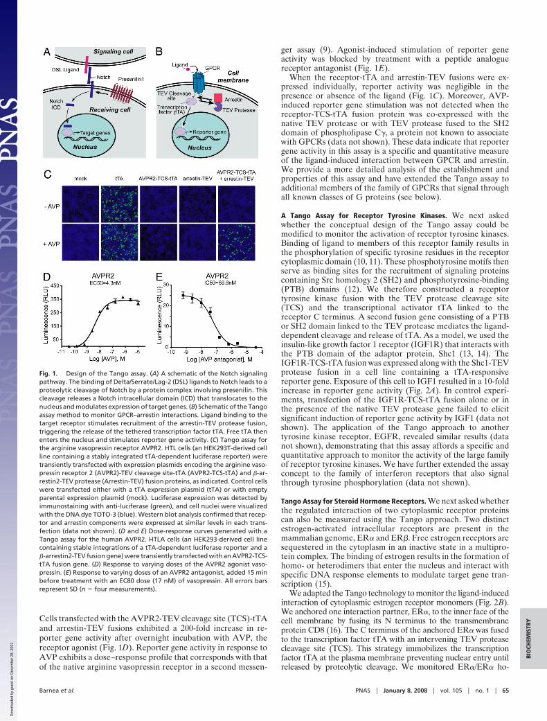
Cells transfected with the AVPR2-TEV cleavage site (TCS)-tTAand arrestin-TEV fusions exhibited a 200-fold increase in re-porter gene activity after overnight incubation with AVP, thereceptor agonist (Fig. 1D). Reporter gene activity in response toAVP exhibits a dose–response profile that corresponds with thatof the native arginine vasopressin receptor in a second messen-
ger assay (9). Agonist-induced stimulation of reporter geneactivity was blocked by treatment with a peptide analoguereceptor antagonist (Fig. 1E).
When the receptor-tTA and arrestin-TEV fusions were ex-pressed individually, reporter activity was negligible in thepresence or absence of the ligand (Fig. 1C). Moreover, AVP-induced reporter gene stimulation was not detected when thereceptor-TCS-tTA fusion protein was co-expressed with thenative TEV protease or with TEV protease fused to the SH2domain of phospholipase C�, a protein not known to associatewith GPCRs (data not shown). These data indicate that reportergene activity in this assay is a specific and quantitative measureof the ligand-induced interaction between GPCR and arrestin.We provide a more detailed analysis of the establishment andproperties of this assay and have extended the Tango assay toadditional members of the family of GPCRs that signal throughall known classes of G proteins (see below).
A Tango Assay for Receptor Tyrosine Kinases. We next askedwhether the conceptual design of the Tango assay could bemodified to monitor the activation of receptor tyrosine kinases.Binding of ligand to members of this receptor family results inthe phosphorylation of specific tyrosine residues in the receptorcytoplasmic domain (10, 11). These phosphotyrosine motifs thenserve as binding sites for the recruitment of signaling proteinscontaining Src homology 2 (SH2) and phosphotyrosine-binding(PTB) domains (12). We therefore constructed a receptortyrosine kinase fusion with the TEV protease cleavage site(TCS) and the transcriptional activator tTA linked to thereceptor C terminus. A second fusion gene consisting of a PTBor SH2 domain linked to the TEV protease mediates the ligand-dependent cleavage and release of tTA. As a model, we used theinsulin-like growth factor 1 receptor (IGF1R) that interacts withthe PTB domain of the adaptor protein, Shc1 (13, 14). TheIGF1R-TCS-tTA fusion was expressed along with the Shc1-TEVprotease fusion in a cell line containing a tTA-responsivereporter gene. Exposure of this cell to IGF1 resulted in a 10-foldincrease in reporter gene activity (Fig. 2A). In control experi-ments, transfection of the IGF1R-TCS-tTA fusion alone or inthe presence of the native TEV protease gene failed to elicitsignificant induction of reporter gene activity by IGF1 (data notshown). The application of the Tango approach to anothertyrosine kinase receptor, EGFR, revealed similar results (datanot shown), demonstrating that this assay affords a specific andquantitative approach to monitor the activity of the large familyof receptor tyrosine kinases. We have further extended the assayconcept to the family of interferon receptors that also signalthrough tyrosine phosphorylation (data not shown).
Tango Assay for Steroid Hormone Receptors. We next asked whetherthe regulated interaction of two cytoplasmic receptor proteinscan also be measured using the Tango approach. Two distinctestrogen-activated intracellular receptors are present in themammalian genome, ER� and ER�. Free estrogen receptors aresequestered in the cytoplasm in an inactive state in a multipro-tein complex. The binding of estrogen results in the formation ofhomo- or heterodimers that enter the nucleus and interact withspecific DNA response elements to modulate target gene tran-scription (15).
We adapted the Tango technology to monitor the ligand-inducedinteraction of cytoplasmic estrogen receptor monomers (Fig. 2B).We anchored one interaction partner, ER�, to the inner face of thecell membrane by fusing its N terminus to the transmembraneprotein CD8 (16). The C terminus of the anchored ER� was fusedto the transcription factor tTA with an intervening TEV proteasecleavage site (TCS). This strategy immobilizes the transcriptionfactor tTA at the plasma membrane preventing nuclear entry untilreleased by proteolytic cleavage. We monitored ER�/ER� ho-
Fig. 1. Design of the Tango assay. (A) A schematic of the Notch signalingpathway. The binding of Delta/Serrate/Lag-2 (DSL) ligands to Notch leads to aproteolytic cleavage of Notch by a protein complex involving presenilin. Thiscleavage releases a Notch intracellular domain (ICD) that translocates to thenucleus and modulates expression of target genes. (B) Schematic of the Tangoassay method to monitor GPCR–arrestin interactions. Ligand binding to thetarget receptor stimulates recruitment of the arrestin-TEV protease fusion,triggering the release of the tethered transcription factor tTA. Free tTA thenenters the nucleus and stimulates reporter gene activity. (C) Tango assay forthe arginine vasopressin receptor AVPR2. HTL cells (an HEK293T-derived cellline containing a stably integrated tTA-dependent luciferase reporter) weretransiently transfected with expression plasmids encoding the arginine vaso-pressin receptor 2 (AVPR2)-TEV cleavage site-tTA (AVPR2-TCS-tTA) and �-ar-restin2-TEV protease (Arrestin-TEV) fusion proteins, as indicated. Control cellswere transfected either with a tTA expression plasmid (tTA) or with emptyparental expression plasmid (mock). Luciferase expression was detected byimmunostaining with anti-luciferase (green), and cell nuclei were visualizedwith the DNA dye TOTO-3 (blue). Western blot analysis confirmed that recep-tor and arrestin components were expressed at similar levels in each trans-fection (data not shown). (D and E) Dose-response curves generated with aTango assay for the human AVPR2. HTLA cells (an HEK293-derived cell linecontaining stable integrations of a tTA-dependent luciferase reporter and a�-arrestin2-TEV fusion gene) were transiently transfected with an AVPR2-TCS-tTA fusion gene. (D) Response to varying doses of the AVPR2 agonist vaso-pressin. (E) Response to varying doses of an AVPR2 antagonist, added 15 minbefore treatment with an EC80 dose (17 nM) of vasopressin. All errors barsrepresent SD (n � four measurements).
Barnea et al. PNAS � January 8, 2008 � vol. 105 � no. 1 � 65
BIO
CHEM
ISTR
Y
Dow
nloa
ded
by g
uest
on
Dec
embe
r 28
, 202
1

modimerization by coexpression of the membrane-anchored CD8-ER�-TCS-tTA fusion with a free cytoplasmic ER� fused to theTEV protease. In parallel, we monitored ER�/ER� heterodimer-ization by expressing the anchored CD8-ER�-TCS-tTA fusion witha fusion of TEV protease to ER�. Stimulation with estradiolresulted in a 10-fold induction in reporter gene activity in the homo-and heterodimerization assays (Fig. 2B) with EC50 values in accordwith those of the native receptors (17). Transfection of the CD8-ER�-TCS-tTA alone, or with native TEV protease, resulted inbackground activity that was not stimulated by estradiol (data notshown). These results demonstrate that the Tango protein interac-tion methodology can be readily applied to monitoring ligand-induced signaling events that involve both transmembrane andintracellular receptors.
Extending Tango to the Family of GPCRs. Experiments to extendTango to a larger repertoire of GPCRs revealed that the extentof agonist-induced reporter gene stimulation varied amongdifferent GPCRs. Individual members of the GPCR family differin their affinity for arrestin (18), a characteristic that is largelydetermined by the phosphorylation at serine and threonine sitesin the C-terminal tail of the receptor (9). To extend the use ofthe Tango assay to GPCRs that weakly recruit arrestin uponactivation, we examined whether the addition of the C-terminaltail of a receptor that forms stable complexes with arrestin couldenhance assay performance for those receptors that respondedpoorly in the Tango assay. We observed that the addition of ashort fragment from the C-terminal tail of AVPR2 enhancedassay performance for multiple receptors, including the �-opioidreceptor and the D2 dopamine receptor (SI Fig. 6) but had no
discernible effect on the ligand specificity of the recipientreceptor (SI Fig. 7 and SI Table 1). This C-terminal modificationof the receptor fusion gene extends the Tango assay to receptorsthat weakly recruit arrestin molecules upon activation and hasallowed us to develop assays for 89 GPCR family membersrepresenting all known signaling classes (data not shown).
Agonist and Antagonist Specificity for the Adrenergic Receptor Fam-ily. We next examined the profile of the agonist and antagonistselectivity of a family of related GPCRs using the Tango assay.We established assays for a panel of five well characterized �-and �-adrenergic receptors and monitored receptor activationand inhibition by selective agonists and antagonists. In the Tangoassay, �- and �-adrenergic receptors displayed the predictedspecificity for subtype selective agonists and antagonists (Fig. 3and SI Table 2). In addition, these Tango assays, with signal-to-background ratios that can exceed 1,000-fold, afford greatersensitivity over a larger dynamic range than comparable secondmessenger assays, in which 2- to 5-fold responses are typicallyobserved. These results illustrate that monitoring arrestin re-cruitment in the Tango assay provides an accurate measure ofreceptor activation and demonstrate the general utility of thismethod to evaluate the ligand selectivity profiles of a panel ofGPCRs.
To determine whether the Tango assay mirrored the quanti-tative effects of agonists and partial agonists observed in assaysof the native GPCR, we examined the activation of the �2-ARby a series of �-adrenergic partial agonists, compounds that elicitsubmaximal receptor responses at saturating doses. We observedthat each agonist elicits different levels of activity at saturation(Fig. 3C). The weakest partial agonist, nylidrin, elicited a max-imal response �100 times lower than that produced by a fullagonist, isoproterenol. Moreover, the relative responses of the�2-AR to these partial agonists in the Tango assay were inaccord with data obtained in second messenger assays thatreport native signaling pathways (19). Thus, the stimulation ofreporter gene activity in the Tango assay is not binary; rather, itdistinguishes intermediate levels of receptor activation inducedby partial agonists.
Target Selectivity of the Tango Assay. The Tango assay reconstructsa receptor-mediated signaling cascade with three exogenousgenes. Thus, in contrast to assays of second messenger accumu-lation, the Tango assay should be immune to signaling elicited byactivation of receptors endogenous to the cell. We comparedTango with an assay that monitors receptor-mediated elevationsin intracellular calcium. The Tango assay was performed with theAVPR2-TCS-tTA fusion in HEK293 cells that express endoge-nous muscarinic acetylcholine and purinergic P2Y receptors (20,21). In the Tango assay, luciferase reporter gene activity wasinduced only by vasopressin that activates the exogenousAVPR2-TCS-tTA receptor and not by agonists for the endog-enous receptors (SI Fig. 8A). In contrast, assays for intracellularcalcium reveal a response to vasopressin as well as to carbacholand ATP, agonists for the muscarinic and P2Y receptors,respectively (SI Fig. 8B).
In a second experiment, we introduced AVPR2-TCS-tTA intoa cell along with an unmodified �-opioid receptor and observedreporter gene activity only by agonists selective for AVPR2 (SIFig. 8C). Similarly, cells transfected with �-opioid receptor-TCS-tTA along with an unmodified AVPR2 responded solely to�-opioid agonists. Thus, the Tango assay, unlike most secondmessenger assays, selectively measures the activation of a specificexogenous receptor-TCS-tTA fusion without interference fromendogenous receptor-mediated signaling pathways.
The selectivity of the Tango assay should permit us to monitoragonists for a given receptor in unfractionated tissue extractsthat often contain agonists for endogenous receptors. We ex-
Fig. 2. Tango assays for tyrosine kinase and steroid hormone receptors. (A)Receptor tyrosine kinase signaling. HTL cells were transfected with an IGF-1receptor (IGF1R)–TCS-tTA fusion construct and a Shc1 PTB domain-TEV pro-tease fusion plasmid. Luciferase activity in this IGF1R–Shc1 interaction assaywas stimulated by IGF-1. (B) Ligand-induced homo- and heterodimerization ofestrogen receptors (ERs). HTL cells were transfected with a CD8-ER�-TCS-tTAfusion and either an ER�- or ER�-TEV protease fusion to measure ER� ho-modimerization (light circles) or ER�/ER� heterodimerization (dark squares) asillustrated. Responses were normalized to the maximal response for eachreceptor. All error bars represent SD (n � four measurements).
66 � www.pnas.org�cgi�doi�10.1073�pnas.0710487105 Barnea et al.
Dow
nloa
ded
by g
uest
on
Dec
embe
r 28
, 202
1

amined this hypothesis by monitoring activation of the human�-opioid receptor by crude extracts of bovine hypothalamus.Hypothalamic extract produced dose-dependent increases inreporter gene activity, with a maximum induction �500 times
background (SI Fig. 8D). Pretreatment with a selective �-opioidantagonist, nor-binal torphimine, completely blocked the re-sponse to the hypothalamic extract in this assay, indicating thatthe response we observe derives solely from activation of theexogenous �-opioid receptor. Thus, Tango provides a selectivebioassay to monitor the activation of a receptor by endogenousagonists present in biological extracts.
Identifying Ligands for Orphan GPCRs. We exploited the sensitivityand specificity of the Tango assay to identify a ligand for theorphan GPCR, GPR1 (22). GPR1 is most closely related toCMKLR1 (23, 24), a member of a group of leukocyte chemoat-tractant receptors. The endogenous ligand recognized by GPR1has not been identified, and thus the biological function of thisreceptor remains unknown. Given the sequence similarity be-tween GPR1 and CMKLR1, we used the Tango assay to examinewhether GPR1 responds to the leukocyte chemattractantchemerin, the previously reported endogenous ligand forCMKLR1 (23, 24). We found that GPR1 responded in the Tangoassay to recombinant chemerin protein with an EC50 of 240 pM,compared with 3 nM for CMKLR1 (Fig. 4A). A C-terminalfragment representing amino acids 145–157 of the maturechemerin protein, previously shown to activate CMKLR1 (25),also activated GPR1 with an EC50 of 1 nM, compared with 24 nMfor CMKLR1 (Fig. 4B).
We next examined the ability of chemerin to activate GPR1and CMKLR1 using a calcium mobilization assay. In this assay,addition of chemerin to cells expressing GPR1 resulted in anelevation of intracellular calcium to a level 30% of that observedin cells expressing CMKLR1 (Fig. 4C). Thus, in the Tango assay,chemerin is a more potent agonist for GPR1 than for CMKLR1,whereas in the calcium mobilization assay, chemerin is moreeffective in stimulating CMKLR1. The relatively weak responseof GPR1 to chemerin using a calcium mobilization assay mayexplain a previous study that did not detect chemerin-mediatedactivation of GPR1 (23).
To confirm the identification of GPR1 as a potential chemerinreceptor, we performed radioligand-binding studies using an io-dinated C-terminal peptide fragment of chemerin (25). In theseexperiments, the labeled peptide was added to cells transfected withGPR1 or CMKLR1 as well as to untransfected cells in the presenceof either full length chemerin (Fig. 4D) or unlabeled peptide (Fig.4E) as competitors. We observed specific binding of the chemerinpeptide to cells transfected with GPR1 as well as to cells transfectedwith CMKLR1, whereas untransfected cells showed no specificbinding (data not shown). Saturation-binding analysis performedon GPR1-transfected cells revealed a single binding site with acalculated Kd of 5.3 nM (Fig. 4F), compared with 4.9 nM forCMKLR1-transfected cells (Fig. 4G). Thus, both GPR1 andCMKLR1 appear to bind the chemerin C-terminal peptide withsimilar affinity, whereas the Tango assay, an assay of receptorfunction, suggests that the chemerin peptide may be a more potentactivator of GPR1 than of CMKLR1. These experiments illustratethat the Tango assay may be particularly advantageous for theanalysis of receptors with second messenger pathways that areeither weak or unknown.
DiscussionThe Tango assay introduces three exogenous genes into a cell toconstruct a receptor-mediated signaling cascade. The conceptderives from the mechanism of action of the Notch receptor andrequires the sequestration of a transcription factor to the cellmembrane by physically linking it to a receptor. Activation of thereceptor fusion results in the recruitment of a signaling proteinfused to a protease that then cleaves and releases the transcrip-tion factor to activate reporter genes in the nucleus. Thisexperimental strategy was modified to develop assays for threeclasses of receptors: GPCRs, receptor tyrosine kinases, and
C
Lum
ines
cenc
e (R
LU)
Log [agonist], M
β2-adrenergic
-10 -9 -8 -7 -6 -50
500
1000
1500
isoproterenol, EC50=33.6nMclenbuterol, EC50=31.9nMsalbutamol, EC50=210nMnylidrin, EC50=100nM
Lum
ines
cenc
e (R
LU)
-12 -10 -8 -6 -40
100
200
300
-12 -10 -8 -6 -40
20
40
60
Isoproterenol, EC50>28µMUK-14,304, EC50=4.1nM
Isoproterenol, EC50=30.5nMUK-14,304, EC50=none
Lum
ines
cenc
e (R
LU)
Log [agonist], MLog [agonist], M
α2A-adrenergic β1-adrenergic
-12 -10 -8 -6 -40
100
200
300
400
-12 -10 -8 -6 -40
100
200
300
Log [antagonist], MLog [antagonist], M
Alprenolol, EC50=1.5nMYohimbine, EC50=none
Alprenolol, EC50=noneYohimbine, EC50=1.7nM
B
A
α2A-adrenergic β1-adrenergic
Fig. 3. Using the Tango assay to profile adrenergic receptor agonists andantagonists. (A and B) Agonist and antagonist selectivity profiling usingTango assays for human adrenergic receptors. HTLA cells were transientlytransfected with �2A-, �2B-, �2C-, �1-, or �2-adrenergic receptor-TCS-tTAfusions, each of which contained the C-terminal tail sequence from AVPR2. (A)In these representative Tango agonist assays, �-adrenergic receptors werepreferentially activated by the �-selective agonist UK14,304, and �-adrenergicreceptors were preferentially activated by the �-selective agonist isoprotere-nol. (B) In representative antagonist assays, �- and �-ARs displayed the ex-pected selectivity to the antagonists yohimbine and alprenolol, respectively.All error bars represent SD (n � four measurements). See SI Table 2 forcomplete results. (C) Stimulation of luciferase reporter gene activity in the�2-adrenergic receptor Tango assay by a full agonist, isoproterenol, andpartial agonists salbutamol, clenbuterol, and nylidrin. Agonist-stimulatedluciferase activity was measured in an HEK293T-derived cell line containingstably integrated luciferase reporter, �-arrestin2-TEV, and �2-AR-TCS-tTA(with AVPR2 C-terminal modification) constructs.
Barnea et al. PNAS � January 8, 2008 � vol. 105 � no. 1 � 67
BIO
CHEM
ISTR
Y
Dow
nloa
ded
by g
uest
on
Dec
embe
r 28
, 202
1

steroid hormone receptors. By using a signaling cascade com-posed of exogenous components, the Tango assay is largelyindependent of endogenous second messengers or adaptationmechanisms. This experimental strategy affords features that
distinguish Tango from other assays that measure receptorfunction. The genetic modifications of the receptor in the Tangoassay transform a transient receptor-mediated cellular responseinto the stable transcription of a reporter gene. The amplifica-tion inherent in transcriptional activation provides a sensitiveand quantitative assay of receptor function. Moreover, reportergene activity is immune to signaling events that result fromactivation of endogenous receptors, a feature that can confoundexisting assays of receptor function.
The Tango assay for GPCRs monitors receptor activation byexploiting ligand-mediated arrestin binding. Because virtually allGPCRs associate with arrestin upon activation, we exploited thegenerality of arrestin recruitment to develop assays for a widerange of GPCRs that include receptors that activate all knownG protein classes. GPCR signaling has been assayed previouslyby monitoring the subcellular redistribution of a fluorescentlylabeled arrestin in response to GPCR stimulation (26, 27). Tangoprovides a more quantitative assay that monitors arrestin re-cruitment solely to the modified receptor of interest and istherefore unaffected by the signaling of endogenous receptors.Tango therefore affords a general but selective assay for allGPCRs, independent of the nature of the G protein that theyactivate. Thus, it is particularly advantageous when G proteinactivation of second messengers is weak or if the identity of thedownstream G protein is unknown.
The approach we have devised to monitor the hormone-evoked dimerization of extracellular estrogen receptors demon-strates that Tango can be used more broadly to detect protein–protein interactions. Recently, a protein interaction techniquebased on the complementation of split TEV protease fragmentshas been described (1). Like the Tango method described here,this split TEV approach involves a cleavage step catalyzed byTEV protease, thereby converting transient interactions into along-lasting signaling readout. Our data indicate that splittingTEV protease is not required to limit proteolytic activity beforeinteraction. Instead, we observe that regulated localizationwithin the cell is sufficient to discriminate between free andassociated TEV- and transcription factor-fused partners. Be-cause Tango therefore obviates the need to engineer comple-mentary TEV fragments with low inherent affinity, it affordsenhanced sensitivity and greater generality.
The sensitivity of Tango, coupled with the ability to monitoractivity of a receptor of interest without interference fromendogenous receptor signaling, provides a suitable bioassay foridentifying endogenous ligands for orphan receptors. We haveexploited this feature to identify a candidate endogenous ligandfor the orphan GPCR, GPR1. GPR1 is most closely related toCMKLR1, a recently described receptor for the inflammation-associated leukocyte chemoattractant chemerin. Using theTango assay, we found that GPR1 is also activated by chemerin,and we confirmed these observations by an independent Ca2�
mobilization assay and by radioligand-binding studies. The ex-istence of a second, previously unrecognized receptor forchemerin raises the possibility that at least some of the activitiesof chemerin in inflammation may be mediated through GPR1.These observations illustrate the utility of the Tango method-ology as a bioassay to discover endogenous ligands for elusiveorphan receptors.
MethodsMaterials. Arg8-Vasopressin, [Adamantaneacetyl1, O-Et-D-Tyr2, Val4, Ami-nobutyryl6, Arg8,9]-Vasopressin, dopamine HCl, U-69593, U.K.-14,304, isopro-terenol, alprenolol, carbachol, ATP, nor-binaltorphimine dihydrochloride, re-combinant human IGF-1, and 17-� estradiol were obtained from Sigma.Yohimbine was from Tocris. The adrenergic compound library was obtainedfrom Biomol. Anti-luciferase was from Promega and was used at a dilution of1:500. TOTO-3 was from Molecular Probes (Invitrogen) and was diluted
A
-14 -12 -10 -8 -60
20
40
60
80
100
120GPR1, EC50=0.24nM
CMKLR1, EC50=3nM
log [chemerin], M
Lum
ines
cenc
e (%
max
)
-14 -12 -10 -8 -60
20
40
60
80
100
GPR1, EC50=1nM
CMKLR1, EC50=24nM
log [peptide], MLu
min
esce
nce
(%m
ax)
B
time (sec) time (sec)time (sec)
CMKLR1 + Gα15 GPR1 + Gα15 GPR1 - Gα15
10 60 110 160200
400
600
800
1000
1200
20 70 120 170 220250
350
450
550
30 80 130
Flu
ores
cenc
e (R
FU
)
C
-13 -12 -11 -10 -9 -8 -7 -6
0
20
40
60
80
100
120
GPR1, IC50=0.23 nMCMKLR1, IC50=0.15 nM
log [chemerin], M
Bo
un
d 1
25I-
pe
ptid
e (
%m
ax)
-13 -12 -11 -10 -9 -8 -7 -60
20
40
60
80
100
120
GPR1, IC50=2.3 nMCMKLR1, IC50=1.9 nM
log [peptide], M
D E
F G
0 2 4 6 8 100
1000
2000
3000
4000
5000
6000
7000GPR1, Kd=5.3 nM
Bo
un
d 1
25I-
pe
ptid
e,
cpm
125I-labelled peptide, nM0 2 4 6 8 10
0
200
400
600
800
1000
1200
1400 CMKLR1, Kd=4.9nM
Bo
un
d 1
25I-
pe
ptid
e,
cpm
125I-labelled peptide, nM
550
650
450
350
250
Bo
un
d 1
25I-
pe
ptid
e (
%m
ax)
Fig. 4. Identification of an agonist for the orphan receptor GPR1. (A and B)Dose-response profiles of GPR1 and CMKLR1 Tango assays in response to recom-binant human chemerin protein (A) and a peptide fragment of chemerin(chemerin 145–157) (B). Responses were normalized to the maximal response foreach receptor. (C) Calcium mobilization assay showing activation of CMKLR1 andGPR1 by chemerin peptide (chemerin 149–157, at 1 �M) in the presence of thepromiscuous G protein G�15. Time of ligand addition is indicated by the arrows.The responses of seven representative cells were averaged. All error bars repre-sent SD. (D–G) Binding of radiolabeled chemerin C-terminal peptide to GPR1-andCMKLR1-transfected cells. Shown is displacement of iodinated chemerin C-terminal peptide (chemerin149–157) binding to GPR1- (black circles) and CMKLR1-expressing cells (gray squares) by full-length chemerin peptide (D) or unlabeledchemerin149–157 (E). Shown is saturation binding of 125I-chemerin149–157 to GPR1-transfected cells (F) and CMKLR1-transfected cells (G).
68 � www.pnas.org�cgi�doi�10.1073�pnas.0710487105 Barnea et al.
Dow
nloa
ded
by g
uest
on
Dec
embe
r 28
, 202
1

1:1,000. Recombinant human Chemerin was from R&D Systems, and Chemerinpeptides were from Phoenix Pharmaceuticals.
Plasmid Constructs. See SI Text for details of plasmid construction. Constructswere generated by PCR using high-fidelity DNA polymerases [Platinum TaqHigh Fidelity (Invitrogen) or Expand High Fidelity (Roche)]. All constructs wereverified by sequencing both DNA strands.
Cell Culture and Transfections. Adherent HEK293T cells were cultured in DMEM(Specialty Media/Millipore), supplemented with 10% FBS (HyClone), 2 mML-glutamine, 100 unit/ml penicillin, and 100 �g/ml streptomycin. See SI Text fordetails of stable cell line generation and transfection methods. Twenty-fourhours after transfection, cells were dissociated by using TrypLE Express (In-vitrogen) and plated in assay plates or cryopreserved in freezing medium(Specialty Media/Millipore).
Reporter Gene Assays. Growing or cryopreserved cells were plated in white96-well assay plates at 10,000–20,000 cells per well or in 384-well assay platesat 6,000 cells per well in DMEM, supplemented with 10% FBS, glutamine,penicillin, and streptomycin. For Tango assays with chemerin peptides, cellswere plated instead in serum-free medium (Cambrex Biowhittaker Pro-293).For estrogen receptor Tango assays, cells were plated in phenol red-freeDMEM without serum addition. Test agonists were added 0–6 h after plating.Test antagonists were added 15 min before addition of an EC80 dose ofagonist. Cells were cultured for 8–24 h before measuring reporter geneactivity. �-Galactosidase activity was determined by using the LuminescentBeta-Galactosidase Detection Kit II (BD Biosciences), and luciferase activity wasdetermined by using the Bright-Glo luciferase assay system (Promega), using
the manufacturers’ protocols. Luminescence was measured by using an LMAXII-384 (Molecular Devices), MicroBeta Jet 1450 (Wallac/Perkin–Elmer), or Vic-tor3 (Perkin–Elmer) luminometer.
Calcium Imaging Assays. Unmodified GPR1 and CMKLR1 expression constructsor an AVPR2-tTA fusion construct were transiently transfected into adherentHEK293T cells, together with a G�-15 expression construct at a ratio of 4:1receptor:G protein, using Fugene 6 (Roche). After a 24-h incubation period,cells were washed and loaded with a calcium-sensitive fluorescent dye (FLIPRCalcium 3 assay kit, Molecular Devices), following the vendor’s protocol.Fluorescence imaging was performed with a Zeiss inverted fluorescence mi-croscope equipped with a Lambda DG-4 wavelength switcher (Sutter Instru-ments) and an Orca II digital camera (Hamamatsu). Data were analyzed usingMetaFluor imaging software (Molecular Devices).
Tissue Extracts. For �OPR assays, bovine hypothalamus extract was preparedby using a boiling water/acid extraction method. See SI Text for details.
Radioligand-Binding Assays. A 9-aa peptide (YFPGQFAFS), corresponding toamino acid residues 149–157 of full-length chemerin, was radioiodinated ontyrosine (Phoenix Pharmaceuticals). See SI Text for details of binding studies.
ACKNOWLEDGMENTS. We are grateful to Tanya Henderson, Benjamin Bar-telle, Leah Cohen, Michael Amatulli, Chris Sun, and Mark A. Johnson forexcellent technical assistance and to Leslie Vosshall and Filippo Mancia forcomments on the manuscript. This research was supported in part by theHoward Hughes Medical Institute (R.A.) and the Mathers Foundation (R.A.and G.B.) and by a grant from the Gates Foundation Grand Challenges inGlobal Health (to R.A.).
1. Wehr MC, Laage R, Bolz U, Fischer TM, Grunewald S, et al. (2006) Monitoring regulatedprotein-protein interactions using split TEV. Nat Methods 3:985–993.
2. Struhl G, Adachi A (1998) Nuclear access and action of notch in vivo. Cell 93:649–660.3. Schroeter EH, Kisslinger JA, Kopan R (1998) Notch-1 signalling requires ligand-induced
proteolytic release of intracellular domain. Nature 393:382–386.4. Gossen M, Bujard H (1992) Tight control of gene expression in mammalian cells by
tetracycline-responsive promoters. Proc Natl Acad Sci USA 89:5547–5551.5. Parks TD, Leuther KK, Howard ED, Johnston SA, Dougherty WG (1994) Release of
proteins and peptides from fusion proteins using a recombinant plant virus proteinase.Anal Biochem 216:413–417.
6. Pitcher JA, Freedman NJ, Lefkowitz RJ (1998) G protein-coupled receptor kinases. AnnuRev Biochem 67:653–692.
7. Luttrell LM, Lefkowitz RJ (2002) The role of beta-arrestins in the termination andtransduction of G-protein-coupled receptor signals. J Cell Sci 115:455–465.
8. Kapust RB, Tozser J, Copeland TD, Waugh DS (2002) The P1� specificity of tobacco etchvirus protease. Biochem Biophys Res Commun 294:949–955.
9. Oakley RH, Laporte SA, Holt JA, Barak LS, Caron MG (1999) Association of beta-arrestinwith G protein-coupled receptors during clathrin-mediated endocytosis dictates theprofile of receptor resensitization. J Biol Chem 274:32248–32257.
10. Schlessinger J (2000) Cell signaling by receptor tyrosine kinases. Cell 103:211–225.11. Pawson T, Hunter T (1994) Signal transduction and growth control in normal and
cancer cells. Curr Opin Genet Dev 4:1–4.12. Schlessinger J, Lemmon MA (2003) SH2 and PTB domains in tyrosine kinase signaling.
Sci STKE 2003:RE12.13. Sasaoka T, et al. (1994) Evidence for a functional role of Shc proteins in mitogenic
signaling induced by insulin, insulin-like growth factor-1, and epidermal growthfactor. J Biol Chem 269:13689–13694.
14. Craparo A, O’Neill TJ, Gustafson TA (1995) Non-SH2 domains within insulin receptorsubstrate-1 and SHC mediate their phosphotyrosine-dependent interaction with theNPEY motif of the insulin-like growth factor I receptor. J Biol Chem 270:15639–15643.
15. Dahlman-Wright K, et al. (2006) International Union of Pharmacology. LXIV. Estrogenreceptors. Pharmacol Rev 58:773–781.
16. Littman DR, Thomas Y, Maddon PJ, Chess L, Axel R (1985) The isolation and sequenceof the gene encoding T8: A molecule defining functional classes of T lymphocytes. Cell40:237–246.
17. Barkhem T, et al. (1998) Differential response of estrogen receptor alpha and estrogenreceptor beta to partial estrogen agonists/antagonists. Mol Pharmacol 54:105–112.
18. Oakley RH, Laporte SA, Holt JA, Caron MG, Barak LS (2000) Differential affinities ofvisual arrestin, beta arrestin1, and beta arrestin2 for G protein-coupled receptorsdelineate two major classes of receptors. J Biol Chem 275:17201–17210.
19. Swaminath G, et al. (2005) Probing the beta2 adrenoceptor binding site with catecholreveals differences in binding and activation by agonists and partial agonists. J BiolChem 280:22165–22171.
20. Krupinski J, Lehman TC, Frankenfield CD, Zwaagstra JC, Watson PA (1992) Moleculardiversity in the adenylylcyclase family. Evidence for eight forms of the enzyme andcloning of type VI. J Biol Chem 267:24858–24862.
21. Schachter JB, Sromek SM, Nicholas RA, Harden TK (1997) HEK293 human embryonickidney cells endogenously express the P2Y1 and P2Y2 receptors. Neuropharmacology36:1181–1187.
22. Marchese A, et al. (1994) Cloning of human genes encoding novel G protein-coupledreceptors. Genomics 23:609–618.
23. Wittamer V, et al. (2003) Specific recruitment of antigen-presenting cells by chemerin,a novel processed ligand from human inflammatory fluids. J Exp Med 198:977–985.
24. Meder W, et al. (2003) Characterization of human circulating TIG2 as a ligand for theorphan receptor ChemR23. FEBS Lett 555:495–499.
25. Wittamer V, et al. (2004) The C-terminal nonapeptide of mature chemerin activates thechemerin receptor with low nanomolar potency. J Biol Chem 279:9956–9962.
26. Barak LS, Ferguson SS, Zhang J, Caron MG (1997) A beta-arrestin/green fluorescentprotein biosensor for detecting G protein-coupled receptor activation. J Biol Chem272:27497–27500.
27. Oakley RH, et al. (2002) The cellular distribution of fluorescently labeled arrestinsprovides a robust, sensitive, and universal assay for screening G protein-coupledreceptors. Assay Drug Dev Technol 1:21–30.
Barnea et al. PNAS � January 8, 2008 � vol. 105 � no. 1 � 69
BIO
CHEM
ISTR
Y
Dow
nloa
ded
by g
uest
on
Dec
embe
r 28
, 202
1