the fetal genitourinary disclosures tractaium.s3.amazonaws.com/uls/handouts/17fgu.pdf · •a2-3...
TRANSCRIPT

1
THE FETAL GENITOURINARY
TRACT
Beverly G. Coleman, MD
Emeritus Professor of Radiology
Perelman School of Medicine
University of Pennsylvania
Director of Fetal Imaging
The Children’s Hospital of Philadelphia
Philadelphia, Pennsylvania
Coleman
Beverly G. Coleman, MD
No Relevant Financial Relationships
Coleman
DISCLOSURES
LEARNING OBJECTIVESAfter completing this presentation, the learner will
be able to discuss:
1. The normal sonographic appearance of all of
the organs and structures that constitute the
fetal GU tract
2. A systematic approach to analyzing fetal scans
referred for suspected GU tract anomalies
3. Tips for recognizing renal developmental
variants
4. Distinguishing features of urinary tract
obstruction compared to cystic renal disease
Coleman
OUTLINE
• The Normal Urinary Tract
• Renal Developmental Variants
• Urinary Tract Obstruction
• Cystic Renal Disease
Coleman
THE NORMAL URINARY TRACT
• Viz at 11-13 wks (TV) and 14-16 wks (TA)
• Corticomedullary differentiation at 16-18 wks
• Exponential in size with GA; RC/AC ratio = 0.27- 0.30; Nomograms for renal length 14-42 wks & renal volume 15-42 wks
• Increased conspicuity by 3rd trimester with thicker cortex, renal sinus/perinephric fat, best corticomedullary differentiation
• Color Doppler renal arteries & veins
Coleman
THE NORMAL URINARY TRACT
Normal Fetal Kidneys • 14 weeks 20 weeks 30 weeks
• 8 Mhz TV 9Mhz 12 Mhz
Coleman

2
THE NORMAL URINARY TRACT
Coleman
Normal CM Differentiation at 27 weeks
THE NORMAL URINARY TRACT
Coleman
The Normal Fetal Bladder
• Anechoic, midline, anterior, pear shaped
with a thin wall
• First structure to be visualized at 9-10 wks
on both TA & TV scans
• Fills and empties every 25-30 minutes
• Max volume from mean of 1 mL at 20 wks
to 36 mL at 41 wks
• Color Doppler umbilical arteries
THE NORMAL URINARY TRACT
Coleman
The Normal Fetal Bladder• Collapsed Distended
THE NORMAL URINARY TRACT
Coleman
• Normal Fetal Adrenals
Disc shaped, “ice cream sandwich”Viz at 20-30 wks, length with GANomograms – length, volume, weight, etc.
• Normal Fetal Gender
Male, female identical to 11wks; phallus direction “angle of the dangle”Testicles descend after 25 wks; both >97% after 32 wks; Small hydroceles common in 15% of normal males Uterus can be viz late; ovaries rarely ever viz
THE NORMAL URINARY TRACT
Normal Fetal Adrenals • 2003 2007 2012
Coleman
THE NORMAL URINARY TRACT
The Normal Genitalia• XY 23 wks XX 28 wks
Coleman
3
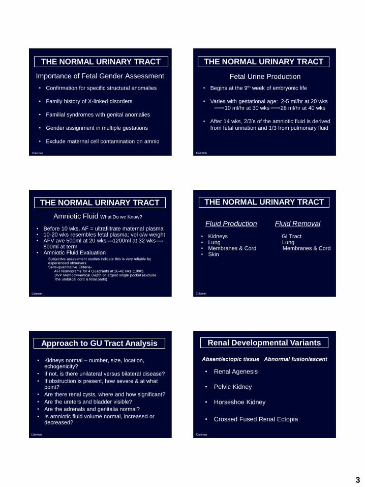
THE NORMAL URINARY TRACT
Importance of Fetal Gender Assessment
Coleman
• Confirmation for specific structural anomalies
• Family history of X-linked disorders
• Familial syndromes with genital anomalies
• Gender assignment in multiple gestations
• Exclude maternal cell contamination on amnio
THE NORMAL URINARY TRACT
Fetal Urine Production
Coleman
• Begins at the 9th week of embryonic life
• Varies with gestational age: 2-5 ml/hr at 20 wks
10 ml/hr at 30 wks 28 ml/hr at 40 wks
• After 14 wks, 2/3’s of the amniotic fluid is derived
from fetal urination and 1/3 from pulmonary fluid
THE NORMAL URINARY TRACT
Amniotic Fluid What Do we Know?
Coleman
• Before 10 wks, AF = ultrafiltrate maternal plasma• 10-20 wks resembles fetal plasma; vol c/w weight• AFV ave 500ml at 20 wks 1200ml at 32 wks
800ml at term• Amniotic Fluid Evaluation
Subjective assessment studies indicate this is very reliable by experienced observersSemi-quantitative Criteria-
AFI Nomograms for 4 Quadrants at 16-42 wks (1990) DVP Method=Vertical Depth of largest single pocket (exclude the umbilical cord & fetal parts)
THE NORMAL URINARY TRACT
Fluid Production Fluid Removal
Coleman
• Kidneys GI Tract• Lung Lung• Membranes & Cord Membranes & Cord• Skin
Approach to GU Tract Analysis
• Kidneys normal – number, size, location, echogenicity?
• If not, is there unilateral versus bilateral disease?
• If obstruction is present, how severe & at what point?
• Are there renal cysts, where and how significant?
• Are the ureters and bladder visible?
• Are the adrenals and genitalia normal?
• Is amniotic fluid volume normal, increased or decreased?
Coleman
Renal Developmental Variants
Absent/ectopic tissue Abnormal fusion/ascent
Coleman
• Renal Agenesis
• Pelvic Kidney
• Horseshoe Kidney
• Crossed Fused Renal Ectopia
4
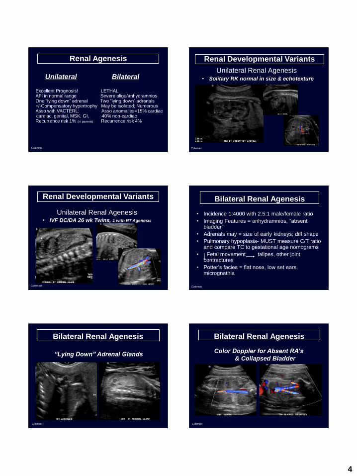
Renal Agenesis
Unilateral Bilateral
Coleman
Excellent Prognosis! LETHALAFI in normal range Severe oligo/anhydramniosOne “lying down” adrenal Two “lying down” adrenals+/-Compensatory hypertrophy May be isolated; NumerousAsso with VACTERL; Asso anomalies=15% cardiac cardiac, genital, MSK, GI, 40% non-cardiacRecurrence risk 1% (nl parents) Recurrence risk 4%
Renal Developmental Variants
Unilateral Renal Agenesis
Coleman
• Solitary RK normal in size & echotexture
Renal Developmental Variants
Unilateral Renal Agenesis
Coleman
• IVF DC/DA 26 wk Twins, 1 with RT Agenesis
Bilateral Renal Agenesis
• Incidence 1:4000 with 2.5:1 male/female ratio
• Imaging Features = anhydramnios, “absent bladder”
• Adrenals may = size of early kidneys; diff shape
• Pulmonary hypoplasia- MUST measure C/T ratio and compare TC to gestational age nomograms
• Fetal movement talipes, other joint contractures
• Potter’s facies = flat nose, low set ears, micrognathia
Coleman
Bilateral Renal Agenesis
Coleman
“Lying Down” Adrenal Glands
Bilateral Renal Agenesis
Color Doppler for Absent RA’s
& Collapsed Bladder
Coleman

5
Bilateral Renal Agenesis
• VACTERL in Di-Di Twins
Coleman
RENAL ECTOPIA
• No kidney observed in the flank, usually unilateral RA (54%)
• Most common forms of renal ectopia:
1. Pelvic kidney (37%)
2. Horseshoe kidneys (5%)
3. Crossed fused (4%)
• +/-Mild to moderate dilatation 2°obst/reflux
• Abnormal morphology 2°malrotation, dysplasia
Coleman
• Empty renal fossa with flattened “lying down” adrenal gland
• Kidney can be missed as often similar to bowel
in texture, superior to bladder, + malrotation
• Normal size or contralateral kidney
• Color Doppler follow RA to pelvis
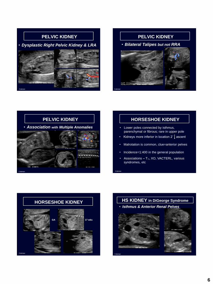
PELVIC KIDNEY
Coleman
PELVIC KIDNEY
• Is One Kidney Truly Missing?
Coleman
PELVIC KIDNEY
• Left Kidney with Vesicoureteral Reflux
Coleman
PELVIC KIDNEY
• Dysplastic LK with Ureterocele
Coleman
6
PELVIC KIDNEY
• Dysplastic Right Pelvic Kidney & LRA
Coleman
PELVIC KIDNEY
• Bilateral Talipes but not RRA
Coleman
PELVIC KIDNEY
• Association with Multiple Anomalies
Coleman
HORSESHOE KIDNEY
• Lower poles connected by isthmus,
parenchymal or fibrous; rare in upper pole
• Kidneys more inferior in location 2˚ ascent
• Malrotation is common, clue=anterior pelves
• Incidence=1:400 in the general population
• Associations – T18, XO, VACTERL, various
syndromes, etc
Coleman
HORSESHOE KIDNEY
Coleman
GA 17 wks
HS KIDNEY in DiGeorge Syndrome
Coleman
• Isthmus & Anterior Renal Pelves
7
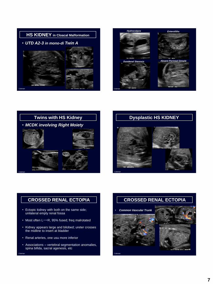
HS KIDNEY in Cloacal Malformation
Coleman
• UTD A2-3 in mono-di Twin A
Hydrocolpos Enteroliths
Duodenal Stenosis Absent Perineal Dimple
Coleman
Twins with HS Kidney
Coleman
• MCDK involving Right Moiety
Dysplastic HS KIDNEY
Coleman
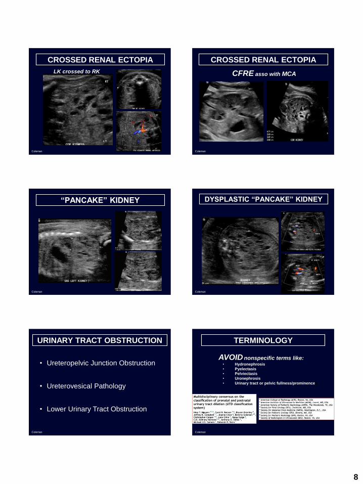
CROSSED RENAL ECTOPIA
Coleman
• Ectopic kidney with both on the same side; unilateral empty renal fossa
• Most often L R, 95% fused; freq malrotated
• Kidney appears large and bilobed; ureter crosses the midline to insert at bladder
• Renal arteries, one usu more inferior
• Associations – vertebral segmentation anomalies, spina bifida, sacral agenesis, etc
CROSSED RENAL ECTOPIA
Coleman
• Common Vascular Trunk
8
CROSSED RENAL ECTOPIA
Coleman
LK crossed to RK
CROSSED RENAL ECTOPIA
Coleman
CFRE asso with MCA
“PANCAKE” KIDNEY
Coleman
DYSPLASTIC “PANCAKE” KIDNEY
Coleman
URINARY TRACT OBSTRUCTION
Coleman
• Ureteropelvic Junction Obstruction
• Ureterovesical Pathology
• Lower Urinary Tract Obstruction
TERMINOLOGY
Coleman
AVOID nonspecific terms like:• Hydronephrosis
• Pyelectasis
• Pelviectasis
• Uronephrosis
• Urinary tract or pelvic fullness/prominencee

9
TERMINOLOGY
Coleman
LOW RISK
Postnatal
uropathy
INCREASED
RISK
Postnatal
uropathy
ANTENATAL CLASSIFICATION SYSTEM
Coleman
UPJ OBSTRUCTION
Coleman
• Most common site for prenatal obstruction, 1:2,000
live births
• Tip: RP + calyces with no dilated ureter or bladder
• Several causes – ischemia, stricture, valves,
anomalous vessels or crossing bands, high ureteral
insertion, muscle abnormality
• Contralateral renal anomaly, 25%; Extra-renal
anomalies occur in 10%
• AFV usually normal; “paradoxical” polyhydramnios
occurs in 33%
• Bilateral UPJ, incidence ~10%
UNILATERAL UPJ
Coleman
A2-3 High Risk
UNILATERAL UPJ
Coleman
A2-3 High Risk, Echogenic Parenchyma
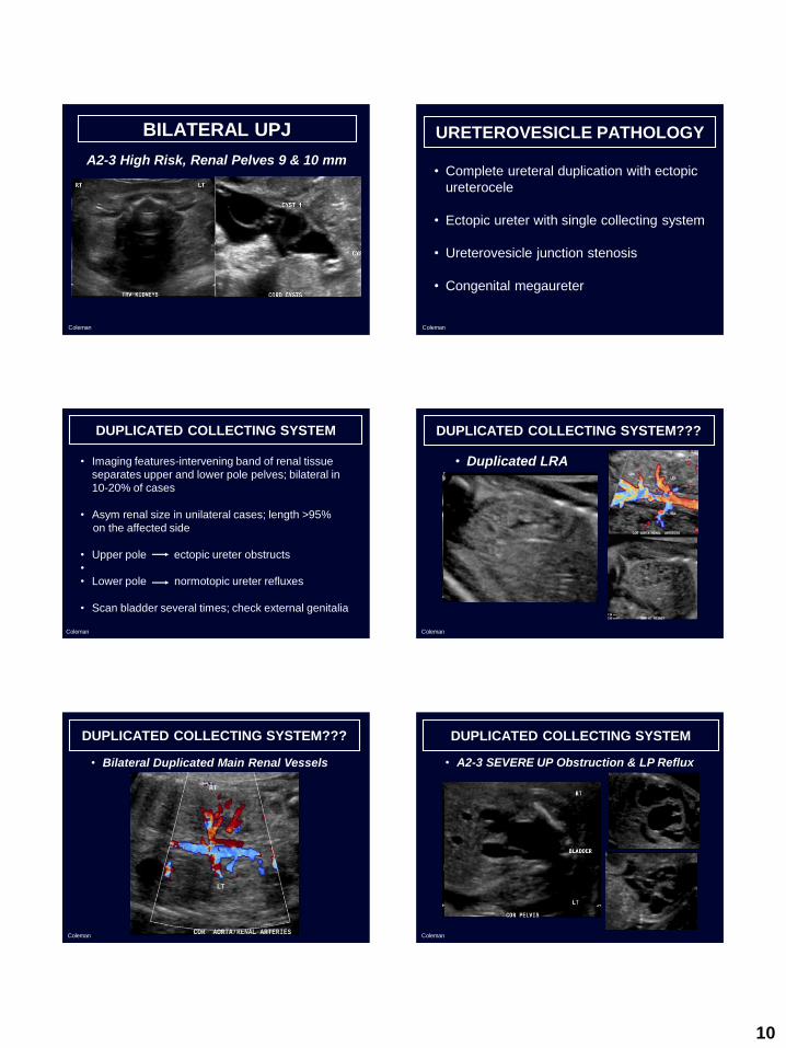
BILATERAL UPJ
Coleman
A2-3 High Risk, Renal Pelves 7 & 11 mm
10
BILATERAL UPJ
Coleman
A2-3 High Risk, Renal Pelves 9 & 10 mm
URETEROVESICLE PATHOLOGY
Coleman
• Complete ureteral duplication with ectopic
ureterocele
• Ectopic ureter with single collecting system
• Ureterovesicle junction stenosis
• Congenital megaureter
DUPLICATED COLLECTING SYSTEM
Coleman
• Imaging features-intervening band of renal tissue
separates upper and lower pole pelves; bilateral in
10-20% of cases
• Asym renal size in unilateral cases; length >95%
on the affected side
• Upper pole ectopic ureter obstructs
•
• Lower pole normotopic ureter refluxes
• Scan bladder several times; check external genitalia
DUPLICATED COLLECTING SYSTEM???
Coleman
• Duplicated LRA
DUPLICATED COLLECTING SYSTEM???
Coleman
• Bilateral Duplicated Main Renal Vessels
DUPLICATED COLLECTING SYSTEM
Coleman
• A2-3 SEVERE UP Obstruction & LP Reflux
11
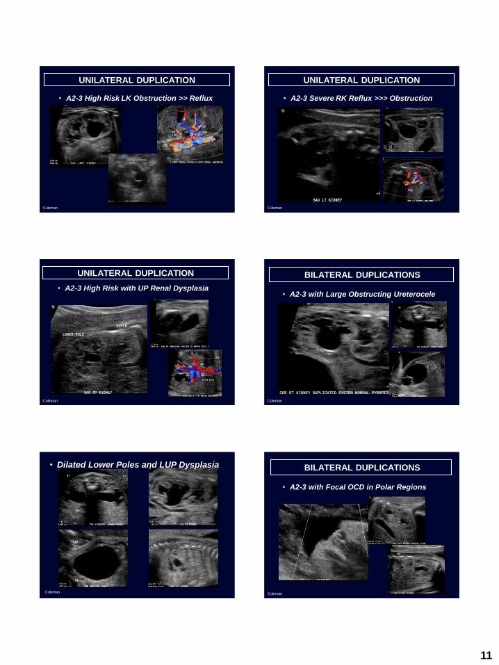
UNILATERAL DUPLICATION
Coleman
• A2-3 High Risk LK Obstruction >> Reflux
UNILATERAL DUPLICATION
Coleman
• A2-3 Severe RK Reflux >>> Obstruction
UNILATERAL DUPLICATION
Coleman
• A2-3 High Risk with UP Renal Dysplasia
BILATERAL DUPLICATIONS
Coleman
• A2-3 with Large Obstructing Ureterocele
Coleman
• Dilated Lower Poles and LUP Dysplasia BILATERAL DUPLICATIONS
Coleman
• A2-3 with Focal OCD in Polar Regions
12
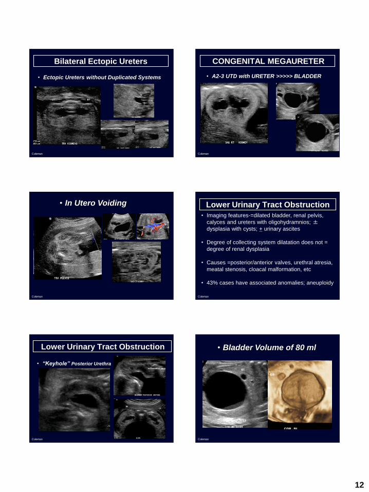
Bilateral Ectopic Ureters
Coleman
• Ectopic Ureters without Duplicated Systems
CONGENITAL MEGAURETER
Coleman
• A2-3 UTD with URETER >>>>> BLADDER
Coleman
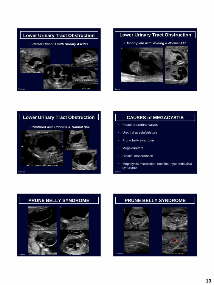
• In Utero Voiding Lower Urinary Tract Obstruction
Coleman
• Imaging features-=dilated bladder, renal pelvis,
calyces and ureters with oligohydramnios; ±dysplasia with cysts; + urinary ascites
• Degree of collecting system dilatation does not =
degree of renal dysplasia
• Causes =posterior/anterior valves, urethral atresia,
meatal stenosis, cloacal malformation, etc
• 43% cases have associated anomalies; aneuploidy
Lower Urinary Tract Obstruction
Coleman
• “Keyhole” Posterior Urethra
Coleman
• Bladder Volume of 80 ml
13
Coleman
• Patent Urachus with Urinary Ascites
Lower Urinary Tract Obstruction Lower Urinary Tract Obstruction
Coleman
• Incomplete with Voiding & Normal AFI
Lower Urinary Tract Obstruction
Coleman
• Ruptured with Urinoma & Normal DVP
CAUSES of MEGACYSTIS
Coleman
• Posterior urethral valves
• Urethral atresia/stricture
• Prune belly syndrome
• Megalourethra
• Cloacal malformation
• Megacystis-microcolon-intestinal hypoperistalsis
syndrome
PRUNE BELLY SYNDROME
Coleman
PRUNE BELLY SYNDROME
Coleman
14
Coleman
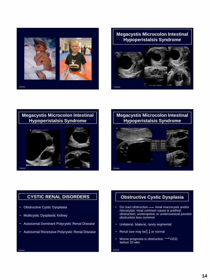
Megacystis Microcolon Intestinal
Hypoperistalsis Syndrome
Coleman
Megacystis Microcolon Intestinal
Hypoperistalsis Syndrome
Coleman
Megacystis Microcolon Intestinal
Hypoperistalsis Syndrome
Coleman
CYSTIC RENAL DISORDERS
Coleman
• Obstructive Cystic Dysplasia
• Multicystic Dysplastic Kidney
• Autosomal Dominant Polycystic Renal Disease
• Autosomal Recessive Polycystic Renal Disease
Obstructive Cystic Dysplasia
Coleman
• GU tract obstruction renal macrocysts and/or microcysts; most common cause is urethral obstruction; ureteropelvic or ureterovesical junction obstruction less common
• Unilateral, bilateral, rarely segmental
• Renal size may be , or normal
• Worse prognosis is obstruction OCD before 20 wks
15
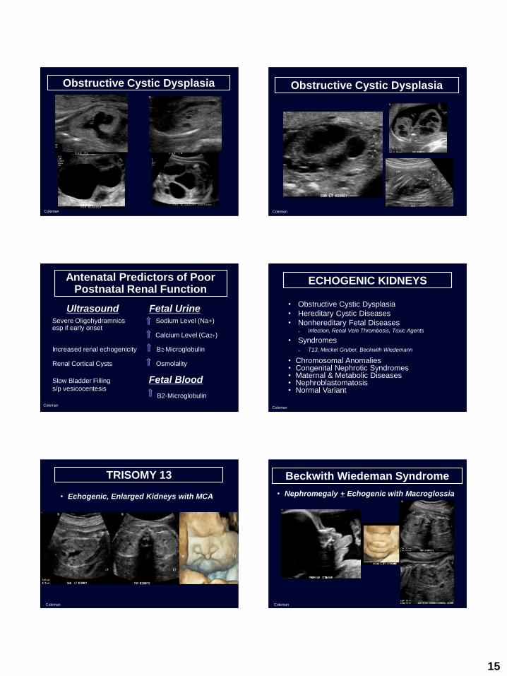
Obstructive Cystic Dysplasia
Coleman
Obstructive Cystic Dysplasia
Coleman
Antenatal Predictors of Poor Postnatal Renal Function
Ultrasound Fetal Urine
Coleman
Severe Oligohydramnios Sodium Level (Na+)esp if early onset
Calcium Level (Ca2+)
Increased renal echogenicity B2-Microglobulin
Renal Cortical Cysts Osmolality
Slow Bladder Filling Fetal Bloods/p vesicocentesis
B2-Microglobulin
ECHOGENIC KIDNEYS
• Obstructive Cystic Dysplasia
• Hereditary Cystic Diseases
• Nonhereditary Fetal Diseases Infection, Renal Vein Thrombosis, Toxic Agents
• Syndromes T13, Meckel Gruber, Beckwith Wiedemann
Coleman
• Chromosomal Anomalies• Congenital Nephrotic Syndromes• Maternal & Metabolic Diseases• Nephroblastomatosis• Normal Variant
TRISOMY 13
Coleman
• Echogenic, Enlarged Kidneys with MCA
Beckwith Wiedeman Syndrome
Coleman
• Nephromegaly + Echogenic with Macroglossia

16
NORMAL VARIANT
Coleman
• Echogenic Kidneys with Normal AFI & Renal Size
9 Mhz
12 Mhz
Sector
Linear
MULTICYSTIC DYSPLASTIC KIDNEY
Coleman
• Non functioning kidney in 90%; may be late
sequelae of renal obstruction
• Variants=pelvic, horseshoe or duplicated kidney
• MCDK usu involutes; rarely complicated by
infection, hypertension, Wilm’s tumor,
• F/U scans to check fluid, contralateral kidney
• Neonatal work up required in EVERY case!
Unilateral MCDK
Coleman
• 80% of cases, L>R renal involvement
Unilateral MCDK
• Cysts vary in size & may enlarge dramatically
Coleman
Cysts Random distribution Calyces in a row
Cyst Size Quite variable Uniform unless compound
Communication None Calyces infundibulum
Parenchyma Islands between cysts Intact, peripheral to calyces
Renal Shape Absent reniform contour Contour preserved
Contour preserved
FEATURE MCDK HYDRONEPHROSIS
Coleman
MULTICYSTIC DYSPLASTIC KIDNEY Unilateral MCDK
Coleman
• Random Cyst Distribution & Patent MRA
17
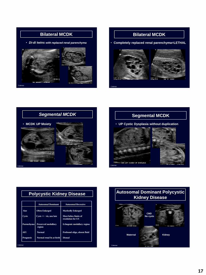
Bilateral MCDK
Coleman
• Di-di twins with replaced renal parenchyma
Bilateral MCDK
Coleman
• Completely replaced renal parenchyma=LETHAL
Segmental MCDK
• MCDK UP Moiety
Coleman
Segmental MCDK
• UP Cystic Dysplasia without duplication
Coleman
Polycystic Kidney Disease
Coleman
Size Often Enlarged Markedly Enlarged
Cysts Cysts +/- viz, usu late Most below limits of
resolution for US
Parenchyma Preserved medullary Echogenic medullary region
region
AFI Normal Profound oligo, absent fluid
Prognosis Normal renal fn at birth Dismal
Autosomal Dominant Autosomal Recessive
Autosomal Dominant Polycystic
Kidney Disease
Coleman
Maternal Kidney
CMD
No cysts

18
Autosomal Dominant Polycystic
Kidney Disease
6 MHz
12 MHz
9 MHzColeman
• Scattered Parenchymal Cysts
Autosomal Dominant Polycystic
Kidney Disease
5 MHz
9 MHz
Coleman
Autosomal Recessive
Polycystic Kidney Disease
Coleman
• Imaging Features = hyperechoic kidneys, ±1-2 mm cysts and echogenic parenchyma; kidneys may look normal up to 20 wks
• Kidney Size > 2 SD above the mean for the expected gestational age
• Oligohydramnios/anhydramnios with non-visualized bladder
• Check for signs of pulmonary hypoplasia such as TC and C/T ratio without cardiomegaly
Autosomal Recessive
Polycystic Kidney Disease
Coleman
• Parents are Known Carriers
12 MHz
5 MHz
Autosomal Recessive
Polycystic Kidney Disease
Coleman
• Severe Oligohydramnios
• Severe Biliary Ectasia
Coleman
19
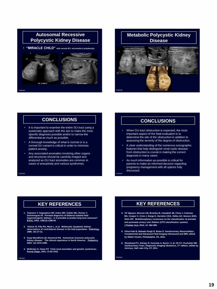
Autosomal Recessive
Polycystic Kidney Disease
Coleman
• “MIRACLE CHILD” with normal AFI, micromelia & polydactyly
Metabolic Polycystic Kidney
Disease
Coleman
CONCLUSIONS
• It is important to examine the entire GU tract using a
systematic approach with the aim to make the most
specific diagnosis possible and/or to narrow the
differential as much as possible.
• A thorough knowledge of what is normal or is a
normal GU variant is critical in order to minimize
patient anxiety.
• Any associated anomalies involving other organs
and structures should be carefully imaged and
analyzed as GU tract anomalies are common in
cases of aneuploidy and various syndromes.
Coleman
CONCLUSIONS
• When GU tract obstruction is expected, the most
important aspect of the fetal evaluation is to
determine the site of the obstruction in addition to
assessing the severity of the degree of obstruction.
• A clear understanding of the numerous sonographic
features that help distinguish renal cystic disease
from obstruction is crucial in making the correct
diagnosis in many cases.
• As much information as possible is critical for
parents to make an informed decision regarding
pregnancy management with all options fully
discussed.Coleman
KEY REFERENCES
1. Tsatsaris V, Gagnadoux MF, Aubry MD, Gubler MC, Dumez Y,
Dommergues M. Prenatal diagnosis of bilateral isolated fetal
hyperechogenic kidneys. Is it possible to predict long term outcome?
BJOG, 2002; 109(12):1388-93.
2. Kleiner B, Filly RA, Mack L, et al. Multicystic dysplastic kidney;
observations of contralateral disease in the fetal population. Radiology,
1986; 161:27-31.
3. Guay-Woodford LM, Desmond RA. Autosomal recessive polycystic
kidney disease: The clinical experience in North America. Pediatrics,
2003; 111:1072-1080.
4. Wellesley D, Howe DT. Fetal renal anomalies and genetic syndromes.
Prenat Diagn, 2001; 21:92-1033.
Coleman
KEY REFERENCES
5. HT Nguyen, Benson CB, Bromley B, Campbell JB, Chow J, Coleman
BG, Cooper C, Crino J, Darge K, Herndon CDA, Odibo AO, Somers MJG,
Stein DR: Multidisciplinary consensus on the classification of prenatal
and postnatal urinary tract dilation (UTD classification system).
J Pediatr Urol, 2014; 10: 982-999.
6. Kline-Fath B, Bahado-Singh R, Bulas D. Genitourinary Abnormalities:
Fundamental and Advanced Fetal Imaging Ultrasound and MRI, edited
by Walter Kluwer, Philadelphia, PA, 2015.
6. Woodward PJ, Sohaey R, Kennedy A, Byrne J L.B, Oh KY, Puchalski MD.
Genitourinary Tract: Diagnostic Imaging Obstetrics, 2nd edition, edited by
Amirsys, Salt Lake City, UT, 2011.
Coleman
20
SAD with My Diagnosis
Coleman
THRILLED with My Diagnosis
Coleman