the eye - sinoe medical association tmsinoemedicalassociation.org/ap4/eyemodels.pdf · the iris is...
TRANSCRIPT

The Eye

Human Anatomy, Frolich, Head II: Throat/Larynx
M&M, fig. 16.4

The eye is formed by three layers, or tunics. From the outside to the inside of the eyeball the three tunics are the
fibrous tunicfibrous tunic, which forms a capsule enclosing and protecting the other components of the eye.
It is subdivided into the sclera, with primarily structural functions, and the cornea, which is part of the optic
apparatus.
vascular tunic, which forms the choroid, the choroid, ciliaryciliarybody and irisbody and iris. This tunic is also called the uveal tract. The
choroid has primarily nutritive functions. The ciliary body generates the aqueous humor of the eye,
but the ciliary muscle also functions in the optic apparatus. The iris is part of the optic apparatus in which it functions
a contractile diaphragm, i.e. the aperture of the eye.neural tunic consists of the retina.
The retina proper forms the photoreceptive layer of the eye. As a double-layered epithelium, the retina also covers
the ciliary process and the posterior surface of the iris, where it has both nutritive and structural functions.
The ciliary and iridial parts of the retina are described together with the ciliary process and iris.


8
some pictures…
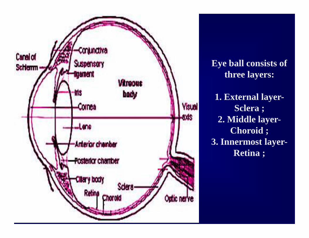
Eye ball consists of three layers:
1. External layer-Sclera ;
2. Middle layer-Choroid ;
3. Innermost layer-Retina ;

Human Anatomy, Frolich, Head II: Throat/Larynx
Movement of eye
Eye movement simulator
(http://cim.ucdavis.edu/eyes/version1/eyesim.ht
m)

11
Innervation

Extrinsic eye musclesMuscle Movement Nerve
Superior oblique
Depresses eye, turns laterally
IV (Trochlear)
Lateral rectus Turns laterally VI (Abducens)
Medial rectus Turns medially III (Oculomotor)
Superior rectus Elevates III (Oculomotor)
Inferior rectus Depresses eye III (Oculomotor)
Inferior oblique Elevates eye, turns laterally
III (Oculomotor)
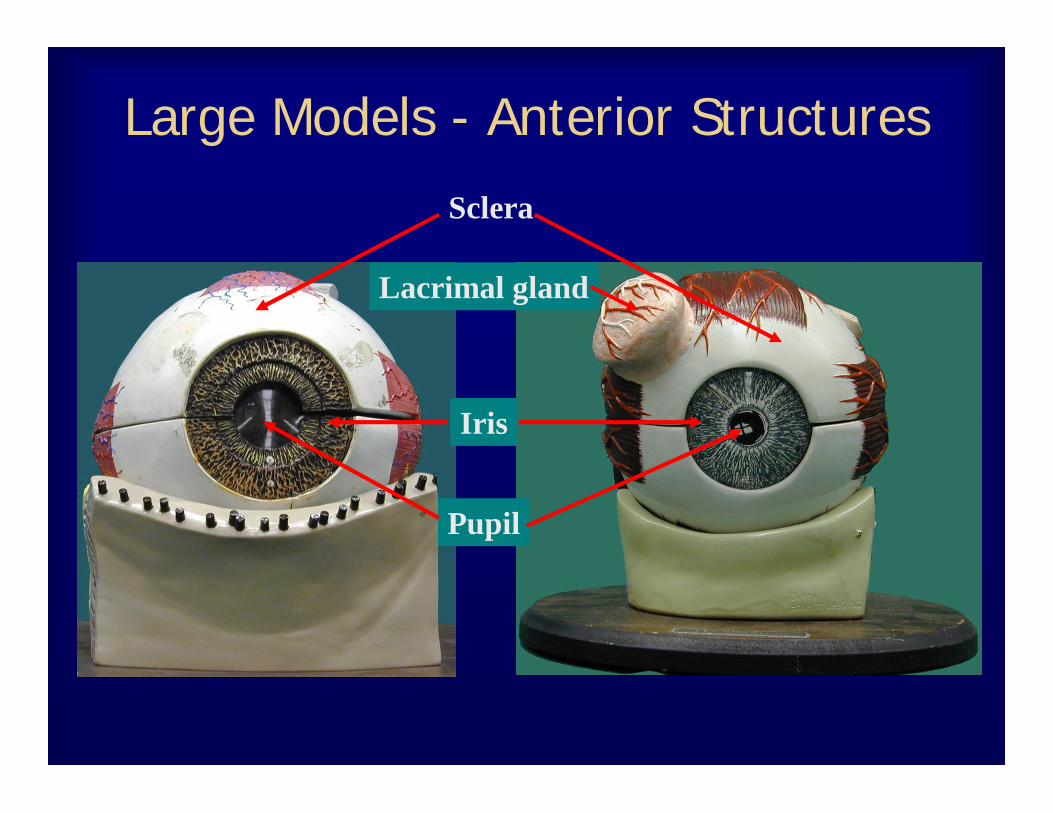
Large Models - Anterior Structures
Lacrimal gland
Sclera
Iris
Pupil

Large Models - Muscles (Anterior)
Medial rectus muscle
Superior rectus muscle
*Tendon of superior oblique muscle
Lateral rectusmuscle
*
Medial rectusmuscle

Small Models - Anterior Structures
Cornea
Sclera
Iris
Pupil
Iris
Sclera
Pupil
Choroid
RetinaModel Copyright ®Somso

Small Models - Muscles (Anterior)
Medial rectus muscle
Superior rectus muscle Tendon of superior oblique muscle
Lateral rectus muscle
Inferior rectus muscleInferior oblique muscle Medial rectus
muscle
Model Copyright ®Somso

Large Models – Medial ViewLacrimal gland
Tendon of Superior oblique muscle
Superior rectus muscle
Medial rectus muscle
Optic nerve
Optic nerve
Sclera
Choroid

Small Models – Medial ViewSuperior rectus muscle Tendon of
superior oblique muscle
Medial rectus muscle
Inferior rectus muscleSclera
Cornea
Inferior oblique muscle
Tendon of superior oblique muscle
Optic nerve
Model Copyright ®Somso

Large Models – Posterior StructuresLacrimal
glandSclera
Optic nerve
Choroid
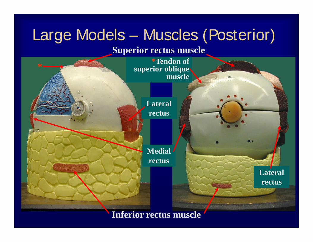
Large Models – Muscles (Posterior)Superior rectus muscle
*Tendon of superior oblique
muscle
Lateral rectus
Lateral rectus
Medial rectus
Inferior rectus muscle
*
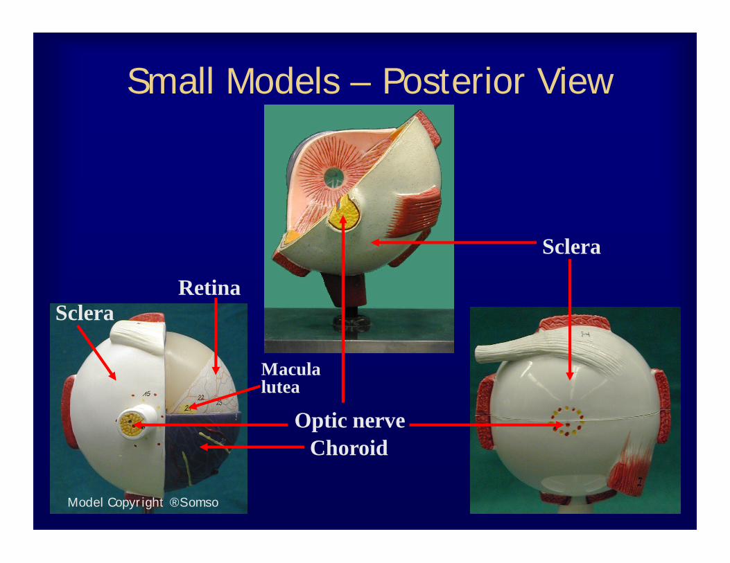
Small Models – Posterior View
Optic nerve
Sclera
Choroid
RetinaSclera
Macula lutea
Model Copyright ®Somso
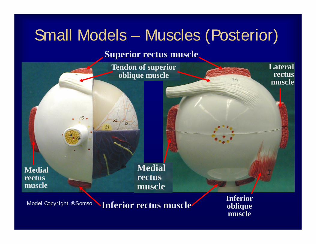
Small Models – Muscles (Posterior)Superior rectus muscle
Tendon of superior oblique muscle
Medial rectus muscle
Inferior rectus muscle
Medial rectus muscle
Inferior oblique muscle
Lateral rectus
muscle
Model Copyright ®Somso

Large Models – LateralSuperior rectus muscle
Inferior oblique muscle
Lateral rectus muscle
Lateral rectus muscle
Lacrimal glandScleraOptic
nerve Optic nerve
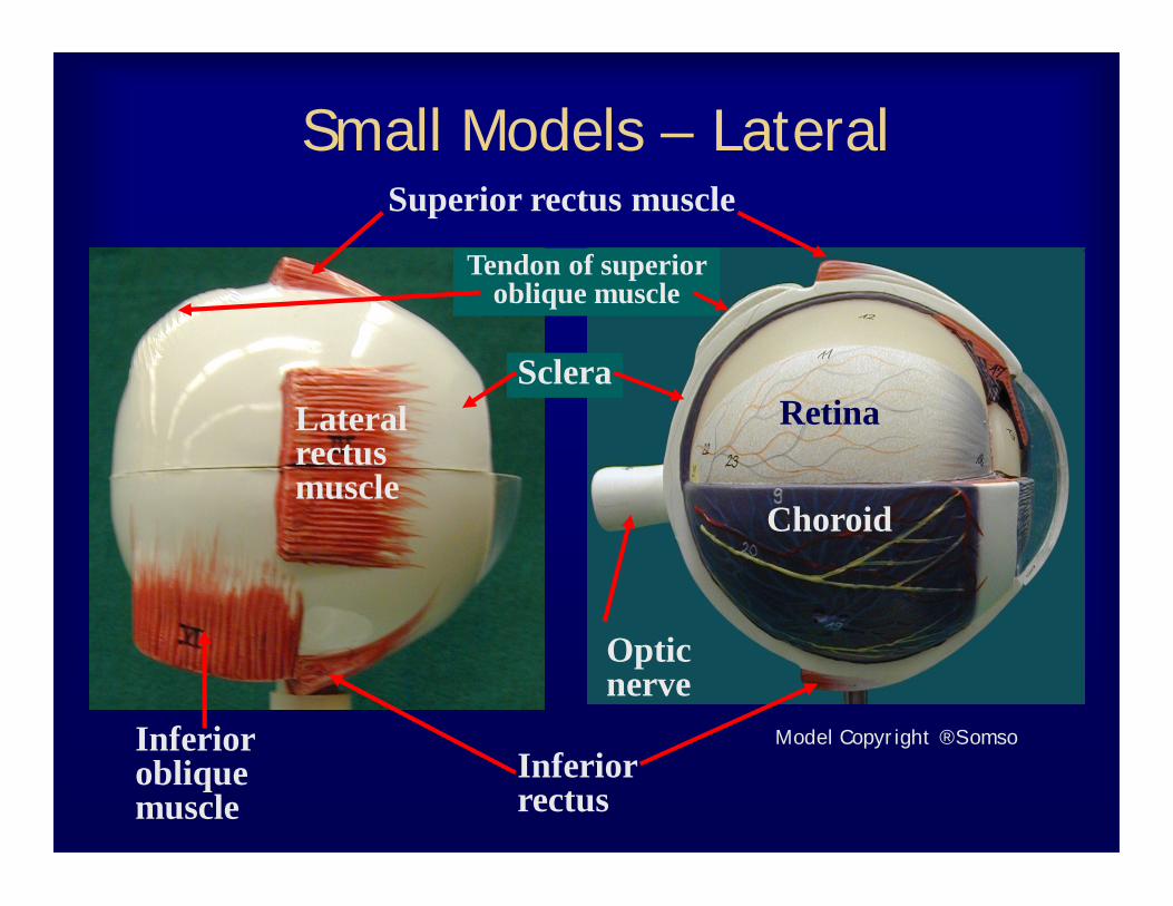
Small Models – LateralSuperior rectus muscle
Tendon of superior oblique muscle
Lateral rectus muscle
Sclera
Optic nerve
Inferior oblique muscle
Inferior rectus
Retina
Choroid
Model Copyright ®Somso

Eye Wall Layers and Chambers
Posterior cavity
Vitreous humor
Sclera
Choroid
Retina
Cornea
CorneaAnterior cavity
Anterior cavity
Model Copyright ®Somso

Internal StructuresCiliary body
Lens
Suspensory ligaments
Fovea centralis
Optic disk
Iris
Model Copyright ®Somso

Cow Eye
Optic nerve
Cornea
• Choroid is darkly pigmented layer• Pupil is opening in iris• Retina is partially detached
Posterior cavity
Iris Iris Retina
ScleraSclera

Sheep Eye
Optic nerve
Cornea
• Choroid is darkly pigmented layer
• Pupil is opening in iris
• Retina is partially detached
Posterior cavity
IrisRetina
Sclera
ScleraOptic nerve
Lens
Lens
Cornea
Cornea

Vision Tests
Snellen-Visual Acuity Chart
Ishihara Color Plates
Astigmatism Chart
Blind Spot Test Figure

Large Eye Model with Lid
http://daphne.palomar.edu/ccarpenter/Models/eye_3b.htmhttp://www.highlands.edu/academics/divisions/scipe/biology/labs/cartersville/2121/senses.htm

31
1. (outer layer) Fibrous: dense connective tissue Sclera – white of the eye Cornea
2. (middle) Vascular: uvea Choroid – posterior, pigmented Ciliary body
Muscles – control lens shape Processes – secrete aqueous
humor Zonule (attaches lens)
Iris
3. (inner layer) Sensory Retina and optic nerve

Figure 15.4a
Central arteryand vein ofthe retina
Optic disc(blind spot)
Optic nervePosterior poleFovea centralisMacula luteaRetinaChoroidSclera
Ora serrata
(a) Diagrammatic view. The vitreoushumor is illustrated only in the
bottom part of the eyeball.
Ciliary body
Ciliary zonule suspensoryligament)
CorneaIris
Anterior polePupil
Anterior segment (contains aqueous humor)
LensScleral venous sinus
Posterior segment(contains vitreous humor)


Posterior View of the Anterior Half of the Eye
Figure 16.9a

The Vascular Tunic