the evolution of the placenta
TRANSCRIPT

REPRODUCTION
© 2016 Society for Reproduction and Fertility DOI: 10.1530/REP-16-0325ISSN 1470–1626 (paper) 1741–7899 (online) Online version via www.reproduction-online.org
REVIEW
The evolution of the placenta
R Michael Roberts1,2, Jonathan A Green2 and Laura C Schulz3
1C.S. Bond Life Sciences Center, University of Missouri, Columbia, Missouri, USA, 2Division of Animal Sciences, University of Missouri, Columbia, Missouri, USA, and 3Department of Obstetrics, Gynecology and Women’s Health, University of Missouri School of Medicine, Columbia, Missouri, USA
Correspondence should be addressed to R M Roberts; Email: [email protected]
Abstract
The very apt definition of a placenta is coined by Mossman, namely apposition or fusion of the fetal membranes to the uterine mucosa for physiological exchange. As such, it is a specialized organ whose purpose is to provide continuing support to the developing young. By this definition, placentas have evolved within every vertebrate class other than birds. They have evolved on multiple occasions, often within quite narrow taxonomic groups. As the placenta and the maternal system associate more intimately, such that the conceptus relies extensively on maternal support, the relationship leads to increased conflict that drives adaptive changes on both sides. The story of vertebrate placentation, therefore, is one of convergent evolution at both the macromolecular and molecular levels. In this short review, we first describe the emergence of placental-like structures in nonmammalian vertebrates and then transition to mammals themselves. We close the review by discussing the mechanisms that might have favored diversity and hence evolution of the morphology and physiology of the placentas of eutherian mammals.Reproduction (2016) 152 R179–R189
10.1530/REP-16-0325
Introduction
Viviparity has evolved independently and seemingly on multiple occasions across a diverse array of animal groups, including invertebrates (Kalinka 2015). It is a phenomenon whereby developing embryos are retained within the reproductive tract, leading to release of live offspring as an alternative to the more fecund egg laying or spawning. One consequence of viviparity is that the retained, fertilized egg must either survive off its own reserves, usually yolk, or obtain some or part of these resources from the mother. The latter situation, of necessity, is expected to lead to increased conflict over how provisions are partitioned between the supplier and the recipient, thus potentially sparking a genetic arms race. In turn, such conflict is expected to drive adaptive changes that lead to a more intimate and possibly more felicitous relationship between offspring and the reproductive tract of the mother that favors the transmission of both maternal and paternal genes to the next generation, despite a loss of overall fecundity.
The transition from oviparity to viviparity and the subsequent emergence of placentation within some vertebrate taxa clearly required major changes in the morphology and physiology of the reproductive tract and has its origins well before the advent of mammals (Van Dyke et al. 2014, Blackburn 2015). A placenta, as defined originally by Mossman, is the ‘apposition or fusion of the fetal membranes to the uterine mucosa for physiological exchange’ (Burton & Jauniaux 2015).
It is a specialized organ whose purpose is to provide continuing support to the developing young through the provision of water, nutrients and gases, and to regulate maternal–fetal interactions often through hormone production. By this definition, placentas have evolved within every vertebrate class other than birds. Although placentation arose once in the common ancestor of mammals, it has arisen independently multiple times within other classes, and even families. The story of vertebrate placentation, therefore, is one of convergent evolution at both the macromolecular and molecular levels. In this short review of placental evolution, we first describe the emergence of placental-like structures in nonmammalian vertebrates and then transition to mammals themselves. We close the review by discussing mechanisms that might have favored diversity and hence evolution of the morphology and physiology of the placentas of eutherian mammals. Of necessity, many important references cannot be cited in a short review of this kind. Instead, we have attempted to direct the reader to scholarly articles that do list the primary source material.
Nonmammalian vertebrates
Cartilaginous fish
Viviparity is the most common mode of reproduction in elasmobranchs. However, there is a wide range in the degree of maternal provisioning of the embryo after
Downloaded from Bioscientifica.com at 10/22/2021 03:22:52PMvia free access

R180 R M Roberts and others
Reproduction (2016) 152 R179–R189 www.reproduction-online.org
ovulation (Wourms & Lombardi 1992), with embryos of some species depending entirely on the yolk sac for all of pregnancy, e.g. the spiny dogfish, which at birth weighs 40% less than the yolk-filled fertilized egg from which it develops. The tiger shark, by contrast, solves such limiting provisioning; in which the dominant embryo eats all of its littermates and any unfertilized eggs before birth (Korsgaard & Weber 1989).
In the elasmobranchs, two placental types are observed. Among stingrays, finger-like projections of the uterine wall, termed trophonemata, provide histotrophic nutrition to the developing embryo (Hamlett et al. 1993), while the epithelium overlying uterine blood vessels thins, lessening the barrier to exchange. The mature embryo of the cownose ray, for example, weighs 3000 times as much as the egg as a result of reliance on the mother rather than the egg yolk (Hamlett et al. 1993). A different placental type, the yolk sac placenta, has evolved multiple times in the ground sharks (Wourms & Lombardi 1992). After the yolk has been consumed, the yolk sac becomes modified into an umbilical cord region and a placental region (Hamlett et al. 1993), which become closely apposed to the epithelium of a highly vascularized oviduct.
Teleosts
In Poeciliopsis fish, eggs are retained within the follicle after fertilization and throughout embryonic development (Wourms & Lombardi 1992). Most species are lecithotrophic, relying on yolk, but in others, a highly folded, follicular epithelium forms a ‘follicular placenta’ with the embryonic pericardial sac. This epithelial layer is lined with microvilli, while the surrounding tissue is highly vascularized, presumably to facilitate maternal–fetal exchange (Kwan et al. 2015). This type of placental structure has arisen several times during Poeciliopsis evolution (Kwan et al. 2015) and has been subsequently lost and regained many times, making it a model for the evolution of placentation (Pollux et al. 2014).
Among syngnathid fish (pipefish and seahorses) the males, and not the females, become pregnant, after the female transfers the fertilized eggs to a brood pouch on the ventral side of either the trunk (Gastrophori) or the tail (Urophori) of the male’s body. The degree of contact and exchange between developing embryos and the father range from minimal to situations that display all the defining features of placentation (Wilson et al. 2001). For example, in the straight-nosed pipe fish Nerophis ophidion, eggs stick to the epithelium of the father’s pouch, which is open to the sea environment (Carcupino et al. 2002), while in the pot-bellied seahorse Hippocampus abdominalis, the pouch is sealed and its epithelium highly folded, thinned and vascularized (Carcupino et al. 2002), thereby allowing exchange of nutrients.
Amphibians
Viviparity has arisen multiple times in amphibians, accompanied in some cases with the evolution of placentas. In frogs, there are examples in which the tadpoles develop in the father’s mouth, in the mother’s stomach, or on the skin of the back (Wake 1993). In the marsupial frogs, development occurs inside a specialized maternal pouch on the back of the animal. Following ovulation, secretory cells of the vascularized pouch enlarge. The pouch epithelium is closely apposed to specialized fetal gills, allowing exchange between maternal and fetal circulations (Savage 2002). This, as A.M. Carter wrote, ‘would satisfy most people’s definition of a placenta’ (Carter 2016). Arguably, however, a structure found outside the reproductive tract, especially following external fertilization that occurs in these frogs and the sea horses described earlier, does not fit the typical definition of a placenta. Nor have any of the ‘gill placentas’ of these endangered frog species yet been shown to be the primary source of fetal nutrient support.
Reptiles
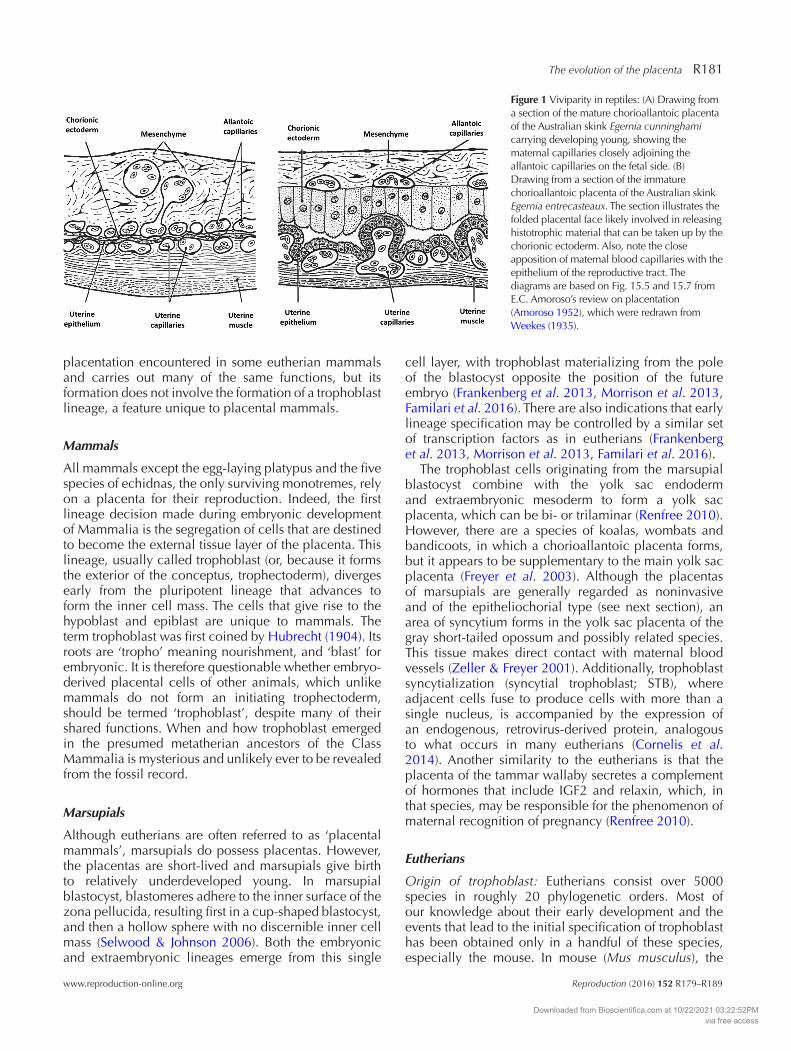
Viviparity occurs in more than 25 families of lizards and snakes (squamate reptiles). Although estimated by some to have arisen independently more than one hundred times, it is conceivable that there was a single origin of viviparity and multiple subsequent reversions to oviparity (Pyron & Burbrink 2014, Van Dyke et al. 2014, Blackburn 2015). In many species, the embryo relies on egg contents for nutrition, but in others a range of adaptations of the female reproductive tract provides a means to exchange gases and nutrients with the conceptus (Stewart 2015). In squamates the placenta is chorioallantoic, but unlike mammals it does not develop from an early arising, extraembryonic trophoblast layer. Most squamate placentas demonstrate a simple interdigitation of the chorioallantois with the uterine epithelium (Stewart 2015), but in some skinks the interface is more intimate (Fig. 1). In the case of the African skink Trachylepis ivensi, there is even a degree of invasive implantation, with direct contact between chorionic projections and maternal capillary endothelium (Blackburn & Flemming 2012).
The chorioallantoic placenta of viviparous lizards can be highly efficient in facilitating exchange of nutrients and gases. Offspring of the Mabuya lizard weigh 500 times more at birth than the egg at fertilization, indicating a nearly complete reliance for growth on maternally supplied materials (Thompson & Speake 2002). There is also evidence for the expression of active transporters at the sites of maternal–fetal apposition in some species (Murphy et al. 2011) and for placental production of steroid hormones (Painter & Moore 2005). Overall, where placentation occurs in squamates, it bears a superficial resemblance to the epitheliochorial
Downloaded from Bioscientifica.com at 10/22/2021 03:22:52PMvia free access
The evolution of the placenta R181
www.reproduction-online.org Reproduction (2016) 152 R179–R189
placentation encountered in some eutherian mammals and carries out many of the same functions, but its formation does not involve the formation of a trophoblast lineage, a feature unique to placental mammals.
Mammals
All mammals except the egg-laying platypus and the five species of echidnas, the only surviving monotremes, rely on a placenta for their reproduction. Indeed, the first lineage decision made during embryonic development of Mammalia is the segregation of cells that are destined to become the external tissue layer of the placenta. This lineage, usually called trophoblast (or, because it forms the exterior of the conceptus, trophectoderm), diverges early from the pluripotent lineage that advances to form the inner cell mass. The cells that give rise to the hypoblast and epiblast are unique to mammals. The term trophoblast was first coined by Hubrecht (1904). Its roots are ‘tropho’ meaning nourishment, and ‘blast’ for embryonic. It is therefore questionable whether embryo-derived placental cells of other animals, which unlike mammals do not form an initiating trophectoderm, should be termed ‘trophoblast’, despite many of their shared functions. When and how trophoblast emerged in the presumed metatherian ancestors of the Class Mammalia is mysterious and unlikely ever to be revealed from the fossil record.
Marsupials
Although eutherians are often referred to as ‘placental mammals’, marsupials do possess placentas. However, the placentas are short-lived and marsupials give birth to relatively underdeveloped young. In marsupial blastocyst, blastomeres adhere to the inner surface of the zona pellucida, resulting first in a cup-shaped blastocyst, and then a hollow sphere with no discernible inner cell mass (Selwood & Johnson 2006). Both the embryonic and extraembryonic lineages emerge from this single
cell layer, with trophoblast materializing from the pole of the blastocyst opposite the position of the future embryo (Frankenberg et al. 2013, Morrison et al. 2013, Familari et al. 2016). There are also indications that early lineage specification may be controlled by a similar set of transcription factors as in eutherians (Frankenberg et al. 2013, Morrison et al. 2013, Familari et al. 2016).
The trophoblast cells originating from the marsupial blastocyst combine with the yolk sac endoderm and extraembryonic mesoderm to form a yolk sac placenta, which can be bi- or trilaminar (Renfree 2010). However, there are a species of koalas, wombats and bandicoots, in which a chorioallantoic placenta forms, but it appears to be supplementary to the main yolk sac placenta (Freyer et al. 2003). Although the placentas of marsupials are generally regarded as noninvasive and of the epitheliochorial type (see next section), an area of syncytium forms in the yolk sac placenta of the gray short-tailed opossum and possibly related species. This tissue makes direct contact with maternal blood vessels (Zeller & Freyer 2001). Additionally, trophoblast syncytialization (syncytial trophoblast; STB), where adjacent cells fuse to produce cells with more than a single nucleus, is accompanied by the expression of an endogenous, retrovirus-derived protein, analogous to what occurs in many eutherians (Cornelis et al. 2014). Another similarity to the eutherians is that the placenta of the tammar wallaby secretes a complement of hormones that include IGF2 and relaxin, which, in that species, may be responsible for the phenomenon of maternal recognition of pregnancy (Renfree 2010).
Eutherians
Origin of trophoblast: Eutherians consist over 5000 species in roughly 20 phylogenetic orders. Most of our knowledge about their early development and the events that lead to the initial specification of trophoblast has been obtained only in a handful of these species, especially the mouse. In mouse (Mus musculus), the
Figure 1 Viviparity in reptiles: (A) Drawing from a section of the mature chorioallantoic placenta of the Australian skink Egernia cunninghami carrying developing young, showing the maternal capillaries closely adjoining the allantoic capillaries on the fetal side. (B) Drawing from a section of the immature chorioallantoic placenta of the Australian skink Egernia entrecasteaux. The section illustrates the folded placental face likely involved in releasing histotrophic material that can be taken up by the chorionic ectoderm. Also, note the close apposition of maternal blood capillaries with the epithelium of the reproductive tract. The diagrams are based on Fig. 15.5 and 15.7 from E.C. Amoroso’s review on placentation (Amoroso 1952), which were redrawn from Weekes (1935).
Downloaded from Bioscientifica.com at 10/22/2021 03:22:52PMvia free access

R182 R M Roberts and others
Reproduction (2016) 152 R179–R189 www.reproduction-online.org
first visible differentiation event, called compaction, occurs between the 8 and 16 cell stage of embryonic development, when the blastomeres polarize to form extended contact zones with their neighbors and an outward-facing apical surface (Posfai et al. 2014). Further symmetrical and asymmetrical divisions follow, so that by the 16 cell morula stage a population of polarized cells exist on the cell surface, which becomes trophectoderm, and another population of cells are positioned inside, which is the precursor of the inner cell mass (ICM). By the time of blastocyst formation, when the mouse conceptus comprises about 32 cells, about two-thirds of which are trophectoderm, the precursors of extraembryonic endoderm (hypoblast) have begun to segregate toward the base of the ICM. From this location, they migrate outward to line the trophectoderm, forming the yolk sac cavity. Later, extraembryonic mesoderm joins with the trophectoderm to form what at this stage is a bilayered chorion. Although there are clear morphological similarities in the steps leading to blastocyst formation in rodents, humans and domestic mammalian species, there are differences in how these events are timed and subsequently played out. In many species, for example, mesoderm differentiates to vascularize the yolk sac, forming a yolk sac placenta that is eventually replaced by the chorioallantoic placenta. Such a placenta does not form in humans, but persists to term in rodents and lagomorphs. Similarly, compared with the mouse, conceptuses from many species undergo at least one further round of cell division before the blastocoel cavity becomes visible. For details on how placentas subsequently develop and begin to diverge in morphology, the reader is directed to the still germane reviews by Amoroso (1952) and Renfree (1982).
Range of placental morphologies: Despite the spectacular diversity encountered in placental structure at both the gross and microanatomical levels (Enders & Carter 2004), there have been attempts over the years to classify placentas and (somewhat unsuccessfully) to relate the outcomes to mammalian phylogeny. For
example, placentas come in a range of shapes and sizes, and have been placed in groups based on the general gross morphologies of the sites where the chorion attaches to the endometrium (Fig. 2). They have also been classified according to how many cell layers separate the maternal and fetal circulations (Fig. 2). A simplified version of this system recognizes three main placental types (Renfree 1982): epitheliochorial, in which there is no erosion of the uterine epithelium; endotheliochorial, where the invading trophoblast reaches but does not penetrate maternal capillaries within the endometrium; and hemochorial, where the trophoblast surface is in direct contact with maternal blood (Fig. 3). A fourth type, synepitheliochorial, has been recently used to describe the placentas of ruminants (Wooding 1992). Here, specialized trophoblast binucleated cells fuse with uterine epithelial cells to form trinucleated cells, a syncytium, or both. The placentas of ruminants, with the exception of the phylogenetically more primitive tragulids, are also characterized as cotyledonary (Fig. 2). Cotyledons are the primary sites of attachment of the chorion to the endometrium and comprise tufts of villous trophoblast that interdigitate and interlock with complementary structures on the endometrium, called caruncles. It is within these sites, often known as placentomes, that binucleated cells, placental and maternal blood capillaries, and placental interchange are most concentrated. Therefore, this kind of placenta is a derived form, secondary to the more superficial, diffuse epitheliochorial type typical of most other artiodactyls.
Superficial vs invasive placentation: Intuitively, it might be inferred that the least invasive types of placenta, the kind where multiple cell layers separate the two blood supplies (Fig. 2B), would be the most inefficient and also the most primitive of all placental types because there are more apparent physical barriers to limit the movement of nutrients and dissolved gases. Also, such placentas have a resemblance to placentas described earlier for nonmammalian species, such as skinks (Fig. 3). However, this is clearly not the case
Figure 2 Cartoon illustrating the diversity of placental morphologies encountered in placental mammals.
Downloaded from Bioscientifica.com at 10/22/2021 03:22:52PMvia free access

The evolution of the placenta R183
www.reproduction-online.org Reproduction (2016) 152 R179–R189
(Wildman et al. 2006). Compare, for example, the relative maturity and independence of a newborn horse, pig or whale, supported during their gestations by diffuse epitheliochorial placentas, to a hapless pup born to a mouse or a baby born to a human, species where the placenta is hemochorial and discoid. Nor is the more superficial form of placenta evolutionarily more ancient than the kinds that are invasive and make direct contact with maternal blood. On the contrary, careful analyses, in which placental features were mapped to mammalian phylogenies, have clearly dispelled this myth and demonstrated that the epitheliochorial placenta is not only a derived form that has evolved from a more invasive placental type, but has arisen independently in three distinct mammalian lineages, including primates (Wildman et al. 2006) (Fig. 4). Moreover, this and similar trees based on other features of the placenta strongly imply that the placenta of the last common eutherian ancestor was discoid, either hemochorial or endotheliochorial (Mess & Carter 2007), and possessed a labyrinth-type of placental interdigitation.
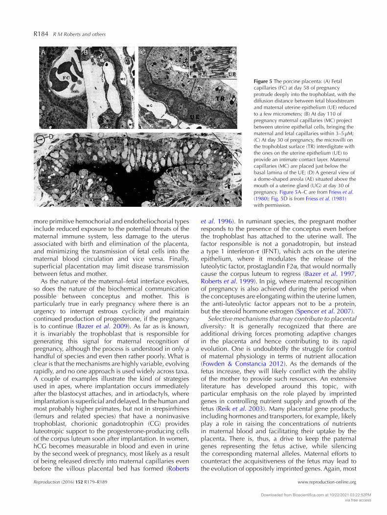
It should be stressed that the perceived disadvantages of extended diffusion distances for dissolved gases and solutes encountered with a superficial placenta, like that of the pig, are largely illusory. As pregnancy proceeds, the surfaces of the uterine and trophoblast epithelial layers become interlocked (Fig. 5B). Capillaries on the maternal and fetal sides come within a few microns of each other (Fig. 5A), often squeezing down to the tight junctions between the trophoblast cells on the one side and uterine epithelial cells on the other, probably allowing efficient exchange of small molecules (Friess et al. 1980). Movement of macromolecules that carry essential components such as metals and vitamins is more problematic, but an alternative strategy has been
employed, namely the delivery of these components not in the blood but in uterine secretions – a process called histotrophic nutrition (Fig. 5C) (Renegar et al. 1982). The uterine glands of the pig, for example, release uteroferrin, a bi-iron containing acid phosphatase to supply iron. Uteroferrin and other proteins with a similar provisioning function are taken up by specialized regions of endocytic trophoblast cells congregated in cup-like structures called areolae, which develop opposite the mouths of each uterine gland (Fig. 5C). From there, the maternal factors are transported via the fetal bloodstream to the liver and other organs for processes such as hematopoiesis (Renegar et al. 1982). The likely advantages of a noninvasive placenta over the
Figure 3 Cartoon indicating how placentas can be classified according to the numbers and kinds of cell layers that separate the bloodstreams of the mother and conceptus.
Figure 4 The evolution of the placental interface in terms of degree of invasiveness of the placental tissue into maternal tissue, with epitheliochorial being the least invasive and hemochorial being most invasive. From Fig. 2 of Wildman et al. (2006), with permission; Copyright (2006) National Academy of Sciences, USA.
Downloaded from Bioscientifica.com at 10/22/2021 03:22:52PMvia free access
R184 R M Roberts and others
Reproduction (2016) 152 R179–R189 www.reproduction-online.org
more primitive hemochorial and endotheliochorial types include reduced exposure to the potential threats of the maternal immune system, less damage to the uterus associated with birth and elimination of the placenta, and minimizing the transmission of fetal cells into the maternal blood circulation and vice versa. Finally, superficial placentation may limit disease transmission between fetus and mother.
As the nature of the maternal–fetal interface evolves, so does the nature of the biochemical communication possible between conceptus and mother. This is particularly true in early pregnancy where there is an urgency to interrupt estrous cyclicity and maintain continued production of progesterone, if the pregnancy is to continue (Bazer et al. 2009). As far as is known, it is invariably the trophoblast that is responsible for generating this signal for maternal recognition of pregnancy, although the process is understood in only a handful of species and even then rather poorly. What is clear is that the mechanisms are highly variable, evolving rapidly, and no one approach is used widely across taxa. A couple of examples illustrate the kind of strategies used in apes, where implantation occurs immediately after the blastocyst attaches, and in artiodactyls, where implantation is superficial and delayed. In the human and most probably higher primates, but not in strepsirrhines (lemurs and related species) that have a noninvasive trophoblast, chorionic gonadotrophin (CG) provides luteotropic support to the progesterone-producing cells of the corpus luteum soon after implantation. In women, hCG becomes measurable in blood and even in urine by the second week of pregnancy, most likely as a result of being released directly into maternal capillaries even before the villous placental bed has formed (Roberts
et al. 1996). In ruminant species, the pregnant mother responds to the presence of the conceptus even before the trophoblast has attached to the uterine wall. The factor responsible is not a gonadotropin, but instead a type 1 interferon-τ (IFNT), which acts on the uterine epithelium, where it modulates the release of the luteolytic factor, prostaglandin F2α, that would normally cause the corpus luteum to regress (Bazer et al. 1997, Roberts et al. 1999). In pig, where maternal recognition of pregnancy is also achieved during the period when the conceptuses are elongating within the uterine lumen, the anti-luteolytic factor appears not to be a protein, but the steroid hormone estrogen (Spencer et al. 2007).
Selective mechanisms that may contribute to placental diversity: It is generally recognized that there are additional driving forces promoting adaptive changes in the placenta and hence contributing to its rapid evolution. One is undoubtedly the struggle for control of maternal physiology in terms of nutrient allocation (Fowden & Constancia 2012). As the demands of the fetus increase, they will likely conflict with the ability of the mother to provide such resources. An extensive literature has developed around this topic, with particular emphasis on the role played by imprinted genes in controlling nutrient supply and growth of the fetus (Reik et al. 2003). Many placental gene products, including hormones and transporters, for example, likely play a role in raising the concentrations of nutrients in maternal blood and facilitating their uptake by the placenta. There is, thus, a drive to keep the paternal genes representing the fetus active, while silencing the corresponding maternal alleles. Maternal efforts to counteract the acquisitiveness of the fetus may lead to the evolution of oppositely imprinted genes. Again, most
Figure 5 The porcine placenta: (A) Fetal capillaries (FC) at day 58 of pregnancy protrude deeply into the trophoblast, with the diffusion distance between fetal bloodstream and maternal uterine epithelium (UE) reduced to a few micrometers; (B) At day 110 of pregnancy maternal capillaries (MC) project between uterine epithelial cells, bringing the maternal and fetal capillaries within 3–5 μM; (C) At day 30 of pregnancy, the microvilli on the trophoblast surface (TR) interdigitate with the ones on the uterine epithelium (UE) to provide an intimate contact layer. Maternal capillaries (MC) are placed just below the basal lamina of the UE; (D) A general view of a dome-shaped areola (AE) situated above the mouth of a uterine gland (UG) at day 30 of pregnancy. Figure 5A–C are from Friess et al. (1980); Fig. 5D is from Friess et al. (1981) with permission.
Downloaded from Bioscientifica.com at 10/22/2021 03:22:52PMvia free access

The evolution of the placenta R185
www.reproduction-online.org Reproduction (2016) 152 R179–R189
speculation on the role of imprinted genes has come from the mouse, with roles inferred for the human, both species with an invasive placenta (Reik et al. 2003). Very little is known about imprinting in those species where there is little or no direct access of the trophoblast to maternal blood, although data are beginning to emerge (Chen et al. 2016).
A second force driving placental evolution is undoubtedly the avoidance of immune rejection (Hemberger 2013), a process still little understood, although speculation abounds. It is assumed that to minimize attention from various arms of the maternal immune system, the trophoblast must continue to evolve counter-measures for its own protection and even advantage. These measures will undoubtedly be subtle and complex and change as the degree of intimacy with maternal system itself evolves. Some clues may emerge by examining candidates encoded by rapidly evolving genes and gene families within individual mammalian clades, some of which are discussed in the next section.
Evolution of placenta-specific genes
Despite the observation that the initial lineage regulators for trophoblast cell fate decisions may be at least partially conserved across mammals, the resulting placental structures and their associated trophoblast cell populations appear not to be governed by particular master genes. There are a few coding genes, e.g., GCM1, a transcription factor that plays a role in controlling the formation of syncytial trophoblast (STB) (Cross et al. 2003) that appear to have an entirely placental function in the sense that they have not been implicated in developmental events outside the trophoblast lineage. However, it seems likely that much of the structural diversity and functional refinements associated with mammalian placentas depend on widely expressed transcription factors operating in a combinatorial, but trophoblast-specific manner and poorly understood processes linked to peculiarities in the epigenetic landscape of trophoblast. There have also been a large number of gene family expansions (Rawn & Cross 2008). These gene families may be performing roles in processes such as maternal recognition of pregnancy, nutrient acquisition and immune protection, thereby contributing to the unique functional needs of different placental forms. It is worth noting that ‘placenta-specific’ in this context means that such genes are disproportionately, but not necessarily uniquely, expressed in the placenta. For example, it is not unusual for some placenta-specific gene products to be elevated in cancer and germ cells.
Placenta-specific gene families
Gene duplication provides a useful opportunity for natural selection to operate (Hughes 1994) and allows
the duplicated genes to acquire new functions, and, through the acquisition or loss of regulatory regions, changes in the temporal, spatial, or magnitude of transcription compared with the original gene. Gene family expansions associated with distinct placental types have arisen repeatedly in eutherians (Rawn & Cross 2008). Here, we provide four examples from the many known placenta-specific gene families. In general, assigning function of individual members within a family will be difficult. Indeed, knockouts in mice performed on single members of gene families have generally not been informative, possibly because phenotype is not obviously impaired within the friendly confines of a vivarium, although it may become so when the pregnant animal is under stress (Ain et al. 2004). Also, products of multigene families likely overlap in function, so that loss of one gene may be compensated by the presence of one or more of its orthologs. Finally, family members may even interact in an epistatic manner, providing a combinatorial benefit not achieved by a single member operating alone.
The growth hormone and prolactin-related proteins are found in primates, rodents and ruminants and, most likely, other taxa. Interestingly, gene family expansions have occurred independently in these different lineages, providing yet another example of convergent evolution. Growth hormone (GH) and prolactin (PRL) themselves arose from a common ancestral gene. In most species, the GH gene is a single copy, but in humans, there are five members, one restricted in expression to the pituitary and the other four to the placenta (Haig 2008). Their likely role is in resource allocation between the fetus and the mother. By contrast, there is only a single copy of the PRL gene in human, whereas there are 23 members in mouse, 24 in rat and 12 in cattle (Haig 1993). All except prolactin itself are expressed in the placenta. In mouse, they have diverse patterns of spatial and temporal expression (Simmons et al. 2008) and appear to target the maternal endometrium (Soares et al. 2007).
The pregnancy-specific glycoproteins (PSGs) are secreted molecules that are abundantly produced by primate and rodent placentas. They are related to the carcinoembryonic antigen cell adhesion molecules (CEACAM) found on the surface of certain tumors and selected normal somatic cells. There are 10 intact PSG genes in the human genome and 17 in mouse (McLellan et al. 2005), although it is almost impossible to assign orthologous members based on sequence identity. Moreover, the organization of the domains in human and mouse PSG proteins are quite different (McLellan et al. 2005), raising the possibility that their roles in primates and humans may not be identical. PSGs are able to enter the maternal circulation and, in human, can accumulate to extraordinarily high concentrations in blood (200–400 µg/mL at term), but their roles remain enigmatic.
Downloaded from Bioscientifica.com at 10/22/2021 03:22:52PMvia free access

R186 R M Roberts and others
Reproduction (2016) 152 R179–R189 www.reproduction-online.org
The interferon τ (IFNτ) proteins are products of the filamentous conceptus of ruminants and have been mentioned earlier in the context of maternal recognition of pregnancy. They have been described in no other taxa (Leaman & Roberts 1992). Within a species they are well conserved, but considerable divergence is evident in inter-species comparisons. They are transcribed from multiple, structurally conserved genes that have probably arisen by duplications and gene conversion events from another type 1 IFN gene, interferon omega (IFNW). Although builds of the bovine genome have suggested IFNτ to be a relatively small gene family (Walker & Roberts 2009), a large number of gene variants have been described in each species and even within single conceptuses, suggesting that the families continue to diverge rapidly. The IFNT proteins are secreted from mononucleated trophoblasts and act on the uterus by binding to type I interferon receptors, with the period of release occurring before the trophoblast has formed definite attachment to the uterine epithelium (Bazer et al. 1997, Roberts et al. 1999). Another notable feature of the IFNτ is that these proteins have retained typical features of other type I interferons, such as potent antiviral activity, although their primary role has shifted to a reproductive function. Conceivably, the progenitor gene product in the common ancestor of present-day ruminants served to protect the conceptus from viral infections.
The pregnancy-associated glycoproteins (PAGs) are products of a gene family present in the placental trophoblasts of swine and ruminants and most other even-toed ungulates (Wallace et al. 2015). They are related to aspartic proteinases, although some notably lack the capacity to cleave protein substrates due to modifications of normally conserved amino acids around the active sites. The PAGs are particularly complex in ruminants; in cattle, for example, there are approximately two dozen members along with some closely related paralogs. Most of the PAGs in ruminants are expressed by specialized polyploid trophoblast cells (binucleate giant cells) discussed earlier. After fusion, the contents of secretory granules within the binucleate cells are disgorged into the uterine stroma, and many of the released PAGs enter the maternal circulation and form the basis of useful pregnancy tests for various ruminants, including dairy cows. The role of PAGs is unknown, but their abundance suggests an important function during pregnancy.
Endogenous retroviruses and retroviral elements
Ancestral retroviral infections have provided another source of novel protein-coding genes that have played a role in placental evolution. To ensure that the viral genes do not replicate, the control elements of endogenous retroviruses (ERVs) are usually highly methylated and
effectively silenced. As a result, the fate of most ERVs is gradual genetic degradation through mutation and homologous recombination. Nevertheless, a minority of ERVs retain a whole or partially intact open reading frame potentially capable of producing a functional protein (Weiss 2016). This point is particularly salient in regard to the placenta, which in many species express a range of ERVs that are involved in trophoblast function (Denner 2016). Of those ERVs expressed in the placenta, the most studied have originated from the envelope (env) elements of the integrated viral DNA, and have been called syncytins (Lokossou et al. 2014).
In humans, ERVW1 (syncytin-1) facilitates the fusion of mononucleated cytotrophoblasts to form multinucleated STBs (Mi et al. 2000). This process involves the interaction of ERVW1 with a ‘receptor’, most probably the neutral amino acid transporter, SLC5A1, on a neighboring cell. The expression of ERVW1 is regulated by the placenta-specific transcription factor GCM1 discussed earlier, via an enhancer element present in the long-terminal repeat (LTR) of the gene (Lin et al. 2005). It is also notable that ERVW1 is not involved in trophoblast fusion in Old World monkeys, despite being present in their genomes (Cáceres et al. 2006). These animals make use of a distinct syncytin, ERVV2, for STB formation. Conversely, ERVV2 is expressed in human placenta but does not appear to be involved in cytotrophoblast fusion (Blaise et al. 2005). An important point illustrated here is that expressed retroviral envelope genes (env) often fulfill similar roles across species, but few represent orthologous genes. Instead, the majority has arisen as a result of independent integration events and coopted for similar functions by different species.
A second env gene from a different endogenous retrovirus (ERVFRD1; syncytin-2) is expressed in the villous trophoblast of the human placenta (Blaise et al. 2003, Malassine et al. 2007) and is also believed to be involved in trophoblast fusion events through interaction with another transporter protein, MFSD2. ERVFRD1 has been proposed to be immunosuppressive, an activity not shared by ERVW1 (Mangeney et al. 2007). Several other ERV proteins, e.g. ERV3-1, and ERVK family members, including an antagonist of cell fusion (Sugimoto et al. 2013) are expressed in the placenta, but the roles of most of them are even more obscure than that of ERVFRD1.
In mouse placenta, the labyrinth layer, which is the functional equivalent of human floating villous trophoblast, forms the transport surface in contact with the maternal bloodstream. It is hemotrichorial, i.e. it has a cytotrophoblast layer overlaid by two STB layers (Dupressoir et al. 2009). Two syncytin genes (Syna & Synb), products of separate integration events, are expressed in this tissue. The SYNB protein is primarily localized to the innermost STB layer, and, like ERVFRD in the human, may have immunosuppressive activity, as well as roles in facilitating cell fusion
Downloaded from Bioscientifica.com at 10/22/2021 03:22:52PMvia free access

The evolution of the placenta R187
www.reproduction-online.org Reproduction (2016) 152 R179–R189
(Mangeney et al. 2007). Neither of these gene products, however, is related to human syncytins, except for the fact they are derived from ERV genes and are expressed placentally.
Finally, endogenous retroviruses, with no obvious homology to each other, have been described in the placentas of a number of other species, including marsupials, ruminants, carnivores and lagomorphs (Denner 2016). In ruminants, they are mainly associated with trophoblast binucleate cells that fuse with uterine epithelial cells, but are assumed in all these species to play roles that are somewhat similar to syncytins in the human placenta.
Conclusions
Placentas, whatever their anatomic features, share a number of common functions, all of which require cooperative interactions with the maternal system. Placentas deal with nutrient and oxygen import, disposal of fetal waste products and gases, help to physically retain the conceptus in the reproductive tract, commandeer the local maternal blood supply in some manner, release factors including metabolic hormones to adjust the needs of the fetus to the resources provided by the mother, and provide immune protection. Yet, despite these apparently conserved functions placentas are arguably the least conserved and the most rapidly evolving mammalian organs. These changes are speculated to be driven, at least in part by maternal–fetal conflict (Haig 1996). Placental divergence has, in turn, been promoted by selection for multiple kinds of genetic changes including (1) duplications and gene conversion events to create large gene families that themselves are continuing to diverge extensively; (2) co-opting of endogenous retrovirus-derived genes and gene control elements; (3) rapid evolution of placenta-specific enhancers and promoter elements; and (4) imprinting. It is also possible that the hypomethylated state of the placental genome (Chuong 2013) has permitted relaxed silencing of gene control elements, including those of ERVs. Perhaps this feature of the placenta sets the stage for increased opportunity for epistasis – the interactions of genes that are not allelic. For example, a harmful mutation in one gene may occasionally become beneficial and confer increased fitness in the presence of a mutation in another gene (sign epistasis) (Weinreich et al. 2005), possibly allowing both mutations to become fixed and the placenta to adapt to the constraints of a changing environment.
Declaration of interest
The authors declare that there is no conflict of interest that could be perceived as prejudicing the impartiality of the research reported.
Funding
Support was received from Missouri Mission Enhancement Funds (L C S), Food for the twenty-first century program at the University of Missouri (R M R & J A G), and a NIH Eunice Kennedy Shriver National Institute of Child Health and Human Development Grant (R01 HD069979) (R M R).
Acknowledgements
The authors thank Dennis Reith for editorial help, and Susan Roberts, Nico Zevallos and Dr Ye Yuan for assistance in preparing the figures.
ReferencesAin R, Dai G, Dunmore JH, Godwin AR & Soares MJ 2004 A prolactin
family paralog regulates reproductive adaptations to a physiological stressor. PNAS 101 16543–16548. (doi:10.1073/pnas.0406185101)
Amoroso E 1952 Placentation. In Marshall’s Physiology of Reproduction, pp 127–311. Ed A Parkes. Boston, MA, USA: Little Brown & Co.
Bazer FW, Spencer TE & Ott TL 1997 Interferon tau: a novel pregnancy recognition signal. American Journal of Reproductive Immunology 37 412–420. (doi:10.1111/aji.1997.37.issue-6)
Bazer FW, Spencer TE, Johnson GA, Burghardt RC & Wu G 2009 Comparative aspects of implantation. Reproduction 138 195–209. (doi:10.1530/REP-09-0158)
Blackburn DG 2015 Evolution of vertebrate viviparity and specializations for fetal nutrition: a quantitative and qualitative analysis. Journal of Morphology 276 961–990. (doi:10.1002/jmor.v276.8)
Blackburn DG & Flemming AF 2012 Invasive implantation and intimate placental associations in a placentotrophic african lizard, Trachylepis ivensi (scincidae). Journal of Morphology 273 137–159. (doi:10.1002/jmor.11011)
Blaise S, de Parseval N, Bénit L & Heidmann T 2003 Genomewide screening for fusogenic human endogenous retrovirus envelopes identifies syncytin 2, a gene conserved on primate evolution. PNAS 100 13013–13018. (doi:10.1073/pnas.2132646100)
Blaise S, de Parseval N & Heidmann T 2005 Functional characterization of two newly identified human endogenous retrovirus coding envelope genes. Retrovirology 2 1–4. (doi:10.1186/1742-4690-2-1)
Burton GJ & Jauniaux E 2015 What is the placenta? American Journal of Obstetrics and Gynecology 213 S6 e1, S6–S8. (doi:10.1016/j.ajoq.2015.07.050)
Cáceres M, NCS Program & Thomas JW 2006 The gene of retroviral origin syncytin 1 is specific to hominoids and is inactive in old world monkeys. Journal of Heredity 97 100–106. (doi:10.1093/jhered/esj011)
Carcupino M, Baldacci A, Mazzini M & Franzoi P 2002 Functional significance of the male brood pouch in the reproductive strategies of pipefishes and seahorses: a morphological and ultrastructural comparative study on three anatomically different pouches. Journal of Fish Biology 61 1465–1480. (doi:10.1111/jfb.2002.61.issue-6)
Carter AM 2016, posting date. Marsupial Frogs. [Online.]Chen Z, Hagen DE, Wang J, Elsik CG, Ji T, Siqueira LG, Hansen PJ &
Rivera RM 2016 Global assessment of imprinted gene expression in the bovine conceptus by next generation sequencing. Epigenetics 11 501–516. (doi:10.1080/15592294.2016.1184805)
Chuong EB 2013 Retroviruses facilitate the rapid evolution of the mammalian placenta. BioEssays 35 853–861. (doi:10.1002/bies.201300059)
Cornelis G, Vernochet C, Malicorne S, Souquere S, Tzika AC, Goodman SM, Catzeflis F, Robinson TJ, Milinkovitch MC, Pierron G et al. 2014 Retroviral envelope syncytin capture in an ancestrally diverged mammalian clade for placentation in the primitive Afrotherian tenrecs. PNAS 111 E4332–E4341. (doi:10.1073/pnas.1412268111)
Cross JC, Baczyk D, Dobric N, Hemberger M, Hughes M, Simmons DG, Yamamoto H & Kingdom JC 2003 Genes, development and evolution of the placenta. Placenta 24 123–130. (doi:10.1053/plac.2002.0887)
Denner J 2016 Expression and function of endogenous retroviruses in the placenta. APMIS 124 31–43. (doi:10.1111/apm.12474)
Downloaded from Bioscientifica.com at 10/22/2021 03:22:52PMvia free access

R188 R M Roberts and others
Reproduction (2016) 152 R179–R189 www.reproduction-online.org
Dupressoir A, Vernochet C, Bawa O, Harper F, Pierron G, Opolon P & Heidmann T 2009 Syncytin-A knockout mice demonstrate the critical role in placentation of a fusogenic, endogenous retrovirus-derived, envelope gene. PNAS 106 12127–12132. (doi:10.1073/pnas.0902925106)
Enders AC & Carter AM 2004 What can comparative studies of placental structure tell us? – A review. Placenta 25 S3–S9. (doi:10.1016/j.placenta.2004.01.020)
Familari M, Au PC, de Iongh RU, Cruz Y & Selwood L 2016 Expression analysis of Cdx2 and Pou5f1 in a marsupial, the stripe-faced dunnart, during early development. Molecular Reproduction and Development 83 108–123. (doi:10.1002/mrd.22597)
Fowden AL & Constancia M 2012 Maternal-fetal resource allocation. Placenta 33 e1–e2. (doi:10.1016/j.placenta.2012.09.005)
Frankenberg S, Shaw G, Freyer C, Pask AJ & Renfree MB 2013 Early cell lineage specification in a marsupial: a case for diverse mechanisms among mammals. Development 140 965–975. (doi:10.1242/dev.091629)
Freyer C, Zeller U & Renfree MB 2003 The marsupial placenta: a phylogenetic analysis. Journal of Experimental Zoology Part A: Comparative Experimental Biology 299A 59–77. (doi:10.1002/(ISSN)1097-010X)
Friess AE, Sinowatz F, Skolek-Winnisch R & Traautner W 1980 The placenta of the pig. I. Finestructural changes of the placental barrier during pregnancy. Anatomy and Embryology 158 179–191. (doi:10.1007/BF00315905)
Friess AE, Sinowatz F, Skolek-Winnisch R & Trautner W 1981 The placenta of the pig. II. The ultrastructure of the areolae. Anatomy and Embryology 163 43–53. (doi:10.1007/BF00315769)
Haig D 1993 Genetic conflicts in human pregnancy. Quarterly Review of Biology 68 495–532. (doi:10.1086/418300)
Haig D 1996 Altercation of generations: genetic conflicts of pregnancy. American Journal of Reproductive Immunology 35 226–232. (doi:10.1111/aji.1996.35.issue-3)
Haig D 2008 Placental growth hormone-related proteins and prolactin-related proteins. Placenta 29 S36–S41. (doi:10.1016/j.placenta.2007.09.010)
Hamlett WC, Eulitt AM, Jarrell RL & Kelly MA 1993 Uterogestation and placentation in elasmobranchs. Journal of Experimental Zoology 266 347–367. (doi:10.1002/(ISSN)1097-010X)
Hemberger M 2013 Immune balance at the foeto-maternal interface as the fulcrum of reproductive success. Journal of Reproductive Immunology 97 36–42. (doi:10.1016/j.jri.2012.10.006)
Hubrecht AA 1904 The trophoblast: a rejoinder. Science 20 367–370. (doi:10.1126/science.20.507.367)
Hughes AL 1994 The evolution of functionally novel proteins after gene duplication. Proceedings of the Royal Society B: Biological Sciences 256 119–124. (doi:10.1098/rspb.1994.0058)
Kalinka AT 2015 How did viviparity originate and evolve? Of conflict, co-option, and cryptic choice. BioEssays 37 721–731. (doi:10.1002/bies.201400200)
Korsgaard B & Weber RE 1989 Maternal-Fetal Trophic and Respiraotry Relationships. Berlin; New York: Springer-Verlag.
Kwan L, Fris M, Rodd FH, Rowe L, Tuhela L & Panhuis TM 2015 An examination of the variation in maternal placentae across the genus Poeciliopsis (Poeciliidae). Journal of Morphology 276 707–720. (doi:10.1002/jmor.v276.6)
Leaman DW & Roberts RM 1992 Genes for the trophoblast interferons in sheep, goat, and musk ox and distribution of related genes among mammals. Journal of Interferon Research 12 1–11. (doi:10.1089/jir.1992.12.1)
Lin C, Lin M & Chen H 2005 Biochemical characterization of the human placental transcription factor GCMa/1. Biochemistry and Cell Biology 83 188–195. (doi:10.1139/o05-026)
Lokossou A, Toudic C & Barbeau B 2014 Implication of human endogenous retrovirus envelope proteins in placental functions. Viruses 6 4609. (doi:10.3390/v6114609)
Malassine A, Blaise S, Handschuh K, Lalucque H, Dupressoir A, Evain-Brion D & Heidmann T 2007 Expression of the fusogenic HERV-FRD Env glycoprotein (syncytin 2) in human placenta is restricted to villous cytotrophoblastic cells. Placenta 28 185–191. (doi:10.1016/j.placenta.2006.03.001)
Mangeney M, Renard M, Schlecht-Louf G, Bouallaga I, Heidmann O, Letzelter C, Richaud A, Ducos B & Heidmann T 2007 Placental syncytins:
genetic disjunction between the fusogenic and immunosuppressive activity of retroviral envelope proteins. PNAS 104 20534–20539. (doi:10.1073/pnas.0707873105)
McLellan AS, Zimmermann W & Moore T 2005 Conservation of pregnancy-specific glycoprotein (PSG) N domains following independent expansions of the gene families in rodents and primates. BMC Evolutionary Biology 5 1–19. (doi:10.1186/1471-2148-5-1)
Mess A & Carter AM 2007 Evolution of the placenta during the early radiation of placental mammals. Comparative Biochemistry and Physiology. Part A: Molecular & Integrative Physiology 148 769–779. (doi:10.1016/j.cbpa.2007.01.029)
Mi S, Lee X, Li X-P, Veldman GM, Finnerty H, Racie L, LaVallie E, Tang X-Y, Edouard P, Howes S et al. 2000 Syncytin is a captive retroviral envelope protein involved in human placental morphogenesis. Nature 403 785–789. (doi:10.1038/35001608)
Morrison JT, Bantilan NS, Wang VN, Nellett KM & Cruz YP 2013 Expression patterns of Oct4, Cdx2, Tead4, and Yap1 proteins during blastocyst formation in embryos of the marsupial, Monodelphis domestica Wagner. Evolution & Development 15 171–185. (doi:10.1111/ede.v15.3)
Murphy BF, Parker SL, Murphy CR & Thompson MB 2011 Placentation in the eastern water skink (Eulamprus quoyii): a placentome-like structure in a lecithotrophic lizard. Journal of Anatomy 218 678–689. (doi:10.1111/j.1469-7580.2011.01368.x)
Painter DL & Moore MC 2005 Steroid hormone metabolism by the chorioallantoic placenta of the mountain spiny lizard Sceloporus jarrovi as a possible mechanism for buffering maternal-fetal hormone exchange. Physiological and Biochemical Zoology 78 364–372. (doi:10.1086/430222)
Pollux BJ, Meredith RW, Springer MS, Garland T & Reznick DN 2014 The evolution of the placenta drives a shift in sexual selection in livebearing fish. Nature 513 233–236. (doi:10.1038/nature13451)
Posfai E, Tam OH & Rossant J 2014 Mechanisms of pluripotency in vivo and in vitro. Current Topics in Developmental Biology 107 1–37. (doi:10.1016/B978-0-12-416022-4.00001-9)
Pyron RA & Burbrink FT 2014 Early origin of viviparity and multiple reversions to oviparity in squamate reptiles. Ecology Letters 17 13–21. (doi:10.1111/ele.2013.17.issue-1)
Rawn SM & Cross JC 2008 The evolution, regulation, and function of placenta-specific genes. Annual Review of Cell and Developmental Biology 24 159–181. (doi:10.1146/annurev.cellbio.24.110707.175418)
Reik W, Constancia M, Fowden A, Anderson N, Dean W, Ferguson-Smith A, Tycko B & Sibley C 2003 Regulation of supply and demand for maternal nutrients in mammals by imprinted genes. Journal of Physiology 547 35–44. (doi:10.1113/jphysiol.2002.033274)
Renegar RH, Bazer FW & Roberts RM 1982 Placental transport and distribution of uteroferrin in the fetal pig. Biology of Reproduction 27 1247–1260. (doi:10.1095/biolreprod27.5.1247)
Renfree MB 1982 Implantation and placentation. In Reproduction in Mammals: Embryonic and Fetal Development, vol 2, pp 26–29. Eds CR Austin & RV Short. Cambridge, UK: Cambridge University Press.
Renfree MB 2010 Review: Marsupials: placental mammals with a difference. Placenta 31 S21–S26. (doi:10.1016/j.placenta.2009.12.023)
Roberts RM, Ealy AD, Alexenko AP, Han CS & Ezashi T 1999 Trophoblast interferons. Placenta 20 259–264. (doi:10.1053/plac.1998.0381)
Roberts RM, Xie S & Mathialagan N 1996 Maternal recognition of pregnancy. Biology of Reproduction 54 294–302. (doi:10.1095/biolreprod54.2.294)
Savage JM 2002 The Amphibians and Reptiles of Costa Rica: A Herpetofauna Between Two Continents, Between Two Seas. Chicago, IL, USA: University of Chicago Press.
Selwood L & Johnson MH 2006 Trophoblast and hypoblast in the monotreme, marsupial and eutherian mammal: evolution and origins. BioEssays 28 128–145. (doi:10.1002/(ISSN)1521-1878)
Simmons DG, Rawn S, Davies A, Hughes M & Cross JC 2008 Spatial and temporal expression of the 23 murine Prolactin/Placental Lactogen-related genes is not associated with their position in the locus. BMC Genomics 9 352. (doi:10.1186/1471-2164-9-352)
Soares MJ, Konno T & Alam SMK 2007 The prolactin family: effectors of pregnancy-dependent adaptations. Trends in Endocrinology & Metabolism 18 114–121. (doi:10.1016/j.tem.2007.02.005)
Spencer TE, Johnson GA, Bazer FW, Burghardt RC & Palmarini M 2007 Pregnancy recognition and conceptus implantation in domestic
Downloaded from Bioscientifica.com at 10/22/2021 03:22:52PMvia free access
The evolution of the placenta R189
www.reproduction-online.org Reproduction (2016) 152 R179–R189
ruminants: roles of progesterone, interferons and endogenous retroviruses. Reproduction, Fertility, and Development 19 65–78. (doi:10.1071/RD06102)
Stewart JR 2015 Placental specializations in lecithotrophic viviparous squamate reptiles. Journal of Experimental Zoology Part B: Molecular and Developmental Evolution 324 549–561. (doi:10.1002/jez.b.v324.6)
Sugimoto J, Sugimoto M, Bernstein H, Jinno Y & Schust D 2013 A novel human endogenous retroviral protein inhibits cell-cell fusion. Scientific Reports 3 1462. (doi:10.1038/srep01462)
Thompson MB & Speake BK 2002 Energy and nutrient utilisation by embryonic reptiles. Comparative Biochemistry and Physiology Part A: Molecular & Integrative Physiology 133 529–538. (doi:10.1016/S1095-6433(02)00188-5)
Van Dyke JU, Brandley MC & Thompson MB 2014 The evolution of viviparity: molecular and genomic data from squamate reptiles advance understanding of live birth in amniotes. Reproduction 147 R15–R26. (doi:10.1530/rep-13-0309)
Wake MH 1993 Evolution of oviductal gestation in amphibians. Journal of Experimental Zoology 266 394–413. (doi:10.1002/(ISSN)1097-010X)
Walker AM & Roberts RM 2009 Characterization of the bovine type I IFN locus: rearrangements, expansions, and novel subfamilies. BMC Genomics 10 1–15. (doi:10.1186/1471-2164-10-1)
Wallace RM, Pohler KG, Smith MF & Green JA 2015 Placental PAGs: gene origins, expression patterns, and use as markers of pregnancy. Reproduction 149 R115–R126. (doi:10.1530/rep-14-0485)
Weekes HC 1935 A review ofplacentation among reptiles with particular regard tofunction and evolution of the placenta. Proceedings of the Zoological Society of London 2 625–640. (doi:10.1111/j.1096-3642.1935.tb01686.x)
Weinreich DM, Watson RA & Chao L 2005 Perspective: sign epistasis and genetic constraint on evolutionary trajectories. Evolution 59 1165–1174. (doi:10.1111/j.0014-3820.2005.tb01768.x)
Weiss RA 2016 Human endogenous retroviruses: friend or foe? APMIS 124 4–10. (doi:10.1111/apm.12476)
Wildman DE, Chen C, Erez O, Grossman LI, Goodman M & Romero R 2006 Evolution of the mammalian placenta revealed by phylogenetic analysis. PNAS 103 3203–3208. (doi:10.1073/pnas.0511344103)
Wilson AB, Vincent A, Ahnesjo I & Meyer A 2001 Male pregnancy in seahorses and pipefishes (Family Syngnathidae): rapid diversification of paternal brood pouch morphology inferred from a molecular phylogeny. Journal of Heredity 92 159–166. (doi:10.1093/jhered/92.2.159)
Wooding FB 1992 Current topic: the synepitheliochorial placenta of ruminants: binucleate cell fusions and hormone production. Placenta 13 101–113. (doi:10.1016/0143-4004(92)90025-O)
Wourms JP & Lombardi J 1992 Reflections on the evolution of piscine viviparity. American Zoologist 32 276–293. (doi:10.1093/icb/32.2.276)
Zeller U & Freyer C 2001 Early ontogeny and placentation of the grey short-tailed opossum, Monodelphis domestica (Didelphidae : Marsupialia): contribution to the reconstruction of the marsupial morphotype. Journal of Zoological Systematics and Evolutionary Research 39 137–158. (doi:10.1046/j.1439-0469.2001.00167.x)
Received 15 June 2016First decision 11 July 2016Revised manuscript received 13 July 2016Accepted 1 August 2016
Downloaded from Bioscientifica.com at 10/22/2021 03:22:52PMvia free access