the embryology of the syncarid crustacean, anaspides tasmaniae · 2 embryology of anaspides method...
TRANSCRIPT
,oitJ/~ PAP. & PROC. ROY. Soc. TASMANIA, 1931) {~, 1937)
1
The Embryology of the Syncarid Crustacean,
Anaspides tasmaniae
By
V. V. HICKMAN, B.A., B.Sc., C.M.Z.S. Ralston Lectu7'er in Biology, The University of Tasmania
PLATES I-XIII
(Read 9th November, 1936)
INTRODUCTION
Within recent years our knowledge of crustacean embryology has been augmented by the work of Prof. H. G. Cannon on the Branchiopoda, and that of Dr. S. M. Manton on the Leptostraca and Mysidacea. Nothing, however, has been published on the embryology of the Syncarida.* An examination of the development of Anaspides tasmaniae Thomson was therefore undertaken. The investigation was carried out at the University of Tasmania during the years 1932-36. The following pages contain an account of the work accomplished up to the end of that period. It is hoped eventually to supplement this research by a further examination of the post-embryonic development.
I desire to express my thanks to the trustees of the John Ralston Bequest, under whose auspices the investigation was made; to Dr. S. M. Manton for copies of her publications; and to my former teacher, Prof. T. T. Flynn, for helpful advice and kindly interest in my work.
* As mentioned above, no previous work on the embryology of a Syncarid crustacean has been published. However, G. Smith (1909, p. 549) has given a brief description of the egg of Anaspides tasmaniae, and has stated that he was 'convinced that no complicated metamorphosis is passed through' during development, but that it was 'possible, however. that the young are hatched out from the egg, not with the complete adult structure.'
Sayee (1907, p. 117) has described an immature form of Koon'ltnga cursor, which differs but slightly from the adult, and Chappuis (1927, p. 602) states that ther,e appears to be no metamorphosis in the development of the Bathynellidae. !
2 EMBRYOLOGY OF ANASPIDES
Method of Obtaining the Eggs
The eggs of Anaspides measure about 1·0 mm. in diameter. They are laid singly, attached to pieces of wood or other debris in the streams and tarns where the shrimp occurs. Sediment and algal growths in the water soon cover the eggs, concealing them from view, and making it almost impossible to find them.
In March, 1932, a small laboratory aquarium was built and stocked with specimens of Anaspides. Little success, however, attended the experiment. It was soon realized that the conditions prevailing in the mountain streams could not be reproduced satisfactorily in the laboratory. Adult specimens of the shrimp rarely lived longer than six to eight weeks under artificial conditions, and no eggs were found in the aquarium. Half-grown specimens could be kept alive for a much longer period, but they died before reaching maturity.
In November, 1932, it was decided to keep a number of shrimps under more natural conditions in a stream on the slopes of Mount Wellington. For this purpose a tributary of the New Town Creek was selected. This stream flows down the north-eastern slopes of the mountain, and empties into the River Derwent at New Town Bay. The bed of the stream is composed of large basaltic rocks and boulders intermingled with coarse gravel and sand. Apart from algal slime there is very little aquatic vegetation in the creek.
During bright, sunny days the adult shrimps spend most of the time hiding in dark recesses among the rocks where the water is flowing rapidly, and it is in such places that they usually lay their eggs. In order to obtain the eggs two small wooden boxes, each measuring about 25 x 20 x 15 cm., were prepared. Through the ends of each box eight holes (15 mm. in diameter) were bored. These were covered on the inside with wire gauze to allow water to circulate freely through the boxes. Two or three stones, some vegetable debris from the bed of the creek, and some pieces of fibrous bark were placed in each box. Fifteen to twenty adult specimens of Anaspides were then introduced and the lid closed. The boxes were next completely submerged in a swiftly flowing part of the stream, a number of heavy stones being placed on top of them to keep them from being washed away.
The two boxes were prepared and stocked with shrimps in the above manner on the 9th November, 1932. The creek was not visited again until the 23rd January, 1933. The boxes had therefore been undisturbed for 3.bout ten weeks, but the spedmens of Anaspides were still alive, and appeared to be quite healthy. The water circulating freely through the boxes had carried sufficient food for the shrimps, which had not only survived the ten weeks' confinement in almost complete darkness, but had also laid a number of eggs. Seven eggs were found in one box and thirty-five in the other. Most of the eggs had been deposited in cracks and crevices in the wood of the boxes,
v, V. HICKMAN
some were on piece;:; of bark, Ollt only Ii single egg' was found attaehed H> a !"tone. It was very difficult t.o remove the e:i1:gs without damaging t.hem, since they adhered firmly to the surface on which they had been laid. The piece's of hark, with eggs attached, WCl'e therefore placed in water and taken to the laboratory for examination.
The boxes were restocked with Anus;delcs on the 28rd February, and examined periodically. It soon b(,carne evident, however, that two boxes were not sufficknt. The nec'essity of leaving the boxes undisturbed over long pel'iods in order to ohtain eggs at advanced stages of development made it imperative to plael' a larger number of boxes in the stream. This pl'OCedUl'(' was also hastened by the fact that 11eavy rain fen on the 4th and 5th October, 1938; the creek was flooded and my two boxes washed away. On 10th October the floodwater had subsided sufficiently to allow a new box to be stocked and placed in the stream. Other boxes 'vere added at intervals, and by 10th January, 1934, eleven boxes had been stocked with shrimps and placed in the stream at slight]~' different altitudes on the mountain. A record of the date, when each box was stocked \vith shrimps and when eggs were found in it, was kept.
If small strips of wood about the size of a microscope slide were lightly tacked on to the lid of the box, leaving a space of about 1,2 mm. between the wooden strip and the jid, the shrimps would often lay their eggs in this spaee. The Pggs adherE'd to the wooden strip, which eould then Ole removed with the eggs attaehed, :v!oss, t'ootlets, bark, and stones were also used as natural substances on which the shrimps might deposit their eggs. OJ these materials fibrous bark proved to be the most satisfactory. Eggs were rarely Jaid on stones. (Fig. 1, PI. L)
Laying Periocl
In order to eliminate as far as possible any effect which long confinement might have on the laying period of A lIuspicies, some of the boxes were stoeked with new shrimps each month, and the eggs were searched for shortly after the introdudion of the shrimps. A fortnight usually elapsed before the shrimps became accustomed to the box and commlenced to lay theil' eggs. Eaeh month of the year eggs "d the two-celled and four-celled stag'es of development were found in the boxes, L;cying is therefore not restrided to any particular 5NtSOn, hut goes on throughout the year. Jt appears, however, to be mcst active during October ana November.
Time (mil Rflle of Delwloprnent
No difficulty ,vas expedencecl in the laboratory in hatching embryos, which had been allowed to complete their gastrulation stage before being removed from the creek, Some were kept in running water; others were placed in water in petJ'i dishes, and the water frequently
4 EMBRYOLOGY OP ANASPIDlcS
danged. Except for short intervals, when the eggs were being e:xamined under the binocular microscope, they were kept in the dark. Some eggs were left to deve.lop under natural conditions in the l11Dlmtain stn'am, so that their time of development might be compaTed with that of eggs kept under laboratory conditions.
Owing to several of my boxes being washed away on four different occasions by floods in the creek, I am not able to give an unbroken series of results for anyone particular yea1'. However, observations carried out over a period of three years have made it possible to determine with reasonable accuracy both the time and the rate of development of the egg of flnaspides.
Some of the eggs found in the boxes on the 2Brd January, 1983, had already reached the egg-nauplius stage. They were kept under observation in the laboratory until the 21st July, 1938, when one of the embryos hatched out. Eggs laid during the spring and summer months, Odober to March, take from eight to nine weeks to reach the egg-nauplius stage. Hence the total time of development in the case of the abovementioned embryo was about 85 weeks. Five eggs laid in one of the boxes on or about 28th November, 1938, were allowed to remain in the creek until 16th April, 1934. They were then 1'emoved to the laboratory. The embryos hatched out of three of the eggs during the second week of July, 193'1, and out of the other two eggs in the following week, the total time of development being 32 to 38 weeks.
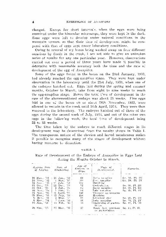
The time taken by the embryo to reach different stages in its development may be determined from the results shown in Table 1. The transparent nature of the chorion and larval membranes makes it possibJe to recognize many of the stages of development without having recourse to dissedion,
]H 16 16 31 i11 16 21 2R
5 8
11j
28
TA!3L1D I.
Rate of DevelopTnent of the Embryo o:f A n.asllides in Eggs Laid during the Months October to March.
I Nov" ':12 2;-; ;jan., ':3:J GR I Early nauplius PI. HI Nov., ':12 <) Mar., '3:, lOR Post naupliu3 Pl. TV
':.12 21 ;July., "}" ,)!l ~~Lt'i ")" 22 D~~e., ' ~-1 ~~ :;2 PI. III
Oct., '~~:3 4 Jan., ':\.1 Gil PI. 111 Nov., '5\:-3 7 Feb., '~4 ," PI. IV Nov", 16 April, "14 146 Nov.; 12 ,]nly, ';i4 22n Mar .. 2 M-ay, ")' 5P, Pi. III Mm>" ':).\ g() Flo IV Jan., 18 '35 ;~3 Pl. III
Feb" '3G 27 Mar., '35 27 PI. IL 10
v, V. HICKMAN
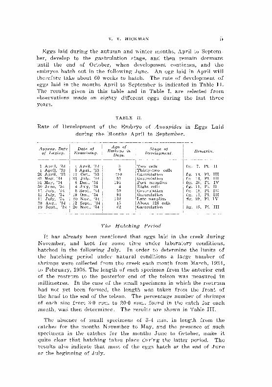
~ggs Jaid during the autumn and winter months, April to Septemh(31", develop to the gastrulation stage, and then remain dormant until the end of October, when development continues, and the embryos hatch out in the following June. An egg laid in April will therefore take about 60 weeks to hatch, The rate of development of eggs laid in the months April to September is indicated in 'rable IL The results given in this table and in Table 1. are selected from observations made on eighty different eggs during the last three years.
TABLE TL
Rate of Development of the Embryo of ibwspides in Eggs Laid during the Months April to September,
.. -----"-~------------ --,---------- ----.-~.-.-- - --- - - -- -- -----
ApP1'OX. Date Da,te of ,.4!Je of S{;age of E1nbrljo in. of Laying. E:l;o/rni'niny. Days. Dc'velopment. Remarks.
------- - ---------.
1 April, '33 4 April, ':-m 3 'rwo eells fig;. 7. PI. II I April, ';3;3
I, S April, '3;j 7 \ Thirty-two cel1s 20 April, ':13 17 Oct .• '3:1 J80 ; Gastrulation fig;. 18, PI. In 31 May, ':J,i 21 .July, -'34 51 I Gastrulation 31 May. ':14 8 Dec., '34 19J I Post nanpliu8 30 .June, '31 4 July, ';J4 T~ight cellt'> 11 July, ';]4 8 SeJJt., '34 59 I Gastrulation 11. .July, '34 10 Oct., '34 i 9] Gastrulation 11 July. '34 20 Nov" '34 1:32 i Late naupiiul:i
fig'. l~. PI. III fig. 29, PI. IV fig'. 11, PI. II fig. 18, PI. IH lig;, 18, Pl. nI ng. 28. PI. IV
28 Aug., ':14 12 Sept., '3;i 15 I Ahout 128 cells 19 Sept., '34 20 Nov., '34 62 Gastntlation fig. IX, PI. III
The Hu,tching PCf'iod
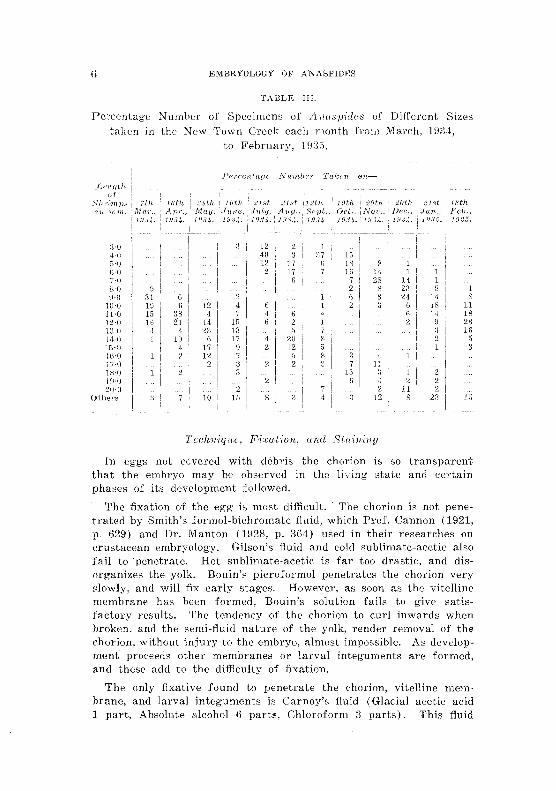
It has aLready been mentioned that eggs laid in the creek during November, and kept for some time under laboratory conditions, hatched in the following-July. In order to determine the limih: of the hatehing period under natural conditions a large number of shrimps were collected from the creek each month from March, 193i1, to February, lH35, The length of each specimen from the anterior end of the rostrum to the posterior end of the telson was measured in millimetres, In the case of the small specimens in which the ro"trum had not yet been formed, the length was taken from the front of thc head to the end of the telson. The percentage number of shrimps of each size from ;3,0 mm, to 20,0 mm" found in the catch for each month, was then determined, 'The results are shown in Table III.
Th,-' absence of small specimens of 3-4, mm, in length from the catches for the months November to May, and the presence of such specimens in thl' catches for the months ,June to October, make it quite dear that hatching takes place during the latter period. The results also indicate that most of the eggs hatch at the end of June or the beginning of July,
EMBRYOLOGY OF ANASpmgS
TABLE HI.
Pel'centage Number of Specimens of linuspidc8 of Different Sizes taken in th" New Town Creek lOath month from March, 198·1,
to February, 193G.
;Hj 4·0 D·n G·O J 7·0 1 8·0 8 1 9·0 G 14 S
l!Hl 6 12 ,] 18 11 IHl 3S 7 14 18 12-0 21 1::'i 28 1·' d 0 1:, Hi 14-0 10 17 5 15-0 ! 9 J!Hi 17·(1 lJ 1B'0 1 ]~H) 2 2O.(] 2 11
Other's 1" 23 1 r~
Technique, Fia:u.t-inrt. and Sfuinill.,q
In B.ggs not eovered with debris the chorion is so transparent that the embryo may be observed in the living state and certain phases of its development followed.
The fixation of the egg is most difficult. The chorion is not penetrated by Smith's for-mol-bichromate fluid, which Prof. Cannon (1921, p. (29) and Dr. Manton (1928, p. 3(4) used in their researches on ernstacean embryology. Gilson's fluid and cold sublimate-acctic also fail to ·penetrate. Hot sublimate-acetic is far too drastic, and disorganizes the yolk. Bouin's picroformol penetrates the chorion very slowly, and will fix early stages. However, as soon as the vitelline membrane has been formed, Bouin's solution fails to give satis·· faetoI'Y results. The tendency of the chorion to cUI'l inwards when broken, and tll(; semi-fluid nature of the yolk, render removal of the chorion, without injury to the embryo, almost impossible. As developmenr proceeds other membranes or larval integuments are formed, and these add to the difficulty of fixation.
The only fixative found to penetrate the chorion, vitelline membrane, and larval integuments is Carnoy's fluid (Glacial acetic acid 1 part, Absolute alcohol 6 parb;, Chloroform ;0, parts). This fiujd

v. V. HICKMAN
penetrates with considerable rapidity, and tends to cause invagination of the chorion, accompanied by distorti.on of the embryo. This may be avoided, however, by making 11 very smaIl perforation in the chorion. Fixation is complete in from 40 to 50 seconds. The eggs are then transferred to llO per cent. alcohol for 24 hours. Trw yolk is thus hardened sufhciently to allovv removal of the chorion without injury to the embryo. It is not necessary to remove the vitelline membrane, but the thick (,horion is an obstacle to accuratE' orientation. Moreover, it becomes hard and brittle during the process of infiltration, cansing trouble later in section cutting. Its removal may be carried out under a binocular dissecting microscope with the aid of needles.
In the one-celled stage the zygote occupies the whole space enclosed by the chorion, and adheres to the latter on fixation. Seetiol1S of this stage, therefore, had to be cut with the chorion in situ. Many failures resulted, but several. complete series of sections of the one-celled stage were thus obtained. In later stages this difficulty is not encountered, and the chorion may be removed in the manner descrihed above.
'1'0 facilitate manipulation and orientation, the eggs were stuck on to small pieces of hardened sheep's brain by the celloidin method, They were then infiltrated with paraffin wax (M.P. fi6° C.). Forty minutes in the wax bath gave satisfactory infiltration without rendering the yolk brittle. 'The wax was changed once before the definitive embedding.
Serial sections were cut at thicknes~es of 8-12 II , and stained with Ehrlich's acid haematoxylin, eosin being sometimes used as a counterstain.
In addition to serial sections, whole mounts of the young germinal disks were made. These were prepared in the following way: After fixation the embryo was stained for about 40 hours in alum-carmine, and then differentiated in acid alcohol for about the same length of time. It was then embedded in paraffin, plated on the mierotome, and all the part, other than the germinal disk, cut away. The disk was then removed from the paraffin block by the aid of xylol, and mounter!.
GENERAL EM.BRYONIC DEVELOPMENT
The Unse.qmented Egg
The newly laid egg of A naspidcs has a faint purple tinge, and is surrounded by a thick transparent chorion. It is almost spherical in shape, and, inelucling the chorion, measures about one millimetre
l~~/rHRYOLOGY OF AN l-\SPIDES
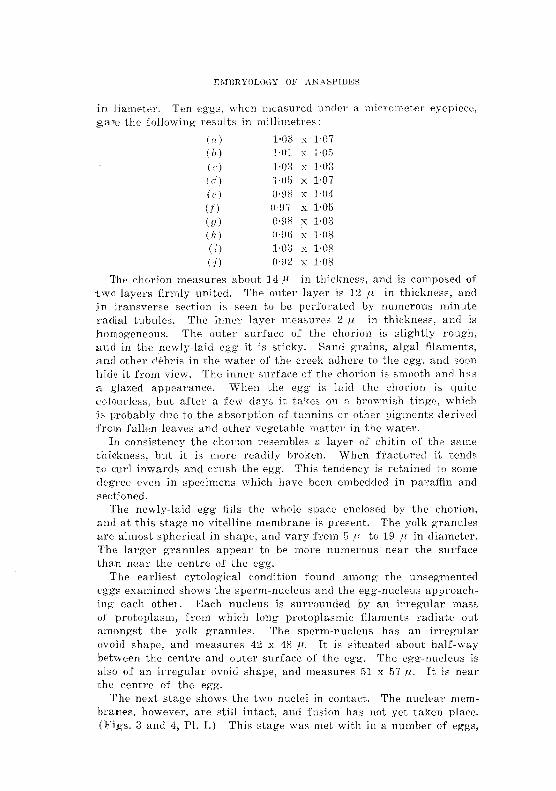
in (lian1t~ter. len eggs, ",yhen nleasu.l'ed under a ga,8 the following results in milJimetI·,,,s:
(n) 1·03 x 1·07 (II ) 1·01 x 1'05 (c) 1-08 x. 1·08 (rI) "Of) Hl7 (e) 0·£18 x ·04 (f) (J-£I7 H)5 iu) CHiS 1-03 (h) O·H() v 1-08 (i) 1-03 x H)S (:i) 0·92 x 1·08
'The chorion rneaSUl'es about 14 /1. in thickness, and is eornposed of two layers firmly united. The outer layer is 12 I_I in thickness, and in transverse section is seen to be perforated hy numerous minute radial tubules. The inner layer measures 2 p in t.hickness, and is DOIllogeneous. The outer surface of the chorion is slightly rough, and in the newly-laid egg it is sticky. Sand grains, algal filaments, and other debris in the water of the ereek adhere to the egg, and soon hide it from view. The inner suI"faee of the chorion is smooth and has a glazed appearance. When the egg' is laid the chorion is (luit", colourless, but after a few days it takes 011 a brownish tinge, whieh is probably due t.o the absorption of tannins or other pigments derived :from fallen leaves and other vegetahle matter in the water.
In consistency the chorion resembles a layer of ehitin of the snr'le thickness, but it is more readily broken. ',Vhen fradul"ed it tends to cud inwards mid 8ru8h the egg. This tendency is retained t.o some degree even in specimens which have been embedded in paraffin and sectioned.
The newly-laid egg fills the whole spaee enclosed by the chorion, and at this stage no vitelline membrane is present. The yolk granules are almost spherical in shape, and vary from 5 p. to 19 /1 in diameter. The larger granules appear to be more numerous near the surface than neal" the centre of the egg.
The earliest eytolog'ic:aI condition found among the unsegmented eggs examined shows the sperm-nucleus and the egg'-nuc!eus approaehing eaeh other. Ii.:aeh nueleus is surrounded by an irregular mass of protoplasm, from which long protoplasmic: filament.s radiate out amongst the yolk granules. The sperm-nucleus has an irregular ovoid shape, and measures 42 x 4S fl·. It is situated about half-way b(etween the centre and outer surfaee of the egg. The egg-nucleus is also of an irregular ovoid shape, and measures 51 x 57 rl. It is near the centre of the egg.
'rhe next stage shows the two nuclei in contact. The nuclear memhra"tles, however, are still intaet, and fusion has not yet taken place. (Figs. a and 4, PI. 1.) This stage was met with in a numbc"1" of eggs,
V. V. HICKMAN 9
and it is probable that the two germ-nuclei remain in contact for some time before actual fusion occurs. Eventually, however, the intervening nuclear membranes break down, and the two nuclei coalesce to form a zygote nucleus at the centre of the egg.
While these changes are in progress a marked decrease in the volume of the egg occurs. An egg which, excluding the chorion, measures 1·02 x 1·04 mm. when newly laid, may decrease in size to such a degree, that it measures only 0'65 x 0·71 mm. when the zygote nucleus is formed. As the volume of the egg decreases, the yolk granules, which are somewhat widely separated in the cytoplasm of the newly-laid egg, are brought closer together. The contraction also results in· the formation of a wide space· between the egg and the chorion.
Segmentation
The egg of Anaspides undergoes total segmentation, and, up to the eight-celled stage, the blastomeres are equal in size. The first cleavage furrow is meridional, and divides the egg into two cells, which, at first, are so loosely held together, that they readily fall apart (figs. 5 and 6, PI. I). However, before the next division, the two cells appear partly to fuse again, and the cleavage furrow becomes. shallow (figs. 7 and 8, PI. II). Serial sections through embryos at the two-celled stage show the nucleus of each blastomere to be near the centre of its cell, and surrounded by an irregular mass of protoplasm, from which strands radiate out among the yolk-granules.
The second cleavage takes place in the usual manner, being meridional and at right angles to the first furrow (figs. 9 and 10, PI. II). The third cleavage is equatorial, giving rise to eight cells. At first the four blastomeres at one pole are superposed on those at the other pole, but the cells soon move into a position in which the four blastomeres at one pole alternate with those at the other (fig. 11, PI. II). These eight cells show no tendency to fuse, and after fixation they may be easily separated from one another. Yolk is distributed uniformly through the eight cells.
When segmentation commences, or shortly afterwards, a delicate vitelfine membrane is formed around the embryo. At first this membrane is very thin, and fixatives penetrate it, but, when the eightcelled stage has been reached, the membrane is so thick, that it presents a serious obstacle to proper fixation.
The rate of cleavage is slow. An embryo examined at 8.30 p.m. on 4th April was at the two-celled stage. Twenty-four hours later it had reached the four-celled condition, and at 9 p.m. on the 8th April thirty-two blastomeres had been formed.
At the sixteen-celled stage (fig. 12, PI. II) a small blastocoel has been formed in the centre of the embryo (fig. 13, PI. II). As segmentation continues the size of the blastocoel becomes larger.
10 EMBRYOLOGY OF ANASPIDES
Mesoderm Formation
Segmentation has now become irregular, and appears to be more active at one than at the other pole of the embryo. When moved about, the living embryo usually rotates within the vitelline membrane so that the same pole is always uppermost. It is at this pole that segmentation appears to be more active and the cells smaller than at the other pole. Between the sixteen-celled stage and the thirty-two celled stage a blastomere at the uppermost pole slips into the blastocoel (figs. 13 and 14, PI. II). As segmentation proceeds, further cells from this pole enter the segmentation cavity, with the result that the surface of the embryo at this pole presents a pitted appearance.
The embryo has now assumed the form of a blastula, the wall of which is composed of a single layer of wedge-shaped blastomeres, surrounding a cavity which is partly filled by an inner mass of cells not arranged in any definite manner (figs. 15 and 16, PI. II). This inner cell-mass constitutes the primary mesoderm.
The total number of cells entering the blastocoel could not be determined. Serial sections showed that some cells, which had already entered the sllgmentation cavity, were undergoing mitosis, while 'others were passing inward.
As development proceeds the nuclei of the outer blastomeres gradually approach the surface. All the cells of the embryo are uniformly rich in yolk.
Gastrulation and Endoderm Formation
When the inward migration of cells into the blastocoel to form the primary mesoderm· has ceased, the outer surface of the embryo presents a uniformly segmented appearance. There is very little difference between the blastomeres of the two poles. The pitted appearance of the surface is no longer visible.
The process of gastrulation is initiated by the formation of a crescent-shaped depression occupying a large area at the uppermost pole (figs. 17 and 19, PI. III). The invagination gradually deepens, its opening becoming smaller and somewhat triangular (fig. 18, PI. III). This opening is the blastopore. The invagination becomes so deep that it reaches the mass of mesoderm cells in the blastocoel (fig. 20, Pl. III). It constitutes the endodermal rudiment, and later gives rise to the yolk-sac, mid-gut, and liver lobes. The mesoderm cells are pushed to one side of the blastocoel by the invagination and become somewhat flattened. They eventually spread out under the ectoderm and below the future position of the germinal disk.
Invagination goes as far as possible, and at one stage there is a deep arch enteric cavity (fig. 20, PI. III). It is, however, not a permanent cavity, for cells continue to move inward, crowd together,
v. V. HICKMAN 11
and obliterate its lumen. The blastopore gl'OWS smaller as gastrulation advances, and it finally closes when the cells around its margin come together. HOW(;VE;r,it never disappears entirely, its position being marked by a small depression in the surface of Lhe embryo, This depression is later replaced by the opening of the proctodaeum,
The endoderm now fornm a solid core of cells, which. together with the mesoderm, fills the whole of' the blastocoel. All the cells of the embryo are laden with yolk-granules, and, apart from their position, there is little to distinguish the mesodE;nn cells from those of' the endoderm.
In eggs laid in the spring and summer the close oJ gastrulation is marked by a short period of rest. In autumn and wintel' eggs it is followed by a long period of dormancy.
Forlna/ion of Germinal Disk and Egg-nauplius
After the quiescent period following gastrulation, cell division takes place actively over a wide area immediately in front oJ the blastopore. This active cell division gives rise to the gerrninal disk. Its narrow posterior end terminates at the blastopore, whilst its broader anterior end and its sides merge into the region surrounding them, In eggs laid during spring and summer the embryo takes about 60 days to reach the stage of development in which the germinal disk is fully di1ferentiated.
The disk now becomes thickened laterally, while the cells composing its antero·,median portion be.come thin. In this way the typical V -shaped form of the disk, seen in so many crustacean embryos, is attained (fig. 21, PI. III).
This stage is soon followed by the appearance of the egg-nauplius, The first sign of its development is the formation of the stomodaeum, This appears as a deep creseent-shaped invagination between the arms of the V, and slightly in front of the middle of the disk. The invagination extends backwards below the disk towards the blastopore. At this stage the posterior margin of the stomodaeum is much higher than its anterior margin. The rudiments of the naupliar appendages appear simultaneously as three pairs of thickenings on the latcral arms of the disk. These thiekenings soon stand out in high relief above the surface of the disk. The opening of the stomodaeum is now seen to lie between the rudiments of the antennae (fig. 28, PL Ill). Immediately in front of the blastopore is a pair of small lateral thickenings, which later give rise to the caudal papilla. The portion of the disk in Jront of the stonlOdaeum and between the antennules soon becomes thickened and elevated to form the labrum.
Up to this stage there has been v.ery little indication of the development of the head-lobes, but now, on each side immediately in front of the bases of the antennules, activc cell-proliferation OCCllrs, and the paired rudiments of the head-lobes arc~ established. The labrum
12 W.'vlBRYOLOGY OF ANASPIDES
is gradually raised above the surface of the disk, and comnwnces to project over the opening of the stomodaeum. The paired lateral elevations in front of the bJastopore corne together, forming t.he lobes of the caudal papilla. The extremities of the antennae and mandibles are now distinctly bilobed (fig. 2/(, PI. III).
While these changes have been taking place, a marked contraction in the area of the germinal disk has occuned. "When the rudiments of the naupliar appendages .first appeared, they were widely rernovcd from the median line, and projected beyond the sides of the embryo. N ow they have been drawn towards the median line, and also doser together (fig. 27, PI. IV).
Eciodwl'lnal Te/oblasts
Soon after the establishTllent of the rudiments of the llaupliar appendages the ectodermal teloblasts appear. These at first form an irregular transverse row on either side of the mid-ventral line and immediately in iront of the blastopore. Each row consists of seven cells (fig. 25, PI. IV). The inner ends of the two rows are lwited by a teloblast in the mid-ventral line. As the germinal disc contracts and the bases of the naupliar appendages are brought closer together, the teloblasts tend to form a curved row round the cauda] papilla, which has now been formed (f"ig. 2G, PI. IV). Yolk-granules in the teloblastic cells are rapidly absorbed, and the cell-nuclei become greatly enlarged. This is soon followed by the division of the cells to form the usual tranverse rows of descendants. The teloblasts do not form a complete ring around the caudal papilla until about the fourteenth week. The complete ring consists of nineteen cells, there being one in the mid-ventral line and nine on eac.h side (figs. 53-54, PI. VIII).
During the development of the egg-naupIius a new investing membrane is formed. This is the first lrn'val inte,qu1nent or em/l1'1Jonic cuticle. It eventually becomes very thick and elastic, and plays an important part in the process of hatching.
The embryo is now sluTounded by three investments, namely, the ehorion, the vitelline membrane, and the first larval integument.
Whenever the age of an embryo is mentioned in the following pages, it is to be regarded as having reference to an embryo developed In an egg laid in early summer.
Flurthwl" Changes in the li,'~~teTnal 8Jwpe of the End))'!!o
In eggs laid during early summer the embryo takes about eleven weeks to reach the stage of development shown in fig. 27, PI. IV. In the twelfth week the head-lobE'S develop rapidly and the labrum increases in length and breadth." Little change occurs in the shape of the antennules, but the bilobed nature of the antennae becomes still more pronounced, the exopodite being distinctly longer than the ~.ndo,podite. The mandibles lose their bilobed appearance and become
v, V. HICKMAN
pyrifornl, the narrow distal portion later giving rise to the mandi'oular palp. lVIeanw hill' the caudal papilla has increased in "ize, and the opening of the proctodaeum has become established in the position formerly occupied by the blastopol'e. The first TOW of ectodermal teloblasts partly encircling the papilla appears at this stage (fig. 28, PI. IV).
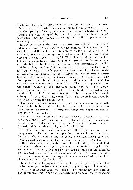
In fourteen weeks the head lobes are dearly defined, and curve outward in front of the base of the antennules, The central cell of each lobe is still visib1e. A rudimentary median eye in the form of a small' pigment-spot has appeared in the apex of the V -shaped area between the head-Jobes (fig. 29, Pl. IV). The tip of the labrum lies between the mandibles. The three basal segments of the antennuleR are established, In the antennae the two basal segments, coxopodite and basipodite, are first dijferentiated at this stage, and there is a marked increase in the length of the two rami, but the exopodite ir, still somewhat longer than the endopodite. The embryo has nO'l,v becoIne distinctly narrower and more elongate, but is wider anteriorly tban posteriorly. Immediately behind and between the mandibles appeal' the rudiments of the maxillules. These are separated from the caudal papilla by the tranvel'se caudal furrow. This furrow and the maxillules are soon hidden by the bending forward of the papilla. The end of the papilla is divided into two blunt lobes, whieh subsequently give rise to the eaudal fork. The proetoda(mm opens In the noteh between the caudal lobes.
The post-mandibular segments of the trunk are formed by growth from teloblasts in front of the blastopore, and arise in succession from before backwards. The limb rudiments also appear in succession from before backwards.
The first larval integument has now bec:omt; relatively thiclc It surrounds the embryo loosely, and is attached only at the ends of the antennules and antennae. A second lan){ll inte,gu'lJIent has been formed, but is not shed until just before hatching.
In about sixteen weeks the central cell of the head-lobes has disappeared. The median eye-spot has become larger and more distinct. The antennules and antennae have eommenced to curve upwards and backwards at the sides of the embryo. The two rami of the antennae are segmented, and the endopodite, which at first was shorter than the exopodite, is now equal to it 1n Jength. The rudiments of the maxillules are now followed by those of the maxiHae and the first three pairs of thoraeic appendages, The caudal papilla has curved forward, and the angle of flexurc has shifted to the third thoracie segment (fig, :30, PI. IV).
In eighteen weeks pigmentation of the paired eyes appears. The median eye-spot has become elongated and pear-shaped, The endopo<lite of the antennules is not yet formed. The antennary endopodite is llOW ctistinetly longer than the exopodite, and as development proceeds
14 Ell\IHHYOLOGY or ld"IASPIDES
the difference in length becomes mon, pronounced owing to l'etanlation in the growth of the exopodite. The rudiments of the first seven pairs of thoracic appendages have bceen formed, and the biramous nature of the first six pairs soon becomes apparent. At first there is very little difference in length between endopodite and exopodite. Eventually, however, the growth of the endopodite exceeds that of the exopodite. The embryo has now completed a little more than half the period which it spends in the egg. Flexure of the body and the increase in length of the embryo have brought the caudal extremity into contact with the front of the labrum. The body tapers gradually from the anterior to the posterior end, there being no sudden diminution in size at any point.
From this stage onward the chief changes in form consist in the further development of the appendages. The eighth thoracic limbs soon appear, and are followed in succession by the abdominal limbs. The epipodial gills of the first seven thoraeic appendages are developed shortly before hatching. The small leaf-like endopodite, which, in the adult, is present on the first four pairs of abdominal limbs, is not developed until after hatching. As they develop the thoraeic limbs are directed downwards, the second, third, and fourth pairs being on the outer side of the others, which, (H'ling to theflexun, of the body, lie between them. The five pairs of abdorninaI appendages and the uropods at first curve upwards at the sides of the abdomen.
At the twenty-second week a large saddle-shaped median dOl'salorgan has been formed. Anteriorly it is bounded by a transverse groove immediately behind the paired eyes, whilst its posterior margin is the jllnetion between the first and second thoracic: segments. Laterally it extends to the level of the paired eyes. In the living' embryo the organ has a yellowish appearance. It seems to reach its maximum development about the twenty-fifth week, and then slowly degenerates, but does not disappear until after hatching. No dorso-Iateral organs are present.
During the last few weeks before hatching the embryo becomes deeply pigmented .on the frout of the head between the eyes, and to a less extent on the back and sides oJ the trunk segments (fig. 31, PI. V).
As stated above, the embryo hatches from eggs laid during early summer in from thirty·-two to t.hirty-flve weeks.
POST-EM.BRYONIC DEVELOPMENT
The Pr-ocesl! of Hntchin,g
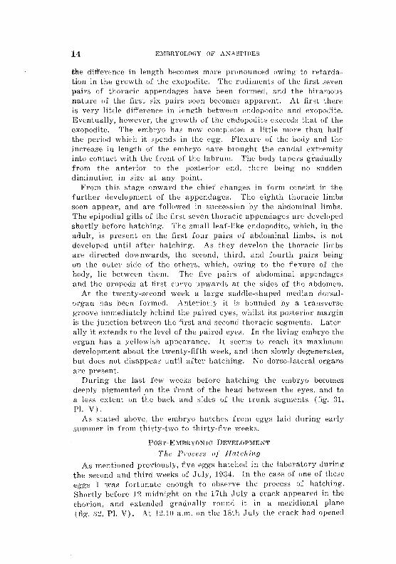
As mentioned previously, five eggs hatched in the laborat.ory during the second and third weeks of July, 19:34. In the ease of one of these eggs 1 was fortunate Emough to observe the process of hatching, Shortly before ] 2 midnight on t.he 17th .J uly a crack appeared in the chorion, and extended gradually round it in a meridional plane (fig. :32, PI. V). At 12.10 a,m. on the 18th .July tlw crack had opened
V. V. HICKMAN 15
and the vitelline membrane had burst. The first larval integument, however, was still intact, but had commenced to bulge out through the opening in the chorion. It was becoming slowly distended by osmosis. The expanding of this membrane gradually forced apart the two portions of the chorion, thus preventing their curling inwards and crushing the embryo (fig. 33, PI. V). The shrimp was now enclosed in a perfectly transparent ovoid sac formed by the first larval integument. This sac measured 2·0 x 1·8 mm. It still had the two portions of the chorion and fragments of the ruptured vitelline membrane attached to its outer surface. The increased space within allowed the shrimp to unflex its body, which up to the present had been strongly curved, with the ventral surface of the telson resting against the front of the head (fig. 34, PI. V). The ovoid sac eventually became quite free from the chorion, and assumed R spherical form. At 1 a.m. it had expanded to a diameter of 2·5 mm.
The shrimp was now moving about actively, and endeavouring to throw off the second larval integument. This had already split on the dorsal side, and was being slowly pushed off the appendages. Ultimately the ecdysis was completed, and the second larval integument lay in a crumpled mass inside the balloon-like first larval integument. At 1.10 a.m. the latter burst, allowing the shrimp to escape.
In another specimen, in which the chorion cracked before 7.30 p.m. on 12th July, the first larval integument did not burst and liberate the shrimp until about 9 p.m. on 13th July.
From the above observations the first larval integument, which is very strong and elastic, seems to serve two important purposes. In the first place, it absorbs water by osmosis', expands, and forces apart the two portions of the fractured chorion. In the second. place, it provides a roomy chamber where ecdysis of the second larval integument may take place in safety. (See also fig. 2, PI. I).
External Charcwters of the Hatched Embryo
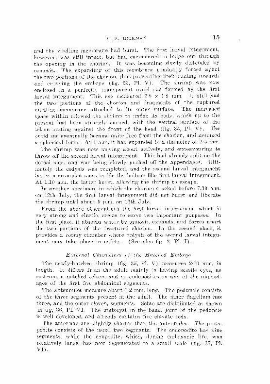
The newly-hatched shrimp (fig. 35, PI. V) measures 2'70 mm. in length. It differs from the adult mainly in having sessile eyes, no rostrum, a notched telson, and no endopodites on any of the appendages of the first five abdominal segments.
The antennules measure about 1·2 mm. long. The peduncle consists of the three segments present in the adult. The inner flagellum has three, and the outer eleven, segments. Setae are distributed as shown in fig, 36, PI. VI. The statocyst in the basal joint of the peduncle is well developed, and already contains five clavate rods.
The antennae are slightly shorter than the antennules. The Pl'otopodite consists of the usual two segments. The endopodite has nine segments, while the exopodite, which, during embryonic life, was relatively large, has now degenerated to a small scale (fig. 37, Pl.. VI).
16 E~vIBRYOLOGY OF ;\.l\;ASPJDFj;-)
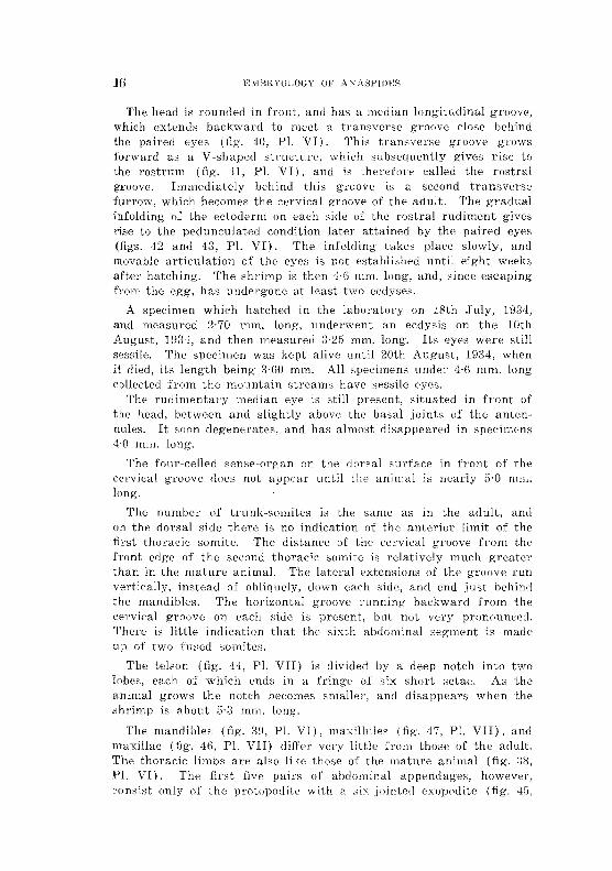
The head IS rounded in front, and h"", a median longitudinal groove, which extends backward to meet a Lransw~rse )!,TO!)Ve close behind the paired eyps (fig. 40, PL VI). This transverse groove grows forward :is a V -shaped strueture, which subsequently gives rise to the rostrum (fig. ,n, PI. VI), and is therefoce taUed the rostral groove. Immediately behind this groove is a second tranClyerSe fllrl'OW, which becomes the cervical grooVi" of the adult. '1'he gradual infolding of the ectoderm on eatb side of the rostral rudiment gives rise to the pedunculated eondition later attained by the paired eyes (figs. 42 and 4;), PI. VI). The infolding bIkes place slowly, and movable articulation of thrc, eyes is H(IL established until (,ight weeks after hatching. The shrimp is then ·Hi mm. long, and, since escaping from the egg, has undergone at least two ecdYBeH.
A specimen which hatched in the laboratory on 18th July, 19:34, and measured 2·70 mm. long, underwent an ecdysis on the 10th August, 19:)4, and then measured :i·2fi mm. long. Its eyes were still ses:sile. The specinwn was kept alive until 20th August, 1934, when it died, its length being 3·60 mm. All specimens under 4·6 mm. long· eollt!cted from the mountain streams have sessile eyes.
The rudimentary median eye is still present, situated in front of the head, between and slightly above the basal joints of the antennules. It soon degenerates, and has almost disappean>d in specimens 4·0 mm. long'.
The four-celled sense-organ on tjl£· dorsal surfacp in front of the cervical groove does not appeal' until the animal is nearly 5·0 mm. long.
The number of trunk-so mites is the same as in the adult, and on the dorsal side there is no indication of the anterior limit ()f the first thoracic somite. The distanee of the C't'rvical groove from the front edge of the second thoracie somite is relatively much greater than in the mature animal. The lateral extensions of the groove run vertieally, instead of obliquely, down eaeh side, and end just behind the mandibles. The horizontal groove running baekwal'd from the cervical groove on each side is present, btlt. not very prouounced. There is little indication that the sixth abdominal segment is made up of two fused somites.
The telson (fig. 44, PI. VII) is divided by a deep r,otch into lobes, each of which ends in a fringe of six: short setae. As tlli' animal grows the notch becomes smaller, and disappears when the shrimp is about fi'3 mm. long.
The mandibles (fig. :i9, PI. VI), maxillu]es (fig. 47, Pl. VB), and rnaxillae (fig. 4G, PI. Vff) ditTer Vel·y little from thos(' of the adult. The thoracic limbs an' also like those of thE' mature animal (fig. :3~l,
Pl. V [). Th,~ first flve pairs of abdominal appendages, however, COTlsist only of the protopociite with a six-jointed f,xopodite (fig. 4;',
PI. VII), The small fiabellate endopodite of tit", nrst rour abdominal limbs does not appear until the animal is 7,0 rnm. long. The have the same form as in the adult.
External dHferences bet".vectl the s.exes do not becoIrte apparent Lmtil about twenty w'2cks after hatching. Indications of the development of the copulatory styled in the male are first visible in specirnens 10·0 mm. long. In such specimens the endopodites of the first two pairs of abdominal limbs are distinctly elongated and narrow, The serrated spines on the basal part of the inner flagellum of the antennules in the' male are not evident until the shrimp is IG,O mm. long. The fiT3t spine to be developed -is a long- pJ:olatel'al SI)ine on sixth seg>ment of the flagdlum,
In the female the first external sexual char'acter to appeal' is the eonical papilla situated between the last pair of thoracic limbs, This papilla makes its earliest appearance in specimens about 12 mm. long, but the opening of the spermatheea upon it is nnt developed until a later stage.
DEVELOPMENT OF ORGANS
F'urther DI'1Jclo1)'})umt oj the klesocl'''J''IJI.
When differentiation of the germ-band is completed the mesoderrnai cells underly it. These cells now constitute the naupliar mesoderm, and are arranged in longitudinal strands below the rudimentc; the nauplial' appendages (fig, 4H, PI. VII). The p()sit)rioJ' ends of the strands lie at the sides of the proctodaemn, A few m;;sodenn cell" are also found in front of the stomodaeum, below the future position of the labrum. At this stage all the cells of the (ombt'yo are still laden with yolk-granules, As development proceeds some mcsodenn cells and many endoderm cells undergo cytolysis, giving rise tc, numerous chromatin granules, which become scatte)l'ed among the' yolk-spherules.
Shortly before the appearance of the caudal papilla, the naupliar mesoderrn cdls at the sides of the proctodaeum grow larger, and their yolk disappears (fig, 50, P1. VIII). As the stomodaeal invagi,e nation is drawli! forward (see below) these ctolls move into position, forming a curved row in front of, and partly sUlTounding:, thf; proctodaeum. These cells form the meBodermal teloblasts, and give rise to descendants from which the mesodermal somites of the post· mandibular trunk-regioll at'e derived (figs. 51 and 52, PL VIII),
M ueh of the naupliar rnesodej'm retainB its yolk until after lhe embryo has hatched.
The mesodermal teloblaste: form an inCOl'llplete ring of t'ight cell" (fig, 54, PI. VIII). Two of the cells are situated one oll each side of the mid·ventral line and between the median ectodermal teloblast and the proctodaeum, The remaini ng' six mesodermal teloblasts an' gitllated thl't:~e on each side of tl"tC' pl'oetodapU111 and to\vards tho
E,-VIBRYOLOGY OF ANASP1Dl!;S
d(wsal side. Frorn th~~se SlX teloblasts the dorsal 1'11t'~;odC'l'In of the t:nlnk S0111ites is -for:'1::;:;d, ""~vhile the tv.Yo vent-rat teloblasts Lhe ventral mesod(~rm (fig-. 5[., PI. IX,l.
l'1S0 to
The descendants of ~h(~ Ine--;Oaernlal tehibJasts fornl paired blocks 01 JYlesodel'rD in each trunk segTnent frofn t.he lYlaxilh~lary segnlcnt to thf.:' sixth ahdo;l1}nal segrnent lnehlS1\7e. /~LS the en1br:\70 lcng,th(~:ns,
t.hl~ preSSl1r~~ of the yolk-sac forces the nlesod(:~rlnal hloeks to aSSUrilO a 1110re 01' less t:dang'ulal' Sf.;lpe in crosH~'8eetlon, and to oceupy a position dorso-lateral to the TH~r\"e-c0rd. "Fron1 its carJiest forlnatiou ,"3ch mesodermal somite is diyisible into dorsal and ventral portions. 'I'11e dorsal part of each lnesode.nnal Bornite gTO\VS uIl\vard bet\vt'en th~ yolk-sac and the ectoderm. From this dorsal mesoderm are derived thB dorsal vessel, pericardial floor, dorsal longitudinal nnu;des, al,d most of the mesodermal investment of the mid-gut,
The ventral part of the mesodermal somites gives risf; to the ventral longit1.1d inal muscles and the musculature of the trunk UIJpendages.
The nauplial' mesoderm extends beyond the head JobeS and in front of the yolk-sae (fig. 64, PL XI), Here it constitutes the preantennulul'y mesoderm. As the (Ombl'Yo lengthens, some of the posterior cells ()f the nanpliar rneSodel'tn ~pread baek"\lval'ds (or ar(~ -forced baekw"ards by the elongating yull.;:-sac). and jieo in the trunk segrnents below and at the sides of the yolk-sac (fig;, PI. XI) I n the posterior segn}ents~ ·\vhel'(~ the yolk-sac does nut press so closely ag~ajlli:-\t the dOL'~al ec:tnderHL sorne of ('ens Jie ul)()\/;? it. In this position they bee orne enclosed by the Up-gTO\V111g: H1esodeTlnal sOTnites, and later are foulld in both thL' cardiac and pericardial ea \Tities (fig-. 56, PI. IX). Here and elsewhen, many of thern lose their yolk granules and become blood corpusc](OS, However, some of those lying below and at the sides of the yolk-sac become applied to it, and give rise to part of the muselllar investment of the mid-gut.
The mesc(j('l'mal investment of the stomodaeum is derived from the preantenDulary mesoderm. Owing' [;0 the larg'" quantity vf yolk present in the embryonie tiswes, I have not been able to recognize with cPl'tainty the eady formation of a pair of preantennulary i~n(:sode:nTlaI Horrlites. I-IoTJ..vevE'l', Lu tr'ansvel'se Bect10ns of a newly hatched en1bI'Yo thel'e lS on caeh side of thp stnrnodaE:1HTl a distinct c:avity in the l1w'10dc>rm. This co.vity cOrl'e8ponds very closely with ~he Jweantennulary ('oelomie ~,pa(ce of .Vebalia (Hee Manton, 1 !)il4,
2:3, .f. 1).
i\s HH!nLioned above, part of the n1€s(,del'rn.Hl investn1e-nt of the mid-gut is formed from nallpliar mesoderm that has spread baek,yards into the trunk. The larger pOl,tioll,howevel', is derived :from the dorsal mesoderm of the c'omites, As the trunk segments are j'o)"med, e"lIs from the dorsal mesoderm grow inwards ol'er the "'Jl'sal side of the yolk-sac, and then spread d(HVmVal'ds to the ventral

v, V, HICKMAl'<
side, forming a delicate drcular band surrounding the gclt, DUl'ing elubryonic life the yolk-sac is of such \vide diarYH::ter, ('ven in the abdominal region, that complete investn;('nt by the mesoclel'm is not etfeeted until aftH hatehing, Complete investment OcellI'S first at the junction of the mid-gut and proctodac'um, then in the preceding abdominal segments in turll from behind forwards; but even in newly hatched specimen it is not well defined further forward than the fourt,h abdominal segment,
Ileu)'t [lud Coelomic CO'vit/es
The development of the dorsal vessel is most dearly observed in the posterior segments, where the yolk-sac does not press so closely against the dorsal ectoderm. As each segment is formed some of the cells in the dorsal part of the mesodermal somites become oriented lengthwise to form the dors'al longitudinal muscles, others creep upward between these muscles and the yolk-sac, finally reaching the dorsal ectoderm (fig. 55, Pi. IX). The mesoderm thus growing upward on either side encl, ses a space above the yolk-sac. This space becomes the cavity of the dorsal v88sel, and the mesodermal upgrowths its lateral walls. Part of the dorsal mesoderm also grows inward over the yolk-sac, the portions from eithcr side meeting near the mid-line to form the floor of the dorsal vessel. In all segments the lateral walls are formed before the Hoor. As the yolk-sac shrinks in size, the mesoderm between it and the dorsal longitudinal muscles b pulled inward, forming a space on each side between the lateral wall of the dorsal vessel and the ectoderm. This space is the pericardial cavity, and its floor the pericardial 110M (fig, 56, PI. IX) The roof of the dorsal vessel is formed by the upper parts of the lateral walls growing inwards and meetmg' in the mid,line below the dorsal ectoderm. In all segments the roof is formed after the walls and the flool'. In front of the second thoracic Regment in a newly hatched embryo the dorsal vessel expands to form H
wide cavity, roofed over by the dorsal organ, and extending' forward as far as the cervical groove (fig. 57, PI. IX).
The mesodermal roof of this cavity is not formed until some time after the dorsai organ has disappeared and the shrimp is about ,1·0 mm, iong'.
Coelomic cavities develop early in tlw ventral part of the pc'ricanlial floor (fig. 59, PI. X), During embryonic life thc'y are compressed in most of the segments by the yolk-sac. They persist, hovvever, until the genital rudiments are formed, and in the newly-hatched embryo they are present ]n the six abdominal segments, and vestiges of them still appear in the posterior thoracic segments. The coelomic space in the preantennulal'Y !11t'soderm at the sides of the stomoda2um has already bet'l1 mentioned,
2() P"MBRYOIcOGY OF ANASPWgS
During the last few weeks of embryonie life the anterior aorta alld lateral eephalie arterim, develop 'in situ as haemocoelic ;;paces in the preantennulary mesoderm (fig. liO, 1'l. Xl. The mesoderm, whieh was originally the anterior part of the nunpliar mesoderm, now lies between the yolk-sae and the brain. It is still richly supplied with yolk granules. The haemoeoelie spaees extend upwards and bllekwards, to unite and open into the wide anterior part of t~e dorsal vessel underlying the dorsal organ,
In the abdominal region behind the first abdominal segment, the wall of the dorsal vessel remains thin and its lumen narrow, hut in. the thoraeie segments the vessel beeomes wider and its walls thieker, thus forming the heart (fig. 58, PI. IX).
The sternal artery, the ventral artery, and the subneural vessel aTe not formed during embryonic life, and their development after hatehing has not been followed .
.fl1inwntnry Canal
(a) FOTe-Yllt. As mentioned above, the ere scent-shaped ectodermal invagination giving rise to the stomodaeum is formed shortly after the differentiation of the germinal disk and before the rudiments of the naupliar appendages appear. The postedor margin of the mouth is at first higher than the anterior rnal'gin, and the invagination extends backwards in tho l11.edian line almost to the proctodaeum (fIg. 61, PI. X). As the labrum is formed and begins to project over the mouth, the stomodaeal invagination becomes bent, and its inner end is drawn forward until it lies between the antero-ventral portion oJ the yolk-sae and the brain (figs. 62-65, PIs. X and Xl). Here it soon indents the yolk-sac. The yolk-globules in the ectodermal cells forming the stomodaeal wall are gradually absorbed. By the seventeenth week the eells toward the inner end of the stomodaeum begin tv f()rm a thick columnar epithelium, which gradually extends to the outer end, However, where the stomodaenm is in contact with the yolk-sac, the cells do not form a columnar epithelium, but remain thin and fiat. The thickening of the wall is aceompanied by an increase in the lumen of the stomodaeum at its inner end, to form the cavity of the ftlture eardiac division of the stomach. At the twenty-fifth week the cavity is partly filled by four longitudinal ridge-like thick(>nings, whieh projeet inwards from its wall, one :from the dorsal surface, one from the v(mtral surface, and one from <'ach side. After hatching, and when the shrimp has reaehed a length of 3·4 111m., the dorsal ridge is diff"erentiated to :fonn the median setose pt'ominence and the two dol' so-lateral ridges of the adult. The two lateral ridges in the embryo give rise to the two ventro-Iateral ridges in the adult, while the ventral ridge in the embryo :forms the median ventral ridge in the mature animal.
V, HICKMAN 21
(II) Mid-gut. At the dose of gastrulation the blastocoel, as mentioned above, is filled with a mass of endodermal yolk-cells, togetlwr with the primary Tnesodel'm (figs. 48 and 49, Pl. VII). The delicauj membrane surrounding each yolk-cell is discernible only unci{;r the most favourable conditions of fixation. If Carnoy':s fluid be allowul to act too long the membrane is destroyed.
With the formation of the germinal disk, many of th(, yolk-cells on the ventral side of the endodermal mass undel'go cytolysis, their nuclei disintegrating into numerous deeply-staining granules, which are scattered among the yolk-globules and finally absorbed (fig'. ,19, PI. VII). Cytolysis continues until about the eightecnth week, and thcn diminishcs in activity. During' the remaining part of embryonic life, however, isolated yolk-cells may be found in which the nuclei are in process of breaking up.
The outermost cells of the endodennal mass constitute the wall of the yolk-sac, theiJ> outer ends forming the yolk-sac membrane (fig'S. 64.-66, PI. XI). They surround an inner core of yolk-cells, which occupy the space destincd to become the lumen of the mid-gut. 'fhese inner yolk-cells seem to fuse with each other and sometimes with the inner ends of the outer cells. They are gradually absorbed as development proceeds.
As the embryo lengtlwns the yolk-sac extends backwards into the caudal papilla (figs. G4-6fi, PI. Xl), It tapers gradually from the anterior to the posterior end, there being no sudden diminution in diameter marking off a cephalo-thoracic part from an abdominal part. The cells throughout its whole length have the nature of yolkcells, and not until just before hatching" do any of them become converted into epithelial cells. Even the cells opposite the stomodaeum and proctodaeum retain the nature of yolk-cells, and do not form epithelial plates at any stage,
As the yolk in the yolk-cells is absorbed the yolk-sac shrinks in diameter, and its cells become smaller. The conversion of the yolkcells into the epithelial cells of the mid-gut COlnmences during' the last week of embryonic life. It first occurs in the cells forming the dorsal wall of the now elongated yolk-sac, immediately in front of the proctodaeum. It then spreads round to the ventral side, advancing forward at the same time (fig. 6G, PI. XI).
Immediately after hatehing, the yolk-cells at the anterior end of the yolk-sac begin to assume an epithelial nature. The indenting· of the yolk-sac by the stomodaeum has helped to form a median dorsal pouch overlying the future cardiac division of the stomach. The yolk-cells forming the wall of this pouch are the first anterior cells of the yolk-sac to be converted into epithelial cells. The pouch becomes the anterior diverticulum of the mid-gut.
22 EMBRYOLOGY OF ANASPlDES
When the embryo hatches, the lumen of the developing mid-gut hehind the lifth thoracic somite is almost empty, but in front it still contains much unabsorbed yolk. This, however, rapidly disappears. and by the time the shrimp is 2·9 mm. long communication of the mid-gut with the stomodaeum and proctodaeum is established. This iB brought about by the absorption of the yolk-cells and the breaking down of the thin ectodermal septa at the points of contact.
When Anas]Jides is 8·0 mm. long all the cells forming the wall of the lengthy yolk-sac have hecome converted into the epithelial cells of the mid-gut. The anterior diverticulum is now w811 developed, and resembles that of the adult.
'l'he rudiment of the second diverticulum of the mid-g'ut appears as a dorsal thickening 01' the gut epithelium in the first abdominal segment when the shrimp is g'5 mm. long. The diverticulum, however, does not attain its pouch-like form until the animal is about 5·5 mm. long.
The third diverticulum is first appaI'ent as a dorsal thickening of the mid-gut epithelium in the fifth abdominal segment of specimens about 4·0 mm. long. It develops very slowly, and does not become pouch-like until illuIS1Jid68 reaches a length of nearly 1,1,0 rn111.
The endodermal yolk-cells give rise not only to the mid-gut and its three diverticula, but also to the lobes of the liver". The origin of these lobes is described below.
(c) H-inci-[Jut. A second invagination of the eetoderm to form the hind-gut does not apPE)ar to take plaee. The cells immediately within the lip of the blastopore form the wall of the proctodaeum, and the blastopore becomes the anal aperture. At a later stage this open-, ing is seen to occupy a postero-dorsal position in the notch between the lobes of the caudal papilla. At first the proctodaeum is very short, and the cells forming its wall are rich in yolk-granules. At the seventeenth week the yolk has been absorbed and the cells have become epithelial. The length of the proctodaeum now increases, and its aperture moves from its postero-dorsal position into the postero-ventl'al position occupied by the anal aperture of the adult. The hind-gut is well developed before hatching, but, as previously mentioned, it; does not communicate with the mid-gilt until some time after the embryo has escaped from the egg.
The livN--lobes are developed from the endodcrmal yolk-sac. Th€ posterior pair of lobees aree the first to he formed. They appeal' about the nineteenth week as a pail' of lateral outgrowths, Olle 011
each sidee of the yolk-sac in tlw maxillary segment. The outgrowths
V. V. HICKMAN 23
soon grow backwards, becoming elongated and fiuger-like. The wan of each lobe consists of endoderIual cells similar to those forming the wall of the yolk-sa\:. A eap of mesodermal ceils covers the end of the lobes (fig. G9, PI. XII). As the lobes lengthen their \:ells gradually bCCOnl€ epitheJiaL 'rhe conversion takes place f-ln~t at the poster-ioT end, and then extends towards the front. The lumen of the lobes is filled with yolk·globules, whieh are slowly absorbed. On hatching, the posterior hepatic lobes have grown baekward as far as the second thoracic segrnent.
Up to the twenty-seeond week of enlbryrmie life the front of the yolk-sac is roundc"cL "When, however, the dorso-ventral mandibu·iar muscles are formed, they pres,; against the front of thf: yolk-sac, forming a vertical groove or indentation on each side. The front of the sac is thus divided into a median lobe and two antcro-lateral lobes. The median lobe eventually forms the anterior diverticulum of the mid-gut. The two antero-lateral lobes give rise to lobes of the liver. The apex of eaeh antero-lateral lobe of the yolk-sac is attached to the dorsal ectoderm by a short ligament. Immediately behind the point of attachment of the ligament eaell lobe grows backwards as a finger-like outgrowth to form the anterior hepatic diverticula. 'rhese hepatic diverticula aTe anterior only in tlw sense that they arise frOTH the antero·Jateral lobes of the yolk-sae. They do not project towards the front, but grow towards the posterior end of the body (fig. 70, PI. XII). They are soon followt>d by another pair of hepatic lobe~, which arise in the same manner immediately behind the point of attachment of t1w dorsal ligament (Iig. 71, PI. XU). The animal usually hatches with two pairs of liver lobes, but in some cases the rudiments of a third pair are also well established. By the time the shrirnp is 7·0 mm. long it has seven pairs of hepatic lobes, and when adult about fifteen pairs.
The genital rudiment appears towards the tend of e.mhcyonic life. Shortly before the embryo hatches germ-cells, having a large oval nudens vvith finely granular chromatin and eon~picl1ous nudeoli, Hn~ found on the floor of the eoelomic pouches in then mt and second thoracic somites. Similar germ-cells appear in succession in the "oeJomie poucheR of the spgments behind the seeond thoracic wmit2 (figs. 72-73, PI. XII), By the time the ernhryo hatch('s these seg:· ruental genital rudin1enLs occur in the eight son1ites of the thorax. Soon after hatching they appear in suecession in the six abdominal segnlents.
As each rudirnent. is ef;tahlished it gro\vs in :length by di.vision of the germ-cells. When the animal is 6·0 mm. long the rudiments in the separate segrnents have Ul1ited to forIn the long gonad of" the
24 ,:lIiIBRYOLOGY OF ,\f\'ASPUlES
adult. Testes and ova!':v develop in a si.rllilar 11lanner. 'rhe duet~ do not appear until much latH in the life of the sh~rimp, no atteIY"lpt vvas 111ade to fol1ovv their deve·loplnDnt.
Whether the genital rudiments in the separate somiws are derived froTn a l)rirnary gern1-eeU, forrnf'd in the early stag~e8 the ;~(~g
mentation of the oiispcnn, could. not he detennlywd. 'l'hcy appear, howcevel', to be derived from the mesodermal (;ells forming th" walls of HlP coelomic pOilches.
Little need be said in regard to thE' nervous system, which arises in much the same way as in other Crustacea, The development of the protocerebrum, with its central cell surrounded by concentric rows of other cells (fig, G8, PI. XI), may be compared with the corresponding structure in the egg-nauplius of Astaclis (Reichenbach, 1886. p, 29).
The median eye at its earliest appearance seems to have a paino!:! ongm, It develops from a group of cells lying between the head-lobes (fig. 67, PL XJ). Two cells in this g'l'OUP, one on each side of th" mid-line, give rise to pigment granules. These granules eventually collect round yolk-spheres in th" cells. As the yolk is used up the pigment granules ferm a dense pyriform black mass. The whole structure is very rudimentary, but, like the median eYt'; of some of the lower Decapoda, it persists as a vestigial organ for some time after hatching. In having a paired origin, the m~edian eye of A'/w8jJ?:deH
resembles that of l'irternia (Mol'off, H112, p, 18). Yolk-granules are found in the plasma of the cells forming the eye in both species. In :fact, Moroff (1912, p. 17) states in reference to .tlrtemia that' de!' ganze Embryo ist von gl'c;sseren und kh'ineren DottHkugeln erfi.il!t, die glcichmassig urn die Zellkerne verteilt sind! This condition is not unlike that of the egg-nauplius of Arwspides.
The lateral eye" in their development l'esemble those of B,'IUlrhi" fJll8 (Claus, 1886, p, GO). During embryonic life, and for several we("ks after hatching, they are sessile. The ectoderm gradually grows in under the eyes, both from the front and the back, but it is until the shrimp is over 4 mm, long that the pedunculated condition is attained (fig. ,n, PI. VI), Hanstrijm (193,;, p. :3G) has l'Gcc'utly shown that the paired eyes resemble thm'e of lvl!f~i" in theil' finer structure.
The statocyst in the basal segment of the antennulary pedunelt develop" as an invagination of the dorsal ,'ctodermaL 'Ihe club"haped rods arise !rom the eetodennal eells forming the outer waH of the invagination. In a newly-hatched emhryo fivf" rods are uSllaH,' prpsent.
v. V. HICK'vlAN
The four-celled sense organ, whieh develops 111 front nl the cei',-ical groove. and which has been compared with a sirnilal' organ the Phyllopoda both by CaIman (lfl.BG, p. 787) and by Hanstriilil (J~J:l·i,
)L &2), does not appear until A IwsJ!idcs is abrmt 4 mm, long'.
!n the development of the nel'Ve C'ord a very distinct ganglion is j',-,l'med behind that of the sixth abdominal segment. This seventh abdominal ganglion is well defined about the twenty-sixth week (Jf
embryonic: life (fig. 74, PI. XlII). As the embryo lengthens tlw sixth abdominal ganglion is pulled away from the fifth, leaving' an intervening space in whil:h yolk-granules co1led (fig, 7\ PL XIII, A t the same time the tension on the nerve cord appears to drag- Lhe ~eventh abdominal ganglion into a position partly overlying the sixth. After hatching the intersegmental bar between the sixth and seventh abdominal ganglia becomes very thin (fig, 76, PL XIII). It finally breaks down and disappeal's, and the two ganglia fuse.
lHW5Clilutlll'e und Endoskeleton
A detailed examination of the devdopment of the musculature and endoskeleton has not been made. The origin <lml development of the trunk musculature and gut musculature have been briefly I1Kl1tione.d in other parts of this paper, The endoskeleton of It na8pides, like that of Nebaliu (Manton, 1934, p. 202), is ffll'med from int<.'r"lcgmental eetodel'mal bars which arise from lateral and ventral inh:ekings. These bars are strongly developed in the mandibular, rnaxillulary, and maxillary segrnents. The rudiment of the mandibular bar appears at about the fifteenth week. The other bars are dcy-eloped in sequence. The greater part of the endoskeleton in the region of the month is formed from the first three post-oral bars. These bars are all nucleated, and remain nucleated even in the adult.
A critieaJ study of the endoskeletons of ilnas]Jides, PnmlWS}lides,
IVebalia, HC'IJ'I'I:mY8is, and l\jyctiph(t'YIes has already been made by Dr. ~\f[anton, As a result of this study the close similarity existing between the endophragmal structures of the Syncal'ida and Leptostl'a<.:a has been demonstrated (Manton, HJ:34, p. 220). This similarity is further emphasized by the dose agreement in the mode of developn10nt.
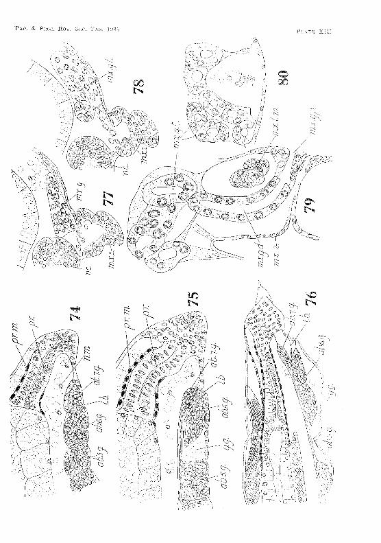
lid ((,l'ilia )'y ()land
The rudiment of the maxillary gland appears about the middJe 01 embryonic life. ]\h'soderm cells begin to form a compact group on each side of the nel'vt~ cord in the maxillary somite (fig, 77, PL XIII), Yolk granules in the vicinity of these cells are rapidly
26 ~~YIBRYOLOGY OF ANASPlDES
absorbed. At first no definite al'l'angement of the cells can be observed, but as development proceeds the nuclei of the eells beeome oriented to form more 01' less regular transverse rows, giving rise to the tubules of the gtand (fig. 78, Pl. XIII). The end-sac of the gland soon appears as a large cavity in the group of mesoderm eells. From the gland a duct leads downward in front of the large transverse adductor muscle in the basal segment of the maxilla, curves under the muscle, and eventually opens neal' the centre of the postel'iOl' surface of the appendage (fig. 79, Pl. XIII). Smith (1909, p. 5;l7), in describing the adult animal, states that the duct 'opens by a pore on the external border of the appendage '. This, however, is not the case. Sections through the maxilla of a mature specimen show quite clearly that the duct opens by a pore on the posterior surfaee . • Just before hatching a small invagination of the ectoderm occurs at this spot, and communicates with the duct from the gland. With the ".xeeption of this ectodermal lining of the aperture, the whole of the gland and its duct are derived from mesodenYl, and are fully developed on hatching (tig. 80, PI. XIII).
Mediam, Dorsal O)"qan
The median dOl'sal organ is not to be confused with the :four-celled Bense organ, which, in young' adults, appears in a median position in front of the cervical gToove, and which CaIman (1£109, p. 16,J) Btates 'may be comparable to an obscure "dorsal organ"'. The fmH-celled sense organ does not develop for some time after hatehing. whereas the median dorsal organ appears at about the twenty-first week of embryonic life. It consists of a marked thickening of the dorsal ectoderm, extending from behind the eyes to the front of the second thoracic segment. The ectodermal cells composing it are long and columnar, but become smaller round the margin of the organ. At first the cells contain yolk-globules, and their boundaries are distinct. As development proceeds the cells degenerate, their walls break down, and in some cells the nuclei disintegrate. In the living embryo the organ has a yellowish colour, and is quite conspicuous. It persists for some time after hatching, rlllt has disappearc'c/ by the time the animal is 8·[) mm. long.
HubUs of Anrts)Jide.s
The newly-hatehed animal moves about aetively, sometimes crawling over rocks and deb'is at the bottom of the water, at other times swimming up to the surface. If it is gently touched when swimming, it will often sink to the bottom, and, lying on its back with its body stretched out in a passive state, remain as if dead for several seconds. This death-feigning habit occurs only in the newly-hatched animal,
V. V. HICKMAN 27
and is not exhibited by the adult. Herrick (1895, p. 184) has described a similar peculiarity in young specimens of Homarus americanus.
Smith (1909, p. 546) states that Anaspides 'will occasionally rise to the surface of the water and turn over on its back in the manner ofa Phyllopod '. I am able definitely to confirm the accuracy of this observation. In quiet pools at the side of the mountain stream, where the water is not disturbed by ripples, Anaspides often comes to the surface, turns over on its back, and, moving about on the underside of the surface-film, searches for food among the small particles which are floating there. The habit is observed more frequently in young than in adult animals, since the weight of the latter is not so easily supported by the surface tension of the water. Small specimens (5'0 mm. long) not only move about on the under side of the surface-film, but also rest there in an inverted position. I have observed specimens resting in this way for over ten minutes.
The animal does not commence feeding until communication between fore-gut, mid-gut, and hind-gut is established. This occurs a few days after hatching. When the young shrimp begins feeding a long cylindrical tube, about equal in length to the mid-gut, is extruded from the anal aperture, and is carried about for some time before being expelled. This tube is probably composed of the inner ends of the yolk-cells forming the wall of the mid-gut, and is cast off when these cells become epithelial. It is certainly not the ectodermal lining of the hind-gut, which is comparatively short, and is cast off during ecdysis.
Mating has not been observed. However, male specimens in the creek are sometimes seen employing the armed basal joint of the inner antennulary flagellum in an attempt to seize hold of the base of one of the antennules in a female.
DISCUSSION
Segmentation of the Egg
The egg of Anaspides, like that of Lucifer (Brooks, 1882, p. 64), exhibits complete and equal cleavage. In both a large segmentation cavity is formed, and into this cavity, shortly after the sixteencelled stage has been reached, a blastomere moves. In Lucifer this blastomere divides as it passes inwards. -In Anaspides it divides after it has entered, and at the same time other blastomeres appear to pass inwards. There is then formed in each case a deep invagination-gastrula with a primitive arch enteric cavity. At first all the cells are equally provided with yolk, but at the commencement
of invagination in Lucifer the yolk has largely disappeared from all
28 EMBRYOLOGY OF ANASPIDES
cells except those in the segmentation cavity. The cells of Anaspides are laden with yolk granules up to a late stage in development.
Brooks (1880, p. 563) originally regarded the cells lying in the segmentation cavity of Lucifer as mesoderm cells. In a later publication (Brooks, 1882, p. 70) he abandoned this opinion in favour of the view that they were yolk pyramids, because they were charged with yolk granules. Unfortunately the subsequent history of these cells was not followed. The corresponding cells in Anaspides constitute the primary mesoderm.
Ectodermal and Mesodermal Teloblasts
The trunk region of Anaspides, like that of the Leptostraca Peracarida, and Decapoda, is formed from rows of ectodermal and mesodermal teloblasts developed immediately in front of the blastopore. The teloblastic regions of Hemimysis and Nebalia have been described in detail by Dr. Manton (1928,p. 370; and 1934, p. 178). Anaspides agrees with both these forms in possessing an ectodermal teloblast in the mid-ventral line. It has been suggested by Dr. Manton (1934, p. 212) that' possibly the Decapoda differ from the Peracarida in possessing no mid-ventral ectodermal teloblast'. Sollaud (1923, p. 93) gives a figure of the telublastic region of Leander in which a mid-ventral teloblast is clearly indicated. It is highly probable that a mid-ventral teloblast will be found in all those Crustacea in which teloblastic development of the trunk occurs.
In Anaspides, Nebalia, and the Decapoda the ectodermal teloblasts form a complete ring around the caudal papilla. In both Anaspides and Nebalia the ring consists of 19 teloblasts.
Four pairs of mesodermal teloblasts are present in Anaspides, Ncbalia, and Hemimysis. Patten (1890, p. 371) records a similar number in Cymothoa.
External FM"m
In the development of the external form of the body, and in the order in which the appendages appear, Anaspides resembles the Leptostraca and Mysidacea. Owing, however, to the much later rupture of the egg-envelopes, the ventral flexure of the body, like tht:t in the Decapoda, is retained until the embryo is hatched. The strong development of both rami of the second antenna during early embryonic life, followed by the gradual degeneration of the exopodite, recalls the similar changes through which this appendage passes in the larval life of certain Decapoda. Thus in the protozooea of Penaeus the second antenna has a three-jointed endopodite and a four-jointed exopodite, but by the time the mysis stage is reached, the exopodite has degenerated into a scale (Muller, 1863, p. 8; and Claus, 1876, p. 11).
V. V. HICKMAN
In the newly-hatched Aunspides the notched tdson, with its six pairs of setae, bears some resemblance to that of a protozooea, and perhaps indicates that the ancestors of .,4naspides passed through some such' larval stage.
The He(wt and Coelom'i!' Sw,s
In its main features the development of the heart resembles that of Hemimysis and Nebnlin. In Nebalin, however, the ventral wall of the dorsal vessel appears first, and later the sides and roof, whereas in Hem:ifnysis and .4naspides the lateral walls are first formed, and then the floor, followed by the roof. As in Hwrnimysis, the pericardial floor is formed from the dorsal mesoderm as it grows upward, and not, as in Estheria, by a later downward growth of the dorsal mesoderm.
Coelomic sacs have been demonstrated in Estheria (Cannon, 1924, p. 425), Chi1'ocephalus (Cannon, 1926, p. 402), Hmnimysis (Manton, 1928, p. 397), and Nebalia (Manton, 1934, p. 227). In Anaspides, as in H emi.1n.ysis and Chirocephalus, the coelomic sacs are early compressed between the yolk-sac and developing pericardium, but while in Ch'i)'occphalus they are soon obliterated, in A Haspides and Hernim.ys·is they persist throughout embryonic life. In A'nlISlYidcR they are large, and most clearly seen in the newly-hatched embryo, when the yolk-sac is somewhat shrunken, and the trunk, released from its flexed condition, has straightened out, The large size of these coelomic sacs is a primitive character comparable to that in Estheria (Cannon, 1924, p. 39,).
AliJlwntury Canal
The position of the anus relative to the blastopore has been reported differently by different authors (see KOl'schelt and Heider, 18lH), p. 173). As pointed out by Dr. Manton (1928, p. 431), it is not always possible to determine with accuracy the relative positions of these two structures, 'sinee the blastopore may be a vague area and is obliterated before the anus appears'. In A naspides, however, the blastopore is a very definite aperture leading into an archenteric cavity, Although the lumen of this cavity is eventually rendered imperceptible by the crowding togdh,·" of the endoderm. celis, the blastopore i,; always well defined. It becomes the anal aperture, and the eeUs irnmediately within its lip give rise to the proctodaeurll.
A critical account of the development of the mid-gut in Crustacea has recently been given (see Manton, 1928, pp. 483,-(86). A repeti, tion of this account is not needed, hut it may be said that generally in the centrolecithal eggs of Crustacea certain cells pass inward from the blastoporal area, absorb yolk, and form yolk-cells, whieh give ris8 partly or <·ntireJy to the mid-gut. In HemimY81'B these yolk-cells
30 El'vlBHYOLUGY OF A?'<ASP1UES
&pread around the yolk, and form the yolk-sac formed into the epithelial cells of the mid-gut.
Later they are transIn Nclm/iu. the yolk-
sac is at first composed of yolk-cells ccmnected posteriorly with an epithelia! endoriermal plate. By proli f'el'ation of its own cells and transformation of the yolk-e.ells along its margin, this plate gradually extends forv\~af'd until an the yolk is 8TICl0Sed in an epitheljul yolksac. A similar proeess probably takes plaee in Leander (see So[]aud, 1928, p. 1(,2L Tn flnaspideo, as in C'0!uchilu8 (Grobben, 1881, p. 243), the celIs fonning- the arc,henterie invag'ination are heavily laden with yolk, and are virtually yolk-eells. They give rise directly to the yolk-sac, and Jater to the mid-gut. In CetodliluH, however, the lumen of the invagination persists, whereas in illwspirle8 it, is obliterated by some of the yolk-tells crowding into its centre and forming a central core. This condition, although orig'inating in a different way, is similar to that appearing in I:l emhnysis after the yolk-cells have formed the secondary yolk-pyramids. The outer cells of the endodel'ma! mass in A nns))idN; form the yolk-sac, and correspond to the secondary yolk-pyramids of I-i em i11l.1II<i8, while the inner core of cells in A.ILU8pideR corresponds to the eentl'al core of yolk in Hem imysis. In both, the eells forming the yoll(-sac become transformed into the epithelium of the mid-gut, the transformation taking place first opposite the stomodaeum and prodoclaeum, but somewhat earlio]' in Hem i11l118i.S than in A. nasjJide8.
Cytolysis .occurs only to a small extent in the yolk-cells of but lat'gely in those of Alw.gpicles. Degeneration of the
yolk-cells occur;; extensi'c1,'in many Decapoela, and ho.8 been l'eeonled in .41Tlhel l g (see Brooks and Herrick, 18H2, p. 425). H OillUI'I18 (see Herriek, 18}!:'i, p. 211), and [,('under (see Sollaud, 1928, p. 8;3).
The liver, or hepatopannroas. of the Crustacea does not always develop in the same way. In Hrmlimys-i8 the posterior liver-lobes 'arise independently of the yolk-sae from a pair of epithelial plates of mesoderm which grow round the yolk-sac' (see Manton, 1928, p. 488). In £1 nllBjJicles these lobes are formed m; out-pushings of the yolk-sac, which open Jatero-ventrally into tbe mid-gut immediately behind the stomodaeum. Sollaud (1828, p. 171) has described a somewhat similar origin :for the liver-lobes of Leander. The hepatic diverticula of Nebcdiu also develop as outgrowths of the endodermal yolk-sac (see Manton, IS8 01, p. 18:.,).
The anterior hepatie lobes of A. nnspitic.s arise in much the same way as those of N rbilliu, except that the yolk-saC' is indented not 'by a single median depression but by two vertical furrows, one on each si.de of the ante1'ior mid-gut diverticulum. Moreover, the anterior lobes do not extend :forward into the bead, but curve over and grow
V. V. HICKMAN 31
towards the tail, the primary pail' of lobes being ~oon followed by f1. seeond pair, whieh develop at the point when, the tlrA pair bend over.
AJrj/,,:i[}tx"yy (}l({/rid
The maxillary gland of A!W8])icies, like that of Estheria (Carmon, 192,:[, p. ,124) and Chil'orcphci/IIB (Cannon, 192fi. p. 406), is of nwsodermal origin. As in Chil'ocephaZuB, the end-sac and prirnordia of the coils appear early. The dose similarity in the adult pattern of the maxillary glands of Chi.roceplwl1l8 and A_nagpides has already been demonstrated (Ree Cannon and Manton, 1927, p. 445). It not surprising, therefore, to find a similarity in their mode of development.
Genital Rudiment
In Ana3picies, as in Nebalia, the gonads arise in the walls of the coelomic pouches at a late stage in embryonic life. Dr. Manton has already pointed out that, in this respect, llle/)alia resembles the Annelids, Peripatlls (Sedgwick, 1887, p. (167) and Sc%pendr(} (Heymons, HJ01, p. 1), A similar origin of thle; gonads is alRo found in spiders, Linlulus, and other Arthropoda.
H atch/7lff Process and Huil1:ts
There is a remarkable similarity in the hatching of AnasjJides and that of certain Cladocera. The winter-egg of Daphl1'ht has no chorion, but is surrounded by a primary ,Yolk membrane or 'Dotterhaul,', which eorresponds to the vitelline membrane in the egg of A nnspides. Below this membrane, during the development of both DUT!hnia and Anaspities, two larval integuments are formed. The first appears at the egg-nauplius stage. It becomes strong, elastic, and semipermeable. The second appears somewhat later, al1d is eonsequently within the first. Now Volmer (1912, p. 6Mi) has "howD that the first larval integument is of the greatest importance in the hatching of Daphnia. By osmosis it absorbs water, and swells out like a balloon, bursting first the' Dotterhaut,' and then the ephipl1iull1. 111 A_nnspides it behaves in a similar manner, bursting first the vitelline membrane, then the chorion. In each ease R long time elapses before the balloon--like structure breaks, jibeJ'a'ting the ,embryo. During this time ",1 ?)uspiries throws off the second larval integument.
According to the observations of Spangenberg, quoted by Oehmichen (1921, p. 242), it appeal'S that the larva of Branchipus is also surTounded by an expanded transparent investment during hatching.
A naspicles not only resembles the Cladocent in its method of hatching, but also in its habit, when young, of moving about in an il1verted position on the under-side of the surface film -in calm water. Wagler (1()27, p. il5;j) gives a figure of SCCl7!holebris in such a position,

El\lBH.YOLnG}:~ Of' j· ... NASPHH::.S
The eggs of A 'nas}J/'dcs laid dUl'ing \\linter l'eseynble the \4'/int<-:1"~
eggs of the Cladoeera in going through the E;arly s~tagcs of segrnen at-luYl. 3(10n aftel; 1aying) and thpn entering upon a long pe-r-iod of ,j_fllTnaney ~
From the above discussion it is c:lear that AnuBpi.des shows in d!!velopment a close re~elllblance to the Leptostraea as exemplified by
the main points of agreement being the following:
(1) The origin and number of ectodernml tdoblasts. (2) Presence of a mid-ventral edodermal teloblast (is) Formation of a compJete l'ing of nineteen ectodermal l;e10-
blasts around the caudal papilla. (4) Presence of four pairs of IlwsodermaJ teloblasts. (5) Irreg'ular nature of the nauplial' mesoderm. (G) Development of the pOf;t-rnandibular segments from before
backwards. (7) Origin of the liver lobes from the endodprmal yolk-sac, (8) Essential features of the devt'Iopmenl of the dorsal v(;sseL 9) The late appearance of the Renita] rudiments in the coelomic
pouches. (10) Presence of a similar type median don;al organ, which
eventually degerwrates. 11) Absenee of a fl'ee-swimming lal'val stage.
III points (1), (2), (4), (5), (G), (8), (10), and (11), Anu.spidcf! and N elm/icc both resemble the Pel'acarida.
Although in An,as}Jides the stages of development following differentiation of the germinal disk aTe mainly Malacostracan in charadeI', the early stages, as in the case of Lucifer, bpar some resemblance to those of certain Entomostraca. Thus the holoblastic segmentation, followed by the formation of a l'elativ(~ly large blastocoel, recalls the similar stages in the Copepod CdochilllS, and also in the Branchiopods Branchillus and BylnoiTephes. The development of an i.nvagination-gastrula, giving rise directly to the mid-gut. is a very rrl'ilnitive condition also seen in Cefochil-us.
AUlispides abo resembles the BranchiojJoda in the following H'speds:
(1) Origin and development of the maxillary gland. (2) Long persir;tence oj' yolk granules in the embryonie tissues. (il) The prolonged dormancy of the embryo in winter eggs. (4) The mode of hatching. (5) The habit of moving about in an inverted position on the
under-side of the surface-fiim in calm water.
i-IfCl{~lAN 33
SlnnrARY
1. ,Ana,sf/ides f:as;'NuY('l(Jc Thun1son lays its eggs singly on rn088, hark> and other forest debrls in the mountain tal>ns and streams of Tasmania.
2. l~gg-Iaying gops on throughout the year, but is most active
d1iI'ing Odob01' and .\lovember.
:L Eggs laid in the spring and summer hatch in about 32 to Hi) 'i}/eeks. ThoiSe la'id in auturnp ~I1d \vinter unclcl'g'O the (~arly stages of segmentation, and then become dormant until spring,
4. Hatching> takes place in the -winter months, June to October.
5, Segmentation is holoblastic, and a large blastocoel is developed.
6. The primary mesoderm is formed by migration of blastomeres into the blastocoeL
7. The formation of the primary mesoderm is followed by the development of a deep invagination-gastrula wit.h a temporary arch
enteric cavity.
8. Th(,invaginatioD-gastl'ula g'ives rise directly to the endodel'mal yolk-sac, and later the mid-gut. .Most of the endoderm ee118 do not beeome epithelial in nature until after hatching.
P. Marked cytolysis occurs in the enelodermal cells, and also t()
some extent in the primary mesoderm.
10. A typical V -shaped germ inal disk is formed, and gives rise to an egg-nauplius of the usual type, All the cells of the embryo at this stage are laden with yolk gl'anules.
l1. The caudal papilla is surrounded by a ring of nineteen ecto-, dermal teloblasts, which give rise to all the post-mandibular segments
of the trunk.
12. A mid-ventral ectodermal teloblast is present.
l~j. The naupliar mesoderm and four pairs of mesodermal telo
blasts are derived from the pl'imal'y mesoderm.
14. The mesodermal teloblasts form paired mesodermal somites, which give rise to the dorsal vessel, the pericardium, the longitudinal muscles of the trunk, the muscles of' the post-mandibular appendages,
and part of' the gut rnuscu!atnre,
15. Coelomic cavities appear in the pericardial floor, and persist
until after hatching.
16. The genital rudiments develop in the walls of the coelomic cavities towards the end of embryonic life.

34 Er"IRRYOLOGY OP ANASPIDES
17. The maxillary g-land is of mesodermal origin, and appears at an early stage.
18. 'l'he liver lobes are formed <iii oatpushing:s or the cndildermal yolle-sac.
lD. The presence of a rudimentary seventh abdominal segment, which becomes fused with the sixth, is indicated by the number of' ganglia in the embryonic nerve cord.
20. A median dorsal organ is present, and persists for some time after hatching.
21. Yolk granules are found in the tissues of the embryo until a late stage of development.
22. The process of hatching resembles that of the Cladoeel·a.
23. The newly-hatehed A nctspi.dcs differs from the adult in having a rudimentary median eye, sessile paired eyes, no rostrum, a forked tehon, no endopodites on any of the appendages of the first five abdominal segments, and no external sex organs.
24. The habits of the young A nasJYides resemble those of certain CladoccTa.
25. A comparison of the development of the Sync arid with that of the Leptostraea, Pel'acarida, Branchiopoda, and Copepoda is made.
REFERENCES
BROOKS. W. K., 18S0.----Zool. A nz. Hd. :3. p. fifi;-(
--~----. lH82.--/Jhil. 'T1'a"1s. Vol. 17!i. pp. f)7-HiT.
BR.OOK~. 'VV', K .. and HEJUUCK, fi'. H., 1892.---lVlmn. Nal;. ~4cad. Sci. 'J'V(ud/,. Vol. 5, pp. 425-42fi.
OALMAN, \V. T., J8~)6.-~Tt'an8. Roy. Soc. Echn ... VoL :'18. DP. 737~R02.
----~---".-.--.~--'---, tB09.--.. A 'TTcat'ise on ZOOlOUH, ed. by E. H:,lj/ Lv.nkester. PI;. 7. FasC'. B. London. pp. 1-:.182.
CANNON, H. G .. ]!}2L-(innrt. ./". l'vUcr. Sci, Vol. t)5. p. G29 .
. --~-------------- ----, 19'1A.----Phil. Tra'n;;. B. Vo1. 212. pp. :J~)5-480.
-------~--.----" .. --., ] 92(L-J. Linn. 00C. Zool. Vol. ~ih. pp. 401-41G.
GANNON, H. G., nnd MANTON, So }yL, 1027.--.1. Lin'!/" Soc. Zoof-. Vol. :if). pp. 48if-45tL
Cl:LA.PPl1l8, P. A .. 1927.---Ann.,;picia.cea, in Kuken1..hal's !1(J/ndbuch i],,:,;T ZoololJiB. Br!. :-L Lf,i.:~f. 6. Berlin. pIJ. 5H3-606.
CL-\.CS, C., 1876"---1 i-nffrsuehungen Z-UT b'Tjorsch'ung dm' ucrM~alf){Ji.-;;;du:;'n G'l"u'-:'iAia.ge de~ Cru,siaccen8JjEitem.,>. Wien .
. ---.----.. ---.-~ l8H6.-,--Ane:;. Ale, Wilj8. V/ien. p. 60.
GnoBREN, C., 18R1.-.i1rb. Zool. rust. lIn+v. }Vit<n. pp. 24:3-2.'<;2.
HANB'l'ROM, n .. l~WLt.---l-lrk. Zoo!.... Stoekhohn. Bd. 26 A. pp. 16-6:).
fl1<~RuICK" F. H., 1895.--· .. ,Bnl. U. 8. F'/.<;h. Com1n. 15, pp. 1-252.
HF.YMONS. R., HWl. -·-ZooIOltl:ca. Stuttgart. VoL VL l). L
KonscHBLT, E., and HBIDER, Ie, lS9H.--}l)rnln·yolo.CJu of In'Ju.~-rtclJ'I'ate-$. Vol. 2. London. pp. Ifl4-:J~L1.
V. V. HICKMAN
lvLi\N'rON .. S. IvLJ 1 92S.---Pl1l1. Tru.-Hs. P VoL 21G, pp. ;·H~;j-,4J;:i.
--~--- -----------, IH34._ ---Ph:il. Trans. B. Vol. 22:1. liP. IG;j-2;"1~C
MOROPF', T'., HH2.-·-~Zool. Anz. Bd. 40< Pf). 16~25<
MlrLLFlR, F.> 18G3.- -/tT~;;h. lV(f,turueH(:h. Berlin. Bel. 2n. p:p. 8-2:-L
Of!.lJIMICHKN, A., 1921.-- -·-Zool. A'n~!. Btl, 53, pp. 241-25;1.
PA~r'I'EN, W., 18HO.-- QUt"LPt. J. lklicr. Sci. Vol. gJ. p._ H71.
RRICHENBACFI,. H., .188f.---A.blL Senckcnu. Oes. Frankfurt tLM Bdo
SAYeE, O. A .. , :l907.---Victor-i(1-'rI. Ivatura.lisl. Vol. 24. p. 117.
SEIocnvlCK,. A., iSS7.------Quart. J. ill ic?', Sc,t. VoL 27. p. 476.
SMITB. G., 190n.---quart. .I. !viic'r. VoL 53. pp. 4E9-$7S.
SOLLAIID. 1(., 1928. -·--RuL Bio!" P',u'uce Nelyiquv. Par.ii::l. Supx.:il. h.
VOLLMr~al, C,,. 1[;12.--Z.<;. 1(:'1:8.<;, Zool. _Leipzig. lid. 102. pp. i_H(-}m['OO.
35
"'lAGLER, E .• IH27.--Hra'Flch-io}70da. Kiikenthal's llandhuch d8r Zoolop/e. Bd. Lief. ;~; and 4. Hedin. pp. 305-gD8.
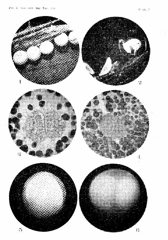
PAP. & PRoe. ROY. Soc. TAS. 1936 PLATE I
PLATE II
Fig. 7.~Two-c€lled stage in which the two blastomeres have partially refu3ed.
Fig. S.-Section through the stage shown in fig. 7.
Fig. 9.~Four-celled stage.
Fig. lO.-Equatorial section through the four-celled stage.
Fig. ll.--Eight-eelled stage.
Fig. 12.-Sixteen-celled stage.
Fig. lit-Section through an emuryo i111mediately after the sixt.een-celled staget
showing a blastomere entering the blastocoel.
Fig. 14.-~Section through a slightly later .stage than that shown in fig. 1:3. The blastomere is now in the blastocoel, and is undergoing mitosis to form the primary mesoderm.
Fig. 15.--Section through a stage more advanced than that shown in fig. 14. The blastocoel is much larger, and now contains a number of primar;y mesoderm cells.
Fig. 16.~Section through a stage ju~t prior to gastrulation.
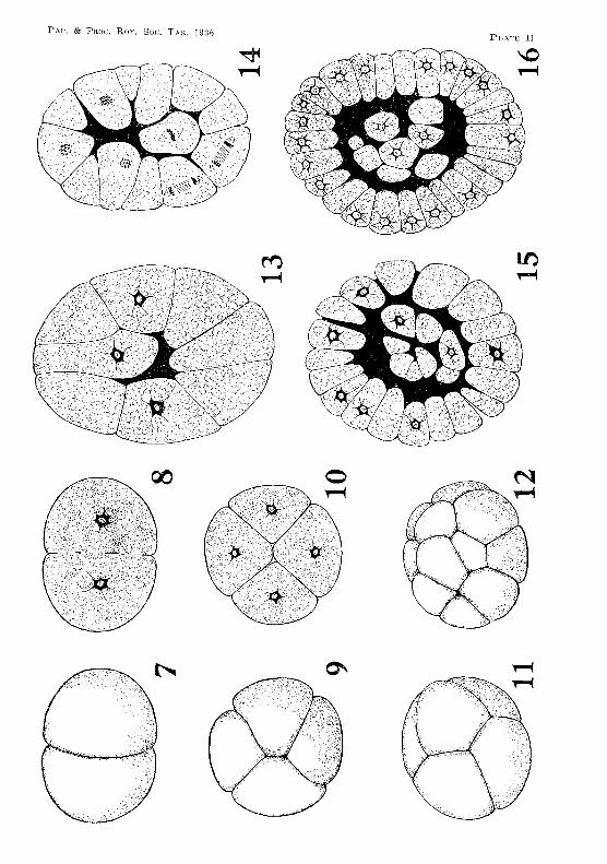
PLATS H
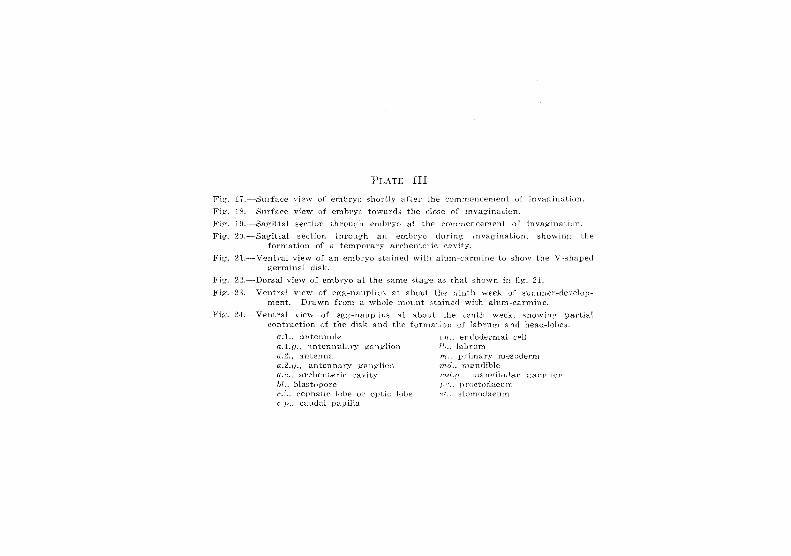
PLATE III
Fig. 17.---Surface viesv of emhryu shortly after the commencement of invagination.
Fig. ] 8.,,--Surface vie\v of enlhryo toward::5 the dose of invagination.
Fig. HI, ---Sagittal section throu,G:'h embryo at the COIllmcncement of invaginat.illJL
Fig. 20.,·,,--Sagittal section through an embryo durin . .;; invagination. shovl'ing t.he formation of a temporary archentc:'lc eav]t.y.
Fig:. 21.-~Ventral view of an embryo stained with al.um-c:i.l'nline to show the V -shaped germinal disk.
Pig', 22.-Dorsal vie,,, of embryo at. the ;Sanl€ stage that shown in fig. 21.
Fig. 2:J.~- VentraJ view of egg-nauplius at about the ninth week of :n~mmer-dev-(~lop
Inent. Drawn from a \vhole mount stained with alum-carmine.
Fig. 2·1.--VentraJ vie"\Y of egg-nauplius at about the lenth \veek, showin,C! partial contraction of the dif'k and the fOrlnatjon uf labrum and head-Iobe3.
a;l .. ~tnt(::nnule
a.I.!!., . .1.ntenJF1iary ganglion n.2., antenna a.2.,lj., antennary g'an.(,.:1io:Ci a.c., archentel'lc cH\+lry bl., blast.opore (~,: .. cephalic lohe 01.' optic h,be c.}) .. cHud<.d papilla.
en .. endodel'lual cpH 1[) .. Iab p um m., prilnary Ines··)derm m(L, mandible m(!.D .. rnanclir.ula.l' g-anrrlion II:'"'., !)i.'c('todaeu.m.
~.'i tom of.heu m
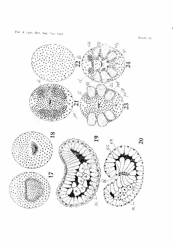

PLATE: IV
Fig. 25.----Surface view of region in the vicinity of the blasio-pol'€ at the first appearance of the ectodermal teloblasto. Drawn "dth the aid of a Canl€l'R lucida frOlu a ,vhole mount stained with alum-carmine.
Fig. 2G.-~Sul'face view of blastoporal region when, by the contraction of the the naupliar appendages have cmne closer together and the row eetoderrnal telohlasts has conunenced to curve round the caudal papilla. (See also fig. 28.)
Fig. 27.--Ventral view of egg-nauplius at the eleventh 'iveek. The disk has now contracted to such an extent t.hat the appendages no longer project beyond the sides of the embryo. The cells of the head-lobes now form concentric c,jrcles.
Pig. 28.-Ventral view of embryo at the thirteenth week. The caudal papilla is noW" \velJ formed.
Pig. 2D.--Ventral view of en1bryo at the fifteent.h '\veek. Median eye and the l'udilllents of the rnaxillules have now appeared.
Fig. 30.--Ventral view of elubryo at the sixteenth week. Endopodite and exopochte of the antennae are now segmented and about equal in length.
n.l.. antennule u. 2.. antenna c.c .. central c.ell of head-1obe c.l., head-lobe C.l}., caudal papilla
ectoderlnal teloblw:;t , median ectodermal teleblast.
(;c.t.1. .. -eG.t.7., later31 ectodermal telohlasts
lb .. labrum m.d., mandible rn.e., median eye 'J'n:-c.1., maxillule m::r.2., maxilla
1>T., proctodaeum. th.Ll., first thoracic. appendage th.l.3 .. third thoracic appendage
© ~ ~@
25
fft!~-------\-e c t 7-
~"'"' ..... ifJ}"", w ~
~ .'" @I f?i.~. '. tJ \ ~"
\pr.
4,
,.... €J\W
:;f0j} eJ (j~ec.t ~
lb.
30
-c ,. C'
~
-0 ~.
~ ~
" "' U1 o p
e' ,.~
v~
¢
;:
>-j r ;...;
'"

Fig'. ~n.-Optie<lJ f,pctinn of .)lJSt hat.ching, rounded by the d"lOl'iof1, vitellil1c~ m.ernbranE'.
S h(!';;,: i H,g
and fil',-;L
the ;'_iltd
ernhr;,-o second
tnt.e;::;-Hn10i1tSo
F'i~.:'. ;32.----Eg,'2: at fi:·~:t. stag'€ of hatch ins;. showing split jn ehorl0R
Fig. :iH. - Sez"ond s-cal,!:'e in the hatchin~': process. The larv"lJ integument is bUlg-lnp: O,-lt tnTo1]g:h the split. in Lhe chorion, -forcing it open.
"Fi!la] stag-e in ha;:'ehin;~. The chorion has burst, and the shl'itnp is surrounded by the disLcTl(1ed lal'val intc.:,rumc11t, to t.he CluteI' f-mrface of ~vl,Thich of the vitelline membrane stiU adhere. Tl~e second Jar>;-aJ integ'unHn:'l ba~~ been l~asl. ofL and lies \vithin 1:1l';':,".
-ThG l1<2.vl"ly hatc1wd :3h;'imp. ~1')(nvjHg t.he sessile hepatic Jobe8, ~lnd -the elon::2:uied yc:lk-sac. The throngh the c:.'b)(lf21"m and mU:3('le~
rh. ehor-io'll 1,'1'1'1 1.,
t\\;I,t
laUe,' 1~ clearl:i i:.he animal..
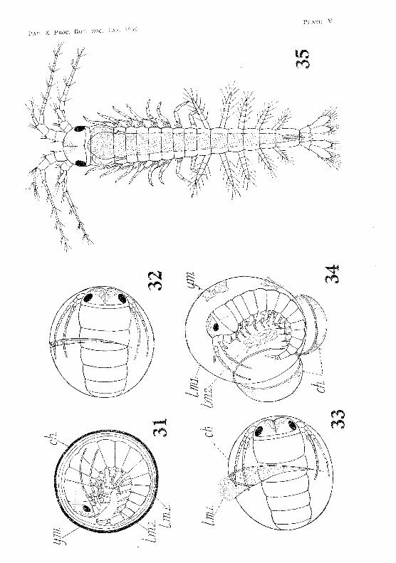
PLATE VI Fil/."s. 36-39.-Appendages of the newly hatched Anaspides:
Fig. 36.-Antennule. Fig. 37.-Antenna. Fig. 38.-Third thoracic appendage. Fig. 39.-Mandibles.
Figs. 40-43.-Dorsal views of the head reg'ion in young specimens of Anaspides to show the gradual development of the rostrum and of the pedunculated condition of the paired eyes:
Fig. 40.-Newly hatched specimen. Fig. 41.-Specimen about 3'4 mm. long. Fig. 42.-Specimen about 4'4 mm. long. Fig. 43.-Specimen about 4·6 mm. long
c.gr ... cervical groove c.s.a., four-celled cervical sense organ m.gT., median longitudinal groove of head T., rostrum r. gr.. ros tral groove th.2., second thoracic segment
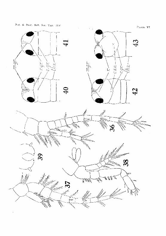
PAP. & PROC. ROY. Soc. TAS. 1936 PLATE VI
:F'i1?~.
PLATE VII
L,dsun lif thce' ne'wly hatehE'd A nasJfid(;,~'.' uds and the notched telson.
Fig. 45.----Third abdomiuaJ pl(7opuu. 46.--Maxilla. 117.--1VIaxillule.
Fig. -ts. -Sagittal sec-tioll throug"h an ernbryo at a stage immediately following ga::-; ~ tl'ulation and just befut'e that shown in fig. 21. The endodermali ('ells have Cl"O\Vned. together, and now fin most of the blastocoel. ThE' blastoPOl"e is closing. and the celli:i within its margin form the wall of the proctudaeum. The :::.tumorlaeal :invagination is commencing. A few rnesoderrn ('ells UlJpear below the fuLlll'€ position of the labr·urn.
Fig. 4-~1, --Tl'anSVE'l'Se seetion of an embryu at the stage ShU\Vll in 1i.!1.'. 2:-1. The tieetion vm;ses thruugh the olJening (Jf lhe slomodaeuUl. The strand:::; of rlle~oderrn l1Il eac:h side at-e seen in c.toss-section Lelow the rudirnellts. of Lhe antenna~, Cytolysi::; bas uenll'red in SOlHf' of the endodeTrnnl cells, p;iving rise to l'.'l'anules of chrornatin.
n,2" alltenna ""fl., ('ndudf'rm~·~l (:elJ 111.11/.., lab Tal mcsodet·lYJ )I. In.; naupli::-1r rnesnL1t'ttl1 )Jr., pJ"odoua(:'um f.;t., ::;tomodaenn1
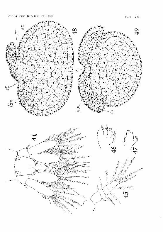
1''\.1'', & Prwc. Hoy. Soc. TAS. lSJ~~!;
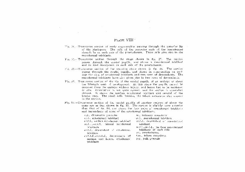
PLA1'E VIII·
:Vig. 50,"- ·Transverse section of carl:{ egg-nauplius paf;~lng through the antE~_rior Up of the blastopore. The cells of the posterior ends of the mesoderrrlal strands lie on each side of thE- proctodaeum. These eells ).~·1 VE:~ rise to {-,he mesodermal teloblastE!.
I,'iig'. 51.-Transverse section through tlle stage shuwn in Jig. 27. The seer ion passes through the caudal papina. and show;~ a Tn€sodernul.i telohlast and its first descendant on eaeh side of the proctodaeum.
Fig. 52.---~Tran5Vel'Se seetion of the nanplius stage shown in fig," 28. The 't:leeUlJn passes throLlgh the caudal papilla, and showl'. in cross-section on side the ring' of ectociE.·:rm.al telobJ<~sts and two rows of descendants. 'rhe mesodern1al teloblasts have also given. rise to two 1'o\vs of descendants.
Fig. 53.~ -Transverse seetlon of the tip of the caudal papilla of an em.bryo at abuut the. fifteenth "yeek of development. At this stage the papilla cannot be removed from the enlbryo without injury. and henee bad to be f3eetioned l~n .sift<. Orientation i~ n~)t quite (,;()YTed, and the sed-ion is s01nevd1<-l.t, oblique. It shows the 111,eciian et--.toderrnal teloblast and se'.reral of lat.eral ones. The sHla!l cell,., funning the tel son mesoderm also appea,' in t.he section.
Fig'. [)-4-.---Transverse sed.ion of the calJl1al of another of ahout the-same age a::-; that shown in fig. The s€ction it; more anterior than that of fig, 53 .. and shows the four pairs of m.es()dermal teJoblast~
and descendants of sorne of the eetodermaJ telobJaRt:::;.
C.(J., dll'omatin ce.t., edodermal telublast ecJ.il., rnedi<l.TI ectodernlaJ t.elohlast e{".t,1.,·cc.t.9." lateral edorlel'lnal
telobJast:3 {l;(s.t .• d.. deseencIan t of eetuderrnal
teloblast
'tn., primary mesocterr!'l rnJ .. , lTIE'sodermal t.elobla,3L '1nJ.d .. desc~ndant of llH::~;CldeT1nal
Leloblast the four TIl€soderrual
of each side pr., proetodaeum t.rn., telsOll nlesocienn Y.,tIo, yolk g;l'anule
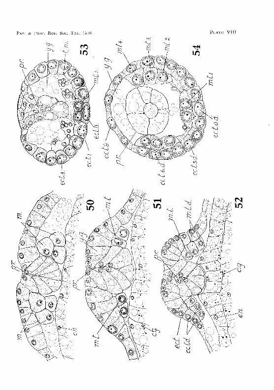
I~I.,A(r}G IX
Pig, 55.---T.rans\'(~n;e section 1)£ th.e The is some'what. fnrr,he-r fOI'V,'8.l'd T,hose The and ventraJ rnesodenn Hre differentiated, the rI(ll'S<-lJ .n1€sode"Jl"t h~1,~
(?oTIlmeneed its upwanl ;,;."rc.'\vth to -[orrn the later::tl w?Jls Hf the' vessel.
F'Ig. 5().-~"1'1'anSVer8e section through the third abdominal segment :!::-\'\."eeJ; embryo. The ;;valls of the dor::;al V(~;C;;Re] are now fonm:~d. and t.he JJe.rl(':ar~
dial cavity it:: (~6tablished b:;r the shrinking avnty of the m(~sor1erm. from the dorso-lateral eetoderm, rrht~ floor ~f the rlorsal ves:"el has heen fanned hy the Inesoflerm, gTGvr'ing' oyer the yolk.-sae. Yolk-granules H nd cens that b::·l,se spreail hackTivard from fne naupHa.r mesodenn are pl'Ci::iCnt the c-avity of the dorsa} \-'eSf4e.l and at th~ sides- of the ~\'1)1kM8a(',.
Fiz· :sr/,,,-·Tn:m,8verse section thnrr~gh t}J'2 nl.R.xHlulary ::3o:rnit.e of ;,1 n(~wly h;}t,(~l1i?d
specimen, showing the 'jal'f2,'e. ~ntei"10r cH\.-it.y of the he1-!rt: roo:fed h;-the dorsal organ.
.Fig, 58, - -'Transverse seetion thr,}up.-h the ffHn'th thorac](: speeimen, sho,ving tb(; thicken'E'd w:::!lJ of the
of 1.1. ne\'\-'i;;: haLL',hf:d
0.),1... prlmary ant€Ylol" of li'-C€l'
d.l.rn., longitudInal muscieF c7.rn., dorsal mesodenn, d.o., d01'S8.,l organ
endcdermal cell genital l'udirnen!
cardiac ea.,city 01' of dor~'58'j vessel
h.j'., fio{'r of ('ardIac
i't!;15.L(., ('oelomic cavil:}" uf ulary t-;egr.acnt
n.m", :naupli<':l.1' r:nef:,.oderr'-"i pericardial ·'2avit.'\'
1)c,/,. perjean1iaJ ti.1)or 1')" posterior lobe of 1.'. m·". ventral l11.csode-rm 't!.l.m,., vent.ral hngir,udi1 181
mn,se.J.er; yolkhgranu IP";
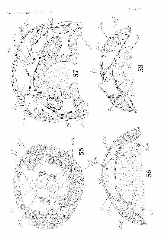
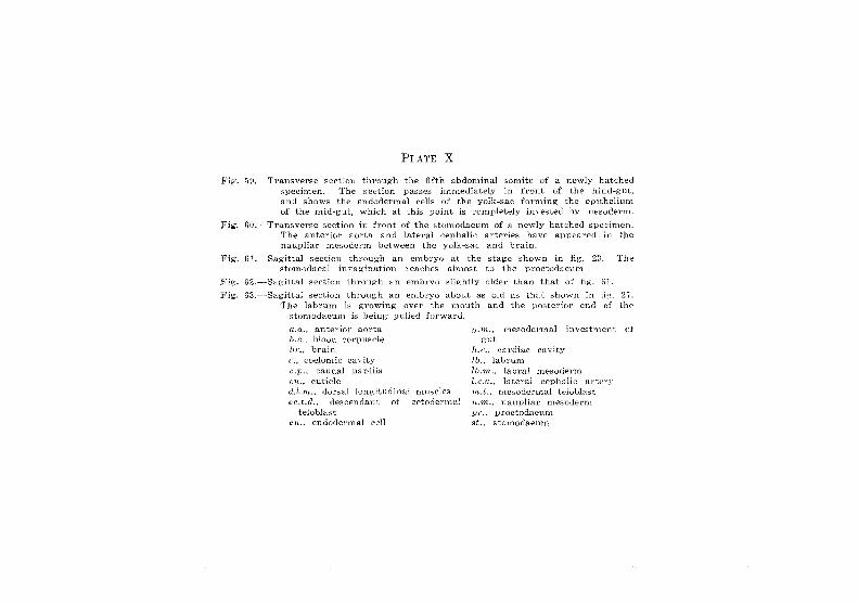
PLATE X
j,""lg. 59.--,·Transverse section through the fifth abdonlinaI sonlite of a newly hatebed Bpecinlen. The section pas;:;es immediately in front of the hind-gut, and shows the endoderlTml cells of the yolk-bac forming the epithelium of the D1id-gut, which at this point is completely invested by luesoderm.
Fig, 60.----Transverse section in front of the slomodaeunl of a newly hatched specimen, The anterior u0rta and lateral cephalic: arterie~ have appeared in the nanpliar .mesodenn between the yolk-sac and brain.
Fig. 61.---Sagittal section throug'h an embryo at the stage shown in fig. 2;:;, The stoITlOdaeal invagination reaches alrnost to the proctodaeum
Pig. G2.-Sagittal section through an embryo slightly older than that of fig. ()].
Fig. 63. --Sagittal section through an embryo about as old as that shown in tig. 27. The labrurn IB growing over the mouth and the posterior end of i..ht"
6tom.odaeurn is being- pulled forward.
a.a.. anterior aorta h,c.. blood eorpusde br .• brain c., coelomic cavity C.p., caudal papilla CU., cutic:Je d.l.Tlt .. , dOl·sal longitudinal rnuscles ec.t.d., descendant. of ,,;,ctodermal
teloblast fhL endodenn.al eel!
fI./H.. mesodel·mal investment of gut
h.e., eardiac eavity lb., labrum lb.m ... labral mesoderm I.e.a., lateral cephalic H.rtel'Y 'In. I,. , mesodertnal teloblast n.m., naupliar mesoderm pr., proctodaeum st., stOlnodaeum
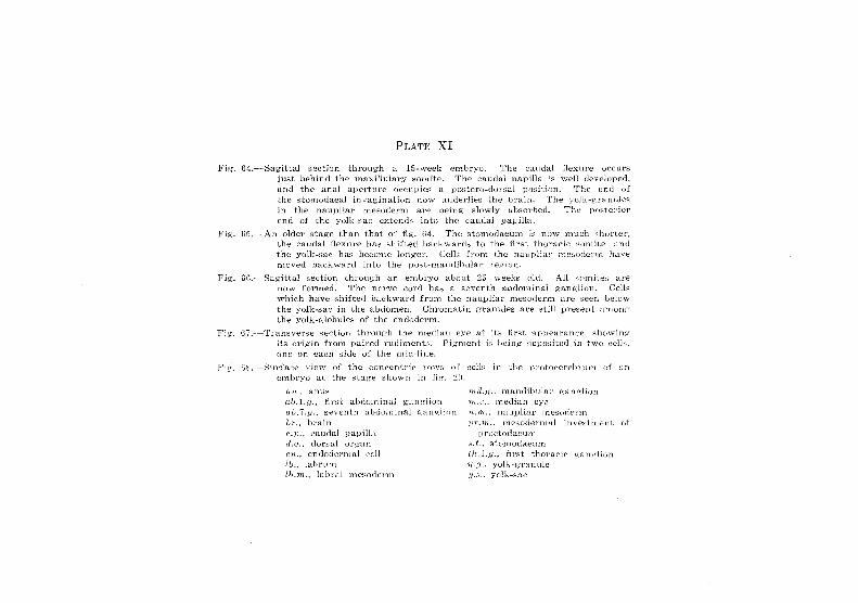
PLATE XI
Fh;. 64.-- Sagittal section through a 15··l,.v-eek €lubryo. The caudal flexure occlirs just behind the maxillulm'Y t'omite. The caudal papilla is \vell devdoped, and the anal aperture occupies a 11osterD-tiOl'sal pnsitl0n. The end of the stomodaeal invagination 110W lmderlies the brain. The ydk-g'rannles in the nanpliar mesorienl1 are being- slowly ubsOlobed. The postel-jor end of the yolk-sac extends into the caudal papilla.
Pi~,,, 6iL--An older sta,ge than that of flg'. U4. 'The stomodaeum is ll;l\V much ::;hortel', the caudal flexure has: shHted bac:kwartb lo the iirst th!..!raeie ;wmiiE.'. and the yolk-sal' has become LOll ReI'. Cell:::; from the nauplial' mesuderm hay!;": moved back\VfUd intu the posL-mandibular regiun.
Fig, f)(i.---Sagittal seetiun throug:h an emLryo about 25 \veeks old. All s0l11iie8 al'~
now formed. The nel've cord has a seventh abdominal ganglion. CeiL,: which have shifted backward frorn the nanpliar mesoderrn are seen below the yolk-sac in the abdomen. Chromatin gTanules are still prei::ient anlong' the yolk-globules of the endoderm.
}'j.~. 67. --Transverse section through the median eye at iis first appeal'an(:e. sh()\\rin~
its orjgin from paired rudiments. Pi.\2:ment is heing jeposited in two ee·jJH, one on eacb side of lhe l1Lid-line.
Fig-, ~i8, .. ~-Surfa('e view uf Lhe coneentric rows; of cells in the pl'ohwel'o2ln"ufrl of an €Tllbryo at the stag-e sho"\vn in fl;~. ~n.
a'n., anu,'; ab.1.y., first abdominal ganglion alJ.7.f/., sevellth alHl{)Hiinal i2:anglioH hr., brain C.1)., cauda] pavilL-l d.o., dorsal 01'i.!,"<ln
(~n., endoJermai celi ll; .. labrum !h.rn .. htbral me.t.::.oG(:rl11
'fnd.l1., mandibulay ~'a11gli\Hl
'if~.e., lYtedian eYl'O 'It.'m., nuuplia)' rTIt.'si)del'l1"l
pr.lfL, mesodennal in\'E'stnlf'rn of l}roelot!aeurn
s.t., stomodaeum th,1.[),., fi ~>st thorac]('. !2:angliDn iJ.g., yolk-granl1lt:' 1/.;':;., yolk-sac
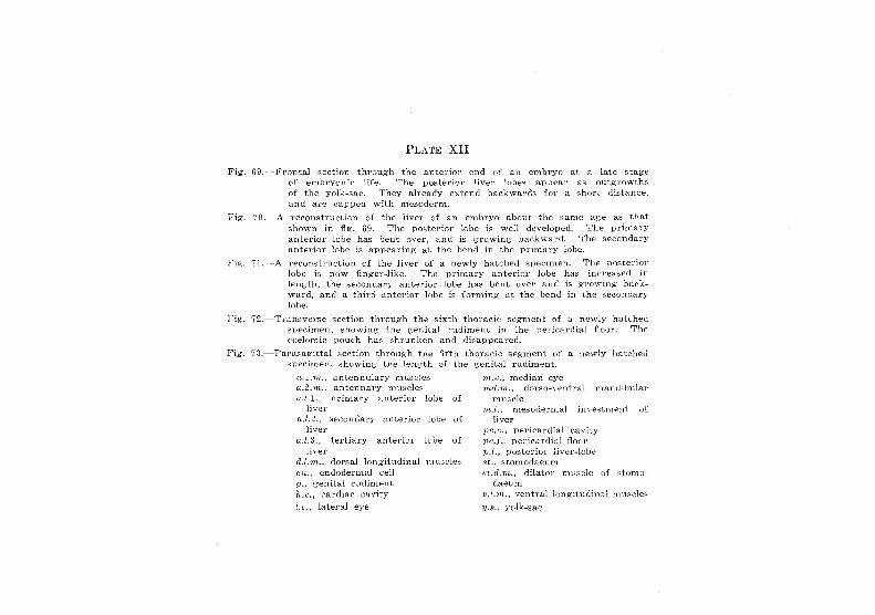
PLATE XII
Fig. 6H.- ~ .. F\rontal sedion through the anterior end of: an embryo at a late l>tage vi elubryonic life. The posterior liver lobes appear as outgrowths of the yolk-sac. They already extend backward::; fur a short distance. and are capped with mesodenl1.
Fig. 70. -A reconstruction of the liver of an elnhryo about the same aRe as that shown in fig. 69. The posterior lobe is well developed. The prirnary anterior lobe has bent over, and is growing oackward. The secondary anterior lobe is appearing at the bend in the prImary lobe.
Fig. '?l.---A reconstruction of the liver of a newly hatched specimen. The posterior lobe is now finger-like. The primary anterior lobe has increased in length, the secondary anterior lobe has bent over and is growing backward, and a third anterior lobe is for-ming at the bend in the secondary lobe.
Fig". '72.~-Transverse Bection through t.he sixth thoracic segment of a newly hatched specimen, showing the genital rudiment in the pericardia1 floor. The coelomic pouch has shrunken and. disappeared.
Fig'. '/8. -Parasagittal section through the fifth thoracic segm_ent of a newly hatched specil11en, showing the length of the genital rudiment.
a.l.rn., antennulary lTIUse:les a.2.'rIl-., antennary museles aL1., primary anterior lobe of'
liver (1.7.2., secondary anteriOl' l()be of
liver ft.l.S., tertiary anterior lobe of
liver d.l.m., dorsa] longitudinal nlu:~des ('-n.. endodermal eel] g.; genital rudiment fcc., cardiac cavity I.e., lateral eye
";n.e., median eye 'J'nd.m., dorso-v~nt.ra] mandibular
muscle 'rn.Z., mesodermal investment or
liver pc.t., pericardial cavity pc.f., pericardial floor ,]).7., posterio!- liver-lobe st., stomodaeum st.d.rn., dilator musele of stomo"·
daeum 1}.l,'m .. ventral longitudinal rnue-cles
y.' .. yolk-sae
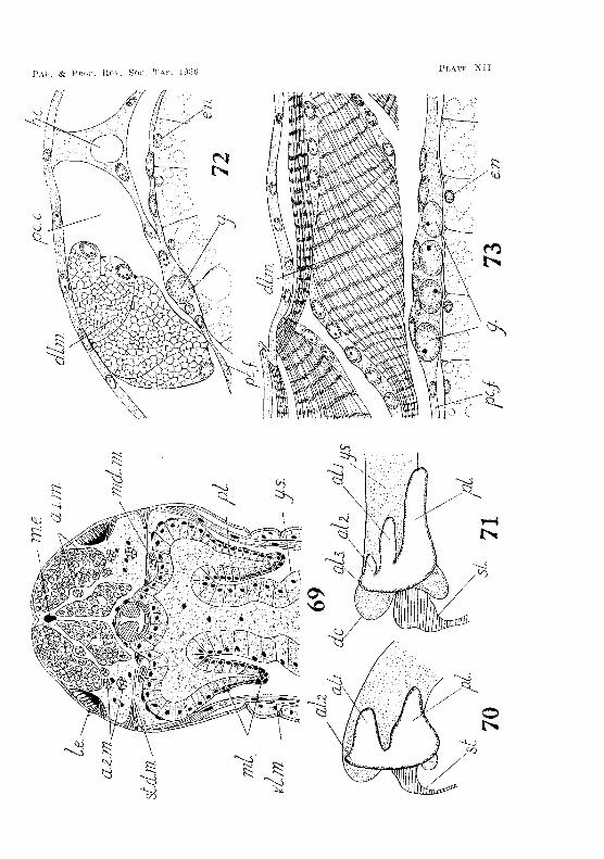
PAl". & PLATR XU
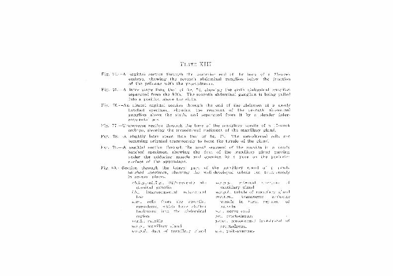
PLATS XIII
Fig .. 7/L---_A s;-lgittal seetion through the puste:ciur end of Lhe Ih)11y of 25-week elTJ:'l"YO, sn(J\ying the 81::venth abdomina! gn,nglioll b(:·lo\y thE' :inndioll of the yolk-sac \vith the prododa8ll.m.
Fig'. 7;"), --A later stag-e than that of shtnving the. sixth [-J.ldOnljn~ll gallf';Eon
Pip:,
Fig·.
;;epal'ated from the -fifth. The s(;!-\-"enth abdominal ganglion is being 1)n11e,-l into a positiun above the sixtr-l.
~'(;.----_AIl almost sagittal section through hatehed specimen, showing: -Lhe ga,ng-liol1 above the slxLh. and :3cgTnental bar.
the end of the abdomen \)1 a n(f-\-vly l'Clnnant of the seventh ab(';ominril
f1'0111 it hy a slE;'JHle," intE'l'-·
77, "-'Tnms\'erse Si..~etiol1 thl'O;Jgh the b:-i~:',E: of the maxillary s{)miie of :a 17-\,-\.'c€>h 2rnln-yo, showing Lhe Dl.(;-sodenn:::tl l'uciimellL of Lhe rnaxillal'Y gland.
tdig-htly latee stage than that of fig". 77. T'lle n1.e~wde1'mal cell::- tHe
beCOD'ling oriented tranf:vel·.~,ely to fOI'111 the tubule of the gluUt\..
79.--A sagittal section through t.he- basal segment of the ill_axilla in a newly hatched specimen, shcn .. vln.ll the duct 1)£ the luaxHlary glaud pas~.,;nj_~
under the ad.duetol' musde and ()1)enillg' by a pore un the rHJSlt·-,·10\
surface of the appendage.
Fhr, ~~fl.- -Section through the h,tel'Hl Phrl the lllaxillal'Y j;~·'jand
hatchE-~d :'i1-)eC~n'len, ShmYlng t.he wl.'l1-developed tl.lb~lie cut Hl pll:-i,CE's.
a!).5.if.,-at.. '( .f;.. fifth-sc'ven t.h dominal g-an>;lia
'i.h.. intel'seJ~:nlenL;:!i ecu,derrnal bar
!/,'iH., ct'~lls frorn the rnesoderrn_, \Yhi('!'~ hav2 , .. :hLllt'(l backward hlHJ the-' region
rn:r.2. .. rna.x.i·lh-i. "i'fl.;";7.,(f., lnaxil"!;1'i')-'
duct \Jf
m~i: . .tj. p., ext.erna.l alh:'!'i:nrc maxillary g-ol"l.nd.
tubule of ntdx1'ilal':'" 'm:(.t.HL, tnlnsverse HddLld~o;
rnu.sde in ba::3:'1J of rnaxllhl
'1/.,('_, lle,."ve C'Ol'd
'))J- •• P1'Ol~tO(j:Leurn
71'f".1'n., n'I€,slHle:nTJal t}lVeiStlt~ent of
P'i:'~lct{Jdaeum .vo!k-S';rar.!ule,";