the effects of acute and chronic nicotine on gaba and
TRANSCRIPT

Brigham Young University Brigham Young University
BYU ScholarsArchive BYU ScholarsArchive
Theses and Dissertations
2011-03-13
The Effects of Acute and Chronic Nicotine on GABA and The Effects of Acute and Chronic Nicotine on GABA and
Dopamine Neurons in the Midbrain Ventral Tegmental Area Dopamine Neurons in the Midbrain Ventral Tegmental Area
Devin Hardy Taylor Brigham Young University - Provo
Follow this and additional works at: https://scholarsarchive.byu.edu/etd
Part of the Psychology Commons
BYU ScholarsArchive Citation BYU ScholarsArchive Citation Taylor, Devin Hardy, "The Effects of Acute and Chronic Nicotine on GABA and Dopamine Neurons in the Midbrain Ventral Tegmental Area" (2011). Theses and Dissertations. 2951. https://scholarsarchive.byu.edu/etd/2951
This Thesis is brought to you for free and open access by BYU ScholarsArchive. It has been accepted for inclusion in Theses and Dissertations by an authorized administrator of BYU ScholarsArchive. For more information, please contact [email protected], [email protected].
The Effects of Acute and Chronic Nicotine on GABA and Dopamine Neurons in the
Midbrain Ventral Tegmental Area
Devin Taylor
A thesis submitted to the faculty of
Brigham Young University
in partial fulfillment of the requirements for the degree of
Master of Science
Scott Steffensen, Ph.D., chair
Dawson Hedges, M.D.
Sterling Sudweeks, Ph.D.
Department of Psychology
Brigham Young University
April 2011
Copyright © 2011 Devin Taylor
All Rights Reserved
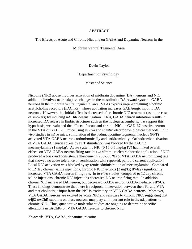
ABSTRACT
The Effects of Acute and Chronic Nicotine on GABA and Dopamine Neurons in the
Midbrain Ventral Tegmental Area
Devin Taylor
Department of Psychology
Master of Science
Nicotine (NIC) abuse involves activation of midbrain dopamine (DA) neurons and NIC
addiction involves neuroadaptive changes in the mesolimbic DA reward system. GABA
neurons in the midbrain ventral tegmental area (VTA) express α4β2-containing nicotinic
acetylcholine receptors (nAChRs), whose activation increases GABAergic input to DA
neurons. However, this initial effect is decreased after chronic NIC treatment (as in the case
of smokers) by inducing nAChR desensitization. Thus, GABA neuron inhibition results in
increased DA release in limbic structures such as the nucleus accumbens. To support this
hypothesis, we evaluated the effects of acute and chronic NIC on GAD-67 positive neurons
in the VTA of GAD GFP mice using in vivo and in vitro electrophysiological methods. In in
vivo studies in naïve mice, stimulation of the peduncopontine tegmental nucleus (PPT)
activated VTA GABA neurons orthodromically and antidromically. Orthodromic activation
of VTA GABA neuron spikes by PPT stimulation was blocked by the nAChR
mecamylamine (1 mg/kg). Acute systemic NIC (0.15-0.5 mg/kg IV) had mixed overall
effects on VTA GABA neuron firing rate, but in situ microelectrophoretic application of NIC
produced a brisk and consistent enhancement (200-500 %) of VTA GABA neuron firing rate
that showed no acute tolerance or sensitization with repeated, periodic current application.
Local NIC activation was blocked by systemic administration of mecamylamine. Compared
to 12 day chronic saline injections, chronic NIC injections (2 mg/kg IP/day) significantly
increased VTA GABA neuron firing rate. In in vitro studies, compared to 12 day chronic
saline injections, chronic NIC injections decreased DA neuron firing rate. In addition,
chronic NIC increased DA neuron, but decreased GABA neuron GABA-mediated sIPSCs.
These findings demonstrate that there is reciprocal innervation between the PPT and VTA
and that cholinergic input from the PPT is excitatory on VTA GABA neurons. Moreover,
VTA GABA neurons are excited by acute NIC and sensitize to chronic NIC, suggesting that
α4β2 nAChR subunits on these neurons may play an important role in the adaptations to
chronic NIC. Thus, quantitative molecular studies are ongoing to determine specific
alterations in nAChRs on VTA GABA neurons to chronic NIC.
Keywords: VTA, GABA, dopamine, nicotine.

iii
CONTENTS
INTRODUCTION .................................................................................................................... 1
BACKGROUND ...................................................................................................................... 2
Nicotine Addiction ................................................................................................................ 2
Dopamine-dependent Mechanisms in the Mesocorticolimbic System ................................. 3
Dopamine-independent Mechanisms in the Mesocorticolimbic System .............................. 4
Nicotine and the Reward System .......................................................................................... 6
GABA Neurons in the Mesocorticolimbic System ............................................................... 9
RATIONALE ABD HYPOTHESES ...................................................................................... 11
METHODS ............................................................................................................................. 12
Animal Subjects .................................................................................................................. 12
Characterization of Neuron Types in vitro .......................................................................... 14
Single-unit Recordings in Anesthetized Rats ...................................................................... 15
Characterization of VTA GABA Neurons in vivo .............................................................. 15
Single-unit Recordings in vivo ............................................................................................ 16
Drug Preparation and Administration in vivo ..................................................................... 17
Chronic Injections ............................................................................................................... 17
Preparation of Brain Slices .................................................................................................. 17
Whole-cell Recordings in vitro ........................................................................................... 18

iv
Single-cell Quantitative RT-PCR ........................................................................................ 19
Statistical Analyses ............................................................................................................. 21
RESULTS ............................................................................................................................... 23
Mixed Effects of Systemic and Local Nicotine and α7 Nicotinic Receptor Agonists on
VTA GABA Neuron Firing Rate ........................................................................................ 23
Acute Nicotine Effects on VTA GABA Neuron Activity in Naïve Mice ........................... 24
Effects of Select nAChR Antagonists on Nicotine Activation of VTA GABA neurons in
Naïve Mice .......................................................................................................................... 25
Cholinergic Inputs to VTA GABA neurons: Effects of α7 Nicotinic Antagonists ............. 26
Chronic Nicotine Effects GABA Neuron Firing Rate and Response to Acute Nicotine .... 27
Chronic Nicotine Effects on VTA DA Neuron Activity ..................................................... 28
Chronic Nicotine Effects on GABAergic synaptic inhibition to Dopamine and GABA
Neurons in the VTA ............................................................................................................ 30
In Vitro Chronic NIC Effects on the Expression of nAChR Subunits in VTA GABA
neurons in GAD GFP Mice ................................................................................................. 31
Discussion ............................................................................................................................... 33
Appendix ................................................................................................................................. 36
Original Experimental plan ................................................................................................. 36
Experiment 1: Acute in vivo studies: effects of acetylcholine and nicotine on VTA GABA
neuron firing rate ................................................................................................................. 36
v
Experiment 2: Acute in vivo studies: effects of buproprion on VTA GABA neuron firing
rate ....................................................................................................................................... 37
Experiment 3: Acute in vitro studies: effects of nicotine on GABA inhibition of VTA DA
Neurons ............................................................................................................................... 38
Experiment 4: Chronic in vitro studies: effects of chronic nicotine ................................... 38
REFERENCES ....................................................................................................................... 39

vi
LIST OF FIGURES
Figure 1. A simplified schematic of the VTA and afferent projections .....................................3
Figure 2. A schematic of the role of nAChRs in the control of VTA DA neuron excitability ..7
Figure 3. Visualization and Patching of GABA neurons in the VTA of GAD GFP mice ......14
Figure 4. Mixed effects of systemic nicotine on VTA GABA neuron firing rate in vivo .......24
Figure 5. Nicotine markedly activates VTA GABA neuron firing rate ...................................25
Figure 6. Effects of nicotinic acytelcholine antagonists ..........................................................26
Figure 7. Stimulation of cholinergic inputs to VTA ................................................................27
Figure 8. Locally administered nicotine effect on chronically treated mice ............................28
Figure 9. Lack of effects of chronic nicotine on dopamine neuron activity ............................29
Figure 10. Nicotine effects on GABA neuron sIPSC frequency .............................................30
Figure 11. Chronic nicotine enhances inhibition to VTA GABA neurons ..............................31
Figure 12. Chronic nicotine effects on nAChR expression .....................................................32

1
The Effects of Acute and Chronic Nicotine on GABA and Dopamine Neurons in the
Midbrain Ventral Tegmental Area
An enormous accretion of research efforts from numerous laboratories across the
country and around the world has been devoted to the study of nicotine (NIC). NIC
addiction specifically is one area of focus worth studying due to its biological nature, abuse
potential and long-term relapsing effects. Tobacco use is the leading cause of preventable
death in the United States (Volkow, 2006). It is associated with the top four leading causes
of death in the country (Volkow, 2006). Cigarette smoking claims the lives of 440,000 US
citizens each year – more than the combined deaths due to alcohol, cocaine, heroin, homicide,
suicide, car accidents, fire, and AIDS (Volkow, 2006). It accounts for four million deaths
annually worldwide (WHO, 1999). If current trends continue, smoking will be the cause of
one-third of all adult deaths globally in 2020 and the mortality rate from tobacco will climb
to ten million a year by 2030 (WHO, 1999). With each puff of a cigarette, a smoker absorbs
over 4,000 chemicals, which will cause damage to nearly every part of the body, from
cataracts to pneumonia, cancer, heart disease, and lung disease. Tobacco has also been
shown to harm unborn children when used by pregnant mothers and is estimated to have
caused 910 infant deaths each year from 1997 to 2001 (Volkow, 2006).
The economic cost of tobacco is not as easy to see. Those who generate tobacco
products clearly see a financial benefit and consumers find some advantage or they would not
be willing to pay for these products. There is no doubt that tobacco producers provide jobs
for many workers. However, when weighing the economic benefits of tobacco production
against the costs of its use, in terms of health care issues and mortality rates, one analysis
2
predicted that for every 1000 metric ton increase in tobacco production, there would be a net
economic loss of 13.6 million dollars per year (WHO, 1999).
Background
Nicotine Addiction
NIC is the major contributor to the maintenance of tobacco use (Benowitz, 1996).
After inhalation, NIC is rapidly absorbed by the blood stream and within 10-19 seconds has
passed through the brain (Benowitz, 1996). Human and animal studies show that NIC-
induced stimulation of neurons in the mesolimbic DA system are central to the reinforcing
effects of NIC use (Benowitz, 1996). The mesolimbic system projects from the VTA of the
midbrain to the nucleus accumbens and is well-known to be involved in reinforcement of
other drugs of abuse.
DA antagonists in the VTA block NIC’s addictive effects and cause cessation of self-
administration in animal models, indicating that NIC must bind to receptors that cause action
potentials and neurotransmitter release in DA neurons. Further research has revealed that
NIC binds to nicotinic acetylcholine receptors (nAChR), causing increased firing rates and
neurotransmitter release of DA neurons.
The objective of this study is to understand the related process from a
neurobiological perspective, supposing that if we understand what neural substrates underlie
NIC’s effects, we will be better equipped to design appropriate therapies and develop suitable
medications for NIC addiction. This thesis contains a review the literature on: NIC
addiction; the mesocorticolimbic dopamine reward system; and pharmacology of nicotinic
effects.
3
Dopamine-dependent Mechanisms in the Mesocorticolimbic System
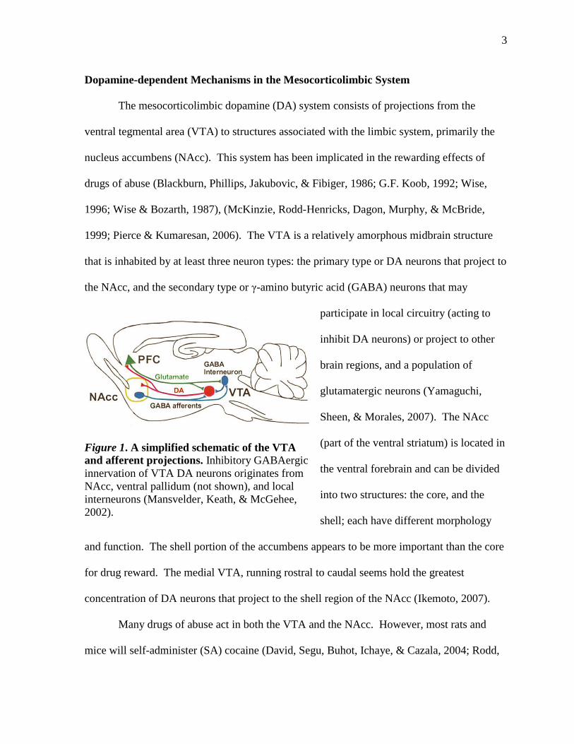
The mesocorticolimbic dopamine (DA) system consists of projections from the
ventral tegmental area (VTA) to structures associated with the limbic system, primarily the
nucleus accumbens (NAcc). This system has been implicated in the rewarding effects of
drugs of abuse (Blackburn, Phillips, Jakubovic, & Fibiger, 1986; G.F. Koob, 1992; Wise,
1996; Wise & Bozarth, 1987), (McKinzie, Rodd-Henricks, Dagon, Murphy, & McBride,
1999; Pierce & Kumaresan, 2006). The VTA is a relatively amorphous midbrain structure
that is inhabited by at least three neuron types: the primary type or DA neurons that project to
the NAcc, and the secondary type or γ-amino butyric acid (GABA) neurons that may
participate in local circuitry (acting to
inhibit DA neurons) or project to other
brain regions, and a population of
glutamatergic neurons (Yamaguchi,
Sheen, & Morales, 2007). The NAcc
(part of the ventral striatum) is located in
the ventral forebrain and can be divided
into two structures: the core, and the
shell; each have different morphology
and function. The shell portion of the accumbens appears to be more important than the core
for drug reward. The medial VTA, running rostral to caudal seems hold the greatest
concentration of DA neurons that project to the shell region of the NAcc (Ikemoto, 2007).
Many drugs of abuse act in both the VTA and the NAcc. However, most rats and
mice will self-administer (SA) cocaine (David, Segu, Buhot, Ichaye, & Cazala, 2004; Rodd,
Figure 1. A simplified schematic of the VTA
and afferent projections. Inhibitory GABAergic
innervation of VTA DA neurons originates from
NAcc, ventral pallidum (not shown), and local
interneurons (Mansvelder, Keath, & McGehee,
2002).

4
et al., 2005), ethanol (Gatto, McBride, Murphy, Lumeng, & Li, 1994; Rodd, et al., 2004),
NIC (Laviolette & van der Kooy, 2003), cannabinoids (Zangen, Solinas, Ikemoto, Goldberg,
& Wise, 2006) and opiates (Bozarth & Wise, 1981; David & Cazala, 1994; Devine & Wise,
1994; Welzl, Kuhn, & Huston, 1989) into the VTA. Taken together, these data suggest that
DA neurons in the VTA that project to the shell of NAcc, and the GABA neurons that may
inhibit these DA neurons locally in the VTA, play an important role in mediating addiction to
various drugs of abuse.
Dopamine-independent Mechanisms in the Mesocorticolimbic System
Early versions of the DA hypothesis for reward suggested that DA might be crucial
for all drug reward, but phencyclidine, morphine, and NIC have both DA-dependent and DA-
independent rewarding effects. It is also questionable whether the rewarding effects of
benzodiazepines, barbiturates, or caffeine are DA-dependent. The emerging view is that DA
is crucial for the rewarding effects of the psychomotor stimulants and is important, but
perhaps not crucial, for the rewarding effects of the opiates, NIC, cannabis and E. So, the
notion that DA-dependent mechanisms are the final common pathway in the processes
mediating drug or natural reward is perhaps too restrictive. In support of DA-independent
mechanisms for reward, a considerable number of electrophysiological studies in freely-
moving animals have shown that only a low percentage of NAcc neurons exhibit discharge
correlations either during heroin, cocaine or ethanol SA, or focused attention (Carelli &
Deadwyler, 1994; Chang, Zhang, Janak, & Woodward, 1997; Peoples & West, 1996). A
lack of DA involvement in drug reinforcement has also been demonstrated for oral ethanol
SA (Rassnick, Stinus, & Koob, 1993), and ethanol conditioned place preference (CPP)
(Cunningham & Noble, 1992; Risinger, Dickinson, & Cunningham, 1992) as well as for

5
cocaine SA (Goeders & Smith, 1983) and cocaine CPP (Mackey & Van der Kooy, 1985;
Spyraki, Fibiger, & Phillips, 1982). The role of DA in cocaine SA has been called into
question by studies demonstrating that DA-transporter knockout mice continue to SA cocaine
(Rocha, et al., 1998). A lack of DA involvement in intracranial self-administration (ICSS)
has also been reported (Kilpatrick, Rooney, Michael, & Wightman, 2000). Furthermore, the
accumulating evidence strongly suggests that GABA neurons in both the VTA and the NAcc
appear to play critical roles in opioid reward (for review see (Xi & Stein, 2002)). More
recently, it has been reported that GABAA receptors in the mammalian VTA serve as a
potential addiction switching mechanism by gating reward transmission through one of two
neural motivational systems: Either a DA-independent (opiate-naive) or a DA-dependent
(opiate-dependent or opiate-withdrawn) system (Laviolette, Gallegos, Henriksen, & van der
Kooy, 2004). After opiate exposure and subsequent withdrawal, the functional conductance
properties of the rat VTA GABAA receptor switch from an inhibitory to an excitatory
signaling mode. Other behavioral studies have shown that chemical destruction of DA
terminals in the NAcc with 6-OHDA had no effect on morphine or heroin SA (Dworkin,
Guerin, Co, Goeders, & Smith, 1988; Ettenberg, Pettit, Bloom, & Koob, 1982; Pettit,
Ettenberg, Bloom, & Koob, 1984). Furthermore, pretreatment with relatively high doses of
haloperidol, a DA receptor antagonist, failed to block the reinstatement of heroin-seeking
behavior upon presentation of stimuli that predicted heroin administration indicating that the
―renewed‖ motivation to seek heroin reinforcement produced by reintroduction of heroin-
predictive cues is not dependent upon DAergic substrates. This finding challenges the
notion, at least for opioids, that DA circuits are critical for drug-seeking behavior (DiChiara
& North, 1992; Fontana, Post, & Pert, 1993; Robinson & Berridge, 1993). Indeed, it has

6
been suggested that DA neurons do not appear to be reward neurons per se, but may be
critical for initiating drug use, and, more importantly, for reinstating drug use during
protracted abstinence (G. F. Koob & Le Moal, 1997). Accordingly, DA neurotransmission
may be only important in mediating the motivational effects of drugs in dependent animals
and that the pedunculopontine tegmental nucleus mediates the rewarding properties of drugs
when animals are non-dependent (Bechara & van der Kooy, 1992; Nader & van der Kooy,
1994). These studies provide evidence for the existence of DA-independent pathways that
also play a role in mediating the reinforcing or rewarding properties of drugs.
Nicotine and the Reward System
Nicotine’s addictive power is largely attributable to actions on DA cell bodies in the
VTA and their projections terminating in the nucleus accumbens (NAcc). Physiologically
revelant NIC concentrations have been shown to activate both pre- and postsynaptic nAChR
(MacDermott, Role, & Siegelbaum, 1999; McGehee, Heath, Gelber, Devay, & Role, 1995;
McGehee & Role, 1995; Wonnacott, 1997). A two state model was offered in explanation of
this phenomenon in 1957, in which NIC could convert active nAChR to a desensitized
conformation (Katz & Thesleff, 1957). This two-state model of nAChR desensitization has
been supported by many experimental studies of peripheral and central nAChRs (Del Castillo
& Webb, 1977; Feltz & Trautmann, 1982; Heidmann, Bernhardt, Neumann, & Changeux,
1983; Ochoa, Chattopadhyay, & McNamee, 1989; Ochoa, Li, & McNamee, 1990; Rang &
Ritter, 1970; Sakmann, Patlak, & Neher, 1980; Walker, Takeyasu, & McNamee, 1982;
Weber, David-Pfeuty, & Changeux, 1975). It has been shown that chronic administration of
NIC results in receptor up-regulation--like a functional antagonist (Marks, Burch, & Collins,
1983; Schwartz & Kellar, 1985; Wonnacott, 1990). These new receptors are fully functional

7
(Marks, et al., 1983; Nguyen, Rasmussen, & Perry, 2004; Schwartz & Kellar, 1983). It is
interesting to note that this same effect can be seen with the administration of acetylcholine
esterase inhibitors (Mansvelder, et al., 2002).
Due to the available number of nAChR subunits, their individual construction can be
quite diverse. Activation and desensitization of these diverse nAChRs may be crucial factors
underlying the effects of NIC on the VTA (Mansvelder, et al., 2002; Mansvelder & McGehee,
2000) and the NAcc (de Rover, Lodder, Kits, Schoffelmeer, & Brussaard, 2002). There is a
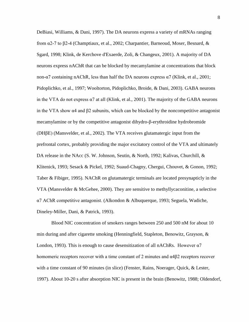
differential distribution of nAChR subtypes on
midbrain neurons and neuronal terminals, i.e.
GABA neurons, glutamate (GLU) terminals, and
DA cell bodies (Mansvelder, et al., 2002;
Mansvelder & McGehee, 2000; Pidoplichko,
Figure 2. A schematic of the role
of nAChRs in the control of VTA
DA neuron excitability. Under
control conditions (upper panel)
non-α7 nAChRs can excite DA and
GABA neurons directly, while α7
receptors can enhance release from
glutamatergic terminals.
Endogenous ACh release from
brainstem cholinergic neurons
contributes to the GABAergic input
to VTA DA neurons. In the
presence of nicotine concentrations
similar to those found in a smoker’s
blood (lower panel), the non-α7
nAChRs desensitize rapidly,
effectively inhibiting GABAergic
inputs to the DA neurons. The α7
nAChRs will not desensitize as
much, which means that
glutamatergic inputs will be
enhanced as the GABAergic inputs
are depressed, thus leading to a net
increase in excitation of the DA
neurons (H. D. Mansvelder and D.
S. McGehee, 2002).
8
DeBiasi, Williams, & Dani, 1997). The DA neurons express a variety of mRNAs ranging
from α2-7 to β2-4 (Champtiaux, et al., 2002; Charpantier, Barneoud, Moser, Besnard, &
Sgard, 1998; Klink, de Kerchove d'Exaerde, Zoli, & Changeux, 2001). A majority of DA
neurons express nAChR that can be blocked by mecamylamine at concentrations that block
non-α7 containing nAChR, less than half the DA neurons express α7 (Klink, et al., 2001;
Pidoplichko, et al., 1997; Wooltorton, Pidoplichko, Broide, & Dani, 2003). GABA neurons
in the VTA do not express α7 at all (Klink, et al., 2001). The majority of the GABA neurons
in the VTA show α4 and β2 subunits, which can be blocked by the noncompetitive antagonist
mecamylamine or by the competitive antagonist dihydro-β-erythroidine hydrobromide
(DHβE) (Mansvelder, et al., 2002). The VTA receives glutamatergic input from the
prefrontal cortex, probably providing the major excitatory control of the VTA and ultimately
DA release in the NAcc (S. W. Johnson, Seutin, & North, 1992; Kalivas, Churchill, &
Klitenick, 1993; Sesack & Pickel, 1992; Suaud-Chagny, Chergui, Chouvet, & Gonon, 1992;
Taber & Fibiger, 1995). NAChR on glutamatergic terminals are located presynapticly in the
VTA (Mansvelder & McGehee, 2000). They are sensitive to methyllycaconitine, a selective
α7 AChR competitive antagonist. (Alkondon & Albuquerque, 1993; Seguela, Wadiche,
Dineley-Miller, Dani, & Patrick, 1993).
Blood NIC concentration of smokers ranges between 250 and 500 nM for about 10
min during and after cigarette smoking (Henningfield, Stapleton, Benowitz, Grayson, &
London, 1993). This is enough to cause desensitization of all nAChRs. However α7
homomeric receptors recover with a time constant of 2 minutes and α4β2 receptors recover
with a time constant of 90 minutes (in slice) (Fenster, Rains, Noerager, Quick, & Lester,
1997). About 10-20 s after absorption NIC is present in the brain (Benowitz, 1988; Oldendorf,
9
1974). Penetration of NIC across the blood-brain barrier occurs by both passive diffusion
and active transport by the choroids plexus (Spector & Goldberg, 1982). Rapid NIC uptake
into tissues and intensive metabolism lead to its quick disappearance from the plasma,
leaving brain concentrations much higher than plasma levels would indicate. It has been
shown in rats that NIC exists in a brain/plasma ration of 3/1 (Benowitz, 1990; Rowell & Li,
1997; Sastry, Chance, Singh, Horn, & Janson, 1995).
The general mechanism of NIC addiction as proposed by Mansvelder et al. states that
all nAChRs become desensitized by NIC. However, the receptors expressing α7 take a
higher dose in order for desensitization for occur, and recover in minutes, as opposed to
hours for all other nAChRs. α7 subunits are expressed on DA neurons and GLU terminals in
the VTA. So the major excitatory input to VTA dopamine cell bodies recovers quickly. This
effectively leaves the dopamine neurons hyper excited and the GABA neurons inhibited for
longer amounts of time.
Finally it has been shown that the majority of endogenous cholinergic inputs into the
VTA appear to contact GABA rather than DA neurons (Fiorillo & Williams, 2000; Garzon,
Vaughan, Uhl, Kuhar, & Pickel, 1999).
GABA Neurons in the Mesocorticolimbic System
We believe that GABA neurons synapse on DA neurons in the VTA, thereby
providing an inhibitory input to regulate DA release. This is in agreement with the previous
finding of GABAergic control over DA neurons in the substantia nigra (Tepper, Paladini, &
Celada, 1998). Inhibition of these inhibitory GABA neurons would result in
hyperexcitability of DA neurons and an increased amount of DA release. This is right in line
with the DA theory mentioned above. Previously it has been shown that Ethanol decreases

10
the firing rate of these GABA neurons with acute administration for up to 2 weeks (Gallegos,
Criado, Lee, Henriksen, & Steffensen, 1999). In line with our theory and the DA theory, the
decreased firing rate of the GABA neurons would result in more DA to be released and a
euphoric state would be followed. After 2 weeks of daily ethanol administration, however,
the firing rate no longer decreases (Gallegos, et al., 1999). This indicates that some sort of
tolerance is occurring with these neurons, resulting in hyperactivity of the GABA neurons
and thus decrease in DA release that leads to a withdrawal state once alcohol is out of the
system.
Another interesting phenomenon we have found in the lab is that high-frequency
stimulation of the internal capsule (IC) causes multiple spike discharge (ICPSDs) of GABA
neurons in the VTA (S. C. Steffensen, Svingos, Pickel, & Henriksen, 1998). These
discharges are blocked by gap junction (GJ) antagonists, suggesting that VTA GABA
neurons are part of a network connected electrically by GJs (S.C. Steffensen, et al., 2003). In
addition, we have shown in the lab that acute ethanol also suppresses VTA GABA neuron
ICPSDs, with an IC50 at a dose of 1.1 g/kg of ethanol (Stobbs, et al., 2004), a moderately
intoxicating dose. We have recently studied VTA GABA neuron firing rate and ICPSDs
during chronic ethanol consumption as well. We have found that neither firing rate nor
ICPSDs adapt to chronic ethanol consumption. This was somewhat surprising to us since
VTA GABA neuron firing rate adapts to chronic ethanol injections. The disparity may lie in
the fact that rats do not become dependent on ethanol in the consumption paradigm while
they are dependent on ethanol on the forced injection paradigm. Thus, it may take
dependence to see physiological adaptation. Nonetheless, in connection with this study we
11
did find that DA D2 receptor expression adapts in connection with chronic ethanol
consumption.
DA, as we have seen thus far, plays an important role in the mesocorticolimbic
system. Acute and local administration of DA activates GABA neurons, increasing their
firing rate 100-200% (Stobbs, et al., 2004). This activation has been shown recently to be
occurring through the D2 receptor, given that antagonists block this activation. As acute
ethanol decreases the firing rate of VTA GABA neurons, but chronic ethanol increases their
firing rate, it would seem logical to look at expression levels of this protein to see if it is also
affected with chronic ethanol.
Rationale and Hypotheses
Current dogma explains that NIC indirectly causes increased firing of GABA neurons.
Once DA is released from DA neurons in the VTA, DA binds to specific receptors (D2) on
GABA neurons, inducing action potentials and increased neuronal firing. However, recent
studies have suggested NIC causes increased firing rates in GABA neurons directly by acting
on specific NIC channels rather than the model of indirect NIC activity (Mansvelder &
McGehee, 2000). The rationale for this study is predicated on the belief that advancement in
the understanding of the brain mechanisms underlying the recreational use and abuse
potential of drugs will pave the way for more effective treatment strategies that would save
lives and resources throughout the world. Each year more people are enslaved by addiction.
It can range from Alcohol and illicit drugs from the street to milder stimulants such as coffee
or non-prescription drugs. Regardless of the drug, the brain’s chemistry is influenced by
these chemicals. Despite current knowledge of the deleterious effects of tobacco use, only
six percent of the 35 million people who try to quit each year remain successful for more
12
than a month (Volkow, 2006). Previous studies reveal that NIC is to blame for the highly
addictive properties of tobacco products. NIC acts within the brain to increase DA levels in
the reward circuits. Scientists have applied this knowledge in the development of gum,
patches, and inhalers infused with NIC which has been fairly successful in alleviation of
some aspects of withdrawal. However, cravings may still persist. Further research is needed
to determine NIC’s actions on neurons in the brain’s pleasure pathway.
By understanding exactly how NIC alters brain activity, we can determine more
effective smoking cessation methods. Improved treatment and quitting aids for smokers
would increase the number of successful quitters each year and dramatically improve the
quality of life for not only the millions of people who would be free of their tobacco
addiction, but the lives of their children, friends, and loved ones who also may suffer from
the second-hand effects of cigarette smoking.
Given the fact that VTA GABA neurons express α4β2 nAChRs I hypothesize that
VTA GABA neurons will be excited by activation of cholinergic inputs in vivo. I
hypothesize that acute nicotinic activation of VTA GABA neurons will result in enhanced
inhibition of VTA DA neurons, but that α4β2 nAChRs will up-regulate or change subunit
composition to chronic NIC exposure. These experiments will go far to elucidating the role
of the VTA in mediating neuroadaptations to NIC.
Methods
Animal Subjects
Male Wistar rats were housed two to a cage from the time of weaning (P25), with ad
libitum access to food and water. The room temperature was controlled (22-25 oC) and
maintained on a reverse 12 hr light/dark cycle with lights ON from 8 PM to 8 AM. Animal

13
care, maintenance and experimental procedures were in accordance with the Brigham Young
University Animal Research Committee and meet or exceeded National Institutes of Health
guidelines for the care and use of laboratory animals.
Male C57BL/6J (black) and CD-1 (white albino) mice were bred and cared for in
accordance with the National Institutes of Health Guide for the Care and Use of Laboratory
Animals. For each methodology employed, animals were treated in strict accordance with
the guidelines of the Animal Research Committee (IACUC) of the Brigham Young
University which incorporate and exceed current NIH guidelines. The Committee has
reviewed and approved the procedures detailed herein. All electrophysiological, behavioral
and molecular methods listed below are currently running in the PI's and Co-Investigators’
(Edwards) laboratories at BYU. Three different mouse strains (PND1–60) were used in this
study. A glutamate decarboxylase-67 (GAD67)-green fluorescent protein (GFP) knock-in
mouse (Tamamaki, et al., 2003) created on a CD-1 inbred strain (Tamamaki, et al., 2003), a
subunit mouse created on a C57BL/6J inbred strain (Mihalek, et al., 1999),
-in mouse created on a C57BL inbred strain (Rudolph,
et al., 1999). We currently have a fully established colony of GAD GFP mice that we have
use for our ongoing grant-related studies on VTA GABA neurons. The heterozygous
GAD67-GFP knock-in mice afforded us the ability to positively identify and record from
GAD65, 67-positive GABA neurons in the VTA via fluorescence microscopy, as
characterizing VTA neurons by electrophysiology alone is problematical. Once weaned at,
all mice were housed in maximum groups of four and given ad libitum access to solid food
and water and placed on a reverse light/dark cycle with lights ON from 8 PM to 8 AM.

14
Characterization of Neuron Types in vitro
Neurons in the VTA of GAD67-GFP mice that exhibit a modest non-cation specific
inward rectifying current (Ih) in combination with low input resistance are assumed to be DA
neurons (Allison, et al., 2006; S.W. Johnson & North, 1992; Margolis, Lock, Hjelmstad, &
Fields, 2006). In GAD67 GFP knock-in mice, GABA neurons will be identified with the aid
of fluorescence microscopy. Only neurons located in the VTA that exhibit robust GFP
fluorescence will be considered GABAergic (Tamamaki, et al., 2003). Figure 3 illustrates
how GAD GFP mice facilitate the visualization of VTA GABA neurons to facilitate their
electrophysiological study.
Figure 3. Visualization and Patching of GABA neurons in the VTA of GAD GFP mice. (A) Superimposed transmitted and FITC GFP 4X magnification confocal images of a coronal
brain slice from a mature GAD GFP mouse showing the distribution of GAD labeling. Inset
shows a cluster of VTA GABA neurons taken at 20X magnification from the area indicated
by the asterisk. No enhancement with antibodies is needed to image GABA neurons in GAD
GFP mice. (B) A cluster of GABA neurons seen at 40X magnification before patching. (C)
IRDIC imaging enables patching of neurons after identification with fluorescent optics.

15
Single-unit Recordings in Anesthetized Rats
Extracellular potentials in Isoflurane (1%) anesthetized adult 250-400 g male Wistar
rats (Charles River Laboratory, Hollister, CA) were recorded by a single 3.0 M NaCl filled
micropipette (1-3 M; 1-2 µm inside diameter), cemented 10-20 µm distal to a 4-barrel
micropipette (20-60 M resistance), and amplified and filtered with a MultiClamp 700A
programmable amplifier (Axon Instruments, Union City, CA). Microelectrode assemblies
were oriented into the VTA [from bregma: 5.6-6.5 posterior (P), 0.5-1.0 lateral (L), 7.0-8.5
ventral (V)] with a piezoelectric inchworm microdrive (Burleigh, Fishers, NY). Single-unit
activity were filtered at 0.3-10 kHz (-3dB) and displayed on Tektronix 2200 digital
oscilloscopes. Square-wave constant current pulses (50-1000 µA; 0.15 msec duration;
average frequency, 0.1Hz) were generated by an IsoFlex constant current isolation unit
controlled by a MASTER-8 Pulse Generator (AMPI, Israel), or by computer. The faciculus
retroflexus (FR; from bregma: -4.0 AP, 0.5 ML, 4.0-4.2 V) was stimulated with insulated,
bipolar stainless steel electrodes. Extracellularly recorded action potentials (min 5:1 signal-
to-noise ratio) were discriminated with WPI-121 (Sarasota, Fl) spike analyzers and converted
to computer-level pulses.
Characterization of VTA GABA Neurons in vivo
All neurons classified as VTA GABA neurons in vivo were located in the VTA, met
the criteria established in previous studies for spike waveform characteristics and response to
IC stimulation (Allison, et al., 2006; S. C. Steffensen, et al., 1998; Stobbs, et al., 2004), and
often were activated and spike-coupled by microelectrophoretic dopamine (Stobbs, et al.,
2004). Presumed VTA GABA neurons were characterized by short-duration (<200 µsec;
measured at half-peak amplitude of the spike), initially negative-going, non-bursting spikes,

16
and were identified by the following IC stimulation criteria (S. C. Steffensen, et al., 1998):
Short latency (i.e., 2-5 msec) antidromic or orthodromic activation via single stimulation of
the IC; and multiple spiking following high-frequency (10 pulses, 200 Hz) stimulation of the
IC (ICPSDs; (Allison, et al., 2006; Lassen, et al., 2007; S. C. Steffensen, et al., 1998; Stobbs,
et al., 2004)). In all studies, stimulation was performed at a level that produced 50%
maximum VTA GABA neuron ICPSDs. This was accomplished by determining the current
needed to produce the maximum number of ICPSDs at 200 Hz and 10 pulses, and then
adjusting the stimulus intensity until 50% ICPSDs was achieved.
Single-unit Recordings in vivo
Single-unit potentials, discriminated spikes, and stimulation events in vivo were
captured by National Instrument’s NB-MIO-16 digital I/O and counter/timer data acquisition
boards (Austin, TX) and processed by customized National Instruments LabVIEW software
in Macintosh-type computers. Potentials were digitized at 20 kHz and 12-bit voltage
resolution. For single-unit activity, all spikes were captured by computer and time stamped.
Spontaneous firing rates were determined on- and off-line by calculating the number of
events over a 5 min epoch, typically 5 min before and at specific intervals after drug injection.
Peri-stimulus and interval-spike histograms were generated off-line using IGOR Pro
(WaveMetrics, Lake Oswego, OR) analysis of the time-stamped data. The duration (msec)
and extent (#events/bin) of post-stimulus permutation of ICPSDs is determined by
rectangular integration at specific time points on the peri-stimulus spike histogram using
IGOR Pro analysis software. The minimum bin width for peri-stimulus spike histograms is
1.0 msec and the number of bins was 1000. These parameters allow for detection of all
phases of pre- and post-stimulus spike activity.

17
Drug Preparation and Administration in vivo
Mecamylamine hydrochloride, bupropion, MG624, nicotine and DHβE were
dissolved in 0.9% saline and administered intravenously through an indwelling jugular
catheter in rats. In mice the same solution was given in an intraperitineal (IP) injection. For
systemic drug studies on VTA GABA neuron responses, drugs were administered
intravenously through a jugular catheter when possible.
Chronic Injections
In chronic studies, mice were treated with an intraperitoneal (IP) injection of saline
(SAL) or nicotine (NIC; 2 mg/kg) administered once-daily (1200 hours) for 12 days and
studied 24 hours after the last dose of SAL or NIC. In behavioral studies, daily injections of
NIC 0.5 mg/kg, IP, for 5 days produced sensitization in locomotor activity (Biala &
Weglinska, 2004), while mice given NIC 2.0 mg/kg, IP, three times each day for 12 days
were significantly less sensitive to NIC challenge than their SAL injected counterparts (Pauly,
Grun, & Collins, 1992). Thus, we chose a 2 mg/kg once daily regimen that fit within these
parameters. Similar studies employing chronic injections of NIC have used a comparable
regimen (Miura, Ishii, Aosaki, & Sumikawa, 2006).
Preparation of Brain Slices
Wistar rats (P21 – 45) and GAD67-GFP mice were anesthetized with Ketamine (60
mg/kg) and decapitated. The brains were quickly dissected and sectioned in ice-cold
artificial cerebrospinal fluid (ACSF), bubbled with 95% O2 / 5% CO2. This cutting solution
consisted of (in mM): 220 Sucrose, 3 KCl, 1.25 NaH2PO4, 25 NaH2CO3, 12 MgSO4, 10
Glucose, 0.2 CaCl2, and 0.4 Ketamine. VTA targeted horizontal slices (~200 µm thick) were
immediately placed into an incubation chamber containing normal ACSF at 34-35°, bubbled

18
with 95% O2 / 5% CO2 at 36° consisting of (in mM): 124 NaCl, 3 KCl, 1.25 NaH2PO4, 26
NaHCO3, 12 glucose, 1.5 MgSO4, 2 CaCl2, pH 7.3, and allowed to incubate for at least 45
minutes prior to being transferred to a recording chamber. Once transferred to a recording
chamber with continuous normal ACSF flow (2.0 ml/min) maintained at 34-35° throughout
the experiment, the slices were then allowed to settle for an additional 15 to 30 minutes
before recordings begins. These incubation and settling periods allowed cells to recover and
stabilize while ketamine is washed out of the tissue. Cells were visualized with either a
Nikon Eclipse FN1 or E600FN microscope in the transmitted de Sénarmont Differential
Interference Contrast (DIC) / infrared (IR) configuration.
Whole-cell Recordings in vitro
Electrodes pulled from borosilicate glass capillary tubes were filled with one of two
types of pipette solutions. For IPSCs, the pipette solution consisted of (in mM): 128 KCl, 20
NaCl, 0.3 CaCl2, 1.2 MgCl2, 10 HEPES, 1 EGTA, 2 Mg-ATP, 0.25 Na-GTP and 4.5 QX314
(pH 7.3). For voltage waveform and current-evoked spiking experiments the pipette solution
consisted of (in mM): 115 K-Gluconate, 9 NaCl, 25 KCl, 10 HEPES, 0.2 EGTA, 1.2 MgCl2,
3 Na-ATP, 1 Na-GTP, and had resistances of 2-4 MΩ. Series resistance (Ra) typically 10 to
20 MΩ, and input resistance (Rm) typically 300 to 400 MΩ, were continuously monitored
with a 10 mV voltage step delivered at 0.1 Hz throughout each experiment and only
experiments that maintain stable Ra and Rm (less than 15% change) were included in this
study. IPSCs were filtered at 2 kHz while voltage waveform-generated currents and current-
drive spikes were filtered at 6 kHz using an Axon Instruments Multiclamp 700A or 700B
amplifier and digitized at 5-20 kHz, respectively, using an Axon 1440A digitizer, and
collected and analyzed using pClamp10 and Igor Pro (Wavemetrics: Oswego, OR) software

19
packages. Evoked and spontaneous IPSCs were recorded in the presence of 100 μM D-L 2-
amino-5-phosphonopentanoic acid (APV), 30 μM 6-cyano-23-dihydroxy-7-nitro-quinoxaline
(CNQX), and 100 nM eticlopride to block NMDA, AMPA, and DA D2-mediated synaptic
currents (Bonci & Williams, 1997), respectively. Miniature IPSCs (mIPSCs) were isolated
from all other spontaneous IPSCs by addition of 0.5 μM TTX. To evoke IPSCs, cells were
stimulated at 0.1 Hz with a stainless steel-platinum/iridium concentric bipolar stimulating
electrode placed ~100 µm rostral to the recording electrode. Evoked IPSCs are inward at the
holding potential of -70 mV and were completely blocked by picrotoxin (100 µM). Evoked
IPSC amplitudes were calculated by taking the difference between the 1.0 msec window
around the peak and the 5.0 msec baseline window immediately preceding the stimulation
artifact. Spontaneous IPSC activity amplitude and frequency was calculated the same for
both sIPSCs and mIPSCs; the average amplitude or frequency during a 2 min period 8-10
min following drug was normalized to the average amplitude or frequency from a 2 min
window prior to drug.
Single-cell Quantitative RT-PCR
Following electrophysiological characterization, putative VTA GABA neurons and
putative DA neurons in mature rats were aspirated under visual observation by application of
suction attached to the recording pipette, and were immediately added to a reverse
transcription (RT) reaction mixture. The iScript cDNA synthesis kit (Biorad) was used for a
total volume of 10 µl per reaction. Reactions are run at 25°C for 10 min, 42°C for 60 min,
and 95°C for 5 min in a PTC-200 thermal cycler (MJ Research Inc., Watertown MA).
Reactions will then be stored at -20°C until running the PCR. A preamplification round of
multiplex PCR was performed by adding iTAQ Supermix with ROX (Biorad) and a cocktail

20
of primers to the completed RT reaction, for a final volume of 50 μL. The reactions were
held at 94°C for 30 seconds then cycled 20 times. Each cycle consists of: 92°C for 15
seconds, 60°C for 20 seconds, and 72°C for 30 seconds. One µl samples of the initial
multiplex PCR was then used as substrate for each reaction in the subsequent real-time
quantitative PCR. Real-Time quantitative PCR using gene specific primers with FAM-
TAMRA TaqMan® probes (Applied Biosystems; TH plus primer:
CTTCCAGTACAAGCACGGTGAA, TH minus primer: AGCGTGACATATA-
CCTCCTTCCA, and TH probe: CCCCATGTGGAATACACAGCGGAAGAG; D2 plus
primer: CGCAGAAAGCTCTCCCAGCAGA, D2 minus primer:
GACTGGTGGGATGTTGCAATCACA and D2 probe: CCATTGTTCTCGGTGTGTTCA;
18s plus primer: GTGCATGGCCGTTCTTAGTTG, minus 18s primer:
GCCACTTGTCCCTCTAAGAAGTTG, and 18s probe:
TGGAGCGATTTGTCTGGTTAATTCCGATAAC) were performed using the iTaq
Supermix with ROX (Bio-Rad) with an iCycler IQ (Bio-Rad) real-time PCR System.
Samples were amplified in triplicate, together with a negative control for each subunit (an
ACSF-only aspiration taken from the brain slice recording chamber when the cells were
aspirated). The amplification protocol is 50°C for 2 minutes, 95°C for 5 minutes, then 50
cycles of 95°C for 15 seconds, 60°C for 20 seconds, and 72°C for 30 seconds. Cycle
threshold (Ct) values were calculated automatically by the iCycler IQ software, with
threshold values set between 5 and 20. Relative fold expression were calculated using the 2-
ΔΔCT method as described in (Livak & Schmittgen, 2001).
21
Statistical Analyses
The results for control and drug treatment groups were derived from calculations
performed on VTA GABA neuron spontaneous firing rate and ICPSDs. Statistical analysis
of data was performed with Microsoft Excel Statistical Analysis Tools or with SPSS. An
analysis of variance (ANOVA) was invoked for comparison across collapsed groups of data
(One Way and Two Way ANOVAs). Comparison among individual means was made by
Newman-Keuls post-hoc tests following ANOVA. Further analyses of saline vs. nicotine
dose-response data or other repeated measures were made by Duncan’s new multiple-range
test to determine the source of detected significance in the ANOVAs. The criterion of
significance was set at p<0.05. For qualitative analysis of the relationship between neural
activity and behavior, two types of graphical representation were constructed. Perievent
histograms that average the pattern of neuronal activity around discrete response events were
plotted. In general, a modulation of VTA GABA neuron firing rate or afferent input that is
temporally associated with a specific behavioral event suggests involvement of the neuron in
coding that behavior. The stronger and sharper the modulation seen related to a given event,
the more likely that that event approximates the "preferred stimulus" of that neuron. The
Kolmogorov-Smirnov one-sample test was used to detect significant deviations (from an
even distribution) in the cumulative frequency distribution of unit activity for designated
epoch intervals before or around the event. For more complicated statistical analyses and
experimental design, we had available the expert services of Dr. Dennis Eggett (BYU
Statistician). The numbers of animals used was based, in part, on experience doing similar
studies and on statistical power analysis. The number of rats used in these experiments was
the minimum necessary to yield scientifically valid data. It has been our experience that 6-9
22
rats are needed to achieve significance for most of the experimental measures outlined in this
application. Our measures involve determinations of electrophysiological responses in VTA
GABA neurons and include firing rate; evoked discharges (electrical coupling), dopamine
modulation of firing rate/coupling and drug effects on these variables. We performed a
Power calculation for the estimation of the number of subjects needed to achieve significance
for a few of the experiments outlined in the application, in particular, those that we have had
many years of experience with and likely represent our most robust measures. For example,
we asked how many animals would be needed to detect a significant difference between
control (i.e., saline) and nicotine on VTA GABA neuron discharges based on archived data
from a similar experiment wherein we studied the effects of one dose of ethanol on ICPSDs
(Stobbs, et al., 2004). The parameters of the Power Analysis were: Mean (control) = 67
discharges; Mean (ethanol) = 35 discharges; SD of difference (effect size) = 21; Alpha =
0.05; Tails (t test) = 2. For the given effect size (SD difference = 21) the power is 0.843 and
the sample size is 6. This means that 84% of studies would be expected to yield a significant
effect if 6 pairs of animals were studied (i.e., control vs. drug). Power values indicate the
probability of obtaining the stated effect size given the sample size indicated. In general,
power probabilities > 0.50 are considered reliable, because comparison of the experimental
treatments using the sample sizes presented in the table are reasonably robust and produce a
statistically significant outcome. Therefore, based on previous experience doing similar
experiments and power calculations of some representative experiments including the
example above, we used 6 rats to achieve significance, even for robust measures. For other
paradigms we know that not all rats will successfully respond to criterion (approximately
25%). Moreover, there are historical issues of unexplained morbidity (approximately 10%)

23
and mortality (2%), as well as technical problems with electrophysiological (e.g., problems
with cranial electrode implants) and behavioral (e.g., patent intravenous catheters) recordings.
Therefore, in some areas of this study, more than 6 rats were used in order to obtain reliable
data.
Results
In the original experimental plan we proposed to test the effects of NIC after α7 nAChR
block with MG624 to determine if NIC effects on α7 nAChRs might counteract the effects of
NIC on non α7 AChRs. We then proposed to test the effects of the α4β2 nAChR agonist
buproprion. It is a drug that interacts with at norepinephrine, dopamine, and acetylcholine.
Thus, we planned experiments involving stimulation of cholinergic inputs to the VTA in
order to rule out the effects of the other neurotransmitters on the action that BP has on VTA
GABA neurons. Next, we planned to study the effects of NIC on membrane properties, as
well as spontaneous and evoked IPSCs in VTA DA neurons then, determine the subtype
expression of VTA GABA neurons using quantitative single-cell RTPCR. Finally, we
proposed testing the neuroadaptations in VTA GABA and DA neurons, in vitro, with chronic
NIC administration. While all experiments proposed were pursued, given our experience
with in vivo electrophysiological studies we knew it was highly likely that some experiments
would suggest that it would be more parsimonious to pursue other studies in order to
maximize our gain. The detailed plan is shown in the appendix.
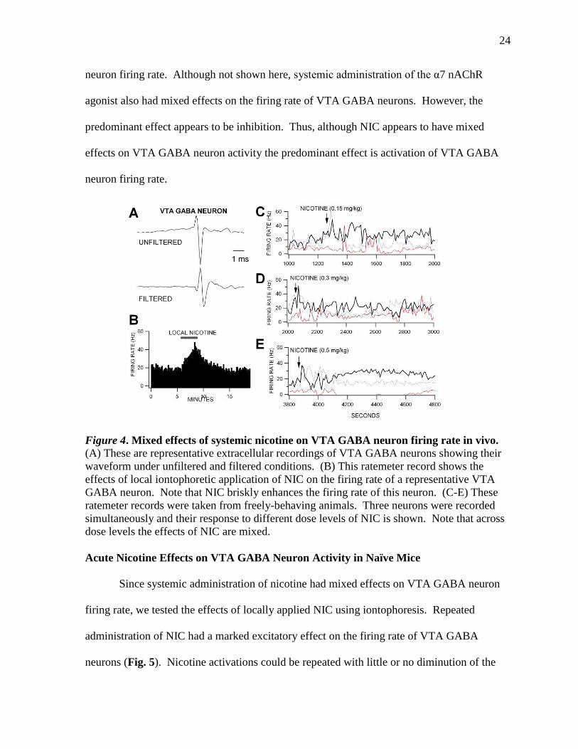
Mixed Effects of Systemic and Local Nicotine and α7 Nicotinic Receptor Agonists on
VTA GABA Neuron Firing Rate
Nicotine appears to only slightly affect firing rate when administered systemically
(Fig. 4). Thus, systemic NIC does not have consistently robust effects on VTA GABA
24
neuron firing rate. Although not shown here, systemic administration of the α7 nAChR
agonist also had mixed effects on the firing rate of VTA GABA neurons. However, the
predominant effect appears to be inhibition. Thus, although NIC appears to have mixed
effects on VTA GABA neuron activity the predominant effect is activation of VTA GABA
neuron firing rate.
Figure 4. Mixed effects of systemic nicotine on VTA GABA neuron firing rate in vivo. (A) These are representative extracellular recordings of VTA GABA neurons showing their
waveform under unfiltered and filtered conditions. (B) This ratemeter record shows the
effects of local iontophoretic application of NIC on the firing rate of a representative VTA
GABA neuron. Note that NIC briskly enhances the firing rate of this neuron. (C-E) These
ratemeter records were taken from freely-behaving animals. Three neurons were recorded
simultaneously and their response to different dose levels of NIC is shown. Note that across
dose levels the effects of NIC are mixed.
Acute Nicotine Effects on VTA GABA Neuron Activity in Naïve Mice
Since systemic administration of nicotine had mixed effects on VTA GABA neuron
firing rate, we tested the effects of locally applied NIC using iontophoresis. Repeated
administration of NIC had a marked excitatory effect on the firing rate of VTA GABA
neurons (Fig. 5). Nicotine activations could be repeated with little or no diminution of the

25
response. By using rectangular integration from the baseline firing rate and averaging 3
cycles of NIC application we found that iontophoretic application of NIC significantly
increased the firing rate of VTA GABA neurons (353.3%; P=0.001, t(2,21)=2.8; n=22).
Figure 5. Nicotine markedly activates VTA GABA neuron firing rate. (A) This ratemeter
record shows the firing rate of a representative VTA GABA neuron. Its baseline firing rate
was approximately 30 Hz. We tested the effects of local application of NIC on the activity of
this neuron. In situ microelectrophohretic application of NIC (+50 nA) markedly activated
this neuron. (B) This graph summarizes the effects of local NIC on VTA GABA neuron
firing rate.
Effects of Select nAChR Antagonists on Nicotine Activation of VTA GABA neurons in
Naïve Mice
Although systemic NIC had mixed effects on VTA GABA neuron activity, when
locally administered NIC clearly activates these neurons. Thus, we performed experiments
to determine which nAChRs might be mediating the activation and whether NIC was acting
directly on VTA GABA neurons or indirectly disinhibiting them via inhibition of some
inhibitory afferent. If an increase in firing rate of VTA GABA neurons was produced by
local administration of NIC, then it could be blocked by systemic administration of a NIC
antagonist. Our next experiment showed this to be true. We further attempted to show the
26
effect that the α4β2 nAChR antagonist dihydro-β-erythroidine (DHβE) had on NIC activation
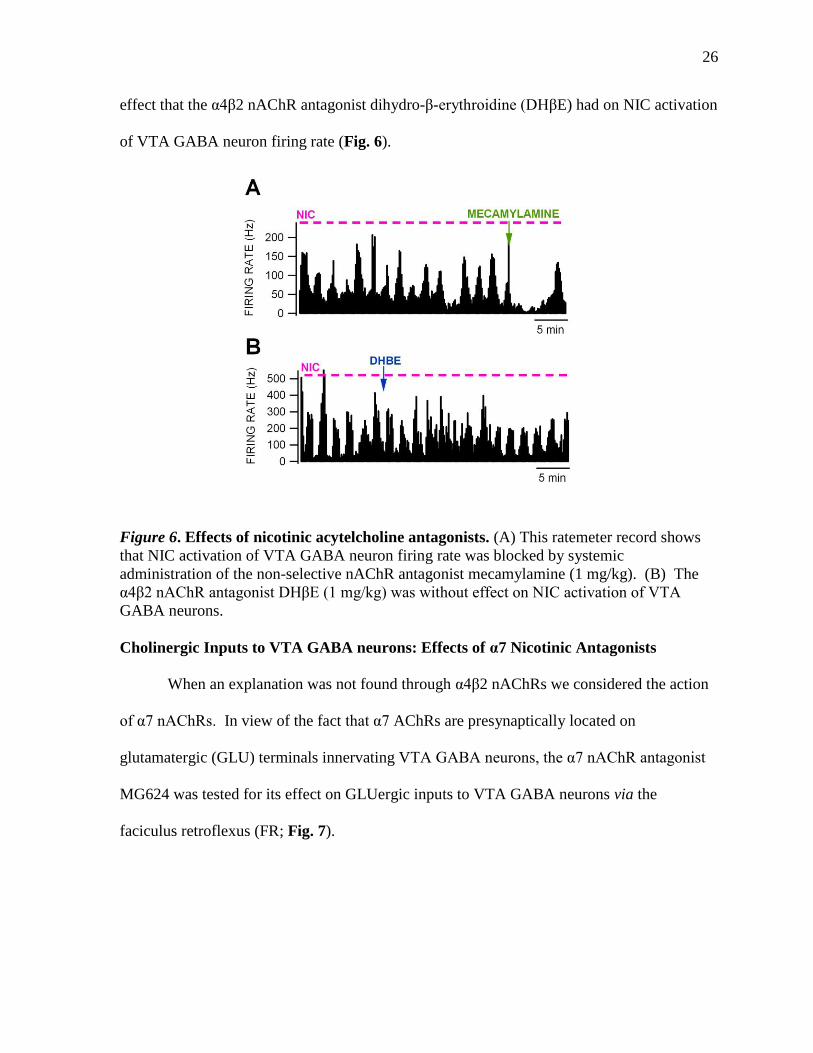
of VTA GABA neuron firing rate (Fig. 6).
Figure 6. Effects of nicotinic acytelcholine antagonists. (A) This ratemeter record shows
that NIC activation of VTA GABA neuron firing rate was blocked by systemic
administration of the non-selective nAChR antagonist mecamylamine (1 mg/kg). (B) The
α4β2 nAChR antagonist DHβE (1 mg/kg) was without effect on NIC activation of VTA
GABA neurons.
Cholinergic Inputs to VTA GABA neurons: Effects of α7 Nicotinic Antagonists
When an explanation was not found through α4β2 nAChRs we considered the action
of α7 nAChRs. In view of the fact that α7 AChRs are presynaptically located on
glutamatergic (GLU) terminals innervating VTA GABA neurons, the α7 nAChR antagonist
MG624 was tested for its effect on GLUergic inputs to VTA GABA neurons via the
faciculus retroflexus (FR; Fig. 7).

27
Figure 7. Stimulation of cholinergic inputs to VTA. (A) The peri-stimulus spike
histogram (PSH; top) shows that high frequency (10 pulses, 200 Hz) stimulation of the
fasciculus retroflexus (FR) activates cholinergic inputs to the VTA from the habenula that
inhibit the firing rate of VTA GABA neurons, which is blocked by the α7 antagonist MG624
(3 mg/kg). (B) Single stimulation of the PPT activates VTA GABA neurons both
orthodromically (left) and antidromically (right). The PSH (top) shows multiple spike
discharges produced by high frequency PPT stimulation. The PSH bottom shows that
mecamylamine (1 mg/kg) blocks PPT activation of VTA GABA neurons.
Chronic Nicotine Effects GABA Neuron Firing Rate and Response to Acute Nicotine
Since NIC exhibited robust excitatory effects on VTA GABA neurons when
administered locally and acutely, with little or no rapid desensitization with repeated
application, we tested the effects of chronic NIC administration on NIC activation of VTA
GABA neurons to evaluate any potential neuroadaptive effects of nAChRs in VTA GABA
neurons or in their afferents. This required both in vivo and in vitro electrophysiological and
molecular studies. The effect of local in vivo NIC in mice who have been chronically
administered NIC or saline was tested (Fig. 8). In chronic studies, mice were treated with an

28
intraperitoneal (IP) injections of saline (SAL) or nicotine (NIC; 2 mg/kg) administered once-
daily for 12 days and studied 24 hours after the last dose of SAL or NIC. Surprisingly, there
was no significant difference in baseline firing rate of VTA GABA neurons (P=0.92,
t(2,23)=0.11; n=19,14; Fig. 8B). While NIC activated VTA GABA neurons in cSAL-treated
mice it did not activate them in cNIC-treated mice (Fig. 8C). There was a significant
difference between the two treatments (P=0.02, t(2,14)=2.53; n=19,14; Fig. 8C).
Figure 8. Locally
administered nicotine
effect on chronically
treated mice. (A) The
ratemeter (top) shows
NIC activation of VTA
GABA neuron firing
rate in a saline-treated
mouse (cSAL). The
ratemeter (bottom)
shows lack of NIC
activation of a VTA
GABA neuron recorded
in a chronic NIC-treated
mouse (cNIC). (B)
There was no significant
difference in the
baseline firing rate of
VTA GABA neurons in
cNIC vs cSAL-treated
mice. (C) There was a
significant reduction in
NIC activation of VTA
GABA neuron firing
rate in NIC-treated mice.

29
Chronic Nicotine Effects on VTA DA Neuron Activity
Our in vivo studies demonstrated that VTA GABA neurons desensitized with chronic
NIC. Since the dogma is that VTA GABA neurons inhibit DA neurons we evaluated the
effects of chronic NIC on DA neuron activity. We performed these studies in vitro in GAD
GFP mice where GABA and DA neurons can be visualized and more easily characterized.
Also, DA neurons are typically not spontaneously active under Isoflurane anesthesia, which
is what we use to record GABA neurons. Most other anesthetics suppress the firing rate of
VTA GABA neurons. We evaluated the effects of chronic NIC on spontaneous and current-
evoked spikes in DA neurons of cSAL versus cNIC mice (Fig. 9). Unexpectedly, DA
neurons in cSAL and cNIC mice showed no significant differences between the groups
(P=0.65, t(2,16)=0.46; n=11,9; Fig. 9B).
Figure 9. Lack of effects
of chronic nicotine on
dopamine neuron
activity. (A) These are
representative recordings
of dopamine neuron
spikes in SAL and NIC-
treated mice. Note the
mild decrease in spike
frequency in the NIC-
treated mouse. (B)
Although dopamine
neuron firing rates tended
to be lower in NIC-
treated (cNIC) mice, they
were not significantly
different on average from
SAL-treated (cSAL) mice.
(C) Depolarizing current steps activate dopamine neuron spike activity (shown here at 150
pA). Note the slow frequency and spike accommodation characteristic of current-evoked
spiking in dopamine neurons. (D) This graph summarizes the effects of current ejection on
the firing frequency of dopamine neurons. There was no significant difference in dopamine
neuron spiking frequency between cNIC and cSAL mice.

30
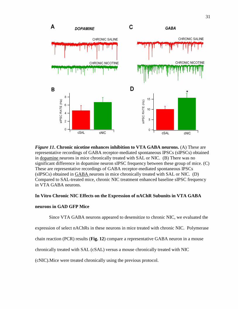
Chronic Nicotine Effects on GABAergic synaptic inhibition to Dopamine and GABA
Neurons in the VTA
In order to determine the mechanism of action of NIC on VTA GABA neurons and
their desensitization with chronic NIC, we studied spontaneous inhibitory postsynaptic
currents (sIPSCs) in DA and GABA neurons in both cSAL and cNIC mice. However, we
needed to first determine the effects of acute NIC on GABA neuron sIPSCs. Figure 10
summarizes the effects of NIC on GABA neuron sIPSC frequency, a measure of the GABA
input to VTA GABA neurons. Nicotine reduced sIPSCs, but not significantly (P=0.22,
t(2,31)=1.25; n=20,13; Fig. 9B).
Since acute NIC inhibited sIPSCs, we wanted to evaluate the chronic effects of NIC
on sIPSCs to further evaluate the mechanism of action of NIC desensitization. Figure 11
summarizes the effects of chronic NIC on both GABA and DA neuron sIPSC frequency.
While there was no difference in acute NIC effects on DA neurons in cSAL vs cNIC mice
(P>0.05), NIC significantly increased sIPSC frequency in GABA neurons in cNIC mice
compared to cSAL mice (P=0.03, t(2,231)=2.25; n=17,13; Fig. 11D).
Figure 10. Nicotine effects
on GABA neuron sIPSC
frequency. Superfusion of 1
uM NIC reduced sIPSCs
recorded in GABA neurons
visualized in GAD GFP mice
31
Figure 11. Chronic nicotine enhances inhibition to VTA GABA neurons. (A) These are
representative recordings of GABA receptor-mediated spontaneous IPSCs (sIPSCs) obtained
in dopamine neurons in mice chronically treated with SAL or NIC. (B) There was no
significant difference in dopamine neuron sIPSC frequency between these group of mice. (C)
These are representative recordings of GABA receptor-mediated spontaneous IPSCs
(sIPSCs) obtained in GABA neurons in mice chronically treated with SAL or NIC. (D)
Compared to SAL-treated mice, chronic NIC treatment enhanced baseline sIPSC frequency
in VTA GABA neurons.
In Vitro Chronic NIC Effects on the Expression of nAChR Subunits in VTA GABA
neurons in GAD GFP Mice
Since VTA GABA neurons appeared to desensitize to chronic NIC, we evaluated the
expression of select nAChRs in these neurons in mice treated with chronic NIC. Polymerase
chain reaction (PCR) results (Fig. 12) compare a representative GABA neuron in a mouse
chronically treated with SAL (cSAL) versus a mouse chronically treated with NIC
(cNIC).Mice were treated chronically using the previous protocol.

32
Figure 12. Chronic nicotine effects on nAChR expression. (A) Chronic saline-treated mice
expressed Cx36, but not TH, transcripts as shown previously. They expressed α4, α6 and β2
nAChR subunits. (B) Chronic NIC-treated mice expressed the α6 subunit.

33
Discussion
As previously stated, systemic administration of nicotine had mixed effects on VTA
GABA neuron firing rate (Fig. 4). Because systemic administration of a drug causes it to be
circulated throughout the entire brain, NIC could be influencing neurons directly or indirectly
through activation of nAChRs on afferents to the VTA. We know that there are afferents to
VTA GABA neurons from the NAcc, ventral pallidum, medial prefrontal cortex and from the
PPT. Thus, NICs systemic effects need to be evaluated in light of its possible net effects on
GABA neurons and its afferents.
Nicotine, iontophoretically applied, instead of systemically, had a robust excitatory
effect on VTA GABA neuron firing rate. Nicotine activations, achieved with cyclic local
application of NIC (Fig. 5), were found consistently and with profound excitation. We were
able to show that this response did not seem to diminish with repeated NIC exposure. We
were surprised that there was no acute desensitization with repeated application of NIC. A
high rate, approximately 75%, of the neurons we studied, were activated by NIC in this
manner. In an article by Mansvelder et al. (Mansvelder, et al., 2002) it was reported that they
have found an increase in GABA neuron firing rate, in vitro, which is supportive of our
effects in vivo. There is some concern, however, that the neurons they studied were not
GABA neurons, as they relied on electrophysiological criteria (i.e., Ih current—DA neurons
being Ih+ and GABA neurons being Ih-), which have been shown more recently to not be
valid (Margolis, et al., 2006). Mansvelder et al. (Mansvelder, De Rover, McGehee, &
Brussaard, 2003) also report that non-α7 nAChR will desensitize within minutes after
exposure to NIC, in vitro. This desensitization is said to occur on heteromeric α4β2 nAChRs,
located on GABAergic neurons, more quickly than the more complex homomeric α7
34
receptors on DA neurons (Barik & Wonnacott, 2009). α7 homomeric receptors are said to
recover, after exposure to NIC, with a time constant of 2 minutes and α4β2 receptors recover
with a time constant of 90 minutes (in slice) (Fenster, et al., 1997) As previously stated, we
did not see this effect in vivo; even after extended periods of exposure to NIC, VTA GABA
neurons showed little or no desensitization to the drug.
In order to show a mechanism of action for the excitation of VTA GABA neurons by
NIC, we tested the effect of systemic nAChR antagonists on these neurons during cyclic
application of NIC. Mecamylamine, an antagonist to all nAChR except the α7, consistently
blocked the activation of NIC on VTA GABA neuron firing rate, confusingly the α4β2
antagonist DHβE did not (Fig. 6). We also found it difficult to administer a DHβE dose in
mice that the literature suggested was large enough to have an effect, without being terminal
to the animal. It was later found that systemic administration of DHβE could not block NIC
activations because the drug cannot cross the blood brain barrier, answering the question as
to why NIC activations were not blocked and making the systemic dose problem in mice
moot. Further investigation into another α4β2 nAChR antagonist that can be used
systemically is ongoing. Others have shown the blockade of NIC firing rate by DHβE in
vitro (Mansvelder, et al., 2002). Thus, we are confident that we will be able to show this
effect in vivo as well. We are currently considering the use of a two barrel iontophoretic
technique with NIC and DHβE in order to test this hypothesis.
The PPT is sending substantial cholinergic input to the VTA (Good & Lupica, 2009).
The Mansvelder et al. (2002) results suggest that α4β2 nAChRs on GABA neurons are
postsynaptic to this input. We showed with stimulation studies that GABA neurons receive
direct input (orthodromic and short latency) and also project to the PPT (Fig. 7B). In

35
addition, there appears to be an indirect cholinergic input from the fasciculus retroflexus
which inhibits VTA GABA neurons via α7 receptors, perhaps via a feedback loop onto VTA
GABA neurons from DA neurons (Fig. 7A).
Although we did not see NIC desensitization of VTA GABA neurons with acute
administration in naive animals in vivo (Fig.5), we did find that NIC desensitizes after
chronic NIC treatment (Fig. 8). There was no significant difference in the baseline firing rate
of VTA GABA neurons in both groups. Nicotine activations were still profound in the cSAL
group, yet not in the cNIC. This differs from Mansvelder, as they showed acute
desensitization with NIC on ACh currents (Mansvelder, et al., 2002). Mansvelder et al.
(2002) has shown direct effects of ACh on VTA GABA neurons (i.e., ACh current) mediated
via non-α7 nAChRs. We did observe a direct NIC effect in some VTA GABA neurons, but
it was equivocal.
Chronic NIC treatment has also been reported by some groups to lead to the increased
release of DA (Benwell & Balfour, 1992; Gaddnas, Pietila, & Ahtee, 2000), yet others report
that DA release is unchanged by chronic NIC (Damsma, Day, & Fibiger, 1989). We also
showed no significant difference in the firing frequency of DA neurons in cSAL and cNIC
mice (Fig. 9). However, we did found that NIC enhances sIPSCs to GABA neurons (Fig.
11). This data is consistent with the data of Mansvelder et al. (2002) in the effects on DA
neurons acutely.
Finally, we examined the expression of nAChR subunits in VTA GABA neurons to
which the local NIC had been applied. PCR results (Fig. 12) hold a more detailed insight
into the mechanism of action taken by NIC after chronic treatment. The cNIC group appears
to be expressing α6 subunits, not present in the cSAL group. We are currently furthering our

36
chronic NIC study by looking for α6 antagonists that can be tested in chronically treated
animals. So far we have been not able to find an α6 antagonist for systemic use, but are
again considering a two barrel ionophoretic approach, as with the DHβE planned experiment.
Appendix
Original Experimental plan
The studies proposed in this thesis are organized into what might represent a journal article
for publication. This is consistent with the expectancies for a Master’s thesis. The studies
proposed below are somewhat ambitious and cover a broad range of experiments. While all
experiments proposed will be pursued, given our experience with in vivo
electrophysiological studies it is highly likely that some experiments will suggest that it
would be more parsimonious to pursue other studies in order to maximize our gain. For
example, we have already performed some experiments with the α7 nAChR agonist JN403
and have seen little or no evidence that might suggest we pursue studies related to α7
nAChRs on VTA GABA neurons.
Experiment 1: Acute in vivo studies: effects of acetylcholine and nicotine on VTA
GABA neuron firing rate
We have already performed a handful of experiments on the effects of systemic
nicotine on VTA GABA neurons. The effects of systemic nicotine have been inconsistent
and equivocal. This may obtain due to complex nature of nAChR hodology in the VTA.
Notwithstanding the lack of effects of systemic NIC, we will test the effects of NIC after α7
nAChR block with MG624, α7 nAChRs are located on GLU terminals to DA neurons. We
have shown previously that DA excites VTA DA neurons through an unknown mechanism.
These studies will enable us to determine if NIC effects on α7 nAChRs might counteract the
37
effects of NIC on non α7 AChRs, given the potential for DA neurons to modulate GABA
neuron activity. We will perform 2-3 experiments to probe this effect. If NIC has consistent
effects on VTA GABA neuron firing rate in the presence of α7 nAChR block we will pursue
dose-response studies with systemic NIC. Also, in association with these studies we will test
the effects of local administration of NIC via in situ microelectrophoresis. Ten rats will be
needed for Experiment 1.
Experiment 2: Acute in vivo studies: effects of buproprion on VTA GABA neuron
firing rate
We have already performed a handful of experiments on the effects of the α4β2
nAChR agonist buproprion. We have found consistent, but complex, effects of this non-
specific drug, which is also an inhibitor of the DA transporter (DAT). We have seen an
initial transient inhibition followed by a prolonged enhancement of VTA GABA neuron
firing rate. We now need to test the effects of α4β2 antagonists to see what is the primary
effect of α4β2 nAChR activation, as VTA GABA neurons express these receptors that are
likely postsynaptic to the cholinergic input from the PPT. We need to rule out the DAT
effects of buproprion. Accordingly, we will also stimulate the PPT in hopes of observing
modulation of VTA GABA neuron activity. We expect PPT stimulation will excite VTA
GABA neurons. In addition, there is a less well-known cholinergic input from the habenula
via the fasciculus retroflexus. We have performed some experiments in this regard.
Interestingly, stimulation of the fasciculus retroflexus inhibits VTA GABA neuron activity
with possible antagonism by α7 nAChR antagonist. Thus, the cholinergic input, which is
typically excitatory, at least with direct input to nAChRs, appears to be operating indirectly
to inhibit VTA GABA neurons. We will use pharmacological antagonists to try to decipher

38
these disparate cholinergic inputs to VTA GABA neurons. Twenty rats will be needed for
Experiment 2.
Experiment 3: Acute in vitro studies: effects of nicotine on GABA inhibition of VTA DA
Neurons
There appear to be no studies in the literature on the effects of NIC or nAChR
agonists on inhibitory GABA input to VTA DA neurons. Thus, we will study the effects of
NIC on membrane properties, as well as spontaneous and evoked IPSCs in VTA DA neurons.
These studies will be accomplished in the presence of GLU blockers which will enable us to
determine if NIC is acting directly on VTA GABA neurons and whether the effect is pre- or
post-synaptic. Moreover, in anticipation of chronic NIC studies (Exp. 4) we will determine
the subtype expression of VTA GABA neurons using quantitative single-cell RTPCR. Dr.
Sterling Sudweeks will assist with these molecular studies given his expertise with nAChRs.
Twelve rats will be needed for these studies.
Experiment 4: Chronic in vitro studies: effects of chronic nicotine
Nicotinic receptor desensitization is a well-known phenomena, as described in the
Introduction. VTA GABA neurons express α4β2 nAChRs. We will evaluate the effects of
chronic NIC administration on neuroadaptations in VTA GABA and DA neurons. This will
be accomplished by quantitative RTPCR studies of VTA GABA neurons and physiological
studies of DA neurons as per the acute experiments outlined in Experiment 3.

39
References
Alkondon, M., & Albuquerque, E. X. (1993). Diversity of nicotinic acetylcholine receptors in
rat hippocampal neurons. I. Pharmacological and functional evidence for distinct
structural subtypes. Journal of Pharmacology and Experimental Therapeutics, 265(3),
1455-1473.
Allison, D. W., Ohran, A. J., Stobbs, S. H., Mameli, M., Valenzuela, C. F., Sudweeks, S. N.,
et al. (2006). Connexin-36 gap junctions mediate electrical coupling between ventral
tegmental area GABA neurons. Synapse, 60(1), 20-31.
Barik, J., & Wonnacott, S. (2009). Molecular and cellular mechanisms of action of
nicotine in the CNS. Handbook of Experimental Pharmacology, (192), 173-207. doi:
10.1007/978-3-540-69248-5_7
Bechara, A., & van der Kooy, D. (1992). A single brain substrate mediates the motivational
effects of both opiates and food in non-deprived, but not in deprived animals.
Behavioral Neuroscience, 106, 351-363.
Benowitz, N. L. (1988). Drug therapy. Pharmacologic aspects of cigarette smoking and
nicotine addiction. The New England Journal of Medicine, 319(20), 1318-1330.
Benowitz, N. L. (1990). Pharmacokinetic considerations in understanding nicotine
dependence. Ciba Foundation Symposium, 152, 186-200; discussion 200-189.
Benowitz, N. L. (1996). Pharmacology of nicotine: addiction and therapeutics. Annual
Review of Pharmacology and Toxicology, 36, 597-613. doi:
10.1146/annurev.pa.36.040196.003121
40
Benwell, M. E., & Balfour, D. J. (1992). The effects of acute and repeated nicotine treatment
on nucleus accumbens dopamine and locomotor activity. British Journal of
Pharmacology, 105(4), 849-856.
Biala, G., & Weglinska, B. (2004). Calcium channel antagonists attenuate cross-sensitization
to the locomotor effects of nicotine and ethanol in mice. Polish Journal of
Pharmacology, 56(4), 391-397.
Blackburn, J. R., Phillips, A. G., Jakubovic, A., & Fibiger, H. C. (1986). Increased dopamine
metabolism in the nucleus accumbens and striatum following consumption of a
nutritive meal but not a palatable non-nutritive saccharin solution. Pharmacology
Biochemistry and Behavior, 25(5), 1095-1100.
Bonci, A., & Williams, J. T. (1997). Increased probability of GABA release during
withdrawal from morphine. The Journal of Neuroscience, 17, 796-803.
Bozarth, M. A., & Wise, R. A. (1981). Intracranial self-administration of morphine into the
ventral tegmental area. Life Sciences, 28, 551-555.
Carelli, R. M., & Deadwyler, S. A. (1994). A comparison of nucleus accumbens neuronal
firing patterns during cocaine self-administration and water reinforcement in rats. The
Journal of Neuroscience, 14, 7735-7746.
Champtiaux, N., Han, Z. Y., Bessis, A., Rossi, F. M., Zoli, M., Marubio, L., et al. (2002).
Distribution and pharmacology of alpha 6-containing nicotinic acetylcholine
receptors analyzed with mutant mice. The Journal of Neuroscience, 22(4), 1208-1217.
Chang, J.-Y., Zhang, L., Janak, P. H., & Woodward, D. J. (1997). Neuronal responses in
prefrontal cortex and nucleus accumbens during heroin self-administration in freely
moving rats. Brain Research, 754, 12-20.

41
Charpantier, E., Barneoud, P., Moser, P., Besnard, F., & Sgard, F. (1998). Nicotinic
acetylcholine subunit mRNA expression in dopaminergic neurons of the rat substantia
nigra and ventral tegmental area. Neuroreport, 9(13), 3097-3101.
Cunningham, C. L., & Noble, D. (1992). Conditioned activation induced by ethanol: role in
sensitization and conditioned place preference. Pharmacology Biochemistry and
Behavior, 43(1), 307-313.
Damsma, G., Day, J., & Fibiger, H. C. (1989). Lack of tolerance to nicotine-induced
dopamine release in the nucleus accumbens. European Journal of Pharmacology,
168(3), 363-368.
David, V., & Cazala, P. (1994). A comparative study of self-administration of morphine into
the amygdala and the ventral tegmental area in mice. Behavioural Brain Research,
65(2), 205-211.
David, V., Segu, L., Buhot, M. C., Ichaye, M., & Cazala, P. (2004). Rewarding effects
elicited by cocaine microinjections into the ventral tegmental area of C57BL/6 mice:
involvement of dopamine D1 and serotonin1B receptors. Psychopharmacology (Berl),
174(3), 367-375. doi: 10.1007/s00213-003-1767-5
de Rover, M., Lodder, J. C., Kits, K. S., Schoffelmeer, A. N., & Brussaard, A. B. (2002).
Cholinergic modulation of nucleus accumbens medium spiny neurons. European
Journal of Neuroscience, 16(12), 2279-2290.
Del Castillo, J., & Webb, G. D. (1977). Rapid desensitization of acetylcholine receptors of
eel electroplaques following iontophoretic application of agonist compounds. The
Journal of Physiology, 270(2), 271-282.
42
Devine, D. P., & Wise, R. A. (1994). Self-administration of morphine, DAMGO, and
DPDPE into the ventral tegmental area of rats. The Journal of Neuroscience, 14,
1978-1984.
DiChiara, G., & North, R. A. (1992). Neurobiology of opiate abuse. Trends in
Pharmacological Sciences, 13, 185-193.
Dworkin, S. I., Guerin, G. F., Co, C., Goeders, N. E., & Smith, J. E. (1988). Lack of an effect
of 6-hydroxydopamine lesions of the nucleus accumbens on intravenous morphine
self-administration. Pharmacology Biochemistry and Behavior, 30, 1051-1057.
Ettenberg, A., Pettit, H. O., Bloom, F. E., & Koob, G. F. (1982). Heroin and cocaine
intravenous self-administration in rats: mediation by separate neural systems.
Psychopharmacology, 78, 204-209.
Feltz, A., & Trautmann, A. (1982). Desensitization at the frog neuromuscular junction: a
biphasic process. The Journal of Physiology, 322, 257-272.
Fenster, C. P., Rains, M. F., Noerager, B., Quick, M. W., & Lester, R. A. (1997). Influence of
subunit composition on desensitization of neuronal acetylcholine receptors at low
concentrations of nicotine. The Journal of Neuroscience, 17(15), 5747-5759.
Fiorillo, C. D., & Williams, J. T. (2000). Cholinergic inhibition of ventral midbrain
dopamine neurons. The Journal of Neuroscience, 20(20), 7855-7860.
Fontana, D. J., Post, R. M., & Pert, A. (1993). Conditioned increases in mesolimbic
dopamine overflow by stimuli associated with cocaine. Bain Research, 629, 31-39.
Gaddnas, H., Pietila, K., & Ahtee, L. (2000). Effects of chronic oral nicotine treatment and
its withdrawal on locomotor activity and brain monoamines in mice. Behavioural
Brain Research, 113(1-2), 65-72. doi: S0166432800002011 [pii]
43
Gallegos, R. A., Criado, J. R., Lee, R. S., Henriksen, S. J., & Steffensen, S. C. (1999).
Adaptive responses of GABAergic neurons in the ventral tegmental area to chronic
ethanol. Journal of Pharmacology and Experimental Therapeutics, 291, 1045-1053.
Garzon, M., Vaughan, R. A., Uhl, G. R., Kuhar, M. J., & Pickel, V. M. (1999). Cholinergic
axon terminals in the ventral tegmental area target a subpopulation of neurons
expressing low levels of the dopamine transporter. Journal of Comparative
Neurology, 410(2), 197-210.
Gatto, G. J., McBride, W. J., Murphy, J. M., Lumeng, L., & Li, T.-K. (1994). Ethanol self-
infusion into the ventral tegmental area by alcohol-preferring (P) rats. Alcohol, 11(6),
557-564.
Goeders, N. E., & Smith, J. E. (1983). Cortical dopaminergic involvement in cocaine
reinforcement. Science, 221, 773-775.
Good, C. H., & Lupica, C. R. (2009). Properties of distinct ventral tegmental area synapses
activated via pedunculopontine or ventral tegmental area stimulation in vitro. Journal
of Physiology, 587(Pt 6), 1233-1247. doi: jphysiol.2008.164194 [pii]
10.1113/jphysiol.2008.164194
Heidmann, T., Bernhardt, J., Neumann, E., & Changeux, J. P. (1983). Rapid kinetics of
agonist binding and permeability response analyzed in parallel on acetylcholine
receptor rich membranes from Torpedo marmorata. Biochemistry, 22(23), 5452-5459.
Henningfield, J. E., Stapleton, J. M., Benowitz, N. L., Grayson, R. F., & London, E. D.
(1993). Higher levels of nicotine in arterial than in venous blood after cigarette
smoking. Drug and Alcohol Dependence, 33(1), 23-29.

44
Ikemoto, S. (2007). Dopamine reward circuitry: Two projection systems from the ventral
midbrain to the nucleus accumbens-olfactory tubercle complex. Brain Research
Reviews, 56(1), 27-78. doi: S0165-0173(07)00075-6 [pii]
10.1016/j.brainresrev.2007.05.004
Johnson, S. W., & North, R. A. (1992). Two types of neurone in the rat ventral tegmental
area and their synaptic inputs. The Journal of Physiology, (Lond), 450, 455-468.
Johnson, S. W., Seutin, V., & North, R. A. (1992). Burst firing in dopamine neurons induced
by N-methyl-D-aspartate: role of electrogenic sodium pump. Science, 258(5082),
665-667.
Kalivas, P. W., Churchill, L., & Klitenick, M. A. (1993). GABA and enkephalin projection
from the nucleus accumbens and ventral pallidum to the ventral tegmental area.
Neuroscience, 57(4), 1047-1060.
Katz, B., & Thesleff, S. (1957). A study of the desensitization produced by acetylcholine at
the motor end-plate. The Journal of Physiology, 138(1), 63-80.
Kilpatrick, M. R., Rooney, M. B., Michael, D. J., & Wightman, R. M. (2000). Extracellular
dopamine dynamics in rat caudate-putamen during experimenter-delivered and
intracranial self-stimulation. Neuroscience, 96(4), 697-706.
Klink, R., de Kerchove d'Exaerde, A., Zoli, M., & Changeux, J. P. (2001). Molecular and
physiological diversity of nicotinic acetylcholine receptors in the midbrain
dopaminergic nuclei. The Journal of Neuroscience, 21(5), 1452-1463.
Koob, G. F. (1992). Dopamine, addiction and reward. Seminars in Neuroscience., 4, 139-148.
Koob, G. F., & Le Moal, M. (1997). Drug abuse: hedonic homeostatic dysregulation. Science,
278(5335), 52-58.

45
Lassen, M. B., Brown, J. E., Stobbs, S. H., Gunderson, S. H., Maes, L., Valenzuela, C. F., et
al. (2007). Brain stimulation reward is integrated by a network of electrically coupled
GABA neurons. Brain Research, 1156, 46-58.
Laviolette, S. R., Gallegos, R. A., Henriksen, S. J., & van der Kooy, D. (2004). Opiate state
controls bi-directional reward signaling via GABAA receptors in the ventral
tegmental area. Nature Neuroscience, 7(2), 160-169.
Laviolette, S. R., & van der Kooy, D. (2003). The motivational valence of nicotine in the rat
ventral tegmental area is switched from rewarding to aversive following blockade of
the alpha7-subunit-containing nicotinic acetylcholine receptor. Psychopharmacology
(Berl), 166(3), 306-313. doi: 10.1007/s00213-002-1317-6
Livak, K. J., & Schmittgen, T. D. (2001). Analysis of relative gene expression data using
real-time quantitative PCR and the 2(-Delta Delta C(T)) Method. Methods, 25(4),
402-408.
MacDermott, A. B., Role, L. W., & Siegelbaum, S. A. (1999). Presynaptic ionotropic
receptors and the control of transmitter release. Annual Review of Neuroscience, 22,
443-485.
Mackey, W. B., & Van der Kooy, D. (1985). Neuroleptics block the positive reinforcing
effects of amphetamine but not morphine as measured by place conditioning.
Pharmacology Biochemistry and Behavior, 22, 101-105.
Mansvelder, H. D., De Rover, M., McGehee, D. S., & Brussaard, A. B. (2003). Cholinergic
modulation of dopaminergic reward areas: upstream and downstream targets of
nicotine addiction. European Journal of Pharmacology, 480(1-3), 117-123. doi:
S0014299903023938 [pii]

46
Mansvelder, H. D., Keath, J. R., & McGehee, D. S. (2002). Synaptic mechanisms underlie
nicotine-induced excitability of brain reward areas. Neuron, 33(6), 905-919.
Mansvelder, H. D., & McGehee, D. S. (2000). Long-term potentiation of excitatory inputs to
brain reward areas by nicotine. Neuron, 27(2), 349-357.
Margolis, E. B., Lock, H., Hjelmstad, G. O., & Fields, H. L. (2006). The ventral tegmental
area revisited: is there an electrophysiological marker for dopaminergic neurons? The
Journal of Physiology, 577(Pt 3), 907-924. doi: jphysiol.2006.117069
[pii]10.1113/jphysiol.2006.117069
Marks, M. J., Burch, J. B., & Collins, A. C. (1983). Effects of chronic nicotine infusion on
tolerance development and nicotinic receptors. Journal of Pharmacology and
Experimental Therapeutics, 226(3), 817-825.
McGehee, D. S., Heath, M. J., Gelber, S., Devay, P., & Role, L. W. (1995). Nicotine
enhancement of fast excitatory synaptic transmission in CNS by presynaptic receptors.
Science, 269(5231), 1692-1696.
McGehee, D. S., & Role, L. W. (1995). Physiological diversity of nicotinic acetylcholine
receptors expressed by vertebrate neurons. Annual Review of Physiology, 57, 521-546.
McKinzie, D. L., Rodd-Henricks, Z. A., Dagon, C. T., Murphy, J. M., & McBride, W. J.
(1999). Cocaine is self-administered into the shell region of the nucleus accumbens in
Wistar rats. 877, 788-791.
47
Mihalek, R. M., Banerjee, P. K., Korpi, E. R., Quinlan, J. J., Firestone, L. L., Mi, Z. P., et al.
(1999). Attenuated sensitivity to neuroactive steroids in gamma-aminobutyrate type A
receptor delta subunit knockout mice. Proceedings of the National Academy of
Sciences of the United States of America, 96(22), 12905-12910.
Miura, M., Ishii, K., Aosaki, T., & Sumikawa, K. (2006). Chronic nicotine treatment
increases GABAergic input to striatal neurons. Neuroreport, 17(5), 537-540. doi:
10.1097/01.wnr.0000204984.21748.e3
00001756-200604030-00016 [pii]
Nader, K., & van der Kooy, D. (1994). The motivation produced by morphine and food is
isomorphic: approaches to specific motivational stimuli are learned. Psychobiology,
22, 68-76.
Nguyen, H. N., Rasmussen, B. A., & Perry, D. C. (2004). Binding and functional activity of
nicotinic cholinergic receptors in selected rat brain regions are increased following
long-term but not short-term nicotine treatment. Journal of Neurochemistry, 90(1),
40-49.
Ochoa, E. L., Chattopadhyay, A., & McNamee, M. G. (1989). Desensitization of the
nicotinic acetylcholine receptor: molecular mechanisms and effect of modulators.
Cellular and Molecular Neurobiology, 9(2), 141-178.
Ochoa, E. L., Li, L., & McNamee, M. G. (1990). Desensitization of central cholinergic
mechanisms and neuroadaptation to nicotine. Molecular Neurobiology, 4(3-4), 251-
287.
Oldendorf, W. H. (1974). Lipid solubility and drug penetration of the blood brain barrier.
Proceedings of the Society for Experimental Biology and Medicine, 147(3), 813-815.

48
Pauly, J. R., Grun, E. U., & Collins, A. C. (1992). Tolerance to nicotine following chronic
treatment by injections: a potential role for corticosterone. Psychopharmacology
(Berl), 108(1-2), 33-39.
Peoples, L. L., & West, M. O. (1996). Phasic firing of single neurons in the rat
nucleusaccumbens correlated with the timing of intravenous cocaine self-
administration. The Journal of Neuroscience, 16, 3459-3473.
Pettit, H. O., Ettenberg, A., Bloom, F. E., & Koob, G. F. (1984). Destruction of dopamine in
the nucleus accumbens selectively attenuates cocaine but not heroin self-
administration in rats. Psychopharmacology, 84(167), 167-173.
Pidoplichko, V. I., DeBiasi, M., Williams, J. T., & Dani, J. A. (1997). Nicotine activates and
desensitizes midbrain dopamine neurons. Nature, 390(6658), 401-404.
Pierce, R. C., & Kumaresan, V. (2006). The mesolimbic dopamine system: the final common
pathway for the reinforcing effect of drugs of abuse? Neuroscience and
Biobehavioral Reviews, 30(2), 215-238.
Rang, H. P., & Ritter, J. M. (1970). On the mechanism of desensitization at cholinergic
receptors. Molecular Pharmacology, 6(4), 357-382.
Rassnick, S., Stinus, L., & Koob, G. F. (1993). The effects of 6-hydroxydopamine lesions of
the nucleus accumbens on oral self-administration of ethanol in the rat. Molecular
Brain Research, 623, 16-24.
Risinger, F. O., Dickinson, S. D., & Cunningham, C. L. (1992). Haloperidol reduces ethanol-
induced motor activity stimulation but not conditioned place preference.
Psychopharmacology, 107, 453-456.
49
Robinson, T. E., & Berridge, K. C. (1993). The neural basis of drug craving: an incentive-
sensitization theory of addiction. Brain Research Reviews, 18, 244-291.
Rocha, B. A., Fumagalli, F., Gainetdinov, R. R., Jones, S. R., Ator, R., Giros, B., et al.
(1998). Cocaine self-administration in dopamine-transporter knockout mice. Nature
Neuroscience, 1(2), 132-137.
Rodd, Z. A., Bell, R. L., Kuc, K. A., Zhang, Y., Murphy, J. M., & McBride, W. J. (2005).
Intracranial self-administration of cocaine within the posterior ventral tegmental area
of Wistar rats: evidence for involvement of serotonin-3 receptors and dopamine
neurons. Journal of Pharmacology and Experimental Therapeutics, 313(1), 134-145.
doi: jpet.104.075952 [pii]
10.1124/jpet.104.075952
Rodd, Z. A., Bell, R. L., Melendez, R. I., Kuc, K. A., Lumeng, L., Li, T. K., et al. (2004).
Comparison of intracranial self-administration of ethanol within the posterior ventral
tegmental area between alcohol-preferring and Wistar rats. Alcoholism: Clinical and
Experimental Research, 28(8), 1212-1219. doi: 00000374-200408000-00010 [pii]
Rowell, P. P., & Li, M. (1997). Dose-response relationship for nicotine-induced up-
regulation of rat brain nicotinic receptors. Journal of Neurochemistry, 68(5), 1982-
1989.
Rudolph, U., Crestani, F., Benke, D., Brunig, I., Benson, J. A., Fritschy, J. M., et al. (1999).
Benzodiazepine actions mediated by specific gamma-aminobutyric acid(A) receptor
subtypes. Nature, 401(6755), 796-800. doi: 10.1038/44579

50
Sakmann, B., Patlak, J., & Neher, E. (1980). Single acetylcholine-activated channels show
burst-kinetics in presence of desensitizing concentrations of agonist. Nature,
286(5768), 71-73.
Sastry, B. V., Chance, M. B., Singh, G., Horn, J. L., & Janson, V. E. (1995). Distribution and
retention of nicotine and its metabolite, cotinine, in the rat as a function of time.
Pharmacology, 50(2), 128-136.
Schwartz, R. D., & Kellar, K. J. (1983). Nicotinic cholinergic receptor binding sites in the
brain: regulation in vivo. Science, 220(4593), 214-216.
Schwartz, R. D., & Kellar, K. J. (1985). In vivo regulation of [3H]acetylcholine recognition
sites in brain by nicotinic cholinergic drugs. Journal of Neurochemistry, 45(2), 427-
433.
Seguela, P., Wadiche, J., Dineley-Miller, K., Dani, J. A., & Patrick, J. W. (1993). Molecular
cloning, functional properties, and distribution of rat brain alpha 7: a nicotinic cation
channel highly permeable to calcium. The Journal of Neuroscience, 13(2), 596-604.
Sesack, S. R., & Pickel, V. M. (1992). Prefrontal cortical efferents in the rat synapse on
unlabeled neuronal targets of catecholamine terminals in the nucleus accumbens septi
and on dopamine neurons in the ventral tegmental area. The Journal of Comparative
Neurology, 320(2), 145-160.
Spector, R., & Goldberg, M. J. (1982). Active transport of nicotine by the isolated choroid
plexus in vitro. Journal of Neurochemistry, 38(2), 594-596.
Spyraki, C., Fibiger, H. C., & Phillips, A. G. (1982). Cocaine-induced place preference
conditioning: lack of effects of neuroleptics and 6-hydroxydopamine lesions. Brain
Research, 253, 195-203.

51
Steffensen, S. C., Stobbs, S. H., Allison, D. W., Lassen, M. B., Brown, J. E., & Henriksen, S.
J. (2003). Ventral tegmental area GABA neurons form a network of dopamine-
sensitive electrical synapses: Role in brain stimulation reward. Society for
Neuroscience Abstract, 29, 679.674.
Steffensen, S. C., Svingos, A. L., Pickel, V. M., & Henriksen, S. J. (1998).
Electrophysiological characterization of GABAergic neurons in the ventral tegmental
area. The Journal of Neuroscience, 18(19), 8003-8015.
Stobbs, S. H., Ohran, A. J., Lassen, M. B., Allison, D. W., Brown, J. E., & Steffensen, S. C.
(2004). Ethanol suppression of ventral tegmental area GABA neuron electrical
transmission involves NMDA receptors. Journal of Pharmacology and Experimental
Therapeutics, 311(1), 282-289.
Suaud-Chagny, M. F., Chergui, K., Chouvet, G., & Gonon, F. (1992). Relationship between
dopamine release in the rat nucleus accumbens and the discharge activity of
dopaminergic neurons during local in vivo application of amino acids in the ventral
tegmental area. Neuroscience, 49(1), 63-72.
Taber, M. T., & Fibiger, H. C. (1995). Electrical stimulation of the prefrontal cortex
increases dopamine release in the nucleus accumbens of the rat: modulation by
metabotropic glutamate receptors. The Journal of Neuroscience, 15(5 Pt 2), 3896-
3904.
Tamamaki, N., Yanagawa, Y., Tomioka, R., Miyazaki, J., Obata, K., & Kaneko, T. (2003).
Green fluorescent protein expression and colocalization with calretinin, parvalbumin,
and somatostatin in the GAD67-GFP knock-in mouse. The Journal of Comparative
Neurology, 467(1), 60-79.
52
Tepper, J. M., Paladini, C. A., & Celada, P. (1998). GABAergic control of the firing pattern
of substantia nigra dopaminergic neurons. Advances in Pharmacology, 42, 694-699.
Volkow, N. D. (2006). Tobacco Addicton (U. S. D. o. H. a. H. Sevrices, Trans.). In
http://www.drugabuse.gov/researchreports/nicotine/nicotine.html (Ed.): National
Institute on Drug Abuse.
Walker, J. W., Takeyasu, K., & McNamee, M. G. (1982). Activation and inactivation
kinetics of Torpedo californica acetylcholine receptor in reconstituted membranes.
Biochemistry, 21(22), 5384-5389.
Weber, M., David-Pfeuty, T., & Changeux, J. P. (1975). Regulation of binding properties of
the nicotinic receptor protein by cholinergic ligands in membrane fragments from
Torpedo marmorata. Proceedings of the Academy of Sciences of the United States of
America, 72(9), 3443-3447.
Welzl, H., Kuhn, G., & Huston, J. P. (1989). Self-administration of small amounts of
morphine through glass micropipettes into the ventral tegmental area of the rat.
Neuropharmacology, 28(10), 1017-1023.
WHO. (1999). Combating the tobacco epidemic. . The World Health report 1999, Making a
difference. Geneva, Switzerland: World Health Organization. (Available
through:http://www.who.int/.).
Wise, R. A. (1996). Addictive drugs and brain stimulation reward. Annual Review of
Neuroscience, 19, 319-340.
Wise, R. A., & Bozarth, M. A. (1987). A psychostimulant theory of addiction. Psychological
Review, 94(1-24).

53
Wonnacott, S. (1990). The paradox of nicotinic acetylcholine receptor upregulation
by nicotine. Trends in Pharmacological Sciences, 11(6), 216-219.
Wonnacott, S. (1997). Presynaptic nicotinic ACh receptors. Trends in Neuroscience, 20(2),
92-98.
Wooltorton, J. R., Pidoplichko, V. I., Broide, R. S., & Dani, J. A. (2003). Differential
desensitization and distribution of nicotinic acetylcholine receptor subtypes in
midbrain dopamine areas. The Journal of Neuroscience, 23(8), 3176-3185.
Xi, Z. X., & Stein, E. A. (2002). GABAergic mechanisms of opiate reinforcement. Alcohol,
37(5), 485-494.
Yamaguchi, T., Sheen, W., & Morales, M. (2007). Glutamatergic neurons are present in the
rat ventral tegmental area. European Journal of Neuroscience, 25(1), 106-118. doi:
EJN5263 [pii]
10.1111/j.1460-9568.2006.05263.x
Zangen, A., Solinas, M., Ikemoto, S., Goldberg, S. R., & Wise, R. A. (2006). Two brain sites
for cannabinoid reward. The Journal of Neuroscience, 26(18), 4901-4907. doi:
26/18/4901 [pii]
10.1523/JNEUROSCI.3554-05.2006