the effect of traumatic brain injury on exposure therapy
TRANSCRIPT

University of Central Florida University of Central Florida
STARS STARS
Electronic Theses and Dissertations, 2004-2019
2015
The Effect of Traumatic Brain Injury on Exposure Therapy in The Effect of Traumatic Brain Injury on Exposure Therapy in
Veterans with Combat-related Posttraumatic Stress Disorder Veterans with Combat-related Posttraumatic Stress Disorder
Kathleen Ragsdale University of Central Florida
Part of the Clinical Psychology Commons
Find similar works at: https://stars.library.ucf.edu/etd
University of Central Florida Libraries http://library.ucf.edu
This Doctoral Dissertation (Open Access) is brought to you for free and open access by STARS. It has been accepted
for inclusion in Electronic Theses and Dissertations, 2004-2019 by an authorized administrator of STARS. For more
information, please contact [email protected].
STARS Citation STARS Citation Ragsdale, Kathleen, "The Effect of Traumatic Brain Injury on Exposure Therapy in Veterans with Combat-related Posttraumatic Stress Disorder" (2015). Electronic Theses and Dissertations, 2004-2019. 1244. https://stars.library.ucf.edu/etd/1244
THE EFFECT OF TRAUMATIC BRAIN INJURY ON EXPOSURE THERAPY IN
VETERANS WITH COMBAT-RELATED POSTTRAUMATIC STRESS DISORDER
by
KATIE A. RAGSDALE
B.S. Florida State University, 2009
M.S. University of Central Florida, 2012
A dissertation submitted in partial fulfillment of the requirements
for the degree of Doctor of Philosophy
in the Department of Psychology
in the College of Sciences
at the University of Central Florida
Orlando, Florida
Summer Term
2015
Major Professor: Deborah Beidel

ii
© 2015 Katie A. Ragsdale
iii
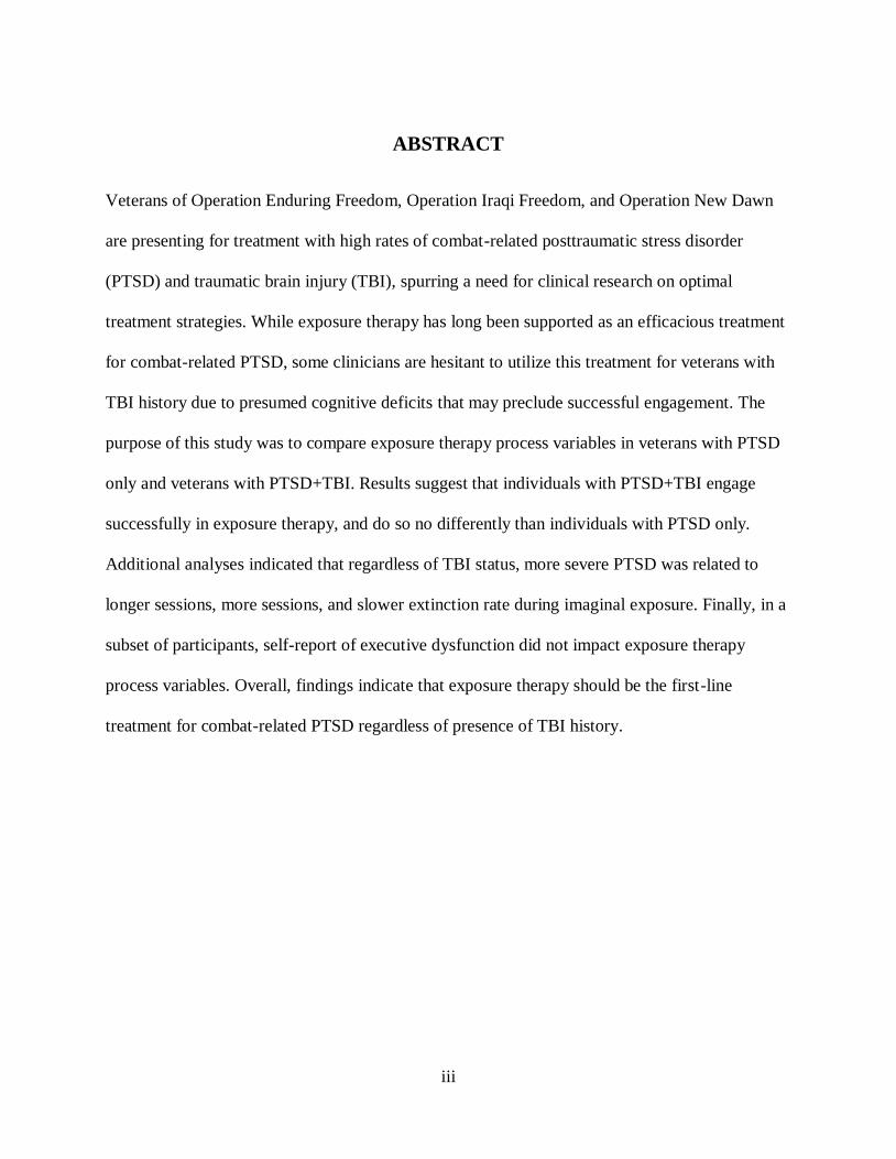
ABSTRACT
Veterans of Operation Enduring Freedom, Operation Iraqi Freedom, and Operation New Dawn
are presenting for treatment with high rates of combat-related posttraumatic stress disorder
(PTSD) and traumatic brain injury (TBI), spurring a need for clinical research on optimal
treatment strategies. While exposure therapy has long been supported as an efficacious treatment
for combat-related PTSD, some clinicians are hesitant to utilize this treatment for veterans with
TBI history due to presumed cognitive deficits that may preclude successful engagement. The
purpose of this study was to compare exposure therapy process variables in veterans with PTSD
only and veterans with PTSD+TBI. Results suggest that individuals with PTSD+TBI engage
successfully in exposure therapy, and do so no differently than individuals with PTSD only.
Additional analyses indicated that regardless of TBI status, more severe PTSD was related to
longer sessions, more sessions, and slower extinction rate during imaginal exposure. Finally, in a
subset of participants, self-report of executive dysfunction did not impact exposure therapy
process variables. Overall, findings indicate that exposure therapy should be the first-line
treatment for combat-related PTSD regardless of presence of TBI history.
iv
TABLE OF CONTENTS
LIST OF FIGURES .................................................................................................................. vi
LIST OF TABLES ................................................................................................................... vii
CHAPTER ONE: INTRODUCTION ..........................................................................................1
Posttraumatic Stress Disorder ..................................................................................................1
Treatment for Posttraumatic Stress Disorder ............................................................................3
Traumatic Brain Injury ............................................................................................................4
Treatment for Traumatic Brain Injury ......................................................................................6
Posttraumatic Stress Disorder with a History of TBI................................................................6
Treatment for Posttraumatic Stress Disorder with a History Traumatic Brain Injury ................7
CHAPTER TWO: METHOD .................................................................................................... 12
Participants ........................................................................................................................... 12
Traumatic Brain Injury Validation Procedure ........................................................................ 13
Measures ............................................................................................................................... 15
Subjective Units of Distress Scale (SUDS) ........................................................................ 15
Procedure .............................................................................................................................. 16
CHAPTER THREE: RESULTS ................................................................................................ 18
Preliminary Analyses ............................................................................................................ 18
Impact of TBI on Process of Exposure Therapy ..................................................................... 18
Initial Fear Activation ........................................................................................................ 19
Overall Fear Activation ..................................................................................................... 19
Length of Exposure Sessions ............................................................................................. 20
Number of Sessions ........................................................................................................... 20
Extinction Rate .................................................................................................................. 20
Overall Extinction ............................................................................................................. 21
Exploratory Analyses ............................................................................................................ 21
Overall within-session habituation ..................................................................................... 21
Impact of Poor Executive Functioning on Process of Exposure Therapy ............................ 22
Pre-treatment PTSD Severity ............................................................................................. 23
CHAPTER FOUR: DISCUSSION ............................................................................................ 25
Limitations and Future Directions ......................................................................................... 28
Conclusion ............................................................................................................................ 30

v
APPENDIX A: TABLES .......................................................................................................... 31
APPENDIX B: FIGURES ......................................................................................................... 35
APPENDIX C: IRB APPROVAL LETTER .............................................................................. 43
REFERENCES ......................................................................................................................... 45
vi
LIST OF FIGURES
Figure 1: Average Peak SUDS across Sessions ......................................................................... 36
Figure 2: Average First, Middle, and Final Peak SUDS ............................................................. 37
Figure 3: Scatterplot of PTSD Severity and Overall Fear Activation.......................................... 38
Figure 4: Scatterplot of PTSD Severity and Initial Fear Activation ............................................ 39
Figure 5: Scatterplot of PTSD Severity and Average Session Length ........................................ 40
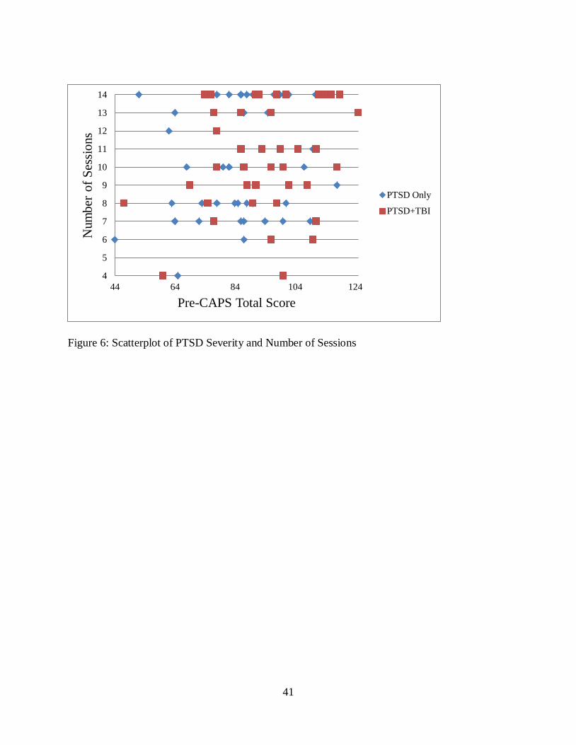
Figure 6: Scatterplot of PTSD Severity and Number of Sessions ............................................... 41
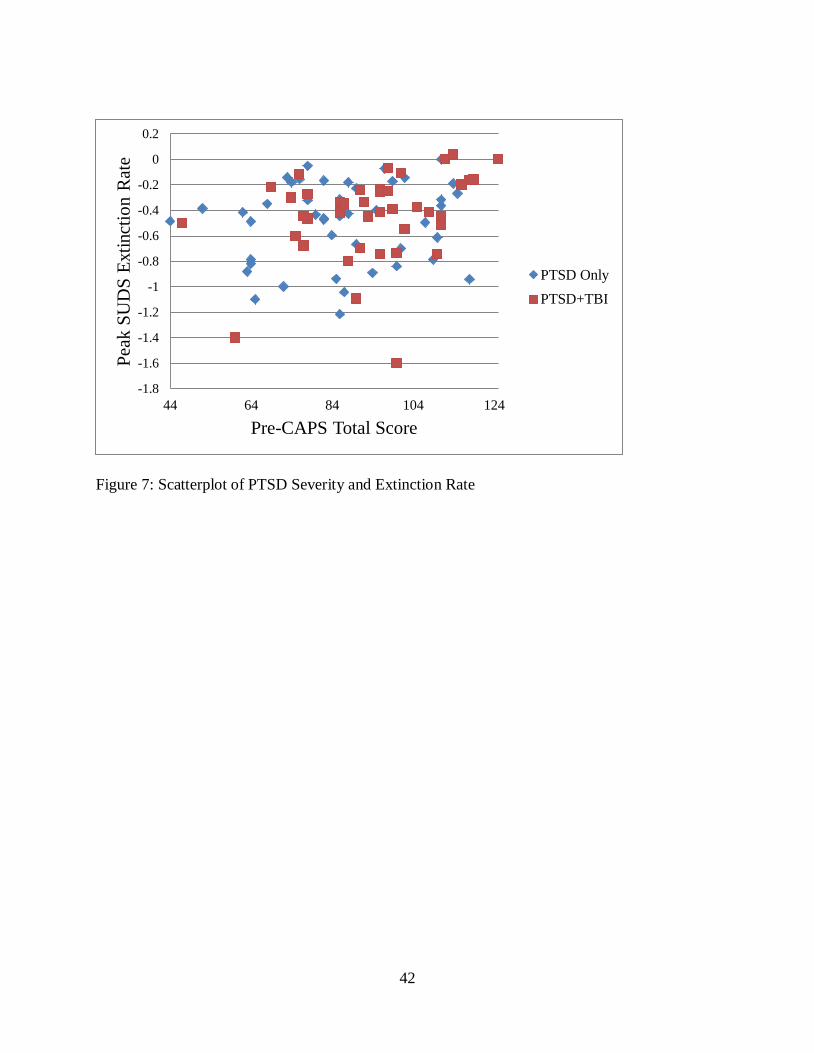
Figure 7: Scatterplot of PTSD Severity and Extinction Rate ...................................................... 42

vii
LIST OF TABLES
Table 1: TBI Group Differences on Treatment Process Variables (N = 90) ................................ 32
Table 2: BRIEF Group Differences on Treatment Process Variables (N = 24) ........................... 33
Table 3: Group Difference Controlling for PTSD Severity (N = 89) .......................................... 34
1
CHAPTER ONE: INTRODUCTION
Operation Enduring Freedom (OEF), Operation Iraqi Freedom (OIF), and Operation New Dawn
(OND) present a unique and urgent healthcare challenge: the assessment and treatment of war
related diseases and disorders. High rates of combat-related posttraumatic stress disorder (PTSD)
and traumatic brain injury (TBI) have appeared in an unprecedented number of war fighters,
spurring a need for clinical research and treatment. While advances in military protective gear
and medical care have resulted in increasing rates of survival, these advances have also led to the
development of higher rates of PTSD and mild traumatic brain injury (mTBI) (McNally & Frueh,
2013; Shively & Perl, 2012; Vasterling, Verfaellie, & Sullivan, 2009). In particular, a number of
individuals have returned from these conflicts with both PTSD and a history of TBI, making it
increasingly necessary to understand these clinical conditions and specifically, how their co-
occurrence may impact symptom presentation and treatment outcome.
Posttraumatic Stress Disorder
PTSD represents a cluster of symptoms that occurs as a result of exposure to a traumatic
event in which the person is exposed to actual or threatened death, serious injury, or threat to
physical integrity of self or others (American Psychiatric Association [APA], 2013). Many
different traumatic events can lead to the onset of PTSD (e.g., sexual assault, natural disasters,
and physical attack) and the disorder occurs in both civilian and military populations. Ensuing
symptoms are classified into four symptom clusters which include intrusion (e.g., flashbacks and
intrusive memories), avoidance (e.g., avoidance of thoughts and feelings associated with the
traumatic event), negative alterations in cognitions and mood (e.g., restricted range of affect),
and alterations in arousal and reactivity (e.g., irritability and hypervigilance). Although the
2
community-based lifetime prevalence for PTSD in United States adults is approximately 6%
(Pietrzak, Goldstein, Southwick, & Grant, 2011), rates vary considerably by sample (APA,
2013). Military combat populations yield some of the highest rates of PTSD (APA, 2013), likely
due in part to exposure to traumatic events encountered in the combat arena. Since 2002, nearly
312,000 veterans have received diagnoses of PTSD through the Department of Veterans Affairs
(VA; Veterans Affairs, 2014). However, this sample may be conservatively biased as it consists
of honorably discharged veterans seeking healthcare at the VA and does not include active duty
military personnel, VA Vet Center veterans, individuals initially diagnosed with other disorders
(e.g., acute stress disorder), or those not enrolled or entitled to VA health care. Thus, the
prevalence of PTSD in OEF/OIF/OND service members varies widely due to discrepant
assessment measures and military populations, with estimates ranging from 2.2 to 17.3%
(Hermann, Shiner, & Friedman, 2012). However, best estimates are closer to 8% (Richardson,
Frueh, & Acierno, 2010; Smith et al., 2008).
Combat-related PTSD can lead to significant impairment in functioning and considerable
distress. In a recent review, PTSD in OEF/OIF veterans was related to homelessness,
unemployment, lower work functioning, higher levels of self-reported impairment in work,
home, and interpersonal relationships, poorer role functioning due to physical and emotional
problems, increased psychosocial difficulties, reduced marital satisfaction, and reduced overall
life satisfaction (Schnurr, Lunney, Bovin, & Marx, 2009). Combat-related PTSD not only
accrues significant personal cost, but also represents a significant healthcare cost. Among nearly
250,000 OIF/OEF veterans accessing VA healthcare between 2001 and 2007, utilization of both
inpatient and outpatient medical care was higher for veterans with PTSD compared to veterans
with other psychiatric diagnoses, with rates twice as high for veterans with PTSD compared to
3
veterans with no psychiatric diagnosis (Cohen et al., 2010). Clearly, combat-related PTSD results
in significant personal, societal, and healthcare costs, thereby emphasizing the need for
efficacious treatments.
Treatment for Posttraumatic Stress Disorder
Cognitive-behavioral therapy (CBT) is a well-accepted and empirically supported
treatment for anxiety disorders (Chambless & Ollendick, 2001; Deacon & Abramowitz, 2004;
Norton & Price, 2007) including PTSD (Bradley, Greene, Russ, Dutra, & Westen, 2005;
Sherman, 1998; Van Etten & Taylor, 1998). A recent review of randomized controlled trials
suggests that trauma-focused CBT, or repeated exposure and/or cognitive restructuring, is an
efficacious and specific treatment for PTSD (Ponniah & Hollon, 2009). Exposure therapy is a
procedure whereby the individual is placed in contact (either through imagination or real life)
with the anxiety provoking stimuli in a controlled, clinician assisted manner. The goal of
exposure is habituation, or a consistent decline in behavioral, physiological, and psychological
responses, and thus extinction of anxiety. In contrast, cognitive restructuring focuses more
specifically on challenging and modifying maladaptive cognitions associated with the trauma.
The addition of cognitive restructuring does not appear to enhance treatment outcome over and
above the exposure component alone (Foa et al., 2005; Marks, Lovell, Noshirvani, Livanous, &
Threasher, 1998; Paunovic & Öst, 2001).
The theory behind the mechanism of action for exposure therapy is that exposure
weakens the conditioned fear response associated with the trauma, thus allowing new learning
(i.e., extinction learning) to occur (Foa, 2011; Foa, Steketee, & Rothbaum, 1989). This learning
is achieved through systematic, controlled exposure to the trauma cues associated with the
original traumatic event, which activates the fear complex (Foa & Kozak, 1986). As the patient

4
engages in exposure to the traumatic cues without the subsequent negative event, habituation
occurs and new neural associations are formed. In in-vivo (real life) exposures, similar
habituation and new learning occurs in response to associated stimuli. Simply, repeated
presentation of traumatic cues without the traumatic outcome allows habituation and learning to
occur, which leads to extinction of the anxiety/fear response.
Extant literature has also provided support for efficacious treatment for combat-related
PTSD specifically (Frueh, Turner, & Beidel, 1995), with exposure based treatments proving to
be the most effective (Goodson et al., 2011). In fact, the Institute of Medicine (IOM) reported
exposure therapy was the only treatment with sufficient evidence to conclude its efficacy for
combat-related PTSD treatment (IOM, 2007). One such treatment developed specifically for
combat-related PTSD is Trauma Management Therapy (TMT), a multicomponent behavioral
treatment utilizing exposure therapy (Frueh, Turner, Beidel, Mirabella, & Jones, 1996; Turner,
Beidel, & Frueh, 2005). In fact, a recent randomized controlled trial of TMT demonstrated its
efficacy for Vietnam-era combat veterans with chronic PTSD (Beidel, Frueh, Uhde, Wong, &
Mentrikoski, 2011), and a similar trial with OEF/OIF veterans is underway, which provided the
treatment sample examined in the current study.
Traumatic Brain Injury
Although combat-related PTSD is a significant healthcare concern, TBI has been coined
the “signature injury” of the Afghanistan and Iraq wars. TBI is defined by either a penetrating or
closed head injury that results in temporary or permanent neurological dysfunction (Marshall et
al., 2012) and may result from a foreign object penetrating the brain (i.e., penetrating head
injury), blunt force trauma, acceleration or deceleration of the brain, or blast injury (Department
of Veterans Affairs and Department of Defense, 2009). Immediate resulting neurological
5
dysfunction can include loss or decreased level of consciousness, memory loss before or after the
injury, alteration in mental status (e.g., slowed thinking, confusion, or disorientation),
neurological deficits (e.g., weakness, change in vision, loss of balance), and/or brain lesions
(Department of Veterans Affairs and Department of Defense, 2009).
TBI severity classifications range from mild to severe based on the length of time of the
resulting dysfunction. According to the Department of Defense (2014), mTBI is defined by
confusion or disorientation for less than 24 hours, loss of consciousness for up to 30 minutes,
memory loss for less than 24 hours, and normal structural brain imaging. Moderate TBI is
defined by confusion or disorientation for more than 24 hours, loss of consciousness for more
than 30 minutes, memory loss greater than 24 hours but not more than seven days, and normal or
abnormal structural brain imaging. Severe TBI is defined by confusion or disorientation for more
than 24 hours, loss of consciousness for more than 24 hours, memory loss for more than seven
days, and normal or abnormal structural brain imaging. Approximately 75% of all TBI cases are
mild (Lu, Gary, Neimeier, Ward, & Lapane, 2012), with a similar prevalence rate found for the
United States military population (77%; Marshall et al., 2012).
It is important to note that TBI is a historical event defined by the injury sustained. The
resulting postconcussive symptoms (PCS) are defined as self-reported somatic, cognitive, and
affective symptoms occurring post injury (Morissette et al., 2011) and can significantly vary
between individuals (Hoge et al., 2008; Riggio & Wong, 2009). PCS of mTBI can include
headaches, poor sleep, dizziness, balance problems, irritability, and concentration, and memory
difficulties (Hoge et al., 2008; Shively & Perl, 2012).
A review of meta-analytic studies supports the presence of impaired cognitive abilities
during the acute phase of an mTBI (i.e., within three months of the injury); however, a debate

6
exists regarding permanent or chronic effects (Ruff, 2011). Nonetheless, a portion of individuals
with mTBI do experience symptom persistence three months post injury (Belanger, Curtiss,
Demery, Lebowitz, & Vanderploeg, 2005). Further, research indicates that individuals with TBI
present with more severe PTSD symptoms than individuals without a history of TBI (Barnes,
Walter, & Chard, 2012; Davis, Walter, Chard, Parkinson, & Houston, 2013; Ragsdale, Neer,
Beidel, Frueh, & Stout, 2013), suggesting that the historical event of the brain injury alone may
in fact influence psychological functioning long term.
Treatment for Traumatic Brain Injury
Once patients are medically stabilized, treatment of TBI transitions to restoration of
functioning (Lu et al., 2012), which may include psychological, educational, supportive, and
pharmacological interventions. Systematic reviews of psychological (e.g., education and
cognitive rehabilitation) treatments for mTBI suggest that educational interventions may be
somewhat helpful if provided early; however, authors emphasize poor research methodology of
available studies and a general lack of methodological rigor (Borg et al., 2004; Comper,
Bisschop, Carnide, & Tricco, 2005; Snell, Surgenor, Hay-Smith, & Siegert, 2009). Within the
military, treatment of mTBI is centered on symptom management, patient education, rest, and
recovery (Marshall et al., 2012), with a focus on treatment of symptoms regardless of their
etiology (Brenner, Vanderploeg, & Terrio, 2009).
Posttraumatic Stress Disorder with a History of TBI
Veterans with a history of TBI have higher rates of PTSD compared to veterans without a
history of TBI (Carlson et al., 2010; Carlson et al., 2011; Hoge et al., 2008; Morissette et al.,
2011; Walker, Clark, & Sanders, 2010). Comorbidity rates of probable PTSD and probable
mTBI among Iraq and Afghanistan veterans range from 33% to 39% (Carlson et al., 2011). In

7
addition, veterans suffering from PTSD who have a history of mTBI endorse significantly more
severe PTSD symptoms than those with PTSD alone (Barnes et al., 2012; Davis et al., 2013;
Ragsdale at al., 2013), and the increased PTSD severity appears to negatively affect the clinical
presentation, leading to higher overall anxiety and more functional limitations (Ragsdale et al.,
2013). Finally, although the presence of TBI quadruples the median annual medical cost for
veterans, the addition of PTSD results in even further increases in the cost medical care (Taylor
et al., 2012).
Treatment for Posttraumatic Stress Disorder with a History Traumatic Brain Injury
Clearly, veterans of OIF/OEF/OND are presenting with high rates of PTSD with a history
of TBI, forcing treatment of this complex condition to the forefront of clinical practice. Although
well supported for the treatment of PTSD, some clinicians are hesitant to use exposure therapy
with individuals who report a history of TBI due to concerns of cognitive impairment (Sripada et
al., 2013). Indeed, the ability to recall and cognitively process the traumatic event is central to
exposure therapy, as its repeated presentation allows for habituation, new learning, and
extinction of the anxiety/fear response. As such, factors associated with TBI that could impede
fear activation, such as memory difficulties (i.e., difficulty retrieving and/or holding and
processing the memory), poor concentration (i.e., difficulty sustaining attention to imaginal
aspects of the exposure), and/or damage to brain structures involved in the process of extinction
learning could theoretically inhibit effective implementation, which could consequently reduce
efficacy.
The amygdala, hippocampus, and ventromedial prefrontal cortex (vmPFC) are involved
in both fear conditioning and extinction (Moustafa et al., 2013). If damaged by TBI, any
impaired functioning could negatively impact treatment success. For example, both lesions and
8
compromised functions in these brain structures impair fear extinction in animals (Moustafa et
al., 2013). Particularly, lesioning intercalated amygdala neurons (Likhtik, Popa, Apergis-
Schoute, Fidacardo, & Paré, 2008), inactivation of the dorsal hippocampus (Corcoran & Maren,
2001), and lesioning of the ventral prelimbic cortex and the infralimbic cortex of the vmPFC in
rats (Quirk, Russo, Barron, & Lebron, 2000) impair extinction learning, which in turn could
attenuate the impact of exposure therapy.
Unfortunately, brain injuries sustained as a result of TBI are heterogeneous in nature due
to the various locations and mechanisms of injury (e.g., penetrating head injury, blunt force
trauma, acceleration or deceleration of the brain, and blast injury; Department of Veterans
Affairs and Department of Defense, 2009), which makes it difficult to precisely locate brain
injury or disruption. Further, mTBI is defined by normal structural brain imaging (Department of
Defense, 2014), precluding study of potentially affected brain regions. Therefore, examining the
extinction process that occurs during exposure therapy (through measures of fear activation and
habituation) may be one of the few means by which to elucidate how mTBI may affect the
process of extinction learning.
Recent research has begun to investigate the feasibility and outcome of cognitive
behavioral therapies (e.g., prolonged exposure [PE] and cognitive processing therapy [CPT]) in
samples of veterans suffering from PTSD with a history of TBI (Chard, Schumm, McIlvain,
Bailey, & Parkinson, 2011; Sripada et al., 2013; Walter, Barnes, & Chard, 2012; Walter, Kiefer,
& Chard, 2012; Wolf, Strom, Kehle, & Eftekhari, 2012). Several studies (Chard et al., 2011;
Walter et al., 2012a, 2012b) have examined seven and eight week Veterans Administration
PTSD/TBI residential programs incorporating modified CPT (CPT-C; Resick, Nishith, Weaver,
Astin & Feuer, 2002) via manualized group and individual treatment (Chard, Resick, Monson, &
9
Kattar, 2008). These residential treatment programs also incorporated various individual speech
and cognitive therapies, attention training, and psychoeducational groups (e.g., distress tolerance,
cognitive enhancement, and anger management). Unfortunately, various methodological
limitations in these studies preclude conclusions that would inform the current study. Samples
were heterogeneous in nature (e.g., various wars and combat arenas [e.g., Vietnam]) (Chard et
al., 2011; Sripada et al., 2013; Walter et al., 2012a; Walter et al., 2012b) and included non-
combat related psychological and physical traumas [e.g., sports injuries] (Chard et al., 2011),
which preclude conclusion or generalization specific to combat TBIs and combat PTSD.
Additionally, and arguably most importantly, conclusions cannot be drawn regarding the
effectiveness of CBT for individuals with TBI, as the multifaceted treatment programs (Chard et
al., 2011; Walter et al., 2012a, 2012b) obscured the ability to examine the efficacy of any
individual treatment component.
With specific regard to prolonged exposure therapy, two studies have investigated its
feasibility for individuals with PTSD and a history of TBI; however, studies again suffer from
methodological limitations. First, Wolf and colleagues (2012) examined an open trial of PE
treatment in OEF/OIF veterans with chronic PTSD and a history of mild to moderate TBI;
however, sample size was small (N = 10) and no control group was used. Similarly, Sripada and
colleagues (2013) examined PE in 40 veterans with PTSD only and 11 veterans with PTSD and
history of TBI. Unfortunately, the heterogeneous sample (only 32% of the sample served in
Afghanistan or Iraq) and the few participants with TBI blunted the statistical power necessary to
detect group differences.
To summarize, the small body of literature examining CBT for individuals with PTSD
and history of TBI suffers from serious methodological limitations including limited power,
10
small sample size, lack of control groups, and treatment confounds. In particular, extant literature
is currently lacking a large homogenous sample of OEF/OIF/OND veterans with combat-related
PTSD and a history of combat-related TBI. Therefore, examination of the feasibility and impact
of exposure therapy in a carefully diagnosed sample of OEF/OIF/OND veterans suffering from
combat-related PTSD with and without history of TBI is warranted.
Given that veterans with PTSD and a history of mTBI endorse significantly more severe
PTSD symptoms than those with PTSD alone (Barnes et al., 2012; Davis et al., 2013; Ragsdale et
al., 2013), it is crucial to ensure that this population can successfully engage in exposure therapy.
The purpose of this study was to examine the potential impact of TBI history on exposure
therapy for combat-related PTSD using a carefully diagnosed sample of OEF/OIF veterans. The
following hypotheses were tested:
1. Individuals with PTSD and history of TBI will demonstrate significantly higher fear
activation compared to individuals with PTSD only.
2. Individuals with PTSD and history of TBI will show significantly longer session times
compared to individuals with PTSD only, as individuals experiencing higher fear activation
will likely require longer times to habituate within a treatment session.
3. Individuals with PTSD and history of TBI will require more exposure sessions compared to
individuals with PTSD only, as higher fear activation and longer session times will likely
require a greater number of sessions to achieve overall extinction.
4. Individuals with PTSD and history of TBI will be less likely to achieve overall extinction
within a 14-session treatment protocol. Although between-session habituation is considered
the indication of positive treatment outcome, some individuals may not achieve overall
habituation after a prescribed number of sessions.

11
5. Individuals with PTSD and history of TBI will evidence slower extinction rate, or change in
slope of peak subjective distress, across sessions when compared to individuals with PTSD
only.
12
CHAPTER TWO: METHOD
The treatment study from which these data were extracted is an ongoing randomized
controlled trial comparing individual exposure therapy to individual exposure therapy plus group
therapy for combat-related PTSD. The study is located at two sites: the University of Central
Florida and the Medical University of South Carolina/Ralph Johnson Veterans Affairs Medical
Center. All procedures were approved by the Institutional Review Boards at UCF or MUSC, as
well as the United States Army Institutional Review Board.
Participants
Participant recruitment consisted of advertising through clinician referral, radio, various
websites, and public events, and in the case of MUSC, through the PTSD clinic at the Ralph
Johnson VAMC in Charleston, South Carolina. The sample consisted of treatment seeking
individuals with combat-related PTSD who had served in Operation Enduring Freedom (OEF)
and Operation Iraqi Freedom (OIF). All participants included in the current study completed the
individual exposure therapy component of the clinical trial. Ninety-three participants (90.3%
male) with a mean age of 36.27 (SD = 9.60; range = 23–63) were included (UCF; N = 84;
MUSC; N = 9). There were no differences on any demographic variable between the two sites.
The sample was 61.3% Caucasian, 21.5% Hispanic/Latino, 11.8% African American,
2.2% Asian/Pacific Islander, 1.1% Indian subcontinent, and 2.2% “other”. With regard to marital
status, 47.3% were married, 28% were single, 15.1% were divorced, and 9.7% were separated.
With regard to education, 17.2% of participants completed high school only, 57% completed
some college, 20.4% had earned a Bachelor’s degree, and 5.4% had earned a Master’s degree.

13
The majority of the sample served in the Army (69.9%), followed by the Marine Corps (18.3%),
Air Force (7.5%), Navy (3.2%) and as a Civilian Contractor (1.1%).
All participants met the Diagnostic and Statistical Manual of Mental Disorders (4th
ed.,
Rev.; DSM-IV-TR; American Psychiatric Association, 2000) criteria for PTSD as assessed by
the Clinician Administered PTSD Scale (CAPS; Blake et al., 1995). Symptom scores for the
three DSM-IV criterion clusters (Criterion B: Reexperiencing, Criterion C: Avoidance and
Numbing, and Criterion D: Hyperarousal) are derived by summing the frequency and intensity
scores for relevant individual items. Summing the subscale scores provides overall frequency,
intensity, and total PTSD scores. The CAPS has excellent reliability, convergent and
discriminant validity, diagnostic utility, and sensitivity to clinical change (Weathers, Keane, &
Davidson, 2001). The CAPS was administered by licensed clinical psychologists, post-doctoral
fellows, masters level clinicians or supervised senior doctoral students. Ten percent of interviews
were randomly selected and scored by a blinded clinician for inter-relater reliability (total score
ICC = .969; PTSD diagnosis κ = 1.00).
Traumatic Brain Injury Validation Procedure
During the initial diagnostic interview for the treatment study, TBI status was determined
by simple self-report (i.e., “Were you ever diagnosed with a TBI?”). To strengthen the validity of
TBI status for the current investigation, we attempted to re-contact previously treated participants
in order to conduct a more thorough assessment. Each participant was contacted a maximum of
three times by telephone. Participants who were successfully contacted were interviewed using
current TBI status criteria (listed below) (Department of Defense, 2014; Department of Veterans
Affairs and Department of Defense, 2009).
14
1. Have you experienced a blow or jolt to the head such as the head being struck by, or
striking, an object; acceleration or deceleration of the brain; blast or explosion injury; or
object penetration of the brain? If so, following the head injury:
2. Did you experience any alteration in mental state at the time of the injury, such as
confusion, disorientation, or slowed thinking?
Note. Observed signs of neurological or neuropsychological dysfunction (e.g.,
headache, dizziness, or poor concentration) is not sufficient to make a diagnosis of
TBI (i.e., these do not indicate a change in mental state) when loss of or altered
consciousness is not present (Department of Veterans Affairs and Department of
Defense, 2009).
3. Did you experience loss of or a decreased level of consciousness?
4. Did you experience memory loss for events immediately before or after the injury?
Participants who answered affirmatively to question one and at least one additional question
are considered by these criteria to have experienced a TBI.
Of the 93 participants included in the current study, 46 (49%) completed the TBI validation.
The other 47 participants were unable to be reached for a variety of reasons (e.g., they did not
answer the phone or return the calls, had changed their phone number, or their voicemail was full
or not set up). Of the 46 participants who completed the TBI validation protocol, 44 (96%)
remained the same TBI status as determined by their original assessment, whereas two (4%)
changed from a negative to a positive TBI status. These two participants reported being
diagnosed with a concussion during the military, which, perhaps unknown to the participants, is
synonymous with mTBI (Department of Veterans Affairs and Department of Defense, 2009).

15
Although unable to contact the entire sample, it appeared that the participants’ initial self-
reports of TBI history were valid, and that the re-querying did not add significant incremental
validity. Therefore, with the exception of the two participants whose TBI status changed from
negative to positive, participants’ initial TBI statuses were utilized for all analyses.
Across the entire sample, 47% (N= 44) had a history of TBI. There were no significant
TBI group differences on any demographic characteristics with the exception of age. Military
personal with PTSD+TBI (M = 33.77, SD = 10.52) were significantly younger than those with
PTSD only (M = 38.51, SD = 7.84), t(88.08) = 2.48, p = .02, a consistent finding in the literature
(Carlson et al., 2010; Hoge et al., 2008; Ragsdale et al., 2013; Taylor et al., 2012).
Measures
Subjective Units of Distress Scale (SUDS)
Fear activation was determined using the Subjective Units of Distress Scale (SUDS), a
self-report rating of the participant’s subjective fear and anxiety. The scale ranged from 0 (none)
to 8 (extreme) and were assessed every five minutes during exposure therapy. SUDS ratings are
used to determine when within- and between-session habituation occurs. Specific treatment
process variables examined in this study include initial fear activation, overall fear activation,
within-session habituation, overall extinction (or between-session habituation), and extinction
rate. Operational definitions are largely based on prior conceptualizations of these variables
(Turner, Beidel, Long, & Greenhouse, 1992; Craske et al., 2008) and are presented below.
Initial fear activation was operationally defined as the change from the baseline SUDS
to the peak SUDS during the first treatment session.
16
Overall fear activation was operationally defined as the change from the lowest SUDS
to the peak SUDS (peak SUDS – lowest SUDS) across all sessions, which captured each
individual’s maximum increase in anxiety or fear as a result of the exposure.
Within-session habituation was operationally defined as a return to baseline SUDS, or
at least a 50% reduction of fear activation (change from baseline SUDS to peak SUDS, during
each individual session), and therefore was a dichotomous variable for each session.
Overall within-session habituation, the within-session habituation variable used for
analysis purposes, was the percentage of sessions during which habituation occurred (total
number of sessions which achieved habituation/total number of imaginal exposure sessions).
Overall extinction (or between-session habituation) was operationally defined by at least
a 50% reduction from the first session peak SUDS to the final session peak SUDS (e.g., an initial
peak SUDS of 8 and final peak SUDS of 4), and therefore, was a dichotomous variable.
Extinction rate was calculated by determining the slope of peak SUDS over time for
each participant.
Finally, total number of exposure sessions and average length of time of sessions were
examined.
Procedure
Following assessment, eligible participants initiated imaginal exposure therapy. The
imaginal scene used for exposure was constructed during session 1 and was based on the
individual’s most severe trauma. At session 2, the clinician assisted the participant to imagine the
traumatic event by reading the scene, inquiring about SUDS every five minutes. The exposure
continued until the participant evidenced within-session habituation. During subsequent sessions,
the participant verbalized the scene him or herself, which was again continued until the
17
participant evidenced within-session habitation. Throughout all exposures, the clinician
continuously observed the participant’s behaviors and inquired about physiological responses
and cognitions to corroborate SUDS. The clinician’s goal was to assist the participant in
remaining in contact with the feared stimuli (i.e., the traumatic memory) until habituation
occurred. Imaginal exposure was conducted for up to 14 sessions, but was terminated once the
participant evidenced between-session habituation.
18
CHAPTER THREE: RESULTS
Preliminary Analyses
Continuous dependent variables were first examined for violations of normality. The
Kolmogorov-Smirnov test indicated that extinction rate (D = .18, p < .001), overall fear
activation (D = .17, p < .001), initial fear activation (D = .15, p < .001), and total number of
sessions (D = .18, p < .001) deviated significantly from a normal distribution, whereas average
session time (D = .06, p = .20) and pre-CAPS total score (D = .06, p = .20) did not. Attempts to
normalize the relevant data using various transformations (e.g., log transformations) were
unsuccessful. However, given the central limit theorem and the robustness of t-tests, groups were
compared with both parametric (t-tests) and non-parametric (Mann-Whitney U) tests.
Analysis of univariate outliers revealed three statistical outliers. In each case, a
participant’s score on one standardized z-score exceeded three standard deviations from the mean
of the dependent variable. Analysis of multivariate outliers revealed one statistical outlier (i.e.,
Mahalonobis distance value of 28.93 exceeded the critical value of 20.52) who was previously
identified during analysis of univariate outliers. The three participants identified in these analyses
were excluded from all future analyses, leading to a final sample size of 90 (PTSD only = 48,
PTSD+TBI = 42).
Impact of TBI on Process of Exposure Therapy
Following preliminary analyses, groups were compared on exposure therapy process
variables (initial and overall fear activation, average session time, total number of exposures
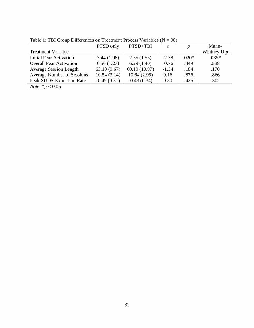
sessions, extinction rate, and overall extinction). See Table 1 for results of all t- and Mann-
Whitney U tests, interpreted below.
19
Initial Fear Activation
The implementation of imaginal exposure therapy requires participants to imagine and
hold the traumatic event in memory. By doing so, fear associated with the traumatic event is
elicited and perceived by the participant, suggesting the presence of fear activation. Fear
activation during the initial treatment session (peak SUDS—lowest SUDS) was compared using
an independent samples t-test. Results indicated that participants with PTSD+TBI (M = 2.55, SD
= 1.53) experienced less fear activation than participants with PTSD only (M = 3.44, SD = 1.96,
t(88) = -2.38, p = .02), which was supported by a Mann-Whitney U test (p = .04). It should be
noted, however, that examination of peak and baseline SUDS revealed that participants with
PTSD+TBI had significantly higher SUDS at baseline (M = 4.71, SD = 1.76) compared to
participants with PTSD only (M = 3.90, SD = 1.92, t(88) = 2.10, p = .04 [Mann-Whitney-U p
=.03]). Thus, the lesser activation in participants with PTSD+TBI most likely resulted from a
ceiling effect, as SUDS contain a finite number of points. When controlling for baseline
differences, a one-way analysis of covariance (ANCOVA) revealed that the two groups did not
differ on initial fear activation, F(1,87) = 1.16, p = .28.
In contrast, the groups did not significantly differ on their peak SUDS rating in the first
treatment session (PTSD+TBI [M = 7.26, SD = 0.91] versus PTSD only [M = 7.33, SD = 0.86],
t(88) = -0.38, p = .70, [Mann-Whitney-U p =.70]), suggesting that individuals in both groups
experienced a high level of distress when imagining the situation.
Overall Fear Activation
Because high levels of anticipatory anxiety are common in the first treatment sessions,
fear activation can sometimes be attenuated. Another way to determine fear activation is to
examine the baseline and peak SUDS regardless of the session in which it occurred. The results
20
of a t-test comparing differences in overall fear activation (overall peak SUDS – overall lowest
SUDS) revealed no difference between participants with PTSD+TBI (M = 6.29, SD = 1.40) and
participants with PTSD only (M = 6.50, SD = 1.27, t(88) = -0.76, p = .45), a finding supported by
a Mann-Whitney U test (p = .54).
Length of Exposure Sessions
A t-test comparing the average treatment session time (in minutes) indicated that the
groups were not significantly different, PTSD+TBI (M = 60.19, SD = 10.97) versus PTSD only
(M = 63.10, SD = 9.67, t(88) = -1.34, p = .18). This finding was supported by a Mann-Whitney U
test (p = .17).
Number of Sessions
The groups were not significantly different on the number of exposure sessions necessary
for extinction (between-session habituation) to occur, PTSD+TBI (M = 10.64, SD = 2.95) versus
PTSD only (M = 10.54, SD = 3.14, t(88) = 0.16, p = .88), a finding supported by a Mann-
Whitney U test (p = .87).
Extinction Rate
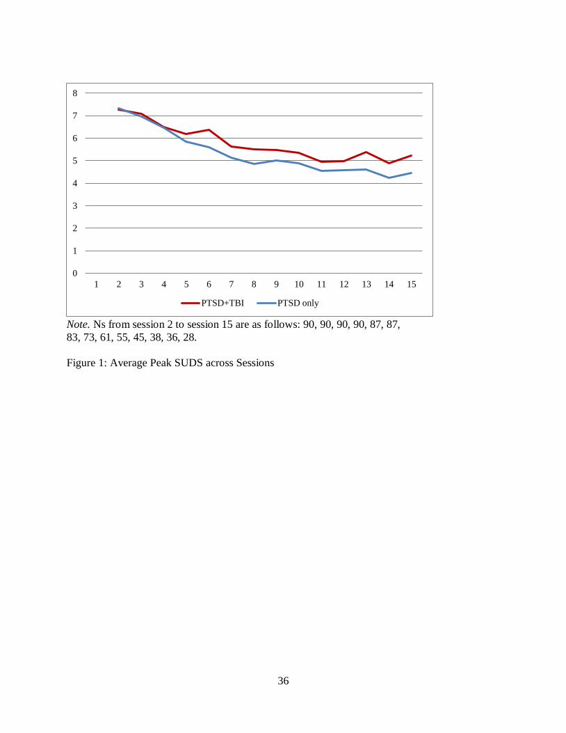
To determine if participants with PTSD+TBI experienced a slower decline in anxiety
during exposure sessions, the extinction rate (slope of peak SUDS) across sessions was compared
using a t-test. There was no significant difference in extinction rate between participants with
PTSD+TBI (M = -0.43, SD = 0.34) and participants with PTSD only (M = -0.49, SD = 0.31; t(88)
= 0.80, p = .43). This finding was supported by a Mann-Whitney U test (p = .30). The average
peak SUDS across all 14 sessions for both groups is depicted in Figure 1.
The treatment protocol dictated that achievement of between-session habituation via
imaginal exposure should be followed by a change to in vivo exposure to address behavioral
21
avoidance. Thus, as some individuals did not require 14 exposure therapy sessions to achieve
between-session habituation, the SUDS for the latter sessions depicted in Figure 1 may be
inflated by the reduced number of participants remaining in the sample. As such, average peak
SUDS were re-examined using each participant’s peak SUDS reported during their initial,
middle, and final imaginal exposure session (see Figure 2). Results indicated that the groups did
not differ on initial peak SUDS (PTSD+TBI [M = 7.27, SD = 0.90] versus PTSD only [M = 7.33,
SD = 0.86], t(88) = -0.32, p = .75), middle peak SUDS (PTSD+TBI [M = 5.98, SD = 1.48] versus
PTSD only [M = 5.66, SD = 1.52], t(88) = 1.03, p = .31), or final peak SUDS (PTSD+TBI [M =
3.83, SD = 1.95] versus PTSD only [M = 3.17, SD = 1.81], t(88) = 1.68, p = .10)
Overall Extinction
Overall, 64.4% of all participants achieved overall extinction (N = 58); 35.6% did not (N
= 32). A Chi-square analysis for independence (with Yate Continuity Correction) was used to
determine if participants with PTSD+TBI achieved overall extinction comparable to participants
with PTSD only. Results indicated no significant association between TBI status and overall
extinction, 2(1, n = 90) = .06, p = .80. Specifically, 67% of PTSD only and 62% of PTSD+TBI
achieved overall extinction.
Exploratory Analyses
Although not part of formal hypothesis testing, the results above suggested additional
data analysis that might inform future investigations. Exploratory analyses are presented here.
Overall within-session habituation
The treatment protocol required within-session habituation for session termination;
however, post hoc analyses revealed that some sessions were terminated prior to the required
criterion for extinction. Some sessions were terminated early for various reasons (e.g., unique or

22
unforeseen time constraints or sudden patient illness). Descriptive analyses revealed that 73.3%
of participants (N = 66) habituated to all imaginal exposure sessions (i.e., achieved within-
session habituation to every session). Remaining participants (N = 24) habituated to 57.1% -
92.86% of sessions.
Restricting the sample to participants who did not achieve within-session habituation
across all sessions, participants with PTSD+TBI (M = 84.02, SD = 10.54) and participants with
PTSD only (M = 77.49, SD = 10.16) did not differ on percentage of individual sessions during
which within-session habituation occurred, t(22) = 1.51, p = .15.
Impact of Poor Executive Functioning on Process of Exposure Therapy
Inasmuch as the results above suggest that a diagnosis of TBI per se does not attenuate
exposure therapy, exploratory analyses examined whether cognitive impairment (i.e., PCS which
might result after a TBI) affected the treatment process. The Behavior Rating Inventory of
Executive Function –Adult Version (BRIEF-A; Roth, Isquith, & Gioia, 2005) was available on a
subset of participants and could be used as an assessment of executive functioning. The BRIEF-
A assesses an individual’s perception of their executive functions in nine areas and provides an
overall summary score, the Global Executive Composite (GEC). A t-score of 65 or greater is
considered clinically significant. The BRIEF-A adequately assesses patients with TBI and
evidences strong reliability in this particular population (0.94 to 0.96; Waid-Ebbs, Wen, Heaton,
Donovan, & Velozo, 2012).
While only a small percentage of the total sample completed a pre-treatment BRIEF (N =
26), two groups were defined based on GEC t-scores (regardless of TBI status). Individuals who
scored a t-score of 65 or higher (N = 14; M = 74.71; SD = 7.44) and individuals who scored a t-
score of 64 or below (N = 12; M = 57.41; SD = 4.14) were compared on treatment process

23
variables. Results indicated that self-reported executive function difficulty did not impact any of
the exposure therapy process variables described above. See Table 2 for descriptive statistics and
results of t-tests.
Pre-treatment PTSD Severity
Consistent with prior investigations (Barnes et al., 2012; Davis et al., 2013), participants
with PTSD+TBI reported significantly higher PTSD symptoms (as measured by the CAPS total
score at pre-treatment) (M = 94.33, SD = 16.88) than participants with PTSD only (M = 86.40,
SD = 17.62, t(87) = 2.16, p = .03). Therefore, a one-way between-groups multivariate analysis of
covariance (MANCOVA) examined group differences on treatment process variables when
controlling for PTSD severity. Overall extinction was converted from a dichotomous to a
continuous variable by calculating the decrease from initial session peak SUDS to final session
peak SUDS. See Table 3 for MANCOVA results, interpreted below.
When controlling for PTSD severity, the overall MANCOVA was statistically significant,
F(6, 81) = 2.44, p = .03; Wilks’ Lambda = .85; ƞp2=.15 . Examination of simple effects revealed
that groups significantly differed on initial fear activation, F(1, 86) = 6.06, p = .02, and average
session length, F(1, 86) = 4.12, p = .046. Results again indicated that participants with
PTSD+TBI experienced less fear activation during the initial treatment session (M = 2.55, SD =
1.53) compared to participants with PTSD only (M = 3.49, SD = 1.94), which held true even
when controlling for session baseline SUDS, F(1, 86) = 4.80, p = .03. While this finding suggests
that individuals with TBI might experience less fear activation during their initial treatment
session, examination of the mean SUDS revealed that the groups differed by only one point, a
difference that may be statistically, but not clinically, significant. Similarly, with regard to
average session length, participants with PTSD only had longer sessions (in minutes) (M = 63.26,

24
SD = 9.71) than participants with PTSD+TBI (M = 60.19, SD = 10.97). However, the average
difference was three minutes, which, while statistically significant, is not clinically significant.
There were no group differences on overall fear activation, average number of sessions,
extinction rate, or overall extinction while controlling for PTSD severity.
In contrast, the MANCOVA results revealed significant relationships between PTSD
severity and average session length, F(1, 86) = 7.44, p = .01, average number of sessions, F(1,
86) = 6.29, p = .01, and extinction rate, F(1, 86) = 4.27, p = .04. In each case, follow-up Pearson
correlations revealed that longer sessions, r(89) = .29, p = .03, more sessions, r(89) = .26, p =
.01, and a slower extinction rates, r(89) = .23, p = .03, were associated with higher PTSD
severity. There were no significant relationships between PTSD severity and initial fear
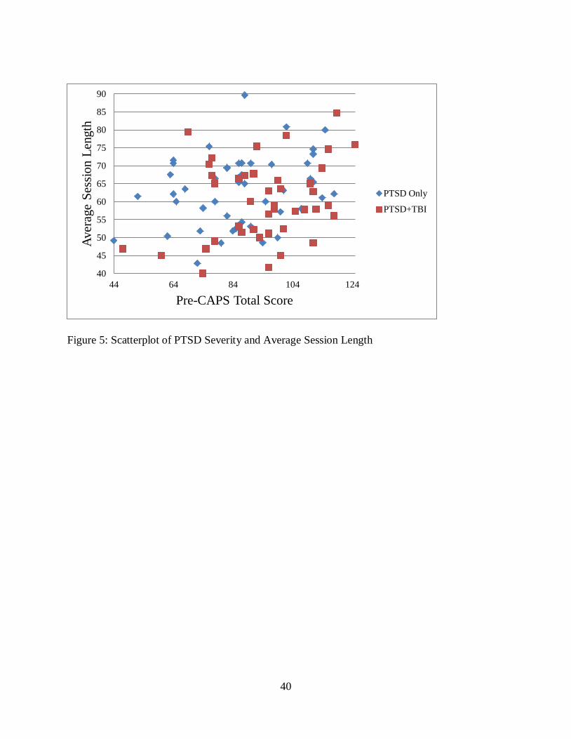
activation, overall fear activation, and overall extinction. See Figures 3-7 for scatterplots of
PTSD severity and each dependent variable for both groups.
25
CHAPTER FOUR: DISCUSSION
Although prior investigations suggested that the presence of TBI does not impact CBT
treatment outcome, treatment confounds and limitations in sample size and composition preclude
application of those findings to substantiate the feasibility of exposure therapy for
OEF/OIF/OND combat veterans with combat-related PTSD+TBI. This study represents the first
time process variables crucial for successful outcome using exposure therapy were examined in a
carefully controlled and diagnosed sample of OEF/OIF/OND combat-veterans with combat-
related PTSD and TBI. The results of this study indicate that TBI history does not impact overall
fear activation, session length, number of sessions, within-session habituation, overall extinction,
or rate of extinction. Individuals with PTSD and a history of TBI do not engage in exposure
differently, or less effectively, than individuals with PTSD only, with one exception. Individuals
with PTSD+TBI experienced less fear activation during their first exposure session, a difference
that was accounted for by higher baseline SUDS. This suggests that the PTSD+TBI group may
experience greater anticipatory anxiety or overall higher general anxiety. The lack of group
differences is actually good news for clinicians as it means that, regardless of TBI status,
exposure therapy for PTSD remains the treatment of choice.
One reason for the reluctance to use exposure therapy when participants report a history
of TBI may be the often misunderstood fact that TBI is an event, and not a disorder. It is unclear
whether individuals, at least with mTBI, continue to experience cognitive difficulties three
months post injury (Ruff, 2011). In fact, research suggests that the majority of these individuals
will fully recover from their head injury within 90-days (Karr, Areshenkoff, & Garcia-Barrera,
2014). As such, TBI status may not influence the exposure therapy process when therapy is
26
initiated after ninety-days. Conversely, a percentage of individuals with mTBI continue to
experience cognitive difficulties three months post injury (Belanger et al., 2005), and chronic
neurocognitive deficits are typical for moderate and severe TBI (Dikmen et al., 2009). Future
investigations may need to specifically identify the subset of individuals with chronic cognitive
difficulties (assessed via more comprehensive neuropsychological examinations) to determine
their ability to fully engage in exposure therapy.
Further complicating the matter, however, is the significant overlap of TBI, PCS, and
PTSD symptoms (Morissette et al., 2011), which suggests that the negative long term effects
may in fact be related to PTSD, and not TBI. For example, extant literature suggests that PCS is
not unique to TBI (Meares et al., 2008; Meares et al., 2011), and that PCS are actually better
predicted by PTSD (Schneiderman, Braver, & Kang, 2008). Given the significant overlap
between PTSD symptoms and PCS (e.g., irritability, sleep difficulty, and impaired
concentration), psychological symptoms occurring post-combat should be treated with
evidenced-based treatments regardless of their presumed etiology (Brenner et al., 2009).
Additionally, TBI status may have had no impact on the exposure therapy process due to
the physiological nature of habituation; that is, exposure therapy does not require higher order
cognitive functions to successfully extinguish fear. Specifically, fear activation and habituation
are the active ingredients of exposure therapy. Given that fear conditioning can occur outside
one’s level of awareness (i.e., some individuals with severe TBI and no memory of traumatic
events develop PTSD) (Bryant, Marosszeky, Crooks, & Gurka, 2000; Bryant, Marosszeky,
Crooks, & Gurka, 2004), it is likely that extinction can as well. These findings suggests that
regardless of the potential long-term cognitive effects of a TBI, individuals who retain basic
27
attentional processes (e.g., ability to pay and to visualize the traumatic event as described by the
therapist) should still be capable of achieving extinction via exposure therapy.
Higher PTSD severity scores are often found in individuals with comorbid mTBI, as
occurred in this sample. After controlling for PTSD severity, participants with PTSD+TBI had
longer treatment sessions than participants with PTSD only; however, session length differed by
approximately three minutes, which is not likely clinically significant. Second, participants with
a history of TBI continued to endorse greater fear activation (one-point higher on a nine-point
SUDS scale) during the initial exposure session, but again, this minimal difference in fear
activation is unlikely to be clinically significant. Overall, findings indicate that TBI status has no
clinically significant effect on the exposure therapy process, even when pre-treatment PTSD
severity is taken into account.
Interestingly, examining the relationship between PTSD severity and exposure therapy
process variables (without regard for TBI history) revealed that severity of the disorder may
impact the length of treatment. More severe PTSD at pre-treatment was related to longer
sessions, more sessions, and slower extinction. These data suggest that consideration of PTSD
symptom severity is necessary for optimal treatment planning and the treatment process, whereas
TBI status does not offer such insights. The findings of this study are similar to prior research
suggesting that TBI and PCS have no effect on functional outcomes (e.g., general health or
missed work days) when PTSD severity is taken into account (Hoge et al., 2008; Polusny et al.,
2011; Schneiderman et al., 2008; Wilk, Herrell, Wynn, Riviere, & Hoge, 2012). Furthermore, the
data from this investigation add a novel understanding of how pre-treatment PTSD severity
influences the process of exposure therapy. Individuals with more severe symptom presentation
may require more sessions, and/or longer sessions, to achieve extinction. Treatment sessions may

28
need to extend beyond 60-minutes to assure sufficient exposure time for patients with the most
severe symptoms. Extending the exposure session until within-session habituation occurs is
clinically important given that some research suggest superior treatment gains when within-
session habituation occurs (see Bluett, Zoellner, & Feeny, 2014 for review).
Limitations and Future Directions
The principle limitation of the current study was the need to rely on patient self-report of
TBI status, particularly when there is no penetrating wound or evidence of injury via brain
imaging. While a patient report of TBI history may initially appear problematic, 96% of
participants who completed the TBI validation process two- to three-years later retained their
initial status. This agreement demonstrates the consistency of the Department of Defense (2014),
Department of Veterans Affairs and Department of Defense (2009) criteria for the TBI, but is
based, as noted above, on the occurrence of an event and not a specific constellation of
symptoms. Our data do indicate that self-report of prior TBI diagnosis may be sufficient to
classify individuals based on the current diagnostic criteria, which supports existing literature
suggesting similar TBI frequency rates for varied assessment methods (Carlson et al., 2011).
What remains for future investigations is the validity of the diagnostic label, whether there can be
a discrete constellation of symptoms identified (unique to TBI), and whether there exists a subset
of individuals with specific cognitive impairments that are contraindicated for exposure therapy.
Further, neither severity nor total number of TBIs was determined for this sample,
variables not yet known to affect PTSD severity and/or the exposure therapy process. During the
TBI screening, the clinician attempted to obtain these clinical markers, but participants had
difficulty reporting the specifics necessary to determine severity. However, none of the
participants in this sample presented with history of penetrating head wounds or other types of
29
injuries more likely to be labeled as moderate or severe TBIs. Nonetheless, future research would
benefit from determining how severity and total number of TBIs affect engagement in exposure
therapy.
This study sheds light on the procedure currently used to determine the presence of a TBI.
Given that individuals only need to experience “a blow or jolt to the head” accompanied by a
single neurological symptom (e.g., alteration in mental state, change in consciousness or memory
loss) (Department of Defense, 2014; Department of Veterans Affairs and Department of Defense,
2009), this diagnosis may be overly inclusive. There are vast differences in the neurological
status between an individual who accidently bumped their head and felt disoriented, compared to
an individual whose brain was penetrated with mortar shrapnel, resulting in days of
unconsciousness. Given this, it is extremely important that clinicians and medical providers are
aware of a specific caveat present in the diagnostic criteria. Specifically, observed signs of
neurological or neuropsychological dysfunction (such as headache, dizziness, or poor
concentration) as a marker for the criterion of alteration of mental state are not sufficient to make
a diagnosis of TBI (i.e., they do not indicate a change in mental state) when loss of or altered
consciousness is not present (Department of Veterans Affairs and Department of Defense, 2009).
This caveat should ameliorate the overpathogizing of minor head injuries and reduce false
positives.
After the results indicated that TBI status did not affect the process of exposure therapy,
we attempted to determine if cognitive impairment might affect engagement in the process.
Unfortunately, the BRIEF-A was only administered to a subsample of participants, and lack of
significant findings may be due to lack of statistical power. More importantly, the BRIEF-A
assesses for perceived difficulty with executive functioning, and is not an objective
30
neuropsychological measure of actual cognitive dysfunction. As such, future investigations
would benefit from examination of neuropsychological dysfunction, both with and without
consideration to TBI status, in order to understand how these factors may play a role in the
exposure therapy process.
Conclusion
Overall, results of this study suggest that individuals with PTSD and a history of TBI can
successfully engage in exposure therapy, and do so no differently than individuals with PTSD
only. Given that exposure based treatments are deemed efficacious for combat-related PTSD
(Goodson et al., 2011; IOM, 2007), a history of TBI should not preclude individuals from
receiving this treatment. In fact, results of this study, coupled with the extant literature, suggests
that exposure therapy should be the first-line treatment for combat-related PTSD regardless of
TBI history. Secondarily, clinicians and medical providers should ensure patients understand that
TBIs are events, and not disorders, and that its prior occurrence does not preclude treatment with
exposure therapy. Adoption of exposure therapy as clinical practice by all clinicians should
ensure that we provide our veterans with the most efficacious and appropriate treatment for their
difficulties post-combat.

31
APPENDIX A: TABLES
32
Table 1: TBI Group Differences on Treatment Process Variables (N = 90)
Treatment Variable
PTSD only PTSD+TBI t p Mann-
Whitney U p
Initial Fear Activation 3.44 (1.96) 2.55 (1.53) -2.38 .020* .035*
Overall Fear Activation 6.50 (1.27) 6.29 (1.40) -0.76 .449 .538
Average Session Length 63.10 (9.67) 60.19 (10.97) -1.34 .184 .170
Average Number of Sessions 10.54 (3.14) 10.64 (2.95) 0.16 .876 .866
Peak SUDS Extinction Rate -0.49 (0.31) -0.43 (0.34) 0.80 .425 .302
Note. *p < 0.05.

33
Table 2: BRIEF Group Differences on Treatment Process Variables (N = 24)
Treatment Variable
t-score > 65
(N = 14)
t-score < 64
(N = 12)
t p
Initial Fear Activation 3.14 (1.61) 3.33 (1.67) -.296 .770
Overall Fear Activation 6.21 (1.53) 6.58 (1.28) -.642 .527
Average Session Length 63.25 (10.29) 65.09 (10.62) -.444 .661
Average Number of Sessions 9.57 (2.59) 11.00 (3.54) -1.19 .248
Peak SUDS Extinction Rate -0.60 (0.27) -0.46 (0.30) -1.26 .221
34
Table 3: Group Difference Controlling for PTSD Severity (N = 89)
Treatment Variable
F (1, 86) p ƞ2
Overall Fear Activation
TBI Status
PTSD Severity
1.07
1.36
.304
.247
.012
.016
Initial Fear Activation
TBI Status
PTSD Severity
6.06
0.01
.016*
.914
.066
.000
Average Session Length
TBI Status
PTSD Severity
4.12
7.44
.046*
.008**
.046
.080
Average Number of Sessions
TBI Status
PTSD Severity
0.09
6.29
.769
.014*
.001
.068
Peak SUDS Extinction Rate
TBI Status
PTSD Severity
0.13
4.27
.719
.042*
.002
.047
Overall Extinction
TBI Status
PTSD Severity
2.55
0.67
.114
.417
.029
.008
Note. *p < 0.05, *p < 0.01.

35
APPENDIX B: FIGURES
36
Note. Ns from session 2 to session 15 are as follows: 90, 90, 90, 90, 87, 87,
83, 73, 61, 55, 45, 38, 36, 28.
Figure 1: Average Peak SUDS across Sessions
0
1
2
3
4
5
6
7
8
1 2 3 4 5 6 7 8 9 10 11 12 13 14 15
PTSD+TBI PTSD only

37
Figure 2: Average First, Middle, and Final Peak SUDS
0
1
2
3
4
5
6
7
8
First Peak SUDS Middle Peak SUDS Final Peak SUDS
PTSD+TBI PTSD Only

38
Figure 3: Scatterplot of PTSD Severity and Overall Fear Activation
0
1
2
3
4
5
6
7
8
44 64 84 104 124
Over
all
Fea
r A
ctiv
atio
n
Pre-CAPS Total Score
PTSD Only
PTSD+TBI

39
Figure 4: Scatterplot of PTSD Severity and Initial Fear Activation
0
1
2
3
4
5
6
7
8
44 64 84 104 124
Init
ial
Fea
r A
ctiv
atio
n
Pre-CAPS Total Score
PTSD Only
PTSD+TBI
40
Figure 5: Scatterplot of PTSD Severity and Average Session Length
40
45
50
55
60
65
70
75
80
85
90
44 64 84 104 124
Aver
age
Ses
sio
n L
ength
Pre-CAPS Total Score
PTSD Only
PTSD+TBI
41
Figure 6: Scatterplot of PTSD Severity and Number of Sessions
4
5
6
7
8
9
10
11
12
13
14
44 64 84 104 124
Num
ber
of
Ses
sio
ns
Pre-CAPS Total Score
PTSD Only
PTSD+TBI
42
Figure 7: Scatterplot of PTSD Severity and Extinction Rate
-1.8
-1.6
-1.4
-1.2
-1
-0.8
-0.6
-0.4
-0.2
0
0.2
44 64 84 104 124
Pea
k S
UD
S E
xti
nct
ion R
ate
Pre-CAPS Total Score
PTSD Only
PTSD+TBI

43
APPENDIX C: IRB APPROVAL LETTER
44

45
REFERENCES
American Psychiatric Association. (2000). Diagnostic and statistical manual of mental
disorders (4th ed., Rev.). Washington, DC: American Psychiatric Association.
American Psychiatric Association. (2013). Diagnostic and statistical manual of mental disorders
(5th ed.). Washington, DC: American Psychiatric Association.
Barnes, S. M., Walter, K. H., & Chard, K. M. (2012). Does a history of mild traumatic brain
injury increase suicide risk in veterans with PTSD?. Rehabilitation Psychology, 57(1),
18-26.
Beidel, D. C., Frueh, B. C., Ulhde, T. W., Wong, N., & Mentrikoski, J. M. (2011).
Multicomponent behavioral treatment for chronic combat-related posttraumatic stress
disorder: A randomized controlled trial. Journal of Anxiety Disorders, 25, 224-231.
Belanger, H., Curtiss, G., Demery, J., Lebowitz, B., & Vanderploeg, R. (2005). Factors
moderating neuropsychological outcomes following mild traumatic brain injury: a meta-
analysis. Journal of the International Neuropsychological Society: JINS, 11(3), 215-
227.
Blake, D., Weathers, F. W., Nagy, L. M., Kaloupek, D. G., Gusman, F. D., Charney, D. S., &
Keane, T. M. (1995). The development of a Clinician-Administered PTSD Scale. Journal
of Traumatic Stress, 8(1), 75-90.
Bluett, E. J., Zoellner, L. A., & Feeny, N. C. (2014). Does change in distress matter?
Mechanisms of change in prolonged exposure for PTSD. Journal of Behavior Therapy
and Experimental Psychiatry, 45(1), 97-104.
Borg, J., Holm, L., Peloso, P., Cassidy, J., Carroll, L., von Holst, H., & ... Yates, D. (2004). Non-

46
surgical intervention and cost for mild traumatic brain injury: results of the WHO
Collaborating Centre Task Force on Mild Traumatic Brain Injury. Journal of
Rehabilitation Medicine: Official Journal of the UEMS European Board of Physical and
Rehabilitation Medicine, (43 Suppl), 76-83.
Bradley, R., Greene, J., Russ, E., Dutra, L., & Westen, D. (2005). A Multidimensional Meta-
Analysis of Psychotherapy for PTSD. The American Journal of Psychiatry, 162(2), 214-
227.
Brenner, L. A., Vanderploeg, R. D., & Terrio, H. (2009). Assessment and Diagnosis of Mild
Traumatic Brain Injury, Posttraumatic Stress Disorder, and Other Polytrauma Conditions:
Burden of Adversity Hypothesis. Rehabilitation Psychology, 54(3), 239-246.
Bryant, R. A., Marosszeky, J. E., Crooks, J., & Gurka, J. A. (2004). Elevated resting heart rate as
a predictor of posttraumatic stress disorder after severe traumatic brain injury.
Psychosomatic Medicine, 66(5), 760-761.
Bryant, R. A. Marosszeky, J. E., Crooks, J., & Gurka, J. A. (2000). Posttraumatic Stress Disorder
After Severe Traumatic Brian Injury. The American Journal of Psychiatry, 157(4), 629-
631.
Carlson, K. F., Kehle, S. M., Meis, L. A., Greer, N., MacDonald, R., Rutks, I., & ... Wilt, T. J.
(2011). Prevalence, assessment, and treatment of mild traumatic brain injury and
posttraumatic stress disorder: A systematic review of the evidence. The Journal of Head
Trauma Rehabilitation, 26(2), 103-115.
Carlson, K. F., Nelson, D., Orazem, R. J., Nugent, S., Cifu, D. X., & Sayer, N. A. (2010).
Psychiatric diagnoses among Iraq and Afghanistan war veterans screened for
deployment-related traumatic brain injury. Journal of Traumatic Stress, 23(1), 17-24.
47
Chambless, D. L., & Ollendick, T. H. (2001). Empirically supported psychological interventions:
Controversies and evidence. Annual Review of Psychology, 52, 685-716.
Chard, K. M., Resick, P. A., Monson, C. M., & Kattar, K. A. (2008). Cognitive processing
therapist group manual: Veterans/military version. Washington DC: U.S. Department of
Veteran’s Affairs.
Chard, K., Schumm, J., McIlvain, S., Bailey, G., & Parkinson, R. (2011). Exploring the efficacy
of a residential treatment program incorporating cognitive processing therapy-cognitive
for veterans with PTSD and traumatic brain injury. Journal of Traumatic Stress, 24(3),
347-351.
Cohen, B. E., Gima, K., Bertenthal, D., Kim, S., Marmar, C. R., & Seal, K. H. (2010). Mental
health diagnoses and utilization of VA non-mental health medical services among
returning Iraq and Afghanistan veterans. Journal of General Internal Medicine, 25(1),
18-24.
Comper, P. P., Bisschop, S. M., Carnide, N. N., & Tricco, A. A. (2005). A systematic review of
treatments for mild traumatic brain injury. Brain Injury, 19(11), 863-880.
Corcoran, K., & Maren, S. (2001). Hippocampal inactivation disrupts contextual retrieval of fear
memory after extinction. The Journal of Neuroscience, 21(5), 1720-1726.
Craske, M. G., Kircanski, K., Zelikowsky, M., Mystkowski, J., Chowdhury, N., & Baker, A.
(2008). Optimizing inhibitory learning during exposure therapy. Behaviour Research and
Therapy, 46(1), 5-27.
Davis, J. J., Walter, K. H., Chard, K. M., Parkinson, R., & Houston, W. S. (2013). Treatment
adherence in cognitive processing therapy for combat-related PTSD with history of mild
TBI. Rehabilitation Psychology, 58(1), 36-42.
48
Deacon, B. J., & Abramowitz, J. S. (2004). Cognitive and behavioral treatments for anxiety
disorders: A review of meta-analytic findings. Journal of Clinical Psychology, 60(4),
429-441.
Department of Defense. (2014). DoD Worldwide Numbers for Traumatic Brain Injury.
Washington, DC. Retrieved from http://dvbic.dcoe.mil/sites/default/files/uploads/2000-
2013_dod-tbi-worldwide-2000-2013-13_02-26-14.pdf.
Department of Veterans Affairs and Department of Defense. (2009). VA/DOD clinical practice
guideline for management of concussion/mild traumatic brain injury. Retrieved from
http://www.healthquality.va.gov/guidelines/Rehab/mtbi/concussion_mtbi_full_1_0.pdf.
Dikmen, S. S., Corrigan, J. D., Levin, H. S., Machamer, J., Stiers, W., & Weisskopf, M. G.
(2009). Cognitive outcome following traumatic brain injury. The Journal of Head
Trauma Rehabilitation, 24(6), 430-438.
Epidemiology Program, Post Deployment Health Group, Office of Public Health, Veterans
Health Administration, Department of Veterans Affairs. (2014). Analysis of VA Health
Care Utilization among Operation Enduring Freedom, Operation Iraqi Freedom, and
Operation New Dawn Veterans, Cumulative from 1st Qtr FY 2002 through 1st Qtr FY
2014. Washington, DC. Retrieved from
http://www.publichealth.va.gov/docs/epidemiology/healthcare-utilization-report-fy2014-
qtr1.pdf.
Foa, E. B. (2011). Prolonged exposure therapy: Past, present, and future. Depression and
Anxiety, 28(12), 1043-1047.
Foa, E. B., Hembree, E. A., & Rothbaum, B. (2007). Prolonged exposure therapy for PTSD:
Emotional processing of traumatic experiences: Therapist guide. New York, NY US:

49
Oxford University Press.
Foa, E. B., & Kozak, M. J. (1986). Emotional processing of fear: Exposure to corrective
information. Psychological Bulletin, 99(1), 20-35.
Foa, E. B., Steketee, G. S., & Rothbaum, B. O. (1989). Behavioral/cognitive conceptualizations
of post-traumatic stress disorder. Behavior Therapy, 20, 155–176.
Frueh, B., Turner, S. M., & Beidel, D. C. (1995). Exposure therapy for combat-related PTSD: A
critical review. Clinical Psychology Review, 15(8), 799-817.
Frueh, B., Turner, S. M., Beidel, D. C., Mirabella, R. F., & Jones, W. J. (1996). Trauma
Management Therapy: A preliminary evaluation of a multicomponent behavioral
treatment for chronic combat-related PTSD. Behaviour Research and Therapy, 34(7),
533-543.
Goodson, J., Helstrom, A., Halpern, J. M., Ferenschak, M. P., Gillihan, S. J., & Powers, M. B.
(2011). The treatment of posttraumatic stress disorder in U.S. combat veterans: A meta-
analytic review. Psychological Reports, 109(2), 573-599.
Hagenaars, M. A., van Minnen, A., & Hoogduin, K. L. (2010). The impact of dissociation and
depression on the efficacy of prolonged exposure treatment for PTSD. Behaviour
Research and Therapy, 48(1), 19-27.
Hermann, B., Shiner, B., & Friedman, M. (2012). Epidemiology and prevention of combat-
related post-traumatic stress in OEF/OIF/OND service members. Military Medicine,
177(8), 1-6.
Hoge, C. W., McGurk, D., Thomas, J. L., Cox, A. L., Engel, C. C., & Castro, C. A. (2008). Mild
traumatic brain injury in U.S. soldiers returning from Iraq. The New England Journal of
Medicine, 358(5), 453-463.
50
Institute of Medicine and National Research Council (2007). Treatment of posttraumatic stress
disorder: An assessment of the evidence. Washington, D.C.: The National Academies
Press.
Karr, J. E., Areshenkoff, C. N., & Garcia-Barrera, M. A. (2014). The neuropsychological
outcomes of concussion: A systematic review of meta-analyses on the cognitive sequelae
of mild traumatic brain injury. Neuropsychology, 28(3), 321-336.
Likhtik, E., Popa, D., Apergis-Schoute, J., Fidacaro, G., & Paré, D. (2008). Amygdala
intercalated neurons are required for expression of fear extinction. Nature, 454 (7204),
642-645.
Lu, J., Gary, K. W., Neimeier, J. P., Ward, J., & Lapane, K. L. (2012). Randomized controlled
trials in adult traumatic brain injury. Brain Injury, 26(13-14), 1523-1548.
McNally, R., & Frueh, B. (2013). Why are Iraq and Afghanistan War veterans seeking PTSD
disability compensation at unprecedented rates?. Journal of Anxiety Disorders, 27(5),
520-526.
Marks, I., Lovell, K., Noshirvani, H., Livanou, M., & Thrasher, S. (1998). Treatment of
posttraumatic stress disorder by exposure and/or cognitive restructuring. Archives of
General Psychiatry, 55,317–325.
Marshall, K. R., Holland, S. L., Meyer, K. S., Martin, E. M., Wilmore, M., & Grimes, J. (2012).
Mild traumatic brain injury screening, diagnosis, and treatment. Military Medicine,
177(8), 67-75.
Meares, S. S., Shores, E. A., Taylor, A. J., Batchelor, J. J., Bryant, R. A., Baguley, I. J., & ...
Marosszeky, J. E. (2008). Mild traumatic brain injury does not predict acute
postconcussion syndrome. Journal of Neurology, Neurosurgery & Psychiatry, 79(3),

51
300-306.
Meares, S., Shores, E., Taylor, A. J., Batchelor, J., Bryant, R. A., Baguley, I. J., & ...
Marosszeky, J. E. (2011). The prospective course of postconcussion syndrome: The role
of mild traumatic brain injury. Neuropsychology, 25(4), 454-465.
Monson, C. M., Gradus, J. L., Young-Xi, U., Schnurr, P. P., Price, J. L., & Schumm, J. A.
(2008). Change in posttraumatic stress disorder symptoms: Do clinicians and patients
agree? Psychological Assessment, 20(2), 131-138.
Morissette, S. B., Woodward, M., Kimbrel, N. A., Meyer, E. C., Kruse, M. I., Dolan, S., &
Gulliver, S. (2011). Deployment-related TBI, persistent postconcussive symptoms,
PTSD, and depression in OEF/OIF veterans. Rehabilitation Psychology, 56(4), 340-350.
Moustafa, A. A., Gilbertson, M. W., Orr, S. P., Herzallah, M. M., Servatius, R. J., & Myers, C.
E. (2013). A model of amygdala–hippocampal–prefrontal interaction in fear conditioning
and extinction in animals. Brain and Cognition, 81(1), 29-43.
Norton, P. J., & Price, E. C. (2007). A meta-analytic review of adult cognitive-behavioral
treatment outcome across the anxiety disorders. Journal of Nervous and Mental Disease,
195(6), 521-531.
Paunovic, N., & Öst, L. G. (2001). Cognitive– behavior therapy vs exposure therapy in the
treatment of PTSD in refugees. Behaviour Research & Therapy, 39, 1183–1197.
Pietrzak, R., Goldstein, R., Southwick, S., & Grant, B. (2011). Prevalence and Axis I
comorbidity of full and partial posttraumatic stress disorder in the United States: results
from Wave 2 of the National Epidemiologic Survey on Alcohol and Related Conditions.
Journal of Anxiety Disorders, 25(3), 456-465.
Polusny, M. A., Kehle, S. M., Nelson, N. W., Erbes, C. R., Arbisi, P. A., & Thuras, P. (2011).
52
Longitudinal effects of mild traumatic brain injury and posttraumatic stress disorder
comorbidity on postdeployment outcomes in National Guard soldiers deployed to Iraq.
Archives of General Psychiatry, 68(1), 79-89.
Ponniah, K., & Hollon, S. D. (2009). Empirically supported psychological treatments for adult
acute stress disorder and posttraumatic stress disorder: A review. Depression and
Anxiety, 26(12), 1086-1109.
Quirk, G. J., Russo, G. K., Barron, J. L., & Lebron, K. (2000). The role of ventromedial
prefrontal cortex in the recovery of extinguished fear. The Journal of
Neuroscience, 20(16), 6225-6231.
Ragsdale, K., Neer, S., Beidel, D., Frueh, B., & Stout, J. (2013). Posttraumatic stress disorder in
OEF/OIF veterans with and without traumatic brain injury. Journal of Anxiety Disorders,
27(4), 420-426.
Resick, P. A., Nishith, P., Waver, T. L., Astin, M. C., & Feuer, C. A. (2002). A comparison of
cognitive processing therapy with prolonged exposure for posttraumatic stress disorder.
Behavior Research and Therapy, 47, 737-743.
Richardson, L. K., Frueh, B. C., & Acierno, R. (2010). Prevalence estimates of combat-related
posttraumatic stress disorder: critical review. Australian and New Zealand Journal of
Psychiatry, 44, 4-19.
Riggio, S., & Wong, M. (2009). Neurobehavioral sequelae of traumatic brain injury. The Mount
Sinai Journal of Medicine, 76(2), 163-172.
Roth, R. M., Isquith, P. K., & Gioia, G. A. (2005). Behavioral Rating Inventory of Executive
Function—Adult version. Lutz, FL: Psychological Assessment Resources, Inc.
Ruff, R. (2011). Mild traumatic brain injury and neural recovery: rethinking the debate.
53
Neurorehabilitation, 28(3), 167-180.
Schneiderman, A., Braver, E. R., & Kang, H. K. (2008). Understanding sequelae of injury
mechanisms and mild traumatic brain injury incurred during the conflicts in Iraq and
Afghanistan: Persistent postconcussive symptoms and posttraumatic stress disorder.
American Journal of Epidemiology, 167, 1446-1452.
Schnurr, P. P., Lunney, C. A., Bovin, M. J., & Marx, B. P. (2009). Posttraumatic stress disorder
and quality of life: Extension of findings to veterans of the wars in Iraq and
Afghanistan. Clinical Psychology Review, 29(8), 727-735.
Sherman, J. J. (1998). Effects of psychotherapeutic treatments for PTSD: A meta-analysis of
Controlled clinical trials. Journal of Traumatic Stress, 11, 413– 435.
Shively, S. B., & Perl, D. P. (2012). Traumatic brain injury, shell shock, and posttraumatic stress
disorder in the military—past, present, and future. Journal of Head Trauma
Rehabilitation, 27(3), 234-239.
Smith, T. C., Ryan, M. A. K., Wingard, D. L., Slymen, D. J., Sallis J. F., & Kritz-Silverstein, D.
(2008). New onset and persistent symptoms of post-traumatic stress disorder self-reported
after deployment and combat exposures: Prospective population based US military cohort
study. British Medical Journal, 336, 366-371.
Snell, D. L., Surgenor, L. J., Hay-Smith, E. C., & Siegert, R. J. (2009). A systematic review of
psychological treatments for mild traumatic brain injury: An update on the
evidence. Journal of Clinical And Experimental Neuropsychology, 31(1), 20-38.
Sripada, R., Rauch, S., Tuerk, P., Smith, E., Defever, A., Mayer, R., & ... Venners, M. (2013).
Mild traumatic brain injury and treatment response in prolonged exposure for PTSD.
Journal of Traumatic Stress, 26(3), 369-375.
54
Taylor, B. C., Hagel, E. M., Carlson, K. F., Cifu, D. X., Cutting, A., Bidelspach, D. E., & Sayer,
N. A. (2012). Prevalence and costs of co-occurring traumatic brain injury with and
without psychiatric disturbance and pain among Afghanistan and Iraq war veteran VA
users. Medical Care, 50(4), 342-346.
Turner, S. M., Beidel, D. C., Long, P. J., & Greenhouse, J. (1992). Reduction of fear in social
phobics: An examination of extinction patterns. Behavior Therapy, 23, 389-403.
Turner, S. M., Beidel, D. C., & Frueh, B. C (2005). Multicomponent behavioral treatment for
chronic combat-related posttraumatic stress disorder: Trauma Management Therapy.
Behavior Modification, 29, 39-69.
Van Etten, M., & Taylor, S. (1998). Comparative efficacy of treatments for posttraumatic stress
disorder: A meta-analysis. Clinical Psychology and Psychotherapy, 5, 126–145.
Vasterling, J. J., Verfaellie, M., & Sullivan, K.D. (2009). Mild traumatic brain injury and
posttraumatic stress disorder in returning veterans: Perspectives from cognitive
neuroscience. Clinical Psychology Review 29(8), 674-684.
Waid-Ebbs, J., Wen, P., Heaton, S., Donovan, N., & Velozo, C. (2012). The item level
psychometrics of the behaviour rating inventory of executive function-adult (BRIEF-A)
in a TBI sample. Brain Injury. 26 (13-14), 1646-1657.
Walker, R. L., Clark, M. E., & Sanders, S. H. (2010). The “Postdeployment multi-symptom
disorder”: An emerging syndrome in need of a new treatment paradigm. Psychological
Services, 7(3), 136-147.
Walter, K., Barnes, S., & Chard, K. (2012a). The influence of comorbid MDD on outcome after
residential treatment for veterans with PTSD and a history of TBI. Journal of Traumatic
Stress, 25(4), 426-432.
55
Walter, K. H., Kiefer, S. L., & Chard, K. M. (2012b). Relationship between posttraumatic stress
disorder and postconcussive symptom improvement after completion of a posttraumatic
stress disorder/traumatic brain injury residential treatment program. Rehabilitation
Psychology, 57(1), 13-17.
Weathers, F. W., Keane, T. M., & Davidson, J. T. (2001). Clinician-Administered PTSD Scale:
A review of the first ten years of research. Depression and Anxiety, 13(3), 132-156.
Wilk, J. E., Herrell, R. K., Wynn, G. H., Riviere, L. A., & Hoge, C. W. (2012). Mild traumatic
brain injury (concussion), posttraumatic stress disorder, and depression in U.S. soldiers
involved in combat deployments: Association with postdeployment symptoms.
Psychosomatic Medicine, 74, 249-257.
Wolf, G. K., Strom, T. Q., Kehle, S. M., & Eftekhari, A. (2012). A preliminary examination of
prolonged exposure therapy with Iraq and Afghanistan veterans with a diagnosis of
posttraumatic stress disorder and mild to moderate traumatic brain injury. The Journal of
Head Trauma Rehabilitation, 27(1), 26-32.