the effect of substituted thiopyrimidine acyclic...
TRANSCRIPT
Available online on www.ijtpr.com
International Journal of Toxicological and Pharmacological Research 2015; 7(1); 28-38
ISSN: 0975-5160
Research Article
*Author for Correspondence
The Effect of Substituted Thiopyrimidine Acyclic Nucleosides and
Their Thioglycoside Analogs as Novel Anti-cancer Agents Targeting
Metastasis and Angiogenesis on N-Nitrosodiethylamine Induced
Hepatocellular Carcinoma in Rats
Mamdouh Moawad Ali1,*, Abeer Hamed Abdel-Halim1, Sherien Kamal Hassan1, Nermin
Mohamed El-Sammad1, Aymn E. Rashad2,3, Soliman M. Saaed4
1Biochemistry Department, Division of Genetic Engineering and Biotechnology, National Research Centre, Dokki 12622,
Giza, Egypt. 2Photochemistry Department, National Research Centre, Dokki 12622, Giza, Egypt
3Faculty of Science and Human Studies, Shaqra University, KSA. 4Radiation Biology Department, National Centre for Radiation Research, Cairo, Egypt.
Available Online: 1st February, 2015
ABSTRACT
Hepatocellular carcinoma is a serious healthcare problem worldwide because of its increasing morbidity and high mortality
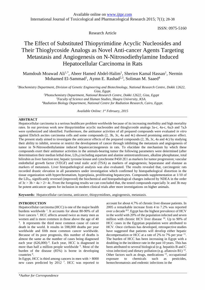
rates. In our previous work new thiopyrimidine acyclic nucleosides and thioglycoside analogs 3a-c, 4a-c, 6a,b and 7a,b
were synthesized and identified. Furthermore, the antitumor activities of all prepared compounds were evaluated in vitro
against Ehrlich ascites carcinoma cells and some compounds (2, 3b, 3c, 4a and 4c) showed promising anticancer effect.
The present study aimed to investigate the anticancer effects of the prepared compounds (2, 3b, 3c, 4a and 4c) by studying
their ability to inhibit, reverse or restrict the development of cancer through inhibiting the metastasis and angiogenesis of
tumor in N-Nitrosodiethylamine induced hepatocarcinogenesis in rats. To elucidate the mechanism by which these
compounds exert their antitumor activities in the animals-bearing tumor the following parameters were determined (after
determination their median lethal dose, LD50) including aspartate and alanine aminotransferases, alkaline phosphatase, total
bilirubin as liver function test; hepatic tyrosine kinase and cytochrome P450 2E1 as markers for tumor progression; vascular
endothelial growth factor (VEGF) and total sialic acid (TSA) as markers of angiogenesis; heparanase and elastase as
markers of metastasis. Liver histopathological analysis was also evaluated. The results revealed that, carcinogenic rats
recorded drastic elevation in all parameters under investigation which confirmed by histopathological distortion in the
tissue organization with hyperchromatism, hyperplasia, proliferating hepatocytes. Compounds supplementation at 1/10 of
the LD50, significantly reversed (improved) the biochemical and histopathological changes induced by NDEA in the order
of 3c > 3b > 4a > 2> 4c. From the foregoing results we can concluded that, the tested compounds especially 3c and 3b may
be potent anticancer agents for inclusion in modern clinical trials after more investigations on higher animals.
Keywords: Hepatocellular carcinoma, anticancer, thiopyrmidines, angiogenesis, metastasis.
INTRODUCTION
Hepatocellular carcinoma (HCC) is one of the major health
burdens worldwide 1. It accounts for about 80-90% of all
liver cancers 2. HCC affects around twice as many men as
women and is more common in those above the age of 40
3. It represents the third most common cause of cancer
death in the world. It results in 598,000 deaths per year
worldwide and fifth most common cancer worldwide.
Because of its poor prognosis, this number of deaths is
almost the same as the number of cases being diagnosed
each year (626,000) 4. Each year, HCC is diagnosed in
more than half a million people worldwide 5. Most of the
burden of the disease (85%) is borne in developing
countries 6.
In Egypt, HCC is third among cancers in men with > 8000
new cases predicted by 2012 7. HCC was reported to
account for about 4.7% of chronic liver disease patients. In
2005 a remarkable increase from 4 to 7.2% was reported
over a decade 8,9. Egypt has the highest prevalence of HCV
in the world with 20% of the population infected and seven
million with chronic HCV liver disease 10. Up to 90% of
HCC cases in the Egyptian population were attributed to
HCV. Once cirrhosis has developed, retrospective studies
have suggested that patients will develop either hepatic
decompensation or HCC at a rate of 2% to 7% per year 11.
The burden of HCC has been increasing in Egypt with a
doubling in the incidence rate in the past 10 years. This has
been attributed to several biological (e.g. hepatitis B and C
virus infection) and dietary pollution (e.g. aflatoxin B1) 12.
Other factors such as drugs, medications 13, occupational
exposure to chemicals such as pesticides,
hemochromatosis and endemic infections in the
Ali et.al. / The Effect of Substituted Thiopyrimidine Acyclic Nucleosides…
IJPTR, Volume 7, Issue 1, February 2015- March 2015, 28-38
Pag
e29
Table 1. In vivo the median lethal dose (LD50) of the
synthesized fused pyrimidine and thiopyrimidine
nucleoside analogs
Compounds LD50 µg/kg b.w.
2 132
3b 108
3c 80
4a 120
4c 160
community, such as schistosomiasis, may have additional
roles in the etiology or progression of the disease 14,15.
Pyrimidines have been recognized as important
heterocyclic compounds due to their diverse biological
activities such as Tie-2 kinase inhibitors, HIV-1 inhibitor,
antimalarial, secretive adenosine A1 receptor antagonist,
antibacterial, anticancer, analgesic, cardiovascular,and
antiallergic activities 16. The thio analogues of pyrimidine
bases, including 2-thiouracil, are minor components of t-
RNA. Their S-, N- or S,N -disubstituted analogs have
shown therapeutic properties, especially antiviral,
antithyroid and antitumor activities due to their
incorporation into polynucleic acids and therefore act as
potential inhibitors of protein and polynucleic acid
syntheses. On the other hand, nucleoside analogs are
structurally, metabolically, and pharmacodynamically
related agents that have diverse biological actions and
Figure 1: Synthesis route of substituted thiopyrimidine derivatives 2–7.

Ali et.al. / The Effect of Substituted Thiopyrimidine Acyclic Nucleosides…
IJPTR, Volume 7, Issue 1, February 2015- March 2015, 28-38
Pag
e30
therapeutic effects including antiviral and antitumor
activities. Furthermore, the glycosylthio heterocycles and
acyclic nucleoside analogues including modifications of
both the acyclic glycon and aglycon parts have stimulated
extensive research as biological inhibitors 16.
Figure 2: Represents the histological changes in liver of
control (A); NDEA-treated rats (B-D) and cancer-
bearing rats after the treatment with the prepared new
thiopyrimidine acyclic nucleosides and thioglycoside
analogs. Histological sections were prepared from
compound 2 (E), compound 3b (F), compound 3c (G),
compound 4a (H), and compound 4c (I). Sections were
stained with hematoxylin and eosin (original
magnification x250).
It is now well established that solid tumor growth is
critically dependent on the growth of new vessels from
preexisting blood vessels surrounding the tumor, a process
called angiogenesis 17. On the basis of this finding, the
development of drugs that inhibit angiogenesis has become
an attractive approach to cancer therapy 18. In addition,
metastasis of cancer cells to distant sites is one of the major
deciding factors in cancer outcome. In fact, prognosis of
cancer is mainly determined by the invasiveness of the
tumors and its ability to metastasize. There is a cascade of
events leading to the metastasis of tumors. These include
separation from the primary site, circulation through blood
or lymph, adhesive to the basement membrane (composed
mainly of heparan sulfate, elastin, and collagen), invasion
and proliferation at distant sites 19. Any compound which
can inhibit one of the steps in the cascade will be useful in
the inhibition of tumor metastasis and tumor growth.
Owing to the above facts, the aim of the present work is to
study the anticancer effect of previously synthesized
thiopyrimidine acyclic nucleosides and thioglycoside
analogs which gave good anticancer effect in vitro (2, 3b,
3c, 4a and 4c) 16 for inhibiting, reversing or restricting the
development of cancer and inhibition of metastasis and
angiogenesis of tumor in the experimental animals
carrying liver cancer induced by N-Nitrosodiethylamine
(NDEA) by studying different biochemical and
histological investigation methods.
MATERIALS AND METHODS
Animals
The animal care and handling was done according to the
guidelines set by the World Health Organization, Geneva,
Switzerland, and according to approval from the ethics
committee for animals care at the National Research
Centre, Egypt (ethic No. 10-230). Adult male Sprague-
Dawley rats (180±20 g, body weight), were purchased
from the animal house of National Research Centre, Egypt.
The animals were housed under standard laboratory
conditions (constant temperature 25-27 °C, with 12 h
light/dark cycle) during the experimental period. The rats
were provided with tap water and commercial diets. The
Table 2. Effect of the synthesized fused pyrimidine and thiopyrimidine nucleoside analogs on serum AST, ALT, ALP
activities and bilirubin level in different studied groups.
Groups AST
(U/L)
ALT
(U/L)
ALP
(U/L)
Bili
(mg/dl)
Control 56.00±5.20 38.00±3.70 110.00±9.30 1.40±0.11
NDEA 110.00±7.80a 95.00±7.00a 215.00±15.70a 2.90±0.20a
2 88.70±7.90 a ,b 80.00±7.0 a ,b 190.00±16.0a ,b 2.70±0.21 a
3b 65.40±7.4 a ,b 63.00±6.11a ,b 120.80±13.4a ,b 1.88±0.21 a ,b
3c 58.00±5.2 b 42.00±4.00 b 100.00±11.0 b 1.50±0.11 b
4a 85.00±7.80 a ,b 75.00±5.90 a ,b 175.0±15.0 a ,b 2.50±0.18 a ,b
4c 95.40±8.60 a 85.00±8.14 a 190.90±17.20a 2.75±0.22 a
Results expressed as Mean ± S.E. a Significantly different from normal control at p < 0.05. b Significantly different from NDEA - treated rats at p < 0.05.
Ali et.al. / The Effect of Substituted Thiopyrimidine Acyclic Nucleosides…
IJPTR, Volume 7, Issue 1, February 2015- March 2015, 28-38
Pag
e31
rats were acclimatized to laboratory condition for 10 days
before commencement of the experiment.
In Vivo the cytotoxicity of prepared thiopyrimidine acyclic
nucleosides and thioglycoside analogs
The median lethal doses (LD50) of the previously prepared
thiopyrimidine acyclic nucleosides and thioglycoside
analogs (2, 3b, 3c, 4a, and 4c) (Figure 1) was determined
in vivo according to Ghosh 20. Briefly, adult male Sprague-
Dawley rats were randomly divided into groups of 10 per
group. Each group was separately administrated once daily
for a period of 4 weeks with doses ranging from 0-500
µg/kg b.w. of the compounds intraperitoneal (i.p.) in a
value of 1 mL/kg body weight. Control animals received
the vehicle alone. The animals were then provided with
food and water immediately after the administration. The
mortality of the animals was observed up to one month
post-treatment. The
LD50 of the prepared compounds was calculated by using
a computer program of probit analysis.
Experimental design
N-Nitrosodiethylamine (NDEA) and carbon tetrachloride
(CCl4) were purchased from Sigma Chemical Co. (St.
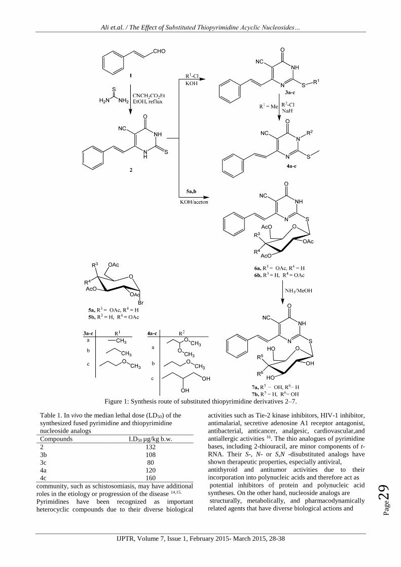
Figure 3: Effect of treatment with the synthesized thiopyrimidine acyclic nucleosides and thioglycoside analogs (2, 3b,
3c, 4a and 4c) on the level of hepatic Cytochrome P450 2E1 (CYP 2E1). Data were expressed as mean ± S.E., a and b
is significant difference from control and NDEA - treated rats respectively at (p < 0.05).
Figure 4: Effect of treatment with the synthesized thiopyrimidine acyclic nucleosides and thioglycoside analogs (2, 3b,
3c, 4a and 4c) on the expression of hepatic tyrosine kinase (TRK). Data were expressed as mean ± S.E., a and b is
significant difference from control and NDEA - treated rats respectively at (p < 0.05).
0
20
40
60
80
100
120
140
160
180
200
220
240
Th
e le
vel
of
CY
P 2
E1
(ng/m
g p
rote
in)
Control NDEA Compound 2 Compound 3b
Compound 3c Compound 4a Compound 4c
a,b
a,b
a,b
a,b
a
b
0255075
100125150175200225250275300325350375400425450
Th
e le
vel
of
TR
K
(pg/m
g p
rote
in)
Control NDEA Compound 2 Compound 3b
Compound 3c Compound 4a Compound 4c
a
a,b
a,b
a,b
a,b
a,b
Ali et.al. / The Effect of Substituted Thiopyrimidine Acyclic Nucleosides…
IJPTR, Volume 7, Issue 1, February 2015- March 2015, 28-38
Pag
e32
Louis, MO, USA). NDEA was dissolved in saline and
injected in a single dose (200 mg/kg, i.p.) to initiate hepatic
carcinogenesis, while CCl4 was used in a single dose (2
mL/kg) by gavage as 1:1 dilution in corn oil to stimulate
liver cell proliferation and regeneration 21. The experiment
continued for 32 weeks.
Adult male Sprague-Dawley rats were divided into groups
with 10 animals in each group. Group 1 (untreated control
group): animals were fed on a standard diet and given
water throughout the course of the experiment. Group 2
(NDEA treated group): Rats were injected with a single
dose of NDEA (200 mg/kg, i.p.) and 2 week later received
a single dose of CCl4 (2 mL/kg) by gavage as 1:1 dilution
in corn oil for 32 weeks. Group 3 (NDEA and synthesized
compounds group): Rats from Group 2 were treated daily
with synthesized compounds (2, 3b, 3c, 4a and 4c) by i.p.
treatment at dose of 1/10 of their LD50 values and
continued for 32 weeks.
At the end of the treatment protocol (32 weeks), animals
were anesthetized with ether and blood samples were
drawn from the orbital venous plexus. Serum was
separated by centrifugation for 5 min at 1500 g and stored
at -20C until analysis.
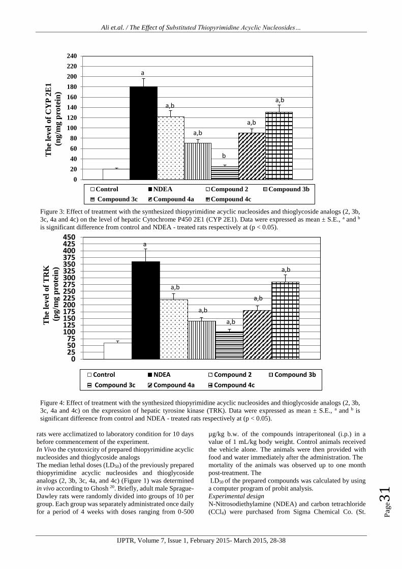
Figure 5: Effect of treatment with the synthesized thiopyrimidine acyclic nucleosides and thioglycoside analogs (2, 3b,
3c, 4a and 4c) on the level of VEGF. Data were expressed as mean ± S.E., a and b is significant difference from control
and NDEA - treated rats respectively at (p < 0.05).
Figure 6: Effect of treatment the synthesized thiopyrimidine acyclic nucleosides and thioglycoside analogs (2, 3b, 3c, 4a
and 4c) on the level of sialic acid (TSA). Data were expressed as mean ± S.E., a and b is significant difference from
control and NDEA - treated rats respectively at (p < 0.05).
0
50
100
150
200
250
300
350
400
450
500T
he
level
of
VE
GF
(pg/m
l)
Control NDEA Compound 2 Compound 3b
Compound 3c Compound 4a Compound 4c
aa
a,b
b
a,ba,b
02.5
57.510
12.515
17.520
22.525
27.530
Th
e le
vel
of
TS
A
(µg/m
l)
Control NDEA Compound 2 Compound 3b
Compound 3c Compound 4a Compound 4c
a
a,b
a,b
b
a
a,b

Ali et.al. / The Effect of Substituted Thiopyrimidine Acyclic Nucleosides…
IJPTR, Volume 7, Issue 1, February 2015- March 2015, 28-38
Pag
e33
All animals were sacrificed by decapitation and their livers
were rapidly excised, weighed, washed with saline and
blotted with a piece of filter paper. Portion of the liver was
immediately fixed in 10% formalin for histological
analysis according to Conn et al. 22 using a standard
method of hematoxylin and eosin. Another portion of liver
was homogenized using a Branson Sonifier (250 VWR
Scientific, Danbury, Conn., USA) in cold sucrose buffer
(0.25 M). All the investigation will carry out in fresh 10%
homogenate. The freshly prepared homogenates were then
centrifuged at 30,000 xg for 30 min at 4C to obtain the
supernatant, which used for biochemical assays and the
protein level was determined as described by Lowry et al.
23.
Aspartate and alanin-aminotransferase (AST and ALT),
alkaline phosphatase (ALP) activities as well as the level
of total bilirubin (Bili) were determined
spectrophotometrically according to the manufacturer's
instructions, using reagent kits obtained from Biomerieux
(France(.
Cytochrome P450 2E1 (CYP 2E1) assay
The effect of synthesized compounds on the expression of
Cytochrome P450 2E1 (CYP 2E1) was determined in
tissue homogenates based on a double-antibody sandwich
enzyme-linked immunosorbent assay (ELISA) of
Cytochrome P450 2E1 kit purchase from Cloud-Clone
Crop. (Houston, TX 77082, USA). The concentration of
CYP 2E1 in the samples is determined by comparing the
O.D. of the samples to the standard curve.
Tyrosine kinase assay
The effect of synthesized compounds on the expression of
tyrosine kinase (TRK) was determined in the tissue
homogenates based on a double-antibody sandwich
enzyme-linked immunosorbent assay (ELISA) of tyrosine
kinase kit purchase from Glory Science Co., Ltd (Del Rio,
TX 78840, USA) according to the manufacturer’s
instructions. The chroma of color and the concentration of
the human TRK of sample were positively correlated and
the optical density was determined at 450 nm. The level of
TRK in samples was calculated as triplicate determinations
from the standard curve
Estimation of VEGF concentration
VEGF concentration in the serum was determined using
ELISA kit obtained from Koma Biotech Inc., Korea. This
assay depends on binding VEGF antigen to a specific
immobilized antibody. The formed immune complex binds
to avidin-peroxidase conjugate, and a color developed in
proportion to the amount of VEGF bound which was
measured at 450 nm.
Total sialic acid (TSA)
TSA was estimated in the serum by periodate-resorcinol
microassay as described by Surangkul et al. 24. The
principle of this method is based on a periodate-resorcinol
reaction with sialic acid found in the sample. The
absorbance was measured at 620 nm immediately by a
microtiter plate reader then N-acetyl neuraminic acid
standard curve was used to calculate TSA concentration.
Determination of heparanase (HPSE) activity
Heparanase activity in tissue homogenates was determined
based on a double-antibody sandwich enzyme-linked
immunosorbent assay (ELISA) of heparanase kit purchase
from Glory Science Co., Ltd (Del Rio, TX 78840, USA)
according to the manufacturer’s instructions. The kit assay
HPSE activity in the sample, use purified HPSE to coat
Figure 7: Effect of treatment with the synthesized thiopyrimidine acyclic nucleosides and thioglycoside analogs (2, 3b,
3c, 4a and 4c) on the activity of heparanase and elastase. Data were expressed as mean ± S.E., a and b is significant
difference from control and NDEA - treated rats respectively at (p < 0.05).
0
0.5
1
1.5
2
2.5
3
3.5
4
4.5
5
5.5
6
6.5
7
7.5
Heparanase Elastase
Act
ivit
y (U
/mg
pro
tein
)
a,b
a,b
a
a,b
a
a
aa a,b
a,ba,b a

Ali et.al. / The Effect of Substituted Thiopyrimidine Acyclic Nucleosides…
IJPTR, Volume 7, Issue 1, February 2015- March 2015, 28-38
Pag
e34
mictotiter plat wells, make solid-phase antibody, then add
HPSE to wells, combined HPSE which with enzyme
labeled, become antibody-antigen-enzyme-antibody
complex, after washing completely, add substrate solution,
the color change is measured spectrophotometrically at a
wavelength of 450 nm. The activity of HPSE in the
samples is then determined by comparing the absorbance
of the samples to the standard curve. The activity was
determined as U/mg protein.
Estimation of elastinolytic activity
The elastase activity is determined in the tissue
homogenates by its catalytic effect on the N-succinyl-
trialanyl-p-nitroanilide substrate releasing p-nitroaniline
which is measured photometrically at 405 nm 25. The
elastase activity was determined as U/mg protein.
Statistical analysis
The results were reported as Mean ± Standard error (S.E.).
Statistical differences were analyzed by one way ANOVA
test followed by student's t test wherein the differences
were considered to be significant at p < 0.05.
RESULTS
In Vivo the cytotoxicity of the synthesized compounds
The LD50 (the median lethal dose resulted in 50% mortality
of the animals) of each compound was determined.
Compounds 2, 3b, 3c, 4a, and 4c showed marked acute
toxicity (Table 1). The concentrations required by 2, 3b,
3c, 4a, and 4c for 50% mortality of the animals were found
to be 132, 108, 80, 120 and 160 µg/kg body weight,
respectively. From the foregoing results it is clear that
compounds 3b and 3c were the best compounds in this
series.
The effect of the synthesized fused pyrimidine and
thiopyrimidine nucleoside analogues on liver tissue
NDEA-treated rats showed significant (p<0.05) increase in
serum AST, ALT and ALP activities, along with
significant (p<0.05) increase in total bilirubin level
compared to control. The administration 2, 3b, 3c, 4a, and
4c at a dose of 1/10 of the LD50 values in the NDEA-treated
rats resulted in normalization in AST, ALT and ALP
activities as well as the total bilirubin level compared to
NDEA-treated group (Table 2). The tested compounds has
resulted in decreasing in the level of liver function test
follows the order 3c > 3b > 4a > 2> 4c. It is clear that, 3b
and 3c were the best compounds in this series.
In this study histological examination of rat liver sections
was consistent with the results obtained from biochemical
studies. Liver of control animals as presented in (Fig. 2a)
revealed normal architecture of hepatic strands around the
central veins. The liver showed intact hepatocytes with
normal sinusoids in between. The hepatic cells are
polygonal in shape with one or two rounded nuclei. Liver
of rats treated with NDEA alone showed distortion in the
tissue organization with hyperchromatism, hyperplasia,
proliferating hepatocytes (Fig. 2b), both hepatic and portal
with significant tumor thrombi within portal vessels, tumor
cells are slightly larger have more irregular nuclei and
numerous mitotic figures with malignant nuclei (Fig. 2c).
Some section showed megalocytosis, hyperchromatic
nuclei as well as nuclear vacuolization and nuclear
prominence, dissolution of hepatic cords which appeared
as empty vacuoles aligned by strands of necrotic
hepatocytes (Fig. 2d). Liver of the NDEA-rats treated with
compound 2 showed diffuse hepatocyte necrosis with
congested central vein (Fig. 2e). Liver sections from rats
treated with compound 3b exhibited improved the
hepatocellular architecture with more regular and less
altered hepatocytes when compared to group treated with
NDEA alone (Fig. 2f). Liver sections from rats treated with
compound 3c showed normal appearance with normal
appearing hepatocytes cords (Fig. 2g). Liver section from
rats treated with compound 4a exhibited moderate
ballooning degeneration of hepatic cells; focal fatty change
of hepatocytes (Fig. 2h). Liver sections from rats treated
with compound 4c showed areas of aberrant hepatocellular
congested central vein with moderated focal hepatocyte
necrosis. Severe infiltration of portal tract by inflammatory
cells Hepatocytes showed ground glass appearance (fig.
2i).From the foregoing results it is clear that, there are
improvement in the histological changes in the order of 3c
> 3b > 4a > 2> 4c.
Effect of prepared compounds on cytochrome P450 (CYP)
2E1 and tyrosine kinase expression
The effect of the prepared compounds on both CYP 2E1
and TRK which implicated in cancer growth was
illustrated in figure 3 and 4. The expressions of both CYP
2E1 and TRK were significantly increased in NDEA-
treated as compared to normal control. While the
administration of compounds 2, 3b, 3c, 4a, and 4c at a dose
of 1/10 of the LD50 values in the NDEA -treated rats
resulted in significantly inhibitory potential against both
CYP 2E1 and TRK for all the compounds comparing with
the NDEA group. Compounds 3c and 3b were the most
potent inhibitor against CYP 2E1 and TRK expression.
Effect of prepared compounds on VEGF and TSA
The effect of the prepared compounds on VEGF and TSA
as markers of angiogenesis was illustrated in figure 5 and
6. In this study, it was found that the levels of both VEGF
and TSA in the NDEA-treated group was very highly
significant increase as compared to control group while the
treatment with 2, 3b, 3c, 4a, and 4c in the NDEA-treated
rats, causes decrease in the level of VEGF and TSA as
compared with NDEA-treated rats, while the VEGF and
TSA level showed significant decrease in compounds 3c
and 3b -treated group reaching to its control level.
Effect of prepared compounds on heparanase and elastase
activity
In the present study the activity of heparanase and elastase
as marker for metastasis of tumor was investigated, the
results showed that the activity of heparanase and elastase
enzymes was very highly significantly increased in
NDEA-treated group. The treatment with 2, 3b, 3c, 4a, and
4c in the NDEA -treated rats, resulted in decrease in the
activity of heparanase and elastase enzymes as compared
with NDEA-treated group especially compounds 3c and 3b
(Figure 7).
DISCUSSION
In our previous work new thiopyrimidine acyclic
nucleosides and thioglycoside analogs 3a-c, 4a-c, 6a,b and
Ali et.al. / The Effect of Substituted Thiopyrimidine Acyclic Nucleosides…
IJPTR, Volume 7, Issue 1, February 2015- March 2015, 28-38
Pag
e35
7a,b were synthesized starting with cinnamaldehyde, ethyl
cyanoacetate and thiourea in ethanol. Furthermore, the
antitumor activities of all prepared compounds were
evaluated in vitro against Ehrlich ascites carcinoma cells
and some compounds (2, 3b, 3c, 4a and 4c) showed
promising anticancer effect 16.
Experimental liver cancer in rodents induced by NDEA, an
environmental and dietary hepatocarcinogen 26, has been
considered as one of the best characterized experimental
models of HCC, allowing the screening of potential
anticancer compounds on various phases of neoplastic
transformation and development 27. NDEA-induced
preneoplastic foci and preneoplastic and neoplastic nodule
formation in rodents closely mimics HCC development in
humans 28. Recently, a cross-species comparison of gene
expression patterns has established that NDEA-induced
liver tumors in rodents closely resemble a subclass of
human HCC 28, which allows extrapolating potential
chemopreventive effects of a candidate agent in clinical
setting.
In the present study, we have investigated the preventive
effect of prepared new thiopyrimidine acyclic nucleosides
and thioglycoside analogs including 2, 3b, 3c, 4a and 4c on
the appearance of early hepatic preneoplastic events,
utilizing a two-stage model of hepatocarcinogenesis
initiated with NDEA and promoted by carbon
tetrachloride. An understanding of how cancer may be
prevented is one of the key objectives of the recent
researches. This can be achieved to some extent by using
chemopreventive agents, naturally occurring or synthetic,
that can suppress or prevent the processes of tumor
development. Therefore, it is essential to identify agents as
well as to evaluate their efficacy and to elucidate their
mechanisms of action. In the present study, serum obtained
from tumor bearing rats showed significant increase in
AST, ALT and ALP activities along with significant
increase in total bilirubin compared to control animals. The
elevation of these enzyme activities was indicative of the
toxic effect of NDEA on the liver tissue associated with
sever histological distortions (figure 2b-d). It is known that
N-nitroso compounds act as strong carcinogens in various
mammals including primates 29. NDEA has been shown to
be metabolized by cytochrome P-450 IIE1 (CYP 2E1) to
its active ethyl radical metabolite, which could interact
with DNA causing mutation and carcinogenesis 30.
Administration of prepared new thiopyrimidine acyclic
nucleosides and thioglycoside analogs including 2, 3b, 3c,
4a and 4c to NDEA treated rats showed restoration of AST,
ALT and ALP activities and total bilirubin level towards
normal especially in group treated with 3b and 3c. Such
reverse in serum enzyme activities could be attributed to
the ability of these compounds to inhibit CYP 2E1
expression (as shown in figure 3), presumably by serving
as a competitive inhibitor, leading to a decrease in the
formation and/or bioactivation of these nitrosamines. The
improvement in the biochemical parameters was
accompanied with improvement in the histopathological
abnormalities especially in 3b and 3c (Figure, 2f and g)
which showing hepatocytes maintaining near-normal
architecture while the other compounds revealed moderate
improvement of hepatic histopathology over NDEA group.
Tyrosine kinases play a critical role in the modulation of
growth factor signaling. Activated forms of these enzymes
can cause increases in tumor cell proliferation and growth,
induce antiapoptotic effects, and promote angiogenesis
and metastasis. In addition to activation by growth factors,
protein kinase activation by somatic mutation is a common
mechanism of tumorigenesis 31. In consistent with the
above fact, our results showed that highly significant
increase in the expression of TRK in NDEA-treated group,
while the administration of prepared compounds (2, 3b, 3c,
4a and 4c) in the NDEA-treated rats resulted in
significantly inhibitory potential against TRK for all the
tested compounds comparing with the NDEA group.
Compounds 3b and 3c were the most potent inhibitor
against TRK expression.
Tumor cell transformation is a multistage process. An In
Situ tumor after a period of time abruptly sparks the
formation of new blood vessels from the preexisting
vasculature a process termed as angiogenesis or
neovascularization. Tyrosine kinase plays an important
role in this process 32. This process though occurs normally
during embryonic development, female reproductive cycle
or wound healing is found as a crucial step in tumor
transition from benign to malignant form, capable of
spreading throughout the body 33. Antiangiogenic drugs
stops new vessels from forming around a tumor and break
up the existing network of abnormal capillaries that feeds
the cancerous mass, thus shrinks the tumor by limiting
blood supply 34. To study the anti-angiogenesis effect of
the prepared compounds we measured the level of VEGF
and our results showed that there was over production of
VEGF after administration NDEA. This is in concurrently
with Torimura et al.35 who stated that VEGF was over
expressed intoxicated with NDEA. Inhibition of tumor
growth by neutralization of VEGF has been verified by
treatment with the prepared compounds which showed
decreased amount of VEGF in the cancer-bearing animals,
thereby inhibiting the formation of new blood vessel and
tumor growth in the order of 3c > 3b > 4a > 2> 4c.
It has been proposed that sialic acid appears to be highly
sensitive marker for the progression of tumor growth and
its angiogenesis 36. Previously, Rachesky et al. 37 have
been reported an increased level of glycoproteins in
animals exposed to carcinogen NDEA. In the present
study, serum total sialic acid level was estimated and found
to be significantly elevated (p<0.05) in NDEA-treated rats
(figure 6). The exact cause in rising of sialic acid levels in
tumorigenesis is not known, however, various theories are
attributed to such increment as: alterations in the cell
surface during cell transformation, stimulation of the liver
by tumor growth to synthesize glycoproteins or increased
glycosylation 38. Also, neoplastic transformation could
lead to sialic acid elevation through the shedding of sialic
acid from the tumor cell surface or possibly as a product of
the tumor itself 39. Administration of prepared compounds
leads to significant decrease in TSA level especially in 3b
and 3c groups. Consequently, this suggests that these
Ali et.al. / The Effect of Substituted Thiopyrimidine Acyclic Nucleosides…
IJPTR, Volume 7, Issue 1, February 2015- March 2015, 28-38
Pag
e36
compounds played an important role against NDEA-
induced hepatocarcinogenesis by maintaining TSA status.
In the present study, results showed that the activities of
heparanase and elastase enzymes were very highly
significantly increased in NDEA-treated group as
compared with control. The treatment with prepared
compounds (2, 3b, 3c, 4a and 4c) resulted in decrease in
the activity of both enzymes especially 3b and 3c (Figure,
7). In the meantime, several studies suggested that
targeting the activity of heparanase and elastase might be
a beneficial antitumor therapy for liver cancer 40. Few
studies on the potency of heparanase as a marker for HCC
were found in the literature 41. Heparanase is a heparan
sulfate (HS) degrading endoglycosidase participating in
extracellular matrix degradation and remodeling. Apart of
its well-characterized enzymatic activity, heparanase was
noted to exert also enzymatic-independent functions,
which include enhanced adhesion of tumor-derived cells
and primary T-cells 42. Heparanase seems to modulate two
critical systems involved in tumor progression, namely
vascular epidermal growth factor (VEGF) expression and
epidermal growth factor receptor (EGFR) activation.
Neutralizing heparanase enzymatic and non-enzymatic
functions is therefore expected to profoundly affect tumor
growth, angiogenesis and metastasis 43. A large number of
publications clearly link heparanase expression to the
process of tumorigenesis in a wide number of cancers,
which were reviewed by Zhang et al. 44. Collectively, they
suggest that heparanase plays a fundamental role in
sustaining the pathology of malignant diseases and
therefore it may provide a potential target for anti-cancer
therapy 45.
Elastase is another broad-range proteolytic enzyme
thought to be a tumor promoter involved in increasing
tumor cell invasiveness by facilitating cell motility and
transendothelial migration as it has the ability to degrade
basement membrane and ECM glycoproteins such as
elastin, fibronectin, as well as adhesive molecules and
junctional cadherins 46. Moreover, elastase considered to
be the only protease that is able to degrade insoluble
elastin, a structural component of elastic tissues such as
blood vessel, skin, lung, liver and breast tissues 47.
Furthermore, Taniguchi et al.48 postulated that increased
elastase destroy the barrier between tumor and the local
circulatory system, either lymphatic or hematogenous, and
result in at least loco-regional metastases.
In conclusion, the results of our study clearly indicate a
beneficial effect of prepared new thiopyrimidine acyclic
nucleosides and thioglycoside analogs including 2, 3b, 3c,
4a, and 4c on chemically-induced rat liver tumorigenesis.
To our knowledge, this is the first experimental evidence
of the chemopreventive activity of thiopyrimidine acyclic
nucleosides and thioglycoside analogs. Under our
experimental conditions, the tested compounds especially
3b and 3c exert their antitumor activity through affecting
the process of angiogenesis and metastasis and may be
potent anticancer agents for inclusion in modern clinical
trials after more investigations on higher animals.
REFERENCES
1. Lyer P, Zekri AR, Hung CW, Schiefelbein E, Ismail K,
Hablas A, Seifeldin IA and Soliman AS (2010)
Condcordance of DNA methylation pattern in plasma
and tumor DNA of Egyptian hepatocellular carinoma
patients. Experimental and Molecular Pathology
88:107-111.
2. Foster GR (2000) Hepatitis C. Billiere’s Ciinical
Gastroenterology 14:327-339.
3. Thompson-Coon J, Rogers G, Hewson P, Wright D,
Anderson R, Cramp M, Jackson S, Ryder S, Price A
and Stein K (2007) Surveillance of cirrhosis for
hepatocellular carcinoma: Systematic review and
economic analysis. Health Technology Assessment
11:1-206.
4. But DYK, Lai CL and Yuen MF (2008) Natural history
of hepatitis-related hepatocellular carcinoma. World J
Gastroenterol 14:1652-1656.
5. El-Serag HB (2011) Hepatocellular carcinoma. New
England Journal of Medicine 365(12):1118–1127.
6. Harnois DM (2012) Hepatitis C virus infection and the
rising incidence of hepatocellular carcinoma.
Mayo Clinic Proceedings 87(1):7–8.
7. Ezzat S, Abdel-Hamid M, Eissa SA, Mokhtar N, Labib
NA, El-Ghorory L, Mikhail NN, Abdel-Hamid A,
Hifnawy T, Strickland GT and Loffredo CA (2005)
Associations of pesticides, HCV, HBV, and
hepatocellular carcinoma in Egypt.
International Journal of Hygiene and
Environmental Health 208:329-339.
8. El-Zayadi AR, Badran HM, Barakat EM, Attia Mel-D,
Shawky S, Mohamed MK, Selim O and Saeid A (2005)
Hepatocellular carcinoma in Egypt: A single center
study over a decade. World J Gastroenterol 11:5193-
5198.
9. Hussein MM, Ibrahim AA, Abdella HM, Montasser IF
and Hassan MI (2008) Evaluation of serum squamous
cell carcinoma antigen as a novel biomarker for
diagnosis of hepatocellular carcinoma in Egyptian
patients. Indian Journal of Cancer 45:167-172.
10. Eassa S, Eissa M, Sharaf SM, Ibrahim MH and
Hassanein OM (2007) Prevalence of hepatitis C virus
infection and evaluation of a health education program
in el-ghar village in zagazig, Egypt. Journal of
Egyptian Public Health Association 82:379-404.
11. Goldman R, Ressom HW, Abdel-Hamid M, Goldman
L, Wang A, Varghese RS, An Y, Loffredo CA, Drake
SK, Eissa SA, Gouda I, Ezzat S and Moiseiwitsch FS
(2007) Candidate markers for the detection of
hepatocellular carcinoma in low-molecular weight
fraction of serum. Carcinogenesis 28:2149-2153.
12. Van Rensburg SJ, Cook-Mozaffari P, Van Schalkwyk
DJ, Van der Watt JJ, Vincent TJ and Purchase IF (1985)
Hepatocellular carcinoma and dietary aflatoxin in
Mozambiqe and Transkei. British Journal of Cancer
51:713-726.
13. Hassan MM, Hwang LY, Hatten CJ, Swaim M, Li D,
Abbruzzese JL, Beasley P and Patt YZ (2002) Risk
factors for hepatocellular carcinoma: Synergism of
alcohol with viral hepatitis and diabetes mellitus.
Hepatology 36:1206-1213.

Ali et.al. / The Effect of Substituted Thiopyrimidine Acyclic Nucleosides…
IJPTR, Volume 7, Issue 1, February 2015- March 2015, 28-38
Pag
e37
14. Bradbear RA, Bain C, Siskind V Schofield FD, Webb
S, Axelsen EM, Halliday JW, Bassett ML and Powell
LW (1985) Cohort study of internal malignancy in
genetic hemochromatosis and other chronic
nonalcoholic liver diseases. Journal National Cancer
Institute 313:1256-1262.
15. Anwar WA, Khaled HM, Amra HA, El-Nezami H and
Loffredo CA (2008) Changing pattern of hepatocellular
carcinoma (HCC) and its risk factors in Egypt:
Possibilities for prevention. Mutation Research
659:176-184.
16. El-Sayed WA, Rashad AE, Awad SM and Ali MM
(2009) Synthesis and in Vitro antitumor activity of new
substituted thiopyrimidine acyclic nucleosides and
their thioglycoside analogs. Nucleosides, Nucleotides
and Nucleic Acids 28:261-274.
17. Zetter BR (1998) Angiogenesis and tumor
metastasis. Annual Review of Medicine 49:407-424.
18. Sunassee K and Vile R (1997) Hitting cancer where it
hurts. Current Biology 7: R282-R285.
19. Pantel K and Brakenhoff RH (2004) Dissecting the
metastatic cascade. National Review of Cancer 4:448-
456.
20. Ghosh MN (1984) Toxicity studies. In: Ghosh MN
(ed.), fundamentals of experimental pharmacology.
Scientific Book Agency. Calcutta, India.
21. Al-Rejaie SS, Aleisa AM., Al-Yahya AA., Saleh AB.,
Abdulmalik A, Amal GF, Al-Shabanah OA and
Mohamed MSA (2009) Progression of
diethylnitrosamine-induced hepatic carcinogenesis in
carnitine-depleted rats. World J Gastroenterol
15(11):1373-1380.
22. Conn HJ, Darrow MA and Emmel VM (1960) Staining
procedure used by biological stain commission, 2nd ed.,
Williams and Winlkins Co., Baltimore.
23. Lowry OH, Rosebrough NJ, Farr AL and Randall RJ
(1951) Protein measurement with the folin phenol
reagent. J Biol Chem 193:265-275.
24. Surangkul D, Pothacharoen P, Suttajit M and
Kongtawelert P (2001) A periodate-resorcinol
microassay for the quantitation of total sialic acid in
human serum. Chiang Mai Med Bull 40(3):111-1118.
25. Zay K, Loo S, Xie C, Devine DV, Wright J and Churg
A (1999) Role of neutrophils and alpha-1-antitrypsin in
coal- and silica-induced connective tissue breakdown.
Am J Physiol 276:L269-L279.
26. Gupta C, Vikram A, Tripathi DN, Ramarao P and Jena
GB (2010) Antioxidant and antimutagenic effect of
quercetin against DEN induced hepatotoxicity in rat.
Phytother Res 24(1):119-128.
27. Miao B, Li J, Fu X, Ding J and Geng M (2005) T-cell
receptor (TCR)/CD3 is involved in sulfated
polymannuroguluronate (SPMG)-induced T
lymphocyte activation. Int Immunopharmacol 5:1171-
1182.
28. Wu XZ and Chen D (2006) Effects of sulfated
polysaccharides on tumour biology. West Indian Med J
55(4):270-273.
29. Swenberg JA, Hoel DG and Magee PN (1991)
Mechanistic and statistical insight into the large
carcinogenesis bioassays on N-nitrosodiethylamine
and N-nitrosodimethylamine. Cancer Res 51:6409-
6414.
30. Anis KV, Rajesh Kumar NV and Kuttan R (2001)
Inhibition of chemical carcinogenesis by biberine in
rats and mice. J Pharm Pharmacol 53:763-768.
31. Krause DS and Van Etten RA (2005) Tyrosine kinases
as targets for cancer therapy. New England Journal of
Medicine 353:172-187.
32. Cohen P (1999) The development and therapeutic
potential of protein kinase inhibitors. Curr Opin Chem
Biol 3:459-465.
33. Kerbel RS (1997) A cancer therapy resistant to
resistance. Nature 390: 335-336.
34. Paul MK and Mukhopadhyay AK (2004) Tyrosine
kinase – Role and significance in cancer. Int J Med Sci
1(2):101-105.
35. Torimura T, Sata M, Ueno T, Kin M, Tsuji R, Suzaku
K, Hashimoto O, Sugawara H and Tanikawa K (1998)
Increased expression of vascular endothelial growth
factor is associated with tumor progression in
hepatocellular carcinoma. Hum Pathol 29:986-991.
36. Süer Gökmen S, Kazezoğlu C, Tabakoğlu E, Altıay G.,
Güngör Ö and Türe M (2004) Serum total sialic acid
levels in lung cancer patients of different histological
types with and no extrapulmonary metastases. Turk J
Biochem 29(4):262-267.
37. Rachesky MH, Hard GL and Glick MC (1983)
Membrane glycopeptides from chemically transformed
cells. Cancer Res 43:39-42.
38. Aranganathan S, Senthil K and Nalini N (2005) A case
control study of glycoprotein status in ovarian
carcinoma. Clinical Biochemistry 38:535-539.
39. Suresh K, Manoharan S, Panjamurthy K and Senthil N
(2007) Modifying effects of Annona squamosa on
glycoconjugates levels in 7,12-
dimethylbenz(a)anthracene induced hamster buccal
pouch carcinogenesis. J Med Sci 7:100-105.
40. Sanderson RD, Yang Y, Suva LJ and Kelly T (2004)
Heparan sulfate proteoglycans and heparanase-partners
in osteolytic tumor growth and metastasis. Matrix Biol
23:341-352.
41. Chen G, Dang YW, Luo DZ, Feng, ZB and Tang XL
(2008) Expression of heparanase in hepatocellular
carcinoma has prognostic significance: A tissue
microarray study. Oncol Res 17:183-189.
42. Nadir Y, Vlodavsky I and Brenner B (2008)
Heparanase, tissue factor, and cancer. Semin Thromb
Hemost 34:187-194.
43. Cohen-Kaplan V, Doweck I, Naroditsky I, Vlodavsky
I and Ilan N (2008) Heparanase augments epidermal
growth factor receptor phosphorylation: correlation
with head and neck tumor progression. Cancer
Research 68:10077-10085.
44. Zhang ZH, Chen Y, Zhao HJ, Ding J and Hou YT
(2007) Silencing of heparanase by siRNA inhibits
tumor metastasis and angiogenesis of human breast
cancer in vitro and in vivo. Cancer Biol Ther 6:587-595.
Ali et.al. / The Effect of Substituted Thiopyrimidine Acyclic Nucleosides…
IJPTR, Volume 7, Issue 1, February 2015- March 2015, 28-38
Pag
e38
45. McKenzie EA (2007) Heparanase: A target for drug
discovery in cancer and inflammation. Br J Pharmacol
151:1-14.
46. Zelvyte I, Stevens T, Westin U and Janciauskiene S
(2004) Alpha-1-antitrypsin and its C-terminal fragment
attenuate effects of degranulated neutrophil-
conditioned medium on lung cancer HCC cells in vitro.
Cancer Cell International 4:4-7.
47. Ginzberg HH, Cherapanov V, Dong Q, Cantin A,
McCulloch AG, Shannon PT and Downey GP (2001)
Neutrophil-mediated epithelial injury during
transmigration: Role of elastase. Am J Physiol
Gastrointest Liver Physiol 281:G705-G717.
48. Taniguchi K, Yang P, Jett J, Bass E, Meyer R, Wang
Y, Deschamps C and Liu W (2002) Polymorphisms in
the promoter region of the neutrophil elastase gene are
associated with lung cancer development. Clinical
Cancer Research 8:1115-1120.