the effect of dipyridamole on vascular cell-derived
TRANSCRIPT
JPET #89987
1
The Effect of Dipyridamole on Vascular Cell-Derived Reactive Oxygen Species
Subrata Chakrabarti, Olga Vitseva, David Iyu,
Sonia Varghese, and Jane E. Freedman
Whitaker Cardiovascular Institute and Evans Department of Medicine, Boston
University School of Medicine, Boston, MA (S.C., O.V., S.V., J.E.F.) and
Department of Physiology, University of Murcia Medical School, Murcia, Spain
(D.I.)
JPET Fast Forward. Published on July 26, 2005 as DOI:10.1124/jpet.105.089987
Copyright 2005 by the American Society for Pharmacology and Experimental Therapeutics.
This article has not been copyedited and formatted. The final version may differ from this version.JPET Fast Forward. Published on July 26, 2005 as DOI: 10.1124/jpet.105.089987
at ASPE
T Journals on A
pril 2, 2022jpet.aspetjournals.org
Dow
nloaded from

JPET #89987
2
Running title: Dipyridamole and Reactive Oxygen Species
Address all correspondence to: Jane E. Freedman, Boston University School of
Medicine, 715 Albany Street, W507, Boston, MA 02118
617-638-4022; 617-638-4066 (fax)
email:[email protected]
Number text pages: 31
Number of tables: 0
Number of figures: 6
References: 31
Total word count: 4433
Abstract word count: 189
Introduction word count: 257
Discussion Word count: 489
Nonstandard abbreviations: DCFDA, 2',7'-Dichlorodihydrofluorescein diacetate;
DPPH, 1,1 diphenyl-2-picrylhydrazyl; TRAP1-6, trombin receptor activating
peptide; Ferrozine, (3-(2-Pyridyl)-5,6-Bis(4-Phenylsulfonic Acid)-1, 2,4 triazine;
sCD40L, soluble CD40 ligand.
Recommended section assignment: Cardiovascular
This article has not been copyedited and formatted. The final version may differ from this version.JPET Fast Forward. Published on July 26, 2005 as DOI: 10.1124/jpet.105.089987
at ASPE
T Journals on A
pril 2, 2022jpet.aspetjournals.org
Dow
nloaded from

JPET #89987
3
ABSTRACT
Platelet and vascular stimulation leads to release of reactive oxygen
species (ROS) that are known to influence vascular reactivity and thrombosis.
Dipyridamole is a vasodilator and platelet inhibitor that has previously been
shown to have direct antioxidant properties. The antioxidant effects of
dipyridamole on vascular cell-derived ROS are not known, therefore,
dipyridamole was incubated with endothelial cells and platelets and cellular redox
status and release of endogenous ROS were assessed. Dipyridamole decreased
intracellular basal ROS generation from endothelial cells as measured by 2’,7’-
dichlorodihydrofluorescein diacetate (DCFDA) oxidation. Incubation of
endothelial cells with dipyridamole also attenuated t-butylhydroperoxide induced
oxidative stress. Using a redox sensitive fluorescent dye, dipyridamole improved
cellular activity after treatment with t-butylhydroperoxide. Incubation with
dipyridamole did not alter platelet release of nitric oxide or hydrogen peroxide but
significantly attenuated superoxide release. Using flow cytometry and confocal
microscopy, dipyridamole decreased platelet ROS generation. Dipyridamole also
suppressed platelet soluble CD40 ligand (sCD40L) release. In summary, at
therapeutically relevant concentrations, dipyridamole suppresses formation of
ROS in platelets and endothelial cells, and improves cellular redox status. These
data suggest that the redox-dependent properties of dipyridamole have a direct
effect on vascular cells.
This article has not been copyedited and formatted. The final version may differ from this version.JPET Fast Forward. Published on July 26, 2005 as DOI: 10.1124/jpet.105.089987
at ASPE
T Journals on A
pril 2, 2022jpet.aspetjournals.org
Dow
nloaded from

JPET #89987
4
INTRODUCTION
Dipyridamole is a platelet inhibitor that is primarily recognized as an
antithrombotic agent. In addition, through the generation of adenosine,
dipyridamole evokes vasodilation and through the combination of these
antiplatelet and vasodilator functions likely improves tissue perfusion. The
European Stroke Prevention Study 2, involving over 6600 patients with transient
ischemic attacks or stroke, demonstrated that treatment with dipyridamole was
as effective as low-dose aspirin in the reduction of stroke risk(Diener et al., 1996)
and combination therapy with dipyridamole and aspirin was more than twice as
effective as aspirin alone. Because dipyridamole is a weak direct platelet
inhibitor, clinical observations such as this suggest that dipyridamole may have
additional beneficial vascular effects.
Dipyridamole has been reported to have antioxidant properties(Iuliano et
al., 1989) but the direct effect on vascular cells is not known. Dipyridamole is a
highly efficient chain breaking antioxidant with fluorescence that is quantitatively
quenched upon reaction with peroxyl radicals(Iuliano et al., 2000). Serving as
oxygen-derived free radical scavenger, dipyridamole has been shown to prevent
membrane and mitochondrial lipid peroxidation as well as oxidative modification
of low-density lipoprotein(Selley et al., 1994; Iuliano et al., 1996). When used as
a superoxide scavenger, dipyridamole prevented pyrogallol-induced stimulation
of platelets(De la Cruz et al., 1992). The clinical relevance of these antioxidant
effects is suggested by the attenuation of cerebral oxidative stress associated
This article has not been copyedited and formatted. The final version may differ from this version.JPET Fast Forward. Published on July 26, 2005 as DOI: 10.1124/jpet.105.089987
at ASPE
T Journals on A
pril 2, 2022jpet.aspetjournals.org
Dow
nloaded from

JPET #89987
5
with human carotid endarterectomy occurring with pretreatment with
dipyridamole(Kusmic et al., 2000a).
It is unknown whether dipyridamole’s antioxidant properties directly alter
endogenous release of reactive oxygen and nitrogen species in the vasculature.
As vessel patency, tone, and thrombosis may all be affected by endogenous
formation of vascular ROS, the redox-dependent properties of dipyridamole were
determined in both platelets and endothelial cells.
This article has not been copyedited and formatted. The final version may differ from this version.JPET Fast Forward. Published on July 26, 2005 as DOI: 10.1124/jpet.105.089987
at ASPE
T Journals on A
pril 2, 2022jpet.aspetjournals.org
Dow
nloaded from

JPET #89987
6
MATERIALS AND METHODS
2',7'-Dichlorodihydrofluorescein diacetate (DCFDA), 5-(and-6)-chloromethyl-2',
7’-dichlorodihydrofluorescein diacetate, acetyl ester (CMDCFDA), sodium
pyruvate, and phorbol 12-myristate 13-acetate (PMA) were obtained from Sigma
Chemical Company (St. Louis, MO). Dipyridamole (2,6-bis(diethanolamino)-4,8-
dipiperidino-pyrimido(5,4-d) pyrimidine) was obtained from Boehringer Ingelheim
(Ridgefield, CT). Alamar Blue dye was purchased and is a proprietary product
from Biosource International (US patent #5,501,959) (Camarillo, CA).
Dulbecco′s Modified Eagle Medium (DMEM), fetal bovine serum (FBS), and
glutamine were obtained from GIBCO-BRL (Gaithersburg, MD). Ferrozine (3-(2-
Pyridyl)-5,6-Bis(4-Phenylsulfonic Acid)-1, 2,4 triazine, monosodium salt) was
obtained from Bio Vectra (Oxford, CT). 1, 1 Diphenyl-2-picrylhydrazyl (DPPH)
was purchased from Wako Chemicals (Richmond, VA). Amplex Red assay kit for
hydrogen peroxide measurements was obtained from Molecular Probes
(Eugene, OR). Fibrinogen and human thrombin were purchased from Enzyme
Research Laboratories (South Bend, IN). Thrombin receptor activation peptide
(TRAP1-6 ) was procured from Peninsula Laboratories (San Carlos, CA). T-
butylhydroperoxide was obtained from Aldrich Chemical Company (Milwaukee,
WI). Dihydrorhodamine (2-(3, 6 diamino-9H-xanthen-9-yl)-benzoic acid, methyl
ester) and Spermine nonoate (1, 3-propanediamine, N-[4-[1-(3-aminopropyl)-2-
hydroxy-2-nitrosohydrazino] butyl]) were procured from Cayman Chemicals (Ann
Arbor, MI).
Direct Antioxidant Properties of Dipyridamole
This article has not been copyedited and formatted. The final version may differ from this version.JPET Fast Forward. Published on July 26, 2005 as DOI: 10.1124/jpet.105.089987
at ASPE
T Journals on A
pril 2, 2022jpet.aspetjournals.org
Dow
nloaded from

JPET #89987
7
The antioxidant activity of dipyridamole was determined by two methods:
by measuring its effect on the scavenging capacity of free radical DPPH and the
ferric reducing antioxidant power (FRAP) assay(Aaby et al., 2004; Shon et al.,
2004; Tavridou and Manolopoulos, 2004; Xu et al., 2004; Firuzi et al., 2005).
Diphenyl-2-picryl-hydrazyl (DPPH) is a stable free radical and DPPH reduction
has been used for the determination of efficacy of antioxidant compounds(Aaby
et al., 2004; Alvarez-Gonzalez et al., 2004; Gandhi and Nair, 2004; Ligeret et al.,
2004; Shon et al., 2004; Tavridou and Manolopoulos, 2004; Xu et al., 2004). The
reduction of stable free radical DPPH was determined as previously
described(Ligeret et al., 2004) by adding DPPH to increasing concentrations of
dipyridamole and measuring the decrease in absorption at 515 nm over time.
The measurement of Fe3+ reduction using the FRAP assay(Aaby et al.,
2004; Xu et al., 2004; Firuzi et al., 2005) determined the antioxidant property of
dipyridamole by monitoring ferrous ferrozine complex formation following
conversion of Fe3+ to Fe2+ as previously described(Ligeret et al., 2004). Briefly,
using ferrozine (100 µM final concentration) and ferric chloride (100 µM final
concentration), the reaction was initiated by the addition of increasing
concentrations of dipyridamole and the absorbance of the Fe2+-ferrozine complex
formation monitored spectrophotometrically at 560 nm as a function of time.
Control samples were run in parallel with DMSO and/or dipyridamole without
FeCl3 or ferrozine.
Platelet Isolation and Endothelial Cell Culture
This article has not been copyedited and formatted. The final version may differ from this version.JPET Fast Forward. Published on July 26, 2005 as DOI: 10.1124/jpet.105.089987
at ASPE
T Journals on A
pril 2, 2022jpet.aspetjournals.org
Dow
nloaded from

JPET #89987
8
Blood from healthy human volunteers on no medications or vitamin
supplements was drawn in a syringe containing 10% sodium citrate. The Boston
Medical Center Ethics Committee on Human Research approved the study
protocol and all patients gave their written, informed consent to participate.
Platelet rich plasma was prepared following centrifugation of blood
(150xg). Washed platelets were prepared as previously described(Freedman et
al., 1996a; Freedman et al., 1996b). Platelet pellets were resuspended in
HEPES buffer (pH 7.4) for subsequent analysis. Platelet counts were
determined in a Coulter Counter (model ZM, Coulter Electronics, Miami, Fl).
Bovine aortic endothelial cells (BAEC) were cultured in DMEM containing
10% FBS, 2.8 mM L-glutamine, 100 u/ml penicillin, and 100 µg/ml Streptomycin
as previously described(Freedman et al., 1995).
Measurement of Reactive Oxygen Species
Confluent endothelial cells washed with fresh DMEM were incubated with
DMEM pre-mixed with DCFDA (10 µM) with and without dipyridamole and
incubated for 2 hours and fluorescence readings were taken immediately after
adding fresh DMEM. Fluorescence was recorded in a microplate reader
(Molecular Devices) over time following excitation at 485 nm and emission at 535
nm. To normalize fluorescence, cellular proteins were measured using the
Biorad Dc protein assay kit (Biorad, Hercules, CA).
Influence of dipyridamole on stimulation induced generation of reactive oxygen
species from endothelial cells was also verified by confocal microscopy.
Confluent BAECs were incubated for 45 minutes at 37oC in Krebs buffer in
This article has not been copyedited and formatted. The final version may differ from this version.JPET Fast Forward. Published on July 26, 2005 as DOI: 10.1124/jpet.105.089987
at ASPE
T Journals on A
pril 2, 2022jpet.aspetjournals.org
Dow
nloaded from

JPET #89987
9
presence of 10µM CMDCFHDA (carboxymethyl derivative of DCFDA) along with
DMSO (vehicle control) or 10µM dipyridamole. Cells were subsequently washed
and then treated with 1u/ml thrombin and images were captured using two-
photon confocal microscopy and processed by using NIH image software.
Treatment of Endothelial Cells with t-Butylhydroperoxide and Evaluation of
Redox state by using Alamar Blue
Confluent BAECs were washed once with DMEM and incubated with and
without dipyridamole. Increasing concentrations of t-butylhydroperoxide were
added and incubated for 2 hours and readings were recorded over time. To
study the resulting redox state as originating from cellular growth, cells were
incubated with redox sensitive water soluble nontoxic dye Alamar Blue(Ahmed et
al., 1994; Collins and Franzblau, 1997; Franzblau, 2000). Following incubation
with t-butylhydroperoxide, cells were washed with fresh DMEM and Alamar Blue
in DMEM (10% Alamar Blue final concentration) was added. Readings were
taken (550nm excitation/570nm emission) in a fluorescent microplate reader.
Cells were normalized by protein content as described above using the Biorad
protein assay kit. Potential endothelial cell toxicity due to t-butylhydroperoxide
was assessed using a lactate dehydrogenase (LDH) assay(Lautraite et al.,
2003).
Measurement of Platelet Nitric Oxide Production and Aggregation
This article has not been copyedited and formatted. The final version may differ from this version.JPET Fast Forward. Published on July 26, 2005 as DOI: 10.1124/jpet.105.089987
at ASPE
T Journals on A
pril 2, 2022jpet.aspetjournals.org
Dow
nloaded from

JPET #89987
10
We adapted a NO-selective micro-electrode (Inter Medical Co., Ltd.,
Nagoya, Japan) for use in a standard platelet aggregometer (Payton Associates,
Buffalo, N.Y.) in order to monitor platelet NO production and aggregation
simultaneously, as previously described(Freedman et al., 1996a; Freedman et
al., 1996b). Platelet NO production was rigorously quantified as the integrated
area under the curve of the signal detected by the micro-electrode following
platelet activation with 20 µM TRAP. As compared to measuring peak height,
this approach allows enhanced quantification and comparison of relative signal.
TRAP aggregation induced platelet NO generation was determined after
standard calibration with spermine nonoate (Cayman Chemicals, Ann Arbor,
Michigan).
Measurement of Platelet Superoxide Production
Lucigenin-derived chemiluminescence was used to estimate aggregation-
dependent superoxide production from stimulated platelets using a
lumiaggregometer (whole blood lumiaggregometer, model 500-CA, Chronolog
Corp., Haveltown, PA) as previously detailed(Freedman and Keaney, 1999).
Washed platelets (4 x 10⁵ platelets/µL), preincubated with lucigenin (bis-N-
Methylacridinium Nitrate) (250 µM) in Hepes buffer for 3 minutes, were placed
into the lumiaggregometer (aggregometer with luminescence detection). After
stabilization of the signal, PMA (0.1 µM)-stimulated superoxide production and
aggregation were simultaneously measured while stirring at 1000 rpm at 37oC.
Washed human platelets were treated with increasing concentrations of
dipyridamole, stimulated with PMA, and compared with vehicle control (DMSO).
This article has not been copyedited and formatted. The final version may differ from this version.JPET Fast Forward. Published on July 26, 2005 as DOI: 10.1124/jpet.105.089987
at ASPE
T Journals on A
pril 2, 2022jpet.aspetjournals.org
Dow
nloaded from

JPET #89987
11
Platelet Generation of Hydrogen Peroxide
In situ generation of stimulation induced platelet hydrogen peroxide was
carried out in presence of dipyridamole using a sensitive hydrogen peroxide
detection kit (Amplex Red, Molecular Probes, Eugene,OR). Reactions were
carried out in HEPES buffer with 20 µM dipyridamole. Amplex red reagent was
added, readings were taken at 560nm excitation/590nm emission, and
fluorescence was monitored as a function of time following addition of 0.1µM
PMA. In separate experiments, catalase was utilized to verify assay specificity.
Assessment of ROS by DCFDA and Platelet Flow Cytometry and Confocal
Microscopy
Freshly isolated washed platelets from healthy volunteers were incubated
with the fluorescent probe DCFDA for 5 minutes in presence of vehicle control or
varying concentrations of dipyridamole. After TRAP stimulation for 3 minutes,
the samples were immediately analyzed in a Fluorescence Activated Cell Sorter
(Dako Cytomation). 10–50,000 platelet specific events were collected for each
set of data points. The activation induced platelet fluorescence induction was
calculated using appropriate gating of the platelet population.
Confocal images were captured in a two photon confocal microscopy. Briefly,
freshly isolated platelets in HEPES buffer were incubated in presence of redox
sensitive probe dihydrorhodamine (DHR, 10µM) for 10 minutes in presence and
absence of 20µM dipyridamole. Confocal images were captured at different time
points before and after TRAP (20µM) stimulation. Images were processed
following laser background subtraction using NIH image software.
This article has not been copyedited and formatted. The final version may differ from this version.JPET Fast Forward. Published on July 26, 2005 as DOI: 10.1124/jpet.105.089987
at ASPE
T Journals on A
pril 2, 2022jpet.aspetjournals.org
Dow
nloaded from

JPET #89987
12
Platelet Soluble CD40 Ligand Release
Washed human platelets (2x10⁸/ml) were stimulated with thrombin (0.2
u/ml) for 2 hrs in the presence of varying concentration of dipyridamole at room
temperature. Platelet supernatant was collected by centrifugation. The released
sCD40L was measured by ELISA (Bender Med Systems, San Bruno, CA).
Statistical Analysis
Differences between groups were determined using an unpaired Student’s
t test. The effects of interventions were analyzed using a paired t test. A
statistically significant difference was assumed with a value of P<0.05. All data
are expressed as the mean ± SEM.
This article has not been copyedited and formatted. The final version may differ from this version.JPET Fast Forward. Published on July 26, 2005 as DOI: 10.1124/jpet.105.089987
at ASPE
T Journals on A
pril 2, 2022jpet.aspetjournals.org
Dow
nloaded from
JPET #89987
13
RESULTS
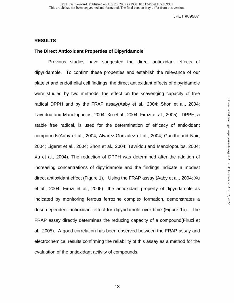
The Direct Antioxidant Properties of Dipyridamole
Previous studies have suggested the direct antioxidant effects of
dipyridamole. To confirm these properties and establish the relevance of our
platelet and endothelial cell findings, the direct antioxidant effects of dipyridamole
were studied by two methods; the effect on the scavenging capacity of free
radical DPPH and by the FRAP assay(Aaby et al., 2004; Shon et al., 2004;
Tavridou and Manolopoulos, 2004; Xu et al., 2004; Firuzi et al., 2005). DPPH, a
stable free radical, is used for the determination of efficacy of antioxidant
compounds(Aaby et al., 2004; Alvarez-Gonzalez et al., 2004; Gandhi and Nair,
2004; Ligeret et al., 2004; Shon et al., 2004; Tavridou and Manolopoulos, 2004;
Xu et al., 2004). The reduction of DPPH was determined after the addition of
increasing concentrations of dipyridamole and the findings indicate a modest
direct antioxidant effect (Figure 1). Using the FRAP assay,(Aaby et al., 2004; Xu
et al., 2004; Firuzi et al., 2005) the antioxidant property of dipyridamole as
indicated by monitoring ferrous ferrozine complex formation, demonstrates a
dose-dependent antioxidant effect for dipyridamole over time (Figure 1b). The
FRAP assay directly determines the reducing capacity of a compound(Firuzi et
al., 2005). A good correlation has been observed between the FRAP assay and
electrochemical results confirming the reliability of this assay as a method for the
evaluation of the antioxidant activity of compounds.
This article has not been copyedited and formatted. The final version may differ from this version.JPET Fast Forward. Published on July 26, 2005 as DOI: 10.1124/jpet.105.089987
at ASPE
T Journals on A
pril 2, 2022jpet.aspetjournals.org
Dow
nloaded from

JPET #89987
14
The Effect of Dipyridamole on Endothelial Cellular Redox State and
Generation of Reactive Oxygen Species
To determine if the antioxidant effects of dipyridamole alter endothelial cell
release of reactive oxygen species, BAECs were incubated with increasing
concentrations of dipyridamole and DCFDA fluorescence was determined.
DCFDA is a fluorescence-based probe that has been recently developed to
detect intracellular production of ROS. DCFDA diffuses passively into cells and
trapped inside generating DCFH after deacetylation by intracellular esterases. It
is subsequently oxidized to a fluorescent product in presence of intracellular
ROS. Oxidation of DCFDA is conveniently monitored for the determination of
intracellular oxidative stress(Halliwell and Whiteman, 2004). As seen in Figure
2a, there is a significant dose-dependent reduction of basal DCFDA fluorescence
as a result of dipyridamole incubation. These effects were seen immediately
after washing out the dipyridamole and persisted as well as increased at 1, 2,
and 3 hours after washing (data not shown). Representative confocal images
are shown in Figure 2b.
t-Butylhydroperoxide is used to generate oxidative stress in various
biological systems(Lautraite et al., 2003). As shown in Figure 3a, the addition of
t-butylhydroperoxide leads to enhanced oxidative stress and this effect is
attenuated following incubation with dipyridamole. Dipyridamole also improves
endothelial metabolic activity after exposure to t-butylhydroperoxide-induced
oxidative stress as shown by the redox sensitive dye Alamar blue (Figure 3b).
Alamar Blue is a redox sensitive dye, reduction of which reflects metabolic
This article has not been copyedited and formatted. The final version may differ from this version.JPET Fast Forward. Published on July 26, 2005 as DOI: 10.1124/jpet.105.089987
at ASPE
T Journals on A
pril 2, 2022jpet.aspetjournals.org
Dow
nloaded from

JPET #89987
15
activity of the cells(Ahmed et al., 1994; Collins and Franzblau, 1997; Franzblau,
2000). To confirm that the t-butylhydroperoxide treatment did not cause cellular
toxicity, endothelial cells that had been incubated with t-butylhydroperoxide were
tested for release of LDH. No significant toxicity was observed at all
concentrations during the periods of incubation (data not shown).
The Effect of Dipyridamole on Activation-induced Platelet Generation of
Reactive Oxygen and Nitrogen Species
Incubation with dipyridamole alters endothelial cell redox status and ROS
generation. To determine if these effects are specific for the endothelial cells or
also relevant in platelets, release of platelet reactive oxygen and nitrogen species
was determined after incubation with dipyridamole. Platelet superoxide release
was measured using a lumiaggregometer after PMA-induced stimulation. PMA is
used as the agonist as it has the most marked effect on platelet superoxide
release(Freedman and Keaney, 1999). Incubation of platelets with 20 µM
dipyridamole led to a marked suppression of platelet release of superoxide
(Figure 4a). This effect was noted over a range of dipyridamole concentrations
(2-100 µM; data not shown).
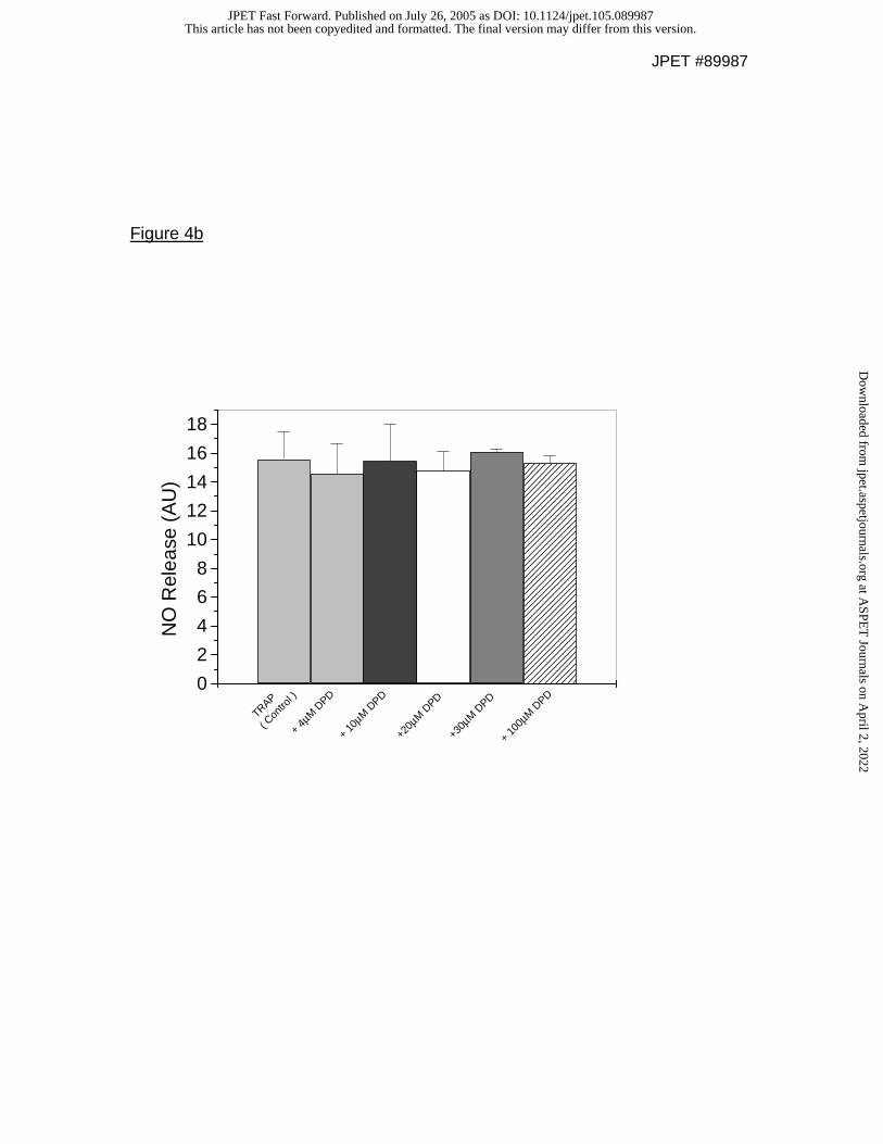
To determine if dipyridamole alters release of other endogenous reactive
species, platelet levels of NO and hydrogen peroxide were also measured.
There was no significant effect of dipyridamole on platelet release of NO as
measured by microelectrode (Figure 4b) or hydrogen peroxide (data not shown)
as measured by the Amplex Red Assay over a range of concentrations. Platelet
This article has not been copyedited and formatted. The final version may differ from this version.JPET Fast Forward. Published on July 26, 2005 as DOI: 10.1124/jpet.105.089987
at ASPE
T Journals on A
pril 2, 2022jpet.aspetjournals.org
Dow
nloaded from

JPET #89987
16
generation of ROS following stimulation as measured by DCFDA oxidation by
flow cytometry was studied. The generation of ROS was significantly attenuated
as a function of increasing dipyridamole concentration (Figure 5a). Washed
platelets were also incubated in presence of redox sensitive probe
dihydrorhodamine (DHR, 10µM) for 10 minutes in presence and absence of
20µM dipyridamole. Confocal images (Figure 5b) were captured at different time
points before and after TRAP (20µM) stimulation (displayed images were taken
10 minute after TRAP stimulation). Presence of dipyridamole attenuated TRAP
activation induced platelet fluorescence induction (Figure 5b).
To determine if the change in redox status of the platelet by dipyridamole
has an effect on the platelet-inflammatory response, platelet release of sCD40L
was determined. Platelets were stimulated with thrombin for 2 hrs in presence of
increasing concentrations of dipyridamole. Platelet supernatant was collected
and the released sCD40L were measured by ELISA. As seen in Figure 6,
incubation with dipyridamole leads to a modest but significant suppression of
sCD40L release from activated platelets.
This article has not been copyedited and formatted. The final version may differ from this version.JPET Fast Forward. Published on July 26, 2005 as DOI: 10.1124/jpet.105.089987
at ASPE
T Journals on A
pril 2, 2022jpet.aspetjournals.org
Dow
nloaded from

JPET #89987
17
DISCUSSION
Platelet and vascular stimulation leads to release of ROS that are known
to induce vasoconstriction, platelet activation, and stimulate the atherothrombotic
processes. Dipyridamole is a vasodilator and platelet inhibitor that has
previously been shown to have direct antioxidant properties, however, its effects
on vascular cells are unknown. Therefore, we studied the effect of dipyridamole
on platelet- and endothelial cell-derived release of ROS. Consistent with
previous studies(Iuliano et al., 1989), the direct antioxidant properties of
dipyridamole were confirmed. Additionally, at therapeutically relevant
concentration (3.5µM) (Aktas et al., 2003), dipyridamole suppressed stimulation–
dependent endothelial ROS formation and platelet release of soluble CD40
ligand. Previous studies using higher concentration of dipyridamole (≥20µM,
peak concentration) reached in blood after chronic intake of the habitual dose
diminished platelet–neutrophil interaction (De La Cruz et al., 2000) and also
attenuated neutrophil superoxide generation (Vargas et al., 2003). Our present
study, with a dose range of 0.5-20µM dipyridamole, displayed a wide range of
antioxidant/anti-inflammatory properties both in platelets and endothelial cells.
Data suggests that the antioxidant effect of dipyridamole is related to its
partition in the lipid phase of the mitochondrial membrane and not to a specific
interaction with membrane proteins. This protection may be due either to a direct
inhibition of the propagation steps or a scavenger effect on the radical species
that would trigger the peroxidative process(Nepomuceno et al., 1999). Our
This article has not been copyedited and formatted. The final version may differ from this version.JPET Fast Forward. Published on July 26, 2005 as DOI: 10.1124/jpet.105.089987
at ASPE
T Journals on A
pril 2, 2022jpet.aspetjournals.org
Dow
nloaded from

JPET #89987
18
findings that specifically showed a suppression of superoxide (Figure 4) would
support the previous suggestions as well as our own findings (Figures 1a and 1b)
that dipyridamole has a direct antioxidant effect.
Oxidant stress can lead to endothelial dysfunction and this, in turn,
contributes to the genesis of the atherothrombotic plaque. Therefore, it is
plausible that dipyridamole promotes vascular protection and improves
endothelial function through its antioxidant actions. Dipyridamole had been
shown to enhance inhibition of platelet function by amplifying the effect of
exogenous NO donors suggesting enhancement of the NO/cGMP pathway(Aktas
et al., 2003). While dipyridamole did not alter endogenous release of platelet
derived NO(Figure 4b), the findings of our study are consistent with these
previous observations(Aktas et al., 2003) as the dipyridamole-dependent
enhancement of exogenous NO could be mediated through the suppression of
platelet superoxide release (Figure 4a) and thus limiting bioavailable NO.
However, further in vitro and in vivo studies are warranted to characterize the
role of dipyridamole in influencing endothelial NO and superoxide generation.
Although the effects on endothelial cells and platelets have not been
investigated, dipyridamole was previously shown to scavenge ROS secreted by
activated neutrophils(Vargas et al., 2003). Dipyridamole has also been shown to
protect erythrocyte membranes from oxidation(Kusmic et al., 2000b) and
neuronal cells from chemically mediated oxidative damage(Blake, 2004). In
addition, dipyridamole was recently shown to prevent damage in a liver ischemia-
reperfusion model(Taniguchi et al., 2004). These redox specific effects may
This article has not been copyedited and formatted. The final version may differ from this version.JPET Fast Forward. Published on July 26, 2005 as DOI: 10.1124/jpet.105.089987
at ASPE
T Journals on A
pril 2, 2022jpet.aspetjournals.org
Dow
nloaded from

JPET #89987
19
extend to other cellular antioxidant/inflammatory interactions that are known to be
important in acute and chronic cardiovascular disease. Flow cytometry data
generated in the AGATE study(Serebruany et al., 2004) indicates that subjects
taking Aggrenox have a depression (beyond aspirin alone) of receptors important
in cell-cell interactions. Our results show that low concentrations of dipyridamole
(≥1µM) induce suppression of the platelet inflammatory protein sCD40L, although
we cannot conclusively say that this effect is mediated through the antioxidant
properties of this drug. Thus, further study of adhesion molecules and
inflammatory mediated cellular cross talk may be warranted.
Recent studies show that reduction of NO bioavailability is associated
with an increase in endothelial production of superoxide (Kalinowski and
Malinski, 2004). Superoxide in both the platelet and endothelial cell is generated
by NAD(P)H oxidase and may trigger eNOS uncoupling contributing to the
endothelial balance between NO and superoxide(Kalinowski and Malinski, 2004).
Release of several ROS, including superoxide, hydroxyl radical, and H2O2 from
platelets are reported, both from unstimulated and after stimulation with agonists
such as collagen or thrombin (Krotz et al., 2004). Several enzymatic systems
contribute to the production of ROS, thereby influencing platelet activity. In the
endothelium, NAD(P)H-oxidase, cyclooxygenase isoforms 1 and 2, cytochrome
P450 epoxygenase isoform 2C9 (CYP2C9), xanthine oxidase (XO), uncoupled
endothelial NO synthase (eNOS), and mitochondrial respiration contribute to the
production of superoxide, H2O2 and hydroxyl radicals. Like endothelium-derived
ROS, platelet-derived ROS potentially stem from enzymatic sources, including
This article has not been copyedited and formatted. The final version may differ from this version.JPET Fast Forward. Published on July 26, 2005 as DOI: 10.1124/jpet.105.089987
at ASPE
T Journals on A
pril 2, 2022jpet.aspetjournals.org
Dow
nloaded from

JPET #89987
20
cyclooxygenase-1, xanthine oxidase, mitochondrial respiration, or uncoupled
eNOS. The platelet isoform of NAD(P)H-oxidase has gained the most attention
because it can be activated by by platelet activation. Platelets have been
reported to possess NAD(P)H-oxidase activity (Seno et al., 2001; Krotz et al.,
2002) and many of its subunits have been found at the protein level(Krotz et al.,
2004). Dipyridamole may modulate NADPH oxidase activity as dipyridamole
induced inhibition of NADPH oxidase activity has been demonstrated in a
diabetic animal model (Onozato et al., 2003).
A recent study demonstated that platelet NADPH oxidase subunit, gp91phox,
regulates the expression of CD40L(Pignatelli et al., 2004). Thus, it is possible
that dipyridamole, through its inhibitory property on NADPH oxidase, may alter
platelet ROS generation as well as platelet release of sCD40L.
In summary, at therapeutically relevant concentrations, dipyridamole
suppresses stimulation–dependent platelet superoxide generation, formation of
ROS in platelets and endothelial cells, and improves cellular redox status. These
data suggest that the antioxidant properties of dipyridamole have a direct effect
on vascular cells and suppress the endogenous release of vascular reactive
oxygen and inflammatory species shown to be relevant in the development of
atherothrombotic diseases.
This article has not been copyedited and formatted. The final version may differ from this version.JPET Fast Forward. Published on July 26, 2005 as DOI: 10.1124/jpet.105.089987
at ASPE
T Journals on A
pril 2, 2022jpet.aspetjournals.org
Dow
nloaded from

JPET #89987
21
ACKNOWLEDGEMENTS
The authors would like to thank Dr. Wolfgang G. Eisert for his helpful
discussions.
This article has not been copyedited and formatted. The final version may differ from this version.JPET Fast Forward. Published on July 26, 2005 as DOI: 10.1124/jpet.105.089987
at ASPE
T Journals on A
pril 2, 2022jpet.aspetjournals.org
Dow
nloaded from

JPET #89987
22
REFERENCES
Aaby K, Hvattum E and Skrede G (2004) Analysis of flavonoids and other
phenolic compounds using high-performance liquid chromatography with
coulometric array detection: relationship to antioxidant activity. J Agric
Food Chem 52:4595-4603.
Ahmed SA, Gogal RM, Jr. and Walsh JE (1994) A new rapid and simple non-
radioactive assay to monitor and determine the proliferation of
lymphocytes: an alternative to [3H]thymidine incorporation assay. J
Immunol Methods 170:211-224.
Aktas B, Utz A, Hoenig-Liedl P, Walter U and Geiger J (2003) Dipyridamole
enhances NO/cGMP-mediated vasodilator-stimulated phosphoprotein
phosphorylation and signaling in human platelets: in vitro and in vivo/ex
vivo studies. Stroke 34:764-769.
Alvarez-Gonzalez I, Madrigal-Bujaidar E, Martino-Roaro L and Espinosa-Aguirre
JJ (2004) Antigenotoxic and antioxidant effect of grapefruit juice in mice
treated with daunorubicin. Toxicol Lett 152:203-211.
Blake AD (2004) Dipyridamole is neuroprotective for cultured rat embryonic
cortical neurons. Biochem Biophys Res Commun 314:501-504.
Collins L and Franzblau SG (1997) Microplate alamar blue assay versus
BACTEC 460 system for high-throughput screening of compounds against
Mycobacterium tuberculosis and Mycobacterium avium. Antimicrob
Agents Chemother 41:1004-1009.
This article has not been copyedited and formatted. The final version may differ from this version.JPET Fast Forward. Published on July 26, 2005 as DOI: 10.1124/jpet.105.089987
at ASPE
T Journals on A
pril 2, 2022jpet.aspetjournals.org
Dow
nloaded from

JPET #89987
23
De La Cruz JP, Blanco E and Sanchez de la Cuesta F (2000) Effect of
dipyridamole and aspirin on the platelet-neutrophil interaction via the nitric
oxide pathway. Eur J Pharmacol 397:35-41.
De la Cruz JP, Garcia PJ and Sanchez de la Cuesta F (1992) Dipyridamole
inhibits platelet aggregation induced by oxygen-derived free radicals.
Thromb Res 66:277-285.
Diener HC, Cunha L, Forbes C, Sivenius J, Smets P and Lowenthal A (1996)
European Stroke Prevention Study. 2. Dipyridamole and acetylsalicylic
acid in the secondary prevention of stroke. J Neurol Sci 143:1-13.
Firuzi O, Lacanna A, Petrucci R, Marrosu G and Saso L (2005) Evaluation of the
antioxidant activity of flavonoids by "ferric reducing antioxidant power"
assay and cyclic voltammetry. Biochim Biophys Acta 1721:174-184.
Franzblau S (2000) A rapid, microplate-based assay for evaluating the activity of
drugs against Mycobacterium leprae, employing the reduction of Alamar
Blue. Lepr Rev 71 Suppl:S74-75; discussion S76.
Freedman JE, Fabian A and Loscalzo J (1995) Impaired EDRF production by
endothelial cells exposed to fibrin monomer and FDP. Am J Physiol
268:C520-526.
Freedman JE, Farhat JH, Loscalzo J and Keaney JF, Jr. (1996a) alpha-
tocopherol inhibits aggregation of human platelets by a protein kinase C-
dependent mechanism. Circulation 94:2434-2440.
Freedman JE and Keaney JF, Jr. (1999) Nitric oxide and superoxide detection in
human platelets. Methods Enzymol 301:61-70.
This article has not been copyedited and formatted. The final version may differ from this version.JPET Fast Forward. Published on July 26, 2005 as DOI: 10.1124/jpet.105.089987
at ASPE
T Journals on A
pril 2, 2022jpet.aspetjournals.org
Dow
nloaded from

JPET #89987
24
Freedman JE, Loscalzo J, Benoit SE, Valeri CR, Barnard MR and Michelson AD
(1996b) Decreased platelet inhibition by nitric oxide in two brothers with a
history of arterial thrombosis. J Clin Invest 97:979-987.
Gandhi NM and Nair CK (2004) Radiation protection by diethyldithiocarbamate:
protection of membrane and DNA in vitro and in vivo against gamma-
radiation. J Radiat Res (Tokyo) 45:175-180.
Halliwell B and Whiteman M (2004) Measuring reactive species and oxidative
damage in vivo and in cell culture: how should you do it and what do the
results mean? Br J Pharmacol 142:231-255.
Iuliano L, Colavita AR, Camastra C, Bello V, Quintarelli C, Alessandroni M,
Piovella F and Violi F (1996) Protection of low density lipoprotein oxidation
at chemical and cellular level by the antioxidant drug dipyridamole. Br J
Pharmacol 119:1438-1446.
Iuliano L, Ghiselli A, Alessandri C, Bonavita MS and Violi F (1989) Superoxide
anion scavenging property of dipyridamole. Thromb Haemost 61:149.
Iuliano L, Piccheri C, Coppola I, Pratico D, Micheletta F and Violi F (2000)
Fluorescence quenching of dipyridamole associated to peroxyl radical
scavenging: a versatile probe to measure the chain breaking antioxidant
activity of biomolecules. Biochim Biophys Acta 1474:177-182.
Kalinowski L and Malinski T (2004) Endothelial NADH/NADPH-dependent
enzymatic sources of superoxide production: relationship to endothelial
dysfunction. Acta Biochim Pol 51:459-469.
This article has not been copyedited and formatted. The final version may differ from this version.JPET Fast Forward. Published on July 26, 2005 as DOI: 10.1124/jpet.105.089987
at ASPE
T Journals on A
pril 2, 2022jpet.aspetjournals.org
Dow
nloaded from

JPET #89987
25
Krotz F, Sohn HY, Gloe T, Zahler S, Riexinger T, Schiele TM, Becker BF,
Theisen K, Klauss V and Pohl U (2002) NAD(P)H oxidase-dependent
platelet superoxide anion release increases platelet recruitment. Blood
100:917-924.
Krotz F, Sohn HY and Pohl U (2004) Reactive oxygen species: players in the
platelet game. Arterioscler Thromb Vasc Biol 24:1988-1996.
Kusmic C, Petersen C, Picano E, Busceti C, Parenti G, Pasini FL and Barsacchi
R (2000a) Antioxidant effect of oral dipyridamole during cerebral
hypoperfusion with human carotid endarterectomy. J Cardiovasc
Pharmacol 36:141-145.
Kusmic C, Picano E, Busceti CL, Petersen C and Barsacchi R (2000b) The
antioxidant drug dipyridamole spares the vitamin E and thiols in red blood
cells after oxidative stress. Cardiovasc Res 47:510-514.
Lautraite S, Bigot-Lasserre D, Bars R and Carmichael N (2003) Optimisation of
cell-based assays for medium throughput screening of oxidative stress.
Toxicol In Vitro 17:207-220.
Ligeret H, Barthelemy S, Zini R, Tillement JP, Labidalle S and Morin D (2004)
Effects of curcumin and curcumin derivatives on mitochondrial
permeability transition pore. Free Radic Biol Med 36:919-929.
Nepomuceno MF, de Oliveira Mamede ME, Vaz de Macedo D, Alves AA,
Pereira-da-Silva L and Tabak M (1999) Antioxidant effect of dipyridamole
and its derivative RA-25 in mitochondria: correlation of activity and
location in the membrane. Biochim Biophys Acta 1418:285-294.
This article has not been copyedited and formatted. The final version may differ from this version.JPET Fast Forward. Published on July 26, 2005 as DOI: 10.1124/jpet.105.089987
at ASPE
T Journals on A
pril 2, 2022jpet.aspetjournals.org
Dow
nloaded from

JPET #89987
26
Onozato ML, Tojo A, Goto A and Fujita T (2003) Effect of combination therapy
with dipyridamole and quinapril in diabetic nephropathy. Diabetes Res Clin
Pract 59:83-92.
Pignatelli P, Sanguigni V, Lenti L, Ferro D, Finocchi A, Rossi P and Violi F (2004)
gp91phox-dependent expression of platelet CD40 ligand. Circulation
110:1326-1329.
Selley ML, Czeti AL, McGuiness JA and Ardlie NG (1994) Dipyridamole inhibits
the oxidative modification of low density lipoprotein. Atherosclerosis
111:91-97.
Seno T, Inoue N, Gao D, Okuda M, Sumi Y, Matsui K, Yamada S, Hirata KI,
Kawashima S, Tawa R, Imajoh-Ohmi S, Sakurai H and Yokoyama M
(2001) Involvement of NADH/NADPH oxidase in human platelet ROS
production. Thromb Res 103:399-409.
Serebruany VL, Malinin AI, Sane DC, Jilma B, Takserman A, Atar D and
Hennekens CH (2004) Magnitude and time course of platelet inhibition
with Aggrenox and Aspirin in patients after ischemic stroke: the AGgrenox
versus Aspirin Therapy Evaluation (AGATE) trial. Eur J Pharmacol
499:315-324.
Shon MY, Choi SD, Kahng GG, Nam SH and Sung NJ (2004) Antimutagenic,
antioxidant and free radical scavenging activity of ethyl acetate extracts
from white, yellow and red onions. Food Chem Toxicol 42:659-666.
This article has not been copyedited and formatted. The final version may differ from this version.JPET Fast Forward. Published on July 26, 2005 as DOI: 10.1124/jpet.105.089987
at ASPE
T Journals on A
pril 2, 2022jpet.aspetjournals.org
Dow
nloaded from

JPET #89987
27
Taniguchi M, Magata S, Suzuki T, Shimamura T, Jin MB, Iida J, Furukawa H and
Todo S (2004) Dipyridamole protects the liver against warm ischemia and
reperfusion injury. J Am Coll Surg 198:758-769.
Tavridou A and Manolopoulos VG (2004) Antioxidant properties of two novel 2-
biphenylmorpholine compounds (EP2306 and EP2302) in vitro and in vivo.
Eur J Pharmacol 505:213-221.
Vargas F, Rivas C, Diaz Y, Contreras N, Silva A, Ojeda LE, Velasquez M and
Fraile G (2003) Antioxidant properties of dipyridamole as assessed by
chemiluminescence. Pharmazie 58:817-823.
Xu JZ, Yeung SY, Chang Q, Huang Y and Chen ZY (2004) Comparison of
antioxidant activity and bioavailability of tea epicatechins with their
epimers. Br J Nutr 91:873-881.
This article has not been copyedited and formatted. The final version may differ from this version.JPET Fast Forward. Published on July 26, 2005 as DOI: 10.1124/jpet.105.089987
at ASPE
T Journals on A
pril 2, 2022jpet.aspetjournals.org
Dow
nloaded from

JPET #89987
28
Footnotes
This work has been supported in part by NIH grants NIH RO1AG08226 (J.F.),
NIH RO1HL62267 (J.F.), and an Established Investigator Award from the
American Heart Association (J.F.). These studies were also funded by an
unrestricted grant from Boehringer-Ingelheim.
This article has not been copyedited and formatted. The final version may differ from this version.JPET Fast Forward. Published on July 26, 2005 as DOI: 10.1124/jpet.105.089987
at ASPE
T Journals on A
pril 2, 2022jpet.aspetjournals.org
Dow
nloaded from

JPET #89987
29
FIGURE LEGENDS
Figure 1a. The antioxidant property of dipyridamole effectively reduces the stable
free radical DPPH. The reduction of DPPH (100 µM) was monitored in presence
of increasing concentrations of dipyridamole. The absorption change at 515 nm
is plotted for the 60 minutes data point (n=5; *P<0.05).
Figure 1b. The effect of dipyridamole on Fe3+ reduction using the FRAP assay.
Reduction of ferric (Fe3+) ions was monitored by ferrous-ferrozine complex
formation. Increasing concentrations of dipyridamole were added to a solution
containing FeCl3 and ferrozine. Absorption at 560 nm is plotted as a function of
incubation time (n=4).
Figure 2a. Dipyridamole decreases basal ROS generation in endothelial cells.
Confluent BAECs were treated with increasing concentrations of dipyridamole
and DCFDA for two hours and DCFDA fluorescence was measured immediately
after washing (n=3; *P<0.001 vs. control) and two hours after washing.
Figure 2b. Presence of dipyridamole attenuates stimulation dependent
endothelial reactive oxygen species generation. Representative confocal images
of endothelial cells in presence of 10µM CMDCFDA after stimulation (1u/ml
thrombin, 10min) in presence of either vehicle control or 10µM dipyridamole.
This article has not been copyedited and formatted. The final version may differ from this version.JPET Fast Forward. Published on July 26, 2005 as DOI: 10.1124/jpet.105.089987
at ASPE
T Journals on A
pril 2, 2022jpet.aspetjournals.org
Dow
nloaded from

JPET #89987
30
Images were captured in two photon confocal microscopy and processed using
NIH image software and displayed with pseudo color assignment.
Figure 3a. Dipyridamole attenuates t-butylhydroperoxide-induced oxidative stress
in endothelial cells. BAECs were incubated for 2 hours with t-butylhydroperoxide
and DCFDA (10µM) in presence and absence of 5 µM dipyridamole. DCFDA
fluorescence was measured over time (0-2 hrs. with the T=0 time period shown;
n=5; *P=0.007, ** P=0.001). Similar results were observed after 1 hr and 2 hr
incubation (data not shown).
Figure 3b. Dipyridamole improves endothelial activity after exposing to
t-butylhydroperoxide-induced oxidative stress. t-Butylhydroperoxide treated
endothelial cells were evaluated for metabolic activity using the redox sensitive
dye Alamar blue (10%). Dipyridamole attenuates the t-butylhydroperoxide-
induced oxidative stress (550nm/570nm; n=5; *P<0.001 vs. control).
Figure 4a. The effect of dipyridamole on stimulation-dependent platelet
superoxide release. Washed human platelets (4x10⁸/ml) were incubated with
either vehicle control (DMSO) or 20 µM dipyridamole for 10 minutes. Platelet
aggregation induced superoxide release was measured in a lumiaggregometer
where 0.1 µM PMA was used as agonist (n=5; *P<0.001).
This article has not been copyedited and formatted. The final version may differ from this version.JPET Fast Forward. Published on July 26, 2005 as DOI: 10.1124/jpet.105.089987
at ASPE
T Journals on A
pril 2, 2022jpet.aspetjournals.org
Dow
nloaded from

JPET #89987
31
Figure 4b. The effect of dipyridamole on platelet aggregation induced nitric oxide
(NO) release. Washed human platelets were incubated with different
concentrations of dipyridamole (DPD) or vehicle control for 5 minutes followed by
measurement of TRAP (Thrombin Receptor Activation Peptide) induced NO
release using a NO selective microelectrode (n=5; P=ns).
Figure 5a. Dipyridamole decreases stimulation induced platelet reactive oxygen
species generation in a concentration dependent manner. By flow cytometry,
there is an increased median platelet fluorescence following TRAP stimulation
(20 µM). Presence of dipyridamole from 20 µM to 100 µM significantly attenuates
this ROS generation (* P<0.05, n=3).
Figure 5b. Confocal images of platelets using redox sensitive probe
dihydrorhodamine (DHR) demonstrate attenuation of reactive oxygen species
generation by dipyridamole. Confocal images were captured after TRAP (20µM,
10min) stimulation in the presence or absence of dipyridamole (DPD, 20µM).
Figure 6. Dipyridamole attenuates sCD40L release from thrombin activated
platelets. Washed human platelets (2x10⁸/ml) in HEPES buffer were incubated
for 2 hours at room temperature with 0.2 u/ml thrombin in presence of varying
concentration of dipyridamole. Platelet supernatants were measured for sCD40L
release (n=6, +P=0.007 vs. thrombin, * P<0.001 vs. thrombin).
This article has not been copyedited and formatted. The final version may differ from this version.JPET Fast Forward. Published on July 26, 2005 as DOI: 10.1124/jpet.105.089987
at ASPE
T Journals on A
pril 2, 2022jpet.aspetjournals.org
Dow
nloaded from

JPET #89987
Figure 1a
0
5
10
15
20
25
30
35
40
*
**
100 µ
M Dipy
ridam
ole
50 µM D
ipyrid
amole
20 µM
Dipy
ridam
ole
2 µM D
ipyrid
amole
None
% D
PP
H R
educ
tion
This article has not been copyedited and formatted. The final version may differ from this version.JPET Fast Forward. Published on July 26, 2005 as DOI: 10.1124/jpet.105.089987
at ASPE
T Journals on A
pril 2, 2022jpet.aspetjournals.org
Dow
nloaded from

JPET #89987
Figure 1b
0 10 20 30 40 50 60
0.00
0.05
0.10
0.15
0.20
0.25
0.30
DMSO
DPD 1 µM
DPD 5 µM
DPD 10 µM
Fe2+
-Fer
rozi
ne (
AU
) (A
bsor
banc
e at
560
nm
)
Time (minutes)
This article has not been copyedited and formatted. The final version may differ from this version.JPET Fast Forward. Published on July 26, 2005 as DOI: 10.1124/jpet.105.089987
at ASPE
T Journals on A
pril 2, 2022jpet.aspetjournals.org
Dow
nloaded from

JPET #89987
Figure 2a
10
20
40
60
80
100
120
140
+DPD 2
0 µM
+DPD 1
0 µM
+DPD 5
µM
+DPD 0
.5 µM
+DPD 0
.1 µM
Contro
l
*
*
*
*
Flu
ores
cenc
e/m
g pr
otei
n
This article has not been copyedited and formatted. The final version may differ from this version.JPET Fast Forward. Published on July 26, 2005 as DOI: 10.1124/jpet.105.089987
at ASPE
T Journals on A
pril 2, 2022jpet.aspetjournals.org
Dow
nloaded from

JPET #89987
Figure 2b
ECs+Thrombin Thrombin+10µM DPD
This article has not been copyedited and formatted. The final version may differ from this version.JPET Fast Forward. Published on July 26, 2005 as DOI: 10.1124/jpet.105.089987
at ASPE
T Journals on A
pril 2, 2022jpet.aspetjournals.org
Dow
nloaded from

JPET #89987
Figure 3a
10
100
200
300
400
500**
**
*
0.25
mM
tBHP
+ D
PD 5 µM
0.25
mM
tBHP
0.1 m
M tB
HP
+ D
PD 5 µM
0.1
mM
tBHP
Contro
l
+ D
PD 5 µM
Contro
l
Flu
ores
cenc
e/m
g pr
otei
n
This article has not been copyedited and formatted. The final version may differ from this version.JPET Fast Forward. Published on July 26, 2005 as DOI: 10.1124/jpet.105.089987
at ASPE
T Journals on A
pril 2, 2022jpet.aspetjournals.org
Dow
nloaded from

JPET #89987
Figure 3b
10
2000
4000
6000
8000
10000
12000
14000
16000
*
*
Contro
l / +D
PD
t-BHP 0
.25
mM
+
DPD 5 µM
t-BHP 0
.25
mM
t-BHP 0
.1 m
M
+ D
PD 5 µM
t-BHP 0
.1 m
M
Flu
ores
cenc
e/m
g pr
otei
n(E
x 55
0 nm
/ Em
570
nm
)
This article has not been copyedited and formatted. The final version may differ from this version.JPET Fast Forward. Published on July 26, 2005 as DOI: 10.1124/jpet.105.089987
at ASPE
T Journals on A
pril 2, 2022jpet.aspetjournals.org
Dow
nloaded from

JPET #89987
Figure 4a
0
2
4
6
8
10
12
14
16
18
Dipyrid
amole
( 2
0 µM
)Con
trol
*
Sup
erox
ide
Rel
ease
(A
.U.)
This article has not been copyedited and formatted. The final version may differ from this version.JPET Fast Forward. Published on July 26, 2005 as DOI: 10.1124/jpet.105.089987
at ASPE
T Journals on A
pril 2, 2022jpet.aspetjournals.org
Dow
nloaded from
JPET #89987
Figure 4b
10
2
4
6
8
10
12
14
16
18
+ 10
0µM
DPD
+30µ
M D
PD
+20µ
M D
PD
+ 10
µM D
PD
+ 4
µM D
PD
TRAP
( Con
trol )
NO
Rel
ease
(A
U)
This article has not been copyedited and formatted. The final version may differ from this version.JPET Fast Forward. Published on July 26, 2005 as DOI: 10.1124/jpet.105.089987
at ASPE
T Journals on A
pril 2, 2022jpet.aspetjournals.org
Dow
nloaded from

JPET #89987
Figure 5a.
10.0
0.2
0.4
0.6
0.8
1.0
1.2
1.4
***
+100
µM DPD
+40
µM D
PD
+20 µ
M DPD
+4 µM
DPD
Con
trol
(+20
µMTRAP)
Rel
ativ
e F
luor
esce
nce
(Rat
io o
f Med
ian
Fl.
w.r
.t. C
ontr
ol)
This article has not been copyedited and formatted. The final version may differ from this version.JPET Fast Forward. Published on July 26, 2005 as DOI: 10.1124/jpet.105.089987
at ASPE
T Journals on A
pril 2, 2022jpet.aspetjournals.org
Dow
nloaded from

JPET #89987
Figure 5b
Platelets+TRAP TRAP + DPD
This article has not been copyedited and formatted. The final version may differ from this version.JPET Fast Forward. Published on July 26, 2005 as DOI: 10.1124/jpet.105.089987
at ASPE
T Journals on A
pril 2, 2022jpet.aspetjournals.org
Dow
nloaded from

JPET #89987
Figure 6
0 20 40 60 80 100
0
2
4
6
8
10
12
*
***+ *
[sC
D40
L] (
ng/m
l)
[Dipyridamole] (µM)
This article has not been copyedited and formatted. The final version may differ from this version.JPET Fast Forward. Published on July 26, 2005 as DOI: 10.1124/jpet.105.089987
at ASPE
T Journals on A
pril 2, 2022jpet.aspetjournals.org
Dow
nloaded from