the effect of congenital deafness on cerebral asymmetry in the perception of emotional and...
TRANSCRIPT
Acta Psychologica 79 (1992) 45-57
North-Holland
45
The effect of congenital deafness on cerebral asymmetry in the perception of emotional and non-emotional faces
Elzbieta Szelag * and Ryszard Wasilewski Polish Academy of Sciences. Warsaw, Poland
Accepted October 199 1
Functional hemispheric specialization in recognizing faces expressing emotions was investigated
in 18 normal hearing and 18 congenitally deaf children aged 13-14 years. Three kinds of faces
were presented: happy, to express positive emotions, sad, to express negative emotions, and
neutral. The subjects’ task was to recognize the test face exposed for 20 msec in the left or right
visual field. The subjects answered by pointing at the exposed stimulus on the response card that
contained three different faces. The errors committed in expositions of faces in the left and right
visual field were analyzed. In the control group the right hemisphere dominated in case of sad and neutral faces, There were no significant differences in recognition of happy faces. The
differentiated hemispheric organization pattern in normal hearing persons supports the hypothe-
sis of different processing of positive and negative emotions expressed by faces. The observed
hemispheric asymmetry was a result of two factors: (1) processing of faces as complex patterns
requiring visuo-spatial analysis, and (2) processing of emotions contained in them.
Functional hemispheric asymmetry was not observed in the group of deaf children for any
kind of emotion expressed in the presented faces. The results suggest that lack of auditory
experience influences the organization of functional hemispheric specialization. It can be
supposed that in deaf children, the analysis of information contained in emotional faces takes
place in both hemispheres.
Introduction
Although it is known that human brain hemispheres are anatomi- cally differentiated since birth (Geschwind and Levitsky 1968; Witel- son and Pallie 19731, there are no univocal data as to the effect of factors active in early ontogenesis on the formation of functional
* Correspondence address: E. Szelag, Nencki Institute of Experimental Biology, Dept. of Neurophysiology, Polish Academy of Sciences, Warsaw, 3 Pasteur str., Poland.
0001~6918/92/$05.00 0 1992 - Elsevier Science Publishers B.V. All rights reserved
hemispheric specialization. Among the problems of psychological re- search thcrc is one that concerns the effect of ontogenetic experience, e.g. the effect of auditory experience on functional hemispheric asym- metry. One of the information sources on this subject are results of research rcccntly carried out on persons born deaf who communicate with sign language and lip reading. It is believed that their level of auditory experience connected with speech and non-verbal sounds from the surroundings is very limited. The results of research carried out on this group could tell us if limitation of auditory experience in ontogenesis affects functional organization of brain hemispheres. Re- search so far has not given any univocal answer as to the functional hemispheric asymmetry in deaf-born subjects and concerned mainly verbal material. Panou and Sewell (19841, Poizncr ct al. (1979), and Manning et al. ( 1977) have proven that, similarly to normal hearing, in deaf persons the left hemisphere is more effective in processing verbal material. This result has been confirmed by clinical studies on individ- ual cases of dcafncss where the ability of communicate in sign lan- guage was lost, i.e. manual aphasia after left hemisphere damage (Damasio et al. 1986; Kimura et al. 1976; Sarno et al. 1963; Douglas and Richardson 1959; Tureen et al. 195 1).
Kelly and Tomlinson-Kcasey (198 l), Gibson and Bryden (19841, and Szelag et al. (1991), however, observed the opposite, i.e. right hemi- spheric dominance in processing verbal material, whcrcas McKeever et al. (1976), Wilson (19X3), Scholes and Fischler (1979), and Marcotte and La Barba (19851 observed equal efficiency of both hemispheres in this process. Equally contradictory and unclear data concerning the functional hemispheric asymmetry in deaf persons were observed in the few experiments in which visuo-spatial material was applied. For this material, in a number of experiments on persons with an undam- aged central nervous system the right hemisphere dominated (e.g. Beaumont 19X2; Bryden 1982; Szelag and Czachowska-Sieszycka 1986). In processing this kind of material in deaf persons, Poizner and Lane (1979) found right hemisphere dominance, whereas Phippard (19771, Panou and Sewell ( 1984), and Manning et al. (1977) did not discover any hemispheric differences, and Neville (lY78) found left hemi- spheric dominance.
To summarize the hitherto conducted experiments, it could be said that in deaf persons the hemispheric pattern of asymmetry is the same as that in normal hearing, revcrscd, or lacking asymmetry. As a
follow-up to experiments on verbal perception the formation of hemi- spheric asymmetry for deaf-born subjects in the perception of non- verbal and visuo-spatial information were to be studied. This kind of material was used in order to eliminate verbal processes in informa- tion processing. In the present experiment we exposed photographs of human faces. Such stimuli are often used in experimental psychology as complicated nonverbal patterns are more efficiently analyzed by the right hemisphere (Rizzolatti et al. 1971; Ellis and Shephard 1975; Patterson and Bradshaw 1975; Klein et al. 1976; Marzi and Berlucchi 1977; Suberi and KcKeever 1977; Reynolds and Jevees 1978; Levine and Cahn 1979; Hecaen 1981; Sergent 1982; Szelag and Czachowska- Sieszycka 1986). The previous studies did not specify whether the face was processed as a visuo-spatial or emotional stimulus. Consequently, there is no univocal data as to the specialization of hemispheres in the processing of faces expressing emotions. It is also not clear whether hemispheric dominance depends on the type of emotional load. Suberi and McKeever (19771, Hansch and Pirozzollo (1980), Lavadas et al. (1980), Ley and Bryden ( 1979), Heller and Levy (19811, and Strauss and Moscovitch (1981) were of the opinion that faces expressing emotions are processed more effectively by the right hemisphere, whereas Reuter-Lorenz and Davidson (1981), Reuter-Lorenz et al. (19831, and Tucker et al. (1977) observed a differentiated pattern of hemispheric asymmetry depending on the type of emotions expressed. Faces expressing positive emotions were analyzed more effectively by the left hemisphere and those expressing negative emotions by the right hemisphere. The above differences of views are reflected in clinical experiments on patients with focal brain damage. Some results suggested that structures which analyze emotions are localized only in the right hemisphere (Gardner 1975; Gainotti 1988; Gainotti and Caltagirone 1989; Szelag and Fersten 1991) whereas the other ones are localized in both hemispheres. It would mean that structures which analyze positive emotions are localized in the left hemisphere and those which analyze negative ones are localized in the right hemisphere (Goldstein 1939; Terzian 1964; Gainotti 1972; Kolb and Milner 1981). In previous experiments on deaf persons the hemi- spheric asymmetry in human face recognition was investigated only by Phippard (1977) who did not find any significant hemispheric differ- ences. Literature does not quote any research on the effect of deaf- ness on hemispheric asymmetry in processing emotional material.
That is why we found it interesting to examine the formation of hemispheric asymmetry in congenitally deaf in recognizing neutral faces and to check whether emotions expressed in faces affect the asymmetry pattern.
In our previous research (Szelag et al. 1991) on the recognition of words by deaf-born children we found right hemisphere dominance, i.e. a converse pattern to that observed in children of the same age with normal hearing. Using the same method of stimulus recognition, we then investigated how in the same deaf persons in which the right hemisphere dominated for verbal material, the non-verbal one gets processed. The present experiment would also tell us whether distur- bances of hemispheric asymmetry in perception of verbal information in deaf persons who use different means of communication affect perception of neutral faces and those expressing emotions.
Method
Two groups of children were tcstcd: born deaf and normal. In both groups all children wcrc 13-14 years old, right-handed. tcstcd on the 12.point Briggs and Nebes (1975) qucstionnairc. and had correct vision.
The deaf group consisted of 18 children. 9 boys and 9 girls, selected from a group of 70 pupils of a primary school for the deaf (Institute of Deafness, Warsaw). All children showed a normal lcvcl of mental dcvclopmcnt; their only dcficicncy of the central nervous system was dcafncss. The level of hcaring loss (set table 1) was X0 dB and more. Thcsc children had practically no previous auditory expericnccs of speech ’ nor of cnvironmcntal sounds. The children communicated by sign language and liprcading. It is worth noting that in the Polish system of rehabilitation lipreading has a fundamental significance.
The control group was formed of Iti clcmcntary school pupils with normal hearing (9 girls and 9 boys).
Ma tcrirrl
The material consisted of three kinds of faces cxprcssing positive and negative emotions and neutral ones. Positive emotions wcrc expressed by happy faces and ncgativc ones by sad facts. WC used photographs of six malt facts in black and white without characteristic features such as bard, moustache or glasses. Each of these
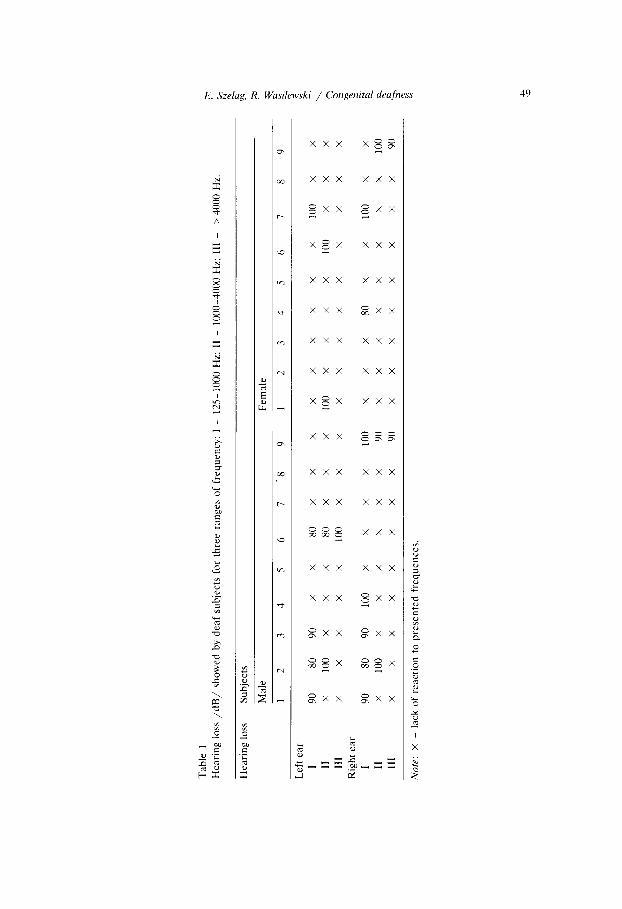
Tab
le
1 H
eari
ng
loss
/d
B/
show
ed
by
deaf
su
bjec
ts
for
thre
e ra
nges
of
fr
eque
ncy:
I
- 12
5-10
00
Hz;
II
-
lOO
f.-
Hz;
II
I -
> 40
00
Hz.
r?
? z
Hea
ring
lo
ss
Subj
ects
2
Mal
e 3
Fem
ale
? 1
2 3
4 5
6 7
-8
9 1
2 3
4 5
6 7
8 9
5
Lef
t ea
r
I
5
90
80
90
x x
80
x x
x x
x x
x x
X
100
x x
P
II
X
100
x X
X
80
x
x x
\ 10
0 x
x x
x 10
0 x
x x
III
X
X
X
X
8 X
10
0 x
x X
X
X
x
x X
X
x
x X
Rig
ht
ear
i;
I $.
90
80
90
10
0 x
x x
x 10
0 x
x x
80
x X
10
0 x
X
II
;;
X
100
x X
X
X
x
x 90
x
x x
x x
X
x x
100
i
111
X
X
X
X
X
X
X
X
90
x x
x x
x x
x x
90
P
Not
e:
x -
lack
of
re
actio
n to
pr
esen
ted
freq
uenc
es.
$
facts cxprcsxcd throc kinds of emotions. Thus 18 test faces wcrc used in the whole cxpcriment.
The synonymity of the evaluation of emotions expressed in those faces was confirmed by a competent judge method: five students wcrc to classify the pho- tographs of facts in the three groups: the ones without any emotion and those cxprcssing positive and ncgativc emotions. The subjects univocally evaluated three kinds of emotions cxprcsscd by facts. The test faces, so classified, were then prcscntcd in a proper experiment in form of slides projected on a screen by a Kodak-Cnrouscl projector cquippcd in clcctronic shutter. The stimuli were prcscntcd right or left of the fixation point which was a black spot (38’ in diameter) placed in the ccntcr of the screen. The distance bctwccn the point of fixation and the inner border of the stimulus was 7” The height of the cxposcd fact was 3”. its width 2”. The lime of presentation of each stimulus was 20 ms.
The cxpcrimcnt was conducted in a sound-proof room. The child was seated at a distance of 1.7 m from the screen on which the faces were presented.
The subject‘s task was to conccntratc his or her vision on the fixation point and to rccognizc the cxposcd test fact. In view of the difficulties deaf children have in concentrating. the subjects were taught to press a button to cxposc each face to concert the best concentration moment with stimulus prcscntation. The subject’s answer consisted in pointing to a chosen test face on a response card containing three vertically situated facts. On each rcsponsc card one face was identical with the test fact. All the facts displayed on the rcsponsc card cxprcssed the same emotions as the pl-cviously shown test fact. Each of the prcscntcd facts had two diffcrcnt response cards. Of the 30 cards used in the experiment each face was placed twice in the top position. twice in the middle position and twice at the bottom. This way the choice of f’accs prcscntcd on cards was fully balanced. The hemispheric differences wcrc mcasurcd by errors committed in left and right visual field prcscntations. The brightness of the stimuli was adjusted individually for each subject to obtain 2OL3OCC of errors. This lcvcl of errors crcatcd a fairly easy situation for the children. That is why the main expcrimcnt was prcccdcd by 21 preliminary series of tests adjusting the conditions of stimuli prcscntation. First. all the faces to be prcscnted in the session wcrc shown in full light for an unlimited period of time. next, in order to preparc the children for a short prcscntation of stimuli. the time of prcscntation was shortened to 100, 40. 20 ms. using the above experimental procedure. The next step of the preliminary scrics was a gradual reduction in light intensity to a 20-304 error level. Then, the proper cxpcrimcnt was started. The preliminary series was similar in length for all subjects and consisted of approximately SO trials. Three sessions lasting approximately 30 minutes and scparatcd by 15minute breaks made the cxpcrimcnt. The only diffcrcncc bctwccn the sessions was in the kind of emotions cxprcssed in the prcscntcd faces. In the first session facts cxprcssing positive emotions (happy) wcrc prcscntcd. in the second session facts cxprcssing negative emotions (sad). and in the third one neutral facts. The facts expressing the particular kinds of emotions were shown in both groups. six times in each session. Each session consisted of 72 trials
half of which were exposed in the right visual field and half in the left visual field. The order of presentation was random. In the experimental sessions the stimuli were presented in 18 element series separated by 2-minute rest breaks. Written instructions were given to both groups. Before each session the children were informed of the kind of emotion expressed by recognized faces.Throughout the cxpcrimcnt one of the researchers monitored the children’s behaviour, especially the subjects’ cyc movc- ments. If eye movements were observed at the moment of stimulus presentation the trial had to be repeated at the end of the series.
Results
The results are described separately for each kind of emotion expressed in the presented faces. Three separate I&factorial analyses of variance applied to mean percentage of errors were used. In each of them the investigated factors were: the group of children (deaf/hearing), visual field (left/right) and sex (boys/girls). After the analysis the Fisher test was applied.
Happy fuces
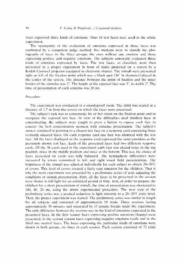
In the analysis of variance none of the tested factors or interactions reached statistical significance. The mean percentage of errors in the left and right visual fields in both groups was similar (see fig. 1). The observed lack of significant differences did not result from a similar error level found in all children in both visual fields, but rather from a considerable variability of the hemispheric pattern in individual subjects. In some children more errors were found in the right visual field while in others more errors were found in the left one. For example, five children of the normal hearing group committed more errors in the right visual field, whereas eight subjects of this group displayed opposite results (more errors in the left visual
HAPPY FACES
deaf
Fig. 1. Mean percent of errors in the left and right visual field in perception of happy faces
52
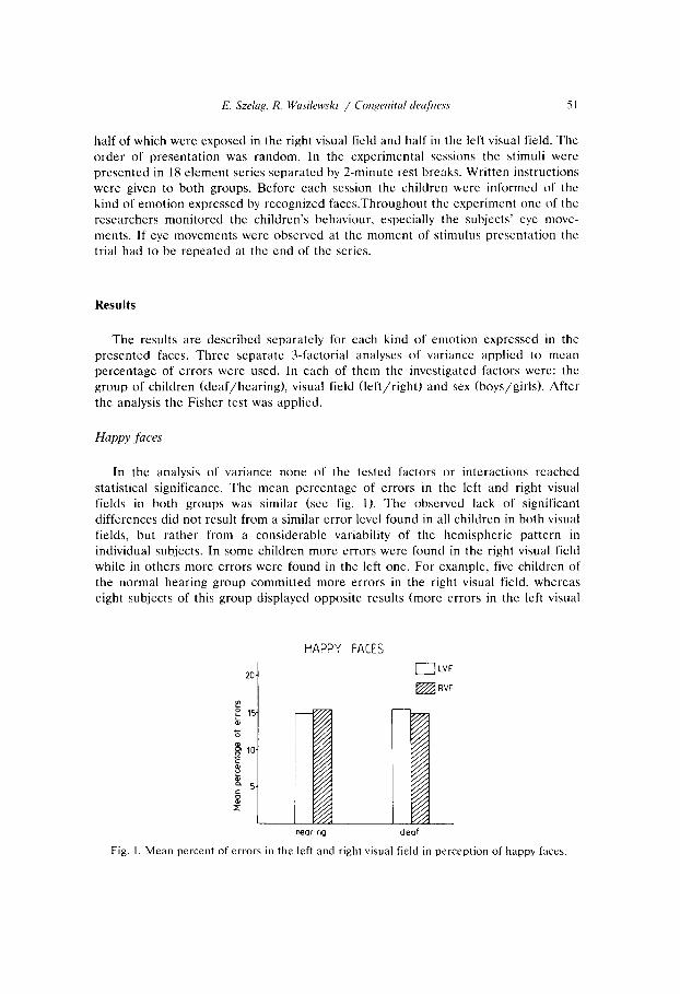
SAD FACES
20. 0 LVF
: m RVF
: 15. 01
heormg deaf
Fig. 2. Mean prrcent of errors in the left and right visual field in perception of sad faces
field) and the five remaining ones had an identical error lcvcl for both visual fields. In the group of deaf subjects. nine children had more errors in the right visual field, tight in the left one, whereas one child showed the identical level of errors in both fields.
Sad Jaws
The analysis of variance rcvcaled statistical significance of (I) visual field (F = 12.07. d/‘= l/36, p < O.OOS); (2) interaction visual field x group of subjects (F = 4.34, df= l/36, p < 0.05). The mean percentage of errors in both groups of children was higher in the right field (13.2%) than in the left one (10.8%, fig. 2). the difference king considerably larger in normal (3.8%) than in deaf children (1.1%). For children with normal hearing the mean percentage of errors in the right visual field (13.1) was significantly higher (p < O.OI - Fisher test) than in the left one (9.3%). Such differences occurred in 15 children. In two subjects the relation was converse (more errors in the left visual field). In one person both visual fields had a similar error Icvel. In deaf children, on the other hand, the difference in error levels between two fields was statistically insignificant (Fisher test). The mean percentage of errors in the right visual field was 13.3% and in the left one 12.2%~. In eight children the mean percentage of errors in the right visual field was higher than in the left one, in seven, the relation was reversed (ix. they committed more errors in the left visual field). In three children error levels wcrc identical in both visual fields.
Similarly to the results observed for sad facts, the analysis of variance also showed statistical significance of (I 1 visual field (F = 14.07. df’= l/36, p < 0.001); (2) interac-
’ Results for neutral faces were presented in our earlier paper (Sselag rt al. 109 1) in which the
hemispheric specialization for verbal and non-verbal material was tested in deaf children.
E. Szelag, R. Wasilewski / Congenital drafnrss 53
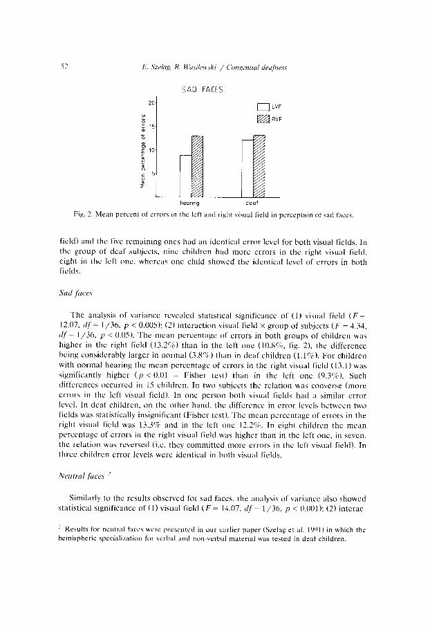
NEUTRAL FACES
DLVF
heorlng deaf
Fig. 3. Mean percent of errors in the left and right visual field in perception of neutral faces
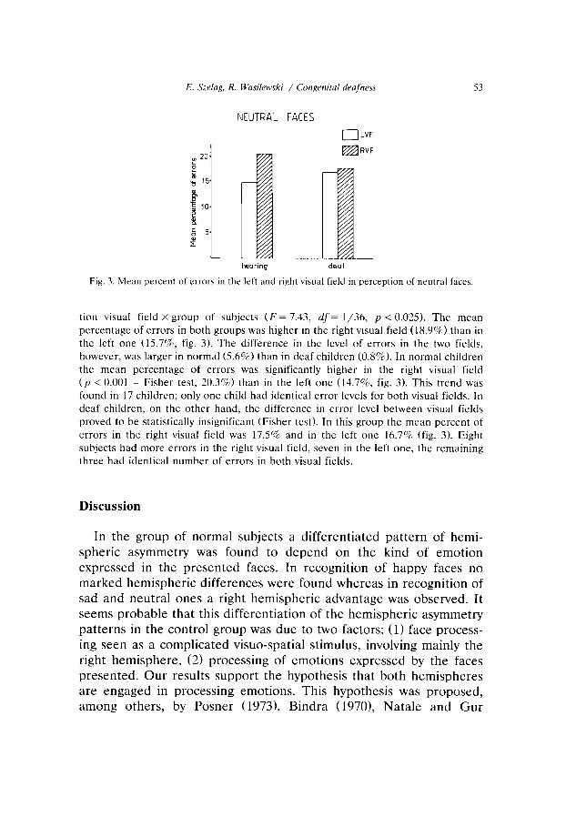
tion visual field x group of subjects (F = 7.43, df‘= l/36, p < 0.025). The mean percentage of errors in both groups was higher in the right visual field (18.9%) than in the left one (15.7’S, fig. 3). The difference in the level of errors in the two fields, however, was larger in normal (5.6%) than in deaf children (0.8%). In normal children the mean percentage of errors was significantly higher in the right visual field (p < 0.001 - Fisher test, 20.3%) than in the left one (14.7%, fig. 3). This trend was found in 17 children; only one child had identical error levels for both visual fields. In deaf children, on the other hand, the difference in error level between visual fields proved to be statistically insignificant (Fisher test). In this group the mean percent of errors in the right visual field was 17.5% and in the left one 16.7% (fig. 3). Eight subjects had more errors in the right visual field, seven in the left one, the remaining three had identical number of errors in both visual fields.
Discussion
In the group of normal subjects a differentiated pattern of hemi- spheric asymmetry was found to depend on the kind of emotion expressed in the presented faces. In recognition of happy faces no marked hemispheric differences were found whereas in recognition of sad and neutral ones a right hemispheric advantage was observed. It seems probable that this differentiation of the hemispheric asymmetry patterns in the control group was due to two factors: (11 face process- ing seen as a complicated visuo-spatial stimulus, involving mainly the right hemisphere, (2) processing of emotions expressed by the faces presented. Our results support the hypothesis that both hemispheres are engaged in processing emotions. This hypothesis was proposed, among others, by Posner (19731, Bindra (1970), Natale and Gur
(19801, and Safer (1981). It could be supposed that the advantage of the right hemisphere observed in recognition of sad faces was due to the fact that both negative emotions and faces (as patterns requiring visuo-spatial analysis) arc processed more effectively by the right hemisphere. Consequently, the lack of hemispheric asymmetry in recognition of happy faces may have resulted from positive emotions being analyzed by the left hemisphere, faces, on the other hand, being processed, as visuo-spatial stimuli by the right hemisphere (as re- ported in the literature). Interrelation of these two opposing factors, processing of positive emotions and processing of faces, may have led to the suppression of hemispheric differences. Our interpretation of control group results is compatible with a suggestion put forward by Safer (1981). He showed that in analyzing facts expressing positive and negative emotions more distinct hemispheric asymmetry then observed is due rather to processing faces as complicated visuo-spatial patterns than due to emotions expressed in those faces.
The pattern of hemispheric asymmetry in the deaf children was found to be different from that in the control group. For all kinds of emotional faces no functional hemisphere differences were observed. The results of our previous experiment (Szelag et al. 1991) could be helpful in explaining the above data. In that experiment, the same group of deaf children showed right hemisphere dominance in word recognition, i.e. a pattern opposite to that found in normal children of the same age. Those data may have resulted from a developmental delay in left hemispheric specialization. Such a delay can occur in deaf children as they arc forced to use visuo-spatial information while communicating with others by sign and gestures. According to the literature, this visuo-spatial, holistic information processing is charac- teristic for the right hemisphere (Bradshaw and Nettleton 19811. It may be assumed that gradual formation of the left hemispheric specialization in deaf children develops as a result of linguistic stimu- lation with the beginning of school education at the age of 6-7 years (lerning of speech articulation, reading and writing). Such specializa- tion formation may last considerably longer in deaf than in normal children. Some support for such an interpretation was provided by Kelly and Tomlinson-Keasey (1981) who showed that deaf children, of the same age as our subjects, displayed a dominance of the right hemisphere in verbal tasks. Probably, at that ontogenetic stage, be- cause of right hemisphere involvement in language processes, the
E. Szelag, R. Wasilewski / Congenital deafness 55
analysis of other materials (e.g. emotions expressed in faces and complex visuo-spatial stimuli) takes place, by way of compensation, in both hemispheres. Although no research data concerning hemispheric representation of emotions have been reported for deaf children, some authors (Panou and Sewell 1984; Gibson and Bryden 1984; Phippard 1977) have proved the lack of hemispheric asymmetry in deaf children in processing non-verbal, visuo-spatial materials differ- ent from ours. That would then support our interpretation. It can also be supposed that in the deaf, both hemispheres are engaged in processing visuo-spatial and emotional material, which is probably caused by right hemispheric involvement in verbal tasks. Gibson is also of this opinion (Gibson and Bryden 1984).
To conclude, our experiment is another attempt to solve the prob- lem of the role of auditory experience in organizing functional hemi- spheric asymmetry in ontogenesis. The results obtained both in the previous and present experiments allow to suppose that hemispheric asymmetry depends on auditory experience. The serious limitation of that experience leads to a formation of a hemispheric specialization pattern different from that found in persons with normal hearing.
References
Beaumont, J.G. (ed.). 1982. Divided visual field studies of cerebral organization. New York:
Academic Press.
Bindra, D., 1970. ‘Emotion and behavior theory: Current research in historical perspective’. In:
P. Block (ed.), Physiological correlates of emotion. New York: Academic Press.
Bradshaw, J.L. and C.N. Nettleton, 1981. The nature of hemispheric specialization in man. The
Behavioral and Brain Sciences 4, 4-57.
Briggs. G.C. and R.D. Nebes. 1975. Patterns of hand performance in a student population.
Cortex 11, 230-238.
Bryden, M.P., 1982. Laterality, functional asymmetry in the intact brain. New York: Academic Press.
Damasio, A.. V. Bellugi, H. Damasio, H. Poizner and J. van Gilder, 1986. Sign language aphasia
during left hemisphere amytal injection. Nature 322, 6077.
Douglas, E. and I.C. Richardson, 1959. Aphasia in a congenitally deaf mute. Brain 83, 68-80.
Ellis, H.D. and I.W. Shephard, 1975. Recognition of upright and inverted faces presented in the
left and right visual fields. Cortex 11, 3-7.
Gainotti, G. 1972. Emotional behavior and hemispheric side lesions. Cortex 8, 41-45. Gainotti, G., 1988. ‘Disorders of emotions and affects in patients with unilateral brain damage’.
In: F. Boiler and J. Grafman (eds.), Handbook of neuropsychology. Amsterdam: Elsevier.
Gainotti, G. and C. Caltagirone, 1989. Emotions and the dual brain. Experiment brain research
series 18. Berlin: Springer-Verlag.
Gardner, H., lY75. The shattered mind. New York: Knopf.
Geschwind, N. and W. Levitsky, 1968. Human brain: Left-right asymmetries in temporal speech
region. Science 1961, 386-1X7.
Goldstein. K.. 1939. The organism. New York: American Book.
Gibson. C.J. and M.P. Bryden. 19X4. Cerebral laterality in deaf and hearing children. Brain and
Language 23, I 12. Hansch, EC. and F.J. Pirozzollo. IYXO. Task relevant effects on the assessment of cerebral
specialization for facial recognition. Brain and Language IO. 5 l-59. Hecaen. H.. 19X1. ‘The neuropsychology of face recognition’. In: G. Davis, H. Ellis and J.
Shephard teds.). Perceiving and remembering faces. New York: Academic Press. pp. 39-54.
Hellcr. W. and J. Levy, 19X1. Perception and expression of emotion in right-banders and
left-banders. Neuropsychologia 19, 263-372.
Kelly. R. and C. Tomlinson-Keasey. 10x1. The effect of auditory input on cerebral laterality.
Brain and Language 1.3, 67777.
Kimura. D., R. Baltison and B. Luhcrt. 1076. Impairment of non-lingutstic hand movcmcnta in
deaf aphasic. Brain and Language 3, 566-571.
Klein, D.. M. Moscovitch and C. Vigna, lY7h. Attentional mechanisms and perceptual asymme-
tries in tachistoscopic recognition of words and faces. Neuropsychologia 14. 55-66.
Kolb, B. and B. Milner, 19X1. Obsetwtions on spontaneous facial expression after focal cerebral
excision and after intracarotid injections of sodium amytal. Neuropsychologia IY. 505-5 14.
Lavadas, E., C. Umilta and P. Ricci-Bitti, IYXO. Evidcncc for sex differences in right hemisphere
dominance for emotion. Neuropsychologia IX. .i~~l~.ih~~.
Levine, SC. and A. Cahn. lY79. Lateral asymmetries in the recognition of words, familiar faces
and unfamiliar faces. Neuropsychologia 17. 619-635.
Ley. R. and M. Bryden. 1979. Hemispheric differences in processing emotions and face\. Brain
and Language 7. 177-13X.
Manning. A.. W. Gable. R. Markman and T. Lahreche. 1977. Lateral cerebral differences in the
deaf in response to linguistic and non-linguistic stimuli. Brain and Language 1. 300-322.
Marcotte. A.C. and R.C. La Barba. IYXS, Cerebral lateralizntion for speech in deaf and normal
children. Brain and Language 26. 24&25X.
Marzi, C.A. and G. Bcrlucchi, 1977. Right visual field superiority for accuracy of recognition ot
famous faces in normals. Ncuropsychologia IS, 7.51-756.
McKeever, W.F.. H.W. Hocmann. V.A. Florian and A.D. van Deventer. lY7h. Evidence of
minimal cerebral asymmetries for processing of English words and American sign language.
Neuropsychologia 14. 313-423.
Natale, M. and R. Cur. lY80. Differential hemispheric lateralization of positive and negative
emotions in normals. Paper given at the Third INS European Conference, Chianciano-Terme.
Italy.
Neville. H.J.. 1978. ‘Electroencephalographic testing of cerebral specialization in normal and
congenitally deaf children: A preliminary report’. In: S.I. Segalowitz and F.A. Gruber teds.),
Language development and neurological theory. New York: Academic Press, pp. 121-131.
Panou. L. and D.F. Scwell. 1984. Cerebral asymmetry in congenitally deaf subjects. Neuropsy-
chologia 22. 3X1-385.
Patterson, K. and J.L. Bradshaw. 1975. Differential hemispheric mediation of nonverbal visual
stimuli. Journal of Experimental Psychology: Human Perception and Performance 1, 246-252.
Phippard. D., 1077. Hemifield differences in visual perception in deaf and hearing subjects.
Neuropaychologia IS. 555~561.
Poizner. H., R. Battison and H. Lane. lY7Y. Cerebral asymmetry for American sign language:
The effects of moving stimuli. Brain and Language 7, 351-362.
Poizner. H. and H. Lane, 197’). Cerebral asymmetry in the perception of American sign
language. Brain and Language 7. 210-226.
E. Szelag, R. Wasilewki / Congenital deyfness 51
Posner, M., 1973. Cognition: An introduction. Glenview. IL: Scott, Toresman.
Reuter-Lorenz, P. and R. Davidson. 1981. Differential contributions of the two cerebral
hemispheres to the perception of happy and sad faces. Neuropsychologia 19, 6099613.
Reuter-Lorenz, P., R. Gives and M. Moscovitch, 1983. Hemispheric specialization and the
perception of emotion: Evidence from right-handers and from inverted and non-inverted left-handers. Neuropsychologia 21, 687-692.
Reynolds, D.M. and M.A. Jevees. 1978. A developmental study of hemisphere specialization for recognition of faces in normal subjects. Cortex 14. 51 l-520.
Rizzolatti. G., C. Umilta and G. Berlucchi, 1971. Opposite superiorities of the right and left
cerebral hemispheres in discriminative reaction time to physionomic and alphabetical mate-
rial. Brain 94, 431-442.
Safer. M.. 1981. Sex and hemisphere differences in accesses to codes for processing emotional
expressions and faces. Journal of Experimental Psychology: General 110, 866100.
Sarno, J.E., L.P. Swisher and M.T. Sarno, 1963. Aphasia in a congenitally deaf man. Cortex 5,
398-414.
Scholes. R.J. and I. Fischler, 1979. Hemispheric function and linguistic skill in the deaf. Brain
and Language 7. 336-350.
Sergent, J.. 1982. About faces: Left-hemisphere involvement in processing physiognomies.
Journal of Experimental Psychology: Human Perception and Performance 8. l-14.
Strauss. E. and M. Moscovitch, 1981. Perceptual asymmetries in processing facial expression and facial identity. Brain and Language 13. 308-332.
Suberi, M. and W. McKeever, 1977. Differential right hemispheric memory storage of emotional
and non-emotional faces. Neuropsychologia 15, 7577768.
Szelag, E. and B. Czachowska-Sieszycka, 1986. Measurement of lateral differences for faces in a
two-response paradigm. Acta Neurobiologiae Experimentalis 46, 2133221.
Szelag, E. and E. Fersten, 1991. Recognition of faces expressing emotions in patients with
unilateral brain damage. Acta Neurobiologiae Experimentalis 51, 45-53.
Szelag. E., R. Wasilewski and E. Fersten, 1991. Hemispheric differences in the perception of
words and faces in deaf and hearing children. Scandinavian Journal of Psychology 32 (in press).
Terzian, H., 1964. Behavioral and EEG effects intracarotid sodium amytal injection. Acta
Neurochir. 12. 230-239.
Tucker. D.M., R.S. Roth, B.A. Arneson and V. Buckingham. 1977. Right hemisphere activation
during stress. Neuropsychologia 15. 697-700.
Tureen. L.L., E.A. Smolik and I.M. Tritt, 1951. Aphasia in a deaf mute. Neurology 1, 237-249.
Wilson, B., 1983. A comparison of deaf, normal and brain damaged adults on a tachistoscopic task. Brain and Language 19, 1X1-190.
Witelson. S.F. and W. Pallie. 1973. Left hemisphere specialization for language in the newborn (neuro-anatomical evidence of asymmetry). Brain 96, 641-646.