the dorsal raphe nucleus and serotonin:...
TRANSCRIPT

G. Di Giovanni, V. Di Matteo & E. Esposito (Eds.)
Progress in Brain Research, Vol. 172
ISSN 0079-6123
Copyright r 2008 Elsevier B.V. All rights reserved
CHAPTER 12
The dorsal raphe nucleus and serotonin: implicationsfor neuroplasticity linked to major depression and
Alzheimer’s disease
Kimmo A. Michelsen, Jos Prickaerts and Harry W.M. Steinbusch�
Department of Neuroscience, Faculty of Health, Medicine and Life Sciences, Maastricht University;European Graduate School of Neuroscience (EURON), PO Box 616, 6200 MD Maastricht, The Netherlands
Abstract: The dorsal raphe nucleus (DRN) is a heterogeneous brainstem nucleus located in the midbrain andpons. Via widespread projections, which target a multitude of brain areas, its neurons utilize manytransmitters to control various physiological functions, including learning, memory and affect. Accordingly,the DRN has been strongly associated with brain dysfunction, especially mood disorders such as depression,but also Alzheimer’s disease. The DRN’s most abundant transmitter, serotonin, has received the mostattention in studies on both normal brain function and disease, and lately its involvement in the regulation ofneuroplasticity has been under particular scrutiny. This chapter begins with a systematic overview of what wecurrently know about the anatomy of the DRN and its neurons, including their ascending projections. Itcontinues with a review of the transmitters of the DRN, followed by a discussion on the connection betweenthe DRN and neuroplasticity. Special emphasis is put on serotonin and its central role in neuroplasticity,which is proving to be of high priority in unraveling the full picture of the cellular mechanisms and theirinterconnections in the etiology of major depression and Alzheimer’s disease.
Keywords: dorsal raphe nucleus; serotonin; neuroplasticity; major depression; Alzheimer’s disease
Introduction
The dorsal raphe nucleus (DRN) is a bilateral,heterogeneous brainstem nucleus. It is located in theventral periaqueductal grey matter of the mesence-phalon, with a caudal tip reaching into the pons. Theneurons of the DRN innervate a multitude of targetsthroughout the brain and utilize many transmitters,of which serotonin is the most abundant andimportant one. The DRN is involved in the control
�Corresponding author. Tel.: +31-43-3881021;
Fax: +31-43-3671096; E-mail: [email protected]
DOI: 10.1016/S0079-6123(08)00912-6 233
of various physiological functions and has beenimplicated in brain dysfunction, especially mooddisorders such as depression. This chapter gives anoverview of the current knowledge about the DRNwith emphasis on its neurons, transmitters andascending projections and on its role in depressionand Alzheimer’s disease (AD).
DRN morphology
The DRN is a bilateral, heterogeneous brainstemnucleus situated in the midbrain and the pons.Most of its cells are located in the ventral part of

234
the periaqueductal grey matter of the midbrain.The nucleus’ most rostral part is at the level of theoculomotor nucleus, whereas its caudal tip lies inthe periventricular grey matter of the rostral pons.From the 1960s to the early 1980s, the morphologyof the DRN was described in the cat (Taber et al.,1960), man (Braak, 1970), the rabbit (Felten andCummings, 1979) and the rat (Steinbusch, 1981).Together with the caudal linear and median raphenucleus, the DRN forms the rostral or superiordivision of the raphe complex. The caudal orinferior division encompasses the raphe obscurus,raphe pallidus and raphe magnus nuclei and partsof the lateral reticular formation, located in themedulla and caudal pons (Steinbusch, 1981;Jacobs and Azmitia, 1992).
Serotonin is the major neurotransmitter of theDRN. The morphology of the serotonergic systemin the DRN was first described in the rat byDahlstrom and Fuxe (1964), using formaldehyde-induced fluorescence (FIF) which had been deve-loped by Falck et al. (1962) for visualization ofmonoamines. The human DRN has been estimatedto contain 235,000713,000 neurons (Baker et al.,1990), of which approximately 165,000734,000 (or70%714%) neurons contain serotonin (Baker etal., 1991). In the cat, the DRN has been estimated tocontain 35,000 neurons, of which up to 70–80% areserotonergic, as demonstrated by the FIF technique(Wiklund et al., 1981; Leger and Wiklund, 1982).The rat DRN has been estimated to containapproximately 35,000 neurons, of which aboutone-third are serotonergic (Descarries et al., 1982).This inconsistency across cat and rat studies may bedue to methodological differences, since the ratresults were based on the measured uptake oftritiated serotonin, and only medium-sized non-indolaminergic neurons were counted in the catstudies.
According to the original nomenclatureby Dahlstrom and Fuxe, the raphe nuclei (includ-ing the brainstem reticular formation) are dividedinto nine subdivisions, B1–B9. The subdivisionswere later renamed and slightly redefined whenthe nuclei were re-examined using an antibodyagainst serotonin, and what is now considered asthe DRN corresponds to the original subdivisionsB6–B7, B6 being the caudal extension. In most
species, the DRN can be divided into fivesubregions, namely the interfascicular, ventral(or ventromedial), ventrolateral (or lateral), dorsaland caudal subregions (Baker et al., 1990). TheDRN is also often divided along the rostrocaudalaxis into a rostral, middle and caudal portion.All five subregions extend from the rostral tothe middle part of the nucleus, except for thecaudal subregion, which is located in the caudalportion of the DRN. Abrams et al. (2004) haveproposed detailed stereotaxic coordinates for theboundaries of the rostral, middle and caudalportion. Accordingly, the rat and mouse DRNwere divided in three equally long parts along therostrocaudal axis. The rostral part of the rat DRNcomprises levels from �6.92 to �7.64, the middleportion levels from �7.73 to �8.45 and the caudalportion levels from �8.54 to �9.26mm bregma. Inmouse, the proposed corresponding values are�4.12 to �4.48, �4.54 to 4.90 and �4.96 to�5.32mm bregma. For both species, values arebased on stereotaxic atlases (Paxinos and Watson,1997; Paxinos and Franklin, 2001) and theauthors’ own immunostainings with tryptophanhydroxylase (TPH) (Abrams et al., 2004). Thesecoordinates deal with the rostrocaudal axis only,but division into the five subregions is fairly easyto make on a morphological basis.
Neuron types
In human, the DRN contains four main morpho-logical neuronal types (+ ¼ average meandiameter) round (+, 2774 mm), ovoid (+,2774 mm; variance 20–39 mm), fusiform (+,1876 mm; variance 9–37 mm) or triangular(1372 mm) cells (Baker et al., 1990). In rat, theyappear similar and have been described as eithersmall round (+, 1073 mm), medium-sized fusi-form and bipolar (+, 2474 mm and 874,respectively), large fusiform (+, 1872 and length3172 mm) and very large multipolar (+,3975 mm) (Steinbusch et al., 1981; Steinbusch,1984).
The four main morphologically different types ofDRN neurons are differentially distributed withinthe DRN, which seems to reflect neurochemical and

235
functional specialization. Indeed, an increasingnumber of studies have supported this notion.Electrophysiological studies in the 1980s led to adivision of rat serotonergic DRN neurons into twotypes, which were named Type I and Type II (ortypical and atypical serotonergic neurons, respec-tively). Type I neurons exhibited a rhythmic firingpattern and were called clock-like neurons, whereasType II neurons fired irregularly and were callednon-clock-like (Nakahama et al., 1981). Morerecently, each type was divided into three distinctclasses based on firing patterns during the sleep–wake cycle as measured by single-unit recordings incats. In addition, non-serotonergic DRN neuronswere divided into three groups as well (Sakai andCrochet, 2001).
Properties
Classes I-A and I-B displayed a regular firingpattern during waking. During waking, dischargerates were higher than during slow-wave-sleep(SWS), whereas almost no firing occurred duringparadoxical sleep (PS, also known as rapid eyemovement (REM) sleep). I-C neurons differedfrom I-A and -B by maintaining a fairly high levelof tonic, rhythmic activity during SWS and PS,Class II-A neurons’ irregular and high dischargerates correlated with motor activity and was highduring active wakefulness (AW; presence of grossbody movements), feeding and grooming, whereasrates were significantly lower or absent duringquiet wakefulness (QW; absence of gross bodymovements), SWS and PS. Class II-B neuronsdisplayed their highest rate of tonic activity duringdeep SWS and very low rates during PS, as well asAW and QW, but were strongly activated duringfeeding and grooming. II-C fired irregularly andslowly during waking periods of no physicalactivity and was reduced during PS (Sakai andCrochet, 2001).
In addition, non-serotonergic neurons wereidentified, and divided into three groups: TypeI-S, Type I-R and phasic neurons. I-R and phasicneurons have a brief action potential and fastfiring rate, whereas I-S neurons have a similardischarge activity as serotonergic neurons except
that their tonic increase during PS compared toSWS (Sakai and Crochet, 2001).
Distribution
About two-thirds of presumed serotonergic neu-rons were confined to classes I-A and I-B, whichwere evenly distributed throughout the DRN.They seemed to be identical to previously identi-fied serotonergic neurons in cat. Only 6% ofserotonergic neurons were confined to Type I-C,and were mainly located in the ventral region ofthe DRN. Type II-A neurons were preferentiallylocated in the middle parts and Type II-B neuronspreferentially in the most rostral and dorsal partsof the DRN. Type II-C neurons were located inthe ventral portion of the DRN, close to themedial longitudinal bundle and the nucleus annul-laris. Each class of Type II neurons constituted8–12% of all serotonergic neurons. To summarize,the clock-like Class I neurons, which are generallywaking-dependent, comprise almost three-fourths of serotonergic DRN neurons, whereasthe non-clock-like Class II neurons, which aregenerally motor-dependent, make up less thanone-fourth (Sakai and Crochet, 2001).
Projections
Efferent projections of the DRN
Serotonergic neurons of the DRN display atopographic organization along the rostrocaudalaxis, with respect to efferent projections (Abramset al., 2004). Thus, neurons located more rostrallyproject to more rostral areas of the brain thanneurons located more caudally in the DRN.
Yet, individual neurons seem to project toseveral distinct but functionally related targetsthrough branched fibres (Lowry, 2002). The firstbranched projections to be discovered run fromthe dorsal DRN along the dorsal raphe corticaltract to the substantia nigra (SN) and caudate-putamen (CP) (van der Kooy and Hattori, 1980a;Imai et al., 1986). Also, single neurons have beenobserved to target hippocampus and entorhinalcortex (Kohler and Steinbusch, 1982), prefrontal

236
cortex and nucleus accumbens (NA) (VanBockstaele et al., 1993), the paraventricularnucleus (PVN) of the thalamus and the lateralparabrachial nucleus (PBN) (Petrov et al., 1992),the central nucleus of the amygdala (CeA) and thePVN (Petrov et al., 1994), distinct sites in thetrigeminal somatosensory pathway (Kirifideset al., 2001) and the vestibular nuclei and CeA(Halberstadt and Balaban, 2006).
This could be a key to understanding the role ofthe DRN as a modulator of complex autonomicfunctions with anatomical correlates in severalparts of the brain. For instance, both the CeA andthe PVN, which are targeted by the same branchedfibres, are involved in anxiety and conditioned fear(Petrov et al., 1992, 1994). These fibres emergefrom well-defined subpopulations of neurons inthe medial part of the middle DRN as well as morecaudal clusters.
However, only a part of the neurons withbranched axons contain serotonin, the reportedrange being between 8% (Petrov et al., 1992) and64% (Halberstadt and Balaban, 2006) dependingon the targets. This serves as a reminder thatserotonin is not the only transmitter utilized by theDRN. For instance, the CeA-PVN projectingsubpopulations mentioned above (where abouthalf the neurons are serotonergic) also containcorticotropin-releasing factor (CRF), which hasbeen associated with anxiety and other mooddisorders. Anxiety-related behavioural changesinduced by serotonergic activity, such as develop-ment of learned helplessness, seem to be CRF-dependent (Maier and Watkins, 2005). However, ithas not been shown, whether the CRF-containingneurons themselves, or the serotonergic DRNneurons they target, send collaterals to CeA andPVN.
Early studies showed that most DRN neuronsproject ipsilaterally and few contralaterally (Milleret al., 1975). Retrograde labelling studies of DRNefferents to the entorhinal cortex indicated that,when present, contralateral terminals are prefer-entially located close to the midline (Kohler andSteinbusch, 1982). Similar results were obtainedrecently in a study by Waselus and co-workers, inwhich all DRN neurons, which sent collaterals tolateral septum and striatum, were located
ventromedially near the midline or slightly lateralto it. Notably, all such collateral neurons wereserotonergic (Waselus et al., 2006). However,single neurons do not seem to project collaterallyto both hemispheres (van der Kooy and Hattori,1980b; Kohler and Steinbusch, 1982). Besides theirtopographic organization, different cell types alsoseem to display different projections. This has,however, not been extensively studied but isreflected in the distribution patterns of differentcell types versus the projections emerging fromdifferent areas.
Fibre morphology
Fibres arising from the DRN are characteristicallyvery fine and have small varicosities, which aregranular or fusiform in shape (so-called type Daxons). This is in contrast to fibres arising fromthe medial raphe nuclei (MRN), which displaylarge, spherical varicosities (so-called typeM axons)and variations in fibre thickness (Kosofsky andMolliver, 1987). Serotonin-immunoreactive fibresdisplay similar variation, which may help togive an indication of the origin of serotonergicfibres in purely immunohistochemical preparations(Kosofsky and Molliver, 1987; Mulligan and Tork,1988). At the light microscopic level, three types ofserotonergic axons can be distinguished. Of these,one arises from the DRN while two are continuouswith each other and arise from the MRN: (1) fine(less than 0.5 mm) DRN fibres with small fusiformvaricosities (generally less than 1 mm in diameter);(2) thick (about 1 mm) and smooth non-varicoseMRN fibres, which travel long distances in straighttrajectories; (3) shorter and thinner varicose fibres(varicosity size 1 mm or more in diameter) arisefrom the thick MRN fibres. The thinner DRNfibres branch frequently and target large, oftendiffuse areas, whereas the thicker fibres branchinfrequently, and are often seen to surround thesomata of single neurons (Mulligan and Tork,1988). On an electron microscopic level, theDRN fibres display small, fusiform boutons andare believed to signal predominantly via volumetransmission, whereas the MRN fibres contacttheir target via large round boutons, often in largenumbers (Tork, 1990). The morphology and origin

237
of the fibres has also been linked to differentialdrug-sensitivity, first demonstrated in the fore-brain, where the neurotoxic amphetamine deriva-tives methylenedioxyamphetamine (MDA) andp-chloroamphetamine (PCA) induce denervationof the fine axons, whereas the thick ones areunaffected by the drugs (Mamounas and Molliver,1988; O’Hearn et al., 1988; Mamounas et al.,1991). A suggested reason for this difference isserotonin transporter (SERT) expression, which,in amygdala, is present in the thick MR drug-insensitive fibres but lacking from the thin DRNdrug-sensitive ones (Brown and Molliver, 2000).
Thus, functionally the serotonergic fibres seemto be organized into two main subsystems, ofwhich the DRN system has a more widespreadinfluence via its highly divergent branches andvolume transmission, while the MRN system hasextensive and direct synaptic contacts with neuro-nal somata.
Pathway overview
The DRN projects along several ascending anddescending pathways, most of which it shareswith one or more of the other raphe nuclei. The
Fig. 1. The three ascending pathways (APs) of the rat DRN and th
cortex; DH, dorsal hypothalamus; GP, globus pallidus; HPC, hippoc
nucleus; LH, lateral hypothalamus; LS, lateral septum; MT, midline
nucleus accumbens; SN, substantia nigra; STN, subthalamic n
hypothalamus. (Reprinted from Michelsen et al., 2007, with permissi
pathway nomenclature differs slightly betweenauthors and the division is not completely con-sistent, with some overlaps and contradictions,especially in the older literature. In this review,we have labelled the pathways according toSteinbusch et al.
Ascending pathways
There are three ascending pathways: the dorsal,medial and ventral ascending pathways (Fig. 1). Ofthese, the dorsal and ventral ascending pathwaysare the two most important efferent projectionsof the DRN. They reach a multitude of targetsthroughout the forebrain, the most important onebeing the CP.
Descending pathways
In addition, four descending projections leave theDRN: the bulbospinal pathway, cerebellar path-way, propriobulbar pathway and one that inner-vates the locus coeruleus, dorsal tegmental nucleusand pontine raphe nucleus. The main targets ofthe descending pathways are cerebellum, the lower
eir main targets. AM, amygdala; CP, caudate-putamen; CTX,
ampus; ILT, intralaminar thalamic nuclei; IP, interpeduncular
thalamic nuclei; NAs, shell of nucleus accumbens; NAc, core of
ucleus; PH, posterior hypothalamus; VMH, ventromedial
on from Elsevier) (See Color Plate 12.1 in color plate section.)

238
brainstem and the spinal chord. These pathwayswill not be further dealt with in this chapter.
The dorsal ascending pathway
The dorsal ascending pathway rises from medialand rostral DRN. Eighty per cent of its fibres areserotonergic. It innervates the striatum and globuspallidus (GP).
Striatum
Anterograde labelling has shown that DRN effer-ents target the ventromedial striatum at caudal tomidlevel (Vertes, 1991). Most striatum-projectingneurons are located in rostral DRN (Steinbusch etal., 1981). Recent retrograde labelling studiesconfirmed a gradient of striatum-projecting neuronsin the DRN. Approximately half of the neuronswere located in the rostral third of the DRN. Threeout of eight neurons were seen in middle DRN,parallell to the midline, with highest concentrationsdorsomedially and ventrally. Only one out of eightneurons were located in caudal DRN, in itsdorsomedial part (Waselus et al., 2006).
The CP is extensively innervated by neurons ofthe DRN. It is the single most important of targetsfor DRN innervation and one of the first to beextensively studied. The earliest anatomical indica-tions for DRN projections to the CP (Anden et al.,1965) were subsequently supported by lesionstudies, which showed a drop in striatal TPHactivity (Geyer et al., 1976) as well as a decrease in[3H]5-HT uptake (Kellar et al., 1977) after DRNleisons, and by in vivo microdialysis, whichshowed that electrical stimulation of the DRNlead to a rise in serotonin dialysate in the CP(McQuade and Sharp, 1997). Meanwhile, moreanatomical data has accumulated. Approximatelyone-third of all serotonergic DRN neurons projectto the CP. This is, however, region-specific: in acluster in dorsomedial DRN, 80–90% of seroto-nergic neurons were found to project to the CP(Steinbusch et al., 1981). In addition, 80% ofDRN neurons that project to the CP are seroto-nergic, and they mainly project ipsilaterally. Theremaining 20% of non-serotonergic CP-projecting
neurons are mostly found in the caudal parts ofventromedian and dorsomedian DRN (see Fig. 2for an overview of DRN anatomy).
The innervation of NA is even higher than that ofthe CP. A majority of the innervation is serotoner-gic. The shell of the NA is more heavily innervatedthan the core, especially in more caudal regionswhere the fibres in the shell are muchmore abundant than elsewhere in the NA. Thecore is innervated exclusively by thin (0.3mm)smooth axons, similar to the rostral shell, which isinnervated predominantly by thin axons and, toa lesser extent, by varicose fibres. In contrast, thecaudal shell is innervated predominantly by thicker,highly varicose (0.5mm between varicosities) sero-tonergic axons (Van Bockstaele and Pickel, 1993;Brown and Molliver, 2000). It has not beendetermined that all the innervation indeed stemsfrom the DRN. However, studies on projections tocerebral cortex and olfactory bulb have shown thatthin drug-sensitive serotonin axons typically arisefrom the DRN and varicose, drug-resistant axonsarise from the MRN (Kosofsky and Molliver, 1987;Mamounas et al., 1991). In NA, the thin fibres ofthe core are more vulnerable to amphetaminederivatives than the thick fibres of the shell,indicating that the DRN innervates the NA core,as suggested by Brown and Molliver (2000).
Globus pallidus
Pallidal afferents from DRN have been demon-strated by tracing studies. Vertes (1991) usedthe retrograde tracer PHA-L in rats and DeVitoet al. (1980) used the anterograde tracerHRP in macaque monkeys. The innervation ofGP is mainly serotonergic, as confirmed by micro-dialysis studies in the rat, where stimulation of theDRN increased serotonin dialysate in the GPby 75%. In the same study, stimulation of theMRN had little or no effect (McQuade and Sharp,1997).
Medial ascending pathway
The main target of the medial ascending pathwayis SN. To a lesser extent, the pathway also
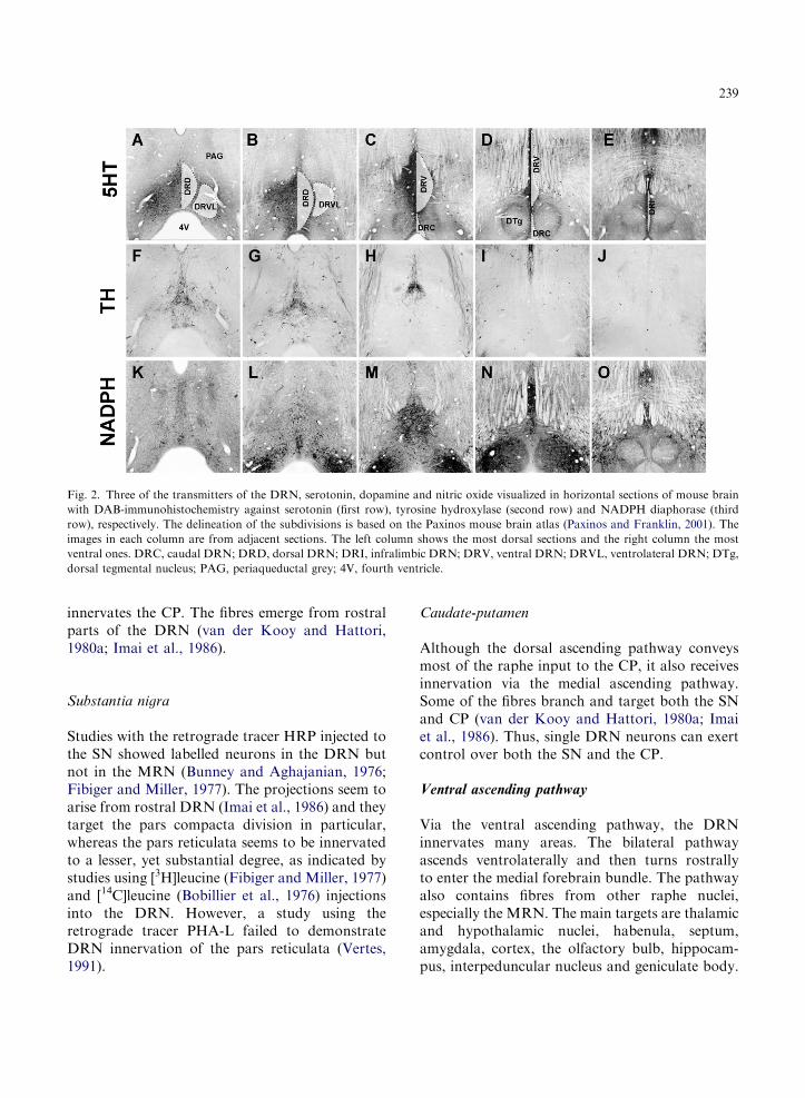
Fig. 2. Three of the transmitters of the DRN, serotonin, dopamine and nitric oxide visualized in horizontal sections of mouse brain
with DAB-immunohistochemistry against serotonin (first row), tyrosine hydroxylase (second row) and NADPH diaphorase (third
row), respectively. The delineation of the subdivisions is based on the Paxinos mouse brain atlas (Paxinos and Franklin, 2001). The
images in each column are from adjacent sections. The left column shows the most dorsal sections and the right column the most
ventral ones. DRC, caudal DRN; DRD, dorsal DRN; DRI, infralimbic DRN; DRV, ventral DRN; DRVL, ventrolateral DRN; DTg,
dorsal tegmental nucleus; PAG, periaqueductal grey; 4V, fourth ventricle.
239
innervates the CP. The fibres emerge from rostralparts of the DRN (van der Kooy and Hattori,1980a; Imai et al., 1986).
Substantia nigra
Studies with the retrograde tracer HRP injected tothe SN showed labelled neurons in the DRN butnot in the MRN (Bunney and Aghajanian, 1976;Fibiger and Miller, 1977). The projections seem toarise from rostral DRN (Imai et al., 1986) and theytarget the pars compacta division in particular,whereas the pars reticulata seems to be innervatedto a lesser, yet substantial degree, as indicated bystudies using [3H]leucine (Fibiger and Miller, 1977)and [14C]leucine (Bobillier et al., 1976) injectionsinto the DRN. However, a study using theretrograde tracer PHA-L failed to demonstrateDRN innervation of the pars reticulata (Vertes,1991).
Caudate-putamen
Although the dorsal ascending pathway conveysmost of the raphe input to the CP, it also receivesinnervation via the medial ascending pathway.Some of the fibres branch and target both the SNand CP (van der Kooy and Hattori, 1980a; Imaiet al., 1986). Thus, single DRN neurons can exertcontrol over both the SN and the CP.
Ventral ascending pathway
Via the ventral ascending pathway, the DRNinnervates many areas. The bilateral pathwayascends ventrolaterally and then turns rostrallyto enter the medial forebrain bundle. The pathwayalso contains fibres from other raphe nuclei,especially the MRN. The main targets are thalamicand hypothalamic nuclei, habenula, septum,amygdala, cortex, the olfactory bulb, hippocam-pus, interpeduncular nucleus and geniculate body.

240
Hypothalamus
Several studies have addressed the projectionsfrom the raphe nuclei to the hypothalamus.In an early autoradiographic study, in which[14C] tracing was used to map DRN efferentsin cat, Bobillier et al. (1976) identified varyingdegrees of DRN innervation in several hypotha-lamic nuclei. Most of these results were laterconfirmed, while some have been contradictedby subsequent experiments. In addition, somestudies have not distinguished between theraphe subnuclei. For instance Steinbusch andNieuwenhuys (1982) demonstrated serotonergicinnervation in nearly all parts of the hypothala-mus, but they did not attempt to locate theprojecting perikarya. Some studies have indicatedthat the MRN is a greater source of hypothalamicserotonin innervation than the DRN (Geyer et al.,1976; Kellar et al., 1977).
The DRN has been reported to innervate theSCN and preoptic area (Bobillier et al., 1976), butlater studies in the rat suggest that DRN does notproject to these structures (van de Kar andLorens, 1979; Meyer-Bernstein and Morin,1996). The dense serotonergic innervation of theSCN and medial preoptic area (Bobillier et al.,1976), and the light-to-moderate serotonergicinnervation of the rest of the anterior hypotha-lamus, seems to emerge from the MRN instead(van de Kar and Lorens, 1979; Hay-Schmidtet al., 2003).
Tracings studies have identified moderate DRNinnervation in posterior hypothalamus (Bobillieret al., 1976) and lesion studies have shown thatthe arcuate nucleus receives innervation fromDRN (van de Kar and Lorens, 1979). Further-more, the lateral hypothalamus receives highinnervation from the DRN. This was shown byearly [14C] tracing studies, such as by Bobillieret al. (1976) and later confirmed with PHA-Ltracing (Vertes, 1991). A recent anterogradetracing study showed that neurons in the centralportion of the rostral DR innervate about 23% ofthe orexinergic neurons of the lateral hypothala-mus, mainly in the lateral parts of the cluster(Yoshida et al., 2006).
Thalamus
Several of the thalamic nuclei receive innervationfrom the DRN. Studies in cat and rat havereported dense innervation in the midline andintralaminar nuclei of the thalamus (includingthe posterior paraventricular, the parafascicular,reuniens, rhomboid, intermediodorsal/mediodor-sal and central medial thalamic nuclei) andmoderate innervation in thalamic paracentraland central lateral intralaminar nuclei (Conradet al., 1974; Bobillier et al., 1976; Vertes, 1991).In addition, the subparafascicular and prethalamicnuclei (Bobillier et al., 1976) have been reported toreceive innervation from the DRN, but confirma-tion by later studies is lacking.
Habenula
The DRN innervates the lateral habenula to amoderate extent, whereas the medial habenuladoes not seem to receive any innervation in rat(Sim and Joseph, 1993), cat (Bobillier et al., 1976)and hamster (Morin and Meyer-Bernstein, 1999).One study, however, reported low innervation inthe medial habenula of rat (Morin and Meyer-Bernstein, 1999). In the same study the hamsterlateral habenula was shown to receive only sparseserotonergic innervation, indicating that the inputfrom DRN is mainly non-serotonergic.
Septum
The DRN sends strong innervation to the lateralseptum, 80% of which is serotonergic (Kohleret al., 1982). The innervation predominantlytargets the medial portions of the lateral septum(Vertes, 1991). Most of the projecting neuronsare located throughout the caudal DRN. In therest of the DRN, neurons are sparse and locatedventromedially. The neuron number decreasestowards the mid-DRN and is very low in rostralDRN, while most of the rostral parts containno septum-projecting neurons at all (Waseluset al., 2006). The medial septum is not generally
241
considered a target of DRN innervation. Micro-dialysis studies have, however, shown that stimu-lation of DRN can increase serotonin dialysate inmedial septum by more than 55%. This suggeststhat the DRN does indeed target the area, but aslong as anatomical evidence is lacking it can not beexcluded that such measurements actually sampleserotonin from the lateral septum (McQuade andSharp, 1997).
Amygdaloid complex
Studies using neuronal tracers, PHA-L in parti-cular, have demonstrated that the basolateral andlateral amygdaloid nuclei, as well as the extendedamygdala (comprising centromedial amygdala + bed
nucleus of stria terminalis and substantia innomi-
nata, as defined by Alheid and Heimer, 1988)receive dense innervation from the DRN (Grove,1988; Vertes, 1991). Also, immunohistochemicaltechniques in rat have shown that the basolateralamygdaloid nuclei receive strong serotonergicinnervation, especially the rostral and medial partsof the basal nucleus, while the caudal part of thebasal nucleus as well as the entire lateral nucleusreceive a lower, yet high density of serotonininnervation. In the centromedial nuclei innervationis very low, except for the posterior part of themedial amygdaloid nucleus and the medial andlateral parts of the posterior nucleus (Steinbusch,1981). The serotonin-immunoreactivity in theamygdaloid has not been directly correlated toDRN efferents. However, in squirrel monkeys, themost abundant serotonergic fibre type is thin, withfusiform or pleiomorphic varicosities, which suggeststhat serotonergic innervation emerges predomi-nantly from the DRN (Sadikot and Parent, 1990).A more recent immunohistochemical study inmacaque monkeys is not consistent with the ratdata, with regard to the relative fibre density inamygdaloid subnuclei, probably due to species diffe-rences. The highest levels were present in lateralsubregions of the central amygdala and dorsolateralbed nucleus of stria terminalis. Levels were high inbasal amygdala and moderate in centromedialamygdaloid nuclei (Freedman and Shi, 2001).
Cerebral Cortex
Several studies have dealt with cortical projectionsof the DRN (Bobillier et al., 1976; O’Hearn andMolliver, 1984; Vertes, 1991). O’Hearn andMolliverdemonstrated that the cortical projections of ratDRN emerge predominantly from the ventralsubnucleus, in particular from immediately dorsalor medial to the medial longitudinal fasciculi. Theseareas account for three-fourths of the DRN inner-vation of the cortex, whereas the dorsal subnucleuscontributes one-fourth. Along the rostrocaudal axis,most neurons are located in the middle DRN, andthe lateral areas of the DRN do not seem to projectto the cerebral cortex at all. More than 80% of theprojections are serotonergic (O’Hearn and Molliver,1984). The ratio of contralateral fibres is 26–35%,and differs between the subnuclei. At least inentorhinal cortex, the contralateral fibres seem topreferentially target medial areas (Kohler andSteinbusch, 1982; O’Hearn and Molliver, 1984).
The frontal cortex receives most of its serotonergicinnervation from the DRN (Kosofsky and Molliver,1987). The density is highest in the dorsal frontalcortex and low in caudal regions, with intermediatedensities in areas in between (Steinbusch, 1981). Thefrontal cortex receives projections from nearly twiceas many DRN neurons as either the parietal oroccipital cortex (O’Hearn and Molliver, 1984). Theentorhinal cortex is targeted by both serotonergic andnon-serotonergic projections (Segal, 1977) and (Koh-ler and Steinbusch, 1982) which for the most partemerge from the DRN (Kohler and Steinbusch,1982). In addition, anterograde labellings with PHA-L have shown that many cortical regions receivedense (the piriform, insular and frontal cortices) ormoderately dense (occipital, entorhinal, perirhinal,frontal orbital, anterior cingulate and infralimbiccortices) projections from the DRN (Vertes, 1991)(Fig. 3).
Hippocampus
DRN projects to the hippocampus (Segal andLandis, 1974; Azmitia and Segal, 1978). DRNefferents to the hippocampus emerge predominantly

Fig. 3. The serotonergic DRN innervation of medial prefrontal cortex (A) and hippocampus (B) are of special interest with regard to
the role of serotonin in depression and AD, respectively. Serotonin was visualized with DAB-immunohistochemistry in coronal
sections of mouse brain, (C) and (D) are details of A and B, respectively.
242
from the most caudal parts of the nucleus, close tothe midline, and is both serotonergic and non-serotonergic (Wyss et al., 1979; Kohler and Stein-busch, 1982). Immunohistochemical stainings havedemonstrated fine serotonergic axons with smallvaricosities throughout the hippocampus (Fig. 3).The fibres’ morphology (Kosofsky and Molliver,1987) suggests that they derive from the DRN(Mamounas et al., 1991). However, lesion studieshave indicated that the MRN and not the DRN isthe major source of hippocampal serotonin innerva-tion (van de Kar and Lorens, 1979).
Olfactory bulb
Tracing studies with radioactively labelled aminoacids in rat (Halaris et al., 1976) and cat (Bobillieret al., 1976) have demonstrated DRN projectionsto the olfactory bulb. The DRN is the primarysource of serotonin in the olfactory bulb, as shownby retrograde transport of [3H]serotonin (Aranedaet al., 1980a, b). Immunohistochemical stainingshave demonstrated serotonergic innervation of alllayers of the olfactory bulb, especially the glome-rular lamina (Steinbusch, 1981).

243
Supraependymal plexus
The supraependymal plexus is a network ofserotonergic fibres, which covers nearly all ventri-cular surfaces with moderate or high density. Theyare most numerous in the third ventricle and theforamina of Monro, fewer in the lateral ventriclesand aqueduct and numerous in the hypothalamicregion of the third ventricle. Areas with low densityor absence of fibres are the third ventricle floor, thepreoptic area, the roof of interventricular foramen,the subfornical organ and the roof of the fourthventricle (Richards et al., 1973; Chan-Palay, 1976;Lorez and Richards, 1982). The plexus wasdiscovered already in the 1920s (Lorez andRichards, 1982) and later identified as being mainlyserotonergic (Richards et al., 1973).
Several studies have indicated that the suprae-pendymal serotonergic fibres ascend from themedial and in particular, dorsal raphe: in rats,supraependymal fibres degenerated after lesions ofthe dorsal and medial raphe (Aghajanian andGallager, 1975), and electrical stimulation of themedial and pontine raphe led to an increase in[3H]5-HT uptake from the intracerebroventricularspace (Chan-Palay, 1976). Also [125I] tetanus toxinwas injected into the lateral ventricles labelledneurons of the medial and dorsal raphe by meansof retrograde transport along the fibres (Richards,1978). Further, there is a direct fibre pathwaybetween the DRN and the aqueduct surface in rats(Steinbusch et al., 1981), and in mice, an exit zonefor fibres to the fourth ventricle has been reportedimmediately dorsal to the DRN (Derer, 1981). Incats, a [3H]-labelled proline injection in DRN andraphe centralis superior labelled supraependymalsurfaces of all ventricles by means of anterogradetransport (Pierce et al., 1976).
Studies on the rat lateral ventricles indicate thatserotonergic fibres do not penetrate the ependyma,but instead enter the ventricles from their rostralpoles. These fibres travel along fibres that travelthrough the median forebrain bundle and turndorsocaudally between the CP and corpus callo-sum. Also, they do not form synaptic contact withependymal cells. They are not found betweenependyma and subependyma, but only in thelateral ventricles (Dinopoulos et al., 1995).
Neurotransmitters
DRN neurons utilize several other neurotransmit-ters (Fig. 3). This chapter will list such transmit-ters, but not those, which are located in afferentfibres to the DRN and synthesized elsewhere.
Serotonin
Serotonin is the main neurotransmitter of theDRN and the first one to be demonstrated there(Dahlstrom and Fuxe, 1964). The serotonergicDRN neurons and their projections have beendescribed in more detail in other parts of thischapter.
Dopamine
Dopamine was one of the first transmitters, to bedemonstrated in DRN neurons, first with histo-fluorescence methods (Lindvall and Bjorklund,1974; Ochi and shimizu, 1978) and later withantibodies against tyrosine hydroxylase (TH) anddopamine-b-hydroxylase (DbH) (Nagatsu et al.,1979). These dopaminergic neurons are locatedpreferentially in ventromedial parts. They mainlytarget the NA and lateral septum, and to a lesserextent medial prefrontal cortex. In addition,very few fibres project to CP (Stratford andWirtshafter, 1990).
GABA
GABAergic neurons were first demonstratedin the DRN by radioautographic tracing andGABA-uptake (Belin et al., 1979). The observa-tion was supported by immunohistochemistrywith an antibody against the GABA-synthesizingenzyme g-aminobutyric acid decarboxylase, orGAD (Mugnaini and Oertel, 1985) and theGABA-degrading enzyme GABA-transaminase,or GABA-T (Nagai et al., 1983). The GABAergicsynapse with serotonergic DRN neurons (Wanget al., 1992). They are markedly smaller than mostserotonergic neurons and fire spikes characterizedby short width and high frequency (Allers andSharp, 2003).

244
Peptide transmitters
Immunohistochemical stainings have shown thatthe DRN harbours neuropeptide Y (NPY) con-taining neurons, most of which are medium-sized,fusiform and bipolar (de Quidt and Emson, 1986).In situ-hybridization has demonstrated the pre-sence of NPY mRNA in the DRN (Pau et al.,1998).
Substance P has been shown to colocalize withserotonin in the DRN in at least rat (Chan-Palayet al., 1978; Hokfelt et al., 1978), cat (Lovick andHunt, 1983; Arvidsson et al., 1994) and human(Baker et al., 1990, 1991). Substance P alsocolocalizes with serotonin in ascending projec-tions, but such fibres have not been shown to arisefrom the DRN (Otake, 2005). However, in anotherstudy no colocalization was seen in ascendingfibres (Rupniak and Kramer, 1999).
Low levels of prepro-galanin mRNA are presentin DRN neurons (Cortes et al., 1990), yet galaninitself has been detected with immunohistochemistryonly after colchicine treatment (Skofitsch andJacobowitz, 1985). Galanin colocalizes with seroto-nin in the DRN. In fact, it has been reported that alarge proportion of serotonergic DRN neurons alsocontain galanin (Melander et al., 1986). Galaninis also present in serotonergic fibres in one of thetarget areas of the DRN, the cortex (Skofitsch andJacobowitz, 1985), but it has not been confirmedthat these projections arise in the DRN.
Enkephalin (ENK)-containing neurons were firstreported in the dorsal and lateral parts of ratDRN, just adjacent to the periventricular greymatter (Hokfelt et al., 1977; Uhl et al., 1979).Immunohistochemical studies showed that ENKis present throughout the cat DRN in neurons ofvariable morphology (Moss et al., 1980, 1981).However, serotonergic double-labelled neuronswere predominantly small and round and locatedat the midline, dorsal to the medial longitudinalfasciculus (Glazer et al., 1981).
CRF immunoreactivity has been demonstratedin DRN neurons after colchicine treatment(Commons et al., 2003). CRF-immunoreactiveneurons were mainly clustered in the dorsomedialsubregion, especially in the middle DRN. Scatteredneurons were seen in the lateral wings, while they
were largely absent from the ventromedial DRN andthe most caudal part of the DRN. Most (B96%) ofCRF-immunoreactive neurons in the dorsomedialDRN were serotonergic, as defined by immuno-reactivity for TPH. Anterograde tracing (PHA-L)indicated that neurons in the middle portion of thedorsomedial DRNmainly target the CeA, the dorsalhypothalamic area and the bed nucleus of the striaterminalis (Commons et al., 2003).
In additional vasoactive intestinal polypeptide
(VIP) has been demonstrated in neurons of bothrat and mouse DRN (Sims et al., 1980) andcholecystokinin (CKK)-containing neurons in therat DRN have been shown to innervate the PVN ofthe thalamus (Bhatnagar et al., 2000; Otake, 2005).
Glutamate
Phosphate-activated glutaminase (PAG) has been de-monstrated in TH-, DbH- or phenylethanolamine-N-methyltransferase (PNMT)-immunoreactive neurons,suggesting that glutamate is formed from glutaminein serotonergic and catecholaminergic neurons of theDRN (Kaneko et al., 1990).
Nitric oxide
The presence of nitric oxide (NO) in DRN wasfirst demonstrated by immunohistochemistryagainst the NO synthesis reaction product citrul-line (Pasqualotto et al., 1991) and against argini-nosuccinate synthetase which turns citrulline intoargininosuccinate (Nakamura et al., 1991). Subse-quently, the presence of NO in both serotonergicand non-serotonergic DRN neurons was demon-strated by colocalization of serotonin-immuno-reactivity with immunoreactivity for NO synthase(NOS) (Dun et al., 1994; Rodrigo et al., 1994) orwith NADPH diaphorase activity (Johnson andMa, 1993; Wotherspoon et al., 1994). The NO-synthesizing neurons are predominantly clusteredin medioventral and mediodorsal parts of DRN(Wang et al., 1995). In the medial subnuclei,between 23 and 38% of serotonergic neuronsappear to synthesize NO, whereas 60–77% ofNO-synthesizing neurons are serotonergic. Inthe lateral subregions, NADPH diaphoraseactivity is present, but its activity does not overlap

245
with serotonergic neurons (Wotherspoon et al.,1994).
Transient presence of additional transmitters
At least two additional neurotransmitters havebeen reported in the developing, but not adult,DRN. Histamine is present in neurons of rat andmouse DRN during embryonic development, butdisappears before birth, as demonstrated by thepresence of histamine-immunoreactivity and histi-dine decarboxylase (the histamine-synthesizingenzyme) mRNA (Auvinen and Panula, 1988;Nissinen and Panula, 1995; Nissinen et al., 1995;Karlstedt et al., 2001). Recent studies have shownthat the gastrointestinal peptide secretin is alsopresent in the DRN during mouse embryonicdevelopment (Lossi et al., 2004).
Plasticity
During the last few decades the traditional view ofthe adult brain as a static network of cells andfibres has given way for increased understandingof the importance of plasticity in the CNS.Neuroplasticity is now seen as an indispensabletrait, which allows the CNS to adjust to itsenvironment, as a result of experience or followinginjury, by undergoing adaptive changes at severalstructural and functional levels. It encompasses,for instance, the outgrowth or shrinkage ofdendrites and axons, as well as neurochemicalchanges at the synapse. In addition, neurogenesishas fairly recently been added to the list.
Among the vast number of functions attributedto serotonin, is regulation of many forms of neuralplasticity. It has been proposed that the seroto-nergic system of the DRN and other raphe nuclei,with its plastic properties, is a key player in thebrain’s integration with the rest of the body andthe environment (Azmitia, 1999). The proposalis in line with, but extends beyond, previousconcepts, and states that the serotonergic systemhas a unique and wide homeostatic role, whichinvolves feedback regulation by a variety ofneuronal and non-neuronal factors in the body,
including neurotransmitters, glucocorticoids,steroids and oxygen.
It is by now well established that serotoninregulates sprouting, synaptic plasticity and neuro-genesis and thereby seems to be deeply involved inthe regulation of most forms of neuroplasticity.
Sprouting
Serotonergic fibres are plastic in the adult brain.Lesioning studies have shown that serotonergicaxons are able to regenerate very fast, as firstshown in the spinal chord (Nobin et al., 1973) andhypothalamus (Frankfurt and Azmitia, 1984).Serotonergic neurons also affect fibre plasticity intheir target cells and areas, and can have bothpromoting and inhibiting effects, although moststudies have demonstrated that serotonin stimu-lates sprouting. For instance, serotonin triggeredgrowth cone retraction in the chick dorsal rootganglion (Igarashi et al., 1995) and inhibitedneurite outgrowth in goldfish retina (Lima et al.,1994). In addition, PC12 cells developed neuritesin the presence of serotonin (Severin and Kon-dratyev, 1988) and serotonin enhanced neuriteoutgrowth in thalamic mouse (Lotto et al., 1999)and rat (Lieske et al., 1999) neurons in vitro.
Synaptogenesis
Serotonin has a stimulating effect on synaptogen-esis. Serotonin depletion by treatment with theTPH inhibitor p-chlorophenylalanine (PCPA)leads to synapse loss in adult rat cortex, hippo-campus and hypothalamus (Chen et al., 1994;Azmitia et al., 1995), as well as in early postnatalrat hippocampus (Mazer et al., 1997). TPHinhibition also leads to learning deficits in rat(Mazer et al., 1997).
Neurogenesis
The traditional view of the mammalian nervoussystem as being entirely postmitotic, has onlyrecently been challenged, and changed, by thediscovery of neurogenesis in the adult mammalianbrain (Eriksson et al., 1998). Already in the1960s, 3H-autoradiography studies identified new

246
neurons in the rat brain (Altman and Das, 1965)but, for a long time, the finding did not receivethe attention it would have deserved. Almost twodecades later, neurogenesis was demonstratedconvincingly in the canary forebrain (Goldmanand Nottebohm, 1983), and during the 1990s itbecame accepted that in humans, primates androdents, proliferation and neurogenesis take placein two areas of the adult brain: the subgranularlayer of the dentate gyrus of the hippocampus andthe subventricular zone (Momma et al., 2000).Both areas are targeted by fibres from the raphenuclei and a multitude of evidence points toserotonin as a key player in the regulation ofneurogenesis. For instance, lesions to the raphenuclei lead to decreased cell proliferation inthe hippocampus, but the effect can be counter-acted by a raphe transplant (Brezun andDaszuta, 2000) and selective serotonin reuptakeinhibitors (SSRI) increase neurogenesis (Malberg,2004).
A putative link between serotonin and neuro-genesis is brain-derived neurotrophic factor(BDNF). BDNF promotes cell survival, synapticplasticity and neurogenesis, and acts in concertwith serotonin: BDNF enhances the survival andgrowth of serotonergic neurons (Mamounas et al.,1995) whereas serotonin stimulates BDNF expres-sion. (Jankowsky and Patterson, 1999; Mattsonet al., 2004). BDNF is a target for the cyclicadenosine monophosphate (cAMP) responsiveelement (CREB) of the cAMP-signalling cascadeand, consequently, cAMP and CREB have alsobeen implicated in serotonin-mediated neurogen-esis (D’Sa and Duman, 2002; Manji et al., 2003).
Major depression and Alzheimer’s disease
Via its ascending projections, the DRN plays animportant role in the regulation of many physio-logical functions. These include learning, memoryand affect. Consequently, a dysfunctional seroto-nergic system has been implicated in disordersrelated to these functions, for instance majordepression and AD. We shall focus on these twodiseases, including a suggested link between majordepression and AD.
Major depression
Major depression is one of the most commonpsychiatric diseases. It has an incidence of about4% and a lifetime prevalence of 12–20% in Europe(Alonso et al., 2004; Paykel et al., 2005) and thus, adeeper understanding of its mechanisms is of highclinical importance. Dysfunction of the serotoner-gic system has been linked to depression, andalthough a dysfunctional serotonin system alonecannot explain the full pathophysiology, it isconsidered a key factor in depression.
The first implication of a connection betweenserotonin and depression was made in the early1960s, when the first antidepressant, iproniazid, wasfound to inhibit the enzymeMAO B, which degradesserotonin and other monoamines. Subsequently, thesearch for drugs, which would selectively enhancethe transmission of a single monoamine, led to thedevelopment of SSRI. SSRI enhance serotonergicsignalling by inhibiting the reuptake of the transmit-ter from the synaptic cleft and constitute the mostsuccessful antidepressants today.
As a major source of serotonergic input to theforebrain, the DRN has naturally received muchattention in depression research. Recent evidenceincludes a postmortem study, which found a 31%decrease in overall neuron number in the DRNof depressed patients with a mean age of 50 years(Baumann et al., 2002). On the other hand, anotherstudy found no decrease in DRN neuron numberand pathology in elderly people who had sufferedfrom depression (Hendricksen et al., 2004). Thismay reflect differences in the aetiology betweendepression among middle-aged and elderly. Inaddition, TPH immunoreactivity and mRNA levelsin the DRN are higher in depressed suicide victimsthan in controls (Boldrini et al., 2005; Bach-Mizrachi et al., 2006). One should not, however,focus only on the DRN itself, but rather on theentire serotonergic system, including the target areasof the fibres emerging from the raphe nuclei.
Neuroplasticity in depression
It has been proposed that major depression could becaused, at least partly, by disturbed neurogenesis

247
(Duman et al., 2000a; Jacobs et al., 2000). Forinstance, imaging studies revealed shrinkage of thehippocampus in the brain of patients with stress-related mood disorders such as major depression(Sheline et al., 1996, 2003).
The hypothesis is also supported by animalstudies, which show that antidepressants, such asSSRI, can increase neurogenesis in the hippocam-pus (Malberg et al., 2000; Duman et al., 2001a, b).Electroconvulsive therapy (ECT), which is widelyused as antidepressant therapy, has a similareffect (Madsen et al., 2000). In addition, it hasbeen shown that hippocampal neurogenesis is aprerequisite of antidepressant-induced behaviouralchanges, because X-irradiation-induced disruptionof hippocampal neurogenesis prevented thebehavioural antidepressant effects of the SSRIimipramine and fluoxetine (Santarelli et al., 2003).
BDNF is considered to be an important linkbetween antidepressant therapy and neurogenesis.As already mentioned, BDNF acts in concertwith serotonin to promote cell survival, synapticplasticity and neurogenesis. Both human andanimal studies provide an increasing amount ofevidence of the interplay between serotonin andBDNF that has accumulated during the last fewyears, as well as evidence linking BDNF functiondirectly to depression: BDNF protein was increasedin the hippocampus of depressed patients who weretreated with antidepressants (Chen et al., 2001) andwas decreased in suicide victims (Karege et al.,2005). Chronic stress decreased the expression ofBDNF mRNA (Smith et al., 1995) and BDNFprotein levels (Xu et al., 2002, 2006) in thehippocampus of rats. In animal models of depres-sion, BDNF itself has been shown to havean antidepressant effect (Siuciak et al., 1997;Shirayama et al., 2002) and antidepressant therapy,including ECT, can antagonize stress-induceddecreases in BDNF levels in normal rats (Nibuyaet al., 1995). The scheme is further supported bystudies showing that neurogenesis in the hippocam-pus and dentate gyrus volume are reduced inBDNF knock-out mice (Lee et al., 2002). As aconclusion, the effect of increased intrasynapticserotonin levels as a result of, for instance, theinhibition of serotonin reuptake by SSRI couldstimulate BDNF expression which, in turn,
promotes cell survival, synaptic plasticity andneurogenesis (Malberg, 2004; Mattson et al.,2004). Serotonergic neurons are also affected byBDNF, thereby creating a positive plasticity-promoting feedback loop.
The Val66Met (G194A) single nucleotide poly-morphism (SNP) may partially explain the varia-tion in the size of the hippocampal formation(Szeszko et al., 2005). This might be linked to thefinding that this SNP is associated with majordepression, although it is probably not itselfdirectly responsible for an increased susceptibilityfor major depression (Schumacher et al., 2005).This SNP has also been associated with anxiety-related behaviour, just as the C281A SNP in theBDNF promotor. Heterozygous carriers of theC281A polymorphism seemed to be less anxiousthan persons, who did not carry the polymorphismat all. The Val66Met polymorphism, on the otherhand, was most abundant in persons with a historyof both anxiety and major depression (Jiang et al.,2005). This is agreement with an animal study,where transgenic mice carrying two alleles of theVal66Met SNP displayed increased anxiety-relatedbehaviour, and did not respond to SSRI treatment(Chen et al., 2006). Finally, two independent linesof a conditional forebrain BDNF homozygote(�/�) knock-out mice have revealed sex differ-ences in depression-related behaviour: femaleBDNF knock-outs displayed increased depres-sion-related behaviour, whereas no similar effectwas observed in males, which only displayedincrease locomotor activity (Monteggia et al.,2007). Interestingly, female BDNF knock-outsshowed increased anxiety-like behaviour whereasmales were normal. Thus, these findings implicateforebrain BDNF in a depression-type found inwomen who are hyporesponsive to environmentalstimuli (Monteggia et al., 2007).
Duman and coworkers have suggested thatthe entire signalling cascade cAMP — CREB —BDNF should be considered (D’Sa and Duman,2002; Duman and Monteggia, 2006). This signaltransduction cascade is upregulated after long-termantidepressant treatment, presumably in order tostimulate neuroplasticity, including neurogenesis(D’Sa and Duman, 2002; Hashimoto et al.,2004). Of note, in contrast to the hippocampus,

248
stimulation of this pathway in NA or amygdalaproduces a prodepressant effect (Newton et al.,2002; Wallace et al., 2004). Activation of CREB,BDNF expression and subsequent neurogenesis is afairly slow process, which is consistent with thenotion that antidepressant treatment induces long-term cellular changes. Consequently, this mightexplain why antidepressants are effective only afterprolonged treatment (Duman et al., 2000b; Frickeret al., 2005).
Serotonin receptors
Many studies on the mechanisms behind depres-sion have focused on serotonin receptors. The5HT1A receptor has received particular attention.Patients suffering from major depression showreduced hippocampal 5HT1A receptor mRNAlevels and receptor binding (Cheetham et al.,1990; Lowther et al., 1997; Lopez-Figueroa et al.,2004) and a PET imaging study found that 5HT1Areceptor binding was reduced by more than 40%in the raphe nuclei in untreated depressed patientsas compared to healthy controls. A decrease inbinding was also observed in, e.g. the mediotem-poral cortex, but nowhere was it as large as inthe raphe (Drevets et al., 1999). Furthermore,depressed patients were twice as likely as controlsto carry two copies of a polymorphism in the5HT1A receptor promoter (C1019G). Amongsuicide victims, homozygotes, with respect to thesame allele, were four times as common as amongcontrols (Lemonde et al., 2003).
5HT1A receptor densities correlate withhypothalamic-pituitary-adrenal (HPA) axis acti-vity and responsiveness in the rat (Burnet et al.,1992), and 5HT1A gene expression is under tonicinhibition of corticosterone (Burnet et al., 1992;Chalmers et al., 1994). 5HT1A receptor-mediatedactivation of the serotonergic system has beenshown to be involved in antidepressant-inducedneurogenesis in adult mice: treatment with the5HT1A receptor agonist 6OH-DPAT increasedcell proliferation in the hippocampus and wasantidepressant in normal mice, but the effectwas absent in knock-out mice, which lackedthe 5-HT1A receptor (Santarelli et al., 2003).
In reserpine-treated rats, which is a model ofdepression, 5-HT1A receptor immunoreactivitywas decreased in the pyramidal cell layer of thehippocampus (Iritani et al., 2006). Interestingly, inBDNF homozygote (�/�) knock-out mice, the5HT1A receptor function, but not the number ofthe 5-HT1A receptors was decreased (Hensleret al., 2007). Of note, this was only studied inmale mice which do not show clear anxiety- anddepression-related behaviour yet as observed infemale BDNF (�/�) knock-out mice. The anxio-lytic effects of 5HT1A receptor agonists (Griebel,1995) and increased anxiety-like behaviour of5HT1A knock-out mice further support the notionthat the receptor activation mediates anxiolyticbehaviour as well (Ramboz et al., 1998).
In addition, both 5HT1A and 5HT1B receptorsregulate cell proliferation in the subgranular layerof the hippocampus (Banasr et al., 2004). A recentstudy by Svenningsson et al. (2006) also points tothe involvement of 5HT1B receptors. The levels ofthe protein p11, which recruits 5HT1B receptors tothe cell membrane (Svenningsson and Greengard,2007), was shown to be decreased in an animalmodel of depression and in human postmortembrains from depressed patients. In addition,p11 can be increased by antidepressants andelectroconvulsive treatment in the normalrodent brain and p11 knock-out mice display adepression-like phenotype despite having increasedlevels of serotonin. Further, the distributionof p11 is similar to that of 5HT1B receptorsand is present, e.g. in the DRN and some of itstarget areas. Interestingly, a treatment-induced(imipramine) rise in p11 did not occur in theDRN, where 5HT1B receptors function as auto-receptors, but only in the forebrain (e.g. cortex)where it is associated with 5HT1B heteroreceptors(Svenningsson et al., 2006).
Other serotonergic receptors have also beenlinked to depression, albeit to a lesser extent. Forinstance decreased 5HT2A receptor binding hasbeen demonstrated in depressed suicide victims(Cheetham et al., 1988; Rosel et al., 1998, 2000,2004) and in patients with major depression(Mintun et al., 2004). However, a recent studyfound no differences in 5HT2A mRNA expressionbetween patients with a history of major

249
depression and controls (Lopez-Figueroa et al.,2004). In rats, 5HT2A and 5HT2C receptors in thesubgranular layer of the dentate gyrus and thesubventricular zone, respectively, have been impli-cated in depression: 5HTA receptor blockade leadsto a decrease in cell proliferation in the subgra-nular layer of the rat hippocampus, and 5HT2Areceptor loss correlates with anxiety (Chen et al.,2000). Activation of 5HT2C produces an increasein proliferation in the subventricular zone(Banasr et al., 2004) and it has been suggestedthat a decrease in 5HT2C and increase in 5HT3receptors may fasten the onset of antidepressants(Dremencov et al., 2006).
Studies by Graeff et al. (1993) suggested theimportance of a balance between 5HT1A and5HT2A receptor activation in the regulation ofconditioned fear. When serotonin was injected intothe amygdala and the periaqueductal grey,conditioned fear was enhanced and inhibited,respectively. The authors have suggested thatserotonergic DRN projections to the amygdalamediate anxiogenic effects by activating the5HT2A receptors whereas MRN projections tothe hippocampus, as well as DRN projections tothe periventricular and aqueductal grey matter,suppress the expression of fear (flight/fight reac-tions) via activation of the 5HT1A receptors(Graeff et al., 1993, 1996).
Tryptophan hydroxylase 2
A loss of function C1473G SNP in one of thehuman serotonin-synthesizing enzymes, TPH2 hasbeen linked to major depression. In mice the samemutation led to a 50–70% reduction in the rate ofserotonin synthesis in cortex and striatum and a40% reduction in serotonin levels in homozygouscarriers. When expressed in cell cultures, themutant allele caused a 55% decrease in serotoninlevels (Zhang et al., 2004). The same laboratoryhas identified another SNP (Arg441His) in elderly(W60 years) unipolar depression patients.Approximately 10% carried the mutant allele,against only 1.3% in a control group. All of thethree control subjects carrying the allele, sufferedform either generalized anxiety symptoms or mild
depression, and a high ratio of the depressedpatients seemed to suffer from a severe form of thedisease. In cells expressing the mutant form ofTPH2, the serotonin levels were 80% lower than incontrols (Zhang et al., 2005a). However, severalother groups have failed to find the allele altogetherdespite very large sample sizes (see Blakely, 2005and other comments in the same journal issue;Delorme et al., 2006). The discrepancy remains tobe elucidated, but raises the possibility that theArg441His mutation is related to a rare, severeform of late-onset depression (Zhang et al., 2005b).No TPH2 knock-out mouse is yet available.
Serotonin transporter
Serotonin transporter polymorphisms seem tounderlie at least a few per cent of affective disorders(Heils et al., 1997). Of two common alleles, a shortone (s) leads to decreased SERT mRNA expressionand serotonin reuptake in vitro as compared to thelong one (l). In healthy humans the s allele has beenassociated with anxiety-related features and in anfMRI study, individuals carrying the s alleledisplayed a stronger amygdala response to fearfulstimuli than those homozygous for the l allele(Lesch et al., 1996; Hariri et al., 2002). In addition,a longitudinal study by Caspi et al. (2003) foundthat homozygous carriers of s allele were morelikely to develop depression as a result of childhoodmaltreatment, than those carrying the l allele only.Thus, it seems that the SERT s allele leads todisturbed serotonergic signalling, which magnifiesthe impact of adverse life events on the brain (Leschet al., 1996; Caspi et al., 2003).
In parallel to the SERT polymorphisms inhuman, mutant mice for the SERT have beengenerated. SERT homozygote (�/�) knock-outmice displayed increased anxiety-like behaviour(Holmes et al., 2003) as well as depression-likebehaviour (Carroll et al., 2007). Also the antide-pressant effect of the selective serotonin reuptakeinhibitor fluoxetine was abolished in these mice(Holmes et al., 2002). Double mutant mice withSERT�/� and BDNF+/� showed an increasedanxiety-like behaviour when compared with boththe single knock-out mutants (Ren-Patterson et al.,
250
2005). Moreover, they had about a 30% reductionin dendrites of hippocampal neurons in comparisonto the wild-type mice (Ren-Patterson et al., 2005).Interestingly, transgenic mice overexpressing theSERT also showed an increased anxiety-likebehaviour (Jennings et al., 2006). Thus, changes in5-HT transmission, either due to increased ordecreased extracellular 5-HT levels, will eventuallyhave effective consequences.
CRF and the HPA axis
CRF activates the HPA axis and mediates theacute stress response. After a stressful event, CRFlevels decrease and HPA axis function returns tonormal. However, if the presence of a stressor isprolonged, chronic stress maintains high CRFsecretion, which has been implicated in anxietyand depression (Leonard, 2005). The effect ofCRF is mediated by two (CRF1 and 2) receptorswhich seem to have opposite effects: mice that aredeficient in CRF2 receptors show an increase inanxiety and stress responses (Kishimoto et al.,2000), whereas CRF1 deficiency has the oppositeeffect in rodents (Smith et al., 1995).
CRF is linked to serotonin and the DRN systemin two ways. Firstly, the CRF-mediated stressresponse and glucocorticoids have major effects onserotonin 5HT1A and 5HT2A receptors. Bothreceptors subtypes have been implicated in depres-sion (see the above section ‘Serotonin receptors’)and it has been suggested that a deficiency in theactivity of either receptor type following HPA axisactivation, may lead to anxiety-related pathology,e.g. by sensitization and desensitization of 5HT1Aand 5HT2A receptors, respectively (Leonard,2005). Secondly, some serotonergic neurons inthe dorsomedial portion of the DRN are CRF-immunoreactive (Commons et al., 2003), raisingthe possibility that the DRN neurons may modifythe HPA axis directly via CRF output, and notonly via serotonin. The target areas of thedorsomedial DRN include amygdala and hypotha-lamus, which also contain CRF2 receptors. Inaddition, collateral DRN efferents have been shownto target both areas simultaneously (Petrov et al.,1994). At least in macaque monkeys, DRN neurons
also innervate the bed nucleus of stria terminalis(Freedman and Shi, 2001), through which amygda-loid CRF neurons project to several targets,including the DRN (Gray, 1993; Leonard, 2005).
It has been proposed that GABAergic DRNneurons are involved in the CRF-mediated regula-tion of serotonergic neurons in the DRN. CRFcould activate GABAergic neurons by binding toCRF1 receptors. As a consequence, the inhibitorytone on serotonergic neurons would increase.CRF2 receptor activation would inhibit GABAer-gic neurons and thus have the opposite result(Valentino and Commons, 2005).
It has also been proposed that substance P,acting via NK1 receptors in the dorsal DRN,could selectively activate CRF/serotonin-immunoreactive neurons in the same area, whileinhibiting more ventrally located serotonergicDRN neurons (Valentino and Commons, 2005).Since the CRF/serotonin-neurons target the amyg-dala (Commons et al., 2003), substance P couldactivate this pathway, while inhibiting otherefferent DRN projections. Indeed, NK1 receptoractivation has similar anxiogenic effects as CRFadministration to the amygdala, whereas NK1antagonists have anxiolytic effects (Gray andBingaman, 1996). Administration of NK1 receptorantagonists has little effect on neural discharge inthe DRN, which indicates that substance P is notreleased in large quantities under basal conditions(Haddjeri and Blier, 2001; Valentino et al., 2003).Instead, it could be activated under certainphysiological conditions, possibly leading toincreased anxiogenic CRF-output to the amyg-dala, as suggested by Valentino and Commons(2005). Interestingly, the projections from theDRN seem to have a facilitatory effect on HPAaxis activity, whereas median raphe efferentsmediate the opposite effect (Lowry, 2002).
Alzheimer’s disease
Neuroplasticity in the ageing brain
Several studies show that neuronal plasticity isdecreased in normal ageing. For instance, youngrats recover faster and more completely fromischaemic damage (Yager et al., 2006). In addition,

251
the expression of the genes encoding for plasticity-related molecules, such as BDNF and its high-affinity receptor trkB, is decreased in normalageing (Croll et al., 1998). The same is true forthe gene encoding the NR1 subunit of the NMDAreceptor, involved in LTP (Eckles-Smith et al.,2000).
Interestingly, such changes seem to be absent inthe brains of mice subjected to dietary restriction.Dietary restriction, i.e. access to sufficient, but notexcessive amounts of food, can extend the lifespanof rodents and keep them, including their brain,healthier in old age (McCay et al., 1935; Sohal andWeindruch, 1996). Gene-expression measurementshave shown that BDNF gene expression is 1.8-foldhigher in 30-month old dietary restricted mice ascompared to mice, with access to food ad libitum,and thus similar to the levels in normal young mice(Lee et al., 2000; Duan et al., 2001; Prolla andMattson, 2001). At least one additional plasticity-related protein, neuroserpin, is also upregulated(1.9-fold) in the same dietary restricted mousemodel (Prolla and Mattson, 2001). In addition, theage-related decrease in the expression of the NR1NMDA receptor subunit can be prevented bydietary restriction (Eckles-Smith et al., 2000).
Exercise also leads to a marked increase inBDNF transcription and increase in neurogenesis(Neeper et al., 1996; Russo-Neustadt et al., 1999,2000). This could have important implications inthe recovery from ischaemia through exercise, assuggested by rat experiments (Kim et al., 2005) butalso in the appreciation of physical activityto stimulate plasticity in the healthy ageing brain.An enriched environment also seems to stimulateneuroplasticity (Mattson et al., 2004). Forinstance, age-related decrease in synaptic densitycan be counteracted by maintaining rats in anenriched environment (Saito et al., 1994) androdents with access to many objects to play withwhen growing up, display increased neurogenesis(Kempermann et al., 1997). In order to test thecombined effect of physical exercise and anenriched environment, Mahncke and coworkersdesigned a training programme for the elderly,aimed at maintaining brain plasticity. It containeddemanding sensory, cognitive and motor activities,which older adults could engage in without
supervision. The programme resulted in improve-ments in memory. Such brain plasticity basedprogrammes could prove to be useful on a broaderscale, as suggested by Mahncke et al. (2006).
As a conclusion, neuroplasticity undergoes age-related decrease, which can be slowed down orhalted with dietary restriction, sensory stimulationand exercise. Optimally, a combination of allthree components in the daily life of older adultscould postpone brain ageing and improve thequality of life.
Neuroplasticity in Alzheimer’s disease
DRN neurons decrease in number in AD (Curcioand Kemper, 1984; Yamamoto and Hirano, 1985;Aletrino et al., 1992). Chen et al. (2000), whorecorded a 41% neuron loss in the DRN of ADpatients, found no correlation between DRNpathology and cognitive decline or non-cognitivebehavioural change in these patients. As suggestedby the authors, a possible explanation is thatthe remaining serotonergic neurons are able totake over the function of the lost neurons. Never-theless, AD is strongly associated with neurode-generation in the DRN. Neuron loss has beenreported to be most severe in the caudal DRN(Zweig et al., 1988). From here, neurons innervateat least the lateral septum and the hippocampus,which is the most affected structure in AD interm of neurodegeneration and amyloid plaquesformation.
Another indication for increased neurodegen-eration and decreased neuroplasticity in the brainof AD patients comes from the observation thatBDNF mRNA and BDNF protein levels aredecreased in different brain structures includingthe hippocampus (Phillips et al., 1991; Narisawa-Saito et al., 1996; Connor et al., 1997). More indetail, neuritis surrounding senile plaques havehigh BDNF levels further underlining the involve-ment of BDNF in neuronal degeneration and/orcompensatory mechanisms (Murer et al., 1999).Similar findings have been observed in severaltransgenic mouse models of AD (Burbach et al.,2004; Wolf et al., 2006; Wu et al., 2006). It can besuggested that the reduction of BDNF in brain

252
structures such as the hippocampus may alsocontribute to the observed cognitive deficits inAD as conditional forebrain-restricted BDNFhomozygote knock-out mice showed profoundimpairments in spatial learning, which is hippo-campus-dependent (Gorski et al., 2003).
The Val66Met (G194A) SNP within the BDNFgene, which modifies neuronal BDNF secretion,has been associated with major depression asmentioned before, but it is also linked to memoryimpairments (Bath and Lee, 2006) and the deve-lopment of sporadic AD (Matsushita et al., 2005).A C270T SNP within the BDNF gene has alsobeen linked to the onset of AD (Kunugi et al.,2001). Of note, there are ethnic differences inwhether such SNPs increase the risk of AD. SinceBDNF is related to major depression as well, ithas been suggested that BDNF could be a bridgebetween AD and major depression, explainingboth the depressive symptoms in AD and thecognitive impairment in major depression (Tsai,2003).
Serotonergic transmission
Serotonergic transmission is impaired in AD.Studies on human postmortem material from ADpatients have consistently shown that the seroto-nergic neuron density in the DRN of AD patientsis reduced by approximately 40–50% (Zweig et al.,1988; Chen et al., 2000; Hendricksen et al., 2004).The disease has also been associated withdecreased serotonin receptor binding in severalbrain areas (Cross et al., 1984). Studies onneocortical biopsy samples showed that serotoninand 5-hydroxyindoleacetic acid concentrations,serotonin uptake and serotonin release weredecreased (Palmer et al., 1987). In addition, SERTactivity was decreased in the DRN and thehippocampus (Tejani-Butt et al., 1995).
The HPA axis
The hippocampus provides an inhibitory input tothe HPA axis. An impaired hippocampal functionwould implicate an affected HPA axis function.Hippocampal damage is one of the major
hallmarks of AD and it has been observed thatHPA axis function is disturbed in AD. This leadsto elevated cortisol levels in AD patients (Daviset al., 1986; Masugi et al., 1989). High cortisollevels in AD patients are inversely related tocognitive performance and associated with hippo-campal dysfunction (Pomara et al., 2003), andexcess cortisol administration in healthy humans isassociated with impaired memory (Newcomeret al., 1999; de Quervain et al., 2000). Thus, theeffects of high cortisol seem to resemble some ofAD-related pathology, which makes the HPA axisan interesting study-object in AD research as wellwith possible implications for affective disorders(see also below).
Depression as a risk factor for Alzheimer’sdisease and vice versa
There is ample evidence to suggest that a history ofdepression constitutes a risk factor for developingAD later in life. Although some studies haveyielded contrasting results, most studies havefound a higher incidence of AD among patientswho have suffered from depression. A meta-analysis involving 20 case-control or case studies,recently confirmed that such a correlation exists(Ownby et al., 2006). It has been suggested thatdepression may, in some cases, constitute an earlysign for AD. However, this seems not to be thecase, as the interval between first diagnoses ofdepression and AD is positively related to the riskfor developing AD (Green et al., 2003; Ownbyet al., 2006).
In a study on AD patients, a history ofdepression also correlated with an increasedformation of the classical pathological hallmarksof AD, namely amyloid plaques and neurofibril-lary tangles, in the hippocampus (Rapp et al.,2006). Of these patients, those who at the time ofAD diagnosis were suffering from depression hadan even higher number of plaques and tangles. Thesame study showed that a history of depressionalso correlated with the rate of cognitive decline,indicating that the neurodegenerative processwas accelerated (Rapp et al., 2006). A history ofdepression has also been associated with loss ofhippocampal volume in the elderly (Sheline et al.,
253
2003). A possible underlying mechanism is thatdepression leads to a downregulation of normalhippocampal neurogenesis, thereby resulting inneuron loss over time. However, at least twohuman postmortem studies have failed to detecthippocampal neuron loss, but instead attribute thevolume loss to pathological synaptic reorganiza-tion (Muller et al., 2001) or increased packingdensity of neurons and glia (Stockmeier et al.,2004). The neurogenesis hypothesis is supportedby studies using a transgenic mouse model for AD(APPK670NM671NL), which overexpresses a mutantform of the APP gene. Such mice display decreasedhippocampal cell proliferation (Haughey et al.,2002; Dong et al., 2004); an effect, which isfurther amplified by isolation stress. Interestingly,isolation stress also accelerated amyloid plaquedeposition in the hippocampus (Dong et al., 2004).
Some studies suggest that depression leads toincreased DRN neuron loss in AD patients.Among AD patients, those who had suffered fromdepression had significantly fewer neurons inDRN (and LC) than non-depressed AD patients(Zweig et al., 1988). In a more recent study onelderly subjects, no depression-associated differ-ences in the number of serotonergic neurons werefound in the DRN of AD patients, regardlessof whether they had also suffered from AD(Hendricksen et al., 2004). This discrepancy issomewhat puzzling but it is possible that thenumber of non-serotonergic, but not serotonergic,neurons in the DRN is decreased in depression.Furthermore, the patient groups used byHendricksen and coworkers were not age-matchedand thus, age was used as a covariate. Curiously,before age-correction, AD patients with depres-sion showed a similar 34% decrease in neuronnumber as compared to AD patients withoutdepression. This must, however, be seen as aconsequence of the depressive patients’ older age.
Not only is depression a risk factor for AD, butthe incidence of depression is higher amongpatients with AD than age-matched controls(Olin et al., 2002). One possible explanation couldbe disturbances in regional cerebral blood flow(rCBF) as a consequence of AD-related changes inthe serotonergic system. Such changes couldinclude loss of serotonergic DRN neurons, which
would lead to decreased serotonergic innervationof DRN target areas of the forebrain. Interest-ingly, serotonergic fibres surround major cerebralarteries and pial microvessels (Chan-Palay, 1976)and play a potent vasoconstrictor role (Parsons,1991) while depression strongly correlates withabnormal rCBF (Soares and Mann, 1997; Drevets,2001; Mayberg, 2003). The role of rCBF indepression is supported by studies, which showthat some antidepressant treatments normalizeblood flow in specific areas in depressive patients(Drevets, 2000; Mayberg, 2003; Zobel et al., 2005).This suggests that there may be a link betweenAD-related depression and decreased cerebralblood flow due to serotonergic dysfunction.Should this be the case, it could have importantimplications on the treatment of depressive ADpatients.
The pathological hallmarks of AD may alsohave an impact on depression. A link betweentangles and depression has been suggested in astudy with a transgenic mouse model, carrying amutant form of human tau (R406W), which causesaccumulation of neurofibrillary tangles, in whichtau protein is a main component (Egashira et al.,2005). The transgenic mice displayed increaseddepression-like behaviour, which was counteractedby the SSRI fluvoxamine. The drug also elevatedthe abnormally low serotonin levels in the brain ofthe transgenic mouse, suggesting that tangleformation impairs the functioning of the seroto-nergic system (Egashira et al., 2005).
The involvement of HPA axis dysfunction inboth depression and AD, as reviewed above, maybe a key to elucidating the causal relationshipbetween the two diseases. Rat experimentshave indicated that glucocorticoids aggravateexcitotoxicity-induced tangle formation in thehippocampus (Elliott et al., 1993) and affectAb-processing (Budas et al., 1999). Furthermore,administration of glucocorticoids to triple trans-genic mice with APPKM670/671NLPS1M146Vtau301Lmutations resulted in increased hippocampalamyloid deposition and tau accumulation(Green et al., 2006). In addition, hippocampal-dependent learning in the water escape task wasimpaired in these mice (Nelson et al., 2007).Interestingly, the same study demonstrated that

254
this effect and the increased amyloid accumulationwere attenuated after 5 months of treatmentwith the SSRI paroxetine. Thus, hippocampaldamage due to plaque and tangle formation maydisrupt the negative feedback to the HPA axis,which augments cortisol levels, creating a viciouscircle where plaque and tau formation is increasedfurther (Pomara et al., 2003). This effect mightbe most relevant during the early stages of thedisease, as suggested by a study by Swanwick et al.(1998), where HPA axis dysfunction did notbecome worse over time and with increasedcognitive decline in AD patients (Swanwicket al., 1998). Thus, HPA axis dysfunction maybe an early trigger for subsequent developmentof AD.
Taken together, depression seems to be a riskfactor for the development of AD, and AD seemsto be linked to a higher incidence of depression.The prime purpose of an effective treatment ofpatients with depression is, of course, to alleviatetheir suffering from that disease, but given the linkbetween these two diseases, the role of depressionas a risk factor should further motivate the fastand effective treatment of depressive patients, inorder to decrease their risk of developing AD.Concurrently, depression among AD patientsshould be diagnosed and viewed upon as aseparate, treatable disease.
Acknowledgements
Some parts of this chapter have been published inMichelsen et al. (2007) with permission fromElsevier. KAM and JP are supported by EuropeanUnion Framework 6 Integrated Project NEW-MOOD Grant LSHM-CT-2004-503474 and KAMby grants from Helsingin Sanomain 100-vuotis-saatio, Alfred Kordelinin yleinen edistys- jasivistysrahasto, Orionin tutkimussaatio and K.Albin Johanssons stiftelse.
References
Abrams, J.K., Johnson, P.L., Hollis, J.H. and Lowry, C.A.
(2004) Anatomic and functional topography of the dorsal
raphe nucleus. Ann. N.Y. Acad. Sci., 1018: 46–57.
Aghajanian, G.K. and Gallager, D.W. (1975) Raphe origin of
serotonergic nerves terminating in the cerebral ventricles.
Brain Res., 88: 221–231.
Aletrino, M.A., Vogels, O.J., Van Domburg, P.H. and ten
Donkelaar, H.J. (1992) Cell loss in the nucleus raphes dorsalis
in Alzheimer’s disease. Neurobiol. Aging, 13: 461–468.
Alheid, G.F. and Heimer, L. (1988) New perspectives in basal
forebrain organization of special relevance for neuropsychia-
tric disorders: the striatopallidal, amygdaloid, and cortico-
petal components of substantia innominata. Neuroscience,
27: 1–39.
Allers, K.A. and Sharp, T. (2003) Neurochemical and
anatomical identification of fast- and slow-firing neurones
in the rat dorsal raphe nucleus using juxtacellular labelling
methods in vivo. Neuroscience, 122: 193–204.
Alonso, J., Angermeyer, M.C., Bernert, S., Bruffaerts, R.,
Brugha, T.S., Bryson, H., de Girolamo, G., Graaf, R.,
Demyttenaere, K., Gasquet, I., Haro, J.M., Katz, S.J.,
Kessler, R.C., Kovess, V., Lepine, J.P., Ormel, J., Polidori,
G., Russo, L.J., Vilagut, G., Almansa, J., Arbabzadeh-
Bouchez, S., Autonell, J., Bernal, M., Buist-Bouwman, M.A.,
Codony, M., Domingo-Salvany, A., Ferrer, M., Joo, S.S.,
Martinez-Alonso, M., Matschinger, H., Mazzi, F., Morgan,
Z., Morosini, P., Palacin, C., Romera, B., Taub, N. and
Vollebergh, W.A. (2004) Prevalence of mental disorders in
Europe: results from the European study of the epidemiology
of mental disorders (ESEMeD) project. Acta. Psychiatr.
Scand. Suppl., 21–27.
Altman, J. and Das, G.D. (1965) Autoradiographic and
histological evidence of postnatal hippocampal neurogenesis
in rats. J. Comp. Neurol., 124: 319–335.
Anden, N.E., Dahlstrom, A., Fuxe, K. and Larsson, K. (1965)
Mapping out of catecholamine and 5-hydroxytryptamine
neurons innervating the telencephalon and diencephalon.
Life Sci., 4: 1275–1279.
Araneda, S., Bobillier, P., Buda, M. and Pujol, J.F. (1980a)
Retrograde axonal transport following injection of [3H]ser-
otonin in the olfactory bulb. I. Biochemical study. Brain Res.,
196: 405–415.
Araneda, S., Gamrani, H., Font, C., Calas, A., Pujol, J.F. and
Bobillier, P. (1980b) Retrograde axonal transport following
injection of [3H]-serotonin into the olfactory bulb. II.
Radioautographic study. Brain Res., 196: 417–427.
Arvidsson, U., Cullheim, S., Ulfhake, B., Luppi, P.H.,
Kitahama, K., Jouvet, M. and Hokfelt, T. (1994) Quantita-
tive and qualitative aspects on the distribution of 5-HT and
its coexistence with substance P and TRH in cat ventral
medullary neurons. J. Chem. Neuroanat., 7: 3–12.
Auvinen, S. and Panula, P. (1988) Development of histamine-
immunoreactive neurons in the rat brain. J. Comp. Neurol.,
276: 289–303.
Azmitia, E.C. (1999) Serotonin neurons, neuroplasticity, and
homeostasis of neural tissue. Neuropsychopharmacology, 21:
33S–45S.
Azmitia, E.C., Rubinstein, V.J., Strafaci, J.A., Rios, J.C. and
Whitaker-Azmitia, P.M. (1995) 5-HT1A agonist and dexa-
methasone reversal of para-chloroamphetamine induced loss

255
of MAP-2 and synaptophysin immunoreactivity in adult rat
brain. Brain Res., 677: 181–192.
Azmitia, E.C. and Segal, M. (1978) An autoradiographic analysis
of the differential ascending projections of the dorsal and
median raphe nuclei in the rat. J. Comp. Neurol., 179: 641–667.
Bach-Mizrachi, H., Underwood, M.D., Kassir, S.A.,
Bakalian, M.J., Sibille, E., Tamir, H., Mann, J.J. and
Arango, V. (2006) Neuronal tryptophan hydroxylase mRNA
expression in the human dorsal and median raphe nuclei:
major depression and suicide. Neuropsychopharmacology,
31: 814–824.
Baker, K.G., Halliday, G.M., Hornung, J.P., Geffen, L.B.,
Cotton, R.G. and Tork, I. (1991) Distribution, morphology
and number of monoamine-synthesizing and substance
P-containing neurons in the human dorsal raphe nucleus.
Neuroscience, 42: 757–775.
Baker, K.G., Halliday, G.M. and Tork, I. (1990) Cytoarchi-
tecture of the human dorsal raphe nucleus. J. Comp. Neurol.,
301: 147–161.
Banasr, M., Hery, M., Printemps, R. and Daszuta, A. (2004)
Serotonin-induced increases in adult cell proliferation and
neurogenesis are mediated through different and common
5-HT receptor subtypes in the dentate gyrus and the sub-
ventricular zone. Neuropsychopharmacology, 29: 450–460.
Bath, K.G. and Lee, F.S. (2006) Variant BDNF (Val66Met)
impact on brain structure and function. Cogn. Affect. Behav.
Neurosci., 6: 79–85.
Baumann, B., Bielau, H., Krell, D., Agelink, M.W., Diekmann,
S., Wurthmann, C., Trubner, K., Bernstein, H.G., Danos, P.
and Bogerts, B. (2002) Circumscribed numerical deficit of
dorsal raphe neurons in mood disorders. Psychol. Med., 32:
93–103.
Belin, M.F., Aguera, M., Tappaz, M., McRae-Degueurce, A.,
Bobillier, P. and Pujol, J.F. (1979) GABA-accumulating
neurons in the nucleus raphe dorsalis and periaqueductal
gray in the rat: a biochemical and radioautographic study.
Brain Res., 170: 279–297.
Bhatnagar, S., Viau, V., Chu, A., Soriano, L., Meijer, O.C. and
Dallman, M.F. (2000) A cholecystokinin-mediated pathway
to the paraventricular thalamus is recruited in chronically
stressed rats and regulates hypothalamic-pituitary-adrenal
function. J. Neurosci., 20: 5564–5573.
Blakely, R.D. (2005) Overview: a rare opportunity or just one
less reason to be depressed. Neuron, 48: 701–702.
Bobillier, P., Seguin, S., Petitjean, F., Salvert, D., Touret, M.
and Jouvet, M. (1976) The raphe nuclei of the cat brain stem:
a topographical atlas of their efferent projections as revealed
by autoradiography. Brain Res., 113: 449–486.
Boldrini, M., Underwood, M.D., Mann, J.J. and Arango, V.
(2005) More tryptophan hydroxylase in the brainstem
dorsal raphe nucleus in depressed suicides. Brain Res.,
1041: 19–28.
Braak, H. (1970) Uber die Kerngebiete de menschlichen
Hirnstammes. II Die Raphekerne. Z. Zellforsch. Mikrosk.
Anat., 107: 123–141.
Brezun, J.M. and Daszuta, A. (2000) Serotonergic reinnerva-
tion reverses lesion-induced decreases in PSA-NCAM
labeling and proliferation of hippocampal cells in adult rats.
Hippocampus, 10: 37–46.
Brown, P. and Molliver, M.E. (2000) Dual serotonin (5-HT)
projections to the nucleus accumbens core and shell: relation
of the 5-HT transporter to amphetamine-induced neurotoxi-
city. J. Neurosci., 20: 1952–1963.
Budas, G., Coughlan, C.M., Seckl, J.R. and Breen, K.C. (1999)
The effect of corticosteroids on amyloid beta precursor
protein/amyloid precursor-like protein expression and pro-
cessing in vivo. Neurosci. Lett., 276: 61–64.
Bunney, B.S. and Aghajanian, G.K. (1976) The precise
localization of nigral afferents in the rat as determined by a
retrograde tracing technique. Brain Res., 117: 423–435.
Burbach, G.J., Hellweg, R., Haas, C.A., Del Turco, D., Deicke,
U., Abramowski, D., Jucker, M., Staufenbiel, M. and Deller,
T. (2004) Induction of brain-derived neurotrophic factor in
plaque-associated glial cells of aged APP23 transgenic mice.
J. Neurosci., 24: 2421–2430.
Burnet, P.W., Mefford, I.N., Smith, C.C., Gold, P.W. and
Sternberg, E.M. (1992) Hippocampal 8-[3H]hydroxy-2-(di-n-
propylamino) tetralin binding site densities, serotonin recep-
tor (5-HT1A) messenger ribonucleic acid abundance, and
serotonin levels parallel the activity of the hypothalamopi-
tuitary-adrenal axis in rat. J. Neurochem., 59: 1062–1070.
Carroll, J.C., Boyce-Rustay, J.M., Millstein, R., Yang, R.,
Wiedholz, L.M., Murphy, D.L. and Holmes, A. (2007)
Effects of mild early life stress on abnormal emotion-related
behaviors in 5-HTT knockout mice. Behav. Genet., 37:
214–222.
Caspi, A., Sugden, K., Moffitt, T.E., Taylor, A., Craig, I.W.,
Harrington, H., McClay, J., Mill, J., Martin, J., Braithwaite,
A. and Poulton, R. (2003) Influence of life stress on
depression: moderation by a polymorphism in the 5-HTT
gene. Science, 301: 386–389.
Chalmers, D.T., Lopez, J.F., Vazquez, D.M., Akil, H. and
Watson, S.J. (1994) Regulation of hippocampal 5-HT1A
receptor gene expression by dexamethasone. Neuropsycho-
pharmacology, 10: 215–222.
Chan-Palay, V. (1976) Serotonin axons in the supra- and
subependymal plexuses and in the leptomeninges; their roles
in local alterations of cerebrospinal fluid and vasomotor
activity. Brain Res., 102: 103–130.
Chan-Palay, V., Jonsson, G. and Palay, S.L. (1978) Serotonin
and substance P coexist in neurons of the rat’s central
nervous system. Proc. Natl. Acad. Sci. U.S.A., 75: 1582–1586.
Cheetham, S.C., Crompton, M.R., Katona, C.L. and Horton,
R.W. (1988) Brain 5-HT2 receptor binding sites in depressed
suicide victims. Brain Res., 443: 272–280.
Cheetham, S.C., Crompton, M.R., Katona, C.L. and Horton,
R.W. (1990) Brain 5-HT1 binding sites in depressed suicides.
Psychopharmacology (Berl.), 102: 544–548.
Chen, B., Dowlatshahi, D., MacQueen, G.M., Wang, J.F. and
Young, L.T. (2001) Increased hippocampal BDNF immuno-
reactivity in subjects treated with antidepressant medication.
Biol. Psychiatry, 50: 260–265.
Chen, C.P.L., Eastwood, S.L., Hope, T., McDonald, B.,
Francis, P.T. and Esiri, M.M. (2000) Immunocytochemical

256
study of the dorsal and median raphe nuclei in patients with
Alzheimer’s disease prospectively assessed for behavioural
changes. Neuropathol. Appl. Neurobiol., 26: 347–355.
Chen, L., Hamaguchi, K., Ogawa, M., Hamada, S. and Okada,
N. (1994) PCPA reduces both monoaminergic afferents and
nonmonoaminergic synapses in the cerebral cortex. Neurosci.
Res., 19: 111–115.
Chen, Z.Y., Jing, D., Bath, K.G., Ieraci, A., Khan, T., Siao,
C.J., Herrera, D.G., Toth, M., Yang, C., McEwen, B.S.,
Hempstead, B.L. and Lee, F.S. (2006) Genetic variant BDNF
(Val66Met) polymorphism alters anxiety-related behavior.
Science, 314: 140–143.
Commons, K.G., Connolley, K.R. and Valentino, R.J. (2003) A
neurochemically distinct dorsal raphe-limbic circuit with a
potential role in affective disorders. Neuropsychopharmacol-
ogy, 28: 206–215.
Connor, B., Young, D., Yan, Q., Faull, R.L., Synek, B. and
Dragunow, M. (1997) Brain-derived neurotrophic factor is
reduced in Alzheimer’s disease. Brain Res. Mol. Brain Res.,
49: 71–81.
Conrad, L.C., Leonard, C.M. and Pfaff, D.W. (1974) Connec-
tions of the median and dorsal raphe nuclei in the rat: an
autoradiographic and degeneration study. J. Comp. Neurol.,
156: 179–205.
Cortes, R., Ceccatelli, S., Schalling, M. and Hokfelt, T. (1990)
Differential effects of intracerebroventricular colchicine
administration on the expression of mRNAs for neuropep-
tides and neurotransmitter enzymes, with special emphasis on
galanin: an in situ hybridization study. Synapse, 6: 369–391.
Croll, S.D., Ip, N.Y., Lindsay, R.M. and Wiegand, S.J. (1998)
Expression of BDNF and trkB as a function of age and
cognitive performance. Brain Res., 812: 200–208.
Cross, A.J., Crow, T.J., Ferrier, I.N., Johnson, J.A., Bloom,
S.R. and Corsellis, J.A. (1984) Serotonin receptor changes in
dementia of the Alzheimer type. J. Neurochem., 43:
1574–1581.
Curcio, C.A. and Kemper, T. (1984) Nucleus raphe dorsalis in
dementia of the Alzheimer type: neurofibrillary changes and
neuronal packing density. J. Neuropathol. Exp. Neurol., 43:
359–368.
D’Sa, C. and Duman, R.S. (2002) Antidepressants and
neuroplasticity. Bipolar Disord., 4: 183–194.
Dahlstrom, A. and Fuxe, K. (1964) Evidence for the existence
of monoamine-containing neurons in the central nervous
system. I. Demonstration of monoamines in the cell bodies of
brain stem neurons. Acta Physiol. Scand., 62(Suppl.): p. 55.
Davis, K.L., Davis, B.M., Greenwald, B.S., Mohs, R.C.,
Mathe, A.A., Johns, C.A. and Horvath, T.B. (1986) Cortisol
and Alzheimer’s disease, I: basal studies. Am. J. Psychiatry,
143: 300–305.
de Quervain, D.J., Roozendaal, B., Nitsch, R.M., McGaugh,
J.L. and Hock, C. (2000) Acute cortisone administration
impairs retrieval of long-term declarative memory in humans.
Nat. Neurosci., 3: 313–314.
de Quidt, M.E. and Emson, P.C. (1986) Distribution of
neuropeptide Y-like immunoreactivity in the rat central
nervous system-II. Immunohistochemical analysis. Neuro-
science, 18: 545–618.
Delorme, R., Durand, C.M., Betancur, C., Wagner, M.,
Ruhrmann, S., Grabe, H.J., Nygren, G., Gillberg, C., Leboyer,
M., Bourgeron, T., Courtet, P., Jollant, F., Buresi, C., Aubry,
J.M., Baud, P., Bondolfi, G., Bertschy, G., Perroud, N. and
Malafosse, A. (2006) No human tryptophan hydroxylase-2
gene R441H mutation in a large cohort of psychiatric patients
and control subjects. Biol. Psychiatry, 60: 202–203.
Derer, P. (1981) The supraependymal fibres (SEF) of the mouse
brain as visualized by the Golgi method. J. Physiol. (Paris),
77: 211–218.
Descarries, L., Watkins, K.C., Garcia, S. and Beaudet, A.
(1982) The serotonin neurons in nucleus raphe dorsalis of
adult rat: a light and electron microscope radioautographic
study. J. Comp. Neurol., 207: 239–254.
DeVito, J.L., Anderson, M.E. and Walsh, K.E. (1980) A
horseradish peroxidase study of afferent connections of the
globus pallidus in Macaca mulatta. Exp. Brain Res., 38: 65–73.
Dinopoulos, A., Dori, I. and Parnavelas, J.G. (1995) Seroto-
nergic innervation of the lateral geniculate nucleus of the rat
during postnatal development: a light and electron micro-
scopic immunocytochemical analysis. J. Comp. Neurol., 363:
532–544.
Dong, H., Goico, B., Martin, M., Csernansky, C.A., Bertchume,
A. and Csernansky, J.G. (2004) Modulation of hippocampal
cell proliferation, memory, and amyloid plaque deposition in
APPsw (Tg2576) mutant mice by isolation stress. Neuro-
science, 127: 601–609.
Dremencov, E., Weizmann, Y., Kinor, N., Gispan-Herman, I.
and Yadid, G. (2006) Modulation of dopamine transmission
by 5HT2C and 5HT3 receptors: a role in the antidepressant
response. Curr. Drug Targets, 7: 165–175.
Drevets, W.C. (2000) Neuroimaging studies of mood disorders.
Biol. Psychiatry, 48: 813–829.
Drevets, W.C. (2001) Neuroimaging and neuropathological
studies of depression: implications for the cognitive-emo-
tional features of mood disorders. Curr. Opin. Neurobiol.,
11: 240–249.
Drevets, W.C., Frank, E., Price, J.C., Kupfer, D.J., Holt, D.,
Greer, P.J., Huang, Y., Gautier, C. and Mathis, C. (1999)
PET imaging of serotonin 1A receptor binding in depression.
Biol. Psychiatry, 46: 1375–1387.
Duan, W., Lee, J., Guo, Z. and Mattson, M.P. (2001) Dietary
restriction stimulates BDNF production in the brain and
thereby protects neurons against excitotoxic injury. J. Mol.
Neurosci., 16: 1–12.
Duman, R.S., Malberg, J. and Nakagawa, S. (2001a) Regula-
tion of adult neurogenesis by psychotropic drugs and stress.
J. Pharmacol. Exp. Ther., 299: 401–407.
Duman, R.S., Nakagawa, S. and Malberg, J. (2001b) Regula-
tion of adult neurogenesis by antidepressant treatment.
Neuropsychopharmacology, 25: 836–844.
Duman, R.S., Malberg, J., Nakagawa, S. and D’Sa, C. (2000a)
Neuronal plasticity and survival in mood disorders. Biol.
Psychiatry, 48: 732–739.

257
Duman, R.S., Malberg, J., Nakagawa, S. and D’Sa, C. (2000b)
Neuronal plasticity and survival in mood disorders. Biol.
Psychiatry, 48: 732–739.
Duman, R.S. and Monteggia, L.M. (2006) A neurotrophic
model for stress-related mood disorders. Biol. Psychiatry, 59:
1116–1127.
Dun, N.J., Dun, S.L. and Forstermann, U. (1994) Nitric oxide
synthase immunoreactivity in rat pontine medullary neurons.
Neuroscience, 59: 429–445.
Eckles-Smith, K., Clayton, D., Bickford, P. and Browning,
M.D. (2000) Caloric restriction prevents age-related deficits
in LTP and in NMDA receptor expression. Brain Res. Mol.
Brain Res., 78: 154–162.
Egashira, N., Iwasaki, K., Takashima, A., Watanabe, T., Kawabe,
H., Matsuda, T., Mishima, K., Chidori, S., Nishimura, R. and
Fujiwara, M. (2005) Altered depression-related behavior and
neurochemical changes in serotonergic neurons in mutant
R406W human tau transgenic mice. Brain Res., 1059: 7–12.
Elliott, E.M., Mattson, M.P., Vanderklish, P., Lynch, G.,
Chang, I. and Sapolsky, R.M. (1993) Corticosterone
exacerbates kainate-induced alterations in hippocampal tau
immunoreactivity and spectrin proteolysis in vivo. J. Neuro-
chem., 61: 57–67.
Eriksson, P.S., Perfilieva, E., Bjork-Eriksson, T., Alborn, A.M.,
Nordborg, C., Peterson, D.A. and Gage, F.H. (1998)
Neurogenesis in the adult human hippocampus. Nat. Med.,
4: 1313–1317.
Falck, B., Hillarp, N.A., Thieme, G. and Torp, A. (1962)
Fluorescence of catecholamines and related compounds with
formaldehyde. J. Histochem. Cytochem., 10: 348–354.
Felten, D.L. and Cummings, J.P. (1979) The raphe nuclei of the
rabbit brain stem. J. Comp. Neurol., 187: 199–243.
Fibiger, H.C. and Miller, J.J. (1977) An anatomical and
electrophysiological investigation of the serotonergic projec-
tion from the dorsal raphe nucleus to the substantia nigra in
the rat. Neuroscience, 2: 975–987.
Frankfurt, M. and Azmitia, E. (1984) Regeneration of
serotonergic fibers in the rat hypothalamus following
unilateral 5,7-dihydroxytryptamine injection. Brain Res.,
298: 273–282.
Freedman, L.J. and Shi, C. (2001) Monoaminergic innervation
of the macaque extended amygdala. Neuroscience, 104:
1067–1084.
Fricker, A.D., Rios, C., Devi, L.A. and Gomes, I. (2005)
Serotonin receptor activation leads to neurite outgrowth and
neuronal survival. Brain Res. Mol. Brain Res., 138: 228–235.
Geyer, M.A., Puerto, A., Dawsey, W.J., Knapp, S., Bullard,
W.P. and Mandell, A.J. (1976) Histologic and enzymatic
studies of the mesolimbic and mesostriatal serotonergic
pathways. Brain Res., 106: 241–256.
Glazer, E.J., Steinbusch, H., Verhofstad, A. and Basbaum, A.I.
(1981) Serotonin neurons in nucleus raphe dorsalis and
paragigantocellularis of the cat contain enkephalin. J.
Physiol. (Paris), 77: 241–245.
Goldman, S.A. and Nottebohm, F. (1983) Neuronal produc-
tion, migration, and differentiation in a vocal control nucleus
of the adult female canary brain. Proc. Natl. Acad. Sci.
U.S.A., 80: 2390–2394.
Gorski, J.A., Balogh, S.A., Wehner, J.M. and Jones, K.R.
(2003) Learning deficits in forebrain-restricted brain-
derived neurotrophic factor mutant mice. Neuroscience,
121: 341–354.
Graeff, F.G., Guimaraes, F.S., De Andrade, T.G. and Deakin,
J.F. (1996) Role of 5-HT in stress, anxiety, and depression.
Pharmacol. Biochem. Behav., 54: 129–141.
Graeff, F.G., Silveira, M.C., Nogueira, R.L., Audi, E.A. and
Oliveira, R.M. (1993) Role of the amygdala and periaque-
ductal gray in anxiety and panic. Behav. Brain Res., 58:
123–131.
Gray, T.S. (1993) Amygdaloid CRF pathways. Role in
autonomic, neuroendocrine, and behavioral responses to
stress. Ann. N.Y. Acad. Sci., 697: 53–60.
Gray, T.S. and Bingaman, E.W. (1996) The amygdala:
corticotropin-releasing factor, steroids, and stress. Crit. Rev.
Neurobiol., 10: 155–168.
Green, K.N., Billings, L.M., Roozendaal, B., McGaugh, J.L.
and LaFerla, F.M. (2006) Glucocorticoids increase
amyloid-beta and tau pathology in a mouse model of
Alzheimer’s disease. J. Neurosci., 26: 9047–9056.
Green, R.C., Cupples, L.A., Kurz, A., Auerbach, S., Go, R.,
Sadovnick, D., Duara, R., Kukull, W.A., Chui, H., Edeki, T.,
Griffith, P.A., Friedland, R.P., Bachman, D. and Farrer, L.
(2003) Depression as a risk factor for Alzheimer disease: the
MIRAGE study. Arch. Neurol., 60: 753–759.
Griebel, G. (1995) 5-Hydroxytryptamine-interacting drugs in
animal models of anxiety disorders: more than 30 years of
research. Pharmacol. Ther., 65: 319–395.
Grove, E.A. (1988) Neural associations of the substantia
innominata in the rat: afferent connections. J. Comp.
Neurol., 277: 315–346.
Haddjeri, N. and Blier, P. (2001) Sustained blockade of
neurokinin-1 receptors enhances serotonin neurotransmis-
sion. Biol. Psychiatry, 50: 191–199.
Halaris, A.E., Jones, B.E. and Moore, R.Y. (1976) Axonal
transport in serotonin neurons of the midbrain raphe. Brain
Res., 107: 555–574.
Halberstadt, A.L. and Balaban, C.D. (2006) Serotonergic and
nonserotonergic neurons in the dorsal raphe nucleus send
collateralized projections to both the vestibular nuclei and the
central amygdaloid nucleus. Neuroscience, 140: 1067–1077.
Hariri, A.R., Mattay, V.S., Tessitore, A., Kolachana, B., Fera,
F., Goldman, D., Egan, M.F. and Weinberger, D.R. (2002)
Serotonin transporter genetic variation and the response of
the human amygdala. Science, 297: 400–403.
Hashimoto, K., Shimizu, E. and Iyo, M. (2004) Critical role of
brain-derived neurotrophic factor in mood disorders. Brain
Res. Brain Res. Rev., 45: 104–114.
Haughey, N.J., Nath, A., Chan, S.L., Borchard, A.C., Rao,
M.S. and Mattson, M.P. (2002) Disruption of neurogenesis
by amyloid beta-peptide, and perturbed neural progenitor
cell homeostasis, in models of Alzheimer’s disease. J.
Neurochem., 83: 1509–1524.

258
Hay-Schmidt, A., Vrang, N., Larsen, P.J. and Mikkelsen, J.D.
(2003) Projections from the raphe nuclei to the supra-
chiasmatic nucleus of the rat. J. Chem. Neuroanat., 25:
293–310.
Heils, A., Mossner, R. and Lesch, K.P. (1997) The human
serotonin transporter gene polymorphism: basic research and
clinical implications. J. Neural. Transm., 104: 1005–1014.
Hendricksen, M., Thomas, A.J., Ferrier, I.N., Ince, P. and
O’Brien, J.T. (2004) Neuropathological study of the
dorsal raphe nuclei in late-life depression and Alzheimer’s
disease with and without depression. Am. J. Psychiatry, 161:
1096–1102.
Hensler, J.G., Advani, T. and Monteggia, L.M. (2007)
Regulation of serotonin-1A receptor function in inducible
brain-derived neurotrophic factor knockout mice after
administration of corticosterone. Biol. Psychiatry, 62:
521–529.
Hokfelt, T., Ljungdahl, A., Steinbusch, H., Verhofstad, A.,
Nilsson, G., Brodin, E., Pernow, B. and Goldstein, M. (1978)
Immunohistochemical evidence of substance P-like immuno-
reactivity in some 5-hydroxytryptamine-containing neurons
in the rat central nervous system. Neuroscience, 3: 517–538.
Hokfelt, T., Ljungdahl, A., Terenius, L., Elde, R. and Nilsson,
G. (1977) Immunohistochemical analysis of peptide pathways
possibly related to pain and analgesia: enkephalin and
substance P. Proc. Natl. Acad. Sci. U.S.A., 74: 3081–3085.
Holmes, A., Yang, R.J., Lesch, K.P., Crawley, J.N. and
Murphy, D.L. (2003) Mice lacking the serotonin transporter
exhibit 5-HT(1A) receptor-mediated abnormalities in tests
for anxiety-like behavior. Neuropsychopharmacology, 28:
2077–2088.
Holmes, A., Yang, R.J., Murphy, D.L. and Crawley, J.N.
(2002) Evaluation of antidepressant-related behavioral
responses in mice lacking the serotonin transporter. Neuro-
psychopharmacology, 27: 914–923.
Igarashi, M., Li, W.W., Sudo, Y. and Fishman, M.C. (1995)
Ligand-induced growth cone collapse: amplification and
blockade by variant GAP-43 peptides. J. Neurosci., 15:
5660–5667.
Imai, H., Steindler, D.A. and Kitai, S.T. (1986) The organiza-
tion of divergent axonal projections from the midbrain raphe
nuclei in the rat. J. Comp. Neurol., 243: 363–380.
Iritani, S., Tohgi, M., Arai, T. and Ikeda, K. (2006)
Immunohistochemical study of the serotonergic neuronal
system in an animal model of the mood disorder. Exp.
Neurol., 201: 60–65.
Jacobs, B.L. and Azmitia, E.C. (1992) Structure and function of
the brain serotonin system. Physiol. Rev., 72: 165–229.
Jacobs, B.L., Praag, H. and Gage, F.H. (2000) Adult brain
neurogenesis and psychiatry: a novel theory of depression.
Mol. Psychiatry, 5: 262–269.
Jankowsky, J.L. and Patterson, P.H. (1999) Cytokine and
growth factor involvement in long-term potentiation. Mol.
Cell. Neurosci., 14: 273–286.
Jennings, K.A., Loder, M.K., Sheward, W.J., Pei, Q., Deacon,
R.M., Benson, M.A., Olverman, H.J., Hastie, N.D., Harmar,
A.J., Shen, S. and Sharp, T. (2006) Increased expression
of the 5-HT transporter confers a low-anxiety phenotype
linked to decreased 5-HT transmission. J. Neurosci., 26:
8955–8964.
Jiang, X., Xu, K., Hoberman, J., Tian, F., Marko, A.J.,
Waheed, J.F., Harris, C.R., Marini, A.M., Enoch, M.A. and
Lipsky, R.H. (2005) BDNF variation and mood disorders: a
novel functional promoter polymorphism and Val66Met are
associated with anxiety but have opposing effects. Neurop-
sychopharmacology, 30: 1353–1361.
Johnson, M.D. and Ma, P.M. (1993) Localization of NADPH
diaphorase activity in monoaminergic neurons of the rat
brain. J. Comp. Neurol., 332: 391–406.
Kaneko, T., Akiyama, H., Nagatsu, I. and Mizuno, N. (1990)
Immunohistochemical demonstration of glutaminase in
catecholaminergic and serotoninergic neurons of rat brain.
Brain Res., 507: 151–154.
Karege, F., Vaudan, G., Schwald, M., Perroud, N. and La
Harpe, R. (2005) Neurotrophin levels in postmortem
brains of suicide victims and the effects of antemortem
diagnosis and psychotropic drugs. Brain Res. Mol. Brain
Res., 136: 29–37.
Karlstedt, K., Nissinen, M., Michelsen, K.A. and Panula, P.
(2001) Multiple sites of l-histidine decarboxylase expression
in mouse suggest novel developmental functions for hista-
mine. Dev. Dyn., 221: 81–91.
Kellar, K.J., Brown, P.A., Madrid, J., Bernstein, M., Vernikos-
Danellis, J. and Mehler, W.R. (1977) Origins of
serotonin innervation of forebrain structures. Exp. Neurol.,
56: 52–62.
Kempermann, G., Kuhn, H.G. and Gage, F.H. (1997) More
hippocampal neurons in adult mice living in an enriched
environment. Nature, 386: 493–495.
Kim, M.W., Bang, M.S., Han, T.R., Ko, Y.J., Yoon, B.W.,
Kim, J.H., Kang, L.M., Lee, K.M. and Kim, M.H. (2005)
Exercise increased BDNF and trkB in the contralateral
hemisphere of the ischemic rat brain. Brain Res., 1052: 16–21.
Kirifides, M.L., Simpson, K.L., Lin, R.C. and Waterhouse,
B.D. (2001) Topographic organization and neurochemical
identity of dorsal raphe neurons that project to the trigeminal
somatosensory pathway in the rat. J. Comp. Neurol., 435:
325–340.
Kishimoto, T., Radulovic, J., Radulovic, M., Lin, C.R., Schrick,
C., Hooshmand, F., Hermanson, O., Rosenfeld, M.G. and
Spiess, J. (2000) Deletion of crhr2 reveals an anxiolytic role
for corticotropin-releasing hormone receptor-2. Nat. Genet.,
24: 415–419.
Kohler, C., Chan-Palay, V. and Steinbusch, H. (1982) The
distribution and origin of serotonin-containing fibers in the
septal area: a combined immunohistochemical and fluores-
cent retrograde tracing study in the rat. J. Comp. Neurol.,
209: 91–111.
Kohler, C. and Steinbusch, H. (1982) Identification of serotonin
and non-serotonin-containing neurons of the mid-brain
raphe projecting to the entorhinal area and the hippocampal
formation. A combined immunohistochemical and fluores-
cent retrograde tracing study in the rat brain. Neuroscience,
7: 951–975.
259
Kosofsky, B.E. and Molliver, M.E. (1987) The serotoninergic
innervation of cerebral cortex: different classes of axon
terminals arise from dorsal and median raphe nuclei.
Synapse, 1: 153–168.
Kunugi, H., Ueki, A., Otsuka, M., Isse, K., Hirasawa, H.,
Kato, N., Nabika, T., Kobayashi, S. and Nanko, S. (2001) A
novel polymorphism of the brain-derived neurotrophic factor
(BDNF) gene associated with late-onset Alzheimer’s disease.
Mol. Psychiatry, 6: 83–86.
Lee, J., Duan, W., Long, J.M., Ingram, D.K. and Mattson,
M.P. (2000) Dietary restriction increases the number of newly
generated neural cells, and induces BDNF expression, in the
dentate gyrus of rats. J. Mol. Neurosci., 15: 99–108.
Lee, J., Duan, W. and Mattson, M.P. (2002) Evidence that
brain-derived neurotrophic factor is required for basal
neurogenesis and mediates, in part, the enhancement of
neurogenesis by dietary restriction in the hippocampus of
adult mice. J. Neurochem., 82: 1367–1375.
Leger, L. and Wiklund, L. (1982) Distribution and numbers of
indoleamine cell bodies in the cat brainstem determined with
Falck–Hillarp fluorescence histochemistry. Brain Res. Bull.,
9: 245–251.
Lemonde, S., Turecki, G., Bakish, D., Du, L., Hrdina, P.D.,
Bown, C.D., Sequeira, A., Kushwaha, N., Morris, S.J.,
Basak, A., Ou, X.M. and Albert, P.R. (2003) Impaired
repression at a 5-hydroxytryptamine 1A receptor gene
polymorphism associated with major depression and suicide.
J. Neurosci., 23: 8788–8799.
Leonard, B.E. (2005) The HPA and immune axes in stress: the
involvement of the serotonergic system. Eur. Psychiatry,
20(Suppl. 3): S302–S306.
Lesch, K.P., Bengel, D., Heils, A., Sabol, S.Z., Greenberg,
B.D., Petri, S., Benjamin, J., Muller, C.R., Hamer, D.H. and
Murphy, D.L. (1996) Association of anxiety-related traits
with a polymorphism in the serotonin transporter gene
regulatory region. Science, 274: 1527–1531.
Lieske, V., Bennett-Clarke, C.A. and Rhoades, R.W. (1999)
Effects of serotonin on neurite outgrowth from thalamic
neurons in vitro. Neuroscience, 90: 967–974.
Lima, L., Matus, P. and Urbina, M. (1994) Serotonin inhibits
outgrowth of goldfish retina and impairs the trophic effect of
taurine. J. Neurosci. Res., 38: 444–450.
Lindvall, O. and Bjorklund, A. (1974) The organization of the
ascending catecholamine neuron systems in the rat brain as
revealed by the glyoxylic acid fluorescence method. Acta.
Physiol. Scand. Suppl., 412: 1–48.
Lopez-Figueroa, A.L., Norton, C.S., Lopez-Figueroa, M.O.,
Armellini-Dodel, D., Burke, S., Akil, H., Lopez, J.F. and
Watson, S.J. (2004) Serotonin 5-HT1A, 5-HT1B, and
5-HT2A receptor mRNA expression in subjects with
major depression, bipolar disorder, and schizophrenia. Biol.
Psychiatry, 55: 225–233.
Lorez, H.P. and Richards, J.G. (1982) Supra-ependymal seroto-
ninergic nerves in mammalian brain: morphological, pharma-
cological and functional studies. Brain Res. Bull., 9: 727–741.
Lossi, L., Bottarelli, L., Candusso, M.E., Leiter, A.B., Rindi,
G. and Merighi, A. (2004) Transient expression of secretin in
serotoninergic neurons of mouse brain during development.
Eur. J. Neurosci., 20: 3259–3269.
Lotto, B., Upton, L., Price, D.J. and Gaspar, P. (1999)
Serotonin receptor activation enhances neurite outgrowth
of thalamic neurones in rodents. Neurosci. Lett., 269: 87–90.
Lovick, T.A. and Hunt, S.P. (1983) Substance P-immunoreactive
and serotonin-containing neurones in the ventral brainstem of
the cat. Neurosci. Lett., 36: 223–228.
Lowry, C.A. (2002) Functional subsets of serotonergic
neurones: implications for control of the hypothalamic-
pituitary-adrenal axis. J. Neuroendocrinol., 14: 911–923.
Lowther, S., De Paermentier, F., Cheetham, S.C., Crompton,
M.R., Katona, C.L. and Horton, R.W. (1997) 5-HT1A
receptor binding sites in post-mortem brain samples
from depressed suicides and controls. J. Affect Disord., 42:
199–207.
Madsen, T.M., Treschow, A., Bengzon, J., Bolwig, T.G.,
Lindvall, O. and Tingstrom, A. (2000) Increased neurogenesis
in a model of electroconvulsive therapy. Biol. Psychiatry, 47:
1043–1049.
Mahncke, H.W., Bronstone, A. and Merzenich, M.M. (2006)
Brain plasticity and functional losses in the aged: scientific
bases for a novel intervention. Prog. Brain Res., 157: 81–109.
Maier, S.F. and Watkins, L.R. (2005) Stressor controllability
and learned helplessness: the roles of the dorsal raphe
nucleus, serotonin, and corticotropin-releasing factor. Neu-
rosci. Biobehav. Rev., 29: 829–841.
Malberg, J.E. (2004) Implications of adult hippocampal
neurogenesis in antidepressant action. J. Psychiatry Neu-
rosci., 29: 196–205.
Malberg, J.E., Eisch, A.J., Nestler, E.J. and Duman, R.S.
(2000) Chronic antidepressant treatment increases neurogen-
esis in adult rat hippocampus. J. Neurosci., 20: 9104–9110.
Mamounas, L.A., Blue, M.E., Siuciak, J.A. and Altar, C.A.
(1995) Brain-derived neurotrophic factor promotes the
survival and sprouting of serotonergic axons in rat brain.
J. Neurosci., 15: 7929–7939.
Mamounas, L.A. and Molliver, M.E. (1988) Evidence for dual
serotonergic projections to neocortex: axons from the dorsal
and median raphe nuclei are differentially vulnerable to the
neurotoxin p-chloroamphetamine (PCA). Exp. Neurol., 102:
23–36.
Mamounas, L.A., Mullen, C.A., O’Hearn, E. and Molliver,
M.E. (1991) Dual serotoninergic projections to forebrain
in the rat: morphologically distinct 5-HT axon terminals
exhibit differential vulnerability to neurotoxic amphetamine
derivatives. J. Comp. Neurol., 314: 558–586.
Manji, H.K., Gottesman, I.I. and Gould, T.D. (2003) Signal
transduction and genes-to-behaviors pathways in psychiatric
diseases. Sci. STKE, 2003: p. e49.
Masugi, F., Ogihara, T., Sakaguchi, K., Otsuka, A., Tsuchiya,
Y., Morimoto, S., Kumahara, Y., Saeki, S. and Nishide, M.
(1989) High plasma levels of cortisol in patients with senile
dementia of the Alzheimer’s type. Methods Find. Exp. Clin.
Pharmacol., 11: 707–710.
Matsushita, S., Arai, H., Matsui, T., Yuzuriha, T., Urakami,
K., Masaki, T. and Higuchi, S. (2005) Brain-derived

260
neurotrophic factor gene polymorphisms and Alzheimer’s
disease. J. Neural. Transm., 112: 703–711.
Mattson, M.P., Maudsley, S. and Martin, B. (2004) BDNF and
5-HT: a dynamic duo in age-related neuronal plasticity and
neurodegenerative disorders. Trends Neurosci., 27: 589–594.
Mayberg, H.S. (2003) Modulating dysfunctional limbic-cortical
circuits in depression: towards development of brain-based
algorithms for diagnosis and optimised treatment. Br. Med.
Bull., 65: 193–207.
Mazer, C., Muneyyirci, J., Taheny, K., Raio, N., Borella, A.
and Whitaker-Azmitia, P. (1997) Serotonin depletion during
synaptogenesis leads to decreased synaptic density and
learning deficits in the adult rat: a possible model of
neurodevelopmental disorders with cognitive deficits. Brain
Res., 760: 68–73.
McCay, C.M., Crowell, M.F. and Maynard, L.A. (1935) The
effect of retarded growth upon the length of the lifespan and
upon the ultimate body size. J. Nutr., 10: 63–79.
McQuade, R. and Sharp, T. (1997) Functional mapping of dorsal
and median raphe 5-hydroxytryptamine pathways in forebrain
of the rat using microdialysis. J. Neurochem., 69: 791–796.
Melander, T., Hokfelt, T., Rokaeus, A., Cuello, A.C., Oertel,
W.H., Verhofstad, A. and Goldstein, M. (1986) Coexistence
of galanin-like immunoreactivity with catecholamines,
5-hydroxytryptamine, GABA and neuropeptides in the rat
CNS. J. Neurosci., 6: 3640–3654.
Meyer-Bernstein, E.L. and Morin, L.P. (1996) Differential
serotonergic innervation of the suprachiasmatic nucleus and
the intergeniculate leaflet and its role in circadian rhythm
modulation. J. Neurosci., 16: 2097–2111.
Michelsen, K.A., Schmitz, C. and Steinbusch, H.W. (2007) The
dorsal raphe nucleus: from silver stainings to a role in
depression. Brain Res. Rev., 55: 329–342.
Miller, J.J., Richardson, T.L., Fibiger, H.C. and McLennan, H.
(1975) Anatomical and electrophysiological identification of
a projection from the mesencephalic raphe to the caudate-
putamen in the rat. Brain Res., 97: 133–136.
Mintun, M.A., Sheline, Y.I., Moerlein, S.M., Vlassenko, A.G.,
Huang, Y. and Snyder, A.Z. (2004) Decreased hippocampal
5-HT2A receptor binding in major depressive disorder: in
vivo measurement with [18F]altanserin positron emission
tomography. Biol. Psychiatry, 55: 217–224.
Momma, S., Johansson, C.B. and Frisen, J. (2000) Get to know
your stem cells. Curr. Opin. Neurobiol., 10: 45–49.
Monteggia, L.M., Luikart, B., Barrot, M., Theobold, D.,
Malkovska, I., Nef, S., Parada, L.F. and Nestler, E.J. (2007)
Brain-derived neurotrophic factor conditional knockouts
show gender differences in depression-related behaviors. Biol.
Psychiatry, 61: 187–197.
Morin, L.P. and Meyer-Bernstein, E.L. (1999) The ascending
serotonergic system in the hamster: comparison with projec-
tions of the dorsal and median raphe nuclei. Neuroscience,
91: 81–105.
Moss, M.S., Glazer, E.J. and Basbaum, A.I. (1980) Enkephalin
neurons in the raphe dorsalis and periaqueductal grey of
the cat: a comparison with substance P. Anat. Rec., 196:
131A.
Moss, M.S., Glazer, E.J. and Basbaum, A.I. (1981) Enkephalin-
immunoreactive perikarya in the cat raphe dorsalis. Neurosci.
Lett., 21: 33–37.
Mugnaini, E. and Oertel, W.H. (1985) An atlas of the
distribution of GABAergic neurons and terminals in the
rat CNS as revealed by GAD immunohistochemistry. In:
Bjorklund A. and Hokfelt T. (Eds.), Handbook of Chemical
Neuroanatomy, Vol. 4. GABA and Neuropeptides in the
CNS. Part IElsevier Science Publishers, B.V., Amsterdam,
pp. 436–608.
Muller, M.B., Lucassen, P.J., Yassouridis, A., Hoogendijk, W.J.,
Holsboer, F. and Swaab, D.F. (2001) Neither major depression
nor glucocorticoid treatment affects the cellular integrity of the
human hippocampus. Eur. J. Neurosci., 14: 1603–1612.
Mulligan, K.A. and Tork, I. (1988) Serotoninergic innervation
of the cat cerebral cortex. J. Comp. Neurol., 270: 86–110.
Murer, M.G., Boissiere, F., Yan, Q., Hunot, S., Villares, J.,
Faucheux, B., Agid, Y., Hirsch, E. and Raisman-Vozari, R.
(1999) An immunohistochemical study of the distribution of
brain-derived neurotrophic factor in the adult human brain,
with particular reference to Alzheimer’s disease. Neuro-
science, 88: 1015–1032.
Nagai, T., McGeer, P.L. and McGeer, E.G. (1983) Distribution
of GABA-T-intensive neurons in the rat forebrain and
midbrain. J. Comp. Neurol., 218: 220–238.
Nagatsu, I., Inagaki, S., Kondo, Y., Karasawa, N. and
Nagatsu, T. (1979) Immunofluorescent studies on the
localization of tyrosine hydroxylase and dopamine-a-
hydroxylase in the mes-, di-, and telencephalon of the rat
using unperfused fresh frozen sections. Acta Histochem.
Cytochem., 12: 20–37.
Nakahama, H., Shima, K., Yamamoto, M. and Aya, K. (1981)
Regularity of the spontaneous discharge of neurons in the
nucleus raphe dorsalis of the cat. Neurosci. Lett., 23: 161–165.
Nakamura, H., Saheki, T., Ichiki, H., Nakata, K. and
Nakagawa, S. (1991) Immunocytochemical localization of
argininosuccinate synthetase in the rat brain. J. Comp.
Neurol., 312: 652–679.
Narisawa-Saito, M., Wakabayashi, K., Tsuji, S., Takahashi, H.
and Nawa, H. (1996) Regional specificity of alterations in
NGF, BDNF and NT-3 levels in Alzheimer’s disease.
Neuroreport, 7: 2925–2928.
Neeper, S.A., Gomez-Pinilla, F., Choi, J. and Cotman, C.W.
(1996) Physical activity increases mRNA for brain-derived
neurotrophic factor and nerve growth factor in rat brain.
Brain Res., 726: 49–56.
Nelson, R.L., Guo, Z., Halagappa, V.M., Pearson, M., Gray,
A.J., Matsuoka, Y., Brown, M., Martin, B., Iyun, T.,
Maudsley, S., Clark, R.F. and Mattson, M.P. (2007)
Prophylactic treatment with paroxetine ameliorates beha-
vioral deficits and retards the development of amyloid and
tau pathologies in 3xTgADmice. Exp. Neurol., 205: 166–176.
Newcomer, J.W., Selke, G., Melson, A.K., Hershey, T., Craft,
S., Richards, K. and Alderson, A.L. (1999) Decreased
memory performance in healthy humans induced by
stress-level cortisol treatment. Arch. Gen. Psychiatry, 56:
527–533.

261
Newton, S.S., Thome, J., Wallace, T.L., Shirayama, Y.,
Schlesinger, L., Sakai, N., Chen, J., Neve, R., Nestler, E.J. and
Duman, R.S. (2002) Inhibition of cAMP response element-
binding protein or dynorphin in the nucleus accumbens
produces an antidepressant-like effect. J. Neurosci., 22:
10883–10890.
Nibuya, M., Morinobu, S. and Duman, R.S. (1995) Regulation
of BDNF and trkB mRNA in rat brain by chronic
electroconvulsive seizure and antidepressant drug treatments.
J. Neurosci., 15: 7539–7547.
Nissinen, M.J., Karlstedt, K., Castren, E. and Panula, P. (1995)
Expression of histidine decarboxylase and cellular histamine-
like immunoreactivity in rat embryogenesis. J. Histochem.
Cytochem., 43: 1241–1252.
Nissinen, M.J. and Panula, P. (1995) Developmental patterns of
histamine-like immunoreactivity in the mouse. J. Histochem
.Cytochem., 43: 211–227.
Nobin, A., Baumgarten, H.G., Bjorklund, A., Lachenmayer, L.
and Stenevi, U. (1973) Axonal degeneration and regeneration
of bulbo-spinal indolamine neurons after 5,6-dihydroxytryp-
tamine treatment. Brain Res., 56: 1–24.
Ochi, J. and Shimizu, K. (1978) Occurence of dopamine-
containing neurons in the midbrain raphe nuclei of the rat.
Neurosci. Lett., 8: 317–320.
O’Hearn, E., Battaglia, G., De Souza, E.B., Kuhar, M.J. and
Molliver, M.E. (1988) Methylenedioxyamphetamine (MDA)
and methylenedioxymethamphetamine (MDMA) cause
selective ablation of serotonergic axon terminals in fore-
brain: immunocytochemical evidence for neurotoxicity.
J. Neurosci., 8: 2788–2803.
O’Hearn, E. and Molliver, M.E. (1984) Organization of raphe-
cortical projections in rat: a quantitative retrograde study.
Brain. Res. Bull., 13: 709–726.
Olin, J.T., Katz, I.R., Meyers, B.S., Schneider, L.S. and
Lebowitz, B.D. (2002) Provisional diagnostic criteria for
depression of Alzheimer disease: rationale and background.
Am. J. Geriatr. Psychiatry, 10: 129–141.
Otake, K. (2005) Cholecystokinin and substance P immuno-
reactive projections to the paraventricular thalamic nucleus in
the rat. Neurosci. Res., 51: 383–394.
Ownby, R.L., Crocco, E., Acevedo, A., John, V. and
Loewenstein, D. (2006) Depression and risk for Alzheimer
disease: systematic review, meta-analysis, and metaregression
analysis. Arch. Gen. Psychiatry, 63: 530–538.
Palmer, A.M., Francis, P.T., Benton, J.S., Sims, N.R., Mann,
D.M., Neary, D., Snowden, J.S. and Bowen, D.M. (1987)
Presynaptic serotonergic dysfunction in patients with Alzhei-
mer’s disease. J. Neurochem., 48: 8–15.
Parsons, A.A. (1991) 5-HT receptors in human and animal
cerebrovasculature. Trends Pharmacol. Sci., 12: 310–315.
Pasqualotto, B.A., Hope, B.T. and Vincent, S.R. (1991)
Citrulline in the rat brain: immunohistochemistry and
coexistence with NADPH-diaphorase. Neurosci. Lett., 128:
155–160.
Pau, K.Y., Yu, J.H., Lee, C.J. and Spies, H.G. (1998)
Topographic localization of neuropeptide Y mRNA in the
monkey brainstem. Regul. Pept., 75–76: 145–153.
Paxinos, G. and Franklin, K.B.J. (2001) The Mouse Brain in
Stereotaxic Coordinates. Academic Press, San Diego, CA.
Paxinos, G. and Watson, C. (1997) The Rat Brain in
Stereotaxic Coordinates. Academic Press, San Diego, CA.
Paykel, E.S., Brugha, T. and Fryers, T. (2005) Size and burden
of depressive disorders in Europe. Eur. Neuropsychophar-
macol., 15: 411–423.
Petrov, T., Krukoff, T.L. and Jhamandas, J.H. (1992) The
hypothalamic paraventricular and lateral parabrachial nuclei
receive collaterals from raphe nucleus neurons: a combined
double retrograde and immunocytochemical study. J. Comp.
Neurol., 318: 18–26.
Petrov, T., Krukoff, T.L. and Jhamandas, J.H. (1994) Chemi-
cally defined collateral projections from the pons to the central
nucleus of the amygdala and hypothalamic paraventricular
nucleus in the rat. Cell Tissue Res., 277: 289–295.
Phillips, H.S., Hains, J.M., Armanini, M., Laramee, G.R.,
Johnson, S.A. and Winslow, J.W. (1991) BDNF mRNA is
decreased in the hippocampus of individuals with Alzheimer’s
disease. Neuron, 7: 695–702.
Pierce, E.T., Foote, W.E. and Hobson, J.A. (1976) The efferent
connection of the nucleus raphe dorsalis. Brain Res., 107:
137–144.
Pomara, N., Greenberg, W.M., Branford, M.D. and
Doraiswamy, P.M. (2003) Therapeutic implications of HPA
axis abnormalities in Alzheimer’s disease: review and update.
Psychopharmacol. Bull., 37: 120–134.
Prolla, T.A. and Mattson, M.P. (2001) Molecular mechanisms
of brain aging and neurodegenerative disorders: lessons from
dietary restriction. Trends Neurosci., 24: S21–S31.
Ramboz, S., Oosting, R., Amara, D.A., Kung, H.F., Blier, P.,
Mendelsohn, M., Mann, J.J., Brunner, D. and Hen, R. (1998)
Serotonin receptor 1A knockout: an animal model of
anxiety-related disorder. Proc. Natl. Acad. Sci. U.S.A., 95:
14476–14481.
Rapp, M.A., Schnaider-Beeri, M., Grossman, H.T., Sano, M.,
Perl, D.P., Purohit, D.P., Gorman, J.M. and Haroutunian,
V. (2006) Increased hippocampal plaques and tangles in
patients with Alzheimer disease with a lifetime history of
major depression. Arch. Gen. Psychiatry, 63: 161–167.
Ren-Patterson, R.F., Cochran, L.W., Holmes, A., Sherrill, S.,
Huang, S.J., Tolliver, T., Lesch, K.P., Lu, B. and Murphy,
D.L. (2005) Loss of brain-derived neurotrophic factor gene
allele exacerbates brain monoamine deficiencies and increases
stress abnormalities of serotonin transporter knockout mice.
J. Neurosci. Res., 79: 756–771.
Richards, J.G. (1978) Cytochemistry and autoradiography in
the search for transmitter-specific neuronal pathways. In:
Coupland R.E. and Forssmann W.G. (Eds.), Peripheral
Neuroendocrine Interaction. Springer, Heidelberg, pp. 1–14.
Richards, J.G., Lorez, H.P. and Tranzer, J.P. (1973) Indoleal-
kylamine nerve terminals in cerebral ventricles: identification
by electron microscopy and fluorescence histochemistry.
Brain Res., 57: 277–288.
Rodrigo, J., Springall, D.R., Uttenthal, O., Bentura, M.L.,
Abadia-Molina, F., Riveros-Moreno, V., Martinez-Murillo,
R., Polak, J.M. and Moncada, S. (1994) Localization of nitric

262
oxide synthase in the adult rat brain. Philos. Trans. R. Soc.
Lond. B. Biol. Sci., 345: 175–221.
Rosel, P., Arranz, B., San, L., Vallejo, J., Crespo, J.M.,
Urretavizcaya, M. and Navarro, M.A. (2000) Altered 5-HT(2A)
binding sites and second messenger inositol trisphosphate
(IP(3)) levels in hippocampus but not in frontal cortex from
depressed suicide victims. Psychiatry Res., 99: 173–181.
Rosel, P., Arranz, B., Urretavizcaya, M., Oros, M., San, L. and
Navarro, M.A. (2004) Altered 5-HT2A and 5-HT4 post-
synaptic receptors and their intracellular signalling systems
IP3 and cAMP in brains from depressed violent suicide
victims. Neuropsychobiology, 49: 189–195.
Rosel, P., Arranz, B., Vallejo, J., Oros, M., Crespo, J.M.,
Menchon, J.M. and Navarro, M.A. (1998) Variations in
[3H]imipramine and 5-HT2A but not [3H]paroxetine binding
sites in suicide brains. Psychiatry Res., 82: 161–170.
Rupniak, N.M. and Kramer, M.S. (1999) Discovery of the
antidepressant and anti-emetic efficacy of substance P receptor
(NK1) antagonists. Trends Pharmacol. Sci., 20: 485–490.
Russo-Neustadt, A., Beard, R.C. and Cotman, C.W. (1999)
Exercise, antidepressant medications, and enhanced brain
derived neurotrophic factor expression. Neuropsychophar-
macology, 21: 679–682.
Russo-Neustadt, A.A., Beard, R.C., Huang, Y.M. and
Cotman, C.W. (2000) Physical activity and antidepressant
treatment potentiate the expression of specific brain-derived
neurotrophic factor transcripts in the rat hippocampus.
Neuroscience, 101: 305–312.
Sadikot, A.F. and Parent, A. (1990) The monoaminergic
innervation of the amygdala in the squirrel monkey: an
immunohistochemical study. Neuroscience, 36: 431–447.
Saito, S., Kobayashi, S., Ohashi, Y., Igarashi, M., Komiya, Y. and
Ando, S. (1994) Decreased synaptic density in aged brains and
its prevention by rearing under enriched environment as revea-
led by synaptophysin contents. J. Neurosci. Res., 39: 57–62.
Sakai, K. and Crochet, S. (2001) Differentiation of presumed
serotonergic dorsal raphe neurons in relation to behavior and
wake-sleep states. Neuroscience, 104: 1141–1155.
Santarelli, L., Saxe, M., Gross, C., Surget, A., Battaglia, F.,
Dulawa, S., Weisstaub, N., Lee, J., Duman, R., Arancio, O.,
Belzung, C. and Hen, R. (2003) Requirement of hippocampal
neurogenesis for the behavioral effects of antidepressants.
Science, 301: 805–809.
Schumacher, J., Jamra, R.A., Becker, T., Ohlraun, S., Klopp,
N., Binder, E.B., Schulze, T.G., Deschner, M., Schmal, C.,
Hofels, S., Zobel, A., Illig, T., Propping, P., Holsboer, F.,
Rietschel, M., Nothen, M.M. and Cichon, S. (2005) Evidence
for a relationship between genetic variants at the brain-
derived neurotrophic factor (BDNF) locus and major
depression. Biol. Psychiatry, 58: 307–314.
Segal, M. (1977) Afferents to the entorhinal cortex of the rat
studied by the method of retrograde transport of horseradish
peroxidase. Exp. Neurol., 57: 750–765.
Segal, M. and Landis, S. (1974) Afferents to the hippocampus
of the rat studied with the method of retrograde transport of
horseradish peroxidase. Brain Res., 78: 1–15.
Severin, E.S. and Kondratyev, A.D. (1988) Regulation of
differentiation of PC12 cells by nerve growth factor. Adv.
Enzyme Regul., 27: 357–370.
Sheline, Y.I., Gado, M.H. and Kraemer, H.C. (2003) Untreated
depression and hippocampal volume loss. Am. J. Psychiatry,
160: 1516–1518.
Sheline, Y.I., Wang, P.W., Gado, M.H., Csernansky, J.G. and
Vannier, M.W. (1996) Hippocampal atrophy in recurrent
major depression. Proc. Natl. Acad. Sci. U.S.A., 93:
3908–3913.
Shirayama, Y., Chen, A.C., Nakagawa, S., Russell, D.S. and
Duman, R.S. (2002) Brain-derived neurotrophic factor
produces antidepressant effects in behavioral models of
depression. J. Neurosci., 22: 3251–3261.
Sim, L.J. and Joseph, S.A. (1993) Dorsal raphe nucleus efferents:
termination in peptidergic fields. Peptides, 14: 75–83.
Sims, K.B., Hoffman, D.L., Said, S.I. and Zimmerman, E.A.
(1980) Vasoactive intestinal polypeptide (VIP) in mouse and
rat brain: an immunocytochemical study. Brain Res., 186:
165–183.
Siuciak, J.A., Lewis, D.R., Wiegand, S.J. and Lindsay, R.M.
(1997) Antidepressant-like effect of brain-derived neuro-
trophic factor (BDNF). Pharmacol. Biochem. Behav., 56:
131–137.
Skofitsch, G. and Jacobowitz, D.M. (1985) Immunohistochemi-
cal mapping of galanin-like neurons in the rat central nervous
system. Peptides, 6: 509–546.
Smith, M.A., Makino, S., Kvetnansky, R. and Post, R.M.
(1995) Stress and glucocorticoids affect the expression of
brain-derived neurotrophic factor and neurotrophin-3
mRNAs in the hippocampus. J. Neurosci., 15: 1768–1777.
Soares, J.C. and Mann, J.J. (1997) The functional neuroana-
tomy of mood disorders. J. Psychiatr. Res., 31: 393–432.
Sohal, R.S. and Weindruch, R. (1996) Oxidative stress, caloric
restriction, and aging. Science, 273: 59–63.
Steinbusch, H.W. (1981) Distribution of serotonin-
immunoreactivity in the central nervous system of the
rat-cell bodies and terminals. Neuroscience, 6: 557–618.
Steinbusch, H.W. (1984) Serotonin-immunoreactive neurons
and their projections in the CNS. In: Bjorklund A. and
Hokfelt T. (Eds.), Handbook of Chemical Neuroanatomy,
Vol. 3. Elsevier, New York, pp. 68–125.
Steinbusch, H.W., Nieuwenhuys, R., Verhofstad, A.A. and van
der, K.D. (1981) The nucleus raphe dorsalis of the rat and its
projection upon the caudatoputamen. A combined cytoarchi-
tectonic, immunohistochemical and retrograde transport
study. J. Physiol. (Paris), 77: 157–174.
Steinbusch, H.W.M. and Nieuwenhuys, R. (1982) Localization
of serotonin-like immunoreactivity in the central nervous
system and pituitary of the rat, with special references to the
innervation of the hypothalamus. In: Haber B. and Gabay S.
(Eds.), Serotonin: Current Aspects of Neurochemistry and
Function. Plenum Press, New York, pp. 7–35.
Stockmeier, C.A., Mahajan, G.J., Konick, L.C., Overholser,
J.C., Jurjus, G.J., Meltzer, H.Y., Uylings, H.B., Friedman, L.
and Rajkowska, G. (2004) Cellular changes in the

263
postmortem hippocampus in major depression. Biol. Psy-
chiatry, 56: 640–650.
Stratford, T.R. and Wirtshafter, D. (1990) Ascending dopami-
nergic projections from the dorsal raphe nucleus in the rat.
Brain Res., 511: 173–176.
Svenningsson, P., Chergui, K., Rachleff, I., Flajolet, M.,
Zhang, X., El Yacoubi, M., Vaugeois, J.M., Nomikos,
G.G. and Greengard, P. (2006) Alterations in 5-HT1B
receptor function by p11 in depression-like states. Science,
311: 77–80.
Svenningsson, P. and Greengard, P. (2007) p11 (S100A10): an
inducible adaptor protein that modulates neuronal functions.
Curr. Opin. Pharmacol., 7: 27–32.
Swanwick, G.R., Kirby, M., Bruce, I., Buggy, F., Coen, R.F.,
Coakley, D. and Lawlor, B.A. (1998) Hypothalamic-pitui-
tary-adrenal axis dysfunction in Alzheimer’s disease: lack of
association between longitudinal and cross-sectional findings.
Am. J. Psychiatry, 155: 286–289.
Szeszko, P.R., Lipsky, R., Mentschel, C., Robinson, D.,
Gunduz-Bruce, H., Sevy, S., Ashtari, M., Napolitano, B.,
Bilder, R.M., Kane, J.M., Goldman, D. and Malhotra, A.K.
(2005) Brain-derived neurotrophic factor val66met poly-
morphism and volume of the hippocampal formation. Mol.
Psychiatry, 10: 631–636.
Taber, E., Brodal, A. and Walberg, F. (1960) The raphe nuclei
of the brain stem in the cat. I. Normal topography and
cytoarchitecture and general discussion. J. Comp. Neurol.,
114: 161–187.
Tejani-Butt, S.M., Yang, J. and Pawlyk, A.C. (1995) Altered
serotonin transporter sites in Alzheimer’s disease raphe and
hippocampus. Neuroreport, 6: 1207–1210.
Tork, I. (1990) Anatomy of the serotonergic system. Ann. N.Y.
Acad. Sci., 600: 9–34.
Tsai, S.J. (2003) Brain-derived neurotrophic factor: a bridge
between major depression and Alzheimer’s disease? Med.
Hypotheses, 61: 110–113.
Uhl, G.R., Goodman, R.R., Kuhar, M.J., Childers, S.R. and
Snyder, S.H. (1979) Immunohistochemical mapping of
enkephalin containing cell bodies, fibers and nerve terminals
in the brain stem of the rat. Brain Res., 166: 75–94.
Valentino, R.J., Bey, V., Pernar, L. and Commons, K.G. (2003)
Substance P acts through local circuits within the rat
dorsal raphe nucleus to alter serotonergic neuronal activity.
J. Neurosci., 23: 7155–7159.
Valentino, R.J. and Commons, K.G. (2005) Peptides that fine-
tune the serotonin system. Neuropeptides, 39: 1–8.
Van Bockstaele, E.J., Biswas, A. and Pickel, V.M. (1993)
Topography of serotonin neurons in the dorsal raphe nucleus
that send axon collaterals to the rat prefrontal cortex and
nucleus accumbens. Brain Res., 624: 188–198.
Van Bockstaele, E.J. and Pickel, V.M. (1993) Ultrastructure of
serotonin-immunoreactive terminals in the core and shell of the
rat nucleus accumbens: cellular substrates for interactions with
catecholamine afferents. J. Comp. Neurol., 334: 603–617.
van de Kar, L.D. and Lorens, S.A. (1979) Differential
serotonergic innervation of individual hypothalamic nuclei
and other forebrain regions by the dorsal and median
midbrain raphe nuclei. Brain Res., 162: 45–54.
van der Kooy, D. and Hattori, T. (1980a) Dorsal raphe cells
with collateral projections to the caudate-putamen and
substantia nigra: a fluorescent retrograde double labeling
study in the rat. Brain Res., 186: 1–7.
van der Kooy, D. and Hattori, T. (1980b) Bilaterally situated
dorsal raphe cell bodies have only unilateral forebrain
projections in rat. Brain Res., 192: 550–554.
Vertes, R.P. (1991) A PHA-L analysis of ascending projections
of the dorsal raphe nucleus in the rat. J. Comp. Neurol., 313:
643–668.
Wallace, T.L., Stellitano, K.E., Neve, R.L. and Duman, R.S.
(2004) Effects of cyclic adenosine monophosphate response
element binding protein overexpression in the basolateral
amygdala on behavioral models of depression and anxiety.
Biol. Psychiatry, 56: 151–160.
Wang, Q.P., Guan, J.L. and Nakai, Y. (1995) Distribution and
synaptic relations of NOS neurons in the dorsal raphe
nucleus: a comparison to 5-HT neurons. Brain Res. Bull., 37:
177–187.
Wang, Q.P., Ochiai, H. and Nakai, Y. (1992) GABAergic
innervation of serotonergic neurons in the dorsal raphe
nucleus of the rat studied by electron microscopy double
immunostaining. Brain Res. Bull., 29: 943–948.
Waselus, M., Galvez, J.P., Valentino, R.J. and Van Bockstaele,
E.J. (2006) Differential projections of dorsal raphe nucleus
neurons to the lateral septum and striatum. J. Chem.
Neuroanat., 31: 233–242.
Wiklund, L., Leger, L. and Persson, M. (1981) Monoamine cell
distribution in the cat brain stem. A fluorescence histochem-
ical study with quantification of indolaminergic and locus
coeruleus cell groups. J. Comp. Neurol., 203: 613–647.
Wolf, S.A., Kronenberg, G., Lehmann, K., Blankenship, A.,
Overall, R., Staufenbiel, M. and Kempermann, G. (2006)
Cognitive and physical activity differently modulate disease
progression in the amyloid precursor protein (APP)-23 model
of Alzheimer’s disease. Biol. Psychiatry, 60: 1314–1323.
Wotherspoon, G., Albert, M., Rattray, M. and Priestley, J.V.
(1994) Serotonin and NADPH-diaphorase in the dorsal
raphe nucleus of the adult rat. Neurosci. Lett., 173: 31–36.
Wu, Z.L., Ciallella, J.R., Flood, D.G., O’Kane, T.M.,
Bozyczko-Coyne, D. and Savage, M.J. (2006) Comparative
analysis of cortical gene expression in mouse models of
Alzheimer’s disease. Neurobiol. Aging, 27: 377–386.
Wyss, J.M., Swanson, L.W. and Cowan, W.M. (1979) A study
of subcortical afferents to the hippocampal formation in the
rat. Neuroscience, 4: 463–476.
Xu, H., Chen, Z., He, J., Haimanot, S., Li, X., Dyck, L. and Li,
X.M. (2006) Synergetic effects of quetiapine and venlafaxine
in preventing the chronic restraint stress-induced decrease in
cell proliferation and BDNF expression in rat hippocampus.
Hippocampus, 16: 551–559.
Xu, H., Qing, H., Lu, W., Keegan, D., Richardson, J.S., Chlan-
Fourney, J. and Li, X.M. (2002) Quetiapine attenuates
the immobilization stress-induced decrease of brain-derived
264
neurotrophic factor expression in rat hippocampus. Neurosci.
Lett., 321: 65–68.
Yager, J.Y., Wright, S., Armstrong, E.A., Jahraus, C.M. and
Saucier, D.M. (2006) The influence of aging on recovery
following ischemic brain damage. Behav. Brain Res., 173:
171–180.
Yamamoto, T. and Hirano, A. (1985) Nucleus raphe dorsalis in
Alzheimer’s disease: neurofibrillary tangles and loss of large
neurons. Ann. Neurol., 17: 573–577.
Yoshida, K., McCormack, S., Espana, R.A., Crocker, A. and
Scammell, T.E. (2006) Afferents to the orexin neurons of the
rat brain. J. Comp. Neurol., 494: 845–861.
Zhang, X., Beaulieu, J.M., Sotnikova, T.D., Gainetdinov, R.R.
and Caron, M.G. (2004) Tryptophan hydroxylase-2 controls
brain serotonin synthesis. Science, 305: p. 217.
Zhang, X., Gainetdinov, R.R., Beaulieu, J.M., Sotnikova, T.D.,
Burch, L.H., Williams, R.B., Schwartz, D.A., Krishnan, K.R.
and Caron, M.G. (2005a) Loss-of-function mutation in
tryptophan hydroxylase-2 identified in unipolar major
depression. Neuron, 45: 11–16.
Zhang, X., Gainetdinov, R.R., Beaulieu, J.M., Sotnikova, T.D.,
Burch, L.H., Williams, R.B., Schwartz, D.A., Krishnan, K.R.
and Caron, M.G. (2005b) Response to correspondence: loss-
of-function mutation in tryptophan hydroxylase-2 identified
in unipolar major depression. Neuron, 48: 705–706.
Zobel, A., Joe, A., Freymann, N., Clusmann, H., Schramm, J.,
Reinhardt, M., Biersack, H.J., Maier, W. and Broich, K.
(2005) Changes in regional cerebral blood flow by therapeutic
vagus nerve stimulation in depression: an exploratory
approach. Psychiatry Res., 139: 165–179.
Zweig, R.M., Ross, C.A., Hedreen, J.C., Steele, C., Cardillo,
J.E., Whitehouse, P.J., Folstein, M.F. and Price, D.L. (1988)
The neuropathology of aminergic nuclei in Alzheimer’s
disease. Ann. Neurol., 24: 233–242.