the dissection of vertebrates - breakthelight · pdf filecutaneous respiration. frogs have...
TRANSCRIPT
113
INTRODUCTION
The North American bullfrog, Rana catesbeiana, is amember of the Anura, which together with the Caudata(salamanders and newts) and the Gymnophiona (caecil-ians or apodans) form the Lissamphibia, the only livinggroup of amphibians. Frogs have a long fossil history,with the Early Triassic Triadobatrachus massinotirecognized as the earliest frog. This primitive frog isincluded with the Anura in the Salienta (although someauthorities consider the Salienta and Anura as equiva-lent). As with lissamphibians generally, anurans tend to have permeable, scaleless skin, kept moist by numer-ous mucous glands, that allows for considerable cutaneous respiration. Frogs have highly specializedlocomotory features that make them instantly recognizable. Most obvious perhaps are that the bodyis rigid, short, and wide, the hind limbs are long and,familiarly though not exclusively, used for jumping, and the tail is absent. It is to the last feature that the group owes its name: Anuran is derived from theancient Greek words meaning “without” (an) and “tail”(oura).
These specializations, among others, provide ampleproof of the risks involved in viewing living vertebratesas primitive or somehow intermediate between othervertebrate grades. They (and the other lissamphibians)are in fact highly derived tetrapods. Anurans are themost successful lissamphibians, including more than4,000 species, living on all continents except Antarctica.They have diversified into numerous and markedly dif-ferent ecological types, even within families.
Anurans generally have an aquatic tadpole or larvalstage and undergo metamorphosis to produce the radi-cally different adult form, but different reproductivestrategies have evolved. Some, such as some membersof the Pipidae, a group of specialized aquatic frogs,produce eggs that develop directly into juvenile frogs,whereas other pipids have aquatic larvae. Some speciesof Nectophrynoides (Bufonidae, the true toads) areviviparous, and in Gastrotheca (Hylidae) the juvenilefrogs develop directly in pouches in the female’s skin. Inthe species Rhinoderma darwini (Rhinodermatidae) the
tadpoles complete their development in the vocal sacsof the male.
Long hind limbs used for jumping is the stereotyped froglocomotory behavior. Again, considerable specializationexists among anurans in this regard. For example,pipids are specialized aquatic frogs with webbed feetused for propulsion through water. Several clades (e.g.,Centrolenidae, Hylidae, Rhacophoridae) include arbo-real frogs, which can move by quadrupedal walking orclimbing as well as by leaping. The Hemisotidae includeburrowing frogs that dig headforemost, a behaviorreflected by their heavily ossified skulls. Several terres-trial frogs tend to hop or walk rather than jump, suchas the Bufonidae, which tend to have heavy or robustbodies with relatively short legs.
Although frogs are commonly used in vertebrate dis-section courses, it is worth remembering that, as verte-brates, they are neither primitive nor typical—they arejust readily available. However, their frequent use, par-ticularly for some species, has been a factor in theirdecline in many areas. Rana catesbeiana belongs to theRanidae, although the systematics of this group are notresolved and it may be paraphyletic. R. catesbeiana is anative North American frog with a fairly wide naturaldistribution and has been introduced in Asia, SouthAmerica, and parts of Europe. Bullfrogs vary consider-ably in size, but length tends to be between 10–17 cm,although many will be 20 cm in length. They live inwater, so are found near lakes, ponds, and rivers.
SECTION I—SKELETON
Skull, Mandible, and Hyoid ApparatusThe skeleton of anurans demonstrates quite dramati-cally the misleading assumption that many beginningstudents have about amphibians—that they are inter-mediate between fishes and higher tetrapods, and so aresimpler versions of reptiles and mammals. One glanceat the highly specialized skeleton of a frog should sufficeto dispel such views. The skeleton described here is of Rana catesbeiana, but its features apply to anuransgenerally.
C H A P T E R 6
THE FROG

Living amphibians generally tend to have specializedand reduced (and in some cases largely cartilaginous)skeletons, and that of frogs is no exception (Figure 6.1).There has indeed been considerable loss of bone and decrease in ossification over basal tetrapods (as wellas higher tetrapods), which is clearly evident in thebroad, flattened, and fenestrated skull (Figures 6.1 and6.2). Two particularly large openings are the orbits dor-sally and the interpterygoid vacuities ventrally on thepalate.
Examine the skull in dorsal view (Figure 6.2a). Itsmargin is approximately parabolic. On each side, thismargin is composed of the small median premaxilla, thelong maxilla, and the shorter quadratojugal, in anteriorto posterior order. Ventrally, the premaxilla and maxillabear a single row of small teeth, the premaxillary andmaxillary teeth, respectively (Figures 6.2b–d). The
114 CHAPTER 6 THE FROG
vomers lie anteriorly just behind the premaxillae. Theybear vomerine teeth.
The paired nasals are broad and flattened medially andcontact each other on the dorsal midline (Figures 6.2a,c, d). Each has a narrow process that extends lat-eroventrally, forming the anterior margin of the largeorbits, and contacts an ascending process of the maxilla.The paried frontoparietals are elongated, flattenedbones that meet along the dorsal midline to form mostof the cranial roof. Much of the side and ventral partsof the braincase are formed by the sphenethmoid, whichis exposed mainly in ventral and lateral views (Figures6.2b, c). It is essentially tubular, but its anterior partexpands laterally. Examine the skull in ventral view tosee this anterior part. Here, the palatines extend astransverse bars on either side of the sphenethmoid toreach the maxillae. A small portion of the sphenethmoid
Skull
Dorsalvertebrae
Pelvis
Tarsals
Metatarsals
Phalanges
Humerus
Prepollex
Radio-ulna
Phalanges
MetacarpalsCarpals
Suprascapula
Femur
Sacral vertebra
Rib
Ilium
Urostyle
Pubis
Ischium
Tibio-fibula
Tibiale
Prehallux
Fibulare
FIGURE 6.1 Dorsal view of the skeleton of the frog.

SECTION I—SKELETON 115
Pterygoid
Annular cartilage removed
Pterygoid
Pterygoid
Sphenethmoid
Sphenethmoid
Sphenethmoid
Mandible
Nasal
NasalNasal
DentaryDentary
Mentomeckalian
Angular
Angular
Meckel's cartilage
Coronoid process
Prootic
Exoccipital
Exoccipital
Exoccipital
Occipital condyle
Occipital condyle
Pterygoid
Palatine
Vomer
Interpterygoidvacuity
Foramen magnum
Foramen magnum
Quadratojugal
Quadratojugal
Columella
Squamosal
Squamosal
Columella
Columella
Annular cartilage
Annular cartilage
Squamosal
Squamosal
Maxilla
Orbit
Maxilla
Maxilla
Frontoparietal
Frontoparietal
Frontoparietal
Frontoparietal
Premaxilla
Exoccipital
Maxilla
Premaxilla
Parasphenoid
Parasphenoid
Premaxilla
(b) Skull, ventral view
(a) Skull, dorsal view
(c) Skull and mandible, left lateral view
(e) Skull, left dorsolateral view
(f) Mandible, left medial view(d) Skull and mandible, posterior view
FIGURE 6.2 Skull and mandible of the frog. (a) dorsal and (b) ventral views of the skull; (c) and (d) left lateraland posterior views of the skull and mandible; (e) left dorsolateral view of the skull; (f) dorsal view of themandible.

that helps form the roof of the braincase is exposed dor-sally, wedged between the nasal and frontoparietalbones.
The parasphenoid is approximately cruciate or “t”-shaped (Figure 6.2b). The anteriorly tapering stem ofthe “t” covers the sphenethmoid ventrally. The top partof the stem projects posteriorly toward the occipitalbones. Each transverse arm of the “t” extends laterallytoward a pterygoid bone. The paired occipital bonesform the posterior part of the skull (Figure 6.2e). Theyenclose the foramen magnum, the large opening forpassage of the spinal cord. Ventrally, each exoccipitalbears an occipital condyle for articulation with the atlas.The pterygoid is triradiate, or “y”-shaped, with its threearms extending out to contact other skeletal elements(Figure 6.2b). Its anterior arm extends anteriorly andcontacts the maxilla and nasal, while its posterior armextends posteriorly, curving gently laterally, to the angleof the jaws. The medial arm is shortest. It extendstoward the prootic (see below) and transverse stem ofthe parasphenoid.
Examine the posterior end of the skull in lateral viewand identify the “T”-shaped squamosal (Figures 6.2c,d). Its long stem is oriented posteroventrally toward theangle of the jaw. The top of the “T” is curved, with onearm extending anteroventrally, the other posterodor-sally. Some prepared specimens provide an unobstructedview of the squamosal (Figure 6.2c), due to the absenceof the annular cartilage that supports the tympanicmembrane. Other specimens retain the cartilage, and thedistal end of the columella (Figure 6.2c; see below) canbe seen within the area it circumscribes.
Examine the skull in dorsal view. The irregularly shapedprootics contain the inner ear. The prootic extendsbetween the squamosal laterally, and the exoccipital andfrontoparietal medially. Anteriorly it helps form the pos-terior wall of the orbit and contains a large opening, thetrigeminal foramen, through which the trigeminal nerve(Cranial Nerve V) passes. Posteriorly the prootic andexoccipital form the foramen ovale, the opening justbeside the occipital condyle, for passage of the glos-sopharyngeal and vagus nerves (Cranial Nerves IX andX, respectively).
The mandible is a slender, parabolic, edentulous struc-ture. Each half of the mandible is composed of a carti-laginous element and two bony elements. The largestelement is the angulare, which forms most of the demi-mandible. Its posteromedial surface bears a mediallydirected flange, the coronoid process. The angulare hasa long, trough-like groove on its dorsal surface. Thegroove is occupied by Meckel’s cartilage, much of whichis often unpreserved in most specimens. However, youshould be able to see portions of it, especially at the pos-
116 CHAPTER 6 THE FROG
terior end of the trough, where Meckel’s cartilage formsthe articulation with the upper jaw. The cartilageextends to the anterior end of the demimandible and isossified as a recognizable element, the mentomeckalian.The right and left mentomeckalians are connected by aligamentous attachment. The dentary is a thin flange of bone that covers the anterolateral surface of the demimandible.
The hyoid apparatus is mainly a thin, broad cartilagi-nous plate, or body, on the floor of the oral cavity thatsupports the tongue and larynx (Figure 6.3). Severalprocesses project from it. The anterior cornu initiallyextends anteriorly, but curves sharply posterodorsally toattach to the skull. In Figure 6.3 only part of the leftanterior cornu is preserved. The posterior cornua arebony rods extending from the posterior margin. Theybegin medially but diverge to contact the larynx.
KEY TERMS: SKULL, MANDIBLE, AND HYOID APPARATUS
Anterior process
Alar process
Manubrium
Body
Posterolateralprocess
Posterior cornu
Anterior cornu (broken & incomplete)
FIGURE 6.3 Hyoid of the frog in dorsal view. The anteriorportion of the right side is missing.
maxillary teeth
Meckel’s cartilage
mentomeckalian
nasals
occipital
occipital condyle
orbits
palatines
parasphenoid
posterior cornu
premaxilla
premaxillary teeth
prootic
angulare
annular cartilage
anterior cornu
columella
coronoid process
dentary
foramen magnum
foramen ovale
frontoparietals
hyoid apparatus
interpterygoid vacuities
mandible
maxilla

SECTION I—SKELETON 117
Postcranial SkeletonThe postcranial skeleton also shows evidence of extrememodification, associated mainly with the highly special-ized locomotor mechanism characteristic of mostanurans. The vertebral column is reduced, with onlynine free vertebrae (Figure 6.4). Most anteriorly in thisseries is the atlas, which articulates with the occipitalcondyles of the skull. The last free vertebra is a sacralvertebra, attaching to the pelvic girdle (see below).Extending posteriorly from the sacral vertebra is the
elongated, rod-like urostyle, which is formed by thefusion of several vertebrae. The vertebrae have promi-nent transverse processes, but ribs are lacking.
The appendicular skeleton is well developed. The pec-toral girdle, largely ossified, contains several elements(Figures 6.1 and 6.5). The scapula is a nearly vertical
pterygoid
quadratojugal
skull
sphenethmoid
squamosal
trigeminal foramen
tympanic membrane
vomers
(a) Vertebral column and pelvic girdle, dorsal view
(b) Vertebral column and pelvic girdle, right lateral view
Ischium
Ilium
Acetabulum
Pubis
Urostyle
Transverseprocess
IschiumIlium
Iliac crest
Acetabulum
Pubis
Urostyle
Left femur
Atlas vertebra
Atlas vertebra
2nd vertebra
8th vertebra
Sacral vertebra
Sacral vertebra
FIGURE 6.4 Vertebral column and pelvis of the frog in dorsaland right lateral views.
(a) Pectoral girdle, dorsal view
(b) Pectoral girdle, ventral view
(c) Pectoral girdle, left lateral view
Episternum
Scapula
Clavicle
Glenoid fossa
Omosternum
Suprascapula
Suprascapula
Suprascapula
Procoracoid
Procoracoid
Sternum
Xiphisternum
EpisternumOssified cartilage
Scapula
Clavicle
Omosternum
Procoracoid
Sternum
Xiphisternum
Episternum
Scapula
Glenoid fossa
Omosternum
Sternum
Xiphisternum
Right humerus
Right humerus
FIGURE 6.5 Pectoral girdle of the frog in (a) dorsal, (b) ventral,and (c) left lateral views.

plate-like structure. Extending dorsomedially from it isthe suprascapula, which has a prominent and usuallycalcified cartilaginous portion medially. Ventrally, thereare two large paired elements. The more anterior andslender paired clavicle, a dermal element, extendsalmost directly medially from the scapula. The larger,more posterior procoracoids form a plate-like base tothe girdle. The glenoid fossa, for articulation with thehumerus (see below), is formed mainly by the scapulaand procoracoid. An anterior median element, theomosternum, lies anteriorly. The cartilaginous epister-num extends anteriorly from it. A posterior medianelement, the sternum, articulates with the procoracoids.A cartilaginous xiphisternum extends posteriorly from it.
The forelimb includes three segments, the most proxi-mal of which is the humerus, extending laterally fromthe glenoid fossa (Figures 6.1 and 6.5). The nextsegment includes the radius and ulna, fused to form aradio-ulna (Figures 6.1 and 6.6). The manus includes aproximal series of small, nodular carpals, followed byfour complete digits, including metacarpals II–V andphalanges. The two medial digits each bear two pha-langes, while the lateral two bear three phalanges each.A small prepollex extends medially from the carpals andmay represent a reduced metacarpal.
In the pelvic girdle, the pelvis is formed on each side bythe ilium, ischium, and pubis (Figures 6.1 and 6.4). Theslender ilium is an elongated, anteriorly directedelement, with a well developed iliac crest. The ischiumand pubis together outline a semicircle in lateral view,the ischium forming the more posterior portion. Theacetabulum is a conspicuous depression for articulationwith the femur (see below). The hind limb consists ofthe proximal femur, followed by the tibia and fibula,fused to form the tibio-fibula. The pes has rather typicalmetatarsals and phalanges, but a modification in the
118 CHAPTER 6 THE FROG
tarsal region produces another functional segment to thehind limb, a feature that is related to the saltatory loco-motion of frogs. Here, the two proximal tarsals areelongated to form a medial tibiale (= astragalus) andlateral fibulare (= calcaneum) that are partly fused attheir ends. The distal tarsals have the more typicalnodular form. There are five digits, with the first beingthe shortest and the fourth longest. Digits I and II eachbear two phalanges, III and V three, and IV four. A smallprehallux, simlar to the prepollex, extends mediallyfrom the tarsal region.
KEY TERMS: POSTCRANIAL SKELETON
Articular fossa
RadiusRadio-ulna Ulna
Metacarpals PhalangesCarpals
FIGURE 6.6 Right antebrachium and manus of the frog indorsal view.
acetabulum
atlas
carpals
clavicle
digits
episternum
femur
fibula
fibulare (= calcaneum)
glenoid fossa
humerus
iliac crest
ilium
ischium
manus
metacarpals
metatarsals
omosternum
pectoral girdle
pelvic girdle
pelvis
pes
phalanges
prehallux
prepollex
procoracoids
pubis
radio-ulna
radius
sacral vertebra
scapula
sternum
suprascapula
tibia
tibiale (= astragalus)
tibio-fibula
transverse processes
ulna
urostyle
vertebrae
vertebral column
xiphisternum
SECTION II—EXTERNAL ANATOMY
As with the underlying skeleton, many features of theexternal anatomy are highly modified in associationwith the highly specialized saltatory mode of locomo-tion. The most obvious features are that frogs have veryshort, wide bodies and very large hind limbs and lack atail (Figure 6.7). With respect to the shortness of thetrunk and relative size of the hind limbs, frogs exhibitthe most extreme specializations of any vertebrate. Theforelimb, including the brachium, antebrachium, andmanus, is typical in form and proportions to that ofother tetrapods. In the hind limb, the femur and crus

SECTION II—EXTERNAL ANATOMY 119
are also typical, but the pes is extremely elongated, acharacteristic due to the marked modification of twoproximal tarsals (see page 118), which produces anadditional functional segment. There are four digits inthe manus. The pes has five digits, which are webbed,as in many swimming forms. The thumb hypertrophiesin males during the breeding season to help hold thefemale during amplexus. Claws are absent, as inamphibians generally.
The skin is, as in many anurans, thin and highly glan-dular. These features are associated with the consider-able degree of respiration through the skin in mostanurans. The mouth is very large, but otherwise normal.The external nares are rather small, located anteriorlyand close together on the dorsal surface of the snout.The eyes are fairly large and project out from the topof the head, but in preserved specimens they are oftenretracted and covered by the small eyelids, which arenot independently moveable. A nictitating membrane ispresent (Figure 6.8a).
Posterior to each eye, the conspicuous and circular tym-panic membrane represents the ear externally (Figure6.8a). Actually, the membrane itself lies deep to the skin,and may be separated from it. As noted above, the mem-brane is supported by the annular cartilage (Figure6.8b). Males are easily distinguished from females bythe size of the tympanic membrane. In females it isabout the same size as the eye, whereas in males it ismuch larger than the eye.
The cloaca is present posteriorly. However, due to theabsence of a tail, it appears to be located somewhat dorsally.
Eye
External nares
Tympanic membrane
Pes(5 digits)
Femur
Position ofsacral vertebra
Manus(4 digits)
Antebrachium
Brachium
Crus
FIGURE 6.7 External features of the frog.
Annularcartilage
Tympanicmembrane (cutand removed)
(a) Tympanic membrane and nictitating membrane, right lateral view
(b) Columella with tympanic membrane removed, right lateral view
Opening to Auditory tube
Columella
Nictitating membraneTympanic
membrane
FIGURE 6.8 Auditory region of the frog in right lateral view.

KEY TERMS: EXTERNAL ANATOMY
120 CHAPTER 6 THE FROG
A series of openings enter the oral cavity. The internalchoanae are prominent and lie posterior to the vomer-ine teeth. The large posterolateral openings are the auditory tubes, which lead to the middle ear cavities.The floor of the orbits lies between the internal naresand auditory tubes. Pressure on this area will force theeyeballs up into their open position. Ventrally in the oralcavity, posterior to the tongue, is the slit-like glottis,which leads to the lungs (see below). It is on a smallprojection, the laryngeal prominence, which is formedby cartilages. Laterally on the floor of the oral cavity,on each side of the anterior end of the glottis, is a smallopening in males that lead to a vocal sac. The sacs areused in calling during the mating season, but are diffi-cult to find. The entrance into the esophagus is poste-rior to the glottis. Probe it gently to verify that it doesindeed extend posteriorly.
Break the tympanic membrane to expose the middle earcavity (Figure 6.8b). The columella (see above), whichhas a cartilaginous distal portion, passes through thiscavity to the membrane. Probe the cavity to verify thatit connects with the oral cavity through the auditory tube.
KEY TERMS: MOUTH, ORAL CAVITY, AND PHARYNX
Palatine ridge
Marginal groove
Maxillary teeth
Internal choana
Vomerine teeth
Floor of orbit
Auditory tube
Esophagus
Laryngeal prominence
Glottis
Tongue
FIGURE 6.9 Anterior view of the oral cavity and pharynx ofthe frog.
cloaca
external nares
eyelids
eyes
manus
nictitating membrane
pes
skin
tympanic membrane
SECTION III—MOUTH, ORAL CAVITY,AND PHARYNX
Open the mouth by cutting through the angle of the jawon each side, so that you can reveal the oral cavity andpharynx, as shown in Figure 6.9. Note the large tongueon the floor of the oral cavity. The tongue is attachedanteriorly and folded back into the oral cavity, so thatits distal, bifid end lies posteriorly. The tongue isextended to catch insects by rotating it dorsally aroundits anterior attachment. A single row of small teeth,often easier to feel than to see, lies around the marginof the upper jaw. As described above, these teeth aremostly maxillary teeth. The few premaxillary teeth arenear the midline. The teeth lie on the lateral side of themaxillary groove, which extends around the margin ofthe upper jaw. The pterygoid ridge is medial to thegroove. A row of vomerine teeth is present on each ofthe vomers. These teeth lie farther posteriorly near themidline of the roof of the oral cavity.
columella
esophagus
auditory tube(Eustachian tube)
glottis
internal choanae
laryngeal prominence
lungs
maxillary groove
maxillary teeth
oral cavity
pharynx
premaxillary teeth
pterygoid ridge
tongue
vomerine teeth
SECTION IV—PLEUROPERITONEALCAVITY, VISCERA, ANDUROGENITAL SYSTEM
Using a scalpel and just to one side of the midventralline, make a small, shallow incision—just large enoughto insert a scissor blade—through the skin (and onlythrough the skin), which is very thin. Avoid damagingthe underlying musculature. Using scissors, continue theincision anteriorly approximately to the level of theaxilla, and posteriorly to approximately midway acrossthe width of the hind limb. From its anterior limit,extend the incision laterally to pass just posterior to theforelimb, and reach nearly to the frog’s dorsum. Simi-larly, from the posterior limit, cut the skin around themargin of the hind limb. Repeat these steps for the skin

SECTION IV—PLEUROPERITONEAL CAVITY, VISCERA, AND UROGENITAL SYSTEM 121
on the other side of the body, finally producing two flapsthat can be reflected laterally.
Begin reflecting the skin on one side. It tends to pullaway easily but does adhere more strongly in severalplaces. These represent the attachments of lymphaticsacs. Scrape away the connecting tissue, but stay closeto the skin in doing so. You will reveal the pectoral andabdominal musculature. Proceed cautiously on the ven-trolateral surface, where the musculocutaneous veinextends anteroposteriorly. Leave the vein on the mus-culature; it lies just lateral to the lateral edge of the pec-toralis major (Figure 6.10). Anteriorly, it veers mediallyand passes deep to the pectoralis major. Follow the veinposteriorly as it reflects onto the deep surface of theskin, and note that it is formed by the coalescence ofthe numerous veins draining the skin.
Examine the abdominal musculature. On the midline,you will note the path of the ventral abdominal vein,which actually lies within the abdominal cavity and willbe exposed presently. Make two anteroposterior inci-sions through the musculature. The first will be approx-imately 0.5 cm to one side of the midventral line, toavoid damaging the ventral abdominal vein. For conve-nience, make this incision on the same side of the bodyon which the musculocutaneous vein was exposed.Make the second incision parallel and just medial to thatportion of the musculocutaneous vein on the abdomi-nal wall. Then make a transverse cut anteriorly andanother posteriorly so that you may remove the rectan-gular block of musculature. This will expose approxi-mately half of the pleuroperitoneal cavity. For themusculature of the other side, simply cut transverselythrough the musculature from the middle of the median
Pectoralis major m.(cut and reflected)
Deltoid m.
Right lobe of liver
Oviduct
External oblique m. (cut)
Musculocutaneous v.
Fat body
Left lobe of liver(posterior part)
Left lobe of liver(anterior part)
Ventral abdominal v. (cut)
Ventral abdominal v. (cut)
Ovary
FIGURE 6.10 Pleuroperitoneal cavity of a female frog in ventral view. The very large ovaries of this individualobscure many of the viscera.

anteroposterior incision, and reflect the resulting twoflaps.
Follow the ventral abdominal vein as it passes into the cleft between the right and left lateral lobes of theliver, the large dark mass lying anteriorly in the pleuroperitoneal cavity. The liver is relatively wide and short, conforming to the shape of the body. Its lobes are usually subdivided to varying degrees. The left lobe usually extends further posteriorly due to thedevelopment of its posterior lobe, and the right lobe of the liver usually covers a smaller median lobe of the liver. Spread the lateral lobes of the liver to revealthe spherical, sac-like gall bladder, which lies just posterior to the passage of the ventral abdominal vein (Figure 6.11). Extending from this vein and the liver to the midvental body wall is the falciformligament. Lift the body wall, and break through the falciform ligament to reveal the pericardium, a sac-likestructure that contains the heart (see below) nestled
122 CHAPTER 6 THE FROG
between the anterior ends of the lateral lobes of the liver.
Examine the structures posterior to the liver. In thefemale the irregularly shaped ovaries are generally con-spicuous, “speckled” structures containing developingfollicles that are usually visible. The ovaries vary in size,depending on stage of the reproductive cycle, and maybe massive, occupying a large part of the pleuroperi-toneal cavity (compare Figures 6.10–6.12). The small,ovoid testes of the male are much less apparent, beingconfined to their relatively dorsal position, and thuscovered by other viscera. They will be described shortly.In both sexes, each gonad is associated with a conspic-uous fat body (Figures 6.11–6.13), which is subdividedinto numerous digitiform lobes that are often pressedup against the sides of the pleuroperitoneal cavity.Stored nutrients in the fat bodies are primarily used tonourish the developing gametes. The size of the fatbodies thus varies greatly with the stage of reproductive
Right lobe of liver
Right lung
Ventral abdominal v. (cut)
Ventral abdominal v. (cut)
Falciform ligament
Fat body
Left lobe of liver(posterior part)
Left lung
Pancreas
Small intestine
Spleen
Stomach
Left lobe of liver(anterior part)
Ovary
Fat body
Gallbladder
Right oviduct
Right ovisac
Large intestine
Urinary bladder
FIGURE 6.11 Pleuroperitoneal cavity of a female frog in ventral view. The smaller ovaries expose many of theviscera.

SECTION IV—PLEUROPERITONEAL CAVITY, VISCERA, AND UROGENITAL SYSTEM 123
Archinephric duct
Sciatic plexus
Renal portal v.
Large intestine (cut)
Urinary bladder (cut)
Pelvis (cut)
Posterior venacava (cut)
Gallbladder
Ventral abdominal v. (cut)
Right lobeof liver
Left lobe of liver(posterior part)
Left lobe of liver,anterior part (cut)
Right ovary
Cloaca
Right oviduct
Common iliac a.
Stomach (cut)
Left oviduct (cut)
Ostium of left oviduct
Heart in pericardium
Left lung
Fat body
Left kidney
Mesovarium
Dorsolumbar v.
Left ovisac (cut)
Right ovisac
Renal vv.
Dorsal aorta
FIGURE 6.12 Pleuroperitoneal cavity of a female frog in ventral view. Several structures have been removed fromthe left side to expose the urogenital system.

cycle. In females, the paired oviducts are large, highlyconvoluted tubes occupying much of the rest of theventral part of the pleuroperitoneal cavity. Stretches ofthe digestive tract (see below) may be exposed amongthe coils of the oviducts, but these are generally nar-rower and slightly darker in color. In males largeoviducts will not be present, and the coils belong to thedigestive tract.
124 CHAPTER 6 THE FROG
If your specimen is a female with very large or massiveovaries, remove them to provide a better view of theremaining structures, as shown in Figure 6.12. Grasp anovary, reflect it laterally, and remove it by cuttingthough its mesentery, the mesovarium (Figure 6.12).
Examine the viscera. Find the stomach, tucked deep tothe lateral side of the left lobe of the liver (Figure 6.11).
Systemic a.
Dorsolumbar v.
Musculocutaneous v.
Ventral abdominal v. (cut)
Archinephric duct
Sciatic v.
Iliac v.
Pelvic v.
Femoral v.
Cloaca
Renal portal v.Large intestine (cut)
Urinary bladder
Vesicular v.
Posterior vena cava (cut)
Stomach (cut)Right lung
Brachial v.
Subclavian v.
Adrenal gland
Fat body
Left testis
Efferent ductules
Mesorchium
Spermatic v.
Kidney
Renal vv.
Celiacomesenteric a.
Dorsal aorta
FIGURE 6.13 Pleuroperitoneal cavity of the male frog, with many of the viscera removed to expose urogenitalsystem.

SECTION V—CARDIOVASCULAR SYSTEM 125
It extends posteriorly on the left side of the pleuroperi-toneal cavity. Proximally it leads to the short, thickesophagus. Lift the proximal end of the stomach toreveal the left lung, far anterior in the pleuroperitonealcavity (Figures 6.11 and 6.13). The right lung may befound dorsal to the right lobe of the liver. The lungsappear as small, contracted sacs in preserved specimens,but they are generally larger in live frogs. Return to thestomach. Distally it narrows and turns abruptly to theright and leads to the intestine. The intestine may besubdivided into the narrow, coiled small intestine fol-lowed by a short, wide large intestine that leads to thecloaca (Figure 6.12). The pancreas lies in the mesenterybetween the duodenum, the first part of the small intes-tine, and the stomach (Figure 6.11). The spleen is adark, ovoid body lying in the mesentery farther distallyand dorsal to the small intestine. Examine the posteriorpart of the pleuroperitoneal cavity to find the large, thin-walled urinary bladder. It empties into the ventralsurface of the tube-like cloaca (Figures 6.11 and 6.13).
Examine an oviduct in a female specimen (Figure 6.12).Follow it anteriorly. As it passes anteriorly, it becomesnarrower but remains highly coiled, and then, as itpasses dorsal to the lung, straightens to reach itsopening, the ostium, which lies just lateral to the peri-cardium and faces ventromedially. Ova enter theoviduct through the ostium and then pass posteriorlythrough the oviduct. At its other end, the oviduct widensand straightens to form the ovisac, which may containmasses of eggs. The ovisacs empty into the dorsalsurface of the cloaca, just proximal to the level of theentrance of the urinary bladder.
The prominent kidneys lie on the dorsal wall of the pleu-roperitoneal cavity (Figures 6.12 and 6.13). They aredark, flattened, ovoid structures. The large vesselbetween them is the posterior vena cava (see below).Adrenal glands lie along the ventral surface of thekidneys and usually appear as lighter-colored bands.Along the lateral margin of the posterior end of eachkidney lies an archinephric duct, which leads posteriorlyinto the dorsal surface of the cloaca, and very near theentrance of the ovisac in the female. The archinephricduct transports only urine in the female, but carries bothurine and sperm in the male. The whitish strands thatemerge deep to the kidneys are part of the sciatic plexusand give rise to the nerves of the hind limb. To follow thenerves more anteriorly, break through the peritoneum sothat a kidney may be lifted from the dorsal body wall.
The ovaries in the female have already been identified.Identify the testes in a male. They lie on the ventralsurface of the kidneys, and each is supported by itsmesentery, the mesorchium (Figure 6.13). The testes aresmall, smooth, and ovoid structures, their light color in
sharp contrast with that of the kidneys. Sperm pass fromthe testes through inconspicuous ductuli effentes in themesorchium, and then enter the kidney to reach thearchinephric duct.
KEY TERMS: PLEUROPERITONEAL CAVITY, VISCERA, ANDUROGENITAL SYSTEM
adrenal glands
archinephric duct(Wolffian duct)
cloaca
duodenum
esophagus
falciform ligament
fat body
gall bladder
intestine
kidneys
large intestine
liver, right and leftlateral lobes
lung
mesorchium
mesovarium
musculocutaneous vein
ostium
ovary
ovisac
pancreas
pericardium
posterior vena cava
small intestine
spleen
stomach
testis
urinary bladder
ventral abdominal vein
SECTION V—CARDIOVASCULAR SYSTEM
Remove the musculature ventral to the pericardiumto expose the vessels anterior to the heart, as shown in Figure 6.14. Make a longitudinal, midventral slitthrough the pericardium to open the pericardial cavityand expose the heart. Several of the heart’s componentsare plainly visible in ventral view. Its most prominentstructure is the single ventricle, which lies in the poste-rior half of the pericardial cavity. Lift the ventricle tosee the sinus venosus (Figure 6.15). The right and leftatria (sing., atrium) are the conspicuous structures ante-rior to the ventricle (Figure 6.14). Between them, thebulbus cordis extends from the ventricle anteriorly andslightly to the left. This anterior part of the heart maybe covered by fat and connective tissue. Carefully pickaway at and remove it. In an injected specimen the structures are clearly identifiable and easy to expose. Inuninjected specimens the vessels are harder to identifyand the thin walled atria readily torn, so proceed cautiously.
The bulbus cordis leads into the truncus arteriosus,which continues a short distance anteriorly before bifur-cating into right and left branches, each of whichextends anterolaterally and gives rise to three large
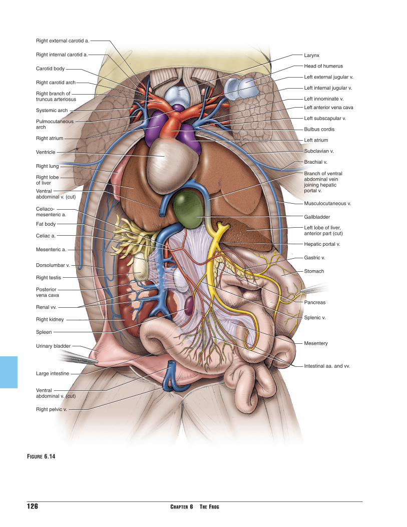
126 CHAPTER 6 THE FROG
Large intestine
Urinary bladder
Spleen
Posterior vena cava
Right lobeof liver
Left lobe of liver,anterior part (cut)
Musculocutaneous v.
Branch of ventralabdominal veinjoining hepatic portal v.
Right lung
Right testisStomach
Splenic v.
Pancreas
Hepatic portal v.
Gastric v.
Mesentery
Intestinal aa. and vv.
Bulbus cordis
Head of humerus
Right carotid arch
Carotid body
Right external carotid a.
Right internal carotid a.
Systemic arch
Right atrium
Right branch of truncus arteriosus
Ventricle
Gallbladder
Celiaco-mesenteric a.
Celiac a.
Mesenteric a.
Brachial v.
Larynx
Left anterior vena cava
Subclavian v.
Left atrium
Pulmocutaneous arch
Left innominate v.
Left subscapular v.
Left internal jugular v.
Left external jugular v.
Fat body
Ventral abdominal v. (cut)
Ventral abdominal v. (cut)
Right pelvic v.
Dorsolumbar v.
Right kidney
Renal vv.
FIGURE 6.14

SECTION V—CARDIOVASCULAR SYSTEM 127
arteries (Figures 6.14 and 6.16). Follow one of thebranches, and identify these arteries and their branch-ing patterns. The most anterior branch is the carotidarch. It divides into a small branch, the external carotidartery, that extends anteriorly to supply the tongue andlower jaw, and a bulbous carotid body that continuesas the internal carotid artery to supply the eye, brain,and upper jaw. The most posterior branch is the pul-mocutaneous arch. It curves sharply posteriorly, givingoff a cutaneous artery extending laterally to supply theskin, and then continues to the lung as the pulmonaryartery. The middle branch is the systemic arch. It sup-plies most of the rest of the body with blood, and so isthe largest of the three branches. Initially the systemicarch passes nearly laterally, but it soon arches stronglydorsally. As it passes dorsal to the lung it curves medi-ally and posteriorly, giving off two branches, the occip-itovertebral artery and the subclavian artery, whichcontinues onto the brachium as the brachial artery(Figure 6.16). After giving off these two branches, thesystemic arch enters the pleuroperitoneal cavity.
Within the cavity each systemic arch passes posterome-dially. Just anterior to the level of the kidneys, the right
and left systemic arches unite to form the dorsal aorta,which continues posteriorly along the middorsal wall ofthe cavity (Figures 6.13 and 6.16). Immediately after itsorigin, the dorsal aorta sends off a large branch, the celi-acomesenteric artery, to the abdominal viscera. Thisvessel soon bifurcates into the celiac artery, whichmainly supplies the liver, gall bladder, stomach, and pan-creas, and the mesenteric artery, which supplies theintestines and spleen.
Between the kidneys, several (usually between four andsix) smaller, paired vessels, the urogenital arteries,extend laterally from the dorsal aorta to supply thekidneys and gonads, as well as fat bodies and urogeni-tal ducts (Figure 6.16). Near the posterior end of thekidneys the dorsal aorta bifurcates into right and leftcommon iliac arteries (Figures 6.12 and 6.16). In thisregion the arteries lie dorsal to the veins, which are alsomore prominent. Follow one of the common iliac arter-ies posteriorly. It gives off two smaller branches in quicksuccession from its lateral surface. These are thehypogastric artery, which mainly supplies musculaturein this region and the urinary bladder, and the femoralartery, which helps supply several muscles and the skinin this region. The common iliac artery then continuesinto the hind limb as the sciatic artery (Figure 6.16). Itsmany branches supply the leg.
Return to the heart, lift the ventricle, and examine thesinus venosus, which leads into the right atrium (Figure6.15). Note the large vessels, the right anterior venacava and the left anterior vena cava, extending alongthe lateral edge of the atria and passing into the sinusvenosus. The venae cavae collect blood from the headand forelimbs, as well as the skin. Many of the vesselsthat enter the venae cavae lie dorsal to the arterialvessels and should be injected with blue latex. In a fewspecimens, however, these will have been infiltrated bythe latex of the arteries and will be partly or completelyred.
Trace an anterior vena cava. The pattern described hereis the general pattern, but there is variation. Indeed, thebranching patterns of the right and left venae cavae mayvary. The anterior vena cava collects blood from severalvessels and empties into the sinus venosus. The mainvessels forming the anterior vena cava are the externaljugular, innominate, and subclavian veins (Figures 6.14and 6.17). These three may join together. The externaljugular vein extends almost directly anteriorly anddrains the tongue and lower jaw. For most of its lengthit passes nearly parallel and just medial to the externalcarotid artery. The innominate vein may be quite shortor it may extend laterally for a longer distance beforereceiving its tributaries, the internal jugular and sub-scapular veins. The internal jugular, draining the eye,brain, and upper jaw, extends anterolaterally, whereas
Left anteriorvena cava
Posteriorvena cava
Hepatic v.
Liver
Ventricle(reflected)
Left atrium
Sinusvenosus
FIGURE 6.15 Heart of the frog in ventral view, with the ven-tricle reflected.

the subscapular vein, draining mainly the muscles asso-ciated with the pectoral girdle, passes nearly laterally.Finally, the subclavian vein passes posterolaterally,formed by the confluence of the brachial vein from theforelimb and the musculocutaneous vein, noted earlier,from the pectoral musculature and the deep surface ofthe skin.
128 CHAPTER 6 THE FROG
The pulmonary veins, returning blood to the heart fromthe lungs, enters the left atrium (Figure 6.17). The pul-monary vein, one on each side, passes dorsal to the anterior vena cava. Right and left pulmonary veins then unite just anterior to the heart to form a shortsingle vessel that extends posteriorly into the left atrium.
External carotid a.
Carotid body
Internal carotid a.
Systemic arch
Cutaneous a.
Pulmonary a.
Occipitovertebral a.
Common iliac a.
Hypogastric a.
Femoral a.
Sciatic a.
Urogential aa.
Dorsal aorta
Ventricle
Left atrium
Subclavian a.
Brachial a.
Right atrium
Bulbus cordis
Truncus arteriosus
Carotid arch
Pulmocutaneous arch
Celiaco-mesenteric a.
Celiac a.
Mesenteric a.
Abdominal skin flapcut and reflected
Kidney
FIGURE 6.16 Schematic illustration of the arterial system of the frog in ventral view superimposed on the bodyoutline.

SECTION V—CARDIOVASCULAR SYSTEM 129
Much of the blood posterior to the heart (that from theskin being the main exception) is returned via the largeposterior vena cava, the large vessel extending betweenthe kidneys (Figures 6.12–6.14). Here it receives severalrenal veins from the kidneys (the fat bodies generallydrain into the more anterior renal veins) and spermatic
veins from the testes or ovarian veins from the ovaries.The posterior vena cava extends anteriorly through theliver, receiving from it several hepatic veins (Figure6.17), to reach the posterior end of the sinus venosus.The posterior vena cava also receives blood from muchof the hind limbs and dorsal body musculature by way
External jugular v.
Internal jugular v.
Innominate v.
Ventral abdominal v.(shifted laterally from midline for clarity)
Pelvic v.
Vesicular v.
Subscapular v.
Subclavian v.
Brachial v.
Branch of ventral abdominal vein joining hepatic portal v.
Femoral v.
Sciatic v.
Communicatingiliac v.
Renal portal v.
Iliac v.
Dorsolumbar v.
Renal vv.
Spermatic/ovarian v.
Left atrium
Right branch of ventral abdominal vein entering liver
Posterior vena cava
Sinus venosus
Right anterior vena cava
Right pulmonary v.
Hepatic vv.
Hepatic portal v.
Gastric v.
Intestinal v.
Musculocutaneous v.
Splenic v.
Kidney
Abdominal skin flapcut and reflected
FIGURE 6.17 Schematic illustration of the venous system of the frog in ventral view superimposed on the bodyoutline. Right atrium, ventricle, and bulbus cordis of heart removed.

of the paired renal portal veins, which enter the kidneys(Figures 6.12, 6.13, and 6.17). This blood then makesits way through the kidneys to reach the posterior venacava.
Examine a renal portal vein. It extends mainly along thedorsolateral surface of the kidney. It is formed posteri-orly by the union of the iliac and sciatic veins (Figures6.13 and 6.17), the latter from the medial side of thethigh. The iliac vein is formed by the femoral vein, thelarge vessel from the lateral side of the thigh, and the pelvic vein. A connection between the femoral andsciatic veins, the communicating iliac vein, extends fromthe femoral, curving dorsally and then medioventrallyto meet the sciatic vein (Figure 6.17). The pelvic veinextends ventromedially to join the pelvic vein from theother side of the body. Their union forms the ventralabdominal vein, already identified above but which willbe described shortly. A small vesicular vein, draining theurinary bladder, enters the ventral abdominal vein justafter its origin. The renal portal vein passes anteriorly,sending numerous branches into the kidney. The dor-solumbar vein, which drains the dorsal and lateralabdominal walls, consists of numerous branches thatcollect usually into a single vessel that enters the renalportal vein at about the midlength level of the kidney(Figures 6.12–6.14 and 6.17).
As noted above, the ventral abdominal vein passes ante-riorly along the midventral wall of the pleuroperitonealcavity and extends between the lobes of the liver. It thenarches dorsally and then posteriorly. It gives off threebranches, two of which enter the right and left laterallobes of the liver, and the third continuing to join thehepatic portal vein, which, as described below, entersthe liver (Figures 6.14 and 6.17). Blood from the hindlimb may thus return to the heart through the posteriorvena cava by several routes. It may pass through therenal portal system or the hepatic veins. In the latterinstance it may pass through the branches of the ventralabdominal vein that enter the liver directly or throughthe hepatic portal vein via that branch of the ventralabdominal vein joining the hepatic portal vein. Thehepatic portal vein drains the abdominal viscera. It isformed mainly by the two following vessels: the gastricvein, which collects several vessels and drains the
130 CHAPTER 6 THE FROG
KEY TERMS: CARDIOVASCULAR SYSTEM
brachial artery
brachial vein
bulbus cordis
carotid arch
carotid body
celiac artery
celiacomesenteric artery
common iliac arteries
cutaneous artery
dorsal aorta
dorsolumbar vein
external carotid artery
external jugular vein
femoral artery
femoral vein
gastric vein
heart
hepatic portal vein
hepatic veins
hypogastric artery
iliac vein
innominate vein
internal carotid artery
internal jugular vein
intestinal vein
kidneys
left anterior vena cava
left atrium
mesenteric artery
musculocutaneous vein
occipitovertebral artery
ovarian veins
pelvic vein
pericardial cavity
pericardium
posterior vena cava
pulmocutaneous arch
pulmonary artery
pulmonary veins
renal portal veins
renal veins
right anterior vena cava
right atrium
sciatic artery
sciatic vein
sinus venosus
spermatic veins
subclavian artery
subclavian vein
subscapular vein
systemic arch
truncus arteriosus
urogenital arteries
ventral abdominal vein
ventricle
stomach and part of the esophagus, and the intestinalvein, formed by vessels that drain most of the smallintestine and large intestine (Figures 6.14 and 6.17). Thehepatic portal then receives a branch from the ventralabdominal vein before entering the liver.