the disposal of acute acid loads in rats and · alter the ph ofintact rat diaphragms in vitro....
TRANSCRIPT
An Extrarenal Role for Parathyroid Hormone inthe Disposal of Acute Acid Loads in Rats andDogs
Donald S. Fraley, Sheldon Adler
J Clin Invest. 1979;63(5):985-997. https://doi.org/10.1172/JCI109399.
Acid infusion studies were performed in nephrectomized rats and dogs with either intactparathyroid glands (intact) or after thyroparathyroidectomy (thyroparathyroidectomized[TPTX]) to determine the role of parathyroid hormone (PTH) in extrarenal disposal andbuffering of acutely administered acid. 29 intact rats given 5 mM/kg HCl and 6 intact dogsgiven 7 mM/kg HCl developed severe metabolic acidosis but all survived. However, each of12 TPTX rats and 4 TPTX dogs given the same acid loads died. Intact rats and dogsbuffered 39 and 50% of administered acid extracellularly, respectively, whereasextracellular buffering of administered acid was 97 and 78% in TPTX rats and dogs,respectively. 17 TPTX rats and 6 TPTX dogs given synthetic PTH 2 h before acid infusionsurvived. The blood bicarbonate and extracellular buffering in these animals, measured 2 hafter acid infusion, was similar to intact animals. Changes in liver, heart, and skeletalmuscle pH determined from [14C]5,5-dimethyl-2,4 oxazolidinedione distribution seemedinsufficient to account for the increased cell buffering of PTH-replaced animals. Indeed,muscle pH in TPTX dogs given PTH and acid was only 0.06 pH units lower than in controldogs given no acid, suggesting that another tissue, presumably bone, was the target forPTH-mediated increased cell buffering. This conclusion was supported by the observationthat PTH did not alter the pH of intact rat […]
Research Article
Find the latest version:
http://jci.me/109399-pdf

An Extrarenal Role for Parathyroid Hormone in the
Disposal of Acute Acid Loads in Rats and Dogs
DONALDS. FRALEYand SHELDONADLER, Department of Medicine, MontefioreHospital, and the University of Pittsburgh School of Medicine,Pittsburgh, Pennsylvania 15213
A B S T RA C T Acid infusion studies were performedin nephrectomized rats and dogs with either intact para-thyroid glands (intact) or after thyroparathyroidectomy(thyroparathyroidectomized [TPTX]) to determine therole of parathyroid hormone (PTH) in extrarenal dis-posal and buffering of acutely administered acid. 29intact rats given 5 mM/kg HCI and 6 intact dogs given7 mM/kg HC1developed severe metabolic acidosis butall survived. However, each of 12 TPTX rats and 4TPTX dogs given the same acid loads died. Intact ratsand dogs buffered 39 and 50% of administered acidextracellularly, respectively, whereas extracellular buf-fering of administered acid was 97 and 78% in TPTXrats and dogs, respectively. 17 TPTX rats and 6 TPTXdogs given synthetic PTH2 h before acid infusion sur-vived. The blood bicarbonate and extracellular buffer-ing in these animals, measured 2 h after acid infusion,was similar to intact animals. Changes in liver, heart,and skeletal muscle pH determined from [14C]5,5-di-methyl-2,4 oxazolidinedione distribution seemed in-sufficient to account for the increased cell bufferingof PTH-replaced animals. Indeed, muscle pH in TPTXdogs given PTHand acid was only 0.06 pH units lowerthan in control dogs given no acid, suggesting thatanother tissue, presumably bone, was the target forPTH-mediated increased cell buffering. This conclu-sion was supported by the observation that PTHdid notalter the pH of intact rat diaphragms in vitro. Theseresults indicate that PTH is necessary for the optimalbuffering of large, acute acid loads presumably by in-creasing bone buffering.
INTRODUCTION
Whereas most metabolic processes in the body producehydrogen ions (1), mechanisms have evolved to prevent
This work was presented in part at the National Meeting ofthe American Federation for Clinical Research and publishedin abstract form in 1976. Clin. Res. 24: 400A.
Received for publication 27 March 1978 and in revisedform 15 December 1978.
their accumulation and the development of life-threat-ening acidosis. These mechanisms include the renalexcretion of the metabolically produced hydrogen ionand the buffering of either endogenously produced orexogenously administered acid. 60 yr ago Van Slykeand Cullen (2) first noted that the buffering of a large,acutely administered acid load was only partially ac-counted for by extracellular buffers. Subsequently, itwas shown in dog (3) and man (4) that at least 50%of anacutely administered acid load was buffered intracellu-larly. Although these studies demonstrated the impor-tance and extent of cellular buffering, they did notdefine the tissues or regulatory mechanisms involvedin the buffering process. Data obtained from acidbalance studies in man (5) and from bone analyses per-formed in acidotic animals (6) suggest that bone is thetissue primarily involved in the cellular buffering ofacid in acute and chronic metabolic acidosis. Accord-ingly, it has been proposed that parathyroid hormonemay be important for the disposal and buffering of acuteacid loads (7), but exact data supporting this hypothesisare not available.
To determine whether parathyroid hormone is re-quired for the normal tissue buffering of acid, acuteacid infusion studies were performed in nephrecto-mized rats and dogs with intact parathyroid glands andin nephrectomized thyroparathyroidectomized rats anddogs. The effect of parathyroid hormone on intracellu-lar skeletal muscle pH was also studied in vitro. Theresults show that parathyroid hormone is required forthe optimal tissue buffering of large, acute acid loadsin both animal species.
METHODSIn vivo rat studies. Male Sprague-Dawley rats weighing
375-500 g were anesthetized with Inactin (Promonta, Ham-burg, West Germany). Femoral artery and central venous cath-eters were inserted and a tracheostomy and bilateral ne-phrectomy were performed. Rats were then placed on a heated,perforated board and rectal temperature was monitoredthroughout the experiment. After a 30-min postoperativestabilization period, two arterial blood samples were obtained
J. Clin. Invest. © The American Society for Clinical Investigation, Inc. 0021-9738/7910510985/13 $1.00 985Volume 63 May 1979 985-997

at 15-min intervals for bloo1( gas determination. Only if acid-base balance and oxygenation proved stable did the animalbegin a 2-h control period. Animals not achieving such stabilityby 1 h were sacrificed. Unless otherwise specified, eachanimal was then given 0.5 N hydrochloric acid, 5 mmilol/kgbody wt, infused intravenously over 30 min followecl bv a 2-hrecovery period. Arterial blood gases and plasma electrolvteswere determined twice during each period. In somiie experi-ments, cell pH was determined at the end of the stabilizationperiod by the intravenous administrationi of 2 ,Ci of [2-'4C]-5,5-dimethyl-2,4 oxazolidinedione (DNM0),1 2 ,uCi of 36Cl, and10 ,uCi of 3H20.
Experiments with the above model were performed in fivegroups: 7 rats with intact parathyroids given no acid inifsioni(intact), 9 rats 1 d after thyroparathyroidectomv given noacid (thyroparathyroidectomized [TPTX]), 29 rats with intactparathyroids given acid (intact plus acid), 12 rats 1 d afterthyroparathyroidectomy giveni acid (TPTX plus acid), and 17rats 1 d after thyroparathyroidectomy given 10 IU/kg body wvtof synthetic (1-34) bovine parathyroid hormone (PTH; ob-tained from Beckman Instrumnents, Inc., Spinco Div., PaloAlto, Calif.) 2 h before acid infusion (TPTX plus PTH plusacid). At the conclusion of the recovery period, rats weresacrificed bv exsanguinationi from the abdominal aorta intoa heparinized syringe. Skeletal muscle (gluteus), cardiac mus-cle, and liver were obtained for determination of intracellularpH. A portion of each sample was used to determine tissueelectrolytes.
Rat diaphragm studies. Intact rat diaphragmls wvere ob-tained from 75- to 90-g non-TPTX Sprague-Dawlev rats andincubated simultaneously in two boxes at 37°C in a modifiedKrebs-Ringer bicarbonate solution conitaininig 100 mg/100 mlof glucose and 6.25 mg/100 ml of chloromycetin as described(8). The solution was gassed with a 4.86% CO2 balanceoxygen gas mixture. Bathing solutions were changedhourly. During the final hour of incubation, 80 mg/100ml of inulin was added to each bath to meassure extra-cellular space, and 25 ACi of [2-'4C]DMO was added toeach solution for calculation of intracelltular pH. At theend of the experiment, diaphragmiis were removed al-ternately from the two boxes for cell pH determination(8). Bath pH was -7.00, achieved by using a bicarbonateconcentration of 10 meq/liter. Incubations lasted foreither 1 or 4 h. The solution in one box conitained either10 or 100 IU/liter synthetic PTH. The other solutioncontained no hormone.
In vivo dog studies. Mongrel dogs weighinig 10-20 kgwere anesthetized with Diabutol (Diamoncd Laboratories,Des Moines, Iowa) intubated, and jugular, femoral venous,and femoral arterial catheters vere inserted. Blood samiiplesvere obtained from the femoral artery. Each animilal
then underwent splenectomny andl bilateral nephrectomvfollowed by a 30-min postoperative stabilizationi periodduiring which two blood samnples were obtainied for bloodgas determiiinationis. If these showed stability of blood acidbase conditions, 50 ,uCi [2-'4C]DMO, 50 ,uCi 36C1, andl 500,uCi 3H20 were given intravenously for measuremiient of extra-cellular space and intracellular pH. A 2-h conitrol periodensued, and blood samples were obtainied at 60 and 120 min.Hydrochloric acid, 7 mmol/kg, given as a 0.3 N solution,v wasinfused intravenously over 90 min. Blood samples, hematocrit,plasma electrolytes, plasmla calcium, and 36Cl measuiremiienitswere determinedl on each blood samiiple. Anlimals were
1 Abbreviations used in this paper: DM0, 5,5-dimethyl-2,4oxazolidinedione; pHI, intracellular pH; PTH, parathyroidhormone; TPTX, thyroparathyroidectomiiizecl.
sacrificed at the end of the recovery period, and skeletalmuscle, liver, anid heart muscle samples were obtained forcell pH anid electrolyte determination. With this nephrecto-mized mPodel four groups were studied: four dogs given noacid infuision (intact control), six dogs given HCl (intact plusacid), fouir dogs TPTX 2 h before the control period and thengiven HCl (TPTX plus acid), and six TPTX dogs given 10 IUof syinthetic PTH/kg bodv wt at the beginning to the controlperiod and(I then infuised with HCl (TPTX plus PTHplus acid).
Anialy tic methods and calculatiotns. The amount of acidinfused wvas calculated from the volume and normality ofthe acid; the latter determined by titration of a primary Trisstanidardl. Blood pH, Pco2 anid Po2 vere measured on a BMS3\IK2 blood micro system (Radiomneter, Copenhagen, Den-mark), anid the bicarbonate concentration was calculatedfrom a stanidardl nomogramii. Plasma and tissue electrolyteswere meeasured in anl IL flamiie photometer (InstrumentationLaboratories, Boston, Mass.) with anl internal lithiumstandard. Calcium was determined on a Corning (CorningGlass Works, Corning, N. Y.) calcium analyzer by photo-metric titration with EGTA as described by Schmidtand Reilly (9). Radioisotopes were counted in a threechannel Packard liquid scintillation counter (PackardInstnmilent Co., Downer's Grove, Ill.). Intracellular pH inthe nephrectomized animlal studies was determined fromdistribution of the three isotopes with the staindard equationsanid method described by Schloerb and Grantham (10). Intra-cellular pH in the rat diaphragm studies was determined as(lescribe(l (11). Chloride space measuremiients were made fre-quently in both control andcl recovery periods, corrected forloss of radioactivity as a result of blood removal, and used tocalculate compartmiienital acidI distribuition. Total extracellularsodiumil, potassiumii, and bicarbonate in control and recovervperiods were calculated as the product of the plasma conl-cenitrationi anid the measured chloride space. At least two setsof determinationis were made in each period. Extracellularbuffering is defined as the reduction in total extracellularbicarbonate during the experiment. Total acid buffering is thesum of the reduction in extracellular bicarbonate plus theincrease in total extracelltular sodium and potassium; the latterreflecting the movement of these ions from inside the cell inexchange for extracellular hydrogen ions (12).
Data atalysis. Data are presented as the mean+SEM. Un-paired t tests vere employed in all the statistical analyses.
RESULTS
Rat experiments
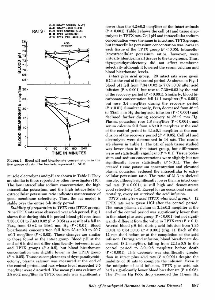
Stability of the rat preparation (intact group).Seven intact rats were prepared as described and ob-served for 6 h. As shown in Fig. 1, blood pH aftersurgery was 7.34±+0.01 and 6 h later had risen to 7.39+0.03 (P < 0.05). Simultaneously, arterial Pco2 de-creased from 48 + I to 39 + 5 mmHg (P < 0.05) and bloodbicarbonate fell from 25.6+0.5 to 23.2+0.8 meq/liter(P < 0.05). These changes were probably the result ofreversible Inactin-induced respiratory depression anda decrease in blood bicarbonate secondary to surgeryand metabolic compensation for the Pco2 decrease (13).Throughout the 6 h, blood Po2 remained constant, vary-ing between 57 and 74 mmHg. Mean plasma calciumconcentration at the end of the control period was4.2+0.2 meq/liter. Liver, skeletal muscle, and heart
986 D. S. Fraley and S. Adler
7.67.57.4
3I 7.3-
a 7.2o 7.10CD 7.0
6.96.8
. 30;zz-0rw 25
w4 20z0C 15
C.)I0
-JCD n L
0-0 INTACT CONTROL(no7)0- INTACT + ACID (n- 29)
RATS: 0-0 TPTX CONTROL(n - 9)U- TPTX+ACID(nsI2)A-- TPTX+PTHeACID(n.17)
ACIDINFUSION
0 60 120 180 240
TIME IN MINUTES
FIGURE 1 Blood pH and bicarbonate concentrations in thefive groups of rats. The brackets represent± 1 SEM.
muscle electrolytes and pH are shown in Table I. Theyare similar to those reported by other investigators (10).The low intracellular sodium concentration, the highintracellular potassium, and the high intracellular toextracellular potassium ratio indicates maintenance ofgood membrane selectivity. Thus, the rat model isstable over the entire 6-h study period.
Stability of preparation in TPTX rats (TPTX group).Nine TPTXrats were observed over a 6-h period. Fig. 1shows that during this 6-h period blood pH rose from7.36±0.01 to 7.40+±0.02 (P < 0.05) as a result of a fall inPco2 from 43+2 to 34+1 mmHg (P < 0.01). Bloodbicarbonate concentartion fell from 23.4+0.9 to 20.7±0.7 meq/liter (P < 0.05). These changes are similarto those found in the intact group. Blood pH at theend of 6 h did not differ significantly between intactand TPTX groups (P > 0.5), but blood bicarbonateconcentration was slightly lower in the TPTX group(P < 0.05). To assess completeness of thyroparathyroid-ectomy, plasma calcium was measured at the end ofthe control period, and rats whose level exceeded 3.5meq/liter were discarded. The mean plasma calcium of2.8±0.2 meq/liter in TPTX controls was significantly
lower than the 4.2±0.2 meq/liter of the intact animals(P < 0.001). Table I shows the cell pH and tissue elec-trolytes in TPTXrats. Cell pH and intracellular sodiumconcentration were the same in intact and TPTXgroups,but intracellular potassium concentration was lower ineach tissue of the TPTX group (P < 0.05). Intracellu-lar:extracellular potassium ratios, however, werevirtually identical in all tissues fo the two groups. Thus,thyroparathyroidectomy did not affect membraneselectivity although it lowered the serum calcium andblood bicarbonate levels.
Intact plus acid group. 29 intact rats were givenHCI at the end of the control period. As shown in Fig. 1blood pH fell from 7.34+0.02 to 7.07±0.02 after acidinfusion (P < 0.001) but rose to 7.30±0.03 by the endof the recovery period (P < 0.001). Similarly, blood bi-carbonate concentration fell 14.1 meq/liter (P < 0.001)but rose 3.4 meq/liter during the recovery period(P < 0.01). Simultaneously, Pco2 decreased from 46±2to 35±1 mmHg during acid infusion (P < 0.001) anddeclined further during recovery to 32±2 mmHg.Plasma potassium rose 1.8 meq/liter (P < 0.001), andserum calcium fell from 4.6±0.2 meq/liter at the endof the control period to 4.1±0.1 meq/liter at the con-clusion of the recovery period (P < 0.05). Cell pH andelectrolytes were determined in 14 rats. The resultsare shown in Table I. The pH of each tissue studiedwas lower than in the intact group, but differenceswere not statistically significant (P > 0.1). Tissue potas-sium and sodium concentrations were slightly but notsignificantly lower statistically (P > 0.1). The de-creased tissue potassium concentration and elevatedplasma potassium reduced the intracellular to extra-cellular potassium ratio. The ratio of 21.3 in skeletalmuscle, although significantly lower than in intact con-trol rats (P < 0.001), is still high and demonstratesgood selectivity (14). Except for an occasional surgicalmortality, every rat survived the acid infusion.
TPTX rats given acid (TPTX plus acid group). 12TPTX rats were given HCI after the control period.The mean plasma calcium of 3.1±0.2 meq/liter at theend of the control period was significantly lower thanin the intact plus acid group (P < 0.001) but not signif-icantly different from the value in TPTX rats (P > 0.1).Arterial blood pH fell during acid infusion from 7.35±0.01 to 6.84±0.03 (P < 0.001) (Fig. 1). Each of the12 rats died before or at the completion of the acidinfusion. During acid infusion, blood bicarbonate de-creased 18.2 meq/liter, falling from 22.1±0.5 in thecontrol period to 3.9+0.6 meq/liter before death(P < 0.001). This decrease was significantly greaterthan in intact plus acid rats (P < 0.001) despite theinability of 10 rats to complete the infusion. Even atthe midpoint of acid infusion, TPTX plus acid ratshad a significantly lower blood bicarbonate (P < 0.05).The 17-mm Hg Pco2 drop exceeded the l1-mm Hg
Role of Parathyroid Hormone in Acute Acid Disposal
I Iv
987

TABLE ICell pH and Electrolytes in the Four Surviving Groups of Rats
Tissue pHi* Na K* K*K+'neal/K+
inieqlliter ineqlliter meqlkg(cell lLater cell wcater dry ct
Intact (n = 7)Skeletal muscle 6.93+0.02 26.3±3.4 178+6.1 493.7+7.6 29.2Liver 7.09+0.03 224.1±7.2 340.6±4.3 36.7Heart muscle 7.08+0.04 26.9+2.6 151.3±5.3 352.7±5.5 24.8
TPTX (n = 9)Skeletal muscle 6.89+0.05 30.3±5.0 162.4±5.34 464.5±12.94 29.5Liver 7.14±0.04 205.1 ±3.8t 303.4 25.34 37.3Heart muscle 7.11±0.04 21.7±3.4 139.1±1.94 327.2±3.54 25.3
Intact + acid (n = 14)Skeletal muscle 6.88+0.02 22.7+4.2 168.3+3.8 475.6±12.2 21.3Liver 7.03±0.03 209.9±5.1 319.8±6.3 26.6Heart muscle 7.03±0.01 26.7±7.0 146.6±3.6 345.2±8.4 18.6
TPTX + PTH + acid (n = 7)Skeletal muscle 6.88±0.03 41.4±+11.74 144.3±10.64 466.4±27.0 20.0Liver 7.21±0.054 197.9±7.2 319.3±2.2 27.5Heart muscle 7.03±0.03 52.4±3.14 121.0+4.84 296.0±16.44 16.8
* Results are expressed as the mean±SEM.4 Significantly different from intact plus acid (P < 0.05).
decrease in the intact plus acid group. The 100%mortality cannot, therefore, be attributed to defectiverespiratory compensation.
TPTX rats given PTHand acid (TPTX plus PTHplusacid group). 17 TPTX rats, given 10 IU/kg body wt ofsynthetic PTH at the beginning of the control period,all survived the acid infusion. The plasma calcium of3.0+0.2 meq/liter before PTH administration was notstatistically different from the other two TPTX groups(P > 0.3). At the end of the experiment, 6 h later, ithad risen insignificantly to 3.2+±0.2 meq/liter (P > 0.3).Thus, toleration of the acid was not because of a PTH-induced rise in plasma calcium. As shown in Fig. 1,control period arterial pH was lower than in the othertwo TPTX groups (P < 0.01). This lower pH was ofrespiratory origin as control bicarbonate concentrationin the three TPTX groups was statistically identical(P > 0.4). Survival of PTH-treated rats, therefore, wasnot the result of elevation of extracellular buffer capac-ity. Through a technical oversight, arterial blood sam-ples were not drawn during acid infusion, but bloodwas obtained at the end of the recovery period. ArterialpH at the end of the recovery period was lower thanin the intact plus acid group, but blood bicarbonateconcentrations were identical statistically in the twogroups (P > 0.4). The absolute decrease in the bloodbicarbonate concentration in the two groups also didnot differ significantly (P > 0.3). Assuming equal acidproduction, tissue buffering of infused acid was the
same, therefore, in intact rats and in TPTX rats givenexogenous PTH.
Acid distribution and cell buffering in intact plusTPTX rats. Extracellular buffering, as calculated fromthe decrease in total extracellular bicarbonate, is shownin Fig. 2. Assuming no endogenous acid production
2.0 k
1.5 ka
4IL0
EE
1.0 H
0.5 H
0.0
INTACT+ ACID (n=19)TPTX +ACID (n-12)
ACID LOADADMINISTERED
I LLCALCULATED CALCULATED
EXTRACELLULAR INTRACELLULARBUFFERING BUFFERING
FIGURE 2 Distribution of acid buffering in intact plus acidand TPTXplus PTHplus acid groups. Extracellular bufferingis the reduction in extracellular bicarbonate obtained by multi-plying the bicarbonate concentration and the chloride space.Calculated intracellular buffering assumes no endogenousacid production. The brackets represent±+1 SEM.
988 D. S. Fraley and S. Adler

during the course of the experiment, 39% of adminis-tered acid was buffered extracellularly and 61%, intra-cellularly in intact rats. These are similar to valuespreviously reported in the dog (3). Extracellular buf-fering in TPTX rats was 97%; significantly higher thanthe 39% found in the intact group (P < 0.001). Thecellular buffering of 3%in the TPTXrats was, of course,significantly lower (P < 0.001). As all TPTX rats diedby the end of acid infusion, no direct 36C1 space meas-urements were obtained. Rather, the control 36C1 spacewas assumed to have increased by the same percentageas in intact rats.
Plasma potassium values were nearly identical inthe two groups surviving the acid infusion. In intactanimals, plasma potassium rose from 6.1+0.1 to 7.9+0.3 meq/liter, whereas in the TPTX group the risewas from 5.5+0.2 to 7.2+0.4 meq/liter. Final plasmapotassium levels in both groups exceeded the 6-h val-ues found in intact and TPTX control rats (P < 0.001).Intracellular sodium and potassium concentrations inthe two surviving TPTX groups did not differ signif-icantly from each other except for heart muscle sodiumand potassium concentration (P < 0.05). Small changes,however, might be missed. Table I shows that thyro-parathyroidectomy per se did not affect cell pH. It alsoshows that despite a slightly lower arterial blood pH,skeletal and heart muscle pH values in the TPTX plusPTHplus acid rats were the same as in intact rats givenacid (P > 0.1). Liver cell pH, however, was higher inthe TPTX plus PTH plus acid group (P < 0.005). Com-pared with TPTX control rats, heart muscle pH wassignificantly lower (P < 0.05) but insignificantly dif-ferent in liver tissue (P > 0.1) and skeletal muscle(P > 0.5). Acid infusion, therefore, lowered heart mus-cle pH in intact and TPTX plus PTH rats and reducedskeletal muscle and liver cell pH in intact rats. How-ever, in TPTX plus PTH rats, skeletal muscle pH didnot fall, and liver cell pH rose. Thus, exogenous PTHdid not appear to increase buffering of administeredacid by skeletal muscle.
Rat diaphragm studies. If PTH enhances skeletalmuscle buffering of administered acid, it would pre-sumably accomplish this by increasing extracellularhydrogen ion movement into cells. Incubation of mus-cle in an acidic medium in the presence of the hormonewould be expected, therefore, to lower muscle cell pH.Rat diaphragms were incubated at an external pH of7.00, a value previously shown in our laboratory toreduce muscle cell pH in vitro and only 0.1 pH unitlower than the blood pH achieved by the intact plusacid rats at the end of the acid infusion period. Theeffect of PTH on diaphragm muscle pH is shown inTable II. In each experiment, the pH of the incubationmedium containing PTHdid not differ from the controlmedium by more than 0.02 pH units. The table showsthat in experiments performed for 1 h at a PTH con-
TABLE IIEffect of PTH on the Intracellular pH of Intact Rat
Diaphragm Muscle In Vitro
Group PTH concentration* pH,.t pH §
lU/liter
1-h incubationControl (n = 6) 0 7.04 7.04+0.03PTH (n = 6) 10 7.03 7.02+0.04
4-h incubationControl (n = 12) 0 7.00 7.00+0.05PTH (n = 12) 10 6.99 7.00+0.02
1-h incubationControl (n = 12) 0 7.04 7.01+0.03PTH (n = 12) 100 7.02 7.00+0.05
* Synthetic (1-34) bovine PTH.$ Extracellular pH.§ Results expressed as the mean+SEM.
centration of 10 IU/liter, there was no significant dif-ference in the pH of tissues incubated with the hormonecompared with those incubated without hormone(P > 0.3). Increasing incubation time fourfold or raisingthe PTH concentration 10-fold to 100 IU/liter had nostatistically significant effect on cell pH (P > 0.5). Tis-sue sodium and potassium concentrations were identi-cal in tissues incubated with or without PTH, whereasthe intracellular:extracellular potassium ratio in eachexperiment exceeded 20. Exogenous PTH, therefore,does not alter diaphragm muscle cell pH in vitro.
Dog experiments
Stability of the dog preparation (intact control).Four nephrectomized, splenectomized dogs with intactparathyroids were followed for 5.5 h. Fig. 3 shows thatblood pH increased insignificantly from 7.36+0.03 to7.41±0.03 (P > 0.05), and the Pco2 also fell insignif-icantly (P > 0.1). Despite minor variations, blood bi-carbonate concentration also was unaltered (P > 0.05).Plasma sodium and potassium concentrations of 144+±3and 3.1±0.6 meq/liter also remained unaltered(P > 0.1). After 5.5 h, animals were sacrificed, andtissues were obtained for measurement of intracellularpH. Liver cell pH was 7.03±0.06, heart pH was 6.92±0.03, and skeletal muscle pH was 6.98±0.02; valuessimilar to ones previously reported in rat, dog, andman (10). Thus, extracellular and intracellular acid baseconditions were stable over 5.5 h.
Acid infusion in intact parathyroid dogs (intact plusacid). Fig. 3 shows that blood pH fell rapidly duringacid infusion (P < 0.001) because of a decrease in bloodbicarbonate of 15.2 meq/liter (P < 0.001). Respiratorycompensation occurred in all animals with a meanPco2 decrease of 15 mmHg (P < 0.001). All six dogs
Role of Parathyroid Hormone in Acute Acid Disposal 989

0-0 INTACT CONTROL(n =4)
DOGS:- INTACT+ACID (n-6)
DOGS.u - TPTXTAACIDC(nD(4)A- TPTX+PTH+ACID(nu6)
7.67.57.4
I 7.3a72a72m 7010J 7.0
6.96.8
.- 30
f 25
w20
z0
Q I oCO o
00 50-j
fi
I-
VTh
I,... I.. ..
0 60 120 165 JO 270TIME IN MINUTES
300 330
FIGURE 3 Blood pH and bicarbonate concentrations in thefour groups of dogs. The brackets represent± 1 SEM.
survived the acid infusion. During the 2-h recoveryperiod, blood pH and bicarbonate rose 0.21 U and 4.5meq/liter, indicating tissue generation of bicarbonate.Plasma sodium concentration decreased from 143±4 to136±3 meq/liter (P < 0.05), whereas plasma potassiumconcentration rose from 3.3+0.2 to 6.2+0.5 meq/liter(P < 0.001). Plasma calcium concentration at the end ofthe control period was 4.2±0.3 meq/liter. At the end ofthe recovery period, liver cell pH was 7.00±0.03, heartmuscle pH was 6.84+±0.02, and skeletal muscle pH was6.82±0.03.
Acid-infused TPTX dogs (TPTX plus acid). FourTPTX dogs were given HCI. At the end of the controlperiod, the mean plasma calcium concentration of 2.3±0.5 meq/liter was significantly lower (P < 0.001) thanin intact animals. Each dog died before completionof the acid infusion despite marked respiratory com-pensatory effort. As shown in Fig. 3, control periodblood pH in these dogs did not significantly differ fromthat of intact control (P > 0.5) or intact plus acid dogs(P > 0.5). Control period blood bicarbonate concentra-tion, however, was significantly lower in the TPTXanimals (P < 0.01). After acid infusion, blood bicarbon-
ate fell 16.9 meq/liter (P < 0.001) to unmeasurablelevels at the time of death. The absolute drop in bloodbicarbonate concentration was greater in the TPTXthan in the intact plus acid dogs (P < 0.02). Controlplasma sodium levels of 133±3 meq/liter were signif-icantly lower than in either intact dog group (P < 0.001),but the plasma potassium level of 3.2+0.5 meq/literdid not differ from the other groups (P > 0.5). All ani-mals died, so no tissues were obtained for cell pH.This 100% mortality was identical to that found inTPTXplus acid rats. To determine whether exogenousPTH would also prevent death in dogs, TPTX dogswere given PTH before receiving the acid infusion.
Acid infusion in TPTX dogs given exogenous PTH(TPTX plus PTHplus acid). Six dogs were given PTHat the beginning of the control period and 2 h latergiven HC1. Fig. 3 shows that control arterial blood pHdid not differ significantly (P > 0.4) from the previousthree groups, whereas control period blood bicarbonateconcentration was the same as in the TPTX plus acidgroup (P > 0.1). After acid infusion, blood pH fell 0.24U (P < 0.001), Pco2 decreased 11 mmHg (P < 0.001),and blood bicarbonate fell 12.2 meq/liter (P < 0.001).All six dogs survived, and during the recovery period,blood pH and bicarbonate rose 0.08 Uand 1.6 meq/liter,respectively. Plasma calcium at the end of the controlperiod was 2.8±0.2 meq/liter; significantly less than inintact dogs (P < 0.001) but not different from the level inthe TPTXgroups (P > 0.05). Plasma sodium concentra-tion fell from 136±1 to 133±1 meq/liter, whereasplasma potassium rose from 3.4±0.4 to 5.4±0.5 meq/liter. Liver cell pH was 7.06±0.02, heart muscle pHwas 6.89±0.04, and skeletal muscle pH was 6.92±0.03at the time of sacrifice. Fig. 3 shows that even midwayduring acid infusion, at least 30 min premortem, bloodbicarbonate decreases in the three acid infused groupsdiffered. In intact plus acid and TPTX plus PTH plusacid dogs, blood bicarbonate had fallen 7.6 and 8.2meq/liter compared with 11.9 meq/liter in the TPTXplus acid group. The 50% greater decrease in the lattergroup's blood bicarbonate at 45 min was significantlydifferent from the two other groups (P < 0.001). Forthe entire experiment, the overall decrease in bloodbicarbonate in intact plus acid and TPTXplus PTHplusacid groups of 10.7 and 10.6 meq/liter was virtuallyidentical (P > 0.5).
Distribution of infused acid in dogs (body buffering).Calculation of extracellular and intracellular bufferingrequires a reproducible measure of extracellular space.Although a small amount of chloride is located intra-cellularly, changes in its distribution reflect changesin extracellular space (15). Whatever substance is used,the results obtained must be reproducible. The re-producibility of the chloride spaces in the present ex-periments is shown in Table III. At the end of the
990 D. S. Fraley and S. Adler
-j ------- 4..
I
(I

TABLE IIIChloride Spaces in the Four Groups of Dogs*
Percent increaseover entire
Group Body weight Period It Period 2§ Period 3"1 experiment
kg ml ml ml %
Intact control 15.7+0.3 5,239±611 5,435±730 5,663±727 8.1Intact + acid 14.9± 1.0 4,757±648 5,465±457 5,614±839 18.0TPTX + acid 15.1±1.7 4,802±596 5,660±624TPTX + PTH
+ acid 17.6±0.9 5,673+1,050 6,564±482 6,672±1,058 17.6
* Values are given as the mean±SEM.t Values were obtained at the end of the 2-h control period and are the mean of two determinationsin each dog.§ Values were obtained after acid infusion in the last three groups and are the mean of two determinationsin each dog.1"Values were obtained at the completion of the recovery period in the second and fourth groups of dogs.They are the mean of three determinations in each dog.
control period, chloride spaces were 33, 33, 32, and32%of body wt, indicating identity among groups. Dur-ing the next 90 min, intact control dogs received amaintenance 5% dextrose infusion while the otherthree groups received hydrochloric acid. Extracellularspace increased 4% in the intact controls and 15% ineach of the three acid-infused groups. Throughout therecovery period, the three surviving groups of dogsreceived a 5% dextrose infusion, and chloride spaceincreased 4.2, 2.7, and 1.7% in these groups.
The buffering distributions shown in Table IV werecalculated from the measured chloride spaces. Intactcontrol animals, although receiving no acid, demon-strated some buffering. Presumably, this was a result ofendogenous acid production in a catabolic postopera-tive dog, although penetration of chloride into cellscannot be completely ruled out. The small change in
total extracellular bicarbonate suggests that this lattereffect, if present, was small. Calculated endogenousacid production rate was _3.5 meq/kg body wt.Buffering in each acid-infused group exceeded theamount of acid administered. Differences betweencalculated buffering and administered acid in the twogroups surviving acid infusion were less than thebuffering shown by intact control animals andpresumably represent endogenous acid production.The greater excess in the TPTX plus acid animals isexplained by leakage of sodium from the erythrocytesof dying animals (16). This conclusion is supported bythe observation that the excess in total buffering wascompletely accounted for by increased extracellularsodium as changes in extracellular potassium wereequal in the three acid-infused groups (P > 0.4). Dogerythrocytes are high in sodium and low in potassium
TABLE IVDistribution of Buffering in the Four Groups of Dogs*
End of acid infusion period End of recovery period
Range of Range ofAmount of administered administered
acid Extracellular Total acid buffered Extracellular Total acid bufferedadministered buffering buffering extracellularly buffering buffering extracellularly
meq meq meq % meq meq %
Intact control (n = 4)t 0.0 7.6+3.3 32.6+21.8 16.1±2.4 53.8± 18.1Intact + acid (n = 6) 117.2+7.5 59.1±3.4 131.2+13.6 (28.2-58.1) 45.9±6.8 141.5±13.0 (21.8-52.5)TPTX + acid (n = 4) 111.0± 19.6 85.3+ 14.2 184.6+29.9 (58.5-87.7)TPTX + PTH + acid
(n = 6) 128.3±11.1 68.7+17.0 153.3±14.8 (30.7-71.4) 57.6+11.5 184.4+13.3 (13.0-61.3)
* All values are given as the mean+SEM.I No acid infusion.
Role of Parathyroid Hormone in Acute Acid Disposal 991

(17), whereas all other cells have a high potassiumcontent, so erythrocytes must have released sodiuminto the extracellular fluid. The amount of acidadministered toTPTX plus acid dogs was less than inthe other two groups because all dogs died beforecompletion of the acid infusion. Table IV shows thatduring the recovery period total extracellular bicarbon-ate decreased by 8.5 meq in the intact controls but rose13.2 and 11.1 meq in the intact plus acid and the TPTXplus PTH plus acid groups, demonstrating cellularbicarbonate generation.
Extracellular buffering differed markedly amonggroups. The percent of administered acid bufferedextracellularly exceeded 50% in all four TPTX plusacid dogs but in only one dog in each of the othertwo acid-infused groups. Fig. 4 shows that -50% ofadministered acid in the intact and PTH-replacedgroups was buffered extracellularly compared withnearly 80% in the TPTX plus acid group (P < 0.001).This difference is not just a premortem phenomenon,as Fig. 3 demonstrates that the extracellular bicarbon-ate concentration at the midpoinit of the infusion periodin the TPTX plus acid group was already significantlylower than in the other two acid-infused groups(P < 0.001). Chloride space changes were similar inthe three groups, so the greater f'all in blood bicarbonatein the TPTX group reflects increased extracellularbuffering. Thus, extracellular buffering was elevated,and cellular buffering diminished in TPTX plus aciddogs. Fig. 4 and Table IV also show that by the end
>, 100-J
-J 75a -ibwcrow<
crcX I- 50-wa a
_ Wcr 25
o- E_OLL0 D
0
ACID ADMINISTEREDEND OF ACID INFUSION
END OF RECOVERYPERIOD
F
h
TINTACT
ACID
TPTX+
ACID
II
TPTX+PTH
ACID
FIGURE 4 Percent of administered acid buffered extracellu-larly at the end of the acid infusion and at the end of therecovery period in the intact plus acid, TPTX plus acid, andTPTX plus PTH plus acid groups of dogs. The brackets repre-sent+l SEM.
of the recovery period extracellular buffering ac-counted for <50%of the administered acid in the intactplus acid and TPTX plus PTH plus acid groups.
The data shown in Table V suggest that the cellularsite of PTH-mediated cellular buffering is not liver,heart, or skeletal muscle. Despite a significant drop inblood pH in intact plus acid and TPTX plus PTH plusacid groups, liver cell pH in these groups did notdiffer significantly from intact controls (P > 0.3). Heartmuscle pH did fall significantly in the intact plus acidgroup but not in the TPTX plus PTH plus acid dogs.Skeletal muscle pH decreased significantly in the twoacid-infused groups, but the decrease in the PTH-re-placed group was only 0.06 pH units (P < 0.05). Itseems unlikely that such small changes are responsiblefor the calculated cellular buffering of 6.4 and 7.3meq/kg body wt (Table V) that occurred in the intactplus acid and the TPTX plus PTH plus acid groups.
DISCUSSION
Daily net acid balance (18) may be upset either by areduction in renal hydrogen ion secretory capacity, asin patients with chronic renal disease (19), or by thesudden presentation of large acid loads exceeding thekidney's ability to sufficiently increase acid excretion.In the latter situation, the body buffers the excess acid.Early experiments suggested that skeletal muscle, andpresumably other tissue membranes, were imperme-able to extracellular hydrogen and(or) hydroxyl ions(20). Subsequent work, however, demonstrated that theinternal pH of muscle was responsive to changes inexternal hydrogen ion activity (11). Indeed, after acuteacid administration >50% of the administered acid isbuffered intracellularly (3, 4). Also, during intermittentacid infusion, although extracellular bicarbonate is themajor buffer, cellular buffers restore extracellular bi-carbonate concentration in the interinfusion periods(21). Yoshimura et al. (22) showed that in the first 24 hafter a large acid infusion, dogs excreted only 25% of
TABLE VCell pH int the Surviving Three Groups of Dogs*
Skeletal Heartmuscle muscle
Group (n value) Liver pHi pH, pH,
Intact control(n = 4) 7.03+0.06 6.98+0.02t 6.92±0.034
Intact + acid(n = 6) 7.00±0.03 6.82±0.03 6.84±0.02
TPTX + PTH+ acid (n = 6) 7.06+0.02 6.92±0.03t 6.89+0.04
* Results are expressed as the mean±SEM.I Significantly different from intact plus acid (P < 0.05).
992 D. S. Fraley and S. Adler
I

the administered acid. Nevertheless, after 24 h, theblood bicarbonate concentration had returned to itscontrol value. Over the next 6 d the remaining acid wasexcreted, suggesting slow release of the acid from in-tracellular buffer sites.
Although these studies demonstrate the extent ofcellular buffering, they do not define its location orregulation. Burnell (23) found decreased bone carbon-ate in dogs with metabolic acidosis, whereas Bergstrom(6) noted reduced bone sodium, calcium, and carbonateconcentrations in metabolic acidosis, suggesting titra-tion of bone buffers by acid. Studies in normal anduremic human volunteers (5) also support the conceptthat bone is a major tisssue buffer. PTH as a majorregulator of bone metabolism (24) might, therefore, beinvolved in bone buffering. Indeed, parathyroid glandsappeared late in evolution, almost 200,000,000 yr afterthe appearance of fish with a calcium skeleton. Wills (7)and Wachmanand Bernstein (25), noting that vitaminD and calcitonin efficiently regulate calcium balancein bony fish, questioned the sudden appearance ofparathyroid glands in amphibia. As amphibia were nolonger able to efficiently transfer hydrogen ions intothe surrounding sea, despite the continued necessityto dispose of metabolic acid, they proposed that theprimary evolutionary function of PTH was to mobilizebone phosphate and carbonate, thereby providing thebody with a reserve of phosphate ions and buffer. PTHalso has many renal actions that affect net acid ex-cretion and acid base balance (26-28), and one studyeven suggests circulating PTH levels are increased inacidosis (29). Finally, bone resorption after PTH ad-ministration may be mediated through changes in localacidity. Acetazolamide blockade of carbonic anhydrase,an enzyme present in rat osteoclasts, prevents PTH-stimulated bone resorption in rats (30). The hyper-calcemic response of rats to dibutyryl cyclic 3'5'-AMPis also blocked by acetazolamide, suggesting that thefinal mediator of PTH-stimulated bone resorption is thelocal pH environment (31). In addition, recent ex-periments by Martin et al. (32) have demonstrated thatboth the uptake and biological effect of PTH in anisolated, perfused canine-bone preparation is enhancedby acidosis.
These experiments support the hypothesis that PTHis important for tissue buffering after acute acid ad-ministration. All 16 TPTX rats and dogs given aciddied, whereas the 35 animals with intact parathyroidsgiven the same acid dose survived. Reduction of extra-cellular bicarbonate was significantly greater in TPTXanimals even after reception of only one-half of the acidload. Differences might have been larger if TPTX ani-mals had received all the acid, but 14 animals diedbefore completion of the infusion. Each animal wasnephrectomized, so changes in acid excretion cannot
explain this observation. There are two possible ex-planations. First, endogenous lactic acid productionmight have increased in TPTX animals. Although thispossibility cannot be totally excluded, it seems un-likely. Oxygen tension in the TPTX animals remainedconstant, CO2retention did not develop, and the aniongap was identical in all groups. Additionally, bloodpressure measurements every 15 min were relativelyconstant, and hypotension developed only near deathlong after bicarbonate had fallen more sharply in theTPTX rats and dogs. The alternative explanation isthat cellular buffering differed in the two groups. Thecalculation of extracellular and cellular bufferingdepicted in Figs. 2 and 4 supports this conclusion. Re-duced tissue buffering in TPTX animals presumablyresulted in depletion of extracellular buffer stores,acidosis, and death. Acid distribution was not preciselymeasured in the TPTX plus acid rats as extracellularspace, and transcellular shifts of sodium and potassiumwere not determined. However, such measurementswere made in the dogs. Cellular buffering is calculatedfrom the difference between total buffering and extra-cellular buffering (3), and determination of the latterrequires a reproducible measure of changes in extra-cellular space. The chloride space values found in thepresent experiments (Table IV) are higher than thosepreviously reported by other investigators (33), a vari-ance for which we have no explanation. This differenceis unimportant, however, as it is the change in extra-cellular space, not the absolute space, that determinesextracellular buffering. Control period chloride spaceswere high but equivalent in each of the four groupsstudied, so reducing the control chloride space from33 to 20% did not significantly affect the extent of cal-culated extracellular buffering. In addition, the changesin chloride space measured after acid administrationwere virtually identical to those reported by otherinvestigators using inulin, radiosulfate, or chloridespaces (3, 21, 22), and the percentage of administeredacid buffered extracellularly in the intact dogs was thesame as that reported in other studies (3, 21, 22).
The reduced cellular buffering induced by thyro-parathyroidectomy could be the result of reduced tissuebuffer capacity, altered plasma calcium concentration,thyroid hormone deficiency, or PTH deficiency. Theeffect of thyroparathyroidectomy on tissue electrolytesand intracellular pH is not known. In this study thryo-parathyroidectomy performed 24 h earlier in controlrats did not significantly alter the cell pH or electrolytecontent of skeletal muscle, cardiac muscle, or liver.Bone pH is not yet measurable (34) so the effect ofthyroparathyroidectomy on bone buffering capacitywas not determined. Given the enormous buffer capac-ity of bone (35), it is unlikely that a change in thisquantity could explain the observed results. Reduced
Role of Parathyroid Hormone in Acute Acid Disposal 993

tissue buffering capacity, therefore, cannot account fordiminished cellular buffering in TPTXanimals. Plasmacalcium was low in TPTX animals, and calcium ionsare involved in a host of biochemical reactions thatmight affect cellular buffering (36). Calcium, for ex-ample, increases the permeability of sea urchin eggsto sodium, increases hydrogen ion efflux, and raises theintracellular pH of the eggs (37). The lowered plasmacalcium level, however, did not appear responsible forthe reduced cell buffering because exogenous PTHrestored cellular buffering in TPTX animals withoutsignificantly altering plasma calcium. For the same rea-son, it is unlikely that thyroid hormone deficiency re-duced tissue buffering. Also, hypothyroidism is unas-sociated with significant abnormalities in acid basebalance. The most likely explanation is that PTH isnecessary for optimal tissue buffering of acutely ad-ministered acid, as TPTX animals given exogenousPTH buffered the infused acid in a manner similar tointact animals. Although other factors not examinedmight be involved in the reduced cellular bufferingseen after thyroparathyroidectomy, it seems that PTHis a prime mediator in the buffering of a large, acuteacid load.
At least two factors determine the extent of cellularbuffering. The first is the time elapsed after acid in-fusion. Schwartz et al. (21) and Yoshimura et al. (22)showed that the percentage of administered acid buf-fered intracellularly increased progressively with time.The cellular generation of bicarbonate during the re-covery period exemplifies this relationship. Inasmuchas blood bicarbonate fell more rapidly in TPTX than inintact dogs (Fig. 3), it seems cell buffers did not becomeincreasingly available in TPTX dogs during the in-fusion period. A second factor that regulates cell buffer-ing is extracellular buffering capacity. The greater theseverity of the metabolic acidosis, the more the bufferrequirement for correction of the acidosis exceeds thevalue predicted from the fall in blood bicarbonate con-centration (38). This is a result of increased cell buf-fering. In addition, the bicarbonate distribution spaceincreases when extracellular buffering capacity islowered by inducing either a metabolic acidosis (39,40)or a respiratory alkalosis (41). Reducing extracellularbuffering capacity, therefore, increases the percentageof cell buffering, and if cell buffering mechanisms areintact, relative cellular buffering will increase as extra-cellular bicarbonate falls. To examine this relationshipin the present experiments, the minimum blood bi-carbonate concentration reached after acid infusion inthe individual animals is plotted as a function of thepercent of administered acid buffered extracellularly.The data are shown in Fig. 5. The horizontal line atthe 50% level was drawn to indicate the usual degreeof extracellular buffering of an acute acid load (3).
cr 0 Intoct Dogs * Intact Rats0 TPTX Dogs 0 TPTX Rats
B 0 TPTX + PTH Dogs 0 TPTX + PTH Rats-J
xw
|Ltoo EbL rF
w 080 O20 0 0'.
o 0
40 0e60-0 ~
S 0 4 8 2 6 20MtNIMUM BLOOD[HCO3l meq/l;ter
FIGURE 5 The percent of administered acid buffered extra-cellularly vs. the minimum blood bicarbonate concentrationachieved in the three groups of dogs ancl three groups of ratswhich received acid infusions.
Points falling below this line indicate increased cellu-lar buffering, whereas those above it suggest reducedcellular buffering. The vertical line drawn at a bicar-bonate concentration of 10 meq/liter is equal to one-halfthe control period value of all the dogs studied. Valuesto the left of this line indicate more severe reductionsin extracellular buffering capacity. Despite the greaterlowering of blood bicarbonate in the TPTX animals,relative extracellular buffering was increased. Of the17 points in the upper left quadrant, representing ani-mals with the greatest reductions in both blood bicar-bonate and cellular buffering, 14 are non-PTH-replacedTPTXanimals. Only two TPTXanimals do not fall intothis quadrant. In contrast, only 3 of 59 intact or PTH-replaced animals are represented in this group.Furthermore, each of the 11 animals whose extracellu-lar buffering exceeded 75%of the acid load were TPTXanimals. As control blood bicarbonate levels are higherin rats than in dogs, we examined the relationship ofcell buffering to the percent reduction in control bi-carbonate. Although not shown, these data are virtuallyidentical to those shown in Fig. 5. Thus, cell bufferingis reduced in both TPTX rats and dogs despite thegreater decrease in extracellular buffering capacity.The large variations between animals probably reflectindividual differences in extracellular and cellularbuffering capacity, body mass, respiratory compensa-tion, penetration of hydrogen ions into cells, or otherfactors.
The data suggest that bone is the target tissue for thecellular buffering action of PTH. All tissues participatein buffering (42) but either because of size or metabolicfunction the major tissue buffers are muscle, liver, and
994 D. S. Fraley and S. Adler

bone. The in vitro rat-diaphragm data suggest that PTHdoes not directly affect skeletal muscle cell pH,although it is recognized that small amounts ofhydrogen ion could be buffered without a measurablechange in cell pH. The conclusion that PTH does notaffect skeletal muscle cell pH is further supported bythe in vivo data showing that skeletal muscle pH wasonly minimally reduced and that heart muscle pH didnot differ between the intact and TPTXplus PTH-acid-infused rats or dogs. Liver pH in dogs also did not differbetween the same groups. Liver cell pH wassignificantly elevated in the TPTXplus PTH plus acidrats compared with intact plus acid rats (Table I). Asthyroparathyroidectomy per se did not alter liver cellpH, the rise in liver cell pH despite a lowerextracellular blood pH suggests that PTH administra-tion influenced liver cell buffering. Cohen et al. (43)have shown that when circulating lactate is taken upand metabolized by the liver, liver cell pH is raised. It ispossible that PTH increased the liver uptake of lactateand prevented the development of lactic acidosis (44).Although such an action of PTH could be important, itdoes not explain the reduction in cellular bufferinginduced by thyroparathyroidectomy. Constancy of cellpH does not rule out buffering by these tissues becausea fall in carbon dioxide tension might maintain cell pHconstant despite a drop in tissue bicarbonate (45).Quantitatively, however, this possibility can beexcluded. Cell water is -35% of body wt (45), most ofwhich is in skeletal muscle. Skeletal muscle bicarbonateconcentration in the control dogs, calculated from thePco2 of 32 mmHg and the intracellular pH (pHi) of 6.98,was 7.2 meq/liter at the conclusion of the experiment.By the end of the acid infusion, the PTH-replaced dogs,with a calculated cell water of 6 liters, had buffered 71meq of administered acid intracellularly. Even if theassumption is made that 50% of tissue buffering is bynonbicarbonate buffers (3) then 35 meqof administeredacid in the TPTXplus PTHplus acid dogs would havetitrated cellular bicarbonate and lowered its concentra-tion to 1 meq/liter. The actual value, however,calculated from the measured Pco2 of 21 mmHg andpHi of 6.92 was 4.5 meq/liter. These calculationsunderestimate the amount of tissue buffering becausethey neglect endogenous acid production. When this isincluded, the theoretical intracellular bicarbonateconcentration approaches zero. In addition, decreasedPco2 lowers tissue bicarbonate concentration (41), andthe fall in Pco2 from 32 to 20 mmHgaccounts for at leastpart of the drop in the muscle bicarbonate concentra-tion after acid infusion. The observed change in musclecell bicarbonate is less than that predicted fromprevious estimates of muscle buffering capacity (14).Roos (46) estimated soft tissue buffering capacity at3.6-4.2 meq/kg body wt. By using this value and the
arterial Pco2 levels in our dogs, it can be calculated thatonly 27-37% of the observed cellular buffering isaccounted for by soft tissue buffering. Althoughchanges in metabolic disposal of acid cannot be ruledout, the data are most consistent with the conclusionthat another tissue, presumably bone, was responsiblefor most of the PTH-induced buffering. ExogenousPTH was administered only 2 h before the acidinfusion; but in bone (47), PTH-stimulated cyclic AMPgeneration occurs well within this time period. As cellpH in the three tissues studied was higher in PTH-replaced dogs than in intact dogs at the end of therecovery period, despite an equivalent degree of cellbuffering, it appears that exogenous PTH increasedbone buffer availability. Whether larger amounts ofPTHwould further enhance tissue buffering cannot bepredicted from the present work. Some studies of theaction of PTH on proximal tubular bicarbonate reab-sorption suggest, for example, that only a minimumamount of hormone needs to be present and that in-creasing amounts have little further effect (48).
PTH-regulated tissue buffering may be importantclinically. Thus, patients with chronic renal failure areoften in positive acid balance (19) and exhibit bonepathology (5). It has been proposed that in these pa-tients the bone is titrated by the retained acid. BecausePTH levels are elevated in chronic renal failure (49),increased PTH could increase bone buffering as it didin the present studies. However, the present work onlyexamined acute metabolic acidosis, and the resultscannot be extrapolated to the chronic state. Also, asmentioned earlier, no data have been obtained showingwhether PTH-mediated buffering is dose related. Stud-ies must still be done to determine whether PTHregulates tissue buffering in chronic as well as acutemetabolic acidosis. The results, however, indicate thatPTH is an important component of the body's defenseagainst life-threatening, acute metabolic acidosis.
ACKNOWLEDGMENTS
The authors are extremely grateful to Miss Barbara Zett andMiss Kathleen Harcharik for their invaluable technical as-sistance and wish to thank Mrs. Elaine R. New for her as-sistance in manuscript preparation.
This work was supported in part by National Institutes ofHealth grant 9 ROI AM17793-04.
REFERENCES1. White, A., P. Handler, and E. L. Smith. 1964. Principles
of Biochemistry. McGraw-Hill Book Company, NewYork.362-557.
2. Van Slyke, D. C., and G. E. Cullen. 1917. Studies of acido-sis. I. The bicarbonate concentration of the blood plasma:its significance, and its determination as a measure ofacidosis.J. Biol. Chem. 30: 289-346.
3. Swan, R. L., and R. F. Pitts. 1955. Neutralization of in-
Role of Parathyroid Hormone in Acute Acid Disposal 995

fused acid by nephrectomized dogs. J. Clin. Invest. 34:205-212.
4. Schwartz, W. B., R. L. Jenson, and A. S. Relman. 1954.The disposition of acid administered to sodium-depletedsubjects: the renal response and the role of the whole bodybuffers.J. Clin. Invest. 33: 587-597.
5. Goodman, A. D., J. Lemann, Jr., E. J. Lennon, and A. S.Relman. 1965. Production, excretion, and net balance offixed acid in patients with renal acidosis. J. Clin. Invest.44: 495-506.
6. Bergstrom, W. H. 1954. The relationship of sodium andpotassium to carbonate in bone. J. Biol. Chem. 206:711-715.
7. Wills, M. R. 1970. Fundamental physiologic role of para-thyroid hormone in acid-base homeostasis. Lancet. II:802-804.
8. Adler, S. 1970. The role of pH, pCO2, and bicarbonatein regulating rat diaphragm citrate content.J. Clin. Invest.49: 1647-1655.
9. Schmidt, R. W., and C. N. Reilly. 1957. New complexonfor titration of calcium in the presence of magnesium.Anal. Chem. 29: 264-267.
10. Schloerb, P. R., and J. J. Grantham. 1965. IntracellularpH measurement with tritiated water, carbon-14 labeled5,5-dimethyl-2,4 oxazolidinedione, and chloride-36. J.Lab. Clin. Med. 65: 669-676.
11. Adler, S., A. Roy, and A. S. Relman. 1965. Intracellularacid-base regulation. I. The response of muscle cells tochange in CO2 tension or extracellular bicarbonate con-centration.J. Clin. Invest. 44: 8-20.
12. Bettic, J. A., and J. L. Gamble. 1975. Skeletal bufferingof acute metabolic acidosis. Am J. Physiol. 229:1618-1624.
13. Collip, J. B., and P. L. Backus. 1920. The effect of pro-longed hyperpnea on the CO2 combining power of theplasma, the carbon dioxide tension of alveolar air and theexcretion of acid and basic phosphate and ammonia by thekidney. Am. J. Physiol. 51: 568-579.
14. Waddell, W. J., and R. G. Bates. 1969. Intracellular pH.Physiol. Rev. 49: 285-329.
15. Weir, E. G., and A. B. Hastings. 1939. The distribution ofbromide and chloride in tissues and body fluids. J. Biol.Chem. 129: 547-553.
16. Glynn, I. M. 1956. Sodium and potassium movements inhuman red cells.J. Physiol. (Lond.). 134: 278-310.
17. Bernstein, R. E. 1954. Potassium and sodium balance inmammalian red cells. Science (Wash. D. C.). 120:459-460.
18. Relman, A. S., E. J. Lennon, and J. Lemann, Jr. 1961.Endogenous production of fixed acid and the measure-ment of the net balance of acid in normal subjects.
J. Clin. Invest. 40: 1621-1630.19. Wrong, O., and H. E. F. Davies. 1959. The excretion of
acid in renal disease. Q. J. Med. 28: 259-313.20. Fenn, W. 0. 1928. The CO2 dissociation curve of nerve
and muscle. Am. J. Physiol. 85: 207-223.21. Schwartz, W. B., K. J. mrning, and R. Porter. 1957. The
internal distribution of hydrogen ions with varying de-grees of metabolic acidosis.J. Clin. Invest. 36: 373-382.
22. Yoshimura, T., M. Fujimoto, 0. Okumura, J. Sugimoto,and T. Kuwada. 1961. Three step regulation of acid-basebalance in body fluids after acid load.]Jpn. J. Physiol. 11:109-125.
23. Burnell, J. M. 1971. Changes in bone sodium and carbon-ate in metabolic acidosis and alkalosis in the dog.J. Clin.Invest. 50: 327-331.
24. Johnston, C. C., Jr., W. P. Deiss, Jr., and R. S. French.1965. Effects of changes in parathyroid status and calciumequilibrium on bone matrix metabolism. Proc. Soc. Exp.Biol. Med. 118: 551-554.
25. Wachman, A., and D. S. Bernstein. 1968. Diet and osteo-porosis. Lancet. I: 958-959.
26. Hiatt, H. H., and D. D. Thompson. 1957. The effects ofparathyroid extract on renal function in man. J. Clin.Invest. 36: 557-565.
27. Kleeman, C. R., D. Bernstein, R. Rockney, J. T. Dowling,and M. H. Maxwell. 1961. Studies on the renal clearanceof diffusable calcium and the role of the parathyroidglands on its regulation. Yale J. Biol. Med. 34: 1-30.
28. Hellman, D. E., NV. Y. W. Au, and F. C. Bartter. 1965.Evidence for a direct effect of parathyroid hormone onurinary acidification. Am. J. Physiol. 209: 643-650.
29. Wachman, A., and D. S. Bernstein. 1970. Parathyroid hor-mone in metabolic acidosis. Its role in pH homeostasis.Clin. Ortho. P. Relat. Res. 69: 252-263.
30. Simasaki, M., and T. Yagi. 1960. Histochemistry ofcarbonic anhydrase with special reference to the osteo-clast. Dent. Bull. Osaka Univ. 1: 89-98.
31. Waite, L. C., W. A. Volkert, and A. D. Kenny. 1970. In-hibition of bone resorption by acetazolamide in the rat.Endocrinology. 87: 1129-1139.
32. Martin, L., J. Freitag, M. Conrades, S. Klahr, and E. Slato-polsky. 1978. Acidosis enhances the uptake and biologiceffect of PTH in isolated perfused bone. Abstracts forthe American Society of Nephrology. 11: 7A.
33. Swan, R. C., H. Madisso, and R. F. Pitts. 1954. Measure-ment of extracellular fluid volume in nephrectomizeddogs.J. Clin. Invest. 33: 1147-1156.
34. Cohen, R. D., and R. A. Iles. 1975. Intracellular pH; meas-urement, control, and metabolic interrelationships. CRCCrit. Rev. Clin. Lab. Sci. 6: 101-143.
35. Bergstrom, W. H., and W. MN. XVallace. 1954. Bone as asodium and potassium reservoir. J. Clin. Invest. 33:867-873.
36. Raisz, L. G. 1972. Calcium, phosphate, magnesium andtrace elements. In Clinical Disorders of Fluid and Elec-trolyte Metabolism. M. Maxwell and C. R. Kleeman, ed-itors. McGraw-Hill Book Company, New York. 2nd edi-tion. 347-400.
37. Epel, S. 1977. The program of fertilization. Sci. Am. 237:128- 138.
38. Waters, W. C., III, J. D. Hall, and W. B. Schwartz. 1963.Spontaneous lactic acidosis: the management of the aci(dbase disturbance and considerations in diagnosis andmanagement. AmJ. Med. 35: 781-793.
39. Garella, S., C. L. Dana, and J. A. Chazan. 1973. Severityof metabolic acidosis as a determinant of bicarbonate re-quirements. N. Engl.J. Med. 289: 121-126.
40. Adrogue, H. J., N. E. Madias, J. Brensilver, and J. J. Cohen.1977. Evidence that acidemia per se is not responsiblefor the increased bicarbonate space in metabolic acidosis.Abstracts for the American Society of Nephrology. 10:94A.
41. Giebisch, G., L. Berger, and R. F. Pitts. 1955. The extra-renal response to acute acid base disturbances of respira-tory origin.J. Clin. Invest. 34: 231-245.
42. Tobin, R. B. 1956. Plasma, extracellular, and muscle elec-trolyte responses to acuite metabolic acidosis.Am.J. Phys-iol. 186: 131-138.
43. Cohen, R. D., R. A. Iles, D. Barnett, M. E. 0. Howell, andJ. Strunin. 1971. The effect of changes in lactate uptake on
996 D. S. Fraley and S. Adler

the intracellular pH of the perfused rat liver. Clin. Sci.41: 159-170.
44. Levitt, M. F., L. B. Turner, A. Y. Sweet, and D. Pandiri.1956. The response ofbone, connective tissue, and muscleto acute acidosis. J. Clin. Invest. 35: 98-105.
45. Skelton, H. 1927. The storage of water by various tissuesof the body. Arch. Intern. Med. 40: 140-152.
46. Roos, A. 1971. Intracellular pH and buffering power ofrat muscle. Am. J. Physiol. 221: 182-188.
47. Chase, L. R., S. A. Fedak, and G. D. Aurbach. 1969. Ac-tivation of skeletal adenyl cyclase by parathyroid hormonein vitro. Endocrinology. 84: 761-768.
48. Morris, R. C., Jr., E. McSherry, and A. Sebastian. 1971.Modulation of experimental renal dysfunction of heredi-tary fructose intolerance by circulating parathyroid hor-mone. Proc. Natl. Acad. Sci. U. S. A. 68: 132-135.
49. Reiss, E., J. M. Canterbury, and A. Kanter. 1969. Circulat-ing parathyroid hormone concentration in chronic renalinsufficiency. Arch. Intern. Med. 124: 417-422.
Role of Parathyroid Hormone in Acute Acid Disposal 997