the diamond hemesep blood processing unit: a real-time
TRANSCRIPT

University of PennsylvaniaScholarlyCommons
Senior Design Reports (CBE) Department of Chemical & BiomolecularEngineering
4-2012
THE DIAMOND HEMESEP BLOODPROCESSING UNIT: A REAL-TIMEMICROFLUIDIC WHOLE BLOODSEPARATION PROCESSDaniel MoonanUniversity of Pennsylvania
Chinmay ParanjapeUniversity of Pennsylvania
Jack TironeUniversity of Pennsylvania
Kristina WangUniversity of Pennsylvania
Follow this and additional works at: http://repository.upenn.edu/cbe_sdr
This paper is posted at ScholarlyCommons. http://repository.upenn.edu/cbe_sdr/35For more information, please contact [email protected].
Moonan, Daniel; Paranjape, Chinmay; Tirone, Jack; and Wang, Kristina, "THE DIAMOND HEMESEP BLOOD PROCESSINGUNIT: A REAL-TIME MICROFLUIDIC WHOLE BLOOD SEPARATION PROCESS" (2012). Senior Design Reports (CBE). 35.http://repository.upenn.edu/cbe_sdr/35

THE DIAMOND HEMESEP BLOOD PROCESSING UNIT: AREAL-TIME MICROFLUIDIC WHOLE BLOOD SEPARATIONPROCESS
AbstractRecent advancements in the field of microfabrication and microfluidics have made possible the design ofseparation devices and clinical diagnostic kits that use relatively smaller volumes of sample material thanexisting technologies. Using this technology, as well as existing technologies in membrane andimmunomagnetic separations, a novel blood processing unit based on microfluidics has been designed. Thisreport will detail the operation and layout of a microfluidic chip that produces three outputs (serum, plasmaand a white blood cell lysate) from a human whole blood input. Microfluidic technology has allowed for thedesign of several distinctive features that make the performance of the blood processing unit comparable toexisting centrifuge technologies available clinically and in research laboratories. Among other features, thechip produces a stabilized white blood cell lysate and is designed to match the blueprint of existing 96-wellplates. In addition to describing the on-board processes and features of the chip, this report will also discussthe components needed for operation of the chip as well as a process to manufacture the product.
This product, known as the Diamond HemeSep blood processing unit, could offer more standardized,efficient blood separation technologies that would benefit health care providers, patients and researchers.Moreover, the product is predicted to have a healthy financial outlook: based on the target market of clinicallaboratories performing preclinical and clinical trials involving numerous samples of blood, we expect to sell 1million cartridges in the first year of production with sales growing to 1.7 million cartridges in the tenth andfinal year. The net present value (NPV) of the proposed project, based on a selling price of $25 a cartridge, isexpected to be $51 million. For the current projections, Series A investors can expect returns of 45%.
This working paper is available at ScholarlyCommons: http://repository.upenn.edu/cbe_sdr/35

Daniel Moonan
Chinmay Paranjape
Jack Tirone
Kristina Wang
Department of Chemical and Biomolecular Engineering
University of Pennsylvania
Philadelphia, PA 19104
March 22, 2012
Professor Leonard Fabiano
Dr. Scott L Diamond
Department of Chemical and Biomolecular Engineering
University of Pennsylvania
Philadelphia, PA 19104
Dear Professors Fabiano and Diamond,
Our group was presented with the task of designing a system, composed of a cartridge and
processing unit that automatically processes a sample of citrated human whole blood. We
succeeded in designing a device that provides three output fraction tubes consisting of at least
100 µL blood plasma, 100 µL serum, and a white blood cell lysate containing at least 10 µg
DNA within 30 minutes. We have named our product the “Diamond HemeSep Cartridge.”
Compared with existing products having similar functions, our product proves to be less
expensive, faster, easier to use and requires less starting volume of whole blood to provide the
same results.
The Diamond HemeSep outputs three different blood fractions: serum, plasma and a white blood
cell lysate. The total market penetration is expected to be 50% after 3 years of total clinical trials
for a blood processing market space of 1000 clinical sites in the country that process 1 million
blood samples per year. With a price of $25 per chip, our project gives a net present value
(NPV) of roughly $51 MM over 10 years, suggesting a profitable project.
Yours sincerely,
Daniel Moonan Chinmay Paranjape
Jack Tirone Kristina Wang


Department of Chemical and Biomolecular Engineering
Senior Design Reports (CBE)
University of Pennsylvania Year 2012
THE DIAMOND HEMESEP BLOOD PROCESSING
UNIT: A REAL-TIME MICROFLUIDIC WHOLE
BLOOD SEPARATION PROCESS
Daniel Moonan Chinmay Paranjape
University of Pennsylvania University of Pennsylvania
Jack Tirone Kristina Wang
University of Pennsylvania University of Pennsylvania


Diamond HemeSep Blood Processing Unit Moonan, Paranjape, Tirone, Wang
1
Table of Contents CHAPTER 1: ABSTRACT ............................................................................................................ 5
CHAPTER 2: INTRODUCTION ................................................................................................... 6
2.1 INTRODUCTION ....................................................................................................................... 6
2.2 PROJECT CHARTER AND SCOPE ............................................................................................ 10
2.3 PURPOSE OF PROCESSING BLOOD ......................................................................................... 10
2.4 INNOVATION MAP ................................................................................................................ 11
CHAPTER 3: MARKET ANALYSIS ......................................................................................... 13
3.1 MARKETS FOR THE DIAMOND HEMESEP .............................................................................. 13
3.2 CUSTOMER REQUIREMENTS ................................................................................................. 14
3.2.1 Critical-to-Quality variables ........................................................................................ 15
3.3 COMPETITION IN THE BLOOD PROCESSING MARKET .............................................................. 15
3.4 DISTINGUISHING FEATURES OF THE DIAMOND HEMESEP ..................................................... 16
3.4.1 House of Quality ........................................................................................................... 18
3.5 MARKET PROJECTION ........................................................................................................... 20
3.6 PATENTS ............................................................................................................................... 21
3.7 SUMMARY ............................................................................................................................ 21
CHAPTER 4: DIAMOND HEMESEP CARTRIDGE ................................................................. 22
4.1 PROCESS OVERVIEW ............................................................................................................. 22
4.1.1 Cartridge Diagram ....................................................................................................... 22
4.1.2 Overall Process Flowsheet ........................................................................................... 25
4.1.3 White Blood Cell Processing Flowsheet ...................................................................... 26
4.1.4 Plasma and Serum Processing Steps ............................................................................ 35
4.1.5 Process Scheduling ....................................................................................................... 40
4.1.6 Flow through the Microfluidic Chip ............................................................................. 44
4.1.7 Process Conditions and Reagent Volume ..................................................................... 45
4.2 MICROFLUIDIC CHANNEL DESIGN ........................................................................................ 46
4.2.1 Introduction to COMSOL ............................................................................................. 46
4.2.2 Design Strategy Using COMSOL ................................................................................. 46
Objective ................................................................................................................................ 46

Diamond HemeSep Blood Processing Unit Moonan, Paranjape, Tirone, Wang
2
4.2.3 Results of COMSOL Simulations .................................................................................. 48
4.3 SERUM FILTER DESIGN ......................................................................................................... 49
4.4 EXPERIMENTAL DATA FROM PROTOTYPE MICROFLUIDIC DEVICE ....................................... 52
4.4.1: Experimental Design and Purpose .............................................................................. 52
4.4.2 Preliminary Qualitative Results ................................................................................... 54
4.4.3 Troubleshooting and Further Qualitative Results ........................................................ 55
CHAPTER 5: BLOOD COMPOSITION ..................................................................................... 59
5.1 INTRODUCTION ..................................................................................................................... 59
5.2 PLASMA ................................................................................................................................ 59
5.3 SERUM .................................................................................................................................. 60
5.4 WHITE BLOOD CELLS ........................................................................................................... 60
5.5 RED BLOOD CELLS ............................................................................................................... 61
5.6 BLOOD COAGULATION ......................................................................................................... 61
5.7 FIBRIN POLYMERIZATION ..................................................................................................... 63
5.8 SUMMARY ............................................................................................................................ 63
CHAPTER 6: PLASMA SEPARATION VIA MICROFLUIDIC DESIGN ............................... 65
6.1 INTRODUCTION ..................................................................................................................... 65
6.2 BIFURCATION LAW (ZWEIFACH-FUNG EFFECT) ................................................................... 65
6.3 FLOW RATE RATIO OPTIMIZATION ......................................................................................... 67
6.4 FLOW RATE RESISTANCE AND USE OF MULTIPLE PARALLEL BIFURCATIONS .......................... 68
6.5 OVERALL DEVICE EFFICIENCY AND THE FAHREUS EFFECT .................................................... 72
CHAPTER 7: WHITE BLOOD CELL SEPARATION VIA IMMUNOMAGNETIC
PRECIPITATION ......................................................................................................................... 75
7.1 INTRODUCTION ..................................................................................................................... 75
7.2 LEUKOCYTE COMMON ANTIGEN ANTIBODY ........................................................................ 76
7.2.1 Justification for Separation .......................................................................................... 76
7.2.2 Background for Separation Antibody Design ............................................................... 77
7.3 MAGNETIC IMMUNOPRECIPITATION DESIGN ........................................................................ 78
CHAPTER 8: SERUM SEPARATION VIA MICROFILTRATION .......................................... 79
8.1 INTRODUCTION ..................................................................................................................... 79
8.2 PHYSICAL CHARACTERISTICS OF FIBRIN CLOT ..................................................................... 79

Diamond HemeSep Blood Processing Unit Moonan, Paranjape, Tirone, Wang
3
8.3 MEMBRANE SELECTION........................................................................................................ 80
8.4 FLOW RATE CALCULATIONS ................................................................................................ 81
CHAPTER 9: MICROFLUIDIC DESIGN PRINCIPLES ........................................................... 82
9.1 INTRODUCTION ..................................................................................................................... 82
9.2 PHOTOLITHOGRAPHY............................................................................................................ 82
9.3 SOFT LITHOGRAPHY.............................................................................................................. 83
9.4 POLYDIMETHYLSILOXANE .................................................................................................... 85
CHAPTER 10: MANUFACTURING CONSIDERATIONS ...................................................... 86
10.1 INTRODUCTION ................................................................................................................... 86
10.2 FABRICATION OF MICROFLUIDIC DEVICE ........................................................................... 87
10.3 ROBOTIC LIQUID HANDLING SYSTEM................................................................................. 89
10.4 MANUFACTURING SCHEDULE ............................................................................................. 90
CHAPTER 11: DIAMOND HEMESEP DEVELOPMENT TIMELINE .................................... 94
11.1 INTRODUCTION ................................................................................................................... 94
11.2 PRODUCT DEVELOPMENT AND PROTOTYPING .................................................................... 94
11.3 FDA APPROVAL ................................................................................................................. 94
11.3.1 Clinical Trials ............................................................................................................. 95
11.3.2 Phase I ........................................................................................................................ 95
11.3.3 Phase II ....................................................................................................................... 96
11.3.4 Phase III ..................................................................................................................... 96
11.4 PREPARATION FOR MANUFACTURING ................................................................................ 97
CHAPTER 12: FINANCIAL ANALYSIS ................................................................................... 98
12.1 MARKET PROJECTION ......................................................................................................... 98
12.2 COSTS SHEET...................................................................................................................... 99
12.3 OPERATING ASSUMPTIONS ............................................................................................... 100
12.4 INVENTORY, WORKING CAPITAL, AND PP&E .................................................................. 100
12.5 INCOME STATEMENT ........................................................................................................ 101
12.6 FREE CASH FLOW ............................................................................................................. 102
12.7 VALUATION AND RETURNS .............................................................................................. 103
12.8 PAYBACK PERIOD ............................................................................................................. 104
12.9 SENSITIVITY ANALYSIS .................................................................................................... 105

Diamond HemeSep Blood Processing Unit Moonan, Paranjape, Tirone, Wang
4
12.9.1 Sensitivity to Market Share ....................................................................................... 105
12.9.2 Sensitivity to Price .................................................................................................... 106
12.9.3 Sensitivity to Number of Clinical Trial Years ........................................................... 106
12.10 SUMMARY ...................................................................................................................... 107
CHAPTER 13: RECOMMENDATIONS AND CONCLUSIONS ............................................ 108
CHAPTER 14: ACKNOWLEDGEMENTS............................................................................... 110
CHAPTER 15: REFERENCES .................................................................................................. 111
CHAPTER 16: APPENDIX ....................................................................................................... 114
16.1 MSDS REPORTS ............................................................................................................... 114
16.1.1 PDMS........................................................................................................................ 114
16.1.2 Thrombin .................................................................................................................. 121
16.1.3 Calcium Chloride ..................................................................................................... 123
16.1.4 Phosphate Buffered Solution (PBS) .......................................................................... 130
16.1.5 Tris-Buffered Solution (TBS) .................................................................................... 139
16.1.6 Cell Lysis Buffer ....................................................................................................... 148
16.1.7 Dynabeads ................................................................................................................ 149
16.1.8 AntiCD45RA Antibody .............................................................................................. 154
16.2 REAGENT VOLUME ........................................................................................................... 159
16.2.1 Serum Filtration Calculations .................................................................................. 159
16.3 CHANNEL BIFURCATION CALCULATIONS ......................................................................... 162
16.4 DARCY’S LAW CALCULATION .......................................................................................... 164
16.5 FINANCIAL APPENDIX ...................................................................................................... 165
16.5.1 Market Projections ................................................................................................... 165
16.5.2 Inventory Costs ......................................................................................................... 166
16.5.3 Operating Assumptions ............................................................................................. 167
16.5.4 Inventory, Working Capital, PP&E .......................................................................... 168
16.5.5 Income Statement ...................................................................................................... 169
16.5.6 Free Cash Flow ........................................................................................................ 169
16.5.7 Valuation and Returns .............................................................................................. 171
16.5.8 Payback Period ......................................................................................................... 172

Diamond HemeSep Blood Processing Unit Moonan, Paranjape, Tirone, Wang
5
Chapter 1: Abstract
Recent advancements in the field of microfabrication and microfluidics have made
possible the design of separation devices and clinical diagnostic kits that use relatively smaller
volumes of sample material than existing technologies. Using this technology, as well as
existing technologies in membrane and immunomagnetic separations, a novel blood processing
unit based on microfluidics has been designed. This report will detail the operation and layout of
a microfluidic chip that produces three outputs (serum, plasma and a white blood cell lysate)
from a human whole blood input. Microfluidic technology has allowed for the design of several
distinctive features that make the performance of the blood processing unit comparable to
existing centrifuge technologies available clinically and in research laboratories. Among other
features, the chip produces a stabilized white blood cell lysate and is designed to match the
blueprint of existing 96-well plates. In addition to describing the on-board processes and
features of the chip, this report will also discuss the components needed for operation of the chip
as well as a process to manufacture the product.
This product, known as the Diamond HemeSep blood processing unit, could offer more
standardized, efficient blood separation technologies that would benefit health care providers,
patients and researchers. Moreover, the product is predicted to have a healthy financial outlook:
based on the target market of clinical laboratories performing preclinical and clinical trials
involving numerous samples of blood, we expect to sell 1 million cartridges in the first year of
production with sales growing to 1.7 million cartridges in the tenth and final year. The net
present value (NPV) of the proposed project, based on a selling price of $25 a cartridge, is
expected to be $51 million. For the current projections, Series A investors can expect returns of
45%.

Diamond HemeSep Blood Processing Unit Moonan, Paranjape, Tirone, Wang
6
Chapter 2: Introduction
2.1 Introduction
Human whole blood is a complex bodily fluid that delivers oxygen and nutrients to the
body’s other organs and tissues. Whole blood is composed of red and white blood cells
suspended in liquid called plasma (as described in detail in Chapter 5). Ordinarily, blood is
separated into fractions of red blood cells, white blood cells and plasma through centrifugation,
which takes advantage of the difference in buoyant densities of these different fractions. Plasma
is then further purified into serum by chemically precipitating out the clotting factors. These
various blood fractions are useful for analysis in many clinical and research settings. Knowledge
about an individual’s ion levels, coagulation system and a full DNA profile from white blood cell
lysate can be useful for appropriate treatment and research during and following surgery,
infection, or for other forms of diagnostics and therapies.
The Diamond HemeSep cartridge, as described in this report, presents an innovative
design for blood processing and separation. Our cartridge uses advancements in microfluidics,
immunomagnetic separation and filtration to provide a stabilized white blood cell lysate, a
plasma fraction and a serum fraction as outputs in a small, self-contained package. With the
aforementioned features, the cartridge’s performance should be comparable to traditional blood
processing technologies such as centrifugation.
Traditional blood processing has been performed using centrifugation (as described in
Chapter 3). However, this procedure introduces unnecessary and inconvenient variability
between samples dependent on the human operator conducting the analysis at the time.
Furthermore, this procedure requires relatively large sample volumes and can be time
consuming. Especially in analyses following a surgery or trauma, more immediate results are

Diamond HemeSep Blood Processing Unit Moonan, Paranjape, Tirone, Wang
7
often desired. In developing the Diamond HemeSep cartridge, we have addressed these concerns
by standardizing the separation process and removing the need for consistent human presence
during the separation procedure while maintaining the reliability and stability of existing
technologies.
The Diamond HemeSep blood processing unit will initially be marketed for use in
clinical research facilities that require blood to be processed for subsequent analysis. In such a
clinical setting, the product may improve reproducibility of results and reduce waiting times in
the labs, ultimately allowing for better patient care and scientific progress. It will do so by
allowing physicians and researchers to acquire information that may be useful from their blood
samples in a more timely fashion.
Chapter 5 describes the basic biology of human whole blood and briefly discusses the
relevant chemistry of blood coagulation for the purpose of serum filtration from blood plasma,
respectively. This chapter also discusses technologies currently in existence to fractionate blood
and prevent and produce clotting. The Diamond HemeSep cartridge will produce the same blood
fractions that current centrifugation technology can produce as well as further processing the
white blood cell fraction to a DNA stabilized cell lysate. A novel benefit of the cartridge is the
ability to produce and process all fractions of blood from a small starting sample volume
simultaneously at the end of the processing time. This means that less blood needs to be
collected from the patient and physicians and researchers do not have to wait for the samples to
be processed in a laboratory by a human technician.
Chapter 6 introduces the scientific background information on the microfluidic design
presented in Chapter 4 for separating plasma from the other blood components. While this
method is not commonly in use, it is capable of producing nearly pure plasma fractions using

Diamond HemeSep Blood Processing Unit Moonan, Paranjape, Tirone, Wang
8
microliter scale samples of blood. The method works based on the Zweifach-Fung effect, which
predicts that blood, when flowing through a main channel into a bifurcation, will fractionate into
a cell fraction and plasma fraction based on the ratio of flow in the two daughter channels. For
reasons described in Chapter 6, the blood cells will tend to flow into the larger channel with the
lower flow rate. Chapter 9 discusses the manufacture of the microfluidic channels using a
technique known as soft lithography.
Chapter 7 introduces the use of an antibody based system for white blood cell isolation
from whole blood. The technique is based upon the affinity of antibodies, proteins produced by
the immune system, for their ligands (in this case, white blood cells). After binding to their
substrate, the antibodies, which are connected to a paramagnetic bead, can be concentrated to the
bottom of a well along with the bound white blood cells to facilitate separation. The selection of
an antibody and a more detailed explanation of the technique are presented.
Filtration as a mechanism for separating blood serum from plasma is discussed in
Chapter 8. In addition, this chapter discusses the basic chemical reactions involved in
coagulating and clotting blood so that the clots may be removed from plasma to produce a serum
fraction.
In chapter 4, we present the layout of the microfluidic portion of the Diamond HemeSep
cartridge, the design of the immunomagnetic separation for white blood cell and the specifics of
the filtration system for filtering plasma into serum. These details include the geometry and
spatial location of the various components and separation units on the cartridge as well as
process flow diagrams that detail scheduling for the liquid handling unit.
A process for the manufacture of the Diamond HemeSep cartridge is outlined in Chapter
10. The microfluidic chips will be designed and prototyped internally, but mass produced

Diamond HemeSep Blood Processing Unit Moonan, Paranjape, Tirone, Wang
9
through an external vendor. However, the chips must be assembled onto the cartridge along with
the tips and reagents needed for the other separation processes prior to packaging and shipping.
Integral to proper performance of the cartridge is ensuring that the appropriate microliter
volumes of sample and reagent can be delivered to their proper locations. To accomplish this, a
liquid handling system will be used. While other engineering specialties, such as mechanical
engineering, will need to be consulted to further develop this processing/handling tower, we have
provided estimates for size and cost of the equipment needed. The manufacture of this tower as
discussed in Chapter 10 will be outsourced to an experienced liquid handling company with
revenues coming from both cartridge and unit sales.
After outlining these design considerations, Chapter 11 briefly elaborates on a proposed
development timeline for the product. Integral to the success of any such medical device is
acquiring FDA approval. The financial analyses presented in Chapter 12 will discuss the
sensitivity of product success on delays in FDA approval.
Finally, the financial analysis examines numerous other scenarios, including the effects
of varying product price and market penetration. Using an assumed selling price of $25 per one
time use cartridge, the NPV of the product is calculated to be roughly $51 M. With further
market research, product sales could grow beyond the assumed values presented herein.

Diamond HemeSep Blood Processing Unit Moonan, Paranjape, Tirone, Wang
10
2.2 Project Charter and Scope
Project Name Microfluidic Blood Processing Unit
Project Champions Scott Diamond, PhD
Project Leaders Dan Moonan, Chinmay Paranjape, Jack Tirone, Kristina Wang
Specific Goals Develop a microfluidic blood processing unit that can separate a 5mL
sample of whole blood into plasma, serum, and white blood cells in 20
minutes.
Project Scope In-scope:
-Basic design of the disposable microfluidic cartridge
-Define separation processes
-Manufacturing procedure
-Economic analysis
-Experiment with designs of microfluidic chip
Deliverables Business opportunity
Market expansion
Technical feasibility
Manufacturing capability assessment
Competitive product analysis
Laboratory data analysis
Timeline -The project feasibility and design stages will take place over the course
of approximately 3 months
2.3 Purpose of Processing Blood
Preclinical and clinical trials often involve the collection and processing of blood to obtain
erythrocyte, leukocyte, and platelet cell counts, as well as stable serum and plasma samples. Techniques
such as proteomics, metabolomics, and DNA analysis often involve assays and require efficient isolation
of specific blood components. Plasma and serum are harvested and stored for analysis of analytes,
biomarkers, etc. As well, the leukocyte samples are often used for later preparation of DNA or for
constructing DNA archives (1).

Diamond HemeSep Blood Processing Unit Moonan, Paranjape, Tirone, Wang
11
2.4 Innovation Map
An innovation map is used to address the need for new technologies when preparing a new
product. As discussed by Seider et al., an innovation map has six levels. Listing these levels from top to
bottom, they are: customer-value proposition, products, product technology, technical differentiation,
process/manufacturing technology, and materials technology. The map connects these levels by stating
which new technological features will be used in the development of the product (1, 2).

Diamond HemeSep Blood Processing Unit Moonan, Paranjape, Tirone, Wang
12
F ig u r e
2.
1: D ia m o n d
H e m e S e p ’s
I n

Diamond HemeSep Blood Processing Unit Moonan, Paranjape, Tirone, Wang
13
Chapter 3: Market Analysis
3.1 Markets for the Diamond HemeSep
As directed in the design statement, the Diamond HemeSep will initially be marketed toward use
in pre-clinical and clinical trials. The Diamond HemeSep will offer significant value to the research
community by eliminating inefficiencies in current practices of processing blood. Our initial target
customers are pharmaceutical companies, biotechnology research and development companies, and
hospital and clinical laboratories. The size of this market has been estimated based on the number of
clinical sites and blood samples. As outlined in the design statement, we assume a market space of 1000
clinical sites that process 1 million blood samples a year in the U.S.
We have also identified other potential applications of the Diamond HemeSep. During product
prototyping, entrance into the other blood markets described below should be considered to explore the
possibility of significantly increasing revenue.
Aside from research purposes, quantification of components in blood, such as leukocytes,
platelets, and serum proteins, is routine for the diagnosis and monitoring of many diseases. For example,
quantification of serum proteins is used as a diagnostic test for diseases such as paraproteinaemias,
hemoglobinopathies, and genetic abnormalities (2). The Diamond HemeSep may allow physicians and
scientists to exercise point-of-care diagnosis and acquire information in a timely manner. Furthermore, in
many surgeries, especially cardiac surgeries undergoing cardiac pulmonary bypass (CPB), there is an
unmet medical need to monitor inflammation by fractionating blood and measuring the concentration of
clinically relevant proteins (1-3). Exposure of blood to non-physiological surfaces of the cardiopulmonary
bypass, hypothermia, surgical trauma, and ischemia-reperfusion of the involved tissues induces complex
inflammatory responses and are considered as main factors causing postoperative complications. These
complications include vital organ dysfunction that can lead to multi-organ failure and even death. The
intensity of the inflammatory response appears to be directly correlated with the severity of CPB-related

Diamond HemeSep Blood Processing Unit Moonan, Paranjape, Tirone, Wang
14
morbidity. Currently, there is no effective method for preventing this systemic inflammatory response
syndrome in cardiac surgery patients undergoing CPB. Therefore, the Diamond HemeSep can potentially
fulfill this unmet medical need by offering a safe and effective therapeutic diagnostic to monitor the
inflammatory response in surgeries.
Thus, in addition to the research market, the Diamond HemeSep may be applicable to various
diagnostic markets. Our other potential targeted customers include physicians’ offices, nursing homes,
and surgery operating rooms, where access to a clinical laboratory is limited.
3.2 Customer Requirements
Considering the needs and features required by potential customers is crucial to designing a new
product and will most likely determine whether the product succeeds or fails. Customer requirements are
determined by analyzing data from the market survey and researching competing products. Once a list of
customer requirements is compiled, each requirement is given a weighting factor to designate its degree
of importance and is also classified as either fitness-to-standard (FTS) or new-unique-difficult (NUD).
Table 3.1 shows the desired customer requirements (2).
Customer Requirement Product Requirements Type Weighting
Factor (%)
Pure blood fractions
Instrument/measurement quality
FTS 20
Reproducible blood fractions Instrument/measurement quality
NUD 20
Minimization of labor
involvement Automation NUD 15
Faster processing time Automation NUD 15
Low whole blood input
volume Low whole blood input volume FTS 5
Portability Instruments that occupy least space FTS 10
Low cost Cost-effective separation method FTS 15
Table 3.1: Customer requirements

Diamond HemeSep Blood Processing Unit Moonan, Paranjape, Tirone, Wang
15
3.2.1 Critical-to-Quality variables
The customer requirements from the previous section must be translated into technical
requirements that can be manufactured and used in the design of the device. These technical requirements
are also called critical-to-quality variables (CTQ) and relate to specific target values. The target values
have been determined by researching competing products and industry standards (2, 6).
Product Requirement Technical Requirement (CTQ) Target
Instrument/measurement quality
Liquid Handling Robot
Microfluidic chip
Immunomagnetic Separation
Microfilter
Efficient Separation
~92-98% purity
>80% yield
Automation Microfluidic chip
Liquid Handling Robot Processing time within 30 min.
Low whole blood input volume Microfluidic chip <10mL
Instruments that occupy least
space
Microfluidic chip
Cartridge
Liquid Handling Robot
No larger than desktop
computer tower
Cost-effective separation
method
Microfluidic chip
Immunomagnetic Separation
Microfilter
Costs under $25
Table 3.1: Critical-to-Quality variables
3.3 Competition in the blood processing market
Conventional plasma and leukocyte separation methods have relied on membrane-based filtration
and centrifugation. Membrane-based filtration uses hydrostatic pressure to force a liquid containing the
biomolecule mixture against a semi-permeable membrane (2, 4). However, due to high cellular fractions
in blood, membrane-based filtration leads to clogging and compromise separation efficiency.
Centrifugation is the process that uses centrifugal force to isolate solid suspended particles from their
surrounding liquid media (2, 4). To separate macromolecules such as proteins and DNA, the solution
usually runs in a special medium that separates into distinct density zones. Traditional bench-top
centrifuges are known to be expensive, time consuming and labor intensive. In an effort to realize
centrifugation on a microscale, disk centrifuges use compact disk-like platforms with manifolds and a

Diamond HemeSep Blood Processing Unit Moonan, Paranjape, Tirone, Wang
16
Figure 3.1 RTS Life Sciences ABF 200
spinning motor plate to achieve centrifugal pumping. However, during centrifugation the sedimented
blood cells can easily lyse, thereby releasing intracellular components that contaminate the plasma sample
(2, 4, 5). Microfluidics has the potential to overcome these limitations. Microfluidics is the science of
studying fluid flow behavior at the microscale and the development of miniaturized analysis systems that
take advantage of the unique physics at these small scales (2, 4). Microfluidics leverages its many distinct
features such as low sample volume, reduction in processing time, automation of processing steps, and
capability to produce reliable and selective outputs. These advantages make microfluidics an attractive
separation method for point-of-care applications and laboratories with high throughput demands.
Currently, there are no microfluidic-based blood processing devices on the market. Our closest direct
competitor is centrifuge-based blood processing machines, such as ones sold by RTS Life Sciences.
RTS Life Sciences ABF 200 is a centrifuge-based blood
processing machine that automates the separation, storage, and
tracking of blood samples. The machine has a proprietary signaling
system that accurately measures the fraction heights of centrifuged
blood in collection tubes. After calculating each volume using the
dimensions of the tube, this information is transferred to a liquid
handling robot to aspirate and dispense the fractions. The machine
processes up to 500 samples a day in either 6ml or 10ml collection tubes and has dimensions of 2m x
2.4m x 1m (2, 4-6). Per quote from sales representative at RTS Life Sciences, the ABF 200 is typically
sold at around $700,000.
3.4 Distinguishing Features of the Diamond HemeSep
A comparative analysis for the Diamond HemeSep and its competing products proves that the
Diamond HemeSep is the superior product. The Diamond HemeSep delivers significant value to our

Diamond HemeSep Blood Processing Unit Moonan, Paranjape, Tirone, Wang
17
customers by saving time, labor, and money, while providing reproducible and high-quality results. Table
3.3 compares major features and prices of our device against competing products.
One main feature of the Diamond HemeSep is the automation of the process that eliminates labor
and saves time. By leveraging the microfluidics technology and liquid handling, the Diamond HemeSep
can separate whole blood into the three desired components in 24 minutes, which is significantly less than
the 60 to 90 minutes under manual processing. Furthermore, automation eliminates the need for consistent
human involvement in the process. This allows staff resources to be better utilized and reduces costs.
Automation also minimizes the exposure to unscreened blood and thus reduces the health risks exposed to
staff. Under manual processing, the separated blood fractions often have highly variable purity.
Automation overcomes this limitation and, through standardization, gives reproducible and accurate
outputs. These advantages ultimately results in greater productivity for our customers.
Other distinct features of Diamond HemeSep differentiate it from competing options and appeal
to our target customers. Unlike the competing products, the Diamond HemeSep only requires small
volume of whole blood input while still achieving efficient separation. This feature is highly desirable in
clinical trials where blood samples are rare, such as in the fields of neonatology and orphan diseases. As
well, the Diamond HemeSep is highly portable and offers the convenience of a relatively small device,
since it does not occupy more space than a desktop computer tower. Unlike competing devices, such as
clinical laboratory centrifuges or the competing product ABF 200, the Diamond HemeSep can be used in
new locations such as physician’s offices that have limited access to clinical laboratory facilities. This
will allow healthcare professionals to acquire information in a timely manner. This feature is also highly
desirable in laboratories where space is limited and real estate costs are high. Moreover, the price of our
product is also significantly lower than the prices of competing products.

Diamond HemeSep Blood Processing Unit Moonan, Paranjape, Tirone, Wang
18
Centrifuge ABF 200 Diamond HemeSep
Exposure to Health Risks Technicians exposed
to unscreened blood
Minimized Minimized
Processing time ~60-90 min. per
10mL whole blood
A rack of 24
vacutainers in <5min.
~24 min. per 5mL
whole blood
Labor Labor intensive; lots
of waiting time
Automated Automated
Consistent/Reproducible
results
No Yes Yes
Quality results No Yes Yes
Space Equipment and
technicians; occupies
most space
2m x 2.4m x 1m 4” x 6” x 3”;
occupies least space
Sample Volume >10ml 6ml or 10ml <5ml
Cost Centrifuge: ~$5000
Labor Costs:
significant
>$700,000 Cartridge: <$25
Processing Unit:
$100,000 Table 3.3: Quality and price comparisons between the Diamond HemeSep blood processing unit and the ABF
200
3.4.1 House of Quality
The House of Quality (HOQ) relates the customer requirements to the overall product
requirements and consists of six sections. The first section is a list of the customer requirements and the
second section lists the technical requirements associated with the customer requirements. The third
section consists of a matrix that shows the relationships between the customer and technical requirements,
showing whether or not the technical requirement exists for a certain customer requirement. The fourth
section, or the top of the house, shows the synergies and conflicts among the technical requirements. In
this section, a plus sign is used to show synergies between both variables while a minus sign is used to
show conflicts between both variables. If no relationship exists between the variables, the space is left
blank. The final section displays the weighting factors for the customer requirements which were already
determined in the customer requirements table (2).

Diamond HemeSep Blood Processing Unit Moonan, Paranjape, Tirone, Wang
19
Figure 3.2: Diamond HemeSep Cartridge’s House of Quality Matrix

Diamond HemeSep Blood Processing Unit Moonan, Paranjape, Tirone, Wang
20
3.5 Market Projection
All revenues in the near future of the Diamond HemeSep will come from its focus on the pre-
clinical and clinical trials market. In the future, other possible markets such as disease diagnostic or
inflammation monitoring in surgeries will be studied. At present, sales in other markets will not be
considered. It has been estimated that there are 1000 total clinical sites in the U.S. and that 1 million
blood samples are processed each year. It was also assumed that the number of clinical sites and blood
samples will grow at a rate equivalent to the growth rate of the blood industry. The average growth rate of
the blood collection and processing industry is 5.5% (37).
Following development of the product, we have assumed that sales of the Diamond HemeSep can
be maintained for 9 years. Diamond HemeSep’s market share is approximated to be 15% in the first year
of production, growing to 30% in the second year of production, and reaching 50% in the third year of
production. It is assumed that Diamond HemeSep will maintain this market share in the rest of production
years. While this kind of market penetration seems ambitious, we believe that it is achievable due to the
quality of our product and the nature of the market. We expect that the Diamond HemeSep’s automation
of blood processing and its other features will provide significant value to clinical laboratories. Since the
technology of our device is novel, there should be little competition. As scientists and healthcare
professionals recognize the value of our product, the use of microfluidic-based blood processing devices
could become a standard within the industry.
Based on these projections, the number of single-use Diamond HemeSep Cartridges we expect to
sell ranges from about 160,000 in 2014 to 900,000 in 2023. Assuming 250 days of production annually
and eight hour work days, manufacturing requirements will range from 80 to 450 cartridges per hour.
Sales for the processing unit were estimated based on assumptions that the processing unit has a
product life of 10 years and that each clinical site uses one unit. Additional revenue from maintenance and
repair services is estimated as 15% of revenue from sales of the processing units.

Diamond HemeSep Blood Processing Unit Moonan, Paranjape, Tirone, Wang
21
3.6 Patents
The intellectual property of the Diamond HemeSep will be limited primarily by patents held by
Bayer Healthcare LLC. Bayer Healthcare’s patent US 7094354 B2, filed on December 19, 2002,
describes the separation of particles using a microfluidic device (38). This should pose no major obstacles
because the Diamond HemeSep will file new patents of novel designs of the microfluidic chip and the
cartridge.
3.7 Summary
Overall, our market analysis showed that the Diamond HemeSep is an innovative
technology with the potential to disrupt the blood processing industry. There is a significant
unmet need for a more efficient and reliable method to fractionate blood. By leveraging its
microfluidic technology and automation process, the Diamond HemeSep can fulfill this need.
Currently, there are no microfluidic-based blood processing devices on the market. Thus, the
Diamond HemeSep has the potential to realize first-mover advantages. Moreover, since it is
portable and relatively low cost, the Diamond HemeSep offers significant value over competing
products. Under base case assumptions, we expect to achieve sales of up to 900,000 single-use
cartridges per year. Additional revenue streams include sales of the processing unit and
maintenance and repair service fees.
Our business model will initially be targeted at the research and clinical trials market. We
will target pharmaceuticals, biotech research and development companies, and hospital and
clinical laboratories. Since the technology of the Diamond HemeSep is theoretically applicable
for diagnostic uses as well, it may be possible to expand into other diagnostic markets for various
diseases.

Diamond HemeSep Blood Processing Unit Moonan, Paranjape, Tirone, Wang
22
Chapter 4: Diamond HemeSep Cartridge
4.1 Process Overview
4.1.1 Cartridge Diagram
Figure 4.1a: 2-D diagram of the Diamond HemeSep cartridge.
A
B
C
H
D
E
F
G
1 32 4 5 6 107 128 119
Blood Reservoir
(5 mL)
WBC Separation
∆P
Tips
WBC
Plasma
Serum
Serum Separation
7 mm
120 ulwhole blood
120 ulwhole blood
120 ulwhole blood
120 ulwhole blood
<66 ulplasma
<66 ulplasma
<66 ulplasma
<66 ulplasma
50 µL CD45RA
ab
100 µL dynabead
400 uL
PBS
400 uL
PBS
300
µL Lysis
Reagents
Ca+2
Thrombin
Blood Cells
Blood
Cells
Blood Cells
Blood Cells
Microfilter<66 ulplasma
Trash
400 uL
PBS
300
µL Lysis
400
uLPBS
Magnet
1 mLWhole blood
Microfluidic Device
Outputs

Diamond HemeSep Blood Processing Unit Moonan, Paranjape, Tirone, Wang
23
Figure 4.1b: 3-D rendition of the Diamond HemeSep cartridge.
Building on the principles outlined in coming chapters, this chapter describes the design
of a cartridge to create a stabilized DNA lysate from WBCs, a plasma output and a serum output.
We sought to design a system that emulates the reliability and reproducibility of clinical
laboratory methods. To do so, we designed three separate, parallel separations processes that use
microfluidics to separate plasma from whole blood, immunoprecipitation to isolate and then lyse
WBCs from whole blood, and a filtration system to separate serum from plasma.
Figure 4.1a is a Microsoft PowerPoint schematic of our cartridge design. Figure 4.1b is a
3-D rendition of the cartridge developed in the CAD software Solidworks. The cartridge has
been designed to the specifications of a standard 96 well microplate for the ease of integrating its
use with existing liquid handling technologies. The patient’s blood sample, collected in a 5 mL
citrated vacutainer, is loaded into the red input labeled “Blood Reservoir” on the cartridge prior
to processing. As described in later sections of the chapter, the original blood sample is initially
aliquoted into samples for plasma separation and white blood cell separation. First, 1 mL of
whole blood is placed in well G10, where the white blood cell processing steps, described later,

Diamond HemeSep Blood Processing Unit Moonan, Paranjape, Tirone, Wang
24
occur in wells G10 and H10 before a final output is created and stored in the larger circle labeled
“WBC”. Somewhat simultaneously, the liquid handler withdraws two aliquots of 120 µL of
whole blood from the original input and deposits the blood to wells E1 –H1. Then the liquid
handling robot applies a constant pressure gradient (∆P in Figure 4.1a) to maintain a blood flow
rate of 10.5 μL/min through each of the four microfluidic bifurcations. This occurs for a total of
20 minutes, with brief interruptions to perform mixing steps as detailed in the WBC processing
section. The flow through the microfluidic chip separates the whole blood into a nearly pure
plasma fraction in wells E5 – H5 and a waste cell, platelet and debris fraction in wells E6 – H6.
This occurs through a phenomenon known as the Zweifach-Fung effect, described in Chapter 6.
A more detailed CAD drawing of the microfluidic portion of the cartridge is provided later.
After the first two aliquots of plasma have been processed, they are transferred to the serum
processing section of the cartridge in well G8. After calcium and thrombin have been added as
detailed further in section 4.2.2, and the plasma has been clotted, the microfilter in G9 is used
along with the liquid handler to remove purified serum from the plasma fractions. The plasma
and serum outputs will be stored in the large red circular areas labeled “Plasma” and “Serum” for
the end-user.

Diamond HemeSep Blood Processing Unit Moonan, Paranjape, Tirone, Wang
25
4.1.2 Overall Process Flowsheet
Mixing well
(batch, 5:19)
Magnet
(batch, 7:51)
Microfluidic
Device
(batch, 20:00)
Filter
(batch, 0:24)
Whole blood
(5 mL)
Whole blood
(1 mL)
Whole blood
(4 x 120 µL)
RBC, WBC, Debris
(4 x ~54 µL, waste)
~100% pure plasma
(2 x ~66 µL)~100% pure plasma
(2 x ~66 µL)
10 µL of 3.5 M Ca2+
10 µL of 0.05 mg/mL thrombin
2 x < 66 µL serum
RBC, Debris
(~1 mL, waste)
> 10 µg DNA in 400
µL Lysis Buffer
25 µL antibody
75 µL dynabeads
3 x 0.7 mL PBS
400 µL Lysis
Buffer

Diamond HemeSep Blood Processing Unit Moonan, Paranjape, Tirone, Wang
26
4.1.3 White Blood Cell Processing Flowsheet
Step 1: Machine picks up 1st tip. Step information: (t = 2 s, tips = 1). Cumulative information: (t = 2 s, tips = 1)
A
B
C
H
D
E
F
G
1 32 4 5 6 107 128 119
Blood Reservoir
(5 mL)
Tips
WBC
Plasma
Serum120 ulwhole blood
120 ulwhole blood
120 ulwhole blood
120 ulwhole blood
<66 ulplasma
<66 ulplasma
<66 ulplasma
<66 ulplasma
50 µL CD45RA
ab
100 µL
dynabead
400 uL
PBS
400 uL
PBS
300 µL
Lysis
Ca+2
Thrombin
Blood Cells
Blood
Cells
Blood
Cells
Blood
Cells
Microfilter
Microfilter
<66 ulplasma
<66 ulplasma
Trash
400 uL
PBS
300 µL
Lysis
400 uL
PBS
Magnet
1 mLWhole blood
Step 2: Draws up 1 mL whole blood. Step information: (t = 2 s, tips = 0). Cumulative information: (t = 4 s, tips = 1)
A
B
C
H
D
E
F
G
1 32 4 5 6 107 128 119
Blood Reservoir
(5 mL)
Tips
WBC
Plasma
Serum120 ulwhole blood
120 ulwhole blood
120 ulwhole blood
120 ulwhole blood
<66 ulplasma
<66 ulplasma
<66 ulplasma
<66 ulplasma
50 µL CD45RA
ab
100 µL
dynabead
400 uL
PBS
400 uL
PBS
300 µL
Lysis
Ca+2
Thrombin
Blood
Cells
Blood Cells
Blood
Cells
Blood Cells
Microfilter
Microfilter
<66 ulplasma
<66 ulplasma
Trash
400 uL
PBS
300 µL
Lysis
400 uL
PBS
Magnet
1 mLWhole blood

Diamond HemeSep Blood Processing Unit Moonan, Paranjape, Tirone, Wang
27
Step 3: Pipette 1 mL whole blood into 1st well, put tip back. Step information: (t = 5 s, tips = 0). Cumulative information: (t = 9 s, tips = 1)
A
B
C
H
D
E
F
G
1 32 4 5 6 107 128 119
Blood Reservoir
(5 mL)
Tips
WBC
Plasma
Serum120 ulwhole blood
120 ulwhole blood
120 ulwhole blood
120 ulwhole blood
<66 ulplasma
<66 ulplasma
<66 ulplasma
<66 ulplasma
50 µL CD45RA
ab
100 µL dynabead
400 uL
PBS
400 uL
PBS
300
µL Lysis
Ca+2
Thrombin
Blood
Cells
Blood Cells
Blood Cells
Blood
Cells
Microfilter
Microfilter
<66 ulplasma
<66 ulplasma
Trash
400 uL
PBS
300
µL Lysis
400 uL
PBS
Magnet
1 mLWhole blood
Step 4: Machine picks up 2nd tip, withdraws 25 μL FlowComp CD45 RA antibody. Step information: (t = 4 s, tips = 1). Cumulative information: (t = 13 s, tips = 2)
A
B
C
H
D
E
F
G
1 32 4 5 6 107 128 119
Blood Reservoir
(5 mL)
Tips
WBC
Plasma
Serum120 ulwhole blood
120 ulwhole blood
120 ulwhole blood
120 ulwhole blood
<66 ulplasma
50 µL
CD45RA ab
<66 ulplasma
<66 ulplasma
<66 ulplasma
100 µL
dynabead
400 uL
PBS
400 uL
PBS
300 µL
Lysis
Ca+2
Thrombin
Blood Cells
Blood Cells
Blood Cells
Blood
Cells
Microfilter
Microfilter
<66 ulplasma
<66 ulplasma
Trash
400 uL
PBS
300 µL
Lysis
400 uL
PBS
Magnet
1 mLWhole blood

Diamond HemeSep Blood Processing Unit Moonan, Paranjape, Tirone, Wang
28
Step 5: Machine pipettes antibody into whole blood, picks up 75 µL bead and pipettes into blood. Step information: (t = 6 s, tips = 1). Cumulative information: (t = 19 s, tips = 2)
A
B
C
H
D
E
F
G
1 32 4 5 6 107 128 119
Blood Reservoir
(5 mL)
Tips
WBC
Plasma
Serum120 ulwhole blood
120 ulwhole blood
120 ulwhole blood
120 ulwhole blood
<66 ulplasma
50 µL
CD45RA ab
<66 ulplasma
<66 ulplasma
<66 ulplasma
100 µL
dynabead
400 uL
PBS
400 uL
PBS
300 µL
Lysis
Ca+2
Thrombin
Blood Cells
Blood Cells
Blood Cells
Blood
Cells
Microfilter
Microfilter
<66 ulplasma
<66 ulplasma
Trash
400 uL
PBS
300 µL
Lysis
400 uL
PBS
Magnet
1 mLWhole blood
A
B
C
H
D
E
F
G
1 32 4 5 6 107 128 119
Blood Reservoir
(5 mL)
Tips
WBC
Plasma
Serum120 ulwhole blood
120 ulwhole blood
120 ulwhole blood
120 ulwhole blood
<66 ulplasma
<66 ulplasma
<66 ulplasma
<66 ulplasma
50 µL
CD45RA ab
100 µL dynabead
400
uLPBS
400
uLPBS
300 µL
Lysis
Ca+2
Thrombin
Blood Cells
Blood
Cells
Blood
Cells
Blood Cells
Microfilter
Microfilter
<66 ulplasma
<66 ulplasma
Trash
400
uLPBS
300 µL
Lysis
400 uL
PBS
Magnet
1 mLWhole blood
Step 6: Mix for 25 s with tip, put tip back and use hand for other processes, come back with same tip, repeat for 5 min total. Step information: (t = 5 min, tips = 0). Cumulative information: (t = 5:19 s, tips = 2)

Diamond HemeSep Blood Processing Unit Moonan, Paranjape, Tirone, Wang
29
Step 7: Transfer bead, antibody, blood mixture to magnetic well using the same tip. Let magnet concentrate WBC for 2 minStep information: (t = 2 min, tips = 0). Cumulative information: (t = 7:19, tips = 2)
A
B
C
H
D
E
F
G
1 32 4 5 6 107 128 119
Blood Reservoir
(5 mL)
Tips
WBC
Plasma
Serum120 ulwhole blood
120 ulwhole blood
120 ulwhole blood
120 ulwhole blood
<66 ulplasma
<66 ulplasma
<66 ulplasma
<66 ulplasma
50 µL CD45RA
ab
100 µL
dynabead
400 uL
PBS
400 uL
PBS
300 µL
Lysis
Ca+2
Thrombin
Blood Cells
Blood
Cells
Blood
Cells
Blood Cells
Microfilter
Microfilter
<66 ulplasma
<66 ulplasma
Trash
400
uLPBS
300 µL
Lysis
400 uL
PBS
Magnet
1 mLWhole blood
Step 8: Machine picks up 3rd tip. Step information: (t = 3 s, tips = 1). Cumulative information: (t = 7:22, tips = 3)

Diamond HemeSep Blood Processing Unit Moonan, Paranjape, Tirone, Wang
30
A
B
C
H
D
E
F
G
1 32 4 5 6 107 128 119
Blood Reservoir
(5 mL)
Tips
WBC
Plasma
Serum120 ulwhole blood
120 ulwhole blood
120 ulwhole blood
120 ulwhole blood
<66 ulplasma
<66 ulplasma
<66 ulplasma
<66 ulplasma
50 µL
CD45RA ab
100 µL dynabead
400
uLPBS
400
uLPBS
300 µL
Lysis
Ca+2
Thrombin
Blood Cells
Blood
Cells
Blood
Cells
Blood Cells
Microfilter
Microfilter
<66 ulplasma
<66 ulplasma
Trash
400
uLPBS
300 µL
Lysis
400 uL
PBS
Magnet
1 mLWhole blood
Step 9: Machine withdraws 0.7 mL supernatant and disposes in waste. Step information: (t = 6 s, tips = 0). Cumulative information: (t = 7:28, tips = 3)
Step 10: Machine puts 3rd tip back, picks up 4th. Step information: (t = 5 s, tips = 1). Cumulative information: (t = 7:33, tips = 4)
A
B
C
H
D
E
F
G
1 32 4 5 6 107 128 119
Blood Reservoir
(5 mL)
Tips
WBC
Plasma
Serum120 ulwhole blood
120 ulwhole blood
120 ulwhole blood
120 ulwhole blood
<66 ulplasma
<66 ulplasma
<66 ulplasma
<66 ulplasma
50 µL CD45RA
ab
100 µL dynabead
400 uL
PBS
400 uL
PBS
300
µL Lysis
Ca+2
Thrombin
Blood Cells
Blood
Cells
Blood
Cells
Blood Cells
Microfilter
Microfilter
<66 ulplasma
<66 ulplasma
Trash
400 uL
PBS
300
µL Lysis
400 uL
PBS
Magnet
1 mLWhole blood

Diamond HemeSep Blood Processing Unit Moonan, Paranjape, Tirone, Wang
31
Step 11: Machine withdraws 0.7 mL PBS. Step information: (t = 2 s, tips = 0). Cumulative information: (t = 7:35, tips = 4)
A
B
C
H
D
E
F
G
1 32 4 5 6 107 128 119
Blood Reservoir
(5 mL)
Tips
WBC
Plasma
Serum120 ulwhole blood
120 ulwhole blood
120 ulwhole blood
120 ulwhole blood
<66 ulplasma
<66 ulplasma
<66 ulplasma
<66 ulplasma
50 µL CD45RA
ab
100 µL dynabead
400 uL
PBS
400 uL
PBS
300
µL Lysis
Ca+2
Thrombin
Blood Cells
Blood
Cells
Blood
Cells
Blood Cells
Microfilter
Microfilter
<66 ulplasma
<66 ulplasma
Trash
400 uL
PBS
300
µL Lysis
400 uL
PBS
Magnet
1 mLWhole blood
Step 12: Gently pipette up and down, move liquid (0.7 mL) to trash, put 4th tip backStep information: (t = 10 s, tips = 0). Cumulative information: (t = 7:45, tips = 4)
A
B
C
H
D
E
F
G
1 32 4 5 6 107 128 119
Blood Reservoir
(5 mL)
Tips
WBC
Plasma
Serum120 ulwhole blood
120 ulwhole blood
120 ulwhole blood
120 ulwhole blood
<66 ulplasma
<66 ulplasma
<66 ulplasma
<66 ulplasma
50 µL
CD45RA ab
100 µL dynabead
400 uL
PBS
400 uL
PBS
300
µL Lysis
Ca+2
Thrombin
Blood
Cells
Blood Cells
Blood Cells
Blood
Cells
Microfilter
Microfilter
<66 ulplasma
<66 ulplasma
Trash
400
uLPBS
300
µL Lysis
400
uLPBS
Magnet
1 mLWhole blood

Diamond HemeSep Blood Processing Unit Moonan, Paranjape, Tirone, Wang
32
Step 13: Machine picks up 5th tip and withdraws 0.7 mL PBSStep information: (t = 5 s, tips = 1). Cumulative information: (t = 7:50, tips = 5)
A
B
C
H
D
E
F
G
1 32 4 5 6 107 128 119
Blood Reservoir
(5 mL)
Tips
WBC
Plasma
Serum120 ulwhole blood
120 ulwhole blood
120 ulwhole blood
120 ulwhole blood
<66 ulplasma
<66 ulplasma
<66 ulplasma
<66 ulplasma
50 µL CD45RA
ab
100 µL dynabead
400
uLPBS
300
µL Lysis
Ca+2
Thrombin
Blood Cells
Blood
Cells
Blood
Cells
Blood Cells
Microfilter
Microfilter
<66 ulplasma
<66 ulplasma
Trash
300
µL Lysis
400 uL
PBS
Magnet
1 mLWhole blood
400
uLPBS
400 uL
PBS
Step 14: Gently pipette up and down, move liquid (0.7 mL) to trash, put 5th tip backStep information: (t = 10 s, tips = 0). Cumulative information: (t = 8:00, tips = 5)
A
B
C
H
D
E
F
G
1 32 4 5 6 107 128 119
Blood Reservoir
(5 mL)
Tips
WBC
Plasma
Serum120 ulwhole blood
120 ulwhole blood
120 ulwhole blood
120 ulwhole blood
<66 ulplasma
<66 ulplasma
<66 ulplasma
<66 ulplasma
50 µL
CD45RA ab
100 µL dynabead
400 uL
PBS
300
µL Lysis
Ca+2
Thrombin
Blood
Cells
Blood Cells
Blood Cells
Blood
Cells
Microfilter
Microfilter
<66 ulplasma
<66 ulplasma
Trash
300
µL Lysis
400
uLPBS
Magnet
1 mLWhole blood
400 uL
PBS
400 uL
PBS

Diamond HemeSep Blood Processing Unit Moonan, Paranjape, Tirone, Wang
33
Step 15: Machine picks up 6th tip and withdraws 400 μL 1X Lysis buffer (Cell SignallingTechnologies)Step information: (t = 5 s, tips = 1). Cumulative information: (t = 8:05, tips = 6)
A
B
C
H
D
E
F
G
1 32 4 5 6 107 128 119
Blood Reservoir
(5 mL)
Tips
WBC
Plasma
Serum120 ulwhole blood
120 ulwhole blood
120 ulwhole blood
120 ulwhole blood
<66 ulplasma
<66 ulplasma
<66 ulplasma
<66 ulplasma
50 µL CD45RA
ab
100 µL
dynabead
400
uLPBS
300 µL
Lysis
Ca+2
Thrombin
Blood Cells
Blood
Cells
Blood
Cells
Blood Cells
Microfilter
Microfilter
<66 ulplasma
<66 ulplasma
Trash
300
µL Lysis
400 uL
PBS
Magnet
1 mLWhole blood
400
uLPBS
400
uLPBS
Step 16: Add buffer to cells and pipette mix for 30 s on / off for 5 minutesStep information: (t = 5 min, tips = 0). Cumulative information: (t = 13:05, tips = 6)
A
B
C
H
D
E
F
G
1 32 4 5 6 107 128 119
Blood Reservoir
(5 mL)
Tips
WBC
Plasma
Serum120 ulwhole blood
120 ulwhole blood
120 ulwhole blood
120 ulwhole blood
<66 ulplasma
<66 ulplasma
<66 ulplasma
<66 ulplasma
50 µL CD45RA
ab
100 µL dynabead
400
uLPBS
300
µL Lysis
Ca+2
Thrombin
Blood Cells
Blood
Cells
Blood
Cells
Blood Cells
Microfilter
Microfilter
<66 ulplasma
<66 ulplasma
Trash
300 µL
Lysis
400 uL
PBS
Magnet
1 mLWhole blood
400 uL
PBS
400
uLPBS

Diamond HemeSep Blood Processing Unit Moonan, Paranjape, Tirone, Wang
34
Step 17: Transfer cell lysate to WBC product wellStep information: (t = 5 s, tips = 0). Cumulative information: (t = 13:10, tips = 6)
A
B
C
H
D
E
F
G
1 32 4 5 6 107 128 119
Blood Reservoir
(5 mL)
Tips
WBC
Plasma
Serum120 ulwhole blood
120 ulwhole blood
120 ulwhole blood
120 ulwhole blood
<66 ulplasma
<66 ulplasma
<66 ulplasma
<66 ulplasma
50 µL CD45RA
ab
100 µL dynabead
400
uLPBS
300
µL Lysis
Ca+2
Thrombin
Blood Cells
Blood
Cells
Blood
Cells
Blood Cells
Microfilter
Microfilter
<66 ulplasma
<66 ulplasma
Trash
300
µL Lysis
400 uL
PBS
Magnet
1 mLWhole blood
400 uL
PBS
400 uL
PBS

Diamond HemeSep Blood Processing Unit Moonan, Paranjape, Tirone, Wang
35
4.1.4 Plasma and Serum Processing Steps
A
B
C
H
D
E
F
G
1 32 4 5 6 107 128 119
Tips
WBC
Plasma
Serum120 ulwhole blood
120 ulwhole blood
120 ulwhole blood
120 ulwhole blood
<66 ulplasma
<66 ulplasma
<66 ulplasma
<66 ulplasma
50 µL
CD45RA ab
100 µL dynabead
400 uL
PBS
400 uL
PBS
300 µL
Lysis
Ca+2
Thrombin
Blood Cells
Blood Cells
Blood Cells
Blood Cells
Microfilter<66 ulplasma
Trash
400
uLPBS
300 µL
Lysis
400
uLPBS
1 mLWhole blood
Step 1: Pick up tip, transfer 120 ul of plasma into 4 wells on microfluidic chip, and put tip back. Step information: (t = 18 s, tips = 0). Cumulative information: (t = 0:18, tips = 1)
5 ml whole blood
Step 2: Bifurcation Process – Run for total of 20 minutes at a flow rate of 10 µL/min. Step information: (t = 20:00, tips = 0). Cumulative information (t = 20:18, tips = 1)
A
B
C
H
D
E
F
G
1 32 4 5 6 107 128 119
Tips
WBC
Plasma
Serum120 ulwhole blood
120 ulwhole blood
120 ulwhole blood
120 ulwhole blood
<66 ulplasma
<66 ulplasma
<66 ulplasma
<66 ulplasma
50 µL
CD45RA ab
100 µL dynabead
400 uL
PBS
400 uL
PBS
300
µL Lysis
Ca+2
Thrombin
Blood Cells
Blood
Cells
Blood
Cells
Blood
Cells
Microfilter
Microfilter
<66 ulplasma
<66 ulplasma
Trash
400 uL
PBS
300 µL
Lysis
400
uLPBS
Magnet
1 mLWhole blood

Diamond HemeSep Blood Processing Unit Moonan, Paranjape, Tirone, Wang
36
Step 3: Pick up tip, transfer <66μL plasma into well for serum separation, return tip. Step information: (t = 0:12, tips = 1). Cumulative information: (t = 20:30, tips = 2)
A
B
C
H
D
E
F
G
1 32 4 5 6 107 128 119
Tips
WBC
Plasma
Serum120 ulwhole blood
120 ulwhole blood
120 ulwhole blood
120 ulwhole blood
<66 ulplasma
<66 ulplasma
<66 ulplasma
<66 ulplasma
50 µL
CD45RA ab
100 µL dynabead
400 uL
PBS
400 uL
PBS
300 µL
Lysis
Ca+2
Thrombin
Blood Cells
Blood Cells
Blood Cells
Blood Cells
Microfilter<66 ulplasma
Trash
400
uLPBS
300 µL
Lysis
400
uLPBS
Magnet
1 mLWhole blood
Step 4: Pick up tip, add 10 μL .05mg/mL thrombin and 10 μL 3.5 M calcium. Step information: (t = 0:06, tips = 1). Cumulative information: (t = 20:36, tips = 3)
A
B
C
H
D
E
F
G
1 32 4 5 6 107 128 119
Tips
WBC
Plasma
Serum120 ulwhole blood
120 ulwhole blood
120 ulwhole blood
120 ulwhole blood
<66 ulplasma
<66 ulplasma
<66 ulplasma
<66 ulplasma
50 µL
CD45RA ab
100 µL dynabead
400
uLPBS
400
uLPBS
300 µL
Lysis
Ca+2
Thrombin
Blood
Cells
Blood Cells
Blood Cells
Blood Cells
Microfilter<66 ulplasma
Trash
400
uLPBS
300 µL
Lysis
400
uLPBS
Magnet
1 mLWhole blood

Diamond HemeSep Blood Processing Unit Moonan, Paranjape, Tirone, Wang
37
Step 5: Use tip to gently mix. Step information: (t = 0:10, tips = 0). Cumulative information (t = 20:46, tips = 3)
A
B
C
H
D
E
F
G
1 32 4 5 6 107 128 119
Tips
WBC
Plasma
Serum120 ulwhole blood
120 ulwhole blood
120 ulwhole blood
120 ulwhole blood
<66 ulplasma
<66 ulplasma
<66 ulplasma
<66 ulplasma
50 µL
CD45RA ab
100 µL dynabead
400 uL
PBS
400 uL
PBS
300 µL
Lysis
Ca+2
Thrombin
Blood Cells
Blood Cells
Blood Cells
Blood Cells
Microfilter<66 ulplasma
Trash
400
uLPBS
300 µL
Lysis
400
uLPBS
Magnet
1 mLWhole blood
Step 6: Transfer solution to well with microfilter, return tip. Step information (t = 0:06, tips = 0). Cumulative information (t = 20:52, tips = 3)
A
B
C
H
D
E
F
G
1 32 4 5 6 107 128 119
Tips
WBC
Plasma
Serum120 ulwhole blood
120 ulwhole blood
120 ulwhole blood
120 ulwhole blood
<66 ulplasma
<66 ulplasma
<66 ulplasma
<66 ulplasma
50 µL
CD45RA ab
100 µL dynabead
400 uL
PBS
400 uL
PBS
300 µL
Lysis
Ca+2
Thrombin
Blood Cells
Blood Cells
Blood Cells
Blood Cells
Microfilter<66 ulplasma
Trash
400
uLPBS
300 µL
Lysis
400
uLPBS
Magnet
1 mLWhole blood

Diamond HemeSep Blood Processing Unit Moonan, Paranjape, Tirone, Wang
38
Step 7: Use liquid handler to push solution through filter. Step information (t = 0:06, tips = 0). Cumulative information (t = 20:58, tips = 3)
A
B
C
H
D
E
F
G
1 32 4 5 6 107 128 119
Tips
WBC
Plasma
Serum120 ulwhole blood
120 ulwhole blood
120 ulwhole blood
120 ulwhole blood
<66 ulplasma
<66 ulplasma
<66 ulplasma
<66 ulplasma
50 µL
CD45RA ab
100 µL dynabead
400 uL
PBS
400 uL
PBS
300 µL
Lysis
Ca+2
Thrombin
Blood Cells
Blood Cells
Blood Cells
Blood Cells
Microfilter<66 ulplasma
Trash
400
uLPBS
300 µL
Lysis
400
uLPBS
Magnet
1 mLWhole blood
Step 8: Pick up tip, transfer serum to serum product well, return tip. Step information: (t = 0:09, tips = 1). Cumulative information (t = 21:07, tips = 4).
A
B
C
H
D
E
F
G
1 32 4 5 6 107 128 119
Tips
WBC
Plasma
Serum120 ulwhole blood
120 ulwhole blood
120 ulwhole blood
120 ulwhole blood
<66 ulplasma
<66 ulplasma
<66 ulplasma
<66 ulplasma
50 µL
CD45RA ab
100 µL dynabead
400 uL
PBS
400 uL
PBS
300 µL
Lysis
Ca+2
Thrombin
Blood Cells
Blood Cells
Blood Cells
Blood Cells
Microfilter<66 ulplasma
Trash
400
uLPBS
300 µL
Lysis
400
uLPBS
Magnet
1 mLWhole blood

Diamond HemeSep Blood Processing Unit Moonan, Paranjape, Tirone, Wang
39
A
B
C
H
D
E
F
G
1 32 4 5 6 107 128 119
Tips
WBC
Plasma
Serum120 ulwhole blood
120 ulwhole blood
120 ulwhole blood
120 ulwhole blood
<66 ulplasma
<66 ulplasma
<66 ulplasma
<66 ulplasma
50 µL
CD45RA ab
100 µL dynabead
400 uL
PBS
400 uL
PBS
300 µL
Lysis
Ca+2
Thrombin
Blood Cells
Blood Cells
Blood Cells
Blood Cells
Microfilter<66 ulplasma
Trash
400
uLPBS
300 µL
Lysis
400
uLPBS
Magnet
1 mLWhole blood
Step 9: Pick up tip, transfer plasma to plasma product well, return tip. Step information (t = 0:09, tips = 1). Cumulative information

Diamond HemeSep Blood Processing Unit Moonan, Paranjape, Tirone, Wang
40
4.1.5 Process Scheduling
In addition to a Gantt chart for the on-cartridge processing steps shown in Figure 4.2,
Tables 4.1 and 4.2 display the exact timing, number of wells/tubes and tips involved in each
separation unit’s steps. This timing was calculated based on channel dimensions as well as the
volumes of reagents needed to try and design a cartridge that had a total processing time of under
30 minutes. It should be noted that the timing displayed in the tables is the time required by each
of the separation units and not the actual cumulative time since the start of cartridge operation.
The Gantt chart displays the real time sequential scheduling of when the liquid handling unit is
occupied by each of the separation processes.
The liquid handling unit pauses during the microfluidic plasma separation, allowing it to
be used to perform mixing steps in the white blood cell processing sequence. Experimentally,
we observed that such starting and stopping of the applied pressure to the microfluidic device
does not interrupt flow through the channels for gaps smaller than approximately 1 minute. This
advantageous feature of the microfluidic chip allows parallel processing to occur on the
cartridge, expediting the total amount of time required to generate the three outputs.

Diamond HemeSep Blood Processing Unit Moonan, Paranjape, Tirone, Wang
41
Figure 4.2: Gantt Chart Displaying Liquid Handler Scheduling During Various Separation Processes

Diamond HemeSep Blood Processing Unit Moonan, Paranjape, Tirone, Wang
42
Table 4.1: White Blood Cell Processing Steps
Step Description Tube/Step Tips/Step Time/step Cumulative Tubes Cumulative Tips Cumulative Time
1 Machine picks up 1st tip 0 1 0:02 0 1 0:02
2 Draw 1 mL whole blood 0 0 0:02 0 1 0:04
3Pipette 1 mL whole blood into 1st
well, put tip back1 0 0:05 1 1 0:09
4Machine picks up 2nd tip, withdraws
25 uL antibody0 1 0:04 1 2 0:13
5
Machine pipettes ab into whole
blood, picks up 75 uL beads and
pipettes into blood
0 0 0:06 1 2 0:19
6
Mix 25 s with tip, put tip back, use
hand for other process, come back
with same tip, repeat 5 min
0 0 5:00 1 2 5:19
7
Transfer bead, ab, blood to magnetic
well using same tip, let magnet
concentrate
1 0 2:00 2 2 7:19
8 Machine picks up 3rd tip 0 1 0:03 2 3 7:22
9Machine withdraws 0.7 mL
supernatant and disposes in waste0 0 0:06 2 3 7:28
10Machine puts 3rd tip back, picks up
4th0 1 0:05 2 4 7:33
11 Machine withrdaws 0.7 mL PBS 0 0 0:02 2 4 7:35
12
Gently pipette up and down, move
liquid (0.7 mL) to trash, put 4th tip
back
0 0 0:10 2 4 7:45
13Machine picks up 5th tip, withdraws
0.7 mL PBS0 1 0:05 2 5 7:50
14
Gently pipette up and down, move
liquid (0.7 mL) to trash, put 5th tip
back
0 0 0:10 2 5 8:00
15Machine picks up 6th tip, withdraws
400 uL 1X lysis buffer0 1 0:05 2 6 8:05
16Add buffer to cells and pipette mix
30 s on/off for 5 min0 0 5:00 2 6 13:05
17Transfer cell lysate to WBC product
well1 0 0:05 3 6 13:10

Diamond HemeSep Blood Processing Unit Moonan, Paranjape, Tirone, Wang
43
Table 4.2: Plasma and Serum Processing Steps
Step Description Tube/Step Tips/Step Time/step Cumulative Tubes Cumulative Tips Cumulative Time
1
Pick up tip, transfer 120 ul of plasma
into 4 wells on microfluidic chip, and
put tip back
4 1 0:18 4 1 0:18
2 Bifurcation Process 0 0 20:00 4 1 20:18
3
Pick up tip, transfer 200 μL plasma
into another well for serum
separation process, return tip
3 1 0:12 7 2 20:30
4Pick up tip, add 10 µL thrombin and
10 µL calcium 0 1 0:06 7 3 20:36
5 Let reagents sit in plasma 0 0 1:40 7 3 22:16
6Transfer soln. to well with microfilter,
return tip1 0 0:06 8 3 22:22
7 Let reagents/plasma sit in filter 0 0 1:40 8 3 24:02
8Use liquid handler to push solution
through filter0 0 0:06 8 3 24:08
9Pick up tip, transfer serum to serum
product well, return tip1 1 0:09 9 4 24:17
10Pick up tip, transfer plasma to plasma
product well, return tip1 1 0:09 10 5 24:28

Diamond HemeSep Blood Processing Unit Moonan, Paranjape, Tirone, Wang
44
4.1.6 Flow through the Microfluidic Chip
To determine the optimal microfluidic design for our plasma separation, six different
geometries were designed in the 2-D CAD software Solidworks and are shown in Figure 4.3
below. Each of the six geometries was tested in COMSOL to ensure that they met the required
4:1 flow rate ratio as described in Chapter 6. For simplicity, only one bifurcation channel was
modeled, although one design incorporated three bifurcation channels. After experimental
testing, design 2 was selected as the optimal design, with a main channel width of 60 µm and a
daughter channel width of 15 µm and a constant depth of 35 µm.
Figure 4.3: Six microfluidic designs constructed in 2-D Solidworks.

Diamond HemeSep Blood Processing Unit Moonan, Paranjape, Tirone, Wang
45
4.1.7 Process Conditions and Reagent Volume
All reagents should be stored at -80 °C until the cartridge is in use. This measure is
intended to preserve biological reagents such as thrombin and the antibodies. Reagents such as
the calcium and lysis buffer are stable indefinitely at room temperature, but for convenience,
may be stored in the cartridge insert with the other reagents at -80 °C.
Thrombin must be rehydrated from its powdered stored form in TBS to obtain a final
concentration of 0.05 mg thrombin/mL. This should be done by the user immediately prior to
use of the cartridge.
The process occurs at room temperature with flow rates as specified elsewhere in and
according to the scheduling presented in this chapter. Reagent volumes are defined on the
cartridge layout.
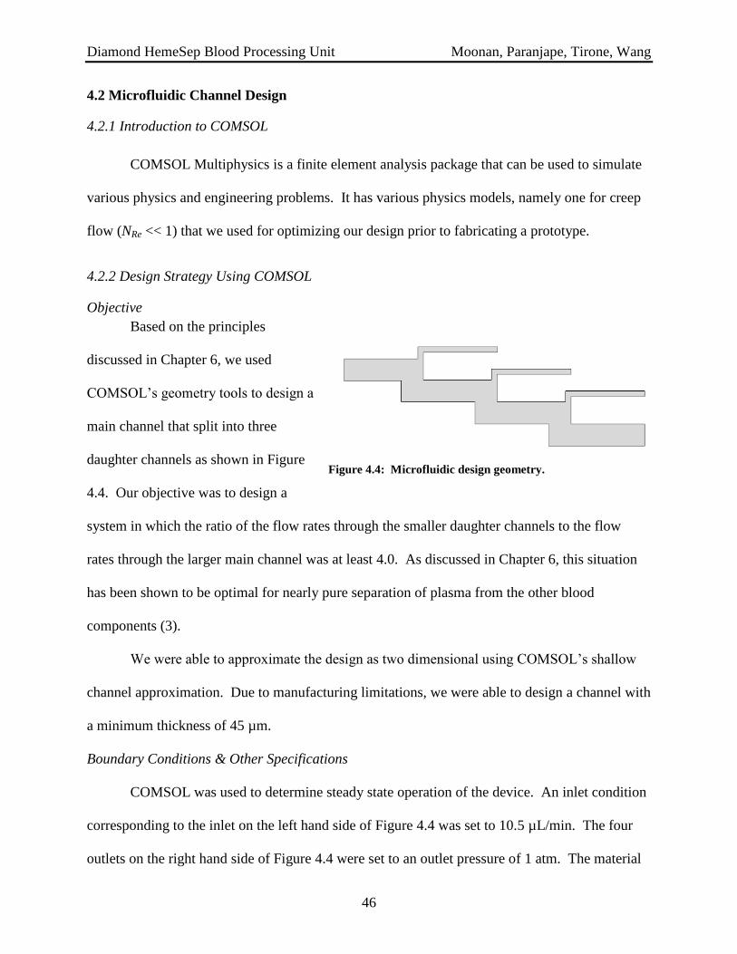
Diamond HemeSep Blood Processing Unit Moonan, Paranjape, Tirone, Wang
46
4.2 Microfluidic Channel Design
4.2.1 Introduction to COMSOL
COMSOL Multiphysics is a finite element analysis package that can be used to simulate
various physics and engineering problems. It has various physics models, namely one for creep
flow (NRe << 1) that we used for optimizing our design prior to fabricating a prototype.
4.2.2 Design Strategy Using COMSOL
Objective
Based on the principles
discussed in Chapter 6, we used
COMSOL’s geometry tools to design a
main channel that split into three
daughter channels as shown in Figure
4.4. Our objective was to design a
system in which the ratio of the flow rates through the smaller daughter channels to the flow
rates through the larger main channel was at least 4.0. As discussed in Chapter 6, this situation
has been shown to be optimal for nearly pure separation of plasma from the other blood
components (3).
We were able to approximate the design as two dimensional using COMSOL’s shallow
channel approximation. Due to manufacturing limitations, we were able to design a channel with
a minimum thickness of 45 µm.
Boundary Conditions & Other Specifications
COMSOL was used to determine steady state operation of the device. An inlet condition
corresponding to the inlet on the left hand side of Figure 4.4 was set to 10.5 µL/min. The four
outlets on the right hand side of Figure 4.4 were set to an outlet pressure of 1 atm. The material
Figure 4.4: Microfluidic design geometry.

Diamond HemeSep Blood Processing Unit Moonan, Paranjape, Tirone, Wang
47
through the entire device was approximated as water, although blood is known to have a slightly
higher viscosity (~5 – 10 cP) than that of water (~1 cP). This assumption was justified since
plasma is 93% water by volume and the ratio of viscosities of blood and water is under ten.
Therefore, we assumed this approximation introduced negligible error while allowing us to
grossly simplify the simulation. After this, the simulation was run to determine steady state
conditions through each section of the microfluidic device.
Analysis of the Results
Results were analyzed primarily to determine the ratio of the flow rate through the
daughter channel to the flow rate through the main channel for each bifurcation. To this effect,
COMSOL was used to determine the average velocity through the daughter channel and main
channel through each bifurcation. The flow rate through a channel is most generally expressed
by Equation 4.1, below where Q is the channel’s volumetric flow rate, u is the linear velocity
through the channel and Across sectional is the cross sectional area of the channel.
(4.1)
Furthermore, the cross sectional area of a rectangular channel is given by Equation 4.2
where Hchannel is the height of the channel in the y-axis direction in Figure 4.4 and Tchannel is the
thickness of the channel in the z-axis (not shown), which was constant for all parts of the device
at 35 µm.
(4.2)
Since the thickness of the channel was constant for the device, the ratio of two flow rates
could be expressed by Equation 4.3.
(4.3)

Diamond HemeSep Blood Processing Unit Moonan, Paranjape, Tirone, Wang
48
This relationship was used to determine the flow rate ratios for a variety of geometries,
some of which are displayed for the reader in the subsequent section.
4.2.3 Results of COMSOL Simulations
Main u [m/s] Daughter u [m/s] Ratio
First Bifurcation 0.047 0.069 1.59
Second Bifurcation 0.031 0.039 1.89
Third Bifurcation 0.024 0.018 3.17
Table 4.3: Ratios for a channel thickness of 10 µm, a daughter height of 15 µm and a main width of 35 µm
Main u [m/s] Daughter u [m/s] Ratio
First Bifurcation 0.054 0.079 1.57
Second Bifurcation 0.040 0.048 1.94
Third Bifurcation 0.032 0.023 3.17
Table 4.4: Ratios for a channel thickness of 10 µm, a daughter height of 15 µm and a main width of 50 µm
Main u [m/s] Daughter u [m/s] Ratio
First Bifurcation 0.071 0.043 3.88
Second Bifurcation 0.064 0.028 5.27
Third Bifurcation 0.061 0.015 9.50
Table 4.5: Ratios for a channel thickness of 35 µm, a daughter height of 15 µm and a main width of 60 µm
Main u [m/s] Daughter u [m/s] Ratio
First Bifurcation 0.064 0.053 2.84
Second Bifurcation 0.053 0.034 3.69
Third Bifurcation 0.048 0.017 6.53
Table 4.6: Ratios for a channel thickness of 35 µm, a daughter height of 20 µm and a main width of 60 µm
For a channel thickness of 35 µm and for a main channel height of 60 µm and daughter channel
height of 15 µm (the design we actually implemented), Figure 4.5 below graphically depicts the
flow pattern

Diamond HemeSep Blood Processing Unit Moonan, Paranjape, Tirone, Wang
49
Figure 4.5: COMSOL velocity profile for microfluidic design geoemtry.
4.3 Serum Filter Design
We considered several designs for the filter unit. Initially, a design in which the filter is
internal to the pipette was favored. However, we decided that keeping the filter on the actual
cartridge fit our needs best. The design of the filter seen in Figure 4.6 is based off of a standard
U-bottom 96 well plate. The membrane and backing is located at the bottom of the well where

Diamond HemeSep Blood Processing Unit Moonan, Paranjape, Tirone, Wang
50
the U-bottom begins. The walls of the plate are slightly narrower here to provide extra support
to the membrane. At the top of the well is a simple rubber septum which has been punctured to
allow the pipette to enter the well. This will provide a seal as the pipette enters the well. This
also has a plastic backing to provide additional support. The filtered serum collects in a
neighboring hood.
The filtering process begins with the transfer of plasma with clotting factors into the well
before the gel point has been reached. After this has fully clotted, the pipette again enters the
well and pushes at 50 psi for 5 seconds. As the serum is pushed through the filter, it will flow
Figure 4.6: Cross section of the filter. All numbers have units of mm

Diamond HemeSep Blood Processing Unit Moonan, Paranjape, Tirone, Wang
51
into the neighboring well, which is positioned slightly below the filter well to reduce holdup.
With a new tip, the fluid handler withdraws the serum from the neighboring well and transfers it
to the final serum container.
The scheduling of the serum filtration is derived from the kinetics of the clotting reaction.
Luckily the kinetics of this complex series of reactions
has been studied extensively (7). Multiple species are
created in multiple reactions, the rates of which impact the
overall structure and properties of the resulting clot.
Although multiple factors have an effect on the rate of this
reaction, the two most important for us are the fibrinogen
and thrombin concentration. Thrombin concentration has
an effect on the rate of coagulation, up to a maximum
concentration of 0.1 NIH unit/mL (an NIH unit is an
arbitrary measure of catalytic activity of an enzyme). At this point, an increase in the
concentration of thrombin does not appreciably accelerate the rate of reaction (Figure 4.7). Thus
we chose to use a concentration of 1 NIH unit/mL thrombin. In addition, CaCl2 is added, as it is
essential for thrombin to function properly.
In our separation we hope to both have a complete clot as quickly as possible, and to
consume all of the fibrinogen and protofibrils. As shown in Figure 4.8, this is accomplished by
letting the reaction proceed for about 225 seconds. We broke this into two sections for two
reasons. First, since fibrinogen and protofibrils are relatively small species, allowing the plasma
to sit in the filter for the full 225 seconds before filtering could allow some of the fibrinogen and
protofibrils to filter through with gravity as the driving force. By transferring to the filter after
Figure 4.7: Turbidity indicating the
progression of clot formation at thrombin
concentrations of 1) 1 NIH unit/mL 2)
.01unit/mL 3) 0.01 unit/mL and 4) 0.001
unit/mL. The rates of 1 and 2 are nearly
identical, with 3 and 4 being significantly
slower

Diamond HemeSep Blood Processing Unit Moonan, Paranjape, Tirone, Wang
52
100 seconds of clotting, all
of the fibrinogen and most
of the protofibrils will be
consumed, but the plasma
will not have reached the
gel phase yet. Second, this
transfer step will mix the
plasma, which will
hopefully speed the reaction.
4.4 Experimental Data from Prototype Microfluidic Device
4.4.1: Experimental Design and Purpose
A prototype of the microfluidic chip of
our cartridge was built in a manner similar to the
soft photolithography previously described. A
photomask was designed in Solidworks and
printed in high resolution (10,000 DPI). Then, a
negative photoresist was spun onto a silicon
wafer at 4000 RPM for 30 seconds to achieve a
thickness of 45 µm. Following a soft bake at 95 °C
for 15 minutes, the wafer was aligned with the
printed photomask in a Karl Suss mask aligner and exposed to 365 nm UV light to achieve 975
mJ/cm2 of exposure energy. Afterwards, a post exposure bake occurred at 95 °C for 3 minutes
and all un-reacted photoresist was removed with a developer for 10 minutes under frequent
Figure 4.8
Figure 4.9: Fabricated PDMS microfluidic
chip attached to a glass slide. As shown here,
a clear plasma fraction (right) and
concentrated blood ouput (red droplet, left)
can be oserved.

Diamond HemeSep Blood Processing Unit Moonan, Paranjape, Tirone, Wang
53
agitation. Then, in a mixture of 1:10 mixture of PDMS and curing agent was poured into the
mask and placed into an 80 °C oven for 2 hours. After this step, the device was removed, cut,
sealed to a glass slide, and ports were punched to allow for outlet collection and pressure input.
The main intention of this testing was for proof of concept purposes. The chip included
six total designs with the same channel dimensions, but different types of bifurcations (as shown
in Figure 4.9). All of these designs had a main channel width of 60 µm, a daughter channel
width of 15 µm and a depth of 45 µm. The same flow rates and fabrication process was used as
would have been for the complete device shown in previous figures. Therefore, testing was
intended to demonstrate three things: 1) that the chip design was capable of being manufactured,
2) that the microfluidic principles underlying the design were effective at separating blood and 3)
that the flowrates being used would be sufficient without either destroying the device or causing
shear of the blood cells.
It should be noted that there were a few important differences between the testing and the
way the cartridge will work in the liquid handling unit. First, lab equipment was used to
substitute functions the liquid handler would normally perform. The liquid handling robotic arm
was substituted with a syringe pump from Harvard Apparatus in order to apply a constant flow
rate (from 10 µL/min to 25 µL/min during testing) across the channels. Blood was collected in a
citrated tube from a willing volunteer and then placed in a syringe to be pumped through the
device. Flow rates ranging from 10 µL/min to 25 µL/min were tested through each of the six
designs. The chip was mounted on an inverted microscope so that pictures of the device in
action could be obtained. Initial trials using the device without modification and with whole
blood showed no plasma separation under any flow rates. However, subsequent trials aimed at
increasing the resistance to flow through the plasma channels demonstrated that the device does

Diamond HemeSep Blood Processing Unit Moonan, Paranjape, Tirone, Wang
54
indeed fractionate the blood into a plasma fraction and a remaining blood fraction. These results
are discussed next.
4.4.2 Preliminary Qualitative Results
The device was first viewed under the microscope to ascertain whether or not the
fabrication process maintained structural fidelity to the SolidWorks mask (Figure 4.3) we
designed. The figures below examine some of the various designs to demonstrate that, indeed,
the channels maintained their structural accuracy through the soft photolithography process. In
the figures below, three designs are examined, a 3-channel bifurcation with rounded square
edges, an angled bifurcation and a “y” bifurcation (a-c, respectively).
Figure 4.10: A – three bifurcations in series with rounded square edges. B – a single, angled bifurcation
pattern. C – a single “y” shaped bifurcation pattern
As mentioned earlier, initial trials of running the device with human whole blood did not
work under any flow rates. The blood simply split its flow but stayed as whole blood as it
flowed through the channel. No images were taken of this process
Furthermore, it was observed that for flow rates up to 15 µL/min, no blood shearing was
observed, indicating that the process could potentially be made faster during year of clinical
testing. Under dilution of the whole blood to 10% hematocrit, flow rates up to 25 µL/min could
be achieved.
Finally, preliminary testing showed that removal of the syringe pump pressure did not
immediately stop flow, because of the pressure buildup in the channels associated with

Diamond HemeSep Blood Processing Unit Moonan, Paranjape, Tirone, Wang
55
microfluidic flow. This observation is critical to correct operation of the device in the cartridge
as shown in the Gantt chart, since the liquid handling unit must take breaks from pushing the
fluid through the microfluidic chip in order to perform parallel mixing steps.
4.4.3 Troubleshooting and Further Qualitative Results
Taken together with our outputs from the COMSOL simulation, preliminary
experimental results suggested that some of the assumptions inherent in our COMSOL model
were incorrect, resulting in a flow pattern that was not conducive to plasma skimming. Namely,
our COMSOL model approximated the
material properties of blood as those of
water. While the viscosities are similar,
since blood is 93% water by volume, this
approximation most likely introduced an
error into calculating the ratio of resistances
through the daughter channels after the
bifurcation. Since it is this ratio that
governs how the blood will split into plasma and cell/particulate fractions, any calculation errors
manifested physically in a flow pattern that did not cause separation of the blood. Furthermore,
the material was assumed to be constant throughout the device domain for the purposes of
simplifying the COMSOL simulation. Again, though the viscosities of plasma and the
concentrated blood fraction are similar, they are not the same, introducing further error in the
resistance calculations. Finally, the COMSOL model did not take into account the total length of
the device in calculating resistances and pressure drops. In normal industry applications where
turbulent flow is common, the effect of pipe length is usually negligible. However, in
Figure 4.11: Flow through a longer path length channel
with a width of 250 µm produced concentrated plasma
towards the channel edges near the channel outlet. This
phenomenon could prove useful in designing a chip to
"skim" plasma from whole blood.

Diamond HemeSep Blood Processing Unit Moonan, Paranjape, Tirone, Wang
56
microfluidics, where creep flow is prevalent, the effect of total channel length is likely not
negligible.
This suggestion is backed up by the following experimental evidence. We tested an
existing microfluidic device with a total path length of almost 10 cm (in comparison to the 1-2
cm path lengths in our prototype). Though the channel width was approximately 250 µm,
making direct comparison with our designs difficult, flow was examined at the outlet of the
channel. As can be seen in Figure 4.11, plasma seems to concentrate towards the edges of the
wall while blood cells migrate towards the center of the flow profile. Future designs could take
advantage of this phenomenon through a three way channel split to skim plasma from the
remainder of whole blood.
Next, we placed a 100 µL droplet of water at the outlet of the plasma channel on our
prototype design with the intention of providing a hydrostatic pressure head to increase
resistance to flow through that channel. This was done for a few different bifurcation styles.
Assuming a completely spherical drop and a density of 1 g/cm3, this droplet corresponded to a
pressure head given by Equations (4.1) and (4.5):
(
) (4.4)
(4.5)
This additional pressure head was sufficient to effect separation of plasma from whole
blood as shown in Figure 4.12 below.

Diamond HemeSep Blood Processing Unit Moonan, Paranjape, Tirone, Wang
57
Figure 4.12: A - angled bifurcation working under increased hydrostatic pressure on plasma channel. B -
rounded square bifurcation working under increased hydrostatic pressure. C - rounded bifurcation in final
design working under increased hydrostatic pressure
The results of this set of experiments fairly conclusively demonstrate that indeed, the
bifurcation principle should hold. Unfortunately, the simplifying assumptions in our COMSOL
model did not allow for the correct calculations of the resistance ratios. However, knowing this
a priori would have been fairly difficult, and with empirical data to help uncover this flaw, the
model can be updated and the design improved during the clinical trial year.
Blood smears were taken for the outputs of both plasma and whole blood to determine
the purity of the plasma fraction obtained. As can be seen in Figure 4.13, the plasma output (A)
has considerably fewer blood cells than the blood output (B) from the channel. Qualitatively,
this demonstrates that the technique is suitable for use in separating plasma from whole blood.

Diamond HemeSep Blood Processing Unit Moonan, Paranjape, Tirone, Wang
58
Figure 1: A - plasma output from device. A nearly pure plasma fraction (free of cells and other blood debris)
is obtained. B - blood ouptut from device contains considerably more cells and other debris from whole blood
Thus, all experimental data adequately supports the aforementioned proof of concept points. With minor
modifications based upon the results of the experiments, the device should be able to be produced in a
manner that successfully accomplishes the point of the product while maintaining the size and costs of the
device mentioned here in the report.

Diamond HemeSep Blood Processing Unit Moonan, Paranjape, Tirone, Wang
59
Chapter 5: Blood Composition
5.1 Introduction
Germane to understanding the point of the Diamond HemeSep is a working knowledge of
the biology and composition of human whole blood. To provide such a basis, this chapter
presents a very simple introduction to the topic.
Human whole blood is an important bodily fluid that
delivers oxygen and nutrients while removing metabolic waste from
the cells and tissues of the body. Blood is composed of three
primary constituents as shown in Figure 5.1: 1) red blood cells
(erythrocytes), 2) white blood cells (leukocytes) suspended in a
liquid 3) plasma. Hematocrit, which is defined as the percentage of
red blood cells by volume in whole blood, is typically around 45%
in humans (8). The three components of blood will be discussed
next.
5.2 Plasma
Human blood cells are suspended in a straw-colored liquid component of blood known as
plasma. Plasma, which is mostly water (93% by volume), also contains dissolved chemical
species. Primarily, plasma transports glucose, clotting factors, minerals, ions, hormones, carbon
dioxide, and proteins such as albumin. As such, plasma serves as a major protein reserve in the
body and also as the major medium for waste removal. Plasma constitutes around 55% of whole
blood by volume (8, 9).
Figure 5.1: Human whole
blood is composed of three
fractions: plasma, white blood
cells (leukocytes) and red
blood cells (erythrocytes) (8)

Diamond HemeSep Blood Processing Unit Moonan, Paranjape, Tirone, Wang
60
Because of its crucial role in regulating blood osmolarity and infection prevention,
plasma is often analyzed for its salt and protein concentrations both clinically and in research.
Consequently, obtaining nearly pure plasma fractions is very important.
5.3 Serum
Blood serum is essentially blood plasma with fibrinogens (proteins used in the clotting
process) removed. Serum does not contain any blood cells, but does contain all of the proteins
not used in the clotting process and otherwise found in the plasma. Like blood plasma, serum is
used in many diagnostic tests both clinically and in research (10).
5.4 White Blood Cells
White blood cells (WBCs), or leukocytes, are the
DNA-containing blood cells that make up the body’s
immune system. These cells protect the body from
infectious disease and foreign agents while also
modulating inflammatory responses. Five different types
of WBCs exist: neutrophils, eosinophils, basophils,
lymphocytes and monocytes. The first three are classified
as granulocytes due to their granular appearance under the
microscope caused by membrane bound enzymes that
engulf and destroy foreign matter. The latter two are characterized as agranular due to the
absence of granular appearance under the microscope (9, 11).
Analysis of white blood cells is useful and necessary in understanding the DNA profile of
a patient and in looking for various markers of disease and health.
Figure 5.2: WBCs are the irregularly
shaped spherical cells whereas RBCs
cells are the larger, donut shaped
cells. (9)

Diamond HemeSep Blood Processing Unit Moonan, Paranjape, Tirone, Wang
61
5.5 Red Blood Cells
Red blood cells (RBCs), or erythrocytes, are the most common type of blood cell. RBCs
deliver oxygen (O2) to the body’s cells and tissues. These cells owe their red appearance and
ability to transport O2 to hemoglobin, an iron containing biomolecule. These cells develop in the
bone marrow and circulate for about 100-200 days before being recycled and regenerated. RBCs
are anucleate, meaning they do not contain a nucleus and, consequently, do not contain DNA.
Per microliter of whole blood, there are roughly 4-6 x 10⁶ RBCs.
RBCs are generated through a process known as erythropoiesis. They are produced in
the bone marrow at a rate of about 2 million cells per second and mature over seven days (12,
13).
5.6 Blood Coagulation
Blood coagulation is the process to form a
clot to quickly prevent excessive blood loss after
injury. While a quick response is essential, the
process must not make clots that impede normal
blood flow. Thus the process is highly regulated by
multiple positive and negative feedback loops.
There are two pathways in the coagulation cascade
(14). Most important in vivo is the extrinsic pathway
in which injury pushes tissue factor from surrounding
subendothelial cells into the blood flow, initiating the
cascade. The intrinsic pathway, however, is much
more important for our purposes. This pathway is activated by an anionic surface such as glass
Figure 5.3: Plasma coagulation. Roman numerals
indicate unactivated coagulation factors, and
activated factors are indicated by a lower case “a”.
Nonenzymatic cofactors are indicated by numerals
in black ovals. White arrows indicate reactions of
the intrinsic pathway

Diamond HemeSep Blood Processing Unit Moonan, Paranjape, Tirone, Wang
62
(15). Contact with an anionic surface produces the active serine protease XIIa from the inactive
form XII. This protease then selectively hydrolyses a portion of the inactive serine protease
factor XI to form the active protease XIa. This in turn activates factor IX in the same way,
which then activates thrombin from prothrombin in the same way. While the rest of the enzyme
factors are involved primarily in regulating coagulation, thrombin is the serine protease actually
responsible for the formation of the clot. It is important to note that many of these factors, and
specifically the interaction of XII with an anionic surface, depend on the presence of calcium
ions. Removal of calcium ions with a chelator such as EDTA or citrate prevents the coagulation
cascade.
Essential in any discussion of blood
coagulation is the final product: a blood clot or
thrombus. There are two main components of
any blood clot. First is an insoluble fibrin
polymer, produced from the soluble protein
monomer fibrinogen. An electron micrograph of
such a clot can be seen in Figure 5.4 (7). This
polymer can vary in its properties depending on
the degree of branching and the thickness of the
individual fibers. The second component is
blood platelets. Platelets are cell fragments about
a quarter of the size of red blood cells. These become ensnared in the network of fibrin and help
plug the flow of blood. Finally, small amounts of the other clotting factors become stuck in the
clot as well. This helps to down-regulate the coagulation response and limit it to the site of the
500 nm
Figure 5.4: Electron micrograph of a blood clot with
bar indicating 500nm

Diamond HemeSep Blood Processing Unit Moonan, Paranjape, Tirone, Wang
63
injury. By removing these clotting factors, the production of thrombin and then fibrin monomer
cease.
5.7 Fibrin Polymerization
The most important process in the coagulation cascade
for our purposes is the polymerization of fibrinogen to form
fibrin. This is a complex process catalyzed by the
serine protease thrombin. Fibrinogen circulates as a
dimer of three chains (AαBβγ)2 which are connected
by a disulfide bond. Figure 5.5b shows a crystal
structure of the A chain of the fibrinogen monomer
(16). The first step in the formation of the insoluble
fibrin clot is the cleavage of part of the Aα chain by
thrombin to form fibrin I monomers. The fibrinogen-
thrombin complex is shown in figure 5.5a (17). These monomers then aggregate to form fibrin I
protofibrils. Third, thrombin cleaves a portion of the Bβ chain to form fibrin II protofibrils.
Finally, these protofibrils rapidly aggregate to form the insoluble fibrin clot.
5.8 Summary
Human whole blood is composed of three main components: 1) red blood cells, 2) white
blood cells and 3) plasma. The cells are suspended in plasma, the major liquid fraction of whole
blood that also contains many other dissolved species. Blood serum is simply plasma with the
fibrinogens clotted out and separated from the rest of the plasma. Each of these blood fractions
Figure 5.5: a) crystal structure of a portion of fibrin bound
to the active site of thrombin. b) crystal structure of fibrin.

Diamond HemeSep Blood Processing Unit Moonan, Paranjape, Tirone, Wang
64
is diagnostically useful in its own right, making blood separation an interesting, essential process
both clinically and in research.

Diamond HemeSep Blood Processing Unit Moonan, Paranjape, Tirone, Wang
65
Chapter 6: Plasma Separation via Microfluidic Design
6.1 Introduction
To induce plasma separation from whole blood without the use of traditional
centrifugation techniques, a microfluidic channel design was implemented based on published
work by Yang, Undar, and Zahn (3). There are many advantages of utilizing microfluidics over
centrifugation, including cost, size, and autonomy of the unit. The efficacy of the device in
separating cells from plasma in whole blood stems from the bifurcation law, or the Zweifach-
Fung Effect.
6.2 Bifurcation Law (Zweifach-Fung Effect)
The bifurcation law states that when red blood cells encounter a bifurcation in a capillary
blood vessel, a large number of cells flow into the daughter vessel that has the higher flow rate,
leaving only a small number of cells to flow into the daughter vessel with the lower flow rate.
This occurs due to the large pressure gradient of the high-flow vessel compared to the low-flow
vessel. Asymmetric shear stresses on the capillary walls also induce a torque on the cell that
causes it to flow into the high-flow vessel. It has been shown that a flow ratio of 2.5:1 is
sufficient to induce this law in channels whose dimensions are approximately the same as a cell.
This preferential bifurcation only occurs, however, when a cell’s centroid falls above a critical
streamline, the stagnation point of the bifurcation. As the ratio of flow in the daughter vessels is
increased, this stagnation point lies closer to the low-flow vessel and effects of this law are more
pronounced. Figure 6.1a below diagrams the effects of pressure and shear rate on the bifurcation
law while Figure 6.1b describes the critical streamline needed for this preferential bifurcation.

Diamond HemeSep Blood Processing Unit Moonan, Paranjape, Tirone, Wang
66
Figure 6.1. Schematic of the bifurcation law (Zweifach-Fung Effect) (3). Figure 1a. shows the effect of
pressure drop differences on the cell (ΔPh > ΔPl). The shear rate differences (γcell_wall_high_flow > γcell_wall_low_flow)
are also presented and are instrumental in producing the Zweifach-Fung Effect. Figure 1b. shows the critical
streamline in a bifurcation channel operating at a 4:1 flow rate ratio and the resulting paths of the cells.
Path’s I and III both flow to the high-flow channel because the centroid of the cell lines above the stagnation
streamline. Path II, however, flows into the low-flow channel.

Diamond HemeSep Blood Processing Unit Moonan, Paranjape, Tirone, Wang
67
6.3 Flow rate ratio optimization
As mentioned above, the choice of flow rate ratio between the daughter channels is a key
design specification in achieving desired separation efficiencies. For use in research
applications, the purity of the plasma should be as high as possible without sacrificing its
integrity. Yang, Undar, and Zahn show in their research that a 4:1 flow ratio between the
channels resulted in a 98.9% recovery efficiency for C8161 Human melanoma cells while ratios
above 6:1 resulted in 100% efficiency (3). Their complete analysis is shown in Figure 6.2
below.
Figure 6.2. Recovery efficiency of 16 um fluorescent particles and 8-10 um C8161 Human melanoma cells. It
can be seen that at flow ratios greater than 4:1 cell recovery efficiency is virtually 100%. Cell efficiency was
consistently greater than the fluorescent particles because the deformity of the cell led to an asymmetric
distribution of cells in the middle of the channel, which increased efficiency (3).

Diamond HemeSep Blood Processing Unit Moonan, Paranjape, Tirone, Wang
68
6.4 Flow rate resistance and use of multiple parallel bifurcations
To achieve greater plasma yields in the microfluidic device, multiple parallel bifurcations
can be incorporated. However, each additional channel causes a drop in flow resistance in the
subsequent channels. To prevent efficiency losses in the subsequent channels, each channel
diameter must be changed to reflect this. Equation 6.1 below demonstrates the relationship
between pressure changes (ΔP), flow rates (Q), and resistance to flow (R):
(6.1)
From this equation it is seen that flow resistances will need to be varied to achieve the desired
pressure changes. These resistances must therefore satisfy the following equation:
(6.2)
Where rQ is the flow rate ratio, Rp,j is the resistance of the jth
plasma channel, and RB,j is the
resistance of the jth
blood cell channel. Yang, Undar, and Zahn performed their analysis on a
microfluidic design that incorporated 5 daughter plasma channels. Figure 6.3 below reproduces
their velocity profiles and analytical results from using computational fluid dynamics.

Diamond HemeSep Blood Processing Unit Moonan, Paranjape, Tirone, Wang
69
Figure 6.3a. CFD velocity profile for microfluidic device. Each split results in a decreasing velocity profile.

Diamond HemeSep Blood Processing Unit Moonan, Paranjape, Tirone, Wang
70
Figure 6.3b. Velocity magnitude at each node for varying channel widths. As blood flows downstream the
velocity magnitude decreases proportionally, and the parabolic peak represents the optimal channel width.

Diamond HemeSep Blood Processing Unit Moonan, Paranjape, Tirone, Wang
71
Figure 6.3c. Flow rate ratio for 1%,5%,15,45% hematocrit using CFD analysis. Bifurcation nodes farther
downstream result in larger flow rate ratios.

Diamond HemeSep Blood Processing Unit Moonan, Paranjape, Tirone, Wang
72
6.5 Overall device efficiency and the Fahreus effect
To test the overall efficiency of the microfluidic device, the percent by volume of plasma
separated is quantified. This value (η) can be expressed with the following equation:
{ (
) (
)
} (6.3)
Where Hmu is the hematocrit levels upstream of the separation region and Hmd is the hematocrit
levels downstream of the separation region. When a higher initial inlet hematocrit is used,
higher upstream and downstream hematocrit levels are observed. These simultaneous increases
lead to higher percent by volume plasma separation, as seen in Equation 6.3. Yang, Undar, and
Zahn quantitatively confirmed Equation 6.3, and the results are reproduced in Figure 6.4 below.
It has been shown, however, that when a large feed inlet is flown into a channel of smaller
diameter, the average hematocrit decreases as channel diameter decreases. This is known as the
Fahreus effect and is shown for a 15 µm channel in Figure 6.5 below (3).

Diamond HemeSep Blood Processing Unit Moonan, Paranjape, Tirone, Wang
73
Figure 6.2. Microchannel hematocrit vs. time for inlet hematocrit levels of 10% and 35%. 4(a) represents
inlet levels of 10% and 4(b) represents inlet levels of 35%. As inlet hematocrit levels increase, the degree of
separation is increased, as expected from Equation6. 3.

Diamond HemeSep Blood Processing Unit Moonan, Paranjape, Tirone, Wang
74
Figure 6.3. Fahreus effect in a 15um microchannel. As inlet hematocrit increases, upstream hematocrit
increases.

Diamond HemeSep Blood Processing Unit Moonan, Paranjape, Tirone, Wang
75
Chapter 7: White Blood Cell Separation via Immunomagnetic
Precipitation
7.1 Introduction
As described in Chapter 5, leukocytes are the only DNA containing cells in human whole
blood. Therefore, it is often of interest to a clinician or researcher to analyze the DNA profile of
these white blood cells for diagnostic purposes. Doing so requires a blood separation technique
capable of producing a nearly pure white blood cell fraction. As described below, an antibody-
based method will be implemented on the Diamond HemeSep Cartridge. Antibodies are proteins
produced by the white blood cells of the body’s immune
system. As shown in Figure 7.1, these antibodies contain
sites that can bind their substrates, the antigens, much like a
lock fits a key (18, 19). The immunomagnetic separation
system, therefore, is based on the extreme fidelity of
antibody binding to their substrate molecules. Selecting an
antibody, chemically linked to a paramagnetic bead, that
may bind exclusively to the white blood cells allows for the
aggregation and subsequent collection of those white blood
cells.
Figure 7.1: Antibodies are proteins
that bind their substrates, antigens,
much like a key fits in a lock (36)

Diamond HemeSep Blood Processing Unit Moonan, Paranjape, Tirone, Wang
76
Immunomagnetic separation is quickly
becoming a favored technique for efficiently
isolating cells from bodily fluids (20). The
technique relies on the high affinity of
antibodies, proteins produced by the immune
system, for their target substrates. The
substrates are typically proteins bound to the
membrane of a target cell. As shown in Figure 7.2, when these antibodies are chemically
conjugated to paramagnetic beads, they can be magnetically concentrated to the bottom of the
well containing the whole blood along with their bound substrate, the white blood cells,
facilitating separation of the bead-antibody-substrate from the remainder of the fluid.
7.2 Leukocyte Common Antigen Antibody
7.2.1 Justification for Separation
The five types of white blood cells (neutrophils, basophils, eosinophils, monocytes and
lymphocytes as discussed in Chapter 4) are responsible for all immunological functions and are
the only nucleated cells in blood. This means that they are the only blood cells containing DNA.
As such, separation of these leukocytes is critical for hematological analyses and clinical
diagnostic tests for studying progression of disease.
Traditional methods for separating these cells take advantage of differences in buoyant
gravity of the different blood components in a density gradient solution such as Ficoll-Paque™
or differences in average diameter between RBCs and WBCs using membrane technology.
However, the former method requires careful attention of a human technician and therefore
Figure 7.2: Antibodies chemically conjugated to
paramagnetic beads facilitate isolation of substrate
(white blood cells).

Diamond HemeSep Blood Processing Unit Moonan, Paranjape, Tirone, Wang
77
requires relatively large volumes of blood while the latter is prone to clogging. Therefore, the
use of immunomagnetic separation presents an interesting and perhaps more efficient, though
slightly more costly method of WBC separation from the rest of the blood components.
7.2.2 Background for Separation Antibody Design
Immunomagnetic separation has several attractive features, including the high degree of
specificity of antibody-substrate binding, the kinetics of antibody binding, and the ability to
standardize a separation procedure using a liquid handling system.
Antibody-substrate specificity refers to the fidelity of binding between the antibody and
its substrate. Ideally for separation purposes, an antibody will bind only to its substrate and not
to other molecules, allowing for the most pure substrate to be obtained after magnetic
concentration.
Antibodies generally bind quickly and specifically to their target substrates, making them
ideal for use in a cartridge such as ours. As shown in Figure 7.3, within about 5 minutes, more
than 60% of antibodies can be expected to
be bound to their targets after gentle mixing
(21). This provides for a quick, reliable and
reproducible method for separating out
white blood cells from whole blood without
having to remove any of the other blood
components first. In our process, the total
processing time for the white blood cells is
Figure 7.3: Antibodies bind quickly and efficiently to
their targets after gentle mixing in a well with human
whole blood (21)

Diamond HemeSep Blood Processing Unit Moonan, Paranjape, Tirone, Wang
78
around 13 minutes with only 5 minutes required for the antibodies to bind to the cells during an
incubation step as discussed in Chapter 4.
7.3 Magnetic Immunoprecipitation Design
In our chip, we utilize an antibody specific for leukocyte common antigen (LCA, also
abbreviated as anti-CD45 antibody). LCA is actually a family of trans-membrane glycoproteins
expressed on the surface of all five types of white blood cells discussed in Chapter 4. Five
isoforms of LCA, named ABC, AB, BC, B and D have been identified and may be recognized by
antibodies known as CD45RA, CD45RB, CD45RC and CD45RD. Our cartridge will utilize a
one of these antibodies to target all five isoforms of LCA.
Because LCA is known to be expressed on all white blood cell types, it is an ideal choice
for use in our separation, since antibodies specific to the various LCA isoforms may precipitate
all types of white blood cells. This simplifies the search for antibodies and increases the yield of
white blood cells obtained from the separation. Furthermore, established kits already exist for
using such antibodies in separating white blood cells from whole blood. The kit used in the
Diamond HemeSep cartridge is the DynaBeads FlowComp Human CD45RA isolation kit from
Invitrogen, with a few modifications to the separation procedure as described in Chapter 4.

Diamond HemeSep Blood Processing Unit Moonan, Paranjape, Tirone, Wang
79
Chapter 8: Serum Separation via Microfiltration
8.1 Introduction
In order to create serum, filtration was the only logical solution. While centrifugation is
by far the most utilized technique, a micro-centrifuge in this setup would be impractical.
Antibody techniques to pull down fibrin also would not work. Antibodies selective for
fibrinogen only remove fibrinogen while leaving all the other clotting factors in the plasma. This
produces defibrinated plasma instead of serum. In addition, filters already exist to help keep
serum separated from a clot when the sample is spun down in a centrifuge. Thus, a filter system
seems quite plausible.
8.2 Physical Characteristics of Fibrin Clot
The design of the filtration system depends on the characteristics of the fibrin clot.
Because of the complexity of the reaction to form the clot, different species are present in the
plasma at various times. Luckily, the kinetics of this complex series of reactions has been
studied extensively (7). Multiple species are created in multiple reactions (Figure 8.1), the rates
of which impact the
overall structure and
properties of the
resulting clot. As
discussed in Chapter 5,
protofibrils rapidly form
after the introduction of
thrombin. These will
Figure 8.1: Normalized concentrations of various species against time

Diamond HemeSep Blood Processing Unit Moonan, Paranjape, Tirone, Wang
80
grow to an approximate length of 0.8µm and a weight of 3,500kDa (22). These are consumed in
the production of the much larger fibers that are characteristic of a clot. These fibers grow to a
diameter of around 0.2 µm and lengths on the scale of 100 µm. Eventually, enough of these
fibers are formed to create a gel. If the filtration is timed correctly, it can take place when
virtually all of the protofibril has been consumed in production of fiber, but before the onset of
the gel.
8.3 Membrane Selection
With these characteristics in mind, a membrane had to be selected. The efficiency of the
filtration depends predominantly on the characteristics of the membrane. The first challenge in
selecting a membrane is determining the required pore size. The pores of the membrane must be
smaller than the particle in order to effectively reject it. In addition, pores for linear
proteins/particles must be smaller than those for globular proteins (23). Most of the fibrin will
be in the form of very large fibers or aggregated into a gel. This suggests using a large pore size.
However, some protofibril may remain. As a result, a pore size of 1 µm was selected. The use
of membranes with this pore size for pre-filtration of serum before its use in easily clogged
instruments validates this choice (24).
With pore size selected, the membrane material was chosen. This polymer must be
hydrophilic, low-protein-binding and biologically inert. A hydrophilic membrane is used to
achieve desired wetting of the membrane by the aqueous solution. In addition, hydrophilic
membranes are less likely to adsorb serum proteins. Although several polymers fit these criteria,
we selected a 200 µm thick polysulfone membrane with a 1 µm pore size. As the membrane by
itself is only rated to 30psi, a porous polypropylene backing was selected for the membrane to
increase rigidity.

Diamond HemeSep Blood Processing Unit Moonan, Paranjape, Tirone, Wang
81
8.4 Flow Rate Calculations
The main difficulty in using filtration is the potential for the membrane to clog.
However, the cartridge is single use, so the filter must work only once. The membrane itself
offers little resistance, but because fibrin is designed to prevent blood loss, it does an excellent
job of impeding flow. The gel that forms on the membrane is the major source of flow
resistance. Neglecting the resistance of the membrane, the flow through the fibrin gel can be
modeled using Darcy’s law.
(8.1)
Values of 2.5 g/L fibrinogen/L plasma, .001 Pa.s for the viscosity of fibrin and 0.2 g/mL for the
density were used (25). The height of the fibrin gel was calculated using the concentration of g
fibrinogen/L in plasma and the fibrin density. This height was used as the depth of the bed (L) in
Darcy’s equation. With a pressure of 50 psi, a flow rate of 59 μL/s was achieved.

Diamond HemeSep Blood Processing Unit Moonan, Paranjape, Tirone, Wang
82
Chapter 9: Microfluidic Design Principles
9.1 Introduction
The design of microfluidic devices poses special challenges over their macro-scale
counterparts. Because the channel dimensions are no more than a few micrometers in size,
highly precise fabrication techniques are needed. Also, since the behavior of fluids is difficult to
predict at these length scales, it is important to choose the channel material that offers the least
amount of interaction between the working fluid. A widely used microfabrication technique
involves the use of photolithography and soft lithography in conjunction with
polydimethylsiloxane (PDMS) and is an ideal fabrication technique when working with
biological fluids.
9.2 Photolithography
The process of photolithography involves the selective corrosion of a substrate into a
desired design. To achieve this, a photomask is designed in a computer-aided drawing (CAD)
software package such as Solidworks, which is then laid on top of a silicon wafer coated with a
light sensitive substrate (known as the “photoresist”) (26). There are two types of photoresists:
positive and negative. A positive photoresist, when exposed to UV light, will be corroded on
portions that are covered by the photomask. Alternatively, a negative photoresist will be
corroded in areas that are not covered by the photomask (27). Both processes can be seen in
Figure 9.1 below.

Diamond HemeSep Blood Processing Unit Moonan, Paranjape, Tirone, Wang
83
Figure 9.1. Process of photolithography. When exposed to UV light, positive resists are corroded in areas
covered by the photomask and negative resists are corroded in areas not exposed by the photomask (27).
Once the photoresist has been completely etched, the entire setup is “hard baked” to harden the
photoresist and adhere it to the wafer surface.
9.3 Soft lithography
To prepare the final microfluidic device, soft lithography must be used in conjunction
with the “master” mold produced from the photolithography process. Soft lithography involves
the use of an elastomeric compound such as PDMS to produce the final design from the master
mold via a series of curing steps. First, PDMS is poured over the master mold, where it

Diamond HemeSep Blood Processing Unit Moonan, Paranjape, Tirone, Wang
84
solidifies when exposed to high temperatures. The solid PDMS is then peeled off the master
mold, resulting in the final microfluidic device (28). This device is then fixed to a glass slide,
creating the channels needed for the microfluidic device to work. Figure 9.2 below shows the
steps in the soft lithography process.
Figure 9.2. Process of soft lithography. Liquid PDMS is poured over the master mold, where it is exposed to
high temperatures and cured into a solid. The PDMS is then removed and is used as the final microfluidic
device. Some common problems with the soft lithography process include pairing and sagging (28).

Diamond HemeSep Blood Processing Unit Moonan, Paranjape, Tirone, Wang
85
9.4 Polydimethylsiloxane
Polydimethylsiloxane (PDMS) is a silicon-based organic compound that is commonly
used in microfluidic device designs. It is optically clear, inert, non-toxic, and non-flammable. It
has the following chemical design:
Figure 9.3: Structure of polydimethyl siloxane (PDMS)
In Figure 9.3, n denotes the number of monomers in the compound. The viscosity of PDMS can
be increased by increasing the number of monomers (increasing n). This can allow PDMS to
take a liquid form (low n) or a rubbery semi-solid (high n) (29). This property makes it a
particularly useful elastomer in soft lithography. Other desirable properties of PDMS is that it
does not swell with humidity (non-hydroscopic), it has high thermal stability, and it can be easily
deformed and reshaped both mechanically and chemically (28).
Diamond HemeSep Blood Processing Unit Moonan, Paranjape, Tirone, Wang
86
Chapter 10: Manufacturing Considerations
10.1 Introduction
The Diamond HemeSep Cartridge will primarily be assembled from original equipment
manufacturer (OEM) parts. Such parts include the actual plastic cartridge containing the input
and output wells, the reagents, and the actual liquid handling unit. The microfluidic portion of
the device will be manufactured in our own facilities, originally using manual labor in the
prototyping phases and eventually moving towards an automated process with human operators.
Originally, we proposed outsourcing microfluidic chip fabrication to an outside company,
especially considering the sizeable cost associated with building a manufacturing facility.
Ultimately, it seemed most sensible to protect the intellectual property of the cartridge by
maintaining complete control over the chip’s fabrication.
As mentioned in the previous chapter, the microfluidic chip is composed of two layers, a
PDMS layer and a glass layer (a glass slide). The interface between these two layers is what
creates the channels through which blood may flow. To create the PDMS layer, soft
photolithography replica molding, as described earlier, was decided upon for use.
While all of the other parts are to be obtained from OEMs, assembly still requires
accurate and efficient delivery of reagents to the snap-in reagents section of the cartridge. In
order to accomplish our eventual goal of 100-200 cartridges per hour, we decided to use a
robotic liquid handling system for delivering the reagents to their plastic wells. A major
consideration in assembly is storing the reagents at the required conditions. Calcium, stored as
CaCl2 in TBS, may be stored indefinitely in solution at room temperature whereas thrombin must
be stored dry at colder temperatures. This means that the user must rehydrate the thrombin prior

Diamond HemeSep Blood Processing Unit Moonan, Paranjape, Tirone, Wang
87
to using the cartridge. This also necessitates delivery of the packaged cartridge on dry ice for
preservation.
10.2 Fabrication of Microfluidic Device
The microfluidic chip will be composed of two layers including a glass slide and a PDMS
layer. PDMS was chosen because of its desirable properties in molding, its optical transparency
to wavelengths as low as 300 nm, its good thermal and chemical stability and its low interfacial
energy.
Soft photolithography was chosen as a method for fabrication of this PDMS portion of
the device. This process was chosen because it can yield a large number of devices with a high
fidelity to the “master.” Using a photosensitive resist, as described in Chapter 9, the PDMS layer
will be patterned by exposure to UV light. As described, this process begins with the creation of
a master, which is fabricated by first creating a resist. Spinning onto a silicon wafer creates this
resist and the pattern is created via etching the imprint using UV irradiation. Then, PDMS molds
may be created from this master, which may be used on large numbers of chips. Despite its
positive features, PDMS has a few known negative attributes, including slight shrinking upon
curing and difficulty in accurate patterning below about 10 μm scales. Despite these few
negatives, the utility and applicability of soft photolithography made it a desirable process for
fabrication of our microfluidic chips.
To fit our goal of 100 – 200 microfluidic chips produced per hour, hundreds of molds
will need to be produced for creation of the PDMS portions of the device. For this, only a few
masters will need to be created. These masters will be created from normal lithography methods.
The channels all have a height of 35 μm, lengths on the order of centimeters and widths ranging

Diamond HemeSep Blood Processing Unit Moonan, Paranjape, Tirone, Wang
88
from 15 to 60 μm. These features are all within the size limits allowed by photolithography and
can therefore be accurately fabricated using this technique.
In the fabrication process, as described earlier, master creation begins with the
application of a positive photoresist onto a silicon wafer and spinning to get a resist thickness of
35 μm. Then, UV light is used to cross link areas not masked that correspond to the channels
and reservoirs. After this, the master is baked to cure unexposed resist and the exposed resist is
washed with an organic developer that dissolves and etches these areas. This causes the
substrate to have channels 35 μm in depth and reservoirs in the positions indicated for our chip.
This process is already diagramed in Figure 9.1 for negative photoresists.
After finishing the master, a negative PDMS mold to the final design pattern may be
created (since a positive photoresist was used). Hundreds of PDMS molds, containing many
parallel master designs, will be created and used as a stamp. Prior studies have shown that
PDMS can be accurately used as a stamp after more than 10 uses (30). This allows each stamp
to have a lifetime of up to two months. These parallel molds will be filled with PDMS and then
thermally cured in a baking process that takes approximately two hours, giving desired PDMS
devices with channels and reservoirs as needed to be sealed to the glass slide layer.
We have decided to outsource the creation of the masters while producing the molds and
actual devices. Each microfluidic device will require approximately 17.5 mL of PDMS. This
assumes less than 1% shrinkage. To accomplish the production of 100-200 chips per hour, we
Figure 10.1: This figures displays the manufacturing process involved in creating PDMS chips. (1) A PDMS
mold is created from a positive photoresist. (2) This mold is filled with PDMS and cured in an oven at 80 C
for 2 hours. (3) the PDMS device is removed from the mold, which may be reused, and is then (4) adhered to
a glass slide, creating the channels and reservoirs as indicated in the microfluidic chip design.

Diamond HemeSep Blood Processing Unit Moonan, Paranjape, Tirone, Wang
89
will begin each day by mixing PDMS with viscous curing agent in a 100 L tank containing 70 L
of PDMS. It will need to be mixed for 10 minutes. Then, the mixture will be deposited onto the
PDMS molds (blue in Figure 10.1) to create the microfluidic chip (gray in Figure 10.1). The
parallel molds will be placed in a curing oven for approximately two hours at 80 °C. Based on
the surface area of 4,000 PDMS molds, the oven needs to have a surface area of approximately
60 ft2. Based on a prior design project, by Abbot, Lee, Kohli and O’Brien, a suitable curing oven
from Wisconsin Oven Corporation, priced at $22,400 may be used to accommodate the needs of
the process.
While the devices cure, reagents are delivered to the plastic cartridge insert as described
in the subsequent section. Deposition of the antibody, Dyna-Beads, PBS, lysis buffer, thrombin
and calcium will each require about 3 seconds for alignment and subsequent deposition of the
liquid, amounting to a total of eighteen seconds. The deposition of the reagents will take place in
a cold environment to protect the reagents from degradation and evaporation.
Once the curing has completed, the PDMS chips are taken from the molds via a robotic
arm and sealed to glass slides pre-coated with Sigmacote, a hydrophobic material, using a PDMS
sealing machine which is estimated to cost $210,000. From this unit, the chips will be shuttled to
an assembly line where a robotic suction arm will assemble the reagent insert and microfluidic
chip into the cartridge, then seal the cartridge and transport them into a freezer for delivery.
Our goal will be to produce approximately 100 – 200 cartridges per hour, corresponding
to approximately 1 million cartridges per year.
10.3 Robotic Liquid Handling System
Although most cartridge parts will be obtained from OEMs, reagents will still need to be
delivered to the cartridge reagent insert prior to packaging. To accomplish the delivery of

Diamond HemeSep Blood Processing Unit Moonan, Paranjape, Tirone, Wang
90
microliter volumes of reagent, a few different options were considered. Namely, we considered
the use of an inkjet handling system and a robotic pin tool. While both of these technologies are
capable of accurately and reproducibly delivering small quantities of liquid on the nanoliter
scale, their use in microliter quantities is far too slow and costly to be of great use. Instead, we
opted to use a robotic liquid handling system. This system is capable of delivering each reagent
to the appropriate well in approximately 1-2 seconds, allowing us to realize our goal of
manufacturing 100-200 cartridges per hour. Since our design is based off of a standard 96 well-
plate micro-assay design, standard liquid handling robots sold may be used to insure accurate
deposition of reagents. This will work by passing the cartridge insert along a conveyer belt
underneath the robotic liquid handling unit, where the unit may be programmed to deliver the
reagents to the standard well plate locations. Such a unit is estimated to cost $35,000 and is
assumed to be able to deliver the reagents in 3 seconds per transfer.
Because of the relatively large volumes (μl scale) of reagents being deposited into the
wells, evaporation of reagents is of little concern. According to the literature, approximately 1 nl
of aqueous solution may be assumed to evaporate in approximately 30 seconds. However,
precautions to reduce the risk are to include more reagent than is necessary during cartridge
operation and to seal the reagent wells with foil shortly following deposition. This problem will
be addressed as needed during the first year of testing.
10.4 Manufacturing Schedule
Using an assumed market size of 1 million chips per year, which is roughly equivalent to
the number of blood draws taken in clinical trials every year, and a market capture of
approximately 50%, we will need to produce 500,000 chips per year.

Diamond HemeSep Blood Processing Unit Moonan, Paranjape, Tirone, Wang
91
To meet this market capture goal, there are several considerations and assumptions to
make. First, we have assumed that the manufacturing facility can operate 8 hours a day, 5 days a
week for 50 weeks of the year (250 total days). Therefore, the number of chips needed for sale is
given by equation 10.1:
(10.1)
Furthermore, it is assumed that roughly 25% of chips manufactured will be used for
quality control testing and therefore, an additional number of chips must be produced. Of these
chips, it is assumed 25% of them will be defective, such that the requisite number of chips is
given by equation 10.2:
(10.2)
This schedule assumes manufacturing starts at time t = 0 (9:00 AM). Mixing and
deposition of PDMS onto the molds is represented by the box labeled “mix/dep”. Mixing takes
approximately 10 minutes and each deposition is assumed to take 3 seconds. It is assumed that
10 chips’ worth of PDMS can be deposited to the molds per deposition yielding the following
amount of time necessary for the step:
(10.3)
The chips are then cured in an oven for a total time of 2 hours. Simultaneously, the
reagents are deposited into the cartridge using the times discussed above:
(10.4)
Because this time exceeds the typical work day, two liquid handling robots will be run in
parallel for a total of 7 hours each. Note that these calculations are for the final year of

Diamond HemeSep Blood Processing Unit Moonan, Paranjape, Tirone, Wang
92
production, and that manufacturing facilities can be modified as necessary during actual
production.
After removing the PDMS layers from the oven, they need to be sealed to the glass layer
to complete the microfluidic device. This process is estimated to take 8 hours (4 hours if split
into two batches) and may begin as soon as the PDMS layers are removed from the oven (i.e.,
simultaneously with reagent loading). Doing so saves time during the process.
Subsequently, the cartridge needs to be assembled and packed. This process is estimated
to take roughly 4 hours (for two batches) and may begin midway through the reagent deposition
as inserts become available to be assembled into cartridges. Doing so allows the cartridges to be
assembled over the course of a typical workday as displayed in Figure 10.2 below.

Diamond HemeSep Blood Processing Unit Moonan, Paranjape, Tirone, Wang
93
Figure 10.2: Scheduling for the manufacturing process

Diamond HemeSep Blood Processing Unit Moonan, Paranjape, Tirone, Wang
94
Chapter 11: Diamond HemeSep Development Timeline
11.1 Introduction
After completing the design stage, we project that the Diamond HemeSep cartridge can
be marketed for use within a year. This time is primarily required for acquiring FDA approval
for the device, although part of the time will be simultaneously used for research and
development to improve on design weaknesses discussed in the report.
11.2 Product Development and Prototyping
We have allocated one year for seeking FDA approval and further optimization of the
cartridge system. Preliminary experimental results showed that the device can successfully skim
plasma from whole blood, but further tuning is required. During this time period, scientists and
engineers will need to address a few key issues to insure the success of the project. Specifically,
experimental results suggested that the channel dimensions chosen can handle flow rates as high
as 25 µL/min as opposed to the 10.5 µL/min assumed previously.
11.3 FDA Approval
The Diamond HemeSep Cartridge is considered to be a medical device, necessitating that
FDA approval be obtained before the product may begin to be distributed to hospitals and
clinical research facilities. As is the case with obtaining FDA approval for any such device, we
must first classify what class the device falls into. Class I devices include basic medical
instruments whose proper functioning can be determined from inspection and whose failure
poses no harm to the patient. Class III devices are devices whose failure could pose significant
or life threatening harm to the patient. An example of a Class I device includes band-aids

Diamond HemeSep Blood Processing Unit Moonan, Paranjape, Tirone, Wang
95
whereas a Class III device would be something like a pacemaker. The Diamond HemeSep
cartridge seems to fall in the middle of these two extremes under Class II (31). The device has a
potential to fail so its safety and efficacy must be substantiated. However, failure of the device
poses no direct threat to the patient. To verify this product classification, product design
specifications, manufacturing protocols and prototypes must be sent to the FDA. After obtaining
this verification, the appropriate paperwork can be filed and clinical trials can commence.
11.3.1 Clinical Trials
In acquiring FDA approval for the device, it will need to be proved that the Diamond
HemeSep Cartridge is both safe and effective. To this end, it may be most beneficial to try and
prove Substantial Equivalence. According to the FDA, proving substantial equivalence means
that, in comparison to a predicate product, the new product: has the same intended use and
different technological characteristics than the predicate, but that do not raise new questions of
safety and effectiveness.
The safety and effectiveness of the Diamond HemeSep Cartridge will be verified through
a series of clinical trials. Test subjects in each Phase of the trials will have multiple blood
samples collected, some of which will be analyzed using conventional centrifugation and the
remainder of which will be processed using the Diamond HemeSep. The former samples will
serve as controls against the latter.
11.3.2 Phase I
Phase I is estimated to take 1 month to run and analyze. Its main purpose will be prove
that the Diamond HemeSep cartridge is safe and does not show a variance in blood plasma
purity, serum purity or DNA profile obtained from blood processing when compared to

Diamond HemeSep Blood Processing Unit Moonan, Paranjape, Tirone, Wang
96
conventional laboratory methods such as centrifugation. To this end, 75 test subjects will be
used. Each subject will have two 5 mL blood samples drawn and tested using both
centrifugation and the Diamond HemeSep. Then, the obtained blood fractions will be examined
using a smear technique for determining plasma and serum purity and the DNA profiles obtained
from the WBC samples will be compared using the centrifuged sample as a control.
11.3.3 Phase II
Phase II will take approximately 5 months to complete and will primarily serve to prove
the efficacy of the device as well as continued safety. To do so, the number of test subjects will
be increased to approximately 300 with each subject being tested three times at one week
intervals as described before. The blood will be tested as in phase I and the results will be
compiled and analyzed for variation in the results due to the different separation methods.
11.3.4 Phase III
Phase III has an estimated duration of 6 months. Its purpose is to prove that the device is
ready for distribution and use in clinical research facilities. In this trial, the number of subjects
will increase to approximately 3000 with each subject being tested exactly as before but for a
total of four times at one week intervals. As before, the data will be compiled and analyzed.

Diamond HemeSep Blood Processing Unit Moonan, Paranjape, Tirone, Wang
97
11.4 Preparation for Manufacturing
After successful results have been obtained from the clinical trial series and while waiting
for FDA approval, manufacturing facilities will be set up. Production space will be leased and
the parts obtained from OEMs will be assembled into Diamond HemeSep Cartridges once FDA
clearance has been received.

Diamond HemeSep Blood Processing Unit Moonan, Paranjape, Tirone, Wang
98
Chapter 12: Financial Analysis
12.1 Market Projection
We have projected the company’s expected revenues over a reasonable time horizon of
10 years and also assumed a product life of 10 years for our base case analysis. Research and
development will commence in 2013 and production will begin in 2014. From consulting with
industry professionals at Becton Dickinson, a reasonable base case price for the cartridge and the
processing unit are $25 and $100,000, respectively. An inflation rate of 2.5% was assumed. This
was based on average annual inflation rate of 2.5% from 2002 to 2012 (32). We have assumed
that the Diamond HemeSep will reach a market share of 50% by the third year of production and
that this share will be maintained for subsequent production years.
Table 12.1 shows total annual revenues. Complete market projections calculations can be
found in Appendix 16.5.1.
End of
Year Total Revenues
2013 $0
2014 $20,275,781
2015 $46,284,540
2016 $84,295,269
2017 $30,141,247
2018 $22,872,920
2019 $24,734,204
2020 $26,746,950
2021 $28,923,483
2022 $31,277,131
2023 $33,822,308 Table 12.1 Total revenues with base case assumptions

Diamond HemeSep Blood Processing Unit Moonan, Paranjape, Tirone, Wang
99
12.2 Costs Sheet
The costs of reagents have been confirmed with sales representatives from suppliers and
a 25% discount was applied to take into account of bulk purchasing prices. Per Dr. Diamond and
consultants, the costs of the microfluidic chip, micro-filter, and magnet are estimated as $5 per
chip, $1 per well, and $100 per well, respectively.
Costs of manufacturing the liquid handler from OEM’s have been estimated as $11,500.
This figure was estimated from considering a price quote of $75,000 from sales representatives
of Becton-Coulter and assuming an 85% mark-up. This estimate lies within the ball-park of
estimates from Dr. Diamond and industry consultants.
Costs of manufacturing machinery and equipment are estimated in Chapter 10.
Laboratory and corporate space costs were estimated at $10/ft2/month and manufacturing
space costs were estimated at $8/ft2/month. Salaries were based on estimates by the design team
(2, 4-6, 32, 33).
Clinical trials are estimated at $2.5 MM for material and human costs. FDA approval,
including application and legal fees is estimated to cost approximately $500,000 (2, 4-6, 32, 33).
R&D start-up costs are a lump-sum to cover materials, utilities, extra equipment, and
anything else our scientists might need. These costs are assumed to be approximate $1 MM per
year pre-production. Once production has begun, 5% of revenues will be re-invested in R&D so
that product offerings can be improved and expanded. Lab information management software
cost was assumed to be $100,000 (2, 4-6, 32, 33).
Cost estimations are detailed in Appendix 16.5.2.

Diamond HemeSep Blood Processing Unit Moonan, Paranjape, Tirone, Wang
100
12.3 Operating Assumptions
Sales, General, and Administrative costs (SG&A), also known as overhead, are assumed
to be a percentage of revenues. Due to a learning curve, overhead functions are expected to
become more efficient over time, so SG&A costs are expected to reduce in subsequent years.
Estimates of laboratory space were based on 250 sq. ft/person. Estimates for corporate
space were based on 100 sq. ft. per person in management and 75 sq. ft. per person for all other
employees. The number of employees required is based on the design team’s best estimates for
equipment operation and company growth (2, 4-6, 32, 33).
The number of pieces for each process machine is based on manufacturing considerations
and is justified in Chapter 10. Operating assumptions are detailed in Appendix 16.5.3.
12.4 Inventory, Working Capital, and PP&E
Inventory was found by summing the total cost of the cartridges and the processing units
needed for that particular year. Since all costs were given in 2012 dollars, adjustments for
inflation were made.
Working capital is found by subtracting current liabilities from current assets. Current
assets are calculated by the sum of accounts receivable and inventory. Accounts receivable is
based on the outstanding amount owed to the company by customers that have not yet paid for
products that is calculated by multiplying total revenues by 1/12, or 8.33%, which is thirty days
out of the year. Inventory is typically held for seven days. To find the working capital cost of
holding on to this inventory, total revenues in a particular year are multiplied by 1.92%, which is
seven days out of the year. Current liabilities consist of accounts payable and cash reserves for
salaries. Accounts payable is the outstanding amount that a company owes to a supplier for

Diamond HemeSep Blood Processing Unit Moonan, Paranjape, Tirone, Wang
101
goods that have been delivered. Accounts payable is calculated by multiplying the cost of
inventory, depreciation, maintenance capital expense (capex), and overhead by 8.33%, which
accounts for thirty days to pay suppliers. Cash reserves for salaries are the amount of cash
needed to pay all employees for one month. Additional working capital for year n is found by
subtracting the working capital for year n-1 from the working capital needed for year n. This
represents a change in free cash flow and can be found as a line item in the free cash flow
section.
Net Plant, Property, and Equipment expenses were calculated by summing the beginning
account, purchases, and maintenance capex and subtracting the accumulated appreciation. Since
the Diamond HemeSep is leasing laboratory, production, and corporate space, these costs are not
included in PP&E. Equipment purchases are the cost of all equipment purchased in a given year.
Maintenance capex consists of equipment needed to maintain previously purchased equipment
and machinery. Maintenance capex was estimated to be 10% of the total cost of equipment from
previous years.
Depreciation was found using the MARCS depreciation schedule. The Diamond
HemeSep’s equipment falls into a class life of 10 years. The basis for depreciation was found by
summing the equipment purchases and the maintenance capex for the respective year.
The breakdown of inventory, working capital, and PP&E are detailed in Appendix 16.5.4.
12.5 Income Statement
The income statement shows how the Diamond HemeSep’s revenues are transformed
into net income. Cost of goods sold (COGS), which are the direct costs attributable to the
production of goods, are subtracted from Revenues to arrive at gross profit. Next, indirect

Diamond HemeSep Blood Processing Unit Moonan, Paranjape, Tirone, Wang
102
expenses are subtracted from gross profit to arrive at earnings-before-interests-and-taxes (EBIT).
To find net income, a typical 37% tax rate was assumed. If the EBIT is negative, a tax benefit
can be applied often referred to as a deferred tax asset. The negative earnings are not taxed, but
instead they are carried forward and deducted from positive taxable income in the future.
Assuming Diamond HemeSep has no debt, taxes are subtracted from EBIT to arrive at Net
Income. Appendix 16.5.5 provides a thorough breakdown of the income statement.
End of Year Gross Profit % Margin EBIT % Margin Net Income % Margin
2013 $ - 0% $ (5,048,125) 0% ($5,048,125) 0%
2014 $ 15,838,316 78% $ 10,826,102 53% $8,688,250 43%
2015 $ 36,687,412 79% $ 29,173,940 63% $18,379,582 40%
2016 $ 66,998,446 79% $ 55,824,601 66% $ 35,169,499 42%
2017 $ 18,976,395 63% $ 12,281,591 41% $ 7,737,402 26%
2018 $ 10,799,528 47% $ 4,795,796 21% $ 3,021,351 13%
2019 $ 11,678,340 47% $ 5,377,751 22% $ 3,387,983 14%
2020 $ 12,628,665 47% $ 6,040,052 23% $ 3,805,233 14%
2021 $ 13,656,322 47% $ 6,810,615 24% $ 4,290,687 15%
2022 $ 14,767,605 47% $ 7,656,240 24% $ 4,823,431 15%
2023 $ 15,969,319 47% $ 8,573,781 25% $ 5,401,482 16%
Table 12.2: Net income statement for the Diamond HemeSep blood processing unit.
12.6 Free Cash Flow
Free cash flow represents the cash that a company is able to generate after the expenses
required to maintain or expand its asset base has been paid. Free cash flow is a basis to determine
the valuation of a company using the Discounted Cash Flow method. It is calculated as operating
cash flow minus capital expenditures. Operating cash flow is net income plus adjustments for
non-cash items such as depreciation and working capital. Capital expenditures are assumed to be
purchases of PP&E (2, 4-6, 32-34). The free cash flow generated each year are shown in
Appendix 16.5.6.

Diamond HemeSep Blood Processing Unit Moonan, Paranjape, Tirone, Wang
103
12.7 Valuation and Returns
The value of the Diamond HemeSep is determined by the Discounted Cash Flow method.
Free cash flow was projected over the ten year horizon. A terminal value was determined at the
end of year ten by using the perpetuity formula and assuming a conservative growth rate of zero.
The free cash flows for each year and the terminal value are then discounted to present value
using a discount rate.
The discount rate was determined using the Capital Asset Pricing Model (CAPM) and
assuming that the ownership of Diamond HemeSep is 100% equity based. Risk free rate was
estimated using 10 year Treasury bill rate. A typical market risk premium of 7% was assumed.
Since the Diamond HemeSep is a novel medical device, a relatively risky venture, a typical beta
for this industry of 2 was assumed. Using CAPM, the equity discount rate is 16%.
We calculated the Internal Rate of Return (IRR) for investors. A company usually goes
through multiple rounds of financing. Since Diamond HemeSep is a product-focused venture
with relatively fast go-to-market time, we have considered one round of financing to estimate
IRR. Series A financing is the first round of financing undergone for a new business venture and
is typically the first time that company ownership is offered to external investors, such as venture
capital and angel investors. Financing is assumed to be provided in the form of convertible
preferred stock (2, 4-6, 32-35).
The NPV of Diamond HemeSep is approximately $51MM and its IRR is approximately
45%. The IRR is greater than the discount rate and falls within investor’s preference of 30%-

Diamond HemeSep Blood Processing Unit Moonan, Paranjape, Tirone, Wang
104
50% IRR for risky companies. Table 12.3 provides a summary of assumptions and results of the
valuation analysis. A thorough breakdown of the valuation analysis is shown in Appendix 16.5.7.
12.8 Payback period
The payback period is the length of time required to recover the cost of an investment.
Usually, the goal of a start-up company is to achieve payback period of two years after
production (2, 4-6, 32-35).
Two methods of calculating the payback period can be used – simple or discounted. The
simple payback period ignores the time value of money, whereas the discounted payback period
takes into account the time value of money. Cumulative present wealth (PW) is calculated from
summing the free cash flows. The payback period occurs when the cumulative PW goes from
negative to positive. While the discounted method is used more often, some analysts still like to
see both methods.
For the Diamond HemeSep, the payback period under the simple and the discounted
methods are one and two years respectively after production has begun. Appendix 16.5.8
provides further details on the calculation behind the payback period.
Perp. Growth rate 0.00%
Risk Free Rate (10 year treasury
bill) 2.23%
Market Risk Premium 7.00%
Beta 2
Discount Rate 16.23%
NPV $50,601,988
IRR to Series A Investor 45%
Table 12.3: NPV and IRR for a Series A investor of
the Diamond HemeSep blood processing unit.

Diamond HemeSep Blood Processing Unit Moonan, Paranjape, Tirone, Wang
105
12.9 Sensitivity Analysis
Sensitivity analysis was performed to study how NPV and IRR fluctuated with respect to
the following variables: market share, price, and number of clinical trial years. For each variable,
worst and best case scenarios were considered.
12.9.1 Sensitivity to Market Share
The base case assumes market share of 15%, 30%, 50% in first three years of production
and market share saturation at 50%. While this assumption is ambitious, we believe that the
Diamond HemeSep is a novel technology that is capable of disrupting the market, has little
competition, and could become an industry standard. However, these values may change
depending on other factors such as later entrants by competitors and sensitivity analysis was
performed to determine the impact on NPV and IRR of the company. Our analysis shows that the
NPV and IRR are quite sensitive to market share. Under the worst case scenario, if the Diamond
HemeSep only achieves a market penetration rate of 25%, the NPV will be $12MM and the IRR
will be 20%. The company would still be profitable, but the investment is certainly not as
attractive. On the other hand, our analysis also shows that the Diamond HemeSep has the
potential to increase value through increased market share. Under the best case scenario, if the
Diamond HemeSep continues to grow its market share at 5% growth rate after 50% penetration,
the NPV will be approximately $69MM and the IRR will be 51%. This analysis demonstrates the
importance of market share. Thus, Diamond HemeSep should try to leverage its first-mover
advantages and aggressively solidify market share early on.

Diamond HemeSep Blood Processing Unit Moonan, Paranjape, Tirone, Wang
106
% Change in Market Share from Base Year
NPV % Change
IRR to
Series A
%
Change
Worst Case
0,5,10,15,20,25,25,25,25,25 $11,858,888 -77% 20% -55%
0,10,20,30,30,30,30,30,30,30 $24,845,293 -51% 32% -29%
Base Case
0,15,30,50,50,50,50,50,50,50 $50,601,988 0% 45% 0%
Best Case
0,15,30,50, grows at 3% $60,630,408 20% 48% 8%
0,15,30,50,grows at 5% $68,526,272 35% 51% 14% Table 12.4: Sensitivity to Market Share for the Diamond HemeSep blood processing unit
12.9.2 Sensitivity to Price
The base case assumes that the cartridge will sell for $25 and $100K for the processing
unit. These prices can vary due to competition and other market dynamics. Our sensitivity
analysis shows that the NPV and IRR still fall within reasonable and attractive values in worst
case scenarios.
NPV % Change
IRR to
Series A
%
Change
Worst Case
-20% change in price $28,052,057 -45% 34% -24%
-10% change in price $39,327,023 -22% 40% -11%
Base Case
0% change in price $50,601,988 0% 45% 0%
Best Case
+10% change in price $61,876,954 22% 49% 9%
+20% change in price $73,151,919 45% 52% 17%
Table 12.5: Sensitivity to Price for the Diamon HemeSep blood processing unit.
12.9.3 Sensitivity to Number of Clinical Trial Years
Under base case assumptions, the Diamond HemeSep will undergo one year of clinical
trials. The actual amount of time required for clinical trials and FDA approvals could vary due to
delays and setbacks. Since the Diamond HemeSep falls under Class II Medical Device category,

Diamond HemeSep Blood Processing Unit Moonan, Paranjape, Tirone, Wang
107
it has a relatively short clinical trial period. In worst case scenario, the Diamond HemeSep might
undergo one additional year of clinical trials, which will result in an NPV of $34MM and IRR of
37%.
NPV % Change
IRR to
Series A
%
Change
Base Case
1 year $50,601,988 45%
Worst Case
2 years $34,371,999 -32% 37% -16%
Table 12.6: Sensitivity to an additional clinical trial year for the Diamond HemeSep blood processing unit
12.10 Summary
Overall, the Diamond HemeSep is expected to be a profitable venture. Under base case
assumptions, the Diamond HemeSep can generate a positive NPV of $50MM and investors can
expect an approximate IRR of 45%. The Diamond HemeSep’s payback period is short and
expected to be within one or two years after production. These metrics suggest that the Diamond
HemeSep is a highly attractive investment.
Our sensitivity analysis revealed that the Diamond HemeSep’s NPV and IRR are quite
sensitive to market share. Our base case assumption that the Diamond HemeSep will achieve
market penetration of 50% after 3 years of production is ambitious, but justified from our
analysis of the market dynamics. Thus, the Diamond HemeSep should take full-advantage of its
first-mover position and secure as much market share early on as possible.

Diamond HemeSep Blood Processing Unit Moonan, Paranjape, Tirone, Wang
108
Chapter 13: Recommendations and Conclusions
The realization of the Diamond HemeSep cartridge system promises to revolutionize the
healthcare industry and provide researchers and physicians with a powerful and efficient
diagnostic tool. However, the design proposed in this report will require adaptations and
enhancements if it is to reach its full potential.
Although our microfluidic design achieved positive results in the lab, modifications must
be made to the device geometry to increase its plasma separation efficiency. Specifically, the
flow rate resistance ratios will need to be increased by either elongating the microfluidic
channels or providing additional micro-valves to increase the pressure drops across the daughter
channel. Another largely unproven component of the Diamond HemeSep cartridge is our serum
separation apparatus. Although the laws that govern the filtration process are well known, the
physical behavior of the device cannot be predicted with complete accuracy and will require
laboratory testing before it can be confidently implemented in the cartridge. These modifications
and testing would be expected to occur during the yearlong research and development stage of
our manufacturing process.
Further study will also need to be focused on proving the efficacy of the Diamond
HemSep cartridge compared to traditional centrifugation processes. Specifically, the
composition and purity of the cartridge outputs must be on par with existing technology and the
reproducibility of the cartridge outputs must be demonstrated as well.
Although our market and economic analysis focuses solely on the research and
development segment, clinical uses of the Diamond HemSep cartridge system can be expected as
well. For instance, it has been found that fatal inflammatory responses can occur in

Diamond HemeSep Blood Processing Unit Moonan, Paranjape, Tirone, Wang
109
postoperative patients that undergo procedures involving cardiopulmonary bypass. These
responses include the secretion of pro-inflammatory cytokines into the blood stream along with
other measurable blood changes. By implementing the Diamond HemeSep system in hospitals,
patients’ blood can be analyzed in real-time during the procedure to quickly respond to and treat
this inflammatory response.

Diamond HemeSep Blood Processing Unit Moonan, Paranjape, Tirone, Wang
110
Chapter 14: Acknowledgements
Our group would like to thank Dr. Scott Diamond for providing us with the project
concept, experimental facilities, and insightful ideas and guidance throughout the semester. We
would also like to thank Ryan Muthard for his assistance in the fabrication and testing of the
microfluidic design. In addition, we thank Joshua Raines and the Mechanical Engineering and
Applied Mechanics department for their assistance in developing and printing our 3-D cartridge
prototype. Special thanks must also go to Professor Leonard Fabiano, Dr. Warren Seider, and all
of the design consultants that provided help to us throughout the semester.

Diamond HemeSep Blood Processing Unit Moonan, Paranjape, Tirone, Wang
111
Chapter 15: References
1. Degrees of automation: Blood fractionation from RTS [Internet].; 2005; cited 3/27/2012]. Available
from: http://www.rts-group.com/html/abf200---500.htm.
2. Seider W, Seader JD, Lewin D, Widagdo S. Product and process design principles: Synthesis, analysis,
and evaluation. 3rd ed. Wiley; 2008.
3. Yang S, Undar A, Zahn J. A microfluidic device for continuous, real time blood plasma separation. Lab
on a Chip. 2006 April 19th, 2006;6:871-80.
4. Highsmith J. Separation systems for commercial biotechnology. Market Research Report. Wellesley,
MA: BCC Research; 2011. Report No.: BIO011E.
5. Hou HW, Bhagat AA, Lee WC, Huang S, Han J, Lim CT. Microfluidic devices for blood fractionation.
Micromachines. 2011;2(3):319.
6. McQuillan A, Sales S. Designing an automated blood fractionation system. Int J Epidemiol.
2008;37(2):51.
7. Weisel J, Nagaswami C. Computer modeling of fibrin polymerization kinetics correlated with
electron microscope and turbidity observations: Clot structure and assembly are kinetically
controlled. Biophys J. 1992;63(1):111-128.
8. Hillyer C, Shaz B, Zimring J, Abshire T. Transfusion medicine and hemostasis: Clinical and laboratory
aspects. 1st ed. Elsevier; 2009.
9. Maton A, Hopkins J, McLaughlin C, Johnson S, Warner M, LaHart D, et al, editors. Human biology
and health. Englewood Cliffs, NJ, USA: ; 1993.
10. Martin E. Concise medical dictionary. 7th ed. Oxford, England: Oxford University Press; 2007.
11. LaFleure-Brooks M. Exploring medical language: A student-directed approach. 7th ed. St. Louis,
MO, USA: Mosby Elsevier; 2008.
12. Pierige F, Serafini S, Rossi L, Magnani M. Cell-based drug delivery. Adv Drug Deliv Rev. 2008 Jan
14;60(2):286-95.
13. Kumar V, Abbas A, Fausto N, Mitchell R. Robbins basic pathology. 8th ed. Saunders.
14. Gailani D, Renné T. Intrinsic pathway of coagulation and arterial thrombosis. Arterioscler Thromb
Vasc Biol. 2007;27:2507-13.
15. Dobrovolsky A, Titaeva E. The fibrinolysis system: Regulation of activity
and physiologic functions of its main components. Biochemistry (Moscow). 2002;67(1):99-108.
16. Yang Z, Kollman J, Pandi L, Doolittle R. Crystal structure of native chicken fibrinogen at 2.7 A
resolution. biochemistry. 2001;40(42):12515-23.
17. Pechik I, Medved L. Structural basis for sequential cleavage of fibrinopeptides upon fibrin
assembly. Biochemistry. 2006;45(11):3588-3597.

Diamond HemeSep Blood Processing Unit Moonan, Paranjape, Tirone, Wang
112
18. Litman GW, Rast JP, Shamblott MJ, Haire RN, Hulst M, Roess W, et al. Phylogenetic diversification
of immunoglobulin genes and the antibody repertoire. Mol Biol Evol. 1993 Jan;10(1):60-72.
19. Janway C. Immunobiology. 5th ed. Garland Publishing; 2001.
20. Rosenberg I. Protein analysis and purification: Benchtop techniques. Springer; 2005.
21. Fisher D, Francis G, Rickwood D, editors. Cell separation: A practical approach. 1st ed. New York,
NY: Oxford University Press; 1998.
22. Weisel JW. Fibrinogen and fibrin. Adv Protein Chem. 2005;70:247-99.
23. Pujar, Zydney. Electrostatic effects on protein partitioning in size exclusion chromatography and
membrane ultrafiltration. J Chromatography A. 1998;796:229-38.
24. Chernysh I, Nagaswami C, Weisel J. Stages of fibrin polymerization visualization and identification
of the structures formed during early blood clots. Journal of the American Society of Hematology.
2011;117(17):4609-14.
25. Ferri F, Greco M, Arcovito G, De Spirito M, Rocco M. Structure of fibrin gels studied by elastic light
scattering techniques: Dependence of fractal dimension, gel crossover length, fiber diameter, and fiber
density on monomer concentration. Phys Rev E Stat Nonlin Soft Matter Phys. 2002 Jul;66(1 Pt
1):011913.
26. Photolithography [Internet]. www.wikipedia.com: Wikipedia; 2012 [updated March 10th, 2012; cited
April 2nd, 2012]. Available from: http://en.wikipedia.org/wiki/Photolithography.
27. Photolithography [Internet].: Georgia Tech, School of Electrical and Computer Engineering; 1999
[updated May 4th, 1999; cited April 2nd, 2012]. Available from:
http://www.ece.gatech.edu/research/labs/vc/theory/photolith.html.
28. Xia Y, Whitesides GM. Soft lithography. Annu Rev Mater Sci. 1998 1998(28):153-84.
29. Polydimethysiloxane [Internet]. Wikipedia: Wikipedia; 2012 [updated April 2nd, 2012; cited April
2nd, 2012]. Available from: http://en.wikipedia.org/wiki/Polydimethylsiloxane.
30. Xia Y, McClelland J, Gupta R, Qin D, Zhao X, Sohn L, et al. Replica molding using polymeric
materials: A practical step toward nanomanufacturing. Advanced Materials. 1997;9:147-9.
31. Medical device classification [Internet]. [updated February 6, 2012. Available from:
http://www.fda.gov/medicaldevices/deviceregulationandguidance/overview/classifyyourdevice/ucm05163
7.htm.
32. Historical inflation rates: 1914-2012, annual and monthly tables [Internet].: Coinnews Media Group;
2012; cited 4/2/2012]. Available from: http://www.usinflationcalculator.com/inflation/current-inflation-
rates/.
33. Beus J, Lee B, Nelson J. PlasmaFluor: A novel approach to blood coagulation analysis. In press 2006.
34. Free cash flow (FCF) definition [Internet].: Investopedia ULC; 2012. Available from:
http://www.investopedia.com/terms/f/freecashflow.asp#axzz1qwrqNn5e.
35. Venture capital: Rounds of financing [Internet].: Macabacus; 2012. Available from:
http://macabacus.com/venture-capital/rounds.

Diamond HemeSep Blood Processing Unit Moonan, Paranjape, Tirone, Wang
113
36. Antibody [Internet]. Available from: http://en.wikipedia.org/wiki/Antibody.
37. Lehr P. The blood industry. Market Research. Wellesley, MA USA: BCC Research; 2011. Report
No.: HLC008G.
38. Pugia M, Profitt J, Schulman L, Blankenstein G, Ralph-Peter P, inventors; Bayer Healthcare LLC,
assignee. Method and separation of particles in a microfluidic device. United States patent US7094354B2.
Aug 22, 2006.

Diamond HemeSep Blood Processing Unit Moonan, Paranjape, Tirone, Wang
114
Chapter 16: APPENDIX
16.1 MSDS Reports
16.1.1 PDMS
Material Safety Data Sheet
Poly(dimethylsiloxane)
ACC# 95130
Section 1 - Chemical Product and Company Identification
MSDS Name: Poly(dimethylsiloxane)
Catalog Numbers: AC178440000, AC178442500, AC178445000
Synonyms: Simethicone; Dimethicone.
Company Identification:
Acros Organics N.V.
One Reagent Lane
Fair Lawn, NJ 07410
For information in North America, call: 800-ACROS-01
For emergencies in the US, call CHEMTREC: 800-424-9300
Section 2 - Composition, Information on Ingredients
CAS# Chemical Name Percent EINECS/ELINCS
9016-00-6 Poly(dimethylsiloxane) 100 unlisted
Section 3 - Hazards Identification
EMERGENCY OVERVIEW

Diamond HemeSep Blood Processing Unit Moonan, Paranjape, Tirone, Wang
115
Appearance: clear liquid.
Caution! May cause eye, skin, and respiratory tract irritation. The toxicological properties of this
material have not been fully investigated.
Target Organs: None known.
Potential Health Effects Eye: May cause eye irritation.
Skin: May cause skin irritation. May be harmful if absorbed through the skin.
Ingestion: May cause irritation of the digestive tract. May be harmful if swallowed.
Inhalation: May cause respiratory tract irritation. May be harmful if inhaled.
Chronic: Adverse reproductive effects have been reported in animals. Animal studies have
reported the development of tumors.
Section 4 - First Aid Measures
Eyes: Immediately flush eyes with plenty of water for at least 15 minutes, occasionally lifting
the upper and lower eyelids. If irritation develo ps, get medical aid.
Skin: Immediately flush skin with plenty of water for at least 15 minutes while removing
contaminated clothing and shoes. Get medical aid if irritation develops or persists.
Ingestion: Do not induce vomiting. Get medical aid if irritation or symptoms occur.
Inhalation: Remove from exposure and move to fresh air immediately. If not breathing, give
artificial respiration. If breathing is difficult, give oxygen. Get medical aid if cough or other
symptoms appear.
Notes to Physician: Treat symptomatically and supportively.
Section 5 - Fire Fighting Measures
General Information: As in any fire, wear a self-contained breathing apparatus in pressure-
demand, MSHA/NIOSH (approved or equivalent), and full protective gear.
Extinguishing Media: Use water spray, dry chemical, carbon dioxide, or appropriate foam.
Flash Point: > 100 deg C (> 212.00 deg F)
Autoignition Temperature: Not applicable.
Explosion Limits, Lower:Not available.
Upper: Not available.
NFPA Rating: (estimated) Health: 1; Flammability: 1; Instability: 0
Section 6 - Accidental Release Measures

Diamond HemeSep Blood Processing Unit Moonan, Paranjape, Tirone, Wang
116
General Information: Use proper personal protective equipment as indicated in Section 8.
Spills/Leaks: Absorb spill with inert material (e.g. vermiculite, sand or earth), then place in
suitable container. Provide ventilation. Do not let this chemical enter the environment.
Section 7 - Handling and Storage
Handling: Use with adequate ventilation. Avoid contact with eyes, skin, and clothing. Avoid
ingestion and inhalation.
Storage: Store in a cool, dry place. Store in a tightly closed container.
Section 8 - Exposure Controls, Personal Protection
Engineering Controls: Facilities storing or utilizing this material should be equipped with an
eyewash facility and a safety shower. Use adequate ventilation to keep airborne concentrations
low.
Exposure Limits
Chemical Name ACGIH NIOSH OSHA - Final PELs
Poly(dimethylsiloxane) none listed none listed none listed
OSHA Vacated PELs: Poly(dimethylsiloxane): No OSHA Vacated PELs are listed for this
chemical.
Personal Protective Equipment Eyes: Wear appropriate protective eyeglasses or chemical safety goggles as described by
OSHA's eye and face protection regulations in 29 CFR 1910.133 or European Standard EN166.
Skin: Wear appropriate protective gloves to prevent skin exposure.
Clothing: Wear appropriate protective clothing to prevent skin exposure.
Respirators: A respiratory protection program that meets OSHA's 29 CFR 1910.134 and ANSI
Z88.2 requirements or European Standard EN 149 must be followed whenever workplace
conditions warrant respirator use.
Section 9 - Physical and Chemical Properties

Diamond HemeSep Blood Processing Unit Moonan, Paranjape, Tirone, Wang
117
Physical State: Liquid
Appearance: clear
Odor: odorless
pH: Not available.
Vapor Pressure: Not available.
Vapor Density: Not available.
Evaporation Rate:Not available.
Viscosity: 100 cSt @ 25 deg C
Boiling Point: > 65 deg C @ 760 mmHg
Freezing/Melting Point:Not available.
Decomposition Temperature:Not available.
Solubility: Insoluble.
Specific Gravity/Density:0.965
Molecular Formula:Not available.
Molecular Weight:Not available.
Section 10 - Stability and Reactivity
Chemical Stability: Stable under normal temperatures and pressures.
Conditions to Avoid: Incompatible materials, excess heat.
Incompatibilities with Other Materials: Strong oxidizing agents, strong acids, strong bases.
Hazardous Decomposition Products: Carbon monoxide, carbon dioxide, silicon dioxide.
Hazardous Polymerization: Will not occur.
Section 11 - Toxicological Information
RTECS#:
CAS# 9016-00-6: TQ2690000
LD50/LC50: Not available.
Carcinogenicity: CAS# 9016-00-6: Not listed by ACGIH, IARC, NTP, or CA Prop 65.
Epidemiology: Tumorigenic effects have been reported in experimental animals.
Teratogenicity: No information found
Reproductive Effects: Adverse reproductive effects have occurred in experimental animals.

Diamond HemeSep Blood Processing Unit Moonan, Paranjape, Tirone, Wang
118
Mutagenicity: No information found
Neurotoxicity: No information found
Other Studies:
Section 12 - Ecological Information
Ecotoxicity: Fish: Rainbow trout: LC50 > 10000 mg/L; 96 Hr; UnspecifiedFish:
Bluegill/Sunfish: LC50 > 10000 mg/L; 96 Hr; Static bioassay Based on the Koc values, this
substance will be immobile in soil and is expected to adsorb to particulates and organic matter in
the water column. Rapid and extensive degradation is expected on dry surface soils. Some
microbial degradation of small compounds is likely. High molecular weight
poly(dimethylsiloxane) may bioconcentrate in aquatic organisms.
Environmental: Poly(dimethylsiloxane) with lower molecular weights exist in the atmosphere
in the vapor and particulate phases. Those with higher molecular weights exist solely in the
particulate phase. Particulate phase poly(dimethylsiloxane) will be removed from the atmosphere
by dry deposition while vapor phase poly(dimethylsiloxane) will be degraded by the reaction
with photochemically-produced hydroxyl radicals with a half-life of 32 hours.
Physical: No information available.
Other: Do not empty into drains.
Section 13 - Disposal Considerations
Chemical waste generators must determine whether a discarded chemical is classified as a
hazardous waste. US EPA guidelines for the classification determination are listed in 40 CFR
Parts 261.3. Additionally, waste generators must consult state and local hazardous waste
regulations to ensure complete and accurate classification.
RCRA P-Series: None listed.
RCRA U-Series: None listed.
Section 14 - Transport Information
US DOT Canada TDG
Shipping Name: Not regulated. Not regulated.

Diamond HemeSep Blood Processing Unit Moonan, Paranjape, Tirone, Wang
119
Hazard Class:
UN Number:
Packing Group:
Section 15 - Regulatory Information
US FEDERAL
TSCA CAS# 9016-00-6 is not listed on the TSCA inventory. It is for research and development use
only.
Health & Safety Reporting List None of the chemicals are on the Health & Safety Reporting List.
Chemical Test Rules None of the chemicals in this product are under a Chemical Test Rule.
Section 12b None of the chemicals are listed under TSCA Section 12b.
TSCA Significant New Use Rule None of the chemicals in this material have a SNUR under TSCA.
CERCLA Hazardous Substances and corresponding RQs None of the chemicals in this material have an RQ.
SARA Section 302 Extremely Hazardous Substances None of the chemicals in this product have a TPQ.
Section 313 No chemicals are reportable under Section 313.
Clean Air Act: This material does not contain any hazardous air pollutants.
This material does not contain any Class 1 Ozone depletors.
This material does not contain any Class 2 Ozone depletors.
Clean Water Act: None of the chemicals in this product are listed as Hazardous Substances under the CWA.
None of the chemicals in this product are listed as Priority Pollutants under the CWA.
None of the chemicals in this product are listed as Toxic Pollutants under the CWA.
OSHA: None of the chemicals in this product are considered highly hazardous by OSHA.
STATE CAS# 9016-00-6 is not present on state lists from CA, PA, MN, MA, FL, or NJ.
California Prop 65

Diamond HemeSep Blood Processing Unit Moonan, Paranjape, Tirone, Wang
120
California No Significant Risk Level: None of the chemicals in this product are listed.
European/International Regulations
European Labeling in Accordance with EC Directives
Hazard Symbols: Not available.
Risk Phrases:
Safety Phrases: S 24/25 Avoid contact with skin and eyes.
WGK (Water Danger/Protection) CAS# 9016-00-6: No information available.
Canada - DSL/NDSL CAS# 9016-00-6 is listed on Canada's DSL List.
Canada - WHMIS This product has a WHMIS classification of D2B.
This product has been classified in accordance with the hazard criteria of the Controlled Products
Regulations and the MSDS contains all of the information required by those regulations.
Canadian Ingredient Disclosure List
Section 16 - Additional Information
MSDS Creation Date: 5/14/1999
Revision #4 Date: 1/11/2008
The information above is believed to be accurate and represents the best information currently
available to us. However, we make no warranty of merchantability or any other warranty,
express or implied, with respect to such information, and we assume no liability resulting from
its use. Users should make their own investigations to determine the suitability of the information
for their particular purposes. In no event shall Fisher be liable for any claims, losses, or
damages of any third party or for lost profits or any special, indirect, incidental, consequential
or exemplary damages, howsoever arising, even if Fisher has been advised of the possibility of
such damages.

Diamond HemeSep Blood Processing Unit Moonan, Paranjape, Tirone, Wang
121
16.1.2 Thrombin

Diamond HemeSep Blood Processing Unit Moonan, Paranjape, Tirone, Wang
122

Diamond HemeSep Blood Processing Unit Moonan, Paranjape, Tirone, Wang
123
16.1.3 Calcium Chloride
Material Safety Data Sheet
Calcium Chloride, Anhydrous, 95%, Irregular Granules
ACC# 95782
Section 1 - Chemical Product and Company Identification
MSDS Name: Calcium Chloride, Anhydrous, 95%, Irregular Granules
Catalog Numbers: AC219170010, AC219170025, AC219170250, AC219175000,
AC300380000, AC300380010 AC300380010, AC300380025, AC300382500
Synonyms: Calpus; Caltac; Dowflake; Liquidow; Peladow; Snowmelt; Superflake Anhydrous.
Company Identification:
Acros Organics N.V.
One Reagent Lane
Fair Lawn, NJ 07410
For information in North America, call: 800-ACROS-01
For emergencies in the US, call CHEMTREC: 800-424-9300
Section 2 - Composition, Information on Ingredients
CAS# Chemical Name Percent EINECS/ELINCS
10043-52-4 Calcium chloride 95% 233-140-8
Hazard Symbols: XI
Risk Phrases: 36
Section 3 - Hazards Identification
EMERGENCY OVERVIEW
Appearance: white solid. May be harmful if swallowed. May cause severe respiratory and
digestive tract irritation with possible burns. May cause severe eye and skin irritation with

Diamond HemeSep Blood Processing Unit Moonan, Paranjape, Tirone, Wang
124
possible burns. May cause cardiac disturbances. Hygroscopic (absorbs moisture from the air).
Warning! Target Organs: Heart.
Potential Health Effects Eye: Contact with eyes may cause severe irritation, and possible eye burns.
Skin: Contact with skin causes irritation and possible burns, especially if the skin is wet or
moist.
Ingestion: May cause severe gastrointestinal tract irritation with nausea, vomiting and possible
burns. May cause cardiac disturbances. May be harmful if swallowed. In very severe cases,
seizures, rapid respiration, slow heartbeat, or death, may result.
Inhalation: May cause severe irritation of the upper respiratory tract with pain, burns, and
inflammation.
Chronic: Effects may be delayed.
Section 4 - First Aid Measures
Eyes: Immediately flush eyes with plenty of water for at least 15 minutes, occasionally lifting
the upper and lower eyelids. Get medical aid.
Skin: Get medical aid. Immediately flush skin with plenty of water for at least 15 minutes while
removing contaminated clothing and shoes. Wash clothing before reuse.
Ingestion: Do NOT induce vomiting. If victim is conscious and alert, give 2-4 cupfuls of milk or
water. Never give anything by mouth to an unconscious person. Get medical aid.
Inhalation: Remove from exposure and move to fresh air immediately. If not breathing, give
artificial respiration. If breathing is difficult, give oxygen. Get medical aid. Do NOT use mouth-
to-mouth resuscitation.
Notes to Physician: Treat symptomatically and supportively.
Section 5 - Fire Fighting Measures
General Information: Wear appropriate protective clothing to prevent contact with skin and
eyes. Wear a self-contained breathing apparatus (SCBA) to prevent contact with thermal
decomposition products.
Extinguishing Media: Use extinguishing media most appropriate for the surrounding fire.
Flash Point: Not applicable.
Autoignition Temperature: Not applicable.
Explosion Limits, Lower:Not available.

Diamond HemeSep Blood Processing Unit Moonan, Paranjape, Tirone, Wang
125
Upper: Not available.
NFPA Rating: (estimated) Health: 2; Flammability: 0; Instability: 0
Section 6 - Accidental Release Measures
General Information: Use proper personal protective equipment as indicated in Section 8.
Spills/Leaks: Vacuum or sweep up material and place into a suitable disposal container. Clean
up spills immediately, observing precautions in the Protective Equipment section. Avoid
generating dusty conditions. Provide ventilation.
Section 7 - Handling and Storage
Handling: Wash thoroughly after handling. Use with adequate ventilation. Minimize dust
generation and accumulation. Keep container tightly closed. Do not get on skin or in eyes. Do
not ingest or inhale. Wash clothing before reuse. Always use cool water when dissolving
Calcium Chloride. Heat evolved is significant.
Storage: Store in a tightly closed container. Store in a cool, dry, well-ventilated area away from
incompatible substances. Store protected from moisture. Store below melting point.
Section 8 - Exposure Controls, Personal Protection
Engineering Controls: Facilities storing or utilizing this material should be equipped with an
eyewash facility and a safety shower. Use adequate ventilation to keep airborne concentrations
low.
Exposure Limits
Chemical Name ACGIH NIOSH OSHA - Final PELs
Calcium chloride none listed none listed none listed
OSHA Vacated PELs: Calcium chloride: No OSHA Vacated PELs are listed for this chemical.
Personal Protective Equipment Eyes: Wear appropriate protective eyeglasses or chemical safety goggles as described by

Diamond HemeSep Blood Processing Unit Moonan, Paranjape, Tirone, Wang
126
OSHA's eye and face protection regulations in 29 CFR 1910.133 or European Standard EN166.
Skin: Wear impervious gloves.
Clothing: Wear appropriate protective clothing to minimize contact with skin.
Respirators: Follow the OSHA respirator regulations found in 29 CFR 1910.134 or European
Standard EN 149. Always use a NIOSH or European Standard EN 149 approved respirator when
necessary.
Section 9 - Physical and Chemical Properties
Physical State: Solid
Appearance: white
Odor: odorless
pH: Not available.
Vapor Pressure: Not available.
Vapor Density: Not available.
Evaporation Rate:Not available.
Viscosity: Not available.
Boiling Point: > 1600 deg C @ 760.00mm Hg
Freezing/Melting Point:782 deg C
Decomposition Temperature:Not available.
Solubility: freely soluble in alcohol
Specific Gravity/Density:2.1500g/cm3
Molecular Formula:CaCl2
Molecular Weight:110.99
Section 10 - Stability and Reactivity
Chemical Stability: Stable.
Conditions to Avoid: Dust generation, excess heat, exposure to moist air or water.
Incompatibilities with Other Materials: Bromine trifluoride, 2-Furanpercarboxylic Acid,
Solutions attack some metals..
Hazardous Decomposition Products: Hydrogen chloride, calcium oxide.
Hazardous Polymerization: Has not been reported.
Section 11 - Toxicological Information

Diamond HemeSep Blood Processing Unit Moonan, Paranjape, Tirone, Wang
127
RTECS#: CAS# 10043-52-4: EV9800000
LD50/LC50: CAS# 10043-52-4:
Oral, mouse: LD50 = 1940 mg/kg;
Oral, rabbit: LD50 = 1384 mg/kg;
Oral, rat: LD50 = 1 gm/kg;<br.
Carcinogenicity: CAS# 10043-52-4: Not listed by ACGIH, IARC, NIOSH, NTP, or OSHA.
Epidemiology: No information found.
Teratogenicity: No information found.
Reproductive Effects: No information found.
Neurotoxicity: No information found.
Mutagenicity: Mutagenic effects have occurred in experimental animals.
Other Studies: See actual entry in RTECS for complete information. </br.
Section 12 - Ecological Information
No information available.
Section 13 - Disposal Considerations
Chemical waste generators must determine whether a discarded chemical is classified as a
hazardous waste. US EPA guidelines for the classification determination are listed in 40 CFR
Parts 261.3. Additionally, waste generators must consult state and local hazardous waste
regulations to ensure complete and accurate classification.
RCRA P-Series: None listed.
RCRA U-Series: None listed.
Section 14 - Transport Information
US DOT IATA RID/ADR IMO
Canada
TDG
Shipping Name: No
information
No
information

Diamond HemeSep Blood Processing Unit Moonan, Paranjape, Tirone, Wang
128
available. available.
Hazard Class:
UN Number:
Packing Group:
Section 15 - Regulatory Information
US FEDERAL
TSCA CAS# 10043-52-4 is listed on the TSCA inventory.
Health & Safety Reporting List None of the chemicals are on the Health & Safety Reporting List.
Chemical Test Rules None of the chemicals in this product are under a Chemical Test Rule.
Section 12b None of the chemicals are listed under TSCA Section 12b.
TSCA Significant New Use Rule None of the chemicals in this material have a SNUR under TSCA.
SARA
CERCLA Hazardous Substances and corresponding RQs None of the chemicals in this material have an RQ.
SARA Section 302 Extremely Hazardous Substances None of the chemicals in this product have a TPQ.
SARA Codes CAS # 10043-52-4: acute, chronic, reactive.
Section 313 No chemicals are reportable under Section 313.
Clean Air Act: This material does not contain any hazardous air pollutants. This material does not contain any
Class 1 Ozone depletors. This material does not contain any Class 2 Ozone depletors.
Clean Water Act: None of the chemicals in this product are listed as Hazardous Substances under the CWA. None
of the chemicals in this product are listed as Priority Pollutants under the CWA. None of the
chemicals in this product are listed as Toxic Pollutants under the CWA.
OSHA:

Diamond HemeSep Blood Processing Unit Moonan, Paranjape, Tirone, Wang
129
None of the chemicals in this product are considered highly hazardous by OSHA.
STATE CAS# 10043-52-4 is not present on state lists from CA, PA, MN, MA, FL, or NJ.
California No Significant Risk Level: None of the chemicals in this product are listed.
European/International Regulations
European Labeling in Accordance with EC Directives
Hazard Symbols: XI
Risk Phrases: R 36 Irritating to eyes.
Safety Phrases: S 22 Do not breathe dust.
S 24 Avoid contact with skin.
WGK (Water Danger/Protection) CAS# 10043-52-4: 0
Canada - DSL/NDSL CAS# 10043-52-4 is listed on Canada's DSL List.
Canada - WHMIS This product has a WHMIS classification of D2B.
Canadian Ingredient Disclosure List
Exposure Limits
Section 16 - Additional Information
MSDS Creation Date: 6/29/1998
Revision #5 Date: 11/19/2001
The information above is believed to be accurate and represents the best information currently
available to us. However, we make no warranty of merchantability or any other warranty,
express or implied, with respect to such information, and we assume no liability resulting from
its use. Users should make their own investigations to determine the suitability of the information
for their particular purposes. In no event shall Fisher be liable for any claims, losses, or
damages of any third party or for lost profits or any special, indirect, incidental, consequential
or exemplary damages, howsoever arising, even if Fisher has been advised of the possibility of
such damages.

Diamond HemeSep Blood Processing Unit Moonan, Paranjape, Tirone, Wang
130
16.1.4 Phosphate Buffered Solution (PBS)
Material Safety Data Sheet
PBS Phosphate Buffered Saline
ACC# 89342
Section 1 - Chemical Product and Company Identification
MSDS Name: PBS Phosphate Buffered Saline
Catalog Numbers: BP661-10, BP661-50, BP665-1
Synonyms: None.
Company Identification: Fisher Scientific
1 Reagent Lane
Fair Lawn, NJ 07410
For information, call: 201-796-7100
Emergency Number: 201-796-7100
For CHEMTREC assistance, call: 800-424-9300
For International CHEMTREC assistance, call: 703-527-3887
Section 2 - Composition, Information on Ingredients
CAS# Chemical Name Percent EINECS/ELINCS
7647-14-5 Sodium Chloride 81.0 231-598-3
7558-79-4 Sodium phosphate, dibasic ~14 231-448-7
7778-77-0 Potassium phosphate, Monobasic ~3.0 231-913-4
7447-40-7 Potassium chloride ~2.0 231-211-8
Section 3 - Hazards Identification

Diamond HemeSep Blood Processing Unit Moonan, Paranjape, Tirone, Wang
131
EMERGENCY OVERVIEW
Appearance: white solid.
Warning! Causes eye irritation. May cause skin and respiratory tract irritation.
Target Organs: Eyes.
Potential Health Effects Eye: Causes eye irritation.
Skin: May cause skin irritation.
Ingestion: Ingestion of large amounts may cause gastrointestinal irritation. May cause gastric
disturbances and electrolytic imbalance. Ingestion of large amounts of sodium chloride may
cause nausea, and vomiting, rigidity or convulsions. Continued exposure can produce coma,
dehydration and internal organ congestion.
Inhalation: Inhalation of dust may cause respiratory tract irritation.
Chronic: No information found.
Section 4 - First Aid Measures
Eyes: Flush eyes with plenty of water for at least 15 minutes, occasionally lifting the upper and
lower eyelids. Get medical aid.
Skin: Flush skin with plenty of water for at least 15 minutes while removing contaminated
clothing and shoes. Get medical aid if irritation develops or persists. Wash clothing before reuse.
Ingestion: Do not induce vomiting. If victim is conscious and alert, give 2-4 cupfuls of milk or
water. Never give anything by mouth to an unconscious person. Get medical aid.
Inhalation: Remove from exposure and move to fresh air immediately. If not breathing, give
artificial respiration. If breathing is difficult, give oxygen. Get medical aid.
Notes to Physician: Treat symptomatically and supportively.
Section 5 - Fire Fighting Measures
General Information: As in any fire, wear a self-contained breathing apparatus in pressure-
demand, MSHA/NIOSH (approved or equivalent), and full protective gear. During a fire,
irritating and highly toxic gases may be generated by thermal decomposition or combustion.
Substance is noncombustible.
Extinguishing Media: Use water spray, dry chemical, carbon dioxide, or chemical foam.
Flash Point: Not applicable.
Autoignition Temperature: Not applicable.
Explosion Limits, Lower:Not available.

Diamond HemeSep Blood Processing Unit Moonan, Paranjape, Tirone, Wang
132
Upper: Not available.
NFPA Rating: (estimated) Health: 2; Flammability: 0; Instability: 0
Section 6 - Accidental Release Measures
General Information: Use proper personal protective equipment as indicated in Section 8.
Spills/Leaks: Vacuum or sweep up material and place into a suitable disposal container. Clean
up spills immediately, observing precautions in the Protective Equipment section. Avoid
generating dusty conditions. Provide ventilation.
Section 7 - Handling and Storage
Handling: Wash thoroughly after handling. Use with adequate ventilation. Avoid contact with
eyes, skin, and clothing. Avoid ingestion and inhalation.
Storage: Store in a tightly closed container. Store in a cool, dry, well-ventilated area away from
incompatible substances.
Section 8 - Exposure Controls, Personal Protection
Engineering Controls: Facilities storing or utilizing this material should be equipped with an
eyewash facility and a safety shower. Use adequate ventilation to keep airborne concentrations
low.
Exposure Limits
Chemical Name ACGIH NIOSH OSHA - Final PELs
Sodium Chloride none listed none listed none listed
Sodium phosphate,
dibasic none listed none listed none listed
Potassium phosphate,
Monobasic none listed none listed none listed

Diamond HemeSep Blood Processing Unit Moonan, Paranjape, Tirone, Wang
133
Potassium chloride none listed none listed none listed
OSHA Vacated PELs: Sodium Chloride: No OSHA Vacated PELs are listed for this chemical.
Sodium phosphate, dibasic: No OSHA Vacated PELs are listed for this chemical. Potassium
phosphate, Monobasic: No OSHA Vacated PELs are listed for this chemical. Potassium chloride:
No OSHA Vacated PELs are listed for this chemical.
Personal Protective Equipment Eyes: Wear appropriate protective eyeglasses or chemical safety goggles as described by
OSHA's eye and face protection regulations in 29 CFR 1910.133 or European Standard EN166.
Skin: Wear appropriate protective gloves to prevent skin exposure.
Clothing: Wear appropriate protective clothing to prevent skin exposure.
Respirators: Follow the OSHA respirator regulations found in 29 CFR 1910.134 or European
Standard EN 149. Use a NIOSH/MSHA or European Standard EN 149 approved respirator if
exposure limits are exceeded or if irritation or other symptoms are experienced.
Section 9 - Physical and Chemical Properties
Physical State: Solid
Appearance: white
Odor: none reported
pH: Not available.
Vapor Pressure: Not available.
Vapor Density: Not available.
Evaporation Rate:Not available.
Viscosity: Not available.
Boiling Point: Not available.
Freezing/Melting Point:Not available.
Decomposition Temperature:Not available.
Solubility: Soluble.
Specific Gravity/Density:Not available.
Molecular Formula:Mixture
Molecular Weight:Not available.
Section 10 - Stability and Reactivity
Chemical Stability: Stable.
Conditions to Avoid: Dust generation, excess heat.

Diamond HemeSep Blood Processing Unit Moonan, Paranjape, Tirone, Wang
134
Incompatibilities with Other Materials: Potassium chloride is incompatible with potassium
permanganate. Sodium chloride is incompatible with dichloromaleic anhydride + urea, lithium,
and nitrogen compounds. Potassium phosphate dibasic and monobasic may react violently with
strong acids.
Hazardous Decomposition Products: Oxides of phosphorus, sodium oxide, oxides of
potassium.
Hazardous Polymerization: Will not occur.
Section 11 - Toxicological Information
RTECS#:
CAS# 7647-14-5: VZ4725000
CAS# 7558-79-4: WC4500000
CAS# 7778-77-0: TC6615500
CAS# 7447-40-7: TS8050000
LD50/LC50: CAS# 7647-14-5:
Draize test, rabbit, eye: 100 mg Mild;
Draize test, rabbit, eye: 100 mg/24H Moderate;
Draize test, rabbit, eye: 10 mg Moderate;
Draize test, rabbit, skin: 50 mg/24H Mild;
Draize test, rabbit, skin: 500 mg/24H Mild;
Inhalation, rat: LC50 = >42 gm/m3/1H;
Oral, mouse: LD50 = 4 gm/kg;
Oral, rat: LD50 = 3000 mg/kg;
Skin, rabbit: LD50 = >10 gm/kg;
.
CAS# 7558-79-4:
Draize test, rabbit, eye: 500 mg/24H Mild;
Draize test, rabbit, skin: 500 mg/24H Mild;
Oral, rat: LD50 = 17 gm/kg;
.
CAS# 7778-77-0:
Skin, rabbit: LD50 = >4640 mg/kg;
.
CAS# 7447-40-7:
Draize test, rabbit, eye: 500 mg/24H Mild;
Oral, mouse: LD50 = 1500 mg/kg;
Oral, rat: LD50 = 2600 mg/kg;
.

Diamond HemeSep Blood Processing Unit Moonan, Paranjape, Tirone, Wang
135
Carcinogenicity: CAS# 7647-14-5: Not listed by ACGIH, IARC, NTP, or CA Prop 65.
CAS# 7558-79-4: Not listed by ACGIH, IARC, NTP, or CA Prop 65.
CAS# 7778-77-0: Not listed by ACGIH, IARC, NTP, or CA Prop 65.
CAS# 7447-40-7: Not listed by ACGIH, IARC, NTP, or CA Prop 65.
Epidemiology: No information found
Teratogenicity: No information found
Reproductive Effects: No information found
Mutagenicity: No information found
Neurotoxicity: No information found
Other Studies:
Section 12 - Ecological Information
No information available.
Section 13 - Disposal Considerations
Chemical waste generators must determine whether a discarded chemical is classified as a
hazardous waste. US EPA guidelines for the classification determination are listed in 40 CFR
Parts 261.3. Additionally, waste generators must consult state and local hazardous waste
regulations to ensure complete and accurate classification.
RCRA P-Series: None listed.
RCRA U-Series: None listed.
Section 14 - Transport Information
US DOT Canada TDG
Shipping Name: Not regulated as a hazardous material No information available.
Hazard Class:
UN Number:

Diamond HemeSep Blood Processing Unit Moonan, Paranjape, Tirone, Wang
136
Packing Group:
Section 15 - Regulatory Information
US FEDERAL
TSCA CAS# 7647-14-5 is listed on the TSCA inventory.
CAS# 7558-79-4 is listed on the TSCA inventory.
CAS# 7778-77-0 is listed on the TSCA inventory.
CAS# 7447-40-7 is listed on the TSCA inventory.
Health & Safety Reporting List None of the chemicals are on the Health & Safety Reporting List.
Chemical Test Rules None of the chemicals in this product are under a Chemical Test Rule.
Section 12b None of the chemicals are listed under TSCA Section 12b.
TSCA Significant New Use Rule None of the chemicals in this material have a SNUR under TSCA.
CERCLA Hazardous Substances and corresponding RQs CAS# 7558-79-4: 5000 lb final RQ; 2270 kg final RQ
SARA Section 302 Extremely Hazardous Substances None of the chemicals in this product have a TPQ.
SARA Codes CAS # 7647-14-5: immediate.
CAS # 7778-77-0: immediate.
CAS # 7447-40-7: immediate.
Section 313 This material contains Potassium chloride (listed as Water Dissociable Nitrate Compounds),
~2.0%, (CAS# 7447-40-7) which is subject to the reporting requirements of Section 313 of
SARA Title III and 40 CFR Part 373.
Clean Air Act: This material does not contain any hazardous air pollutants.
This material does not contain any Class 1 Ozone depletors.
This material does not contain any Class 2 Ozone depletors.
Clean Water Act: CAS# 7558-79-4 is listed as a Hazardous Substance under the CWA.
None of the chemicals in this product are listed as Priority Pollutants under the CWA.
None of the chemicals in this product are listed as Toxic Pollutants under the CWA.
OSHA: None of the chemicals in this product are considered highly hazardous by OSHA.

Diamond HemeSep Blood Processing Unit Moonan, Paranjape, Tirone, Wang
137
STATE CAS# 7647-14-5 is not present on state lists from CA, PA, MN, MA, FL, or NJ.
CAS# 7558-79-4 can be found on the following state right to know lists: California, New
Jersey, Pennsylvania, Massachusetts.
CAS# 7778-77-0 is not present on state lists from CA, PA, MN, MA, FL, or NJ.
CAS# 7447-40-7 is not present on state lists from CA, PA, MN, MA, FL, or NJ.
California Prop 65
California No Significant Risk Level: None of the chemicals in this product are listed.
European/International Regulations
European Labeling in Accordance with EC Directives
Hazard Symbols: Not available.
Risk Phrases:
Safety Phrases:
WGK (Water Danger/Protection) CAS# 7647-14-5: 0
CAS# 7558-79-4: 1
CAS# 7778-77-0: 1
CAS# 7447-40-7: 1
Canada - DSL/NDSL CAS# 7647-14-5 is listed on Canada's DSL List.
CAS# 7558-79-4 is listed on Canada's DSL List.
CAS# 7778-77-0 is listed on Canada's DSL List.
CAS# 7447-40-7 is listed on Canada's DSL List.
Canada - WHMIS This product has a WHMIS classification of D2B.
This product has been classified in accordance with the hazard criteria of the Controlled Products
Regulations and the MSDS contains all of the information required by those regulations.
Canadian Ingredient Disclosure List CAS# 7447-40-7 is not listed on the Canadian Ingredient Disclosure List.
Section 16 - Additional Information
MSDS Creation Date: 7/16/1999
Revision #6 Date: 6/29/2007
The information above is believed to be accurate and represents the best information currently
available to us. However, we make no warranty of merchantability or any other warranty,
express or implied, with respect to such information, and we assume no liability resulting from

Diamond HemeSep Blood Processing Unit Moonan, Paranjape, Tirone, Wang
138
its use. Users should make their own investigations to determine the suitability of the information
for their particular purposes. In no event shall Fisher be liable for any claims, losses, or
damages of any third party or for lost profits or any special, indirect, incidental, consequential
or exemplary damages, howsoever arising, even if Fisher has been advised of the possibility of
such damages.

Diamond HemeSep Blood Processing Unit Moonan, Paranjape, Tirone, Wang
139
16.1.5 Tris-Buffered Solution (TBS)
Close This Browser Window to Return to Data Sheet Index
MATERIAL SAFETY DATA SHEET
Effective Date: July 23, 2010
SECTION 1 PRODUCT AND COMPANY IDENTIFICATION
PRODUCT
NAME:
para-tertiary-
Butylstyrene
CHEMICAL
SYNONYMS:
1-(1,1-Dimethylethyl)-4-
ethenylbenzene
PRODUCT
CODE: TBS
CHEMICAL
FAMILY: Aromatic Hydrocarbon
PRODUCT USE: Specialty Monomer
COMPANY
NAME: Deltech Corporation
11911 Scenic
Highway
Baton Rouge, LA
70807
24 Hour EMERGENCY
PHONE (01) 225 772-0150 CHEMTREC:
1-800 424-
9300
SECTION 2 COMPOSITION/INFORMATION ON
INGREDIENTS

Diamond HemeSep Blood Processing Unit Moonan, Paranjape, Tirone, Wang
140
Component (CAS #) % By Weight Exposure Limits
p-tert-Butylstyrene (1746-23-2) 94-96 Not Established
tert-Butylethylbenzene (37871-
12-8) 2- 3 Not Established
m-tert-Butylstyrene (19789-36-
7) 1- 3 Not Established
This material is Hazardous by definition of Hazard Communication Standard (29 CFR
1910.1200).
SECTION 3 HAZARD IDENTIFICATION
EMERGENCY OVERVIEW
TBS is water-white liquid with aromatic odor. TBS is a moderate eye, respiratory
system and skin irritant. TBS may polymerize and autoaccelerate if exposed to heat or
mixed with catalyst, peroxide, metal halide, acids, or oxidizing agents resulting in
rapid temperature rise and increased volatilization. Combustible. Hot vapors are
extremely flammable and are heavier than air.
POTENTIAL HEALTH EFFECTS:
EYE: Vapors may cause irritation. Liquid is strong eye irritant.
SKIN: Exposure may cause redness and irritation of the skin. May cause drying and flaking
of the skin. A single, prolonged exposure is not likely to result in the material being
absorbed through the skin in harmful amounts.
INGESTION: Substance may be harmful if swallowed, although it is unlikely to have
serious health effects in minute quantities. May cause central nervous system depression
(dizziness, drowsiness), nausea and loss of consciousness.
INHALATION: Avoid breathing vapors or mists, may be harmful if inhaled. Because of
low vapor pressure it is unlikely that TBS will be inhaled in harmful amounts. If heated or
misted, concentrations may be attained that may be hazardous from a single exposure.

Diamond HemeSep Blood Processing Unit Moonan, Paranjape, Tirone, Wang
141
Irritation to the upper respiratory tract and lungs, central nervous system depression
(dizziness, drowsiness), nausea and loss of consciousness are common consequences of
inhalation overexposure
CHRONIC (CANCER) INFORMATION: No Data.
TERATOLOGY (BIRTH DEFECT) INFORMATION: No Data.
REPRODUCTION INFORMATION: No Data.
POTENTIAL ENVIRONMENTAL EFFECTS: No Data. The low volatility should
significantly limit persistence and mitigate the potential to produce adverse environmental
impacts.
SECTION 4 FIRST AID MEASURES
EYES: Flush eyes thoroughly with water for 15 minutes. Consult physician if irritation
persists.
SKIN: Wash affected area with soap and water. Remove contaminated clothing and shoes.
Do not reuse contaminated clothing without laundering. Consult physician if irritation
persists.
INGESTION: Do not induce vomiting; consult physician immediately.
INHALATION: Remove victim to fresh air. Get medical assistance immediately. May
cause chemical pneumonia if aspirated. Administer oxygen if there is difficulty breathing.
Administer artificial respiration if not breathing.
NOTE TO PHYSICIANS
Because rapid absorption may occur through the lungs if aspirated, the decision of whether
to induce vomiting should be made by a physician. If lavage is performed, suggest
endotracheal and/or esophageal control. Danger from lung aspiration must be weighed
against toxicity when considering emptying the stomach. Treat burns as thermal burns.
Treatment based upon judgement of the physician in response to the reactions of the patient.
SECTION 5 FIRE FIGHTING MEASURES

Diamond HemeSep Blood Processing Unit Moonan, Paranjape, Tirone, Wang
142
FLAMMABLE
PROPERTIES:
Flash Point: 177
°F Method: TCC
Flammable. Hot vapors are heavier than air and very flammable. Vapors may travel
considerable distances to ignition source and cause flash fires or explosions.
FLAMMABILITY
LIMITS: (212 °F) Lower Flammablity Limit: 1.0%
Upper Flammablity Limit: 2.7%
AUTOIGNITION
TEMPERATURE: 813°F (434°C)
HAZARDOUS COMBUSTION PRODUCTS: Carbon dioxide, carbon monoxide, and
other toxic fumes of incomplete combustion. During a fire, smoke may contain mists of the
original material.
EXTINGUISHING MEDIA: Carbon dioxide, foam or dry chemical. Water fog or fine
spray; water may be ineffective. General purpose synthetic foams or protein foams are
preferred.
FIRE FIGHTING INSTRUCTIONS: Use water spray to cool fire exposed containers,
protect personnel, and disperse vapors and spills. Dike and collect water due to potential
environmental damage and spread of fire with product carried across water surface. Use
self-contained breathing apparatus and fight fire from safe distance due to explosion
potential.
UNUSUAL HAZARDS ASSOCIATED WITH FIRE: Closed containers of TBS may
build up explosive pressures when exposed to the heat of fires. Closed containers of TBS
exposed to the heat of fires may begin to polymerize in an exothermic manner leading to
autoacceleration and rapid pressure increase and explosion potential.
SECTION 6 ACCIDENTAL RELEASE MEASURES
SMALL SPILL: Absorb spill with an inert material (dry sand) and place in chemical waste
container for disposal (see section 13). Do not use reactive absorbents.
LARGE SPILL(on land): Remove all sparking devices and ignition sources. Contain
spilled liquid with dikes of earth. Pump water into diked area and collect product from the

Diamond HemeSep Blood Processing Unit Moonan, Paranjape, Tirone, Wang
143
top of water. Dispose of hydrocarbon laden water accordingly. Use oil spill collection pads
and booms to contain runoff and seepage from diked areas.
SPILLS INTO WATERWAYS: Contain spill with oil booms and recover product by
vacuum truck or oil collection pads.
REPORTABLE QUANTITY: Not a DOT listed Hazardous Substance. Various state and
local regulations may apply.
SECTION 7 HANDLING AND STORAGE
HANDLING: Avoid contact with eyes. Avoid prolonged or repeated contact with skin.
Keep containers tightly closed and use in well ventilated areas. Avoid prolonged or repeated
breathing of vapors. Use grounding and bonding connections when transferring material to
prevent static discharge, fire or explosion. Use spark proof tools and explosion proof
equipment. Even empty containers may contain vapors. Do not cut, drill, grind or weld on
containers, even if emptied of product.
STORAGE: Store in a cool area or refrigerated tank away from high temperatures, hot
pipes or direct sunlight. Maintain TBS temperature in storage below 90°F (32°C). Maintain
inhibitor concentration above 50 ppm. If storage of more than 6 weeks is required aerate
once per week with dry air to maintain dissolved oxygen above 10 ppm.
SECTION 8 EXPOSURE CONTROLS / PERSONAL
PROTECTION
ENGINEERING CONTROLS: Use local ventilation to maintain airborne concentrations
below exposure limits. Use only with adequate ventilation.
RESPIRATORY PROTECTION: For operations where inhalation exposure may occur, a
NIOSH approved air purifying respirator with organic vapor cartridge(s) or canister may be
permissible. Protection provided by air purifying respirators is limited. Use a positive-
pressure air-supplied respirator if there is any potential for uncontrolled release or any other
circumstances where air-purifying respirators may not provide adequate protection.
SKIN PROTECTION: When contact may occur, use protective clothing and gloves
impervious to hydrocarbon materials. Use of specific items such as face shield, apron,
gloves, boots or body suit is dependent upon operation. Wash hands thoroughly before

Diamond HemeSep Blood Processing Unit Moonan, Paranjape, Tirone, Wang
144
eating, drinking or smoking.
EYE PROTECTION: Use safety glasses when handling small amounts. When splashing
may occur use chemical splash goggles and face shield. If vapors cause eye discomfort use a
full-face, supplied-air respirator.
SECTION 9 PHYSICAL AND CHEMICAL PROPERTIES
BOILING POINT 426°F (219°C)
MELTING POINT: -36°F (-38°C)
VAPOR PRESSURE: 0.18 mmHG @68°F (20°C)
VAPOR DENSITY: 5.55 (air=1)
SOLUBILITY IN WATER: Insoluble (5 ppm)
DENSITY: 0.884 g/cc @ 77°F (25°C)
pH: N/A
ODOR: Strong Aromatic Hydrocarbon
APPEARANCE: Colorless Liquid
SECTION 10 STABILITY AND REACTIVITY
CHEMICAL STABILITY: Stable under recommended storage conditions. Inhibited with
tertiary-butyl-catechol (TBC). Maintain temperature below 90°F (32°C). Maintain oxygen
content above 10 ppm.
CONDITIONS TO AVOID: Do not blanket with inert gas to avoid depleting dissolved
oxygen concentration. Avoid excessive heat and keep away from open flames or ignition
sources. Avoid dead-headed pumps while transferring.
INCOMPATABILITY: Do not use copper or brass tubing or connections. Do not mix
with oxidizing agents, acids, metal halides or peroxides.

Diamond HemeSep Blood Processing Unit Moonan, Paranjape, Tirone, Wang
145
HAZARDOUS DECOMPOSITON PRODUCTS : None known.
HAZARDOUS POLYMERIZATION: Polymerization may occur if exposed to excessive
heat or catalyzed by mixture with incompatible materials. Polymerization is exothermic and
may result in autoacceleration and increased pressure, venting of container, and a fire or
explosion hazard.
SECTION 11 TOXICOLOGICAL INFORMATION
EYE EFFECTS: No Data. Would be expected to be a mild to moderate eye irritant with
toxicity resembling that of styrene.
SKIN EFFECTS: No Data. Would be expected to be a mild to moderate skin irritant with
toxicity resembling that of styrene.
ACUTE ORAL EFFECTS: Oral LD50 in rats is >2000mg/kg.
ACUTE INHALATION EFFECTS: No Data. Would be expected to be a mild to
moderate respiratory irritant with toxicity resembling that of styrene.
SUBCHRONIC EFFECTS: No Data.
CHRONIC EFFECTS / CARCINOGENICITY: No Data.
TERATOLOGY: No Data.
REPRODUCTION: No Data.
MUTAGENICITY: No Data.
SECTION 12 ECOLOGICAL INFORMATION
ECOTOXICITY: The 96 hour static LC50 for fathead minnows is ~2 mg/L. The 48 hour
static LC50 for Daphnia magna is ~1 mg/L. In each case the “No observed adverse effect”
level was approximately 50% of LC50.
CHEMICAL FATE INFORMATION: Biodegradation under aerobic conditions is
expected to be low, however the material is expected to biodegrade in a wastewater
treatment plant. TBS is not expected to bioconcentrate in aquatic systems.

Diamond HemeSep Blood Processing Unit Moonan, Paranjape, Tirone, Wang
146
SECTION 13 DISPOSAL CONSIDERATIONS
DISPOSAL: Do not dump into sewers, on the ground, into any body of water or into
municipal or industrial waste receptacles. Disposal must be in accordance with all local,
state and Federal/Provincial laws and regulations governing the disposal of chemical wastes.
The preferred method of disposal for unusable or contaminated product is sending to a
licensed, permitted incinerator or thermal destruction device. The heat of combustion of the
material in the product form is approximately 18,000 BTUs/lb.
RECYCLING OPPORTUNITIES: Downgraded or unused product could possibly be
returned to Deltech for recycling if certain criteria are met. Contact a Deltech sales
representative for more information.
SECTION 14 TRANSPORT INFORMATION
PROPER SHIPPING NAME Combustible Liquid, N.O.S.
(Para-Tertiary-Butyl Styrene, Stabilized)
NA 1993, PGIII
OTHER DOT
REQUIREMENTS Not regulated in drum quantities (non-bulk)
SECTION 15 REGULATORY INFORMATION
Not meant to be all inclusive.
US FEDERAL / STATE REGULATIONS:
CERCLA: SARA TITLE III SECTIONS 311 AND 312 HAZARD CATEGORY: Acute Health Hazard

Diamond HemeSep Blood Processing Unit Moonan, Paranjape, Tirone, Wang
147
Fire Hazard
Chronic Health Hazard
This notice must not be detached from the MSDS.
TSCA: Listed
PENNSYLVANIA RIGHT-TO-KNOW HAZARDOUS SUBSTANCES (34 Pa. Code
Chap. 301-323) (Threshold 1%): Listed
INTERNATIONAL REGULATIONS:
CANADIAN DSL INVENTORY: Listed
EINCS: This product is on the European Inventory of Existing Commercial Chemical
Substances. EINECS Inventory Number: 2171269
SECTION 16 OTHER INFORMATION
MSDS STATUS:
All sections revised to bring MSDS in compliance with the ANSI Z400.1-1998 Standard
(10/02).
NATIONAL FIRE PROTECTION ASSOCIATION (NFPA) SYSTEM RATING:
The NFPA system rating for this product is: Fire - 2 Health - 2 Reactivity - 2
This material may be regulated by Louisiana's Right-To-Know Law, R.S.30:2361 ET SEQ.
Deltech believes the information and recommendations contained herein to be accurate and
reliable. Since the foregoing is provided without charge and since use conditions and disposal are
not within its control, Deltech assumes no obligation or liability for such information and
recommendations and does not guarantee results from use of products described or other
information contained herein. Deltech makes no warranties, express or implied, including
merchantability or fitness for purpose; nor is freedom from any patent owned by Deltech or
others to be inferred; and Deltech disclaims all liability for any resulting loss or damage.

Diamond HemeSep Blood Processing Unit Moonan, Paranjape, Tirone, Wang
148
16.1.6 Cell Lysis Buffer

Diamond HemeSep Blood Processing Unit Moonan, Paranjape, Tirone, Wang
149
16.1.7 Dynabeads

Diamond HemeSep Blood Processing Unit Moonan, Paranjape, Tirone, Wang
150

Diamond HemeSep Blood Processing Unit Moonan, Paranjape, Tirone, Wang
151

Diamond HemeSep Blood Processing Unit Moonan, Paranjape, Tirone, Wang
152

Diamond HemeSep Blood Processing Unit Moonan, Paranjape, Tirone, Wang
153

Diamond HemeSep Blood Processing Unit Moonan, Paranjape, Tirone, Wang
154
16.1.8 AntiCD45RA Antibody

Diamond HemeSep Blood Processing Unit Moonan, Paranjape, Tirone, Wang
155

Diamond HemeSep Blood Processing Unit Moonan, Paranjape, Tirone, Wang
156

Diamond HemeSep Blood Processing Unit Moonan, Paranjape, Tirone, Wang
157

Diamond HemeSep Blood Processing Unit Moonan, Paranjape, Tirone, Wang
158

Diamond HemeSep Blood Processing Unit Moonan, Paranjape, Tirone, Wang
159
16.2 Reagent Volume
16.2.1 Serum Filtration Calculations
5x10^(-4) mg of thrombin with an activity of 400 NIH units/mg is hydrated with 10
microL of TBS buffer to arrive at a concentration of .05mg thrombin/mL . This is added to 200
microL of plasma to arrive at a concentration of 1NIH unit/mL.
Assuming we need to neutralize it all citrate, it will take 34mg CaCl2. If this is hydrated
with 10microL TBS, you arrive at a concentration of 3.5M, which may not be possible. We can
likely use less, probably ~5-15mg of CaCl2.
Immunomagnetic Precipitation Calculations:
Per the manufacturer’s protocol (attached next), 25 µL of antibody is needed for 2 mL of
whole blood used. However, the protocol has been modified to run under a shorter amount of
time. Based on the antibody binding chart displayed in Chapter 7, we assumed a minimum of
50% antibody binding in 5 minutes. Therefore, for 1 mL of whole blood processed we assumed
25 µL of antibody would be necessary. Conservatively accounting for losses in the wash steps,
this number was doubled to 50 µL. Likewise, the manufacturer’s protocol calls for 75 µL of
beads per 2 mL of whole blood. Under the same assumptions, this number was doubled and then
a small portion added to give 100 µL for 1 mL of whole blood under the time-modified
procedure used in our cartridge.

Diamond HemeSep Blood Processing Unit Moonan, Paranjape, Tirone, Wang
160

Diamond HemeSep Blood Processing Unit Moonan, Paranjape, Tirone, Wang
161

Diamond HemeSep Blood Processing Unit Moonan, Paranjape, Tirone, Wang
162
16.3 Channel Bifurcation Calculations
COMSOL was used to calculate the fluid mechanics involved with blood flowing through
the microfluidic channels. This was done according to the procedure outlined in Equations 4.1
through 4.3. Specifically, the average velocities were calculated by creating cut lines as shown
below:
COMSOL then reported an average velocity across the cutline. These values were used as
outlined in Chapter 4 to calculate the flow rate ratios as in the example trials.
Since we were told to have a plasma output of > 100 µL and a serum output of > 100 µL,
this necessitated that the microfluidic device be capable of outputting at least 200 µL of
relatively pure plasma in the allotted run time. Assuming a flowrate of 10.5 µL/min and, for the
design parameters of a main channel width of 60 µm and daughter channel width of 15 µm, the
following calculations were used to determine total plasma output for a run time of 20 minutes:
(1)
Then, the flowrates through each of the daughter channels was calculated as:
(2)

Diamond HemeSep Blood Processing Unit Moonan, Paranjape, Tirone, Wang
163
The flow rates through each of the daughter channels were then added as follows:
∑ ∑ (3)
The total plasma volume is then given by:
(
) (4)
Since this volume came from a flow rate of 10.5 µL/min, for 20 minutes, 840 µL of whole blood
was required, indicating that for the first bifurcation process, half this amount is needed (420 µL
indicating at least 105 µL are needed in each well). Conservatively, we placed 120 µL of whole
blood in each well for each bifurcation step. The amount of plasma being taken from the whole
blood is therefore 27.8% by volume, which seems reasonable given that blood is nearly 55%
plasma by volume.
These calculations seemed adequate for the given design but can be modified during the
trial year as necessary and according to experimental data.

Diamond HemeSep Blood Processing Unit Moonan, Paranjape, Tirone, Wang
164
16.4 Darcy’s Law Calculation
Area of membrane =
Volume compressed fibrin
= [ ]
compressed fibrin
Height of fibrin gel=
Flow rate through membrane =

Diamond HemeSep Blood Processing Unit Moonan, Paranjape, Tirone, Wang
165
16.5 Financial Appendix
16.5.1 Market Projections

Diamond HemeSep Blood Processing Unit Moonan, Paranjape, Tirone, Wang
166
16.5.2 Inventory Costs

Diamond HemeSep Blood Processing Unit Moonan, Paranjape, Tirone, Wang
167
16.5.3 Operating Assumptions

Diamond HemeSep Blood Processing Unit Moonan, Paranjape, Tirone, Wang
168
16.5.4 Inventory, Working Capital, PP&E

Diamond HemeSep Blood Processing Unit Moonan, Paranjape, Tirone, Wang
169
16.5.5 Income Statement

Diamond HemeSep Blood Processing Unit Moonan, Paranjape, Tirone, Wang
170
16.5.6 Free Cash Flow

Diamond HemeSep Blood Processing Unit Moonan, Paranjape, Tirone, Wang
171
16.5.7 Valuation and Returns

Diamond HemeSep Blood Processing Unit Moonan, Paranjape, Tirone, Wang
172
16.5.8 Payback Period

Diamond HemeSep Blood Processing Unit Moonan, Paranjape, Tirone, Wang
173