the diagnostic workup in children with arthrogryposis
TRANSCRIPT

HAL Id: dumas-02491483https://dumas.ccsd.cnrs.fr/dumas-02491483
Submitted on 26 Feb 2020
HAL is a multi-disciplinary open accessarchive for the deposit and dissemination of sci-entific research documents, whether they are pub-lished or not. The documents may come fromteaching and research institutions in France orabroad, or from public or private research centers.
L’archive ouverte pluridisciplinaire HAL, estdestinée au dépôt et à la diffusion de documentsscientifiques de niveau recherche, publiés ou non,émanant des établissements d’enseignement et derecherche français ou étrangers, des laboratoirespublics ou privés.
The diagnostic workup in children with arthrogryposismultiplex congenita: description of practices through amonocentric cohort and suggestion of recommendations
Pauline Le Tanno
To cite this version:Pauline Le Tanno. The diagnostic workup in children with arthrogryposis multiplex congenita: de-scription of practices through a monocentric cohort and suggestion of recommendations. Humanhealth and pathology. 2020. �dumas-02491483�

AVERTISSEMENT
Ce document est le fruit d'un long travail approuvé par le jury de soutenance et mis à disposition de l'ensemble de la communauté universitaire élargie.
Il n’a pas été réévalué depuis la date de soutenance.
Il est soumis à la propriété intellectuelle de l'auteur. Ceci implique une obligation de citation et de référencement lors de l’utilisation de ce document.
D’autre part, toute contrefaçon, plagiat, reproduction illicite encourt une poursuite pénale.
Contact au SID de Grenoble : [email protected]
LIENS LIENS Code de la Propriété Intellectuelle. articles L 122. 4 Code de la Propriété Intellectuelle. articles L 335.2- L 335.10
http://www.cfcopies.com/juridique/droit-auteur

1
UNIVERSITÉ GRENOBLE ALPES
UFR DE MÉDECINE DE GRENOBLE
Année : 2020
THE DIAGNOSTIC WORKUP IN CHILDREN WITH ARTHROGRYPOSIS MULTIPLEX CONGENITA : DESCRIPTION OF PRACTICES THROUGH A
MONOCENTRIC COHORT AND SUGGESTION OF RECOMMENDATIONS.
THÈSE PRÉSENTÉE POUR L’OBTENTION DU TITRE DE DOCTEUR EN MÉDECINE
DIPLÔME D’ÉTAT
Pauline LE TANNO
THÈSE SOUTENUE PUBLIQUEMENT À LA FACULTÉ DE MÉDECINE DE GRENOBLE Le : 10 février 2020 DEVANT LE JURY COMPOSÉ DE Président du jury : Monsieur le Professeur Pierre-Simon JOUK Membres : Monsieur le Professeur Dominic PÉRENNOU Madame le Docteur Véronique BOURG Monsieur le Docteur Julien FAURÉ Monsieur le Docteur Klaus DIETERICH, Directeur de thèse L’UFR de Médecine de Grenoble n’entend donner aucune approbation ni improbation aux opinions émises dans les thèses ; ces opinions sont considérées comme propres à leurs auteurs.
[Données à caractère personnel]

2

3

4
5

6
TABLE DES MATIÈRES
REMERCIEMENTS……………………………………………………………………………………………………………………
7
LISTE DES ABRÉVIATIONS………………………………………………………………………………………………………. 9
LISTE DES TABLEAUX ET FIGURES…………………………………………………………………………………………… 11
RÉSUMÉ…………………………………………………………………………………………………………………………………. 12
ABSTRACT………………………………………………………………………………………………………………………………. 13
INTRODUCTION……………………………………………………………………………………………………………………… 14
1. Definition and Epidemiology………………………………………………………………………………………. 14
2. Causes and classification…………………………………………………………………………………………….. 15
3. Context and purpose………………………………………………………………………………………………….. 19
MATERIALS AND METHODS…………………………………………………………………………………………………… 21 1. Protocol registration…………………………………………………………………………………………………… 21
2. Cohort building…………………………………………………………………………………………………………… 22
3. Data collection……………………………………………………………………………………………………………. 23
4. Patients’ classification………………………………………………………………………………………………… 23
5. Data analysis in the light of information provided by scientific literature……………………. 24
RESULTS…………………………………………………………………………………………………………………………………. 26 1. Cohort constitution……………………………………………………………………………………………………. 26
2. Etiologies of AMC………………………………………………………………………………………………………. 27
2.A/ Group 1 : Amyoplasia…………………………………………………………………………………………… 27
3.B/ Group 2 : distal arthrogryposis..……………………………………………………………………………. 27
3.C/ Group 3 : other patients..……………………………………………………………………………………… 30
3. Paraclinical investigations performed and results……………………………………………………… 31
3.A/ Other genetic investigations…………………………………………………………………………………. 31
3.B/ Other biological investigations……………………………………………………………………………… 32
3.C/ Neuromuscular investigations………………………………………………………………………………. 34
3.D/ Central Nervous System investigations………………………………………………………………… 37
3.E/ Associated organ impairment assessment……………………………………………………………. 39
DISCUSSION…………………………………………………………………………………………………………………………… 42 1. Comparison of our cohort with literature of AMC patients………………………………………. 42
2. AMC diagnostic assessment : delineating indications for each investigation……………. 45
2.A/ Widely performed and non-invasive investigations……………………………………………… 45
2.B/ CNS explorations (cMRI, tcUS, EEG) are indicated for group 3C only…………………… 47
2.C/ Neuromuscular explorations (MB, ENMG) are invasive and of poor interest……….. 49
2.D/ Reflection about spinal cord explorations (scMRI, scUS)……………………………………… 50
2.E/ More rarely performed investigation that require specific indications………………… 52
3. AMC diagnostic assessment : synthesis of recommendations…………………………………… 57
4. Limits of this study…………………………………………………………………………………………………….. 61
CONCLUSION……………………………………………………………………………………………………………………….... 64
REFERENCES…………………………………………………………………………………………………………………………… 67
SUPPLEMENTAL DATA……………………………………………………………………………………………………………. 75
SERMENT D’HIPPOCRATE………………………………………………………………………………………………………. 84
7
REMERCIEMENTS
À Monsieur le Professeur Pierre-Simon JOUK. Je vous remercie de me faire l’honneur
d’être le président de ce jury, afin de commenter ce travail centré sur une pathologie pour
laquelle vous avez fait beaucoup. Je vous remercie également pour votre bienveillance et
votre humanité qui ont accompagné le début de mon internat.
À Monsieur le Docteur Klaus DIETERICH. Merci de m’avoir proposé ce sujet, ambitieux
mais essentiel. Merci pour ta patience, pour avoir répondu à mes nombreuses sollicitations y
compris sur ton temps personnel.
À Monsieur le Professeur Julien FAURÉ. Merci d’accepter de relire ce travail. La qualité
des interactions entre la génétique clinique et la biologie moléculaire, surtout dans le domaine
de l’arthrogrypose, constitue une part essentielle de la prise en charge de nos patients.
À Madame le Docteur Véronique BOURG. Je te remercie également d’avoir accepté
cette relecture. La MPR constitue en soi un pilier fondamental du bilan arthrogrypose, mais
ton énergie et ton expérience le renforcent.
À Monsieur le Professeur Dominic PÉRENNOU. Merci d’accepter de relire ce travail.
Merci également pour votre soutien dans la promotion des nombreux projets communs aux
équipes de MPR et de génétique.
À mes parents, de m’avoir encouragée si tenacement, et de m’avoir hébergé pour mes
longues heures de révisions qui ont émaillé mon cursus universitaire. Merci pour votre
affection et votre fierté qui sont mes guides.
À ma sœur, mon frère : la petite dernière a enfin fini ses études !
À mes beaux-parents, pour m’accueillir avec autant de générosité et m’accepter
comme leur fille.
À mes amis de médecine, le groupe grenoblois d’abord, David et Morgane et leur
future mite, Flora et Olivier, Valérie et Grégoire, Floriane et Charlie, Claire, Pauline. Double
merci à Floriane pour sa relecture !! Au groupe parisien aussi, qui m’a soutenue sur mes
dernières semaines de travail intensif, Pauline, Alix et Reaksmei. Sans oublier les exilés
bretons, Florie, ou bisontins, Océane et Clément. Je suis ravie d’avoir passé toutes ces années
avec vous, un chemin semé d’épreuves (spéciale pensée au super groupe de conf) mais aussi
de supers souvenirs (vive le MDB). Je suis super fière de voir les médecins et personnes que
vous êtes devenus, vous assurez.
Il en va de même, bien sûr, pour mes amis anciens médecins, Armelle, éduc spé, Pinki,
et pharmaciens, Momo et Natacha, dont l’amitié est d’une importance toute aussi capitale.
À mes plus vieux amis, qui suivez ça de loin, mention spéciale à Cécile et Nora.

8
À mon ancien co-interne Brice, toujours là pour répondre à mes (nombreuses)
questions, et mon seul repère dans cette aventure grenobloise.
À mes co-internes de génétique à travers la France, Pauline, Marine, Julian, Simon et
tant d’autres ; plus largement à la SIGF, qui m’a été d’un soutien indispensable.
À mes autres co-internes croisés dans des stages hors génétiques ou inter-CHU, merci
notamment à mes co-internes de Necker, dont la bonne humeur m’a aidé à traverser ces
quelques semaines difficiles.
À Charles, Françoise, Véronique et Julien d’avoir encadré mes travaux tout au long de
mon externat puis de mon internat. À Florence, Radu, plus récemment Isabelle. Merci à tous
pour votre accueil, votre enseignement. Traverser cet internat à vos côtés a été un plaisir.
À l’équipe de biologie moléculaire, qui m’a accueillie 6 mois, merci notamment à
Nathalie pour sa pédagogie et sa gentillesse, à John et Xenia pour leur implication dans les
arthrogryposes.
À tout le service de génétique, des secrétaires aux techniciens en passant par les
ingénieurs, les conseillères en génétique, ARC et psychologue. À Gipsy, Marjolaine et
Emmanuelle, indispensables au bon fonctionnement du Centre de référence Arthrogrypose.
À tous les médecins de l’HCE, neuropédiatres et MPR aux premières loges. Aux
rééducateurs de MPR, Claire et Véronique, équipe de choc.
À l’équipe du GETI, qui m’a accueillie pendant mon M2.
À tous les patients qui font l’objet de ce travail, et leur famille. A l’Association Alliance
Arthrogrypose, leur énergie et leur force. Les week-ends de l’association sont pour moi des
souvenirs précieux.
Et surtout à mon Hadri, futur mari et relecteur attentif de toutes les thèses, merci pour
ton aide si précieuse, ta patience infinie, et ton amour tout aussi infini je l’espère. J’ai une
chance inouïe.

9
LISTE DES ABRÉVIATIONS
AAA Association Alliance Arthrogrypose
AChR Acetylcholine Receptor
AFM Active Foetal Movements
AMC Arthrogryposis Multiplex Congenita
AMC-NGS Next Generation Sequencing of genes involved in AMC
C Central nervous system involvement
CBD Congenital Bone Disorders
CGH Comparative Genomic Hybridization
CK Creatine Kinase
cMRI Cerebral Magnetic Resonance Imaging
CNS Central Nervous System
CNV Copy Number Variant
DA Distal Arthrogryposis
EEG Electroencephalogram
EKG Electrocardiogram
ENMG Electroneuromyography
FADS Foetal Akinesia Deformation Sequence
ID Intellectual Disability
IUFD Intra Uterine Foetal Death
LRP4 Low‐density lipoprotein Receptor‐related Protein 4
M Muscle
MB Muscle biopsy
MCC Multiple Congenital Contractures
MD1 Myotonic Dystrophy type 1
MG Myasthenia Gravis
mMRI Muscle Magnetic Resonance Imaging
MRC Mitochondrial Respiratory Chain
MRI Magnetic Resonance Imaging
MuSK Muscle Specific Kinase
NGS Next Generation Sequencing

10
NMD Neuromuscular Diseases
NMD-NGS Next Generation sequencing of genes involved in neuromuscular diseases
NMJ Neuromuscular Junction
O Other
PCH Ponto-Cerebellar Hypoplasia
PN Peripheral Nerve
PWS Prader-Willi Syndrome
scMRI Spinal cord Magnetic Resonance Imaging
scUS Spinal cord Ultrasonography
SMA Spinal Muscular Atrophy
SMALED SMA with Lower Extremity predominance
TCS Tethered Cord Syndrome
tcUS Transcranial Ultrasonography
TNMG Transient Neonatal Myasthenia Gravis
TOP Termination of Pregnancy
U Unknown
US Ultrasonography
VUS Variant of Unknown Significance
WES Whole Exome Sequencing
WG Weeks of Gestation
WGS Whole Genome Sequencing

11
LISTE DES TABLEAUX ET FIGURES
Table 1 Typical Amyoplasia criteria……………………………………………………………………………………….. 17
Table 2 Distal arthrogryposis classification……………………………………………………………………………. 18
Table 3 Epidemiological information…………………………………………………………………………………….. 26
Table 4 Paraclinical investigations performed for diagnostic purposes ………………………………… 33
Table 5 Anomalies observed on muscle biopsy examination…………………………………………………. 36
Table 6 Detailed anomalies observed on electroneuromyography……………………………………….. 36
Table 7 Details on anomalies reported on cMRI and/or tcUS……………………………………………….. 38
Table 8 Details on anomalies observed on spinal cord imaging…………………………………………….. 39
Table 9 Details on anomalies reported in cardiological explorations…………………………………….. 41
Table 10 Details on anomalies observed on abdominal ultrasonography………………………………… 41
Figure 1
Simplified scheme of causes and consequences of decreased AFM…………………………..
16
Figure 2 Main steps of present study protocol……………………………………………………………………….. 21
Figure 3 Possible diagnosis in Group 3 with (C) or without (O) CNS involvement……………………. 25
Figure 4 Flowchart of the study and detailed molecular diagnoses obtained in group 2 and 3.. 28
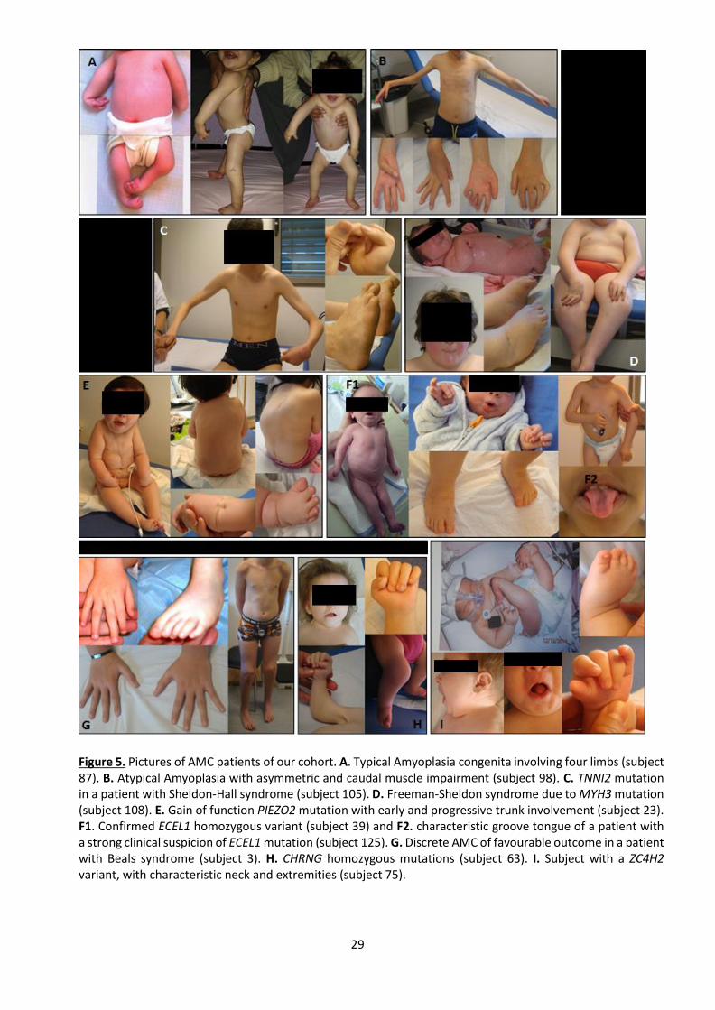
Figure 5 Pictures of AMC patients of our cohort…………………………………………………………………….. 29
Figure 6 Proposition of recommendations in the aetiological assessment of AMC children……. 58
Figure S1
Study information note destined to patients………………………………..…………………………..
75
Table S1
Features that led to the diagnosis of atypical Amyoplasia…………………………………………
77
Table S2 List of the 121 NMD-related genes sequenced in CHU Grenoble-Alpes laboratory…… 78
Table S3 Indications that had motivated prenatal karyotypes………………………………………………… 79
Table S4 Detailed karyotype and array-CGH results (postnatal)………………………………………………. 80
Table S5 Other Cytogenetic analyses performed in our cohort……………………………………………….. 80
Table S6 Other targeted analyses performed in our cohort…………………………………………………….. 81
Table S7 Abnormal creatine kinase dosage results…………………………………………………………………. 81
Table S8 Details on metabolic investigations performed in our patients…………………………………. 82
Table S9 Details on anomalies observed on electroencephalogram………………………………………… 82
Table S10 Metabolic disorders associated with AMC………………………………………………………………… 83
12
RÉSUMÉ
Titre : Le parcours diagnostique des enfants atteints d’Arthrogrypose Multiple Congénitale :
description des pratiques actuelles à travers une cohorte monocentrique, et proposition de
recommandations.
Introduction: L’Arthrogrypose Multiple Congénitale (AMC) correspond à des limitations
articulaires touchant au moins deux niveaux. Sa prévalence est estimée entre 1/3000 et
1/12000. Le diagnostic étiologique est difficile car il en existe plus de 400 causes. L’objectif de
ce travail est de décrire le parcours diagnostique des enfants atteints d’AMC et de proposer
des recommandations afin d’optimiser les pratiques cliniques.
Matériel et méthodes: Nous avons mené une étude rétrospective observationnelle
monocentrique, incluant les enfants évalués au Centre Hospitalier Universitaire de Grenoble
de 2007 à 2019. Nous avons collecté les informations sur leur parcours diagnostique, puis
avons mené une revue de la littérature pour chaque investigation paraclinique afin d’en
évaluer la pertinence à la lumière du diagnostic final des patients, des connaissances
scientifiques, de leur bénéfices, risques et coûts.
Résultats: Nous avons inclus un total de 125 patients, dont 43% cas d’Amyoplasie, 26%
d’arthrogrypose distale et 31% d’autres formes. Un diagnostic étiologique était posé dans 63%
des cas. Nous proposons une procédure diagnostique en deux temps: d’abord des
investigations non invasives permettant d’orienter les patients vers l’un des trois groupes
principaux, puis des investigations plus spécifiques avec des indications précises, en fonction
de leur rendement attendu et de leur caractère invasif.
Conclusion: L’approche diagnostique des enfants avec AMC doit résulter d’un travail
pluridisciplinaire. L’utilisation croissante du séquençage de nouvelle génération facilitera
cette démarche, mais le phénotypage restera essentiel pour guider leur interprétation.
Mots-clefs: Arthrogrypose Multiple Congénitale, démarche diagnostique, cohorte pédiatrique

13
ABSTRACT
Title: The diagnostic workup in children with Arthrogryposis Multiplex Congenita: description
of practices through a monocentric cohort and suggestion of recommendations.
Introduction: Arthrogryposis multiplex congenita (AMC) defines congenital contractures
involving two or more body areas. The prevalence is estimated between 1/3000 and 1/12000.
More than 400 conditions may lead to AMC through foetal hypo/a-kinesia, making the
aetiological diagnosis challenging. The objective of this work was to describe the aetiological
management of children with AMC and to propose recommendations in order to optimize
clinical practices.
Material and methods: We conducted a retrospective single centre observational study.
Patients had been evaluated at least once at a paediatric age in Grenoble University Hospital
from 2007 to 2019. After determining the diagnostic status of these patients, data on their
diagnostic procedure were gathered. A literature review was performed for each paraclinical
investigation to discuss their relevance in the light of patients’ diagnoses, scientific knowledge,
and benefit/risk or cost/benefit ratio.
Results: 125 patients were included, 43% had Amyoplasia, 26% had distal arthrogryposis, and
31% had other forms. A definitive aetiological diagnosis was available for 63% of cases. We
propose a two-time diagnostic process : first, non-invasive investigations that aim at
classifying patients into one of the three groups, and then specific investigations targeting a
subset of patients according to the expected yield and invasiveness.
Conclusion: The diagnostic management of AMC patients has to result from a multidisciplinary
approach. With the use of next generation sequencing, the aetiological assessment will be
facilitated, but a relevant phenotyping will be paramount to guide their interpretation.
Keywords: Arthrogryposis Multiplex Congenita, diagnostic assessment, paediatric cohort.

14
INTRODUCTION
1. Definitions and epidemiology
Etymologically, the term “Arthrogryposis” results from the association of the Greek words
“arthron” (joints), “gryp” (curved) and “osis” (condition) and points to joint contractures with
limited range of motion. The Latin term “Arthrogryposis Multiplex Congenita” (AMC) used
alternatively with the term “Multiple Congenital Contractures” (MCC), more specifically
defines a congenital arthrogryposis, involving at least two body areas (1–3). Contractures are
typically symmetric and although they do not progress to unaffected joints, they may change
in amplitude under the influence of growth and treatment (2).
Although AMC has been first defined as a condition per se, it is now admitted as a
descriptive term to define a symptom, common to a group of heterogeneous congenital
conditions (4). Depending on the cause, contractures location and severity may vary, as well
as the primary involvement of spine and jaw, or the existence of muscle weakness in the
involved body area. AMC can be either isolated or associated to other body systems
impairment such as central nervous system (CNS) as well as respiratory, gastrointestinal and
genitourinary systems. Prognostic factors like response to treatment, importance of deficits,
activity limitation, participation restriction but also neurodevelopment and even life
expectancy may differ widely according to the aetiology (2).
As true prevalence estimation remains challenging, it is thought to stand around 1/3000-
1/12000 (5–7). AMC is thus a rare affection, in contrast with isolated congenital contractures
such as congenital clubfeet that affect 1/500 individuals (1).

15
2. Causes and classification
The common underlying cause of AMC is foetal hypokinesia or decreased active foetal
movement (AFM) (4). AFM begin around 5 or 6 weeks of gestation (WG) and are essential for
the further development of joints. Independently of the underlying cause, their absence may
lead to connective tissue deposition around joints and stretching of tendons, increasing
mobility reduction and resulting in contractures, whose severity depends on the earliness of
hypokinesia (8,9). Foetal hypokinesia leads to a variety of other secondary deformations, that
thus can be encountered in all forms of AMC: craniofacial changes, pulmonary hypoplasia,
polyhydramnios, decreased gut mobility and shortened gut, short umbilical cord, growth
restriction and skin changes (4) (Fig. 1). The whole set of symptoms constitute the most severe
form of AMC called foetal akinesia deformation sequence (FADS) or Pena-Shokeir syndrome,
usually perinatally lethal (10).
More than 400 conditions causing foetal hypokinesia and thus AMC have been described
(4). They can involve any part of the anatomical structures implicated in movement and
include all causes of lack or excess of muscle contraction as well as mechanical restriction.
They can be divided into two categories (11) (Fig. 1) :
- Extrinsic causes : intrauterine foetal space restriction (severe oligohydramnios,
tumoral process, multiple pregnancy, uterine malformation), intrauterine vascular
compromise or environmental conditions (maternal illness including infections and
myasthenia gravis, maternal exposure to toxics, treatment or trauma)
- Intrinsic causes (foetal dysfunction) : myopathic process (congenital myopathies,
congenital muscular dystrophies, myositis...), neuropathic process (CNS impairment
with or without structural anomalies, peripheral nerves impairment including anterior
horn cells diseases, myelin anomalies…), neuromuscular end-plate process, connective
joint tissue abnormalities (restrictive dermopathies or cartilage/bone tissue
disturbances) and metabolic diseases.

16
Figure 1. Simplified scheme of causes and consequences of decreased AFM. Legends : AFM, Active foetal movements; AMC, Arthrogryposis Multiplex Congenita; CNS, central nervous system; M, muscle, NMJ, neuromuscular junction; PN, peripheral nerve.
Although a genetic origin is involved in most cases, the first cause of AMC, involved in
1/3 cases with an estimated prevalence of 1/10 000 individuals, is sporadic : Amyoplasia
congenita (5,12) (Table 1). This entity, first described in 1840, is recognizable by the
characteristic position of upper and lower limbs joints, absence of CNS involvement, absence
of primary spinal and jaw involvement although spinal deformities can be present at birth as
the consequence of foetal hypomobility and clinical muscle atrophy (3). Associated signs are
common and include digestive anomalies, skin defects, loss of digits or limbs, and classic nevus
flammeus of the midforehead (12). Muscular MRI (mMRI) typically evidences a reproducible
pattern (manuscript submitted). A four limbs AMC arise in 60% of cases, while upper or lower
limbs only account for 15% each (12). Orthopaedic outcome is very favourable especially in
comparison with other anterior horn cell diseases (13). Indeed, on the basis of the observation
of clinical similarities with Erb-Duchenne obstetrical palsy and electroneuromyographic
studies, this disorder is thought to result from a neurogenic process, i.e. the impairment of
the anterior horn cells of the spinal cord in early embryonic and foetal life, responsible for the
underdevelopment of muscle tissue and further replacement by fatty or fibrous deposits.
While the exact cause of this impairment is still unclear, the nature of the associated signs and
discordance in monozygotic twins suggest an ischemic event, impacting the spinal cord at the
level of upper and lower limbs expansion, i.e. motor roots C5-C6 and L2-L4 (12,14).

17
Table 1. Typical Amyoplasia criteria
Clinical Major criteria : - Sporadic - Symmetric (all 4 limbs or only UL/LL in a bilateral way) - Contractures distribution and joint positions :
* UL : internal rotation of shoulders, extended elbows, pronation of the forearm, flexed wrist, adducted thumbs and camptodactyly * LL : severe equinovarus feet, no rocker bottom feet
- Muscular atrophy (shoulder girdle muscles when UL involved) - Normal cognitive function, no sign of CNS involvement
Minor criteria : - Shortness of affected limbs - Mild IUGR - Spares the trunk - Dimples overlying involved joint
- Lack of flexion creases on limbs, fingers, hands - Nevus flammeus over craniofacial midline - Gracile, osteoporotic long bones - Good response to physiotherapy
Do not exclude the diagnosis : - Gastrointestinal impairment : abdominal wall defects, inguinal hernia, bowel atresia, gastroschisis - Skin defects - Loss of digits or limbs anomalies
Imaging Absence or severe muscle atrophy of : - UL : biceps and brachialis muscles - LL : gracilis, sartorius and anterior tibialis muscles
Bone X-rays : Gracile, osteoporotic long bones
Adapted from Hall et al., 2014 (12). Legends : CNS, central nervous system; IUGR, intra uterine growth retardation; LL, lower limbs; UL, upper limbs.
A group of affections called distal arthrogryposis (DA) represents another 1/5 to 1/3 of
AMC individuals (15). First described in 1982, DA is a heterogenous group of syndromes where
AMC involve mainly hands (overriding fingers, camptodactyly or pseudocamptodactyly with
hypoplasia of interphalangeal creases, ulnar deviation and/or wrist extension) and feet
(talipes equinovarus, calcaneovalgus deformities, vertical talus, and/or metatarsus varus),
with varying involvement of proximal joints. This arises in the absence of any primary
neurological and/or muscle disease affecting limb function, with thus conserved muscle mass
and cognitive function although muscle weakness and fatigability can occur. Familial cases are
common, with classically a wide intrafamilial variability or even incomplete penetrance.
Transmission if most often autosomal dominant. The DA group comprises ten different
entities, among which many are clinically recognizable (16–18) (Table 2). Genes responsible
for DA were the first described in link with AMC, as soon as 1995 for FBN2 (19), and then for
genes encoding sarcomeric muscle proteins like TNNI2, TNNT3 and TPM2, also involved in
congenital myopathies (20). Mechanisms of AMC in DA are various, either muscle weakness
in sarcomeric and ECEL1 forms, lack of relaxation in PIEZO2 patients, or connective tissues
dysfunction in FBN2 (18,19,21,22).

18
Table 2. Distal arthrogryposis classification
Type Name Genes Key distinguishing features
DA1 Classic DA/digitotalar
dysmorphism
TNNI21, TNNT31,
TPM21, MYH32,
MYBPC13
Extended wrists
Flexed and overlapping
fingers
Ulnar deviation
Feet malposition (club
feet/vertical talus)
No facial involvement
DA2A Freeman-Sheldon syndrome MYH32 Facial contractures, small pursed
mouth, scoliosis
DA2B Sheldon-Hall syndrome TNNI21, TNNT31,
TPM21, MYH32
Facial contractures less severe than in
DA2A, MRI pattern (TPM2)
DA3 Gordon syndrome PIEZO24 Cleft palate, blepharophimosis, ophthalmoplegia, painful muscle
contractures, spine involvement with respiratory impairment,
BMI<18/short stature, dimples
DA4 DA with severe scoliosis uk Scoliosis
DA5 DA with ophthalmoplegia, ptosis
and retinal involvement
PIEZO24, ECEL15 ECEL1 : ptosis without ophthalmoplegia, groove tong, extended
knees, adducted thumbs, MRI pattern
DA6 DA with sensorineural hearing loss
and microcephaly
uk Sensorineural hearing loss, microcephaly
DA7 Trismus-pseudocamptodactyly
syndrome/Hecht syndrome
MYH86 Trismus, facultative finger contractures
DA8 AD Multiple pterygium syndrome MYH37 Multiple pterygium
DA9 CCA/Beals syndrome FBN28 Arachnodactyly and camptodactyly, marfanoid habitus, ear
deformity
DA10 DA with congenital plantar flexion
contractures
uk Plantar flexion contractures
From Bamshad et al., 2009 (16), Hall et al., 2017 (17) and Kimber et al., 2012 (18). Legends : AD, autosomal dominant; BMI, Body Mass index; CCA,
congenital contractural arachnodactyly; DA, distal arthrogryposis; MRI, Magnetic Resonance Imaging; uk, unknown. 1Sung et al., 2003 (20); 2Toydemir
et al., 2006 (23), 3Gurnett et al., 2010 (24), 5Coste et al., 2013 (21),5Dieterich et al., 2013 (22), 6Toydemir et al. 2006 (25), 7Chong et al., 2015 (26), 7Putnam et al., 1995 (19).
Apart from DA genes, more than 400 genes have been reported in association with AMC.
Attempts to classify AMC forms according to genes function have been made, distributing
these genes into 29 different groups standing for different pathways. However, as a single
gene may be involved in more than one pathway, it may be classified in several groups. In
addition, many genes may lead to distinct phenotypes with or without AMC, and conversely a
single phenotype may be caused by different genes (27). Classification is thus arduous. Other
classification systems are based on the aetiological process of foetal akinesia as previously
cited. However, clinical classifications are most useful in daily practice. We can count two of
them, both dividing AMC patients in 3 groups :
- the first one is the most used in the literature and distinguishes three categories of
patients each accounting for 30% : primarily limb involvement (group I), musculoskeletal
involvement associated with other system anomalies (group II), and musculoskeletal
involvement associated with CNS dysfunction, intellectual disability (ID) or lethality (group III)
(28).

19
- the other is much more rarely used but is of great interest for clinical practice and for
family communication. It distinguishes two first groups accounting for more of the half of
cases, Amyoplasia congenita and DA, plus another one where all other aetiologies are
gathered (17).
No fully satisfying classification system exist, they all have their advantages and
drawbacks. However, they may be a useful way to stratify the diagnostic strategy at first meet
of a patient presenting with AMC.
3. Context and purpose
The identification of the aetiological diagnosis is of major importance in AMC, which arises
either sporadically or may be inherited with all transmission mode possible (12). As most
forms are severe, and as AMC is diagnosed at birth with parents in age of expanding their
family, questions arise about reoccurrence risk for future pregnancies, and often for prenatal
diagnosis. In addition, while exhaustive correlations are not established yet, elements of
prognosis, treatment and follow-up optimisation can be provided according to the underlying
cause. Last but not least, as for all rare conditions, patients and their families have often been
through a long and thorough medical evaluation with a long diagnostic wavering, that may
have had a significant psychological impact on individuals and structural consequences in
families. Getting a diagnosis thus helps families to work through their thoughts and feelings,
by providing an answer to their questions about the origin of their child’s condition, by helping
them to see more clearly their future, and by enabling them to get in touch with families who
have a child affected with the same disease.
Considering that AMC remains rare and that causes are numerous and cover a wide
spectrum of severity and clinical presentation, its aetiological diagnostic management is
challenging. Together with the fact that clinicians in charge of AMC patients are from various
medical specialties, their knowledge of diseases associated with AMC might be incomplete.
Lastly, hitherto there is no uniform consensus guidelines regarding diagnostic check-up. All
these elements may lead to heterogeneous medical practices and poor estimation of risk-
benefit or cost-benefit balance of each diagnostic test, leading to a non-optimal diagnostic
management of AMC patients.

20
Reference centres for rare diseases have been created to address this issue, common
to all rare diseases. Grenoble University Hospital has been labelled in 2007 as one of the
national reference centres for developmental anomalies and ID. In this context, our team has
set up a dedicated consultation for AMC patients, first for children, then for adults. Paediatric
dedicated workup comprises consultations with a clinical geneticist, a rehabilitation
professional and an orthopaedic surgeon, full physical therapist and occupational therapist
workup, psychological family evaluation and an imaging work-up when required. Thanks to
the clinical experience acquired so far, the increasing recognition of this work by French
clinicians, and due to the close relationship of our team with the national association of
patient “Association Alliance Arthrogrypose” (AAA) created in 2005, this dedicated
consultation attracts many patients across the country and abroad. As they first come to our
centre with a medical diagnostic background, these patients nicely reflect diagnostic
management of AMC in a national perspective. Adding to these patients those who have a
local follow-up in Grenoble, we have now built a large cohort of AMC patients of various
aetiologies.
Among authors that propose diagnostic management recommendations, only few
drew their conclusions on the basis of a cohort description, a convenient way to adapt
recommendations to existing practices. In addition, these publications are either too ancient,
or their cohort present a selection bias, are of small size or include a large part of perinatally
lethal cases (29,8,30,31).
The aim of this work was thus to describe our single centre cohort of AMC children
patients gathered from 2007 to 2019, with the first objective of drawing a picture of the
aetiologies found in our patients. In the light of this, the second goal was to redraw their
genetic and paraclinical diagnostic work-up, in order to evaluate the relevance of each test
with the help of information available in the literature, looking forward to proposing practices
guidelines for clinicians to optimize AMC patients’ diagnostic management.

21
MATERIAL AND METHODS
We conducted a retrospective single centre observational study in a cohort of AMC
paediatric patients, with the aim of redrawing their diagnostic process in order to evaluate
each investigation in comparison with available information in the literature.
1/ Protocol registration
Standard protocol of the study (Fig. 2), was submitted and approved by the Grenoble-
Alpes University hospital research section, who proceeded to an internal declaration of the
study (No. 2205066v0). As it was a noninterventional retrospective study (MR-004), no CNIL
(i.e. French data protection authority) declaration and no ethics committee approval were
requested (JORF10/05/2017). There was no refusal to participate from any of the patients,
and specific informed consent was obtained for photographs publication. The study
information note destined for patients is provided in Supplemental data (Fig. S1).
Figure 2. Main steps of present study protocol. Legends : AMC, Arthrogryposis Multiplex Congenita.

22
2/ Cohort building
As it was a retrospective observational study, no theorical sample size was calculated.
Cohort building was made using CEMARA, a national database designed for rare diseases
instituted in 2004, whose completion is mandatory for all labelled rare disease reference or
competence centre. Thus, every patient consulting in our reference centre for developmental
anomalies and ID of the South-East, first labelled in 2007 and renewed every five years since
then, has been registered in this application. The consultation purpose is one of the required
information to fill in. Patients consulting for arthrogryposis can thus be easily identified using
the application search tool. Recruitment is either local or national, with patients addressed by
healthcare professionals from other centres, or coming on their own, most often on the advice
of the national association of patients “AAA”.
Hence, we requested from our local CEMARA database the list of all patients for whom
the French term “arthrogrypose” or “Amyoplasia” had been quoted as a consultation motive
since 2007. We then selected patients meeting the following inclusion criteria :
- patients presenting with AMC, as previously defined, with or without associated signs
- born alive
- whose first evaluation had been performed at a paediatric age (i.e. ≤17 years old) in
Grenoble University Hospital
- and who had been evaluated at least once in our reference centre since its labelling
in 2007 and until February 2019. This evaluation had to comprise at least a consultation with
a medical geneticist, or a medical opinion based on numerous and sufficient clinical and
paraclinical data.
Patients presenting with arthrogryposis at a late paediatric age evaluated by our adult
team were excluded in order to homogenize gathered data and avoid a lack of information
about the diagnostic work-up in infancy. Patients presenting with arthrogryposis involving a
single joint level (e.g. isolated equinovarus feet) were also excluded to stick to the definition
of AMC. Finally, we did not include foetal diagnosis of arthrogryposis leading to intra uterine
death (IUFD), termination of pregnancy (TOP) for medical purposes or stillbirths since we
wanted to focus on postnatal diagnostic management. To better reflect the recruitment of
our reference centre for AMC patients, we chose not to select patients according to their
home city or country.

23
3/ Data collection
Data were collected from patient’s paper and informatic files reading. As most patients
had been evaluated in other cities health centres, information had been collected by parental
questioning or written documents transmitted by the parents during medical appointments.
Data were anonymized and an identification number was randomly assigned to each patient.
Information collected were: family history and consanguinity, pregnancy history, birth,
neonatal events and clinical presentation, joints spontaneous position and functional
limitations assessed by the rehabilitation team, associated extra-articular signs, main
treatments conducted and management, outcome of functional limitations, and age at first
and each appointment with the clinical geneticist of the reference centre. More specifically,
diagnostic investigations including biological, functional or imaging investigations performed
pre or postnatally and their results were collected, as well as age at diagnosis when a
molecular diagnosis was available.
4/ Patients’ classification
In order to facilitate data analysis, patients were classified into 3 groups. To do so, we used
the conclusions of the last clinical evaluation in genetic consultation which already classified
patients into one of these categories and checked it or updated it using clinical and biological
collected data since then. In accordance with our clinical habits in the diagnostic query of our
patients during and following the first evaluation, we chose to use an adaptation of the second
clinical classification previously described in the INTRODUCTION section, as following:
Group 1: Amyoplasia congenita
Within this group we distinguished two subgroups :
- typical Amyoplasia (T) as defined in Table 1 if evidences were sufficient to be fully
confident to raise this diagnosis
- atypical Amyoplasia (aT) if the diagnosis was suggestive of Amyoplasia but that atypical
elements were found, making this diagnosis uncertain.

24
Group 2: Distal Arthrogryposis (DA)
DA was defined as explained in the INTRODUCTION section and in Table 2. To better reflect
our ID process, we extended the definition of DA8 to include all other forms of multiple
pterygia syndrome. Patients of group 2 were further subclassified as following:
- peripheral nerve involvement (PN) including patients bearing mutations in genes PIEZO2 and
ECEL1
- neuromuscular end-plate impairment (NMJ): mutations in CHRNG
- muscle involvement (M): mutations in TPM2, TNNT3, TNNI2, MYH3, MYH8 and MYPBC1
- other (O): other genes previously associated with DA (such as FBN2)
- unknown (U): when clinical and paraclinical features were suggestive of DA but that no
molecular diagnosis was available to categorize them into one of the previously defined group
with strong confidence.
Group 3: other types of AMC
In this group we individualized patients presenting with CNS impairment (C) as defined by
the presence of severe hypotonia, decreased alertness, psychomotor delay or ID,
microcephaly, seizures, or other features suggestive often associated with CNS impairment
such as dysmorphic features.
All other patients of group 3 (O) had various causes of AMC that could not be classified
either as Amyoplasia, DA, or other types with central involvement. Possible diagnoses found
in this group are gathered in Fig. 3.
5/ Data analysis in the light of information provided by the scientific literature
Each investigation performed for diagnostic purposes was listed. We determined the
percentage of patients that had been submitted to it, reported the proportion of positive
results and how it helped to guide the diagnosis. Each groups and subgroups of patients were
analysed separately.
Then we compared our data to medical information available in the literature. We
performed a rapid review for each paramedical investigation, using the search engine
PubMed® (NCBI). Articles were selected from abstract analysis, and only relevant publications

25
were further studied. Additional papers, identified through the study of bibliography of firstly
selected publications, if pertinent, were also considered. Review was performed between
April and August 2019. Details on article selection and conclusion may be shared on request.
For some paraclinical investigations, literature review had been previously performed in
Dieterich et al, 2019 (32). In parallel, we completed our analysis by studying publications
describing AMC cohorts or providing diagnostic management recommendations.
Combining conclusions obtained from the analysis of our data and results of our
literature searches, we aimed at establishing clinical recommendations for AMC patients’
diagnostic management.
Figure 3. Possible diagnosis in Group 3 with (C) or without (O) CNS involvement. From Dieterich et al., 2019 (32) and Hall et al., 2014 (4). Legends : ARC, arthrogryposis Renal dysfunction and Cholestasis; ATR-X, Alpha Thalassemia/mental Retardation syndrome; CBD, Congenital Bone Disorders; CDP, Chondrodysplasia; CMD, congenital muscular dystrophies; CMV, cytomegalovirus; CNS, central nervous system; COFS, Cerebro-Facio-Oculo-Skeletal syndrome; LGMD, Limb Girdle Muscular Dystrophies; MD, Myotonic Dystrophy; MEB, Muscle Eye Brain; MG, myasthenia gravis; OFD, oro-facio-digital syndrome; OPD, oto-palato-digital syndrome; PCH, ponto-cerebellar hypoplasia; PWS, Prader-Willi Syndrome; SLO, Smith-Lemli Opitz syndrome; SMA, Spinal Muscular Atrophy; SMALED, SMA Lower Extremities predominant, autosomal dominant; VZV, varicella zona virus; WWS, Walker Warburg Syndrome.

26
RESULTS
1/ Cohort constitution
We gathered 123 paediatric AMC patients over 12 years. Additionally, two patients
that had been evaluated first before 2007 but had been followed up in the reference centre
after its certification were included as well, reaching a total of 125 patients (Fig. 4A). The full
clinical workup proposed to AMC children had been performed for 118 of them. Twenty-five
(20%) had a regular local follow-up, 90 (72%) were followed-up in secondary or tertiary level
care centres in other French cities and 10 (8%) from other countries, mostly from Northern
Africa (n=6). Epidemiological, pregnancy and birth data are summarized in Table 3.
54 patients were classified in group 1 (43.2%), 32 in group 2 (25.6%) and 39 in group 3
(31.2%). In group 1, 41 cases had typical Amyoplasia (75.9%) and in group 3 most patients had
signs of CNS involvement (n=27, 69.2%) (Fig. 4B).
Table 3. Epidemiological information
Epidemiological data Pregnancy data Birth data
Item N° (%) Item N° (%) Item N° (%)
Females 64 (51.2) Multiple pregnancies 42,3 (3.2) Preterm birth 17/104 (16.3) Related parents 8 (6.3) Twins 34 Very premature6 3 Familial cases 12 (9.5) Triplets 1 Post term birth 4/104 (3.8) Parental 8 In vitro fertilization 3 (2.4) Median birth weight (kg) 2.91 (1.03-4.49)/99 Siblings 4 Uterine malformations5 6 (4.8) Median birth height (cm) 47 (38-53)/62 Adoption 3 (2.4) Pregnancy events 14/114 (12.3) Median birth OFC (cm) 35 (28-39.5)/71 Past IUFD/miscarriage 8/89 (9) Vaginal bleeding 4 C-section 38/122 (31.1) Year of birth* 2010 First semester infection 4 Breech presentation 20 Age at first evaluation* 24mo Toxaemia/HBP/oedema 3 Scarred uterus 5 Early death 4 (3.2)1 Vehicle accident 1 Foetal cause7 8 Premature delivery threat 1 Maternal cause8 4 Early membrane rupture 1 Term overtaking 1 Long bone fracture 1 (0.8) Neonatal difficulties 159 (12)
Legends : HBP, high blood pressure; IUFD, intra uterine foetal death; OFC, occipitofrontal circumference; mo, months old; N°, number of cases. *Median. 1 group 3 only; 2and 2 additional early death of a twin;3 2 patients of group 1T, 2 of group 3O; 4 1 monochorial, 1 dichorial, 1 unknown; 5
unicornuate, bicornuate or septate uterus; 6 before 32WG; 7foetal cardiac rhythm anomaly (n=5), laparoschis (n=2), twin-to-twin transfusion syndrome; 8 narrow pelvis, anticoagulation therapy for lupus, septate uterus, cervical cerclage. 91 case of cardiorespiratory arrest.

27
2/ Aetiologies of AMC (Fig. 4B)
2.A/ Group 1: Amyoplasia congenita
Among patients with typical Amyoplasia, 66% had four limbs involvement (n=27), 22%
only upper limbs (n=9) and 12% only lower limbs (n=5) (Fig. 5A). Thirteen cases were qualified
as atypical Amyoplasia based on various clinical evidences (Table S1). Among these patients,
nine had molecular investigations, negative in two and still ongoing in seven. For patient 98,
clinical suspicion was strengthened by electroneuromyography (ENMG) results as we’ll discuss
later (Fig. 5B).
2.B/ Group 2 : Distal arthrogryposis
In this group, 66% of patients had a molecular diagnosis, all obtained by targeted Sanger
or next-generation sequencing (NGS) focused on AMC or neuromuscular diseases (NMD)
related genes (AMC-NGS and NMD-NGS, respectively, whose list of sequenced genes is
provided in Table S2) (Fig. 4B). Mutations in genes encoding proteins of the muscle sarcomere
were the most frequent aetiology, among which half were MYH3 variants including two
familial cases (Fig. 5C-D). Impairment of the peripheral nerve was nearly as frequent with a
large majority of gain of function heterozygous PIEZO2 variants among which two were
identified by specific research of the familial variation previously identified in their affected
sibling; and one familial case of a homozygous ECEL1 variant (Fig. 5E-F). However, we had only
one case of neuromuscular end-plate impairment due to a homozygous CHRNG variant and
one case of Beals syndrome due to FBN2 mutation (Fig. 5G-H).
Among undiagnosed patients, eight NGS analyses were still ongoing. There was a clinical
suspicion of DA2 in six patients including two cases with negative targeted Sanger sequencing
and no further explorations ongoing. Multiple pterygium syndrome was suspected in three
cases, with negative CHRNG sequencing in one. An ECEL1 mutation was negative in a case for
which investigations were ongoing. Paraclinical examinations did not permit to get a formal
diagnosis although it helped to better delineate the phenotype for two patients.

28
A
Figure 4. A. Flowchart of the study and B. Cohort distribution of the three groups, and detailed definitive diagnoses in group 2 (green disk) and 3 (blue disk). Legends : AAA, Association Alliance Arthrogrypose; AMC, Arthrogryposis multiplex congenita; C, CNS involvement; DA,
Distal Arthrogryposis; M, muscle, NMJ, neuromuscular junction; O, other; PN, peripheral nerve; U, unknown.
29
Figure 5. Pictures of AMC patients of our cohort. A. Typical Amyoplasia congenita involving four limbs (subject 87). B. Atypical Amyoplasia with asymmetric and caudal muscle impairment (subject 98). C. TNNI2 mutation in a patient with Sheldon-Hall syndrome (subject 105). D. Freeman-Sheldon syndrome due to MYH3 mutation (subject 108). E. Gain of function PIEZO2 mutation with early and progressive trunk involvement (subject 23). F1. Confirmed ECEL1 homozygous variant (subject 39) and F2. characteristic groove tongue of a patient with a strong clinical suspicion of ECEL1 mutation (subject 125). G. Discrete AMC of favourable outcome in a patient with Beals syndrome (subject 3). H. CHRNG homozygous mutations (subject 63). I. Subject with a ZC4H2 variant, with characteristic neck and extremities (subject 75).

30
2. C/ Group 3 : Other patients
In group 3, a definitive diagnosis was available for 41% and 50% of patients in group
3C and 3O, respectively (Fig. 4B).
In group 3C, ZC4H2 appears to be a frequent cause of AMC with CNS involvement, with
four point mutations identified by targeted sequencing (n=3) or Whole Exome Sequencing
(WES) (n=1), and one deletion evidenced by comparative genomic hybridization array (array-
CGH). One case was familial (Fig. 5I). Chromosomal analysis identified two additional cases in
this group, both first identified on karyotype: a 18p tetrasomy with an isochromosome and a
4p duplication due to an unbalanced translocation der(4;22)(p10;q10), further characterized
by array-CGH. AMC-NGS identified a point mutation in TNNI2, a gene responsible for DA type
2B, in an individual that was initially suspected with DA but had been classified in group 3
because of a cognitive impairment. Looking back to his personal history, we hypothesised that
his cognitive dysfunction could have a distinct cause, i.e. a car accident with head impact in
childhood. Also, NMD-NGS pointed out a heterozygous NALCN variant in a child with cognitive
dysfunction compatible with the diagnosis of CLIFAHDD syndrome (“Congenital contractures
of the LImbs and FAce, Hypotonia and Developmental Delay”) (MIM#616266). Lastly, targeted
sequencing of KAT6B and NSD1 were asked for suggestive presentations of SBBYSSS variant of
Ohdo syndrome (MIM#603736) and Sotos syndrome (MIM#115750), respectively. Among
patients without molecular diagnosis, there was a clinical suspicion of Muscle Eye Brain
disease unexplored to date, and a case of hyperexplexia with negative GLRA1 sequencing.
Concerning patients of group 3O, WES and NMD-NGS identified variants in genes
responsible for the kyphoscoliotic form of Ehlers-Danlos syndrome (MIM#614557) and for
Larsen syndrome (MIM#150250), two conjunctive tissue disorders associated with AMC
respectively due to FKBP14 and FLNB variants. Two other patients had mutations in genes
responsible for syndromic forms of AMC, i.e. nail patella syndrome due to LMX1B variants
(MIM#161200), and Aarskog due to a FGD1 mutation (MIM#305400). FKBP14 and FGD1
variants were identified in familial cases. Interestingly in this group we described two cases of
environmental causes of AMC, one due to severe oligohydramnios secondary to a premature
membrane rupture at 18WG and the other due to a complicated twin pregnancy (early cervical

31
cerclage, prolonged bleeding, premature rupture of membranes inducing extreme preterm
birth).
More than half of these patients did not have any aetiological diagnosis. Among them,
two were diagnosed with constitutional bone disorders (CBD) such as Diastrophic Dysplasia
and Brück syndrome with so far negative analyses. Overall, NMD-NGS or WES analyses results
were still pending in nine patients. As we’ll see, in group 3, paraclinical investigations could be
useful to guide the diagnosis, although it did not allow to conclude formally at time of the
study.
Overall, a definitive aetiological diagnosis of AMC was established for 79 patients, i.e.
63.2% of the cohort, comprising 43 clinical and 36 molecular diagnoses. Median age of
patients at time of molecular diagnosis results was four years old. For the 46 undiagnosed
patients, nine never had any analyses, 13 had previous negative investigations (including two
NMD-NGS and one WES) but no ongoing explorations and 24 had pending NMD-related genes
or untargeted NGS results.
3/ Paraclinical investigations performed and results (Table 4)
3. A/ Other genetic investigations
A karyotype was performed in less than half of our cohort, 48% antenatally (n=26),
41% postnatally (n=22) and 11% (n=6) both antenatally and postnatally.
In the antenatal period, karyotype was performed in a quarter of the cohort (25.6%,
n=32) in equal proportions in the three groups (27.8%, 25% and 23.1% respectively). Half were
performed because of bilateral equinovarus feet, isolated or associated with articular or extra-
articular anomalies. Antenatal array-CGH were rarely performed (4.8%, n=6, 3 from group 3)
and all were normal. Detailed indications are summed up in Table S3. Postnatal karyotypes
(22.4%, n=28) were performed mostly in group 3 (41% (n=16), in contrast with 16.7 and 9.4%
in group 1 and 2, respectively), as was array-CGH. Together, they contributed to three
diagnoses, resulting in a yield of 2.4% in the whole cohort and 11% in group 3C. It also
unravelled incidental findings in six cases, polymorphic copy number variants (CNVs) or

32
Variant of Unknown Significance (VUS) (Table S4). Other type of cytogenetic examinations had
been performed in a limited number of patients (Table S5).
Targeted sequencing on a subset of genes involved in AMC was performed in almost
half of the patients. Among the 48 tested patient, Sanger sequencing of AMC-genes had been
performed in 33 and AMC-NGS in 17, both with a high yield (42 and 33%, respectively). Both
were mostly performed in group 2 for which yield was the best. Finally, 22 patients had a
NMD-NGS, mostly group 3 patients, with a global yield so far of 18.2%, with still a significant
number of pending analyses.
Targeted sequencing of other genes was performed in 12.7% (n=16) of the cohort (Table
S6). It enabled a diagnosis for five patients. Finally, genome-wide investigations, i.e. WES or
whole genome sequencing (WGS), were performed in 11.1% of the cohort, mostly in group 3,
and led to two diagnoses, resulting in a hitherto yield of 15.4%, here again with a significant
number of pending results.
DMPK analysis, Prader Willi 15q11.2q13 locus analysis and SMN1 analysis were all
performed in around 10% of patients. Some were performed prenatally (n=5, n=2 and n=4,
respectively), mostly because of decreased foetal movements. DMPK triplet expansions were
mostly investigated in group 3, SMN1 deletions mainly in group 1 patients, whereas Prader-
Willi was questioned as much in the three groups. Those analyses never turned out positive.
3.B/ Other biological investigations
Creatine Kinase (CK) levels were determined in approximately one out of five
individuals. CK levels were slightly elevated or subnormal only for four patients, especially in
two local patients with ZC4H2 and NSD1 variants for which dosages were performed in
particular circumstances, not mentioned in medical reports and without available control
dosages, suggesting that they had been judged uninterpretable (Table S7). Moreover,
elevated CK are not reported in these disorders and at least one other ZC4H2 mutated patients
had normal values.
33
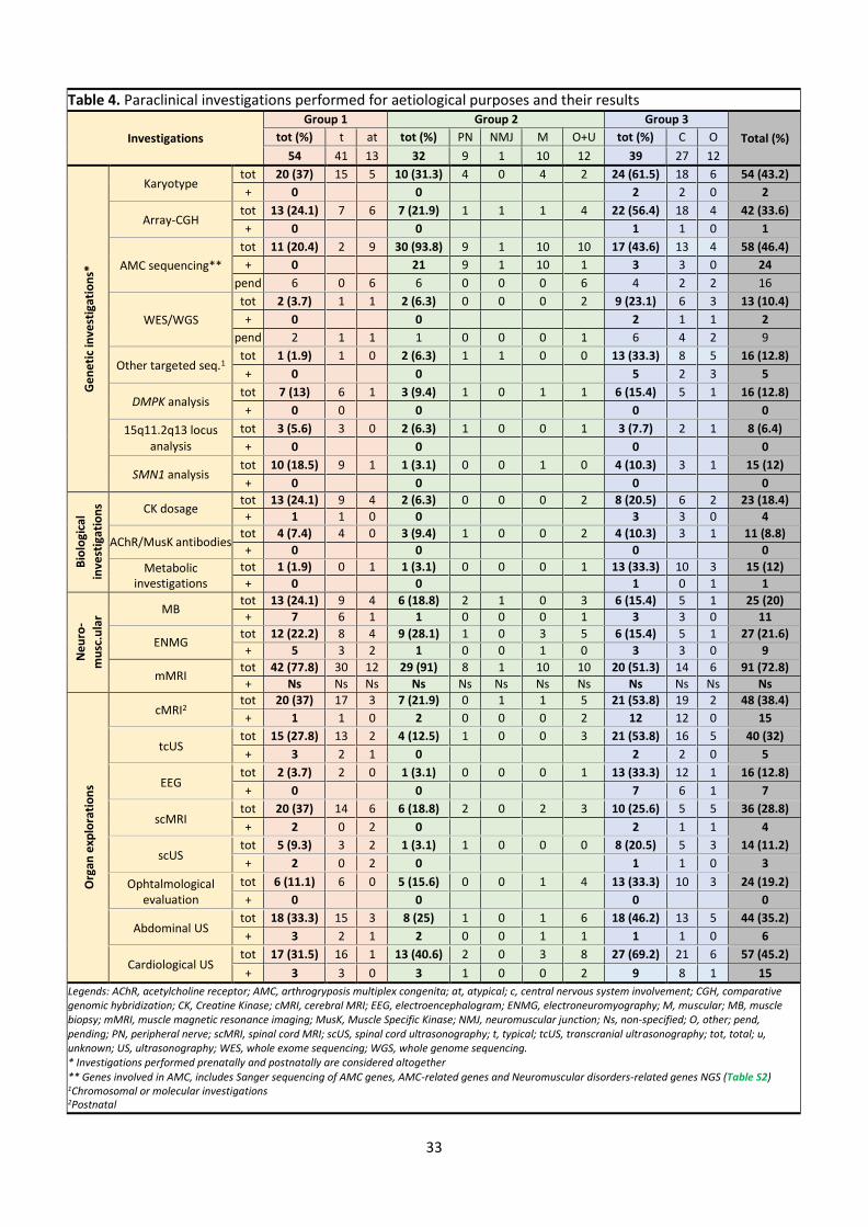
Table 4. Paraclinical investigations performed for aetiological purposes and their results
Investigations
Group 1 Group 2 Group 3 Total (%) tot (%) t at tot (%) PN NMJ M O+U tot (%) C O
54 41 13 32 9 1 10 12 39 27 12
Ge
ne
tic
inve
stig
atio
ns*
Karyotype tot 20 (37) 15 5 10 (31.3) 4 0 4 2 24 (61.5) 18 6 54 (43.2)
+ 0 0 2 2 0 2
Array-CGH tot 13 (24.1) 7 6 7 (21.9) 1 1 1 4 22 (56.4) 18 4 42 (33.6)
+ 0 0 1 1 0 1
AMC sequencing**
tot 11 (20.4) 2 9 30 (93.8) 9 1 10 10 17 (43.6) 13 4 58 (46.4)
+ 0 21 9 1 10 1 3 3 0 24
pend 6 0 6 6 0 0 0 6 4 2 2 16
WES/WGS
tot 2 (3.7) 1 1 2 (6.3) 0 0 0 2 9 (23.1) 6 3 13 (10.4)
+ 0 0 2 1 1 2
pend 2 1 1 1 0 0 0 1 6 4 2 9
Other targeted seq.1 tot 1 (1.9) 1 0 2 (6.3) 1 1 0 0 13 (33.3) 8 5 16 (12.8)
+ 0 0 5 2 3 5
DMPK analysis tot 7 (13) 6 1 3 (9.4) 1 0 1 1 6 (15.4) 5 1 16 (12.8)
+ 0 0 0 0 0
15q11.2q13 locus analysis
tot 3 (5.6) 3 0 2 (6.3) 1 0 0 1 3 (7.7) 2 1 8 (6.4)
+ 0 0 0 0
SMN1 analysis tot 10 (18.5) 9 1 1 (3.1) 0 0 1 0 4 (10.3) 3 1 15 (12)
+ 0 0 0 0
Bio
logi
cal
inve
stig
atio
ns CK dosage
tot 13 (24.1) 9 4 2 (6.3) 0 0 0 2 8 (20.5) 6 2 23 (18.4)
+ 1 1 0 0 3 3 0 4
AChR/MusK antibodies tot 4 (7.4) 4 0 3 (9.4) 1 0 0 2 4 (10.3) 3 1 11 (8.8)
+ 0 0 0 0
Metabolic investigations
tot 1 (1.9) 0 1 1 (3.1) 0 0 0 1 13 (33.3) 10 3 15 (12)
+ 0 0 1 0 1 1
Ne
uro
-
mu
sc.u
lar
MB tot 13 (24.1) 9 4 6 (18.8) 2 1 0 3 6 (15.4) 5 1 25 (20)
+ 7 6 1 1 0 0 0 1 3 3 0 11
ENMG tot 12 (22.2) 8 4 9 (28.1) 1 0 3 5 6 (15.4) 5 1 27 (21.6)
+ 5 3 2 1 0 0 1 0 3 3 0 9
mMRI tot 42 (77.8) 30 12 29 (91) 8 1 10 10 20 (51.3) 14 6 91 (72.8)
+ Ns Ns Ns Ns Ns Ns Ns Ns Ns Ns Ns Ns
Org
an e
xplo
rati
on
s
cMRI2 tot 20 (37) 17 3 7 (21.9) 0 1 1 5 21 (53.8) 19 2 48 (38.4)
+ 1 1 0 2 0 0 0 2 12 12 0 15
tcUS tot 15 (27.8) 13 2 4 (12.5) 1 0 0 3 21 (53.8) 16 5 40 (32)
+ 3 2 1 0 2 2 0 5
EEG tot 2 (3.7) 2 0 1 (3.1) 0 0 0 1 13 (33.3) 12 1 16 (12.8)
+ 0 0 7 6 1 7
scMRI tot 20 (37) 14 6 6 (18.8) 2 0 2 3 10 (25.6) 5 5 36 (28.8)
+ 2 0 2 0 2 1 1 4
scUS tot 5 (9.3) 3 2 1 (3.1) 1 0 0 0 8 (20.5) 5 3 14 (11.2)
+ 2 0 2 0 1 1 0 3
Ophtalmological evaluation
tot 6 (11.1) 6 0 5 (15.6) 0 0 1 4 13 (33.3) 10 3 24 (19.2)
+ 0 0 0 0
Abdominal US tot 18 (33.3) 15 3 8 (25) 1 0 1 6 18 (46.2) 13 5 44 (35.2)
+ 3 2 1 2 0 0 1 1 1 1 0 6
Cardiological US tot 17 (31.5) 16 1 13 (40.6) 2 0 3 8 27 (69.2) 21 6 57 (45.2)
+ 3 3 0 3 1 0 0 2 9 8 1 15
Legends: AChR, acetylcholine receptor; AMC, arthrogryposis multiplex congenita; at, atypical; c, central nervous system involvement; CGH, comparative genomic hybridization; CK, Creatine Kinase; cMRI, cerebral MRI; EEG, electroencephalogram; ENMG, electroneuromyography; M, muscular; MB, muscle biopsy; mMRI, muscle magnetic resonance imaging; MusK, Muscle Specific Kinase; NMJ, neuromuscular junction; Ns, non-specified; O, other; pend, pending; PN, peripheral nerve; scMRI, spinal cord MRI; scUS, spinal cord ultrasonography; t, typical; tcUS, transcranial ultrasonography; tot, total; u, unknown; US, ultrasonography; WES, whole exome sequencing; WGS, whole genome sequencing. * Investigations performed prenatally and postnatally are considered altogether ** Genes involved in AMC, includes Sanger sequencing of AMC genes, AMC-related genes and Neuromuscular disorders-related genes NGS (Table S2) 1Chromosomal or molecular investigations 2Postnatal

34
Investigation of maternal myasthenia antibodies targeting either Acetylcholine
Receptor (AChR) and/or Muscle specific Kinase (MuSK) was even more rarely performed
without differences in the 3 groups and was never found positive. For two patients, detection
was performed during pregnancy because of foetal limb malposition and reduced limb
movements.
Apart from hepatic, phospho-calcic or thyroid metabolism assessment, metabolic
investigations were performed in one patient out of ten, as expected mostly in group 3C. Most
screened patients had a basic checkup looking mainly for organic aciduria, urea cycle deficits
or other aminoacidopathies, energetics disorders of citric acid cycle and fatty acid beta
oxidation dysfunction. Only one patient had slightly abnormal ammonia levels whose cause
remains unknown. Other investigations were performed in two or single patients (Table S8).
3.C/ Neuromuscular investigations
Muscle biopsy (MB) and electroneuromyography (ENMG) were both performed in
about one out of five patients, with a slightly higher proportion in group 1 and 2.
MB was planned or discussed for 11 additional patients but had not yet been
performed to our knowledge. Two patients were biopsied twice. MB was often performed in
parallel of a surgical intervention. Many were interpreted as normal (n=6) or non-contributory
because of lack of muscle tissue (n=5), and rarely results were not available (n=3).
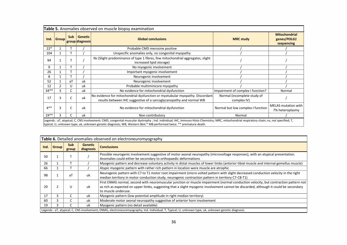
Histopathological examination reports described abnormalities in 11 cases (Table 5).
Surprisingly, six had a clinical diagnosis of typical Amyoplasia and showed various types of
anomalies. A neurogenic involvement was reported in two cases of typical and atypical
Amyoplasia, in accordance with the hypothetic pathogenesis of this disorder, bringing another
evidence toward Amyoplasia for the atypical case. However, two were suggestive of myogenic
involvement, based on the association of poorly specific features, but did not question the
diagnosis which was typical. One patient with a clinical diagnosis of DA had normal
microscopic evaluation but a suspicion of multiminicore myopathy. In this patient (subject 12)
for whom first clinical suspicion was a Freeman-Sheldon syndrome with normal Sanger
sequencing of six DA genes and no other current investigations, this result may certainly help
in the interpretation of future molecular investigations.

35
In group 3C, it appears that MB was mainly conducted to rule out a mitochondrial
dysfunction. Eventually, the five tested individuals had a severe presentation with premature
death in three of them. In the absence of evidence of such disorder on histology, investigations
were completed by an analysis of mitochondrial respiratory chain (MRC) in three patients
unravelling complex I borderline deficiencies and led to targeted genetic analysis in two
individuals that turned out to be negative. Other anomalies were reported on histological
examination of some of these patients but could not help to clarify the diagnosis. One had
discordant results. In group 3O, MB was only performed in the patient with kyphoscoliotic
Ehlers-Danlos syndrome and was non-contributory.
One third of ENMG were considered as abnormal (Table 6). Among patient with
Amyoplasia, only one patient with a typical presentation had a neurogenic pattern, while the
others had either normal ENMG, or a discordant myogenic pattern probably secondary to
muscle disuse or atrophy. Interestingly, ENMG was of great value in an atypical Amyoplasia
patient with unusual contracture location since it evidenced a neurogenic pattern which
involved C7-C8-T1 motor roots instead of C5-C6, in accordance with his clinical presentation.
In this patient it raised the hypothesis of a common pathophysiological mechanism with what
is supposed in Amyoplasia, i.e. vascular compromise, but occurring in this case at a more
caudal metameric location. Furthermore, this presentation is quite similar to Klumpke’s
obstetrical palsy affecting motor roots C8-T1, a similarity that strengthened our hypothesis.
This, together with the fact that this patient had unremarkable NMD-NGS results, lead to
expect a low if ever null recurrence risk in this family, although it would be unwise to
definitively and completely discard a genetic cause due to the lack of data about this
presentation. Finally, ENMG completion helped to delineate the diagnosis of three patients of
group 3 and one patient with DA (subject 20), adding for the latest new evidence toward the
suspected diagnosis of Freeman-Sheldon though not confirmed on Sanger sequencing so far.
Whole-body T1-weighed mMRI was widely performed in this cohort and almost all
patients with atypical Amyoplasia and DA had been investigated. It was planned in five
additional patients, and failed in three others from group 3C. Eighteen individuals underwent
several mMRI at different ages. Analysis of data delivered by this imaging is the purpose of
other researches coordinated by our team and will not be detailed here. Overall, it mainly
36
Table 5. Anomalies observed on muscle biopsy examination
Ind.
Group Sub
group Genetic
diagnosis
Global conclusions
MRC study Mitochondrial genes/POLG1 sequencing
22* 1 T / Probable CMD merosine positive / /
104 1 T / Unspecific anomalies only, no congenital myopathy / /
94 1 T / Ns (Slight predominance of type 1 fibres, few mitochondrial aggregates, slight
increased lipid storage) / /
9 1 T / No myogenic involvement / /
26 1 T / Important myogenic involvement / /
8 1 T / Neurogenic involvement / /
52 1 aT uk Neurogenic involvement / /
12 2 U uk Probable multiminicore myopathy / /
34** 3 C uk No evidence for mitochondrial dysfunction Impairment of complex I function? Normal
17 3 C uk No evidence for mitochondrial dysfunction or myotubular myopathy. Discordant
results between IHC suggestive of a sarcoglycanopathy and normal WB Normal (incomplete study of
complex IV) /
4** 3 C uk No evidence for mitochondrial dysfunction Normal but low complex I function MELAS mutation with
7% heteroplasmy
19** 3 C uk Non contributory Normal /
Legends : aT, atypical; C, CNS involvement; CMD, congenital muscular dystrophy ; Ind. individual; IHC, Immuno Histo Chemistry; MRC, mitochondrial respiratory chain; ns, not specified; T, typical; U, unknown type; uk, unknown genetic diagnosis; WB, Western Blot.* MB performed twice. ** premature death.
Table 6. Detailed anomalies observed on electroneuromyography
Ind. Group Sub
group Genetic
diagnosis Conclusions
50 1 T / Possible neurogenic involvement suggestive of motor axonal neuropathy (microvoltage responses), with an atypical presentation. Anomalies could either be secondary to orthopaedic deformations
26 1 T / Myogenic pattern and decrease voluntary activity in distal muscles of lower limbs (anterior tibial muscle and internal gemellus muscle)
66 1 T / Atypic myogenic pattern with rather rich pattern in location were muscle are atrophic
98 1 aT uk Neurogenic pattern with C7 to T1 motor root impairment (micro volted pattern with slight decreased conduction velocity in the right median territory in motor conduction study, neurogenic contraction pattern in territory C7-C8-T1)
20
2
U
uk First ENMG normal, second with neuromuscular junction or muscle impairment (normal conduction velocity, but contraction pattern not as rich as expected on upper limbs, suggesting that a slight myogenic involvement cannot be discarded, although it could be secondary to muscle underuse.
17 3 C uk Myogenic pattern (low potential amplitude in right median territory)
60 3 C uk Moderate motor axonal neuropathy suggestive of anterior horn involvement
19 3 C uk Myogenic pattern (no detail available)
Legends : aT, atypical; C, CNS involvement; ENMG, electroneuromyography; Ind. Individual; T, Typical; U, unknown type; uk, unknown genetic diagnosis.

37
helped in group 1 patient, where it helped to maintain the diagnosis of Amyoplasia in one, and
led to a classification in the typical group for another patient for who clinical diagnosis was
uncertain. In group 2, mMRI strengthened a suspicion of ECEL1 mutation in one patient, still
under investigation. In group 3, mMRI mostly evidenced normal muscle tissues or diffuse
atrophy without specific pattern, among which several could be secondary to joint limitations.
3.D/ Central nervous system investigations
For more than half of our patients (51.2%, n=64), cerebral MRI (cMRI) and/or
transcranial ultrasonography (tcUS) had been performed. Although it was mostly proposed to
group 3C patients, subjects of the two other groups were also frequently investigated,
including typical Amyoplasia where a subset (n=3) had been performed because of abnormal
tcUS.
Anomalies had been observed in one out of three cMRI performed, mostly in group
3C (Table 7). In this group, five patients had specific lesions such as polymicrogyria or
mesencephalic cleft that will certainly conduct to a molecular diagnosis in the future. Others
had nonspecific features including three cases of group 1 and 2. Notably, one patient
diagnosed as typical Amyoplasia had several associated nonspecific anomalies.
Twenty-two patients had both cMRI and tcUS. Rarely, tcUS reports pointed towards
abnormal and unspecific findings, of which only two were confirmed on cMRI. Conversely, six
patients showing structural brain anomalies on cMRI had unremarkable or only partially
affected tcUS.
Antenatal cMRI were performed for seven patients with equal proportion in the 3
groups because of multiple joint anomalies associated with growth defects, abnormal
amniotic fluid quantity or multi organ involvement. All were normal, and two had postnatal
MRI, normal as well.
Electroencephalogram (EEG) was performed in a small subset of our patients with a
clear predominance in group 3. Indications were abnormal movements (n=4), seizures (n=3),
abnormal muscle tone (n=4, hypotonia in the two patients of group 1), malaises (n=2),
abnormal alertness (n=1), or others (n=2, including the patient of group 2 in whom the
indication remained unclear). Anomalies have been reported in six patients but were relevant

38
only for four of them, to assess or rule out the epileptic nature of seizures and abnormal
movements or to characterize seizures (Table S9).
Spinal cord MRI (scMRI) and spinal cord ultrasonography (scUS) were performed in
nearly 1/3 and 1/10 of our patients, respectively (Table 8). While scMRI was mostly done in
group 1, group 3 had the highest proportion of scUS especially for patients without central
signs, as part of a global organ assessment. Most often, scMRI was performed all at once with
cMRI. For six patients for whom scUS was done, a scMRI was further proposed, and all findings
on scUS were confirmed by scMRI. Conversely, among three cases of scMRI anomalies for
which scUS had been realized, only one was unremarkable. Significant and causal anomalies
were not reported but instead we identified features frequently observed in the general
population such as tethered or low tethered cona medullaris and dilated central canal.
Table 7. Details on anomalies reported on cMRI and/or tcUS
Ind. Group Sub
group Genetic
diagnosis
cMRI tcUS
Age Observed anomalies Observed anomalies
22 1 T / « early » First MRI : white matter anomalies
Second MRI : thin CC splenium, carrefour dilation normal
104 1 T / 6 mo Normal Sub arachnoid stage I
haemorrhage
18
1
T
/
1 mo
Normal
Periventricular frontal and occipital right sided
hyperechoic signal
93 1 aT uk / / Septum pellucidum cyst
3 2 O FBN2 ns Blake pouch cyst
125 2 U uk 8 mo Benign enlargement of subarachnoid space
11 3 C NSD1 2.5 yo Rarefaction of white matter, CC atrophy, patent
septum pellucidum Normal
45 3 C ZC4H2 Nn Delayed myelination Normal
47 3 C 18p
tetrasomy 1 yo
White matter thinning, periventricular hypersignal, lateral ventricles dilation
/
34 3 C uk 1 mo Unilateral ventricle dilation Normal
102
3
C
uk
2 mo 4mo
Polymycrogyria, pachygyria, white matter rarefaction, delayed myelination, doubt on
brainstem hypoplasia, passive ventricle dilation
Ventricle dilation
68 3 C uk 2 yo Polymicrogyria Normal
17 3 C uk Ns Abnormal myelination, CSC atrophy,
mesencephalic cleft Normal
60 3 C uk Nn Very thin CC Thin CC
19 3 C uk Ns CSC atrophy /
6 3 C uk 2 yo CSC atrophy, white matter thinning /
4 3 C uk 10 do 8 mo
White matter hypersignal, delayed myelination, thin vermis, thin CC
Normal
84 3 C uk Nn Punctiform abnormal white matter signal,
subarachnoid haemorrhage of the posterior fossa Normal
Legends : aT, atypical; C, CNS involvement; CC, corpus callosum; cMRI, cerebral MRI; CSC, cortical-subcortical; do, days old; Ind. Individual; mo, months old; MRI, magnetic resonance imaging; nn, neonatal period; ns, not specified; O, other; T, typical: tcUS, transcranial ultrasonography; U, unknown type of distal arthrogryposis; uk, unknown genetic diagnosis; yo, years old. Bold characters : specific anomalies.

39
Table 8. Details on anomalies observed on spinal cord imaging
Ind. Group Sub
group Molecular diagnosis
Abnormality on scMRI Abnormality on scUS
77* 1 aT uk Low-lying and tethered conus medullaris Low-lying and tethered conus medullaris,
no spinal swelling at lumbar level
70 1 aT uk Central canal dilation (conus terminalis) Central canal dilation
116 3 C ZC4H2 / Low-lying conus medullaris
19 3 C uk Tethered conus medullaris /
51 3 O uk Central canal dilation at high thoracic
level, L5 sacralisation normal
Legends : aT, atypical; C, CNS involvement; Ind. individual; O, other; scMRI, spinal cord MRI ; tcUS, transcranial ultrasonography; uk, unknown genetic diagnosis. *AMC involving lower limbs only, suspicion of TRPV4 mutation with pending molecular investigations.
3.E/ Associated organ impairment assessment
Abdominal ultrasonography (US), cardiological and ophthalmological evaluations were
all frequently performed, mostly in group 3.
Ophthalmological evaluation was the most rarely reported. Often, details about this
examination were not mentioned on medical reports. When it was, most of the time an eye
fundus and electrophysiological studies had been realized and all were unremarkable.
However, detailed patient interview and clinical examination revealed pathological findings in
38 patients among who only four belonged to group 1. Most were refractive disorders (n=12)
and strabismus (n=14), common findings in the general population and often associated with
syndromic diseases. Otherwise, more specific features such as abnormal ocular movements
were evidenced (n=17): impairment oculomotor abilities in all but one PIEZO2 patients, and
saccadic movements, nystagmus or oculomotor asynergy predominantly in patients of group
3. Ptosis (n=11) and blepharophimosis (n=4) were also frequent findings, especially in PIEZO2
patients (n=2) and in the individual bearing a KAT6B variant. Of note, no data about
ophthalmological examination were available for our ECEL1 mutated patient.
A cardiologic US was performed in nearly half of the cohort. One out of four
examinations were reported as abnormal (Table 9), with mostly nonspecific and
spontaneously favourable outcome without need for surgical procedure, including in patients
with syndromic forms (NSD1 and KAT6B variants), in line with their diagnosis. More specific
anomalies were found in group 3C, such as a double aortic arch requiring neonatal surgery

40
and a septal hypertrophy. In addition, another patient of this group had an innominate artery
compression syndrome that did not require surgery, but there was no information about how
this anomaly had been assessed. Five patients had other cardiological explorations such as 12-
lead electrocardiogram (EKG) or holter EKG. Only one was reported abnormal.
A third of individuals had an abdominal US, anomalies were found in six patients,
mostly nonspecific and probably independent from AMC aetiology, except for our individual
with a KAT6B variant that had bilateral renal hypoplasia and severe vesicoureteral reflux that
are probably part of his syndrome, although hydronephrosis and kidney cysts are more
common (Table 10). For two patients, renal anomalies had been suspected in utero.
Altogether, global morphological explorations comprising at least a cardiological
examination, abdominal and transcranial US were performed in one patient out of five
patients (21.6%, n=27), 36% of group 3, versus 9% or 18% of group 1 and 2, respectively. An
anomaly was reported in a third of explored patients due to the high proportion of renal and
heart defects. Specific and nonspecific anomalies might have guided - or at least will guide -
the diagnosis in syndromic forms of group 3.

41
Table 9. Details on anomalies reported in cardiological explorations
Ind. Group Sub
group Genetic
diagnosis
Echocardiography EKG
Observed anomalies Outcome
49 1 T / Minor PDA, PFO Ns /
27 1 T / ASD, large PDA benign /
104 1 T / PDA, PFO Sp favorable /
9 1 T / trabecular VSD Ns /
76 2 PN PIEZO2 PDA, PFO Ns /
92 2 U uk normal Incomplet right BBB
119 2 U uk ASD Sp favorable /
125 2 U uk PDA Sp favorable /
116 3 C uk Restrictive PDA
Transient mild tricuspid failure Sp favorable Normal Holter EKG
11 3 C NSD1 ASD ostium secundum Ns /
45 3 C ZC4H2 perimembranous VSD Ns /
75 3 C ZC4H2 PFO Sp favorable /
110 3 C KAT6B ASD, VSD Ns /
17 3 C uk Double aortic arch Neonatal surgery /
102 3 C uk Isolated PFO Ns /
112 3 C uk VSD, PDA Sp favorable /
113 3 C uk Innominate artery compression
syndrome Stable, no
surgery normal
79 3 C uk Asymetric septal hypertrophy Ns normal
89 3 O uk ASD, minor VSD, PFO Sp favorable / Legends : ASD, Atrial Septal Defect; BBB, bundle branch block; ns, non-specified; EKG, electrocardiogram; Ind., Individual; OS, ostium secundum; PDA, patent ductus arteriosus; PFO, patent foramen ovale; PN, peripheral nerve; sp, spontaneously; T, typical; U, unknown form of distal arthrogryposis; uk, unknown genetic diagnosis; VSD, Ventricular Septal Defect.
Table 10. Details on anomalies observed on abdominal ultrasonography
Ind. Group Sub
group Genetic
diagnosis Abdominal US
104 1 T / Hepatic calcifications (normal blood hepatic workup)
65 1 T / Unilateral hydronephrosis
93 1 aT uk Transitory unilateral hydronephrosis
121 2 M MYH3 Unilateral pyelectasis (bilateral in utero)
125 2 U uk Unilateral hydronephrosis (bilateral in utero)
110 3 C KAT6B Bilateral kidney hypoplasia, severe vesicoureteral reflux Legends : aT, atypical; C, CNS involvement; Ind., individual; M, muscle impairment; T, typical; U, unknown form of distal arthrogryposis; uk, unknown genetic diagnosis; US, ultrasognography.
42
DISCUSSION
In this retrospective single centre observational study, we gathered 125 paediatric
AMC patients and analysed their diagnostic pathway in the light of their final diagnosis. The
goal was to establish recommendations about diagnostic strategy and to harmonise clinical
practice.
1/ Comparison of our cohort with literature of AMC patients
Numerous cohorts were described so far worldwide, with various numbers of patients,
enrolment criteria and aims. Our cohort was comparable to previous reports in terms of
familial cases (6-30%) (7,8,29,33,34) and sex ratio (7), even if a smaller French cohort had an
excess of boys (30). Nonetheless, we had less cases of consanguinity in comparison with
previous works where it ranged from 15 to 46% (8,29,30,33), and patients were also mostly
younger at first evaluation (29,34). By choosing to include all patients presenting to our
reference centre with AMC as a consultation motif per se, we may have a wider spectrum of
presentations compared to previous reports whose enrolment went through neuromuscular
reference centres (29,30,35). However, with the aim to describe paediatric diagnostic
assessment, we excluded TOP, IUFD and stillbirth cases, resulting in a lower proportion of
severe cases (5,7,8,29,31). Lastly, as our patients had to be evaluated at least once by a
geneticist of our team, this referral bias on severity was strengthen by the exclusion of the
most severe neurological, neuromuscular and neurometabolic diseases for which paediatric
neurologists or metabolicians are solicited first. In addition, AMC is often not the forefront
symptom in these patients and thus might not have been quoted in the national rare disease
database (CEMARA). Finally, severe syndromic forms might not be addressed for a specialized
consultation in our centre.
Our global diagnostic rate was comparable with previous cohorts where it ranged from 54
to 93% (5,6,8,31,35). Our proportion of typical Amyoplasia (31%) and DA (26%) patients,
grossly respects previous estimations for which each group accounts for 1/3 of all AMC cases
(12,17).

43
In group 2, we underline genes encoding sarcomere proteins as the forefront aetiology of
DA, involving half of molecularly confirmed cases, as previously described (33), MYH3 being
the most frequent. Peripheral nerve involvement arises as the second cause with eight PIEZO2
cases. With the addition of the five patients of our recently published adult cohort (36), gain
of function mutations of this gene represent 7.7% of all AMC patients evaluated in our centre.
To our knowledge, this contribution of PIEZO2 in AMC had never been reported. However, we
had less homozygous ECEL1 and CHRNG mutations with only three and two cases in the whole
cohort in contrast with up to 7% and 9.4% of AMC cases respectively in other publications,
probably due to a much higher proportion of consanguineous families (30,33,37). In this
group, we highlight the high yields of molecular investigations in comparison with the two
others. Indeed, genes responsible for DA had been progressively unravelled since 1995 (19).
As DA phenotypes are recognizable, these genes have long been studied by targeted Sanger
sequencing before the introduction of NGS. In addition, first NGS technics set up in our
laboratory focused on a small group of AMC-related genes among which many were
responsible for DA.
In group 3C, ZC4H2 appears as a frequent cause of AMC with CNS involvement with
five cases in this study and an additional adult case, thus accounting for 3.6% of the whole
cohort. Here, the great interest of array-CGH has to be underlined, since deletions involving
at least the first exon of ZC4H2 represent 29% of all ZC4H2 pathogenic variants in females,
with length ranging from 81kb to 826kb, while it is likely lethal in boys (38–41). As phenotypes
associated with ZC4H2 variants in females are highly variable and may overlap with clinical
features of other patients of group 3C, array-CGH with special focus on ZC4H2 locus may be
preferred to ZC4H2 microdeletion analysis alone. More globally, our rate of chromosomal
aberrations of 2.4% in the cohort was in line with the 3-7% of the literature considering our
referral bias on severity (6,7,31). Notably, chromosomal investigations had a high yield in this
group, contributing to two diagnoses of syndromes for which congenital contractures, most
often distal, had previously been described (42–44). Since the diagnostic value of
chromosomal investigations was null in other groups, and as AMC is a clinical feature of many
chromosomal disorders, most of the time involving cognitive function, we support previous
recommendations suggesting restraining chromosome studies to patients with syndromic
forms of AMC and/or CNS involvement (4,7,32). Selection of patients would also avoid array-

44
CGH incidental findings or complex interpretation of VUS as it happened for a significant
number of patients here. Of note, our cases highlight the contribution of conventional
karyotype. While array-CGH would have identified those defects, karyotype was essential to
unravel causal mechanisms, which is of major importance for genetic counselling.
Nevertheless, with the advent of CNV detection by WGS, karyotype as a first line investigation
remains questionable.
Among patients from group 3O, as a consequence of our referral bias, we had no cases
of molecularly confirmed neuromuscular disorders although it is described as a major cause
of AMC in previous studies (6,33,45). However, we got four cases of congenital conjunctive or
bone tissue disorders including two that had a molecular characterization, whereas this cause
is rarely mentioned in other cohorts. Furthermost, there were two cases of environmental
causes due to severe pregnancy complications, leading to a proportion of 1.6%, whereas
previous reports had rates of 3.2-7.1% (29–31). These higher rates are due to the inclusion of
uterine malformations as a cause of AMC. Here we reported uterine anomalies in six mothers,
in line with the reported frequency of 2 to 4% in other series of AMC patients (34) but also
with the frequency in the general women population (46). Previous reports suggested that
these malformations are not a major cause of AMC but that it could instead increase the
severity of previously existing and independent limitations. The diagnosis of environmental
causes has major implications in terms of genetic counselling and osteoarticular prognosis and
must not be hold by excess (46).
Finally, among patients with atypical Amyoplasia, we identified a yet uncharacterized
clinical presentation resulting from a neurogenic pattern which involving C7-C8-T1 motor
roots and resembling Klumpke’s obstetrical palsy. This may suggest a similar
pathophysiological mechanism with what is supposed in Amyoplasia, but other patients need
to be described to conclude formally.
In our cohort, nearly 40% of patients had no definitive diagnosis and this number
reached 56% when excluding patients with a clinical diagnosis, mostly group 3 patients. For a
part of them there was a strong clinical suspicion that had not been molecularly characterized
yet. As wide or untargeted NGS methods became available only recently, few patients had
been tested hitherto. For another subset of patients, results were still pending, mostly
45
because these technics require much time for their complex interpretation. As a consequence,
it was difficult to estimate the yield of NGS in our cohort. In previous studies using WES in
cohorts of AMC patients, diagnostic rate was estimated around 55% (52-58.3%) (33,37,45).
We thus expect to increase soon our proportion of diagnosed patients by 10-20%, since this
technology along with WGS will be of growing use in the future. Apart from solving previously
undiagnosed cases, it will also enable to lower age at diagnosis, improving management and
genetic counselling for parents in age of expanding their family. Finally, it will facilitate
identification of new genes and expand our knowledge of pathophysiological pathways
leading to AMC.
2/ AMC diagnostic assessment : delineating indications for each investigation
Based both on the observation of aetiological diagnostic management of AMC in our
cohort and on a literature review, we attempted to determine the diagnostic value and
relevance of each paraclinical investigation that could be conducted. We classified them into
five main groups.
2. A/ Widely performed and non-invasive investigations
CK dosage
CK levels are used as a diagnosis biomarker for neuromuscular diseases, more specifically
for muscular dystrophies. Their dosage is thus mandatory in case of muscle weakness, doughy
or decreased muscle mass and in case of a progressive course. It has to be performed with
peculiar precaution to avoid false positive results. In this study, dosage was rarely reported,
especially in group 2, probably because DA phenotypes are most often clearly distinguishable
from muscular dystrophies and because of our referral bias. In our literature review, we
determined that CK levels were highly elevated (i.e. more than ten times normal ranges) in
patients with muscular dystrophies, anterior horn cell affections such as SMA Lower Extremity
Dominant (SMALED) type 2 or musculo-contractural Ehlers-Danlos syndrome, while
intermediate levels were detected in patients with TRPV4 neuropathic AMC or AMC

46
associated with congenital myopathy due to TTN variants. Interestingly, DA2B patients bearing
TPM2 variants may also present with intermediate levels (32) but unfortunately, none of our
two patients with TPM2 variants had a CK level determination.
Associated organ impairment assessment
A global morphological check-up was frequently performed in our cohort, with a striking
enrichment in group 3, as expected.
Cardiac, genitourinary and gastro-intestinal malformations are found in many syndromes
associated with AMC. Cardiac anomalies are also present in myopathies and muscular or
myotonic dystrophies but have never been described neither in Amyoplasia nor in DA. Indeed,
here, cardiac explorations unravelled mainly nonspecific features in these groups. While
genitourinary and gastro-intestinal malformations are not common in DA, renal agenesis,
dilated ureter or transient hydronephrosis have been mentioned in Amyoplasia (12). Indeed,
the latest feature was found in two Amyoplasia patients of our cohort, but as it is quite
frequent in the general population and as it was only rarely mentioned in Amyoplasia patients
so far, it did not contribute to establish the diagnosis. Abdominal wall defects are more
common in Amyoplasia, usually evident at birth or antenatally. In contrast, in group 3,
described anomalies were globally less common, more specific or cumulative. For instance,
the association of septal defect and kidney hypoplasia was a further evidence motivating
KAT6B sequencing in subject 110.
While eye fundus and electrophysiological studies were unremarkable, specific
ophthalmological features were found to be a clue to guide the diagnostic process of many
patients. Indeed, ophthalmologic characteristics found in DA have been extensively described
(47) and we now know that epicanthus, blepharophimosis or short palpebral fissures,
ophthalmoplegia, keratoconus and retinopathies are common in PIEZO2 mutated individuals
(21), as we showed here, while patients with ECEL1 mutations may exhibit ptosis,
lagophthalmos, pseudo-exophthalmos and Duane syndrome (22). In addition, other ocular
anomalies of various types have been reported in genetic or infectious disorders with AMC.
As a result, a special attention to these features has to be paid at time of first clinical
evaluation for all AMC cases to best guide the diagnosis. A more thorough evaluation
47
comprising an eye fundus and a slit-lamp examination should be undergone as part of the
global assessment, especially in group 3.
In conclusion, CK dosage and associated organ impairment assessment comprising
cardiological US, abdominal US and ophthalmological explorations are non-invasive, easily
available, cost effective methods. Hence, although they have greatest interest in group 3, we
suggest that they can be used in first line in AMC patient, especially in new-borns.
2. B/ CNS explorations (cMRI, tcUS, EEG) are indicated for group 3C only
Brain explorations were frequently performed in our cohort. We note that although it was
mostly done in group 3C, a significant proportion of the other groups was investigated as well.
Brain imaging
Causes of central forms of AMC are numerous, with miscellaneous pathophysiological
mechanisms involved and genes implied and thus a wide variety of clinical presentation (4).
They comprise syndromic forms of AMC (chromosomal abnormalities or other) as well as
purely neurologic disorders or even neuromuscular disorders. Most often, AMC is not
constant in a given syndrome, but is reported only in the most severe end of the spectrum of
clinical presentations, making the diagnosis even more important for prognosis and genetic
counselling (48). AMC with CNS involvement stands as one of the three groups of Hall
classification, together with lethal forms (4). The proportion of these patients is usually around
30% (5,6,29) but ranges from 11.4 to 48% (7,30) depending on enrolment criteria. From
another point of view, ID with or without structural brain anomalies, is reported in about 25%
of AMC syndromes (1,6,13).
In neurogenic forms, brain anomalies are frequent (49). Specific features clearly guide the
diagnosis. Here, polymicrogyria appears clearly as a frequent sign associated with AMC, as
previously described (30). However, this cortical anomaly is not an aetiological diagnosis per
se since it may result from miscellaneous genetic disorders. As foetal MRI cannot be
performed before the third semester of pregnancy, determining the genetic cause is of major
importance for genetic counselling and early diagnosis. Apart from specific findings,

48
nonspecific anomalies may as well help in the diagnostic procedure, providing additional
evidences toward a diagnosis. For instance, we found enlarged ventricles in a female patient
deleted for ZC4H2, a finding in accordance with recent reports indicating brain atrophy in 23%
of females with ZC4H2 variants (41). Interestingly, structural brain anomalies have been
reported in Amyoplasia but are thought to occur as a consequence of vascular compromise
and prematurity, or due to an independent affection (12,33,50). Brain imaging of patient 22,
diagnosed as typical Amyoplasia and for who several unspecific anomalies were evidenced,
need to be re-analysed to determine whether this hypothesis is plausible or not.
As a summary, we support the realization of a brain imaging for any abnormality at
neurological evaluation, especially poor axial tone or alertness anomaly. If neurological
involvement is clear, a cMRI should be performed straight away while in doubtful situations,
tcUS may be performed to guide the decision and a neurological examination should be
renewed at latter state. Indeed, although non radiant, cMRI is a costly investigation with
restricted access, that may require anaesthesia depending on the age, whereas tcUS is non-
invasive and easily available, although less sensitive for complex malformations or white
mater lesions (51). If required, timing has to be questioned as far as possible. It is important
to keep in mind that while CNS imaging is useful to provide developmental prognosis, it badly
predicts contractures severity. Indeed, in AMC causes involving both central and peripheral
nervous system such as ZC4H2 mutations, contractures are likely to be rather linked to
peripheral nervous system involvement and might progress independently from the central
nervous impairment (48).
Electroencephalogram
Many central forms of AMC are associated with epilepsy. Hence the usefulness of EEG
in this group makes no doubt and was demonstrated here. Epilepsy has never been described
in Amyoplasia nor in DA, except in a doubtful case report of DA2A syndrome without
molecular confirmation (52). Therefore, in those two groups, with the exception of hypotonia,
warning signs that indicate EEG and furthermost positive results may lead to reconsider the
diagnosis. However, as hypotonia is challenging to assess in AMC patients in the neonatal
period, and as EEG is non-invasive, an overuse of this exploration has limited consequences.

49
2. C/ Neuromuscular explorations (MB, ENMG) are invasive and of poor interest
While MB and ENMG were more rarely performed in this study than in previous cohort
with a neuromuscular referral (30), it had been proposed to a significant number of group 1
and group 2 patients.
Muscle biopsy
MB is mostly used in paediatrics to investigate muscle weakness or gross motor delay
with or without elevated CK. In these indications pathological yield reaches 60-80% with
mainly diagnosis of congenital myopathies or muscular dystrophies (53,54). Our literature
review showed that in DA and Amyoplasia, MB were either negative or nonspecific. In other
types of AMC, specific lesions might help to better delineate the diagnosis of congenital
myopathies, peripheral neuropathies and motoneuron disorders (32). In our cohort, MB was
of poor interest if ever contradictory. It slightly narrowed the spectrum of aetiological
hypothesis in two patients, one with atypical Amyoplasia and the other with DA, for whom no
molecular diagnosis was available. In group 3 it contributed to discard mitochondrial disorders
that could be investigated by other means.
MB is an expensive and invasive procedure requiring in children anaesthesia or
sedation and leads to aesthetic sequelae. The choice of muscle is crucial to avoid
uncontributed histology examination because of lack of muscle or a too severely damaged
one, which happened in a significant number of our patients. Given these elements and the
little impact on aetiological diagnosis, we suggest that MB should be performed as an ultimate
diagnostic tool only in patients for whom there is clinical suspicion of an underlying congenital
myopathy, neuropathy, motor neuron disease and in atypical Amyoplasia or DA cases, or in
case of suspicion of mitochondrial dysfunction, when the diagnosis cannot be established
using other less invasive methods. Ultimately, it can sometimes be essential in the
interpretation of VUS identified by NGS. If MB is indicated, a muscle MRI or at least a muscle
echography has to be performed to select the optimal location for biopsy, and it should be
performed at time of surgery, which is frequent in AMC patients.

50
Electroneuromyography
ENMG allows to sort out and diagnose anterior horn cell diseases or neuropathies and
may as well point out toward myopathies. In the literature, ENMG is mostly useful in
neurogenic forms of AMC, with an electrophysiological diagnostic yield of 100% that drops to
34-56% in myopathic forms (32,55,56). Concerning Amyoplasia, while a neurogenic pattern
suggestive of segmental motor neuropathy is expected, a large subset of Amyoplasia patients
have normal ENMG (14,29,55), a heterogeneity probably explained by variable choice of
studied muscles between patients, and illustrated in our cohort.
ENMG is an operator dependant, invasive and painful procedure. Its full completion
requires cooperation of the patient and its interpretation is therefore particularly challenging
in young children, explaining low diagnostic values in this age group (55,57). It might be even
more arduous in patients presenting with severely atrophic or absent muscles, and studied
muscle have to be carefully selected. Nevertheless, we show that ENMG could be of major
interest to corroborate clinical feeling of atypical Amyoplasia, and that it may provide
additional elements to guide the diagnosis. Altogether, we suggest that ENMG should be
performed only in second line in patients with peripheral presentation when the clinical
distinction between a neurogenic and myopathic involvement is doubtful. Atypical
Amyoplasia with suggestive impairment of distal motor roots C7 to D1 is an appropriate
indication. When needed, ENMG should be optimally performed in a child old enough to
cooperate, and by a neurologist used to work with children, in order to optimise diagnostic
yields.
2.D/ Reflection about spinal cord explorations (scMRI, scUS)
Literature suggests that AMC involving spinal cord represents around 23% of cases, among
which anterior horn impairment leading to Amyoplasia is a major cause (6) followed by
infantile or X-linked SMA and pontocerebellar hypoplasia (PCH) type 1 (30). While many
authors published their data on spinal cord autopsy, data on spinal cord imaging in AMC
patients are rare, especially in Amyoplasia. In SMA, scMRI is either found normal (58,59) or
shows localized narrowing (60) while in type 1 PCH, scMRI appears unremarkable (61). More

51
globally, abnormal medullar findings have been described for various, uncharacterized
neurogenic forms of AMC with or without cerebral structural defects, with mainly diffuse or
focal atrophy linked with dysgenesis of the anterior horns (49). Interestingly, whole spinal cord
thinning was reported in cases of Zika virus infection associated with AMC (62). Apart from
purely neurogenic forms, abnormal spinal cord termination level has been reported in cases
of caudal regression syndrome, that may be associated with contractures (63).
In our cohort, spinal cord imaging was mostly performed in group 1, probably due to their
presentation suggestive of anterior horn impairment. It provided findings that did not explain
AMC and that are frequently seen in the general population. Low-lying or tethered conus
medullaris are at risk for tethered cord syndrome (TCS) which causes progressive neurological
symptoms such as back pain, scoliosis, bowel or bladder dysfunctions and neurological motor
or sensitive deficits of the lower extremities that can delay motor milestones or induce foot
deformity (64). The discovery of asymptomatic low-lying or tethered conus medullaris
requires to set up a regular medical follow-up, looking for signs of TCS and, in case of
symptoms, to question surgical management in order to prevent neurologic deterioration
(65). To our knowledge, only one publication reported arthrogryposis as a possible cause of
TCS (66). However, more data are needed and whether AMC is a contributing factor, or a co-
occurring finding remains to be determined.
Localized or generalized dilation of the central canal is a feature that can be transient in
healthy new-borns and that can persist with time without any symptoms in the absence of
other malformations such as Chiari malformation (67). However, a full medullar imaging
followed by regular scMRI associated to a clinical follow up appear to be essential to insure
stability of the lesion and absence of neurological signs (68).
No neural tube defects were observed in our patients, although their association with
congenital contractures of lower limbs have been extensively described, since they lead to
limitation of AFM (4,69,70). In addition, these defects can be part of more global syndromic
forms of AMC such as trisomy 18. Most of them are diagnosed antenatally or at birth in case
of lumbosacral lesion or cutaneous stigmata and might thus skip genetic consultations.
Overall, our data suggest that there is little benefit to perform scMRI in AMC patients
in for diagnostic purposes in the absence of specific suspicion of spinal cord lesions,
52
considering its complexity and high cost. However, if a cMRI is required and before any spine
surgery, a scMRI should be done to look for associated spinal lesions. Otherwise, scUS appears
to be a convenient non-invasive method to propose in the first three months of life of children,
looking either for common spinal lesions that could worsen patients’ prognosis, or for neural
tube defects if contractures are limited to lower limbs, in addition to usual warning signs.
2.E/ More rarely performed investigation that require specific indications
DMPK, 15q11.2q13 locus and SMN1 analyses
These analyses investigate quite frequent genetic disorders and require specific laboratory
methods, often easily available in secondary unit cares. Hence, they are often performed
straight away in neonatal care units for hypotonic children in spite of a low expected
diagnostic yield, as illustrated here.
Myotonic dystrophy type 1 (MD1), or Steinert’s disease, is a frequent cause of foetal hypo
or akinesia, with a prevalence of 1/8000. It results from a CTG expansion in the DMPK gene,
undetectable by NGS methods. Up to 20% of these patients have been reported with AMC
(71). Conversely in AMC cohorts, MD1 is found in 3-7.4% of cases (6,31). Contractures typically
involve the feet but may be present at multiple joint levels even without distal limb
contractures (71,72). DMPK triplet expansion was mostly sought in group 3 where severe
hypotonia is most frequent. Severe generalized hypotonia is a relevant motive to consider this
analysis in AMC patients, especially if other suggesting signs of Steinert’s disease exist, i.e.
respiratory insufficiency, facial diplegia and diminished deep tendon reflexes without
fasciculations. Prenatal history of polyhydramnios, family history and clinical signs in the
mother (facial paresis, myotonia, neuropsychological signs) are also strong arguments for this
clinical hypothesis that should be looked for.
In our study, Prader Willi Syndrome (PWS) was questioned independently of patients’ final
diagnosis, certainly because of the cognitive prognostic and the interest of an early diagnosis
to institute a growth hormone therapy within the first year of life. The occurrence of AMC
seems to be a very rare event in this syndrome and has per se only been described in two

53
patients (73,74). Single level distal contractures such as adducted thumbs or mispositioned
toes have more frequently been described (75,76). Of note, one patient exhibiting PWS was
excluded from the cohort because revision of her medical file indicated that she did not stricto
sensus presented with AMC, since she only had a unilateral talus foot with overlapping toes
that did respond well to physical therapy within a few weeks. While 15q11.2q13 paternal allele
deletions, the major mechanism leading to PWS, would be detected using array-CGH,
methylation anomalies require specific investigations. This should be thus only considered in
severely hypotonic patients of group 3C when contractures are mild and distal, especially if
they are associated to other classical signs of PWS i.e. lethargy, feeding difficulties with poor
sucking and failure to thrive, genital hypoplasia or cryptorchidism in males, or characteristic
facial features.
Deletion analysis of SMN1 was mostly performed in group 1 probably because these
patients present with features suggestive of anterior horn impairment with notably areflexia.
Homozygous deletion of SMN1 leads to typical or 5q Spinal Muscular Atrophy (SMA), the
second most frequent recessive genetic disorder (1/6000 – 10 000 live births, all types
included (77)) with a high prevalence of heterozygous deletion carriers. In SMA, cases for
whom arthrogryposis had been reported in the literature are relatively rare and all presented
with the most severe type 0 SMA (78–80). This type is much rarer (1/300 000 (78)) and
presents at birth with severe hypotonia with respiratory and facial involvement and areflexia.
We therefore suggest limiting SMN1/SMN2 analyses at least to severe neonatal hypotonia,
and furthermore if the other signs of type 0 are present. In case of clinical suspicion, SMN1
needs to be specifically studied since its analysis with NGS is challenging. However, non 5q
SMA due to TRPV4, BICD2 or DYNC1H1 mutations, that can be distinguished from SMN1
proximal SMA on several features (81), are often described in association with arthrogryposis
(81–84). As clinical variability is wide and as some patients may as well have severe neonatal
presentations (84), NGS with specific focus on those genes need to be performed if SMN1
analysis is normal, especially if MRI unravels perisylvian polymicrogyria, a possible feature of
BICD2 and DYNC1H1 cases (85).

54
Other cytogenetic investigations
In respectively three and two patients, 22q11.2 microdeletion and Fragile-X syndromes
were investigated. These are two frequent genetic disorders; one is the most common
interstitial chromosomal microdeletion in humans and the other stands as the first molecular
cause of mental retardation in boys. They have been extensively described in the literature
and joint contractures are not known as a classic feature (86,87). While we did not find
association of AMC with Fragile-X syndrome resulting from a CGG expansion, we found few
occurrences of 22q11.2 locus anomalies. However, all cases were either atypical in terms of
clinical presentation or breakpoints, or there was low evidence to assess AMC. Therefore,
targeted 22q11.2 locus and X-fragile analysis shouldn’t be performed in first line to investigate
AMC patients. Given their frequency, the diagnosis of one of these syndromes in AMC children
has to raise the hypothesis of co-existing disorders, and other investigations need to be
performed to explain AMC.
Of note, only one of our patients had a skin biopsy for karyotyping and 12p tetrasomy
research. Somatic mosaicism has been reported as a frequent feature in arthrogryposis
patients, for chromosomal anomalies as well as monogenic syndromes (4,37,88). As suggested
by some authors, fibroblasts cultures might be useful for patients with central involvement
when no diagnosis is obtained despite thorough genetic investigations (4).
Acetylcholine receptor (AchR) and muscle specific kinase (MuSK) antibodies
Maternal myasthenia gravis (MG) is due to the development of auto antibodies
targeting mainly AChR and more rarely MuSK or low‐density lipoprotein receptor‐related
protein 4 (LRP4) (89,90). Incidence is of 5-10 per million per year, women being more prone
to develop the disease in early adulthood (91). Transplacental transmission of maternal
antibodies may cause, in about 10-15% of cases, a condition called transient neonatal
myasthenia gravis (TNMG), and even more rarely a persistent myopathy called « foetal AchR
inactivation syndrome » (92–94). In addition, antibodies that target specifically the foetal γ
subunit of AChR, that switches at around 30WG for a ε subunit in the adult form (95), can be
associated with cases of foetal arthrogryposis, ranging from mild contractures to perinatal
death (96). The rate of children with skeletal anomalies born from mothers with MG has been

55
recently estimated to be around 3.9% in a cohort of 127 births (94), whereas no case of MCC
were reported in other large cohorts (97). Most authors report a high lethality in the foetal
or neonatal period (92,94,98,99) although mild contractures or favourable skeletal outcomes
are also described (92,93,100,101). Conversely, in large cohorts of FADS or AMC patients,
maternal AChR antibodies are identified in 1.3 to 7.1% of cases (102–104), but may be absent
(7,30) as in our cohort. Concerning MuSK antibodies, arthrogryposis cases have been
described only in three siblings (105) but never in large cohorts (103). Finally, no data was
found about an association between LRP4 antibodies and children with arthrogryposis.
When antibodies are evidenced in a mother, risk for following pregnancies appears to
be very high (92,97) and siblings of an affected child either with TNMG or AMC have an
increased risk to develop either one or the other, independently of MG mother’s clinical state
(98). Potential treatment, such as mother thymectomy and immunoglobulins injections during
pregnancy, might prevent recurrences (97,106,107). Despite the extreme rarity of this
condition, it is thus important not to miss the diagnosis. As clinical presentation is wide, we
suggest considering this diagnosis when no other evident one appears, especially in severe
neonatal presentations or history of previously affected foetuses or children. MG signs in
mother are a supplemental evidence but shouldn’t be considered as an exclusion criterion
since asymptomatic or only mildly affected mother are common (96). Hitherto, data are
insufficient concerning MuSK and LRP4 antibodies, we thus recommend starting with AChR
antibodies, and conserve mothers’ serum for further antibodies explorations in case of strong
clinical suspicion and negative AChR. When performed, AChR antibodies detection should be
done in the mother as early as possible to be able to establish a clear correlation with new-
born’s symptoms.
Metabolic investigations
Only a small part of our cohort was investigated for metabolic diseases. This proportion
was much higher in other cohorts of patients enrolled by neuromuscular reference centers
(30). This reflects our selection bias, further illustrated by normal results in all patients. A lower
rate of consanguinity than previous reports may participate.
56
Metabolic disorders that first manifest in utero may lead to fetal hypo or akinesia and thus
to AMC. Through a literature review, we aimed at determining for each type of metabolic
disease, weather AMC cases have been described or not (Table S10).
Intoxication disorders are usually asymptomatic immediately at birth but rather start
suddenly in the neonatal period after a symptom-free stage. Neurological symptoms are
predominant for most of those severe disorders, and hyperammonia is the key warning sign.
While urea cycle disorders seem to have never been associated to AMC, we found few cases
of organic aciduria or aminoacidopathies.
Concerning energy disorders, fatty acid beta oxidation disorders have not been associated
with arthrogryposis, whereas few citric acid cycle’s related disorders have, all in perinatally
lethal cases. In contrast, many mitochondrial disorders may affect intra uterine development
and thus lead to AMC, notably Leigh syndrome for which clinical skeletal presentation may
suggest Amyoplasia congenita before the onset of neurological symptoms (108). In a
retrospective study of 300 patients with proven respiratory chain enzyme deficiency,
congenital contractures have been reported in two patients and AMC in only one (109). Here
AMC is mostly distal but may also present as generalized severe arthrogryposis (110).
Hydrops fetalis may per se lead to arthrogryposis. Non-immune forms may be explained
by lysosomal disorders. In Gaucher disease with hydrops fetalis, arthrogryposis was reported
to occur in around 19.5% of cases and often results in perinatal death (111).
As a global conclusion, metabolic disorders are rare diseases for which investigations are
expensive and time-consuming. Mitochondrial disorders, citric acid cycle deficiency, and
specific subtypes of other metabolic diseases have been associated with AMC. Most were
reported in severe cases with neurologic involvement and most often perinatal death. We
thus recommend considering global metabolic checkup for group 3 patients with severe
neurological presentations, but to question targeted investigations only in strongly suggestive
presentations, on the advice of a paediatrician specialized in metabolic disorders. The global
check-up should comprise blood and urine amino acid analysis, organic acid test, lactate and
pyruvate dosage; and ammonia has to be interpreted with caution if performed. In cases of
muscular or CNS impairment associated with two other organ failures, a MRC dysfunction has
to be investigated using serum and/or cerebrospinal fluid lactate and pyruvate dosage,
57
completed by cMRI with spectroscopy, and as a last resort with skin or muscle biopsy
comprising a study of the MRC.
3/ AMC diagnostic assessment: synthesis of recommendations
Based on those considerations, we propose recommendations for the diagnostic
management intended to all paediatrician and geneticists in charge of AMC patients (Fig. 6).
In this process, we highlight the importance of distinguishing, as soon as possible in
patients’ lives, Amyoplasia and DA patients from the other forms. Indeed, they are most often
recognizable and together account for more than half cases of AMC. But most of all, patients
presenting with these forms do not need further explorations, and indeed we showed here
that many patients of group 1 and 2 had a thorough medical assessment that rarely led to
relevant findings. This reflects both a poor knowledge of AMC condition, common to all rare
diseases, but also the challenging clinical evaluation of AMC new-borns in the early neonatal
period. Indeed, phenotypic presentations of the various forms often overlap, since they all
result from reduced foetal movement. In many forms, there may be neonatal decreased limb
movements, muscle mass or strength and deep tendon reflexes, making difficult to clearly
categorise the patient in one group or the other. In addition, phenotypes may overlap
between the three groups, especially in case of atypical presentation. It is thus tempting to
perform a thorough aetiological assessment, even if most investigations are expected to turn
out normal. In addition, parental reassurance is a motivation per se for a complete check-up.
Here we thus try to provide clues at the time of familial or personal history assessment and
clinical evaluation, but also suggest a set of paraclinical investigations that can be used in first
line because of their harmlessness and cost-effectiveness, as opposed to those that has to be
performed in specific cases, because of their invasiveness, high cost and/or low yields.
Distinguishing group 1 and group 2 straight away has two other advantages. First of all, it
enables to bring a fast answer for genetic counselling: sporadic occurrence for Amyoplasia,
and usually de novo or autosomal dominant transmission for DA with the possibility to reach
a genetic diagnosis quickly thanks to the high yield of genetic investigations in this group.
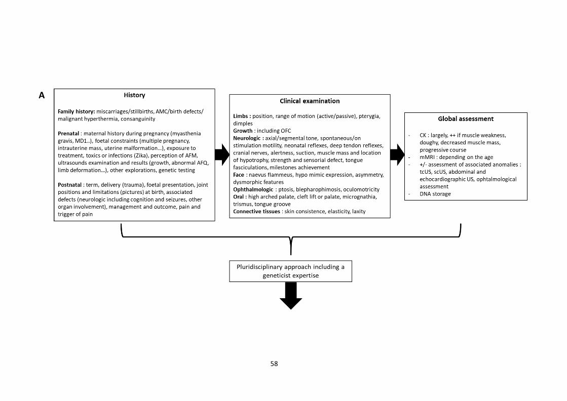
58
A

59
Figure 6. Proposition of recommendations in the aetiological assessment of AMC children. A. General recommendations for assessment of all patients. B. Recommendations according to the conclusions of the global assessment. Legends : AAp, AminoAcidogram in plasma; AAu, Aminoacidogram
in urine: AChR ab, antibodies targeting Acetylcholine receptor; AFM, Active Foetal Movements; AFQ, Amniotic Fluid Quantity; AMC, arthrogryposis multiplex congenita; CGH, comparative genomic hybridization; CK, creatine Kinase; cMRI, cerebral MRI; CNS, Central Nervous System; DNA, deoxyribonucleic acid; EEG, electroencephalogram; ENMG, electroneuromyogram; FSS, Freeman-Sheldon Syndrome; L/P, lactate pyruvate ratio; MB, muscle biopsy; MCP : metacarpophalangeal; MD1 : Myotonic Dystrophy type 1; mMRI, muscle MRI; MRC, mitochondrial chain respiratory study; NGS, next generation sequencing; OAT, organic acid test; OFC, occipitofrontal circumference; scMRI, spinal cord MRI; scUS, spinal cord ultrasounds, SHS, Sheldon-Hall Syndrome; tcUS, transcranial ultrasounds; US, ultrasonography; WES, whole exome sequencing; WGS, whole genome sequencing.
B
60
Secondly, it provides favourable prognosis elements: absence of intellectual impairment for
both, but also favourable orthopaedic outcome in Amyoplasia. This distinction is often
facilitated by the expertise of a clinical geneticist, as part of the multidisciplinary team.
Meeting the genetic team is also essential in guiding and coordinating genetic investigations,
as well as helping in the interpretation of NGS data.
In our centre, a mMRI is systematically proposed at each dedicated consultation for AMC
patients. This method is of increasing use in the diagnostic strategy. Indeed, studies of muscle
mass and infiltration identified reproducible patterns of muscle impairment distribution, first
in various inherited muscular disorders such as muscular dystrophies or myopathies (112), and
more recently in AMC for ECEL1 and CHRNG (22,113) and more strikingly in Amyoplasia
congenita (manuscript submitted). While the usefulness of this tool will soon be demonstrated
in oncoming publications, our team already actively use it as an additional evidence in favour
of a diagnosis especially in Amyoplasia (impairment of biceps brachii and brachialis at the
upper limb, and gracilis, sartorius and tibialis anterior at the lower limbs) and ECEL1 (long head
of biceps femoris and sartorius impairment with rectus femoris conservation). In addition, it
appears as a helpful tool to guide rehabilitation by discriminating completely absent muscle
from thin but residual ones, although it doesn’t seem that there are any correlations between
muscle impairment on imaging and global function. In the future, a further benefit of mMRI
will be to follow the evolution of muscle impairment through time by repeated evaluations in
order to better delineate the prognosis for a given aetiology. Lastly, mMRI is a helpful to select
muscle to biopsy or to study on ENMG in order to improve their yield. Altogether we
recommend it in first instance, but, as it is not validated as a diagnostic tool yet, it should be
only be done when the age of the patient doesn’t require general anaesthesia. In addition, the
expertise of a trained radiologist is mandatory, and age as well as motor milestones
achievement has to be taken in account in the interpretation.
In this work, we focused on postnatal aetiological diagnostic management of AMC.
Actually, this process may even start in utero if AMC is detected. In the absence of family
history of AMC, this antenatal assessment is even more challenging, in contrast with a high
need of genetic counselling at this time. Indeed, hitherto, no clear correlation has been

61
established between antenatal anomalies and specific causes of AMC, and investigations
mostly focus on providing prognostic elements. However, information about course of
pregnancy in AMC children need to be gathered to be able, as soon as the number of
patients will be sufficient, to identify aetiological markers and improve the accuracy of
information delivered to soon-to-be parents.
It is difficult to give precise guidelines in AMC since aetiologies are numerous and every
case has its own issues. The target audience is large, and although we tried to consider all
points of view, especially neonatal clinicians ‘one, the reality in practice is often different from
theorical considerations. The take-home message is to avoid as much as possible invasive low
yield investigations made obsolete by NGS methods and that considerably increase the
negative experience of the patient in this long diagnostic process. In any case, and in the
absence of immediate new parental project, it may be useful to re-evaluate the child at
distance in order to get sufficient hindsight on his neurodevelopment but also on the evolution
of contractures and response to rehabilitation treatment. This will bring further elements that
will facilitate the complex interpretation of NGS data. While an early diagnosis is useful for
young parents, we have to keep in mind that a too early identification of a disease with poor
cognitive prognosis might be deleterious for the establishment of the attachment relationship
between the child and his parents.
4/ Limits of this study
Here, we chose a classification system rarely used by other authors, but that we daily
apply during dedicated AMC patients’ consultations. It nicely reflects our intellectual process
and it is very convenient for communication with families (17). However, it has its own limits.
First, borders that distinguish the different groups of the classification is often difficult
to delineate and categorization may be clinician dependant. Indeed, consensus criteria for
Amyoplasia and DA have been established long ago but remain broad and elusive. Concerning
Amyoplasia, although typical forms are easily recognizable, no criterion is constant. In
addition, as there are no biochemical or molecular diagnostic marker for Amyoplasia,
definition of atypical Amyoplasia is tricky, and this concept has never been clearly defined so

62
far. This is true also for Amyoplasia involving lower limbs only, for which confusing with
neurogenic forms due to TRPV4 mutation is possible.
Concerning DA, definition has much widened now that entities have been better
delineated over the last few years, and we now know that in many presentations, contractures
are not exclusively distal. Indeed, while medium calibre joints or proximal joints involvement
is common in PIEZO2, ECEL1 related AMC or multiple pterygia syndrome, it is also observed in
CCA or even in sarcomeric forms (18). Thus, while typical presentations are easily
recognizable, classification in group 2 is much more arduous for atypical cases without
molecular characterization.
In addition, some symptoms may bring confusion. Clinicians have to be aware of signs
previously described as specific of a given syndrome that may actually arise in many forms,
such as pterygia. Also, as ID or developmental delay is inconstant in some syndromes, they
may lead to classify patients in group 2 or 3O instead of 3C, as it happened in one of our
patients with Aarskog syndrome and in previous studies (5,6). As well, although structural
brain anomalies and ID described in Amyoplasia patients might be due to independent events
(12,50), there may be misclassifications between group 1 and 3C. This may also happen with
DA patients as in our study.
No classification system is fully satisfying, and as genetic basis begin to be uncovered,
classification will probably have to be reconsidered following pathophysiological pathways or
using multi-layered approaches (11,27). This will ultimately facilitate patients comparison
between studies.
Another limit of our study is the fact that most patients were not evaluated at birth,
but after they had received a more or less extensive diagnostic assessment. Thus, diagnosis
was established in the light of results available so far and hence it does not reflect the situation
of a neonatologist first to face the patient. In addition, the retrospective nature of data
collection and the fact that most patients were followed up in other medical care centres than
ours impaired exhaustivity and accuracy of data collection. For non-local patients, we were
dependant on the number, nature and precision of medical sheets provided. One can suppose
that this could be counterbalanced by the fact that patients were mostly coming to our centre
on their own and that they were thus willing to provide the maximum of information.

63
However, despite parental willingness, some common paraclinical investigations that turned
out to be normal, such as CK dosage, might not have been mentioned on these documents.
Lastly, for non-local patients, we had no information about the explorations conducted after
the clinical evaluation in our centre.
To finish, in our cohort French practices representation is questioned by the fact that 8%
of our cases were children coming from other countries. We chose not to exclude them to
better reflect our own practises and recruitment. This actually mainly impacted accuracy of
information concerning pregnancy management, whereas information about diagnosis and
therapeutic postnatal management was often as complete as for French residents. Indeed, for
most of them management was co-coordinated by our team or was conducted in another
medical care centre of France.

64
CONCLUSION
In a retrospective single centre observational study, we gathered clinical and
paraclinical diagnostic data from 125 patients with AMC evaluated at a paediatric age between
2007 and 2019 in Grenoble University Hospital reference centre for developmental anomalies.
Taking advantage of this data and of our clinical experience, we aimed at proposing medical
practical recommendations for the diagnostic procedure of patients with AMC that could
benefit all clinicians in charge of these patients. The nationwide recruitment of our patients,
coming from secondary or tertiary level care centres, associated with the high number of
patients gathered, enabled us to diminish referral biases and to highlight the diversity of the
current medical diagnostic practices. This notably enabled us to unravel the previously
undescribed importance of PIEZO2 mutations in patients with distal arthrogryposis and of
ZC4H2 in patients with central nervous system involvement.
We propose a new classification system that prod into distinguishing straight away,
as soon as possible in patient’s life, the two main causes of AMC, i.e. Amyoplasia congenita
and distal arthrogryposis. We discuss the relevance of all investigations that could be
performed in patients and propose first that despite a poor diagnostic yield with mostly
nonspecific anomalies, non-invasive and cost-effective investigations such as ultrasonography
or CK dosage should be largely proposed, since they may bring further elements to guide the
diagnosis. We also illustrate that invasive investigations, such as muscle biopsy or
electromyoneurography, should be considered only in last intention in specific cases, and that
their realization should be framed by several considerations to increase their yield. We further
question the diagnostic value of other widely performed investigations such as DMPK, SMN1
and 15q11-q13 locus analyses as well as metabolic investigations, whereas we emphasize the
importance of chromosomal investigations, spinal cord and muscle MRI.
In this diagnostic procedure, a multidisciplinary approach is of major importance. In
the next years, the increasing use of next generation sequencing technologies will increase
diagnostic rates by uncovering new genes and revolutionizing our understanding of AMC
physiopathology. However, a relevant phenotyping will remain paramount to facilitate wide
genomic data interpretation.

65
THÈSE SOUTENUE PAR : Pauline LE TANNO
TITRE : “The diagnostic workup in children with Arthrogryposis Multiplex Congenita :
description of practices through a monocentric cohort and suggestion of recommendations.”
CONCLUSION :
Dans cette étude rétrospective, nous avons inclus 125 patients atteints
d’Arthrogrypose Multiple Congénitale (AMC), évalués à un âge pédiatrique au Centre de
Référence des Anomalies du Développement du Centre Hospitalo-Universitaire Grenoble-
Alpes entre 2007 et 2019. Nous avons mis en parallèle les données disponibles sur l’ensemble
des examens paracliniques effectués chez ces patients tout au long de leur parcours
diagnostique, notre expérience clinique, ainsi que les connaissances scientifiques rapportées
dans la littérature. L’objectif était de proposer des recommandations médicales pour guider
la démarche diagnostique chez ces patients, à destination de tout médecin susceptible de les
prendre en charge. Le nombre significatif d’individus réunis ainsi que la variété de leurs
origines, beaucoup ayant été adressés par des centres de soins secondaires ou tertiaires de
toute la France, nous ont permis de réduire certains biais de recrutement et de refléter au
mieux les pratiques médicales actuelles sur notre territoire. Ceci a également permis
d’illustrer la part importante des mutations des gènes PIEZO2 dans les arthrogryposes distales
et ZC4H2 chez les patients présentant une atteinte du système nerveux central, ce qui n’avait
pas été relevé à ce jour.
Dans ce travail, nous proposons une nouvelle classification clinique qui pousse à
distinguer au plus tôt les deux principales causes d’AMC, l’Amyoplasie congénitale et les
arthrogryposes distales, des autres formes. Nous discutons de la pertinence de toutes les
investigations paracliniques qui ont pu être proposées chez ces patients, et suggérons que les
investigations non invasives et peu couteuses, telles que l’échographie ou les dosages de
créatine kinase, soient proposées de façon large en première intention, puisqu’elles sont
susceptibles d’apporter des éléments permettant de réduire le spectre des hypothèses
diagnostiques évoquées. Nous montrons également que certaines investigations, telles que la
biopsie musculaire ou l’électroneuromyographie, sont de faible contribution au regard de leur

66
caractère invasif. Nous proposons qu’elles ne soient réalisées qu’en dernière intention dans
des indications sélectionnées et qu’elles soient encadrées de certaines précautions
permettant d’optimiser leur rendement. Nous précisons les indications d’autres analyses
fréquemment pratiquées telles que l’étude des gènes DMPK, SMN1 et du locus 15q11-q13,
ainsi que celles des investigations métaboliques. Enfin nous attirons l’attention sur la valeur
des investigations chromosomiques et de l’IRM musculaire, et discutons de l’impact
d’analyses plus rarement pratiquées telles que la recherche d’anticorps anti-récepteurs de
l’acétylcholine et de l’IRM médullaire.

67
REFERENCES
1. Hall JG. Arthrogryposis multiplex congenita: etiology, genetics, classification, diagnostic approach, and general aspects. J Pediatr Orthop B. 1997;6(3):159‑ 66.
2. Cachecho S, Elfassy C, Hamdy R, Rosenbaum P, Dahan-Oliel N. Arthrogryposis multiplex congenita definition: Update using an international consensus-based approach. Am J Med Genet C Semin Med Genet. 2019; DOI: 10.1002/ajmg.c.31739
3. Dahan-Oliel N, Cachecho S, Barnes D, Bedard T, Davison AM, Dieterich K, et al. International multidisciplinary collaboration toward an annotated definition of arthrogryposis multiplex congenita. Am J Med Genet C Semin Med Genet. 2019;181(3):288‑ 99. DOI: 10.1002/ajmg.c.31721
4. Hall JG. Arthrogryposis (multiple congenital contractures): diagnostic approach to etiology, classification, genetics, and general principles. Eur J Med Genet. 2014;57(8):464‑ 72. DOI: 10.1016/j.ejmg.2014.03.008
5. Lowry RB, Sibbald B, Bedard T, Hall JG. Prevalence of multiple congenital contractures including arthrogryposis multiplex congenita in Alberta, Canada, and a strategy for classification and coding. Birth Defects Res Part A Clin Mol Teratol. 2010;88(12):1057‑ 61. DOI: 10.1002/bdra.20738
6. Darin N, Kimber E, Kroksmark A-K, Tulinius M. Multiple congenital contractures: birth prevalence, etiology, and outcome. J Pediatr. 2002;140(1):61‑ 7. DOI: 10.1067/mpd.2002.121148
7. Hoff JM, Loane M, Gilhus NE, Rasmussen S, Daltveit AK. Arthrogryposis multiplexa congenita: an epidemiologic study of nearly 9 million births in 24 EUROCAT registers. Eur J Obstet Gynecol Reprod Biol. 2011;159(2):347‑ 50. DOI: 10.1016/j.ejogrb.2011.09.027
8. Navti OB, Kinning E, Vasudevan P, Barrow M, Porter H, Howarth E, et al. Review of perinatal management of arthrogryposis at a large UK teaching hospital serving a multiethnic population. Prenat Diagn. 2010;30(1):49‑ 56. DOI: 10.1002/pd.2411
9. Skaria P, Dahl A, Ahmed A. Arthrogryposis multiplex congenita in utero: radiologic and pathologic findings. J Matern Fetal Neonatal Med. 2019;32(3):502‑ 11. DOI: 10.1080/14767058.2017.1381683
10. Hall JG. Pena-Shokeir phenotype (fetal akinesia deformation sequence) revisited. Birth Defects Res Part A Clin Mol Teratol. 2009;85(8):677‑ 94. DOI: 10.1002/bdra.20611
11. Hall JG, Kimber E, Dieterich K. Classification of arthrogryposis. Am J Med Genet C Semin Med Genet. 2019;181(3):300‑ 3. DOI: 10.1002/ajmg.c.31716
12. Hall JG, Aldinger KA, Tanaka KI. Amyoplasia revisited. Am J Med Genet A. 2014;164A(3):700‑ 30. DOI: 10.1002/ajmg.a.36395
13. Reed SD, Hall JG, Riccardi VM, Aylsworth A, Timmons C. Chromosomal abnormalities associated with congenital contractures (arthrogryposis). Clin Genet. 1985;27(4):353‑ 72. DOI: 10.1111/j.1399-0004.1985.tb02278.x
14. Gaitanis JN, McMillan HJ, Wu A, Darras BT. Electrophysiologic Evidence for Anterior Horn Cell Disease in Amyoplasia. Pediatric Neurology. 2010;43(2):142‑ 7. DOI: 10.1016/j.pediatrneurol.2010.03.018

68
15. Kimber E. Arthrogryposis. Causes, Consequences and Clinical Course in Amyoplasia and Distal Arthrogryposis [dissertation]. Göteborg : University of Gothenburg; 2009. 64 p.
16. Bamshad M, Van Heest AE, Pleasure D. Arthrogryposis: a review and update. J Bone Joint Surg Am. 2009;91 Suppl 4:40‑ 6. DOI: 10.2106/JBJS.I.00281
17. Hall JG, Kimber E, van Bosse HJP. Genetics and Classifications. J Pediatr Orthop. 2017;37 Suppl 1:S4‑ 8. DOI: 10.1097/BPO.0000000000000997
18. Kimber E, Tajsharghi H, Kroksmark A-K, Oldfors A, Tulinius M. Distal arthrogryposis: clinical and genetic findings. Acta Paediatr. 2012;101(8):877‑ 87. DOI: 10.1111/j.1651- 2227.2012.02708.x
19. Putnam EA, Zhang H, Ramirez F, Milewicz DM. Fibrillin-2 (FBN2) mutations result in the Marfan-like disorder, congenital contractural arachnodactyly. Nat Genet. 1995;11(4):456‑ 8. DOI: 10.1038/ng1295-456
20. Sung SS, Brassington A-ME, Grannatt K, Rutherford A, Whitby FG, Krakowiak PA, et al. Mutations in genes encoding fast-twitch contractile proteins cause distal arthrogryposis syndromes. Am J Hum Genet. 2003;72(3):681‑ 90. DOI: 10.1086/368294
21. Coste B, Houge G, Murray MF, Stitziel N, Bandell M, Giovanni MA, et al. Gain-of-function mutations in the mechanically activated ion channel PIEZO2 cause a subtype of Distal Arthrogryposis. Proc Natl Acad Sci USA. 2013;110(12):4667‑ 72. DOI: 10.1073/pnas.1221400110
22. Dieterich K, Quijano-Roy S, Monnier N, Zhou J, Fauré J, Smirnow DA, et al. The neuronal endopeptidase ECEL1 is associated with a distinct form of recessive distal arthrogryposis. Hum Mol Genet. 2013;22(8):1483‑ 92. DOI: 10.1093/hmg/dds514
23. Toydemir RM, Rutherford A, Whitby FG, Jorde LB, Carey JC, Bamshad MJ. Mutations in embryonic myosin heavy chain (MYH3) cause Freeman-Sheldon syndrome and Sheldon-Hall syndrome. Nat Genet. 2006;38(5):561‑ 5. DOI: 10.1038/ng1775
24. Gurnett CA, Desruisseau DM, McCall K, Choi R, Meyer ZI, Talerico M, et al. Myosin binding protein C1: a novel gene for autosomal dominant distal arthrogryposis type 1. Hum Mol Genet. 2010;19(7):1165‑ 73. DOI: 10.1093/hmg/ddp587
25. Toydemir RM, Chen H, Proud VK, Martin R, van Bokhoven H, Hamel BCJ, et al. Trismus- pseudocamptodactyly syndrome is caused by recurrent mutation of MYH8. Am J Med Genet A. 2006;140(22):2387‑ 93. DOI: 10.1002/ajmg.a.31495
26. Chong JX, Burrage LC, Beck AE, Marvin CT, McMillin MJ, Shively KM, et al. Autosomal- Dominant Multiple Pterygium Syndrome Is Caused by Mutations in MYH3. Am J Hum Genet. 2015;96(5):841‑ 9. DOI: 10.1016/j.ajhg.2015.04.004
27. Kiefer J, Hall JG. Gene ontology analysis of arthrogryposis (multiple congenital contractures). Am J Med Genet C Semin Med Genet. 2019; DOI: 10.1002/ajmg.c.31733
28. Hall JG. An approach to congenital contractures (arthrogryposis). Pediatr Ann. 1981;10(7):15‑ 26.
29. Hageman G, Ippel EPF, Beemer FA, Pater JM de, Lindhout D, Willemse J. The diagnostic management of newborns with congenital contractures: A nosologic study of 75 cases. American Journal of Medical Genetics. 1988;30(4):883‑ 904. DOI: 10.1002/ajmg.1320300404

69
30. Wallach E, Walther-Louvier U, Espil-Taris C, Rivier F, Baudou E, Cances C. Arthrogryposis in children: Etiological assessments and preparation of a protocol for etiological investigations. Arch Pediatr. 2018; DOI: 10.1016/j.arcped.2018.05.004
31. Solotareff Fagué L. Arthrogryposes congénitales : étude descriptive de 31 cas au CHRU de Lille, entre 2005 et 2012 [dissertation]. Lille: Université du droit et de la santé 2; 2013. 57 p.
32. Dieterich K, Le Tanno P, Kimber E, Jouk P-S, Hall J, Giampietro P. The diagnostic workup in a patient with AMC: Overview of the clinical evaluation and paraclinical analyses with review of the literature. Am J Med Genet C Semin Med Genet. 2019;181(3):337‑ 44. DOI: 10.1002/ajmg.c.31730
33. Bayram Y, Karaca E, Coban Akdemir Z, Yilmaz EO, Tayfun GA, Aydin H, et al. Molecular etiology of arthrogryposis in multiple families of mostly Turkish origin. J Clin Invest. 2016;126(2):762‑ 78. DOI: 10.1172/JCI84457
34. Valdés-Flores M, Casas-Avila L, Hernández-Zamora E, Kofman S, Hidalgo-Bravo A. Characterization of a group unrelated patients with arthrogryposis multiplex congenita. Jornal de Pediatria. 2016;92(1):58‑ 64. DOI: 10.1016/j.jped.2015.04.008
35. Ambegaonkar G, Manzur AY, Robb SA, Kinali M, Muntoni F. The multiple phenotypes of Arthrogryposis multiplex congenita with reference to the neurogenic variant. European Journal of Paediatric Neurology. 2011;15(4):316‑ 9. DOI: 10.1016/j.ejpn.2011.01.007
36. Dai S, Dieterich K, Jaeger M, Wuyam B, Jouk P-S, Pérennou D. Disability in adults with arthrogryposis is severe, partly invisible, and varies by genotype. Neurology. 2018;90(18):e1596‑ 604. DOI: 10.1212/WNL.0000000000005418
37. Pehlivan D, Bayram Y, Gunes N, Coban Akdemir Z, Shukla A, Bierhals T, et al. The Genomics of Arthrogryposis, a Complex Trait: Candidate Genes and Further Evidence for Oligogenic Inheritance. The American Journal of Human Genetics. 2019;105(1):132‑ 50. DOI: 10.1016/j.ajhg.2019.05.015
38. Hirata H, Nanda I, van Riesen A, McMichael G, Hu H, Hambrock M, et al. ZC4H2 mutations are associated with arthrogryposis multiplex congenita and intellectual disability through impairment of central and peripheral synaptic plasticity. Am J Hum Genet. 2013;92(5):681‑ 95. DOI: 10.1016/j.ajhg.2013.03.021
39. Zanzottera C, Milani D, Alfei E, Rizzo A, D’Arrigo S, Esposito S, et al. ZC4H2 deletions can cause severe phenotype in female carriers. American Journal of Medical Genetics Part A. 2017;173(5):1358‑ 63. DOI: 10.1002/ajmg.a.38155
40. Okubo Y, Endo W, Inui T, Suzuki-Muromoto S, Miyabayashi T, Togashi N, et al. A severe female case of arthrogryposis multiplex congenita with brain atrophy, spastic quadriplegia and intellectual disability caused by ZC4H2 mutation. Brain and Development. 2018;40(4):334‑ 8. DOI: 10.1016/j.braindev.2017.11.011
41. Frints SGM, Hennig F, Colombo R, Jacquemont S, Terhal P, Zimmerman HH, et al. Deleterious de novo variants of X-linked ZC4H2 in females cause a variable phenotype with neurogenic arthrogryposis multiplex congenita. Human Mutation. Dec 2019;40(12):2270-2285.
42. Gonzalez CH, Sommer A, Meisner LF, Elejalde BR, Opitz JM. The trisomy 4p syndrome: case report and review. Am J Med Genet. 1977;1(2):137‑ 56. DOI: 10.1002/ajmg.1320010202

70
43. Sebold C, Roeder E, Zimmerman M, Soileau B, Heard P, Carter E, et al. Tetrasomy 18p: Report of the molecular and clinical findings of 43 individuals. American Journal of Medical Genetics Part A. 2010;152A(9):2164‑ 72. DOI: 10.1002/ajmg.a.33597
44. Karimzad Hagh J, Liehr T, Ghaedi H, Mossalaeie MM, Alimohammadi S, Inanloo Hajiloo F, et al. Prenatal Diagnosis of Mosaic Tetrasomy 18p in a Case without Sonographic Abnormalities. Int J Mol Cell Med. 2017;6(1):61‑ 5.
45. Laquérriere A, Maluenda J, Camus A, Fontenas L, Dieterich K, Nolent F, et al. Mutations in CNTNAP1 and ADCY6 are responsible for severe arthrogryposis multiplex congenita with axoglial defects. Hum Mol Genet. 2014;23(9):2279‑ 89. DOI: 10.1093/hmg/ddt618
46. Hall JG. Uterine structural anomalies and arthrogryposis-death of an urban legend. Am J Med Genet A. 2013;161A(1):82‑ 8. DOI: 10.1002/ajmg.a.35683
47. Pallotta R, Ehresmann T, Fusilli P. Ocular findings in distal arthrogryposis. Ophthalmic Genet. 2001;22(2):125‑ 30. DOI: 10.1076/opge.22.2.125.2232
48. Dieterich K, Kimber E, Hall JG. Central nervous system involvement in arthrogryposis multiplex congenita: Overview of causes, diagnosis, and care. Am J Med Genet C Semin Med Genet. 2019;181(3):345‑ 53. DOI: 10.1002/ajmg.c.31732
49. Fedrizzi E, Botteon G, Inverno M, Ciceri E, D’Incerti L, Dworzak F. Neurogenic arthrogryposis multiplex congenita: clinical and MRI findings. Pediatr Neurol. 1993;9(5):343‑ 8. DOI: 10.1016/0887-8994(93)90102-i
50. Kamien B, Zankl A, Gabbett M. Septo-optic dysplasia and associations with amyoplasia and gastroschisis. Birth Defects Res Part A Clin Mol Teratol. 2010;88(6):497‑ 501. DOI: 10.1002/bdra.20663
51. Llorens-Salvador R, Moreno-Flores A. The ABCs of transfontanellar ultrasound and more. Radiologia. 2016;58 Suppl 2:129‑ 41. DOI: 10.1016/j.rx.2016.02.007
52. Sackey A, Coulter B, Fryer A, Van Velzen D. Epilepsy in the Freeman Sheldon Syndrome. J Child Neurol. 1995;10(4):335‑ 7. DOI: 10.1177/088307389501000421
53. Gibreel WO, Selcen D, Zeidan MM, Ishitani MB, Moir CR, Zarroug AE. Safety and yield of muscle biopsy in pediatric patients in the modern era. J Pediatr Surg. 2014;49(9):1429‑ 32. DOI: 10.1016/j.jpedsurg.2014.02.079
54. Thavorntanaburt S, Tanboon J, Likasitwattanakul S, Sangruchi T, Nishino I, Ngerncham M, et al. Impact of muscle biopsy on diagnosis and management of children with neuromuscular diseases: A 10-year retrospective critical review. J Pediatr Surg. 2018;53(3):489‑ 92. DOI: 10.1016/j.jpedsurg.2017.06.006
55. Kang PB, Lidov HGW, David WS, Torres A, Anthony DC, Jones HR, et al. Diagnostic value of electromyography and muscle biopsy in arthrogryposis multiplex congenita. Ann Neurol. 2003;54(6):790‑ 5. DOI: 10.1002/ana.10769
56. Rabie M, Jossiphov J, Nevo Y. Electromyography (EMG) accuracy compared to muscle biopsy in childhood. J Child Neurol. 2007;22(7):803‑ 8. DOI: 10.1177/0883073807304204
57. Dubowitz V. Muscle disorders in childhood. 2nd edition. London: WB Saunders; 1995. 460 p.

71
58. Rossor AM, Oates EC, Salter HK, Liu Y, Murphy SM, Schule R, et al. Phenotypic and molecular insights into spinal muscular atrophy due to mutations in BICD2. Brain. 2015;138(Pt 2):293‑ 310. DOI: 10.1093/brain/awu356
59. Mercuri E, Messina S, Kinali M, Cini C, Longman C, Battini R, et al. Congenital form of spinal muscular atrophy predominantly affecting the lower limbs: a clinical and muscle MRI study. Neuromuscul Disord. 2004;14(2):125‑ 9. DOI: 10.1016/j.nmd.2003.09.005
60. Russell EJ. Cervical disk disease. Radiology. 1990;177(2):313‑ 25. DOI: 10.1148/radiology.177.2.2217760
61. Görgen-Pauly U, Sperner J, Reiss I, Gehl HB, Reusche E. Familial pontocerebellar hypoplasia type I with anterior horn cell disease. Eur J Paediatr Neurol. 1999;3(1):33‑ 8. DOI: 10.1053/ejpn.1999.0177
62. de Souza AS, de Oliveira-Szjenfeld PS, de Oliveira Melo AS, de Souza LAM, Batista AGM, Tovar- Moll F. Imaging findings in congenital Zika virus infection syndrome: an update. Childs Nerv Syst. 2018;34(1):85‑ 93. DOI: 10.1007/s00381-017-3637-1
63. Bicakci I, Turgut ST, Turgut B, Icagasioglu A, Egilmez Z, Yumusakhuylu Y. A case of caudal regression syndrome: walking or sitting? Pan Afr Med J. 2014;18:92. DOI: 10.11604/pamj.2014.18.92.3683
64. Sanchez T, John RM. Early identification of tethered cord syndrome: a clinical challenge. J Pediatr Health Care. 2014;28(3):e23-33. DOI: 10.1016/j.pedhc.2013.06.007
65. Solmaz I, Izci Y, Albayrak B, Cetinalp E, Kural C, Sengul G, et al. Tethered cord syndrome in childhood: special emphasis on the surgical technique and review of the literature with our experience. Turk Neurosurg. 2011;21(4):516‑ 21. DOI: 10.5137/1019-5149.JTN .4394-11.1
66. McGirt MJ, Mehta V, Garces-Ambrossi G, Gottfried O, Solakoglu C, Gokaslan ZL, et al. Pediatric tethered cord syndrome: response of scoliosis to untethering procedures. Clinical article. J Neurosurg Pediatr. 2009;4(3):270‑ 4. DOI: 10.3171/2009.4.PEDS08463
67. Jinkins JR, Sener RN. Idiopathic localized hydromyelia: dilatation of the central canal of the spinal cord of probable congenital origin. J Comput Assist Tomogr. 1999;23(3):351‑ 3. DOI: 10.1097/00004728-199905000-00004
68. Jones BV. Cord Cystic Cavities: Syringomyelia and Prominent Central Canal. Semin Ultrasound CT MR. 2017;38(2):98‑ 104. DOI: 10.1053/j.sult.2016.07.008
69. Kowalczyk B, Feluś J. Arthrogryposis: an update on clinical aspects, etiology, and treatment strategies. Arch Med Sci. 2016;12(1):10‑ 24. DOI: 10.5114/aoms.2016.57578
70. Westcott MA, Dynes MC, Remer EM, Donaldson JS, Dias LS. Congenital and acquired orthopedic abnormalities in patients with myelomeningocele. Radiographics. 1992;12(6):1155‑ 73. DOI: 10.1148/radiographics.12.6.1439018
71. Schilling L, Forst R, Forst J, Fujak A. Orthopaedic Disorders in Myotonic Dystrophy Type 1: descriptive clinical study of 21 patients. BMC Musculoskelet Disord. 2013;14:338. DOI: 10.1186/1471-2474-14-338
72. Martinello F, Piazza A, Pastorello E, Angelini C, Trevisan CP. Clinical and neuroimaging study of central nervous system in congenital myotonic dystrophy. J Neurol. 1999;246(3):186‑ 92. DOI: 10.1007/s004150050332

72
73. Denizot S, Boscher C, Le Vaillant C, Rozé JC, Gras Le Guen C. Distal arthrogryposis and neonatal hypotonia: an unusual presentation of Prader-Willi syndrome (PWS). J Perinatol. 2004;24(11):733‑ 4. DOI: 10.1038/sj.jp.7211185
74. Bigi N, Faure J-M, Coubes C, Puechberty J, Lefort G, Sarda P, et al. Prader-Willi syndrome: is there a recognizable fetal phenotype? Prenat Diagn. 2008;28(9):796‑ 9. DOI: 10.1002/pd.1973
75. Klinge L, Scott RC, de Sousa C. Neonatal subdural and extradural haemorrhage in Prader-Willi syndrome. Neuropediatrics. 2001;32(4):221‑ 2. DOI: 10.1055/s-2001-17379
76. Oiglane-Shlik E, Zordania R, Varendi H, Antson A, Mägi M-L, Tasa G, et al. The neonatal phenotype of Prader-Willi syndrome. Am J Med Genet A. 2006;140(11):1241‑ 4. DOI: 10.1002/ajmg.a.31223
77. Prior TW, Snyder PJ, Rink BD, Pearl DK, Pyatt RE, Mihal DC, et al. Newborn and carrier screening for spinal muscular atrophy. Am J Med Genet A. 2010;152A(7):1608‑ 16. DOI: 10.1002/ajmg.a.33474
78. Grotto S, Cuisset J-M, Marret S, Drunat S, Faure P, Audebert-Bellanger S, et al. Type 0 Spinal Muscular Atrophy: Further Delineation of Prenatal and Postnatal Features in 16 Patients. J Neuromuscul Dis. 2016;3(4):487‑ 95. DOI: 10.3233/JND-160177
79. Bürglen L, Amiel J, Viollet L, Lefebvre S, Burlet P, Clermont O, et al. Survival motor neuron gene deletion in the arthrogryposis multiplex congenita-spinal muscular atrophy association. J Clin Invest. 1996;98(5):1130‑ 2. DOI: 10.1172/JCI118895
80. Falsaperla R, Romeo G, Di Giorgio A, Pavone P, Parano E, Connolly AM. Long-term survival in a child with arthrogryposis multiplex congenita and spinal muscular atrophy. J Child Neurol. 2001;16(12):934‑ 6. DOI: 10.1177/088307380101601213
81. Guillot N, Cuisset J-M, Cuvellier J-C, Hurtevent J-F, Joriot S, Vallee L. Unusual clinical features in infantile Spinal Muscular Atrophies. Brain Dev. 2008;30(3):169‑ 78. DOI: 10.1016/j.braindev.2007.07.008
82. McEntagart M. TRPV4 axonal neuropathy spectrum disorder. Journal of Clinical Neuroscience. 2012;19(7):927‑ 33. DOI: 10.1016/j.jocn.2011.12.003
83. Scoto M, Rossor AM, Harms MB, Cirak S, Calissano M, Robb S, et al. Novel mutations expand the clinical spectrum of DYNC1H1-associated spinal muscular atrophy. Neurology. 2015;84(7):668‑ 79. DOI: 10.1212/WNL.0000000000001269
84. Storbeck M, Horsberg Eriksen B, Unger A, Hölker I, Aukrust I, Martínez-Carrera LA, et al. Phenotypic extremes of BICD2-opathies: from lethal, congenital muscular atrophy with arthrogryposis to asymptomatic with subclinical features. Eur J Hum Genet. 2017;25(9):1040‑ 8. DOI: 10.1038/ejhg.2017.98
85. Ravenscroft G, Di Donato N, Hahn G, Davis MR, Craven PD, Poke G, et al. Recurrent de novo BICD2 mutation associated with arthrogryposis multiplex congenita and bilateral perisylvian polymicrogyria. Neuromuscul Disord. 2016;26(11):744‑ 8. DOI: 10.1016/j.nmd.2016.09.009
86. de la Chapelle A, Herva R, Koivisto M, Aula P. A deletion in chromosome 22 can cause DiGeorge syndrome. Hum Genet. 1981;57(3):253‑ 6. DOI: 10.1007/bf00278938
87. Hunter JE, Berry-Kravis E, Hipp H, Todd PK. FMR1 Disorders. Dans: Adam MP, Ardinger HH, Pagon RA, Wallace SE, Bean LJ, Stephens K, et al., directeurs. GeneReviews®. [En ligne]. Seattle

73
(WA): University of Washington, Seattle; 1993-2020. Disponible: http://www.ncbi.nlm.nih.gov/books/NBK1384/
88. Li X, Jiang M, Han W, Zhao N, Liu W, Sui Y, et al. A novel TNNI2 mutation causes Freeman- Sheldon syndrome in a Chinese family with an affected adult with only facial contractures. Gene. 2013;527(2):630‑ 5. DOI: 10.1016/j.gene.2013.06.082
89. Eymard B. [Antibodies in myasthenia gravis]. Rev Neurol (Paris). 2009;165(2):137‑ 43. DOI: 10.1016/j.neurol.2008.11.020
90. Zisimopoulou P, Evangelakou P, Tzartos J, Lazaridis K, Zouvelou V, Mantegazza R, et al. A comprehensive analysis of the epidemiology and clinical characteristics of anti-LRP4 in myasthenia gravis. Journal of Autoimmunity. 2014;52:139‑ 45. DOI: 10.1016/j.jaut.2013.12.004
91. Andersen JB, Heldal AT, Engeland A, Gilhus NE. Myasthenia gravis epidemiology in a national cohort; combining multiple disease registries. Acta Neurol Scand, Suppl. 2014;(198):26‑ 31. DOI: 10.1111/ane.12233
92. Polizzi A, Huson SM, Vincent A. Teratogen update: maternal myasthenia gravis as a cause of congenital arthrogryposis. Teratology. 2000;62(5):332‑ 41. DOI: 10.1002/1096- 9926(200011)62:5<332::AID-TERA7>3.0.CO;2-E
93. Hacohen Y, Jacobson LW, Byrne S, Norwood F, Lall A, Robb S, et al. Fetal acetylcholine receptor inactivation syndrome. Neurol Neuroimmunol Neuroinflamm. 2014;2(1). DOI: 10.1212/NXI.0000000000000057
94. Gilhus NE, Hong Y. Maternal myasthenia gravis represents a risk for the child through autoantibody transfer, immunosuppressive therapy and genetic influence. European Journal of Neurology. 2018;25(12):1402‑ 9. DOI: 10.1111/ene.13788
95. Hesselmans LF, Jennekens FG, Van den Oord CJ, Veldman H, Vincent A. Development of innervation of skeletal muscle fibers in man: relation to acetylcholine receptors. Anat Rec. 1993;236(3):553‑ 62. DOI: 10.1002/ar.1092360315
96. Vincent A, McConville J, Farrugia ME, Bowen J, Plested P, Tang T, et al. Antibodies in myasthenia gravis and related disorders. Ann N Y Acad Sci. 2003;998:324‑ 35. DOI: 10.1196/annals.1254.036
97. Hoff JM, Daltveit AK, Gilhus NE. Myasthenia gravis in pregnancy and birth: identifying risk factors, optimising care. Eur J Neurol. 2007;14(1):38‑ 43. DOI: 10.1111/j.1468- 1331.2006.01538.x
98. Hoff JM, Daltveit AK, Gilhus NE. Artrogryposis multiplex congenita -- a rare fetal condition caused by maternal myasthenia gravis. Acta Neurol Scand, Suppl. 2006;183:26‑ 7. DOI: 10.1111/j.1600-0404.2006.00610.x
99. Mikou F, Kaouti N, Ghazli M, El Kerroumi M, Sefrioui O, Morsad F, et al. [Severe neonatal myasthenia with arthrogryposis]. J Gynecol Obstet Biol Reprod (Paris). 2003;32(7):660‑ 2.
100. Cantagrel S, Maury L, Yamamoto A-M, Maheut J, Toutain A, Castelnau P. Akinesia, arthrogryposis, craniosynostosis: a presentation of neonatal myasthenia with fetal onset. Am J Perinatol. 2002;19(6):297‑ 301. DOI: 10.1055/s-2002-34468
74
101. Morel E, Bach JF, Briard ML, Aubry JP. Neonatal myasthenia gravis. Anti-acetylcholine receptor antibodies in the amniotic fluid. J Neuroimmunol. 1984;6(5):313‑ 7. DOI: 10.1016/0165- 5728(84)90020-1
102. Hellmund A, Berg C, Geipel A, Müller A, Gembruch U. Prenatal diagnosis of fetal akinesia deformation sequence (FADS): a study of 79 consecutive cases. Arch Gynecol Obstet. 2016;294(4):697‑ 707. DOI: 10.1007/s00404-016-4017-x
103. Dalton P, Clover L, Wallerstein R, Stewart H, Genzel-Boroviczeny O, Dean A, et al. Fetal arthrogryposis and maternal serum antibodies. Neuromuscul Disord. 2006;16(8):481‑ 91. DOI: 10.1016/j.nmd.2006.05.015
104. Eymard B, Morel E, Dulac O, Moutard-Codou ML, Jeannot E, Harpey JP, et al. [Myasthenia and pregnancy: a clinical and immunologic study of 42 cases (21 neonatal myasthenia cases)]. Rev Neurol (Paris). 1989;145(10):696‑ 701.
105. Vincent A, Waters P, Leite MI, Jacobson L, Koneczny I, Cossins J, et al. Antibodies identified by cell-based assays in myasthenia gravis and associated diseases. Ann N Y Acad Sci. 2012;1274:92‑ 8. DOI: 10.1111/j.1749-6632.2012.06789.x
106. Hamel J, Ciafaloni E. An Update: Myasthenia Gravis and Pregnancy. Neurol Clin. 2018;36(2):355‑ 65. DOI: 10.1016/j.ncl.2018.01.005
107. Midelfart Hoff J, Midelfart A. Maternal myasthenia gravis: a cause for arthrogryposis multiplex congenita. J Child Orthop. 2015;9(6):433‑ 5. DOI: 10.1007/s11832-015-0690-8
108. Wilnai Y, Seaver LH, Enns GM. Atypical amyoplasia congenita in an infant with Leigh syndrome: A mitochondrial cause of severe contractures? American Journal of Medical Genetics Part A. 2012;158A(9):2353‑ 7. DOI: 10.1002/ajmg.a.35533
109. von Kleist-Retzow J-C, Cormier-Daire V, Viot G, Goldenberg A, Mardach B, Amiel J, et al. Antenatal manifestations of mitochondrial respiratory chain deficiency. J Pediatr. 2003;143(2):208‑ 12. DOI: 10.1067/S0022-3476(03)00130-6
110. Laubscher B, Janzer RC, Krähenbühl S, Hirt L, Deonna T. Ragged-red fibers and complex I deficiency in a neonate with arthrogryposis congenita. Pediatr Neurol. 1997;17(3):249‑ 51. DOI: 10.1016/s0887-8994(97)00082-9
111. Mignot C, Gelot A, Bessières B, Daffos F, Voyer M, Menez F, et al. Perinatal-lethal Gaucher disease. Am J Med Genet A. 2003;120A(3):338‑ 44. DOI: 10.1002/ajmg.a.20117
112. Quijano-Roy S, Avila-Smirnow D, Carlier RY, WB-MRI muscle study group. Whole body muscle MRI protocol: pattern recognition in early onset NM disorders. Neuromuscul Disord. 2012;22 Suppl 2:S68-84. DOI: 10.1016/j.nmd.2012.08.003
113. Carrera-García L, Natera-de Benito D, Dieterich K, de la Banda MGG, Felter A, Inarejos E, et al. CHRNG-related nonlethal multiple pterygium syndrome: Muscle imaging pattern and clinical, histopathological, and molecular genetic findings. Am J Med Genet A. 2019;179(6):915‑ 26. DOI: 10.1002/ajmg.a.61122
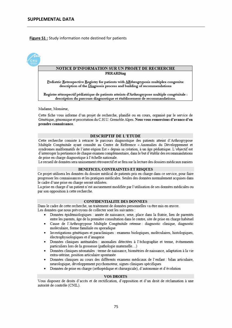
75
SUPPLEMENTAL DATA
Figure S1 : Study information note destined for patients

76

77
Table S1. Features that led to the diagnosis of atypical Amyoplasia
Ind. UL/LL/4L Atypical characteristic Genetic Analysis
5 4L Global loss of muscle function with hypomobility and hypotonia NMD-NGS : negative
16 4L Classic impairment of LL but only distal impairment of UL none
38 LL Atypical LL only presentation NMD-NGS: ongoing
40 4L Global loss of muscle function with hypomobility and hypotonia none
52 LL Cramps and pains NMD-NGS: ongoing
70 UL Cramps and pains NMD-NGS: ongoing
73 LL Abnormally weak response to physiotherapy NMD-NGS: negative WES : ongoing
77 LL Atypical LL only presentation NMD-NGS: ongoing
82 4L Atypical muscle masses and/or mild involvement of trunk NMD-NGS: ongoing
93 LL Complete loss of deep tendon reflexes despite only mild or
moderate muscle atrophy NMD-NGS: ongoing
98 UL Atypical elbow pattern, asymmetry NMD-NGS: negative
114 LL Atypical LL only presentation none
123 4L Reduction abnormality of limbs none
Legends : 4L, four limbs; Ind., individual; LL, lower limbs; NMD NGS, Neuromuscular Disorders-related genes next generation sequencing; UL, upper limbs; WES, Whole Exome Sequencing.

78
Table S2. List of the 121 NMD-related genes sequenced in CHU Grenoble-Alpes laboratory (NMD-NGS)
Gene Disease Inh. Gene Disease Inh. Gene Disease Inh.
ACTA1 CM AD/AR DNM2 FADS/CM/other myo AR/AD MYO18B CM AD
ADAMST2 EDS AR DOK7 FADS/CMS AR MYOD1 FADS AR
ADCY6 FADS AR DPAGT1 CMS AR MYOT Other Myo AD
ADGRG6 FADS AR DYNC1H1 SMALED AD NALCN AMC+CNS AD
AGRN CMS AR ECEL1 DA AR NEB CM/other myo AR
ALG2 CMS AR EMD Other dys XL ORAI1 Other myo AD
ALG14 CMS AR ERBB3 FADS AR PIEZO2 DA AD
ANO5 Other myo AR FBN2 DA AD PIP5K1C FADS AR
ATP2A1 Myotonia AR FBLN5 Other conn AD PLEC CMS AR
BAG3 Other myo, DCM AD FHL1 Other myo XL PLOD1 EDS AR
BICD2 SMALED AD FKBP14 EDS AR PNPLA2 Other myo AR
BIN1 CM AD/AR FLNC Other myo AD PRPL CMS AR
CACNA1S CM/PP AR GFPT1 CMS AR PTPLA CM AR
CASQ1 CM AR GLE1 FADS AR RAPSN FADS/CMS AR
CAV3 LGMD/ Other myo AD GNE Other myo AR RYR1 CM/other myo/MH AR
CCDC78 CM AR HSPG2 Other conn AR SCN4A FADS/CMS/PP/myotonia AR/AD
CFL2 CM AR ITGA7 Other myo AR SEPN1 CM/other myo/dys AR
CHAT CMS AR KBTBD13 CM AD SLC25A1 Other mya AR
CHRNA1 MPS/CMS AR/AD KCNA1 Myotonia AD SLC5A7 CMS/neuropathy AD/AR
CHRNB1 FADS/CMS AR /AD KCNJ2 PP AD SNAP25 CMS ?
CHRND MPS/CMS AR /AD KCNQ2 Other AD SOSTM1 Other myo AD
CHRNE CMS AD/AR KLHL40 CM AR SPEG CM AR
CHRNG MPS/CMS AR KLHL41 CM AR STAC3 CM AR
CLCN1 Myotonia AD/AR LAMA2 Other dys AR STIM1 other myo AD
CNTN1 CM AR LAMB2 CMS AR SYT2 CMS, other mya AD
CNTNAP1 FADS AR LDB3 Other myo AD TCAP LGMD AR
COL1A1 EDS AD LMOD3 CM AR TIA1 Other myo AD
COL1A2 EDS AD/AR LMNA LGMD/other myo/dys AD/AR TNNI2 DA AD
COL3A1 EDS AD/AR LRP4 CMS AR TNNT1 CM AR
COL5A1 EDS AD MAGEL2 Foetal AMC AD TNNT3 DA AD
COL5A3 EDS AD? MATR3 Other myo AD TNXB EDS AR
COL6A1 Other myo/dys AD/AR MEGF10 CM AR TPM2 DA/CM/CM AD
COL6A2 Other myo/dys AD/AR MTM1 CM XL TPM3 CM AD
COL6A3 Other myo/dys AD/AR MUSK FADS/CMS AR TRIM32 CM AR
COL6A6 Other myo ? MYBPC1 FADS AR TRPV4 SMAD AD
COL12A1 Other myo/dys AD/AR MYBPC3 CM AR TTN CM/other myo AD/AR
COL14A1 CMS AR MYH2 DA/CM AD/AR VCP Other myo AD
COLQ CMS AR MYH3 MPS/DA AD ZBTB42 FADS AR
CRYAB Other myo AR/AD MYH7 CM/ other Myo AD ZC4H2 AMC+CNS XL
DES Other myo AR MYH8 DA AD
DNAJB6 LGMD AD MYO9A CMS AR
List of sequenced genes in 2017, other genes might have been added since then.
Legends : AD, autosomal dominant; AMC, Arthrogryposis Multiplex Congenita; AR, autosomal recessive; CM, congenital myopathies; CMS,
congenital Myasthenic Syndrome; CNS, Central Nervous System; Conn, connective tissue disorder; DA, Distal Arthrogryposis; DCM, dilated
cardiomyopathy; Dys, dystrophy; EDS, Ehlers Danlos Syndrome; FADS, Foetal Akinesia Deformation Sequence; LGMD, Limb Girdle Muscular
Dystrophy; MH, Malignant Hyperthermia; MPS, Multiple Pterygia Syndrome; Mya, myasthenia; Myo, myopathy; NMD, neuromuscular diseases;
PP, periodic palsy; SMAD, Spinal Muscular Atrophy, distal; SMALED, Spinal Muscular Atrophy, Lower-Etremity predominant. Bold character : AMC-
related genes list, including foetal or lethal forms. Red character : List of the 11 AMC-related genes previously sequenced in the laboratory (AMC-
NGS).

79
Table S3. Indications that had motivated prenatal karyotypes
Ind. Group Sub
Group
Genetic
diagnosis Indication Details
54 1 T / Isolated BEV
/ 50 1 T /
38 1 aT uk
22 1 T / BEV + other articular anomalies UL and LL
85 1 T / BEV + other articular anomalies
UL and LL 118 3 O uk
89 3 O uk
106
1
T
/
BEV + other articular and extra articular
anomalies UL and LL, decreased AFM, umbilical cyst
64 1 T /
BEV + other articular and extra articular
UL and LL, decreased AFQ, growth delay
23
2
PN
PIEZO2
UL and LL, decreased AFM, abnormal cardiac
hyperechogenicity
74 2 M MYH3 UL and LL, increased AFQ, retrognathism
60 3 C uk UL and LL, increased AFQ, decreased AFM, abnormal
extremities
73 1 aT uk BEV + other extra articular anomalies
Unique umbilical artery
52 1 aT uk Mesenteric cyst
30 2 PN PIEZO2 Growth delay
76 2 PN PIEZO2 Abnormal abdominal image
69 1 T / History of pregnancy with Down syndrome
14 1 T / Increased nuchal transluency
Short long bones, decreased AFQ
58 2 M MYH3 Growth delay, spine anomalies
11 3 C NSD1 Increased first semester dosage
61 3 O LMX1B and unilateral EV
66 1 T / Other articular anomalies
Bilateral club hand
98 1 aT uk Clenched fists, UL muscle amyotrophy
123 1 aT uk Other articular anomalies + other signs Unilateral EV and decreased AFM
116 3 C ZC4H2 Other articular anomalies + other signs Clenched fists, growth delay, increased AFQ
45 3 C ZC4H2 Other articular anomalies + other signs Excess of AFQ
65 1 T / Other
Laparoschisis
55 2 PN PIEZO2 Decreased AFQ and AFM
121 2 M MYH3 Other Increased AFQ, increased OFC, frontal excess of skin
58 2 M TPM2 Other
Decreased AFQ and AFM, growth delay
6 3 C uk Increased AFQ, short long bones, growth delay
Legends : AFM, active foetal movements; AFQ, amniotic fluid quantity; aT, atypical; C, BEV, bilateral Equinovarus feet; CNS involvement; EV,
equinovarus foot; Ind., individual; LL, lower limbs, M, muscle; O, other; OFC, occipitofrontal circumference; PN, peripheral nerve; T, typical; uk,
unknown; UL, upper limbs. Lines coloured in grey indicate patients for which a prenatal array-CGH was also performed.

80
Table S4. Detailed Karyotype and array-CGH results (postnatal)
Ind. Group Sub
group
Genetic
diagnosis Investigation Results
Positive results
45 3 C uk Array-CGH 329kb Xq11.2 deletion involving ZC4H2 (chrX:64,196,201-
64,525,424)
124
3
C
uk
Karyotype 4p trisomy resulting from unbalanced de novo translocation
der(4;22)(p10;q10)
Array-CGH Characterisation of translocation : 4p trisomy
47 3 C uk Karyotype 18p tetrasomy resulting from an isochromosome 18p
Incidental findings/class 1-2-3 CNVs*
49 1 T / Array-CGH 2 inherited deletions
63 2 NMJ CHRNG Array-CGH 10p13 duplication inherited from the mother, polymorphism
119 2 O uk Karyotype 47,XYY
12 2 O uk Array-CGH 16q23.1 de novo duplication, VUS
116 3 C ZC4H2 Array-CGH 2p25.2 de novo deletion, VUS
19 3 C uk Array-CGH 16q21 duplication inherited from the father, polymorphism
Legends : C, CNS involvement; CGH, Comparative Genomic Hybridization; CNVs, Copy Number Variants; Ind., individual; NMJ, neuromuscular junction; O, other; T, typical; uk, unknown; VUS, Variant of Unknown Significance. * According to ACMG classification, i.e. benign, likely benign or VUS
Table S5. Other cytogenetic analyses performed in our cohort
Ind. Group Sub
group Genetic
diagnosis Chromosomal investigations
69 1 T / MLPA for subtelomeric loci study
76 2 PN PIEZO2 MLPA targeting main microdeletions syndromes
11 3 C NSD1 MLPA for subtelomeric loci study*
75 3 C ZC4H2 Fragile X
110 3 C KAT6B MLPA for subtelomeric loci study, 22q11.2 deletion search
17 3 C uk 22q11.2 deletion search, 12p tetrasomy**
60 3 C uk MLPA targeting main microdeletions syndromes
6 3 C uk Fragile-X
4 3 C uk 22q11.2 microdeletion search, 4p duplication
51 3 O uk Wolff-Hirschhorn syndrome
Legends : C, CNS involvement; Ind., individual; MLPA, multiplex Ligation-dependant Probe Amplification; O, other; PN, peripheral nerve; st, subtelomeric; T, typical; uk, unknown; µdel, microdeletions. * polymorphic SHANK3 duplication. ** performed on fibroblasts

81
Table S6. Other targeted analyses performed in our cohort
Ind. Group Sub
group
Genetic
diagnosis Investigations Result
50 1 T / HSPB1, HSPB3, HSPB8 -
55 2 PN PIEZO2 Congenital myasthenia genes -
63 2 NMJ CHRNG ASXL1 Sg -
11
3
C
NSD1
FBN1 -
TGFBR2 -
NSD1 MLPA then Sg + (Sg)
110 3 C KAT6B KAT6B Sg +
34 3 C uk Mt-DNA mutation screening -
POLG1 Sg -
17 3 C uk ARC syndrome genes -
7 3 C uk GLRA1 Sg -
60 3 C uk NM genes* P
SLC12A6 Sg -
4 3 C uk Mt-DNA mutation screening -1
POLG1 Sg -
79 3 C uk CRLF1 Sg -
61 3 O LMX1B LMX1B Sg +
109 3 O FLNB FLNB Sg +
35 3 O FGD1 FGD1 Sg +
2 3 O uk DTDST Sg -
32 3 O uk Brück syndrome genes -
Legends : + positive ; -, negative; ARC, arthrogryposis Renal dysfunction and Cholestasis; C,
CNS involvement; Ind., individual; MLPA, multiplex ligation-dependant probe amplification;
mtDNA, mitochondrial DNA; NM, Neuromuscular; NMJ, neuromuscular junction; O, other;
P, polymorphism; PN, peripheral nerve; Sg, Sanger sequencing; T, typical; uk, unknown;
VUS, Variant of Unknown Significance.
* other analysis than NMD-NGS performed in our laboratory 1 MELAS mutation with 7% of heteroplasmy, absent in several maternal tissues
Table S7. Abnormal creatine kinase dosage results
Ind. Group Sub
group
Genetic
diagnosis CK dosage value
33 1 T / Subnormal (ns)
116 3 C ZC4H2 Light elevation* (463UI/L), post-surgery
11 3 C NSD1 Mild elevation* (1422UI/L) in the neonatal period, context of hypoglycaemia
60 3 C uk Subnormal (263UI/L)
Legends : CK, creatine Kinase; C, CNS involvement; Ind., individual; ns, not specified; T, typical; uk, unknown.
* not mentioned in medical reports, no control dosage available

82
Table S8. Details on metabolic investigations performed in our patients
Metabolic investigation Number of
patients investigated
Results
Amino acids analysis in blood 7 Negative
Amino acids analysis in urine 5 Negative
Organic acids test in urine 8 Negative
Ammonia in blood 6
Hyperammonia (140µmol/L) without hyperlactatemia in patient 17 (group 3C)
Lactate and pyruvate in blood 7 Negative
Acyl carnitine profile in blood 3 Negative
Very long chain fatty acids 2 Negative
Cerebrospinal Fluid investigations : Amino acid analysis Neurotransmitters Not specified
2 1 1 1
Negative
Not specified 3 Negative
Other : - Gaucher disease - Urinary glycosaminoglycans and oligosaccharids, acid hydrolases, sialic acid - Adenylosuccinase deficiency - CDG syndrome - Vacuolated lymphocytes, acid maltase - Urinary pyridinoline and deoxypyridinoline - G6PD deficiency - Cupper dosage
1 for each Negative
Legends : CDG, congenital disorders of glycosylation; G6PD, glucose-6-phosphate deshydrogenase.
Table S9. Details on anomalies observed on electroencephalogram
Ind. Group Sub
group Indications Conclusions
47 3 C* Seizures (myoclonic, absence) Inter ictal anomalies on the right hemisphere
34 3 C Abnormal movements (myoclonia, tremor)
Transitory anomalies on the left anterior region, conclusion of an epileptic origin for abnormal myoclonic movements
102 3 C Seizures (West syndrome) Slow polyspike wave complexes
7
3 C Hypertonia crisis on stimulation (hyperexplexia)
Registering of one clinical crisis without EEG anomalies, normal inter ictal EEG
60
3 C Sucking and deglutition issues, central apnea
Acute wave pattern predominant on the right hemisphere
4
3 C Seizures, epileptic encephalopathy No physiologic pattern, multifocal spikes mostly on central left
region
89 3 C Hypotonia Few bilateral temporal spikes
Legends : C, CNS involvement; EEG, Electroencephalogram; Ind., individual. * 12p tetrasomy

83
Table S10. Metabolic disorders associated with AMC Type of disorder Genes Reference PMID
Intoxication disorders
Organic aciduria :
3-methylmalonique aciduria TMEM70 21147908
Lethal restrictive dermopathy 19102058
Aminoacidopathies :
GLYT1 encephalopathy SLC6A9 27773429
Serine biosynthesis defects PSAT 26960553
Urea cycle deficiencies (none)
Energetic disorders
Fatty acid beta oxidation disorders (none)
Citric acid cycle’s related disorders :
Pyruvate deshydrogenase deficiency PDHA1 28495245
Mitochondrial disorders :
Leigh syndrome GMF2 26016410
SURF1 22887355
IBA57 23462291
Other genes with mitochondrial function ATAD1 29390050
NALCN 27473021
ATPAF2, EFG1, MRPS16 21596636
EIF2S3 25790323
MELAS Mitochondrial 16353243
Complex I deficiency Uc 17162196, 16143021, 12420122, 10669207, 9390702
Complex III deficiency Uc 12970634
Complex V deficiency Uc 14757859, 12970634
Multiple complex deficiencies Uc 10401692
Complexe molecules disorders
Peroxisomal disorders
Rhizomelic chondrodysplasia punctate PEX7, GNPAT, AGPS 987909, 20301447
D-bi functional protein deficiency HSD17B4 11743515
Zellweger (CDP) PEX1, PEX6, PEX12* 24704792, 31369690, 20301621
Lysosomal disorders
Gaucher GBA1 12838552, 26327947, 24704792
Mucopolysaccharidoses (none)
Glycogen storage disease
Type IV GBE1 27546458, 26752647, 23218673
Type VII PFKM 26108272
Type IXb PHKB 8051930
Purine/pyrimidine biosynthesis defects
Adenylsuccinate lyase deficiency ADSL 24704792, 17188615
Glycosylation defects
CDG type Ia, b, d PMM2, MPI, ALG3 24704792, 31369690
CDG Type Ij DPAGT1 26033833
CDG type In RFT1 19267216
ALG14-CDG ALG14 28733338
Other
ARC Syndrome VPSS33B, VIPAS39 15052268, 20190753
Hyaline systemic fibromatosis ANTXR2 20140429
Legends : AMC, Arthrogryposis Multiplex Congenita; ARC, arthrogryposis, renal dysfunction and cholestasis; CDG, Congenital Disorders of Glycosylation; MT, mitochondrial genes; Uc, absence of molecular characterization. * among other PEX family genes

84
SERMENT D’HIPPOCRATE