the diagnosis and management of primary autoimmune ... · provan5 and anita hill1 on behalf of the...
TRANSCRIPT

1
The diagnosis and management of primary autoimmune
haemolytic anaemia
Quentin A Hill1, Robert Stamps2, Edwin Massey3, John D Grainger4, Drew
Provan5 and Anita Hill1 on behalf of the British Society for Haematology.
1Department of Haematology, Leeds Teaching Hospitals, 2NHSBT, Sheffield,
3NHSBT, Bristol, 4Royal Manchester Children’s Hospital, University of
Manchester, Manchester, 5Barts and The London School of Medicine and
Dentistry, London
Correspondence: BSH Secretary, British Society for Haematology, 100 White
Lion Street, London, N1 9PF
e-mail [email protected]
KEYWORDS
Autoimmune haemolytic anaemia, cold haemagglutinin disease, paroxysmal cold
haemoglobinuria, transfusion, rituximab.

2
SCOPE
The objective of this guideline is to provide healthcare professionals with
guidance on the management of patients with primary autoimmune haemolytic
anaemia (AIHA). The guidance may not be appropriate to every patient and in all
cases individual patient circumstances may dictate an alternative approach.
Attempts to categorise autoimmune haemolytic anaemia (AIHA) and define its
response to treatment vary considerably in the published literature. Author
defined criteria have been used in this guideline, but this limits study
comparisons and will have contributed to differences in reported outcome.
Methodology
Literature review details
Recommendations are based on the systematic review of published English
language literature from January 1960 to October 2015 (see supplementary
appendix 1 for further details).
The Grading of Recommendations Assessment, Development and Evaluation
(GRADE) nomenclature was used to evaluate levels of evidence and to assess
the strength of recommendations. The GRADE criteria are specified in the British
Committee for Standards in Haematology (BCSH) guidance pack
(http://www.bcshguidances.com/BCSH_PROCESS/42_EVIDENCE_LEVELS_AN
D_GRADES_OF_RECOMMENDATION.html ) and the GRADE working group
website http://www.gradeworkinggroup.org
Working group membership

3
The guideline group was selected to be representative of UK-based experts in
the diagnosis and management of AIHA.
Review
Review of the manuscript was performed by the BCSH General Haematology
Task Force, BCSH Executive Committee and then a sounding board of the
British Society for Haematology (BSH). This compromises 50 or more members
of the BSH who have reviewed this Guidance and commented on its content and
applicability in the UK setting.
BACKGROUND
AIHA is a decompensated acquired haemolysis caused by the host’s immune
system acting against its own red cell antigens. The incidence is approximately 1
per 100,000/ year (Klein et al, 2010;Pirofsky, 1975). It can occur at any age but
incidence rises with increasing age. Serologically, cases are divided into warm
type (65%), cold type (29% cold haemagglutinin disease, 1% paroxysmal cold
haemoglobinuria) or mixed AIHA (5%). Approximately half are primary
(idiopathic) AIHA and half are secondary to associated disorders (Table I).
Patients with AIHA may present with symptoms of anaemia (weakness 88%,
dizziness 50%, dyspnoea 9%), haemolysis (jaundice 21%, dark urine 3%) or
symptoms of an underlying disorder (Pirofsky, 1975). Without underlying disease,
examination may be unremarkable or reveal mild pallor or splenomegaly. Less
often, severe haemolysis leads to hepatosplenomegaly, haemoglobinuria and
signs of heart failure (Packman, 2008).
Cold haemagglutinin disease (CHAD) can present as a primary chronic clonal
disorder, usually occurring in middle age or in the elderly. Cold-induced
acrocyanosis (dusky blue appearance of toes, fingers, nose tip or ears) or

4
Raynaud’s phenomenon occur in 40‒90% of patients (Berentsen et al,
2006;Swiecicki et al, 2013). Secondary CHAD can be self-limiting, for example
following childhood infection. With its different natural history, secondary CHAD
has also been termed cold agglutinin syndrome (Berentsen & Tjonnfjord, 2012).
Paroxysmal cold haemoglobinuria (PCH) is typically transient, presenting 1-2
weeks after an upper respiratory tract infection or other childhood illness with
acute fever, abdominal, back or leg pain and haemoglobinuria (Gehrs &
Friedberg, 2002). Haemolysis can be severe and intravascular but usually
settles over several weeks.
DIAGNOSTIC APPROACH TO SUSPECTED AIHA
When a patient presents with suspected AIHA, three questions should be
considered. Is there haemolysis; is the haemolysis autoimmune and what is the
type of AIHA?
1. Is there haemolysis?
Typical laboratory findings in patients with haemolysis:
Bilirubin (unconjugated) - increased
Reticulocyte count - increased
Lactate dehydrogenase (LDH) – may be normal or increased
Haptoglobin – reduced
Blood film – spherocytes, agglutination or polychromasia
Urinary haemosiderin - can be detected approximately one week after
onset of intravascular haemolysis
However, there may be confounding factors as these laboratory tests are not
highly specific. Some parameters may be normal, especially with mild
compensated haemolysis.

5
The differential diagnosis of haemolytic anaemia is shown in table II.
2. Is the haemolysis immune?
A positive direct antiglobulin test (DAT) indicates the presence of immunoglobulin
(Ig)G, IgM, IgA or complement (usually C3d) bound to the red cell membrane. In
the presence of haemolysis, this suggests an immune aetiology but clinical
assessment is required before a diagnosis of AIHA can be made. Typically
monospecific anti-IgG and anti-C3d antibodies are used in the initial screening
and these help to determine the type of AIHA.
A positive DAT is not specific and is also associated with a wide range of non-
haemolytic disease states, possibly through passive deposition of
immunoglobulins or immune complexes; examples include liver disease, chronic
infection, malignancy, systemic lupus erythematosus (SLE), renal disorders and
drugs such as intravenous immunoglobulin (IVIg) or antithymocyte globulin.
The DAT: Recommendation
At a minimum, the DAT should include monospecific anti-IgG and anti-
C3d (1C)
DAT positive, evidence of haemolysis.
Before diagnosing AIHA, ask the following 5 questions:
Is there a history of blood transfusion in the last 3 months?
o Consider a delayed haemolytic transfusion reaction (HTR)
Has the patient received a solid organ or allogeneic haematopoietic stem cell
transplant (HSCT)?
6
o Consider alloimmune haemolysis caused by major ABO mismatch
(HSCT) or passenger lymphocyte syndrome (PLS) (solid organ or
HSCT).
In infants, could this be haemolytic disease of the newborn (HDN)?
Has the patient received any relevant drugs?
o Consider drug-induced immune haemolytic anaemia (DIIHA).
Is there another known cause of haemolysis?
o Given the high prevalence of an incidental positive DAT within the
hospital population, consider whether there is an alternative cause of
haemolysis or abnormal laboratory values
DAT-negative AIHA
Rarely, AIHA patients test negative with a tube test DAT, for example due to a
low affinity antibody, low levels of red cell bound antibody or an immunoglobulin
not tested for (e.g. IgA-only AIHA). A gel column agglutination method is a more
sensitive method that is less prone to error than a conventional tube test (Fayek
et al, 2012). AIHA can be diagnosed in 3% of patients testing negative with a gel
card method by using a red cell elution technique (Sachs et al, 2006). Patients
with DAT-negative AIHA generally have a milder anaemia and are steroid
responsive.
Investigation of DAT-negative haemolysis: Recommendation
In patients with unexplained haemolysis and a negative screening DAT,
retest with a column agglutination DAT method that includes
monospecific anti-IgG, anti-IgA and anti-C3d (1B). If also negative,
consider preparing and investigating a red cell eluate (2C).

7
Investigations
The most relevant tests to investigate for an underlying cause for AIHA are
shown in Table III. Although reticulocytopenia can occur in the acute phase of
AIHA; haematinic deficiency, marrow infiltration, aplastic anaemia and parvovirus
B19 infection should be considered if present. Further serological investigation is
required to determine the type of AIHA (e.g. warm, CHAD, PCH) as the approach
to treatment differs. Finally, if the patient requires blood, investigations are
needed to exclude underlying alloantibodies and identify units suitable for
transfusion. In adults, two 7ml EDTA samples are usually sufficient for initial
serological investigation. A clotted sample is also required for investigation of
suspected PCH or DIIHA.
3. What type of AIHA is present?
Typical serological characteristics of AIHA subtypes are shown in table IV.
Although the autoantibody specificity can sometimes be identified, specificity
does not help predict the clinical outcome (Issitt, 1985).
Warm AIHA. Is caused by autoantibodies (usually IgG) that bind red cells
optimally in vitro at 37°C. When tested with anti-C3 and anti-IgG reagents, the
DAT would be positive for: IgG only (35%), IgG + C3 (56%) or C3 only (9%)
(Issitt, 1985). AIHA can be considered warm when there is a consistent clinical
picture and a DAT positive to IgG, C3 or both, when a clinically significant cold
reactive antibody has been excluded (Figure 1).
CHAD. Is caused by autoantibodies (usually IgM) that bind red cells optimally in
vitro at 4°C. Although the DAT is usually positive for C3 only, 21-28% are also
positive with IgG (Berentsen et al, 2006;Swiecicki et al, 2013). Furthermore, only
7-31% with AIHA and a C3-only positive DAT have CHAD.

8
Marked red cell agglutination on the blood film is classically seen in CHAD but
can occur in mixed AIHA and PCH. Milder agglutination sometimes occurs in
warm AIHA and clinically insignificant polyclonal cold agglutinins (CAs) can
cause agglutination on a blood film spread at room temperature. Up to 35% of
patients with warm AIHA have CAs reactive at 20°C (Petz & Garratty, 1980).
CHAD must therefore be distinguished from insignificant CAs. The thermal
amplitude of CAs (the maximum temperature at which antibody binds red cells in
vitro) is usually <25°C. At 4°C, the CA antibody titre is usually only positive with
a dilution <1:64 and it rarely exceeds 1:256. In CHAD, the titre is usually >1:500
at 4°C and the thermal amplitude ≥30°C (but can be as low as 25°C if red cells
are suspended in saline rather than 30% bovine albumin). Defining an absolute
cut off for titre or thermal amplitude is difficult and there are exceptions.
CHAD can be diagnosed in patients with AIHA and a DAT positive to C3 ± IgG,
with a consistent clinical picture and a high titre cold reactive antibody. The
thermal amplitude may be considered as a supportive serological investigation
where diagnostic uncertainty exists.
Primary CHAD. The term “primary” CHAD has been used to describe patients
without other systemic autoimmune disease or infective aetiology and who have
no clinical or radiological evidence of underlying lymphoma. However, with
immunophenotyping, the majority of such cases have evidence of a clonal bone
marrow lymphoproliferative disorder and a circulating IgM monoclonal
paraprotein (Berentsen, 2011). The paraprotein can be detected by serum
electrophoresis and immunofixation in >90% of cases (Berentsen et al, 2006) but
the sample must be kept at 37°C until serum has been separated or the antibody
will remain bound to red cells. All cases of suspected primary CHAD should be
reviewed by an appropriately constituted haemato-oncology multidisciplinary
team (National Institute for Clinical Excellence, 2003).

9
Paroxysmal cold haemoglobinuria (PCH). PCH is caused by a biphasic IgG
antibody which binds to red cells at low temperature and causes complement
mediated lysis as the temperature is raised. The DAT is usually positive to C3
only. There may be agglutination, spherocytes or erythrophagocytosis by
neutrophils on blood film. Reticulocytopenia is common early in PCH evolving
into reticulocytosis with recovery. PCH can be diagnosed in patients with AIHA
and a positive Donath-Landsteiner test. The test can be technically difficult
(Sokol et al, 1999) and false negative results can be avoided by using an indirect
method. Testing should be performed by a specialist lab and a warm separated
serum sample is required. Testing should be considered in patients with AIHA
and a DAT positive for C3 ± IgG, when CHAD has been excluded, and there is
either haemoglobinuria, cold-associated symptoms, atypical serological features
or if the patient is <18 years old.
Mixed AIHA. Mixed AIHA is caused by a combination of a warm IgG antibody
and a cold IgM antibody with a thermal amplitude of at least 30°C. The DAT is
usually positive with IgG and C3. The cold antibody may have a low antibody
titre (e.g. <1:64). Cold-induced haemolysis, Raynaud phenomenon or
acrocyanosis do not appear to be features of mixed AIHA (Shulman et al,
1985;Sokol et al, 1983). Mixed AIHA can be diagnosed in patients with AIHA, a
DAT positive for IgG and C3, a cold antibody with a thermal amplitude ≥30°C,
evidence of a warm IgG antibody and the absence of typical features of CHAD.
Diagnostic pathway
A diagnostic pathway is illustrated in Figure 1. Patients with AIHA and a DAT
positive for C3 ± IgG should be screened for a cold antibody. A direct
agglutination test (DAggT) can be performed as a screening test in the local
transfusion laboratory; a clinically significant cold haemagglutinin can be
excluded if saline suspended normal red cells are not agglutinated by the
patient’s serum after incubation at room temperature for 30-60 minutes (Petz,
10
2008). If this screening is positive, further testing is needed to distinguish
insignificant CAs from CHAD. Samples for titres and thermal amplitude should
be kept at 37°C for transportation. Since this can be challenging, received
ethylenediaminetetra-acetic acid (EDTA)-anticoagulated samples should be
warmed to 37°C in a water bath for 1 hour before testing (Issitt, 1985).
Limitations
The diagnostic algorithm (Figure 1) is a guide and the diagnosis is not always
straightforward. The clinical picture should be considered and advice of a
reference laboratory may be required before a final diagnosis is made. A
limitation of serological testing (cold antibody titres, thermal amplitude and the
Donath-Landsteiner test) is the current absence of a UK External Quality
Assurance (EQA) scheme. Testing should therefore be conducted in laboratories
performing these tests on a regular basis.
Identifying the type of AIHA: Recommendations
Patients with AIHA and a DAT positive for C3 ± IgG should be screened
for a cold antibody using a direct agglutination test (DAggT) at room
temperature (1C)
Patients with a positive cold autoantibody screen should be further
investigated with an antibody titre in a laboratory performing these tests
on a regular basis (2C). Received EDTA-anticoagulated samples should
be warmed to 37°C in a water bath for 1 hour prior to removal of the
plasma for testing (1C).
In patients with suspected CHAD, the clotted sample for protein
electrophoresis and immunofixation should be kept at 37°C until the
serum has been separated (1C).
All cases of suspected primary CHAD should be reviewed by an
appropriately constituted haemato-oncology multidisciplinary team (1C)

11
SEROLOGICAL INVESTIGATION OF PATIENTS REQUIRING TRANSFUSION
Investigation should be guided by sections 6.5 and 7.13 of recent BCSH
guidelines on pre-transfusion compatibility procedures (Milkins et al, 2013). The
main aims of the investigation are to determine ABO, Rh and K status of the
patient and identify alloantibodies if present.
RESCUE (EMERGENCY) THERAPY
General Strategies for all AIHA
Investigations may reveal a treatable underlying cause such as infection.
If drug-induced AIHA is suspected, relevant medication should be
stopped.
Blood transfusion
Full compatibility testing can take 4-6 hours or more (Petz, 2004). Approximately
30% of patients with AIHA have an underlying alloantibody (most commonly Rh
or K) but these are rare if there is no history of previous transfusion or
pregnancy (Petz, 2004). If anaemia is life threatening, transfusion with ABO, Rh
and K matched blood is more appropriate than delaying until full serological
investigations have been completed. In patients with a clinically significant cold
type antibody, the use of a blood warmer and ensuring a warm environment for
transfusion is rational although evidence of benefit limited.
Transfusion: Recommendations
If anaemia is life threatening in the time required for full compatibility
testing, transfuse with ABO, Rh and K matched red cells (1C)
Consider the use of a blood warmer for transfusion in patients with cold
AIHA (CHAD, mixed AIHA and PCH) (2C)

12
Warm AIHA
These are the options available in an emergency situation.
Immunoglobulins
Approximately 40% of patients responded to IVIg 0.4-0.5 g/kg/day for 5 days and
most responders maintained their Hb for ≥3 weeks (Flores et al, 1993).
Response was predicted by low pre-treatment Hb; and IVIg is accepted in UK
Department of Health guidelines as a short term treatment when the Hb is <60 g/l
(but higher in patients with co-morbidities) or as a temporising measure prior to
splenectomy (Wimperis et al, 2011).
Plasma exchange
The evidence for plasma exchange is largely limited to case reports and any
benefit is temporary. Plasma exchange has been used in patients with severe
haemolysis while attempting control with other therapies such as
immunosuppression (Szczepiorkowski et al, 2010;Von, 2003).
Methylprednisolone
The experience of high dose intravenous methylprednisolone is limited to case
reports. Methylprednisolone may have a role in fulminant cases but the risk of
serious infections may also increase (Bay et al, 2007;Everett et al, 2006).
Emergency splenectomy and splenic embolisation
Patients with severe transfusion-dependent haemolysis who have not responded
to immunosuppression may require urgent splenectomy. If the patient is not

13
vaccinated two weeks prior to splenectomy, this should be deferred until 14 days
post-splenectomy as functional antibody responses are improved (Davies et al,
2011). In critically ill patients with warm AIHA deemed unfit for splenectomy
(e.g. severity of anaemia or lack of available blood to transfuse), case reports
have documented success with partial splenic embolisation.
Rescue therapy - warm AIHA: Recommendation
Consider IVIg or plasma exchange for severe or life threatening anaemia
(2C)
Primary CHAD
These are the options available in an emergency situation.
Steroids
The overall response of CHAD to steroids can be disappointing with response
rates of 14-69% in larger series. Responses are often partial, and cannot be
sustained without an unacceptably high steroid dose. However, given limited
therapeutic options, a trial of prednisolone 1 mg/kg/day may be considered as a
rescue therapy.
Plasma exchange
Responses were seen in 4/6 case reports (Von, 2003). However, responses are
often transient (Petz, 2008) and like warm AIHA, its role may be in stabilising
patients with severe disease in conjunction with alternative therapy.
Agglutination can occur within the cell separator and its tubing, especially if the
agglutinin is active at 37°C and the room and extracorporeal circuit may need a
high temperature setting. Daily or alternative day exchange of 1-1.5 times plasma
volume with albumin has been recommended (Szczepiorkowski et al, 2010).

14
Rescue therapy – Primay CHAD: Recommendation
Consider plasma exchange or steroids for severe or life-threatening
anaemia (2C)
NON-EMERGENCY MANAGEMENT
General strategies
Venous thrombo-embolism (VTE) prophylaxis
VTE is an important cause of morbidity and mortality in AIHA and is more likely
when haemolysis is active. In one study of patients with severe AIHA (defined as
Hb <85 g/l), VTE occurred in 6/28 (21%), and was significantly more likely if no
thromboprophylaxis was given (Hendrick, 2003). In another, VTE occurred in
8/40 (20%). Seven had uncompensated haemolysis (Hb range 41-89 g/l) and 6
were outpatients (Lecouffe-Desprets et al, 2015).
Thromboprophylaxis: Recommendation
Thromboprophylaxis with low molecular weight heparin is
recommended for in-patients with an acute exacerbation of
haemolysis (1C) and should be considered in ambulatory patients
during severe exacerbations (Hb <85 g/l) (2C)
Folic acid
Prior to the widespread practice of folic acid supplementation, numerous cases of
megaloblastic anaemia and folate deficiency were reported in patients with
chronic haemolytic anaemia.
Folic acid: Recommendation

15
Patients with AIHA should receive folic acid supplementation (1B)
Gastric “protection”
The incidence of peptic ulcer disease (PUD) in the general population is
approximately 0.1%, with risk of upper gastrointestinal complications increasing
2.2-4.2-fold with corticosteroids. Other risk factors include increasing age, and
previous ulcer. In patients receiving corticosteroids, the highest risk is in those
receiving concomitant NSAIDS or aspirin while previous history of PUD may also
increase risk.
Gastric “protection”: Recommendation
Patients receiving corticosteroids who are at increased risk for
peptic ulcer disease e.g. concomitant thrombocytopenia, prior
history peptic ulcer disease, concurrent use of NSAID, anticoagulant
or antiplatelet agent and age ≥60 years, should receive a proton
pump inhibitor (2C)
Prevention of glucocorticoid induced osteoporosis
Osteoporotic (particularly vertebral) fracture occurs in up to 30-50% of adults
receiving long term glucocorticoids (Rizzoli et al, 2012). Calcium and vitamin D
supplements (typically 1200-1500 mg calcium and 800-1000 U vitamin D) reduce
bone loss and are recommended for all patients receiving corticosteroids (Rizzoli
et al, 2012;Weinstein, 2011). In well performed studies, bone mineral density
was increased by bisphosphonates. Postmenopausal women and men aged ≥50
years starting corticosteroids with an anticipated duration ≥3 months at a dose of
prednisolone ≥7.5 mg/day are considered high risk for osteoporotic fracture and
additional treatment such as a bisphosphonate is recommended (Grossman et
al, 2010;Hansen et al, 2011;Lekamwasam et al, 2012).

16
Osteoporosis prevention: Recommendations
All patients should receive oral calcium and vitamin D supplements
while taking corticosteroids (1A)
Postmenopausal women and men age ≥50 years commencing
corticosteroids should receive a bisphosphonate (1A)
Specific management strategies
These are summarised in Figure 2. For many patients, AIHA is a chronic
condition and the goal of therapy is disease control with minimal side effects.
Patients with mild compensated haemolysis may not require active therapy.
Morbidity and mortality are poorly understood, but while death from uncontrolled
haemolysis can still occur, the relative contribution of infection in patients on
immunosuppression is significant.
Primary warm AIHA
First line treatment
Corticosteroids
Approximately 80% of patients respond to corticosteroids at a dose equivalent to
prednisolone 60-100 mg/day and approximately two-thirds achieve complete
remission (CR). The initial response may take several weeks but absence of
response by 21 days should be considered a steroid failure. In responding
patients, an incremental taper can begin, for example once Hb >100 g/l or after a
maximum of 3 weeks, reducing to 20-30 mg over 4-6 weeks, and then by 5 mg
every month. In a series of 33 primary AIHA cases, relapse was more common if
steroids were tapered to ≤10 mg in less than 2 months and if stopped in less than
6 months (Dussadee et al, 2010). Approximately 20% of patients remain in
remission after steroids are discontinued. Although a further 40% can maintain

17
an acceptable Hb on maintenance prednisolone <15-20 mg, due to the long term
side effects of steroids, second line therapy should be considered.
Dexamethasone Data are limited but do not suggest that dexamethasone is
superior to prednisolone (Ionita 2010 and Meyer 97).
Primary warm AIHA - first line treatment: Recommendations
First line therapy is prednisolone 1 mg/kg/day (1B)
Second line therapy should be considered if (2C):
o No response to 1 mg/kg/day after 3 weeks
o Relapse during or after steroid reduction
Second line treatment
The best studied and most efficacious treatments used are rituximab and
splenectomy. Approximately 70% respond to splenectomy but even higher
response rates are reported with rituximab. Following splenectomy, refractory or
relapsing patients often require immunosuppression and the rate of serious
infection appears higher post-splenectomy (Barcellini et al, 2014;Rivero et al,
1979;Roumier et al, 2014). Given the significance of infection and chronic
course of AIHA, most patients will benefit from an effective well tolerated steroid
sparing agent prior to consideration of splenectomy.
Rituximab
Response rates of 100% have been reported following rituximab for primary
warm AIHA [n=17/17 (Bussone et al, 2009); n=11/11 (D'Arena et al, 2007);
n=14/14 (Barcellini et al, 2012)]. In series including primary and secondary warm
AIHA, 79% responded with CR in 42% (Reynaud et al, 2015). Prior
splenectomy does not adversely affect outcome, although better outcome is
associated with shorter duration of AIHA. In the only prospective randomised
study, first line rituximab and prednisolone was compared to prednisolone

18
monotherapy (Birgens et al, 2013). At 12 months, complete remission rates were
75% vs. 36% (P=0.003) respectively.
Median time to response is approximately 3-6 weeks (range 2-16 weeks). The
long term remission rate is unknown but relapse occurs in 14-25% after a median
of 15-21 months (Barcellini et al, 2012;Bussone et al, 2009) and in 50% by 30
months (Maung et al, 2013). Rituximab is largely well tolerated although severe
neutropenia, transient infusion-related reactions (Bussone et al, 2009) or
infections have been reported. Reactivation of hepatitis B virus (HBV) is a
potentially fatal complication and pre-administration screening with serology for
HBV surface antigen and HBV core antibody is recommended (Roche Products
Ltd, 2014). Progressive multifocal leucoencephalopathy is a rare complication
(Carson et al, 2009).
The standard regimen is 375 mg/m2 weekly for 4 consecutive weeks but low
dose rituximab achieves profound B cell suppression when used for autoimmune
disorders (Provan et al, 2007). Rituximab 100 mg weekly for 4 weeks with
prednisolone, first or second line (Barcellini et al, 2012), produced comparable
response rates. However, rituximab was used at an earlier disease stage than
studies of standard dose therapy, and variable definitions of response and short
follow up further limit comparison.
Primary warm AIHA - second line treatment: Recommendation
Rituximab (1B)
Third line treatment
The treatment options are listed alphabetically so as to show no preference for a
particular therapy.
Azathioprine

19
Approximately 60% of AIHA patients respond to azathioprine 100-200 mg/day
(Worlledge et al, 1968), 2-2.5 mg/day with prednisolone (Pirofsky, 1975) or dose
unstated (Barcellini et al, 2014;Roumier et al, 2014). However, the number
achieving steroid independence and the duration of response is unclear.
Thiopurine methyltransferase (TPMT) deficiency prevents azathioprine
metabolism and should be excluded prior to commencing therapy.
Ciclosporin
Case reports and small series suggest some efficacy in AIHA. Where specified,
the ciclosporin dose was typically 5 mg/kg/day.
Danazol
Six out of 7 patients with primary warm AIHA (3 treated first line) responded to
danazol 200 mg 3-4 times/day, added to prednisolone (Ahn et al, 1985). The
series was later expanded to 13 with a 77% response rate (Ahn, 1990). In a
further study, 3/3 patients with secondary AIHA responded to danazol 200 mg
three times daily (Manoharan, 1987).
Mycophenolate (MMF)
Small series and case reports suggest some efficacy in primary and secondary
AIHA. Most patients had received multiple previous therapies and were treated
with MMF 500 mg twice daily, titrated up to 1 g twice daily. Responses typically
took 3-4 months.
Splenectomy
Larger series of unselected patients with AIHA suggest 50-85% of patients
respond (improved Hb or increased sensitivity to steroids). Response rates
appear higher in primary vs. secondary AIHA. If series are combined, 71%
(61/86) of primary warm AIHA cases responded to splenectomy (Akpek et al,
1999;Allgood & Chaplin, Jr., 1967;CHERTKOW & Dacie, 1956;Dausset &
COLOMBANI, 1959;Ly & Albrechtsen, 1981) with a complete remission (CR) rate

20
of 42% (31/74). Most responses occur within the first few months of surgery but
slower responses (5-6 months) have been reported. Approximately a third of
patients relapse after splenectomy.
Radioisotope scanning. Although early studies suggested that relatively high
splenic uptake would predict a good response to splenectomy, this was not
supported by subsequent studies and scanning has fallen from routine clinical
practice.
Infection. Vaccinations and the re-vaccination schedule should be based on the
latest Department of Health (Public Health England, 2014) or equivalent
guidelines. Prophylactic antibiotics should be started postoperatively and a
course of antibiotics for emergency use provided at discharge (Davies et al,
2011).
Thrombosis. Approximately 2% of unselected patients develop VTE within 90
days of splenectomy with higher risk in those with haemolytic anaemia (Thomsen
et al, 2010). Postoperative portal or splenic vein thrombosis (PSVT) is also more
common in those with haemolytic anaemia, occurring in 8% (4/47) of one series
(van't et al, 2000). Extended low molecular weight heparin (LMWH) prophylaxis
has been proposed on grounds of risk (Mohren et al, 2004) but evidence of
benefit is lacking. Longer term VTE risk is also increased by AIHA and
splenectomy.
Primary warm AIHA - third line treatment: Recommendations
Azathioprine, ciclosporin, danazol, mycophenolate mofetil,
splenectomy (2C)
Patients with AIHA undergoing splenectomy: Recommendations

21
Radioisotope studies to determine the main site of red cell
destruction are not currently recommended when considering
splenectomy (1C)
Patients should be counselled on infection risk and be vaccinated at
least 2 weeks before splenectomy (1C).
There should be a low threshold for investigating patients with post-
operative fever, abdominal pain or ileus with Doppler ultrasound to
exclude portal or splenic vein thrombosis (1B)
Patients without a contra-indication should receive
thromboprophylaxis with LMWH following splenectomy (1C).
Extended prophylaxis following discharge may be considered in
patients considered high risk (2C).
After splenectomy, patients should be discharged on prophylactic
antibiotics, provided with a course of antibiotics for emergency use
and given advice on risk factors for infection. Long term follow up
should be organised for revaccination in primary or secondary care
(1C).
Treatment options for patients failing third line therapies
Alemtuzumab
Case reports suggest some efficacy in AIHA although dosing regimens have
varied.
Cyclophosphamide
Although some success has been reported with low dose oral cyclophosphamide
(e.g. 50-100 mg daily) with or without prednisolone, there are few data on dosing
or efficacy and given its mutagenic potential, oral cyclophosphamide cannot be
recommended over second line steroid-sparing agents. Higher intravenous
doses also appear effective, for example 50 mg/kg/day for 4 days (Moyo et al,
2002) or 1g monthly for 4 months (Thabet & Faisal, 2014).

22
Haematopoietic stem cell transplantation (HSCT)
Since toxicity and treatment-related mortality is significant, HSCT should be
restricted to carefully selected patients with refractory life-threatening disease
following multidisciplinary review. Fewer than 20 patients treated for AIHA have
been reported to the European Group for Blood and Marrow Transplantation
(EBMT). Some prolonged remissions have been reported, particularly with
allogeneic HSCT (Passweg & Rabusin, 2008). Procedures should be performed
in Joint Accreditation Committee of ISCT and EBMT (JACIE)-accredited centres
with expertise in HSCT for patients with autoimmune diseases.
Warm AIHA caused by IgA antibodies
Warm AIHA caused by isolated IgA occurs in 0.1-2.7% of cases and usually
responds to conventional treatment including steroids and splenectomy.
Primary warm AIHA caused by IgA antibodies: Recommendation
The therapeutic approach to warm AIHA is unaffected by
identification of warm IgA antibodies (concurrent with IgM, IgG or as
an isolated cause of AIHA) (2C).
Mixed AIHA
Mixed AIHA is usually described as causing severe haemolysis (Grant et al,
1988). Approximately 50% are primary while secondary cases are often
associated with SLE. Mixed AIHA is steroid responsive but most often leads to
chronic haemolysis. Splenectomy was unsuccessful in 3/4 (Sokol et al, 1983)
and 2/3 patients (Shulman et al, 1985). Occasional success has been reported
with IVIg and plasma exchange for acute haemolysis, with chemotherapy for
underlying lymphoma and with cyclophosphamide.

23
Mixed AIHA: Recommendations
First line therapy for mixed AIHA is prednisolone 1 mg/kg/ day (1C)
If AIHA is secondary, optimize treatment of the underlying disorder
(1C).
If AIHA is primary, consider immunosuppression as second line
therapy similar to primary warm AIHA (2C).
Primary CHAD
For all patients, avoid cold exposure where possible to reduce the risk of severe
exacerbations, dressing to protect the head, face and distal extremities in cold
weather. Therapeutic intervention should be considered for symptomatic
anaemia, severe circulatory symptoms or transfusion dependence (Berentsen &
Tjonnfjord, 2012).
In patients requiring treatment, splenectomy has usually been avoided because
IgM sensitised red cells are not selectively removed in the spleen. Evidence of
efficacy is therefore lacking and splenectomy appears to have a very limited role.
Pharmacological treatment
CHAD is less responsive than warm AIHA. Case reports or small series do not
encourage the use of chlorambucil, cladribine, azathioprine, or
cyclophosphamide. Alpha interferon was effective in some but not all case
reports. Case reports also document a response to eculizumab (Roth et al,
2009), bortezomib (Carson et al, 2010) and rituximab-bendamustine (Gueli et al,
2013).
Rituximab

24
In prospective studies, the overall response rate to rituximab 375 mg/m2 weekly
for 4 weeks was 51% (27/53) [n=4/6 (Berentsen et al, 2001), n=14/27 (Berentsen
et al, 2004), n=9/20 (primary and secondary CHAD) (Schollkopf et al, 2006)] and
treatment was well tolerated. However 57-89% relapsed with a median response
duration of 6.5-11 months. In a prospective study of rituximab combined with
fludarabine, the response rate was 76% and estimated median response
duration >66 months although 44% had grade 3-4 haematological toxicity
(Berentsen et al, 2010).
Treatment of primary CHAD: Recommendations
Patients should be advised to avoid cold exposure where possible
(1C)
Indications for treatment: symptomatic anaemia, severe circulatory
symptoms or transfusion dependence (1C)
First line treatment: rituximab, or if clonality has been demonstrated,
the addition of fludarabine may be considered (1B)
Surgery in patients with CHAD and cold agglutinins
Iatrogenic cooling such as peri- and post-operative hypothermia can precipitate
haemolysis in CHAD patients. Surgery can proceed safely by careful
maintenance of body temperature. Knowing the antibodies thermal amplitude
may help define a minimum temperature threshold. Elective surgery should be
deferred if a transient post-infective CHAD is suspected.
Cardiothoracic surgery on cardiopulmonary bypass (CPB) may involve paralysis
and cooling of the heart to 8-12°C (cold cardioplegia). Clinically insignificant
CAs, might then become significant. In patients with CHAD or CAs identified pre-
operatively, a number of successful strategies such as warm cardioplegia with
systemic normothermia have been employed. Agglutination can also present
intraoperatively with increased pressure in the cardioplegia line (Bracken et al,

25
1993;Fischer et al, 1997) or with visible agglutination in the cardioplegia system.
However, complications appear rare in patients with CAs undergoing CPB, even
without modifications to reduce hypothermia (Jain et al, 2013) and although
serological screening prior to cold cardioplegia is recommended by some, it is
not currently routine practice.
CHAD, Cold agglutinins and surgery: Recommendations
In patients with CHAD, take measures to ensure the patient is
normothermic during and immediately after surgery (1C)
All cardiothoracic units should have a policy for CA screening prior
to cold cardioplegia and for management of unexpected
agglutination detected during cold cardioplegia (2C)
CHILDHOOD AIHA
AIHA can occur at any age during childhood from infancy through to adolescence
but with a peak incidence <5 years. In up to 77% it is a self-limiting illness,
requiring only short term therapy (Buchanan et al, 1976). Warm AIHA
predominates in children followed in frequency by PCH, typically triggered by a
viral infection. CHAD is less common in children compared to adults, and often
follows a mycoplasma infection. Immunological disease (most commonly Evans
syndrome or CVID) is associated with approximately 50% of cases.
Clinical and laboratory features
Most children present with pallor, jaundice, tiredness or dark urine. Less
commonly, there will be fever or abdominal pain and 3% presented with collapse,
coma or acute renal insufficiency due to sudden, severe anaemia (Aladjidi et al,
2011). The laboratory investigations and differential diagnosis are described in
the adult section above and in Tables II and III. In the differential diagnosis,
particular attention should be given to congenital disorders such as Diamond-

26
Blackfan anaemia, transient erythroblastopenia of childhood and parvovirus B19
infection. ALPS and a primary immunodeficiency should be specifically tested for
before commencing steroids or IVIg. Investigations should include serum
immunoglobulins, peripheral T cell subsets and antinuclear antibodies. An
unusual association is giant cell hepatitis (GCH) and liver function tests should
also be checked (Maggiore et al, 2011).
Management
The management of AIHA in children is similar to that described in the adult
sections above. First line therapy is corticosteroids, typically given as
prednisolone at a starting dose of 2 mg/kg/day (Habibi et al, 1974;Heisel &
Ortega, 1983;Naithani et al, 2007) with responses in 81-100% of children with
primary or secondary AIHA. As children compensate better for a falling Hb,
blood transfusion support can be less aggressive until there is evidence of
cardiac decompensation, which is unusual when the Hb is >50g/l (Ware & Rosse,
1998). IVIg may be a useful rescue therapy and in one series, 6/11 (54.5%)
children responded to doses of 0.4-2 g/kg/day for 2‒5 days (Flores et al, 1993).
The best studied second line agent is rituximab with response rates of 75-100%
in children with primary or secondary AIHA. A response to splenectomy was
reported in 3/4 (Buchanan et al, 1976), 3/4 (Sokol et al, 1984) and 12/12 (8 CR, 4
PR) (Habibi et al, 1974) children. However, there was little detail on durability of
response while in another series of 5 patients, none achieved complete
remission but 3 died of sepsis within a year of splenectomy (Heisel & Ortega,
1983). Given that childhood AIHA is often self limiting, splenectomy should
usually be considered a third line treatment option. Small series and case reports
suggest that azathioprine, ciclosporin and danazol may also have some activity
in childhood AIHA. In a recent study, 4/4 children responded to sirolimus given
as second/further-line treatment for primary AIHA (Miano et al, 2015).

27
Childhood AIHA: Recommendations
Transfusion can usually be avoided unless there are signs of cardiac
decompensation (2C)
Test for additional immunological diseases before starting treatment
(1A).
Investigations should also include liver function tests (2C).
PAROXYSMAL COLD HAEMOGLOBINURIA
The most common form of PCH is acute and transient, following an infectious
illness in childhood. PCH may account for up to 40% of AIHA in younger
children. Although the original cases of PCH were described in patients with late
stage or congenital syphilis, chronic cases are now usually idiopathic or follow
infection.
Precipitating factors
Most cases in children have a clear history of a precipitating upper respiratory
infection. Infectious precipitants described include varicella, adenovirus, CMV,
EBV, Haemophilus influenzae, E. coli, M. pneumoniae, measles, mumps and
measles vaccination.
Clinical and laboratory findings
Clinical features are described above. Laboratory findings and diagnostic tests
are described in the section on investigation of AIHA.
Management
Due to the often transient nature of PCH, initial management is supportive. In the
acute phase, intravascular haemolysis can be severe and blood transfusion may

28
be required. P antigen-negative blood is not usually required (Sokol et al, 1999).
Fever should be managed with anti-pyretics but active cooling should be avoided
due to the risk of precipitating haemolysis. Although cold avoidance has been
recommended, there is no evidence to support the active warming of patients.
Patients can make a good recovery without steroids, which are best reserved for
severe or persistent disease. In the setting of life-threatening disease,
plasmapheresis may temporarily reduce the haemolysis.
Recommendations (PCH)
Encourage cold avoidance and avoid active cooling for fever (2C).
Steroids should only be considered in severe or persistent disease
(2C)
AIHA IN PREGNANCY
Diagnosis of AIHA presenting in pregnancy
AIHA has been estimated to occur in approximately 1 in 140,000 pregnacies
(Sokol et al, 1982). Diagnosis and investigation for underlying causes should be
similar to non-pregnant cases but CT imaging usually avoided and pregnancy-
associated causes of haemolysis considered, especially if there is a
thrombocytopenia.
Maternal outcome
Maternal outcome is generally good and many cases improve or resolve after
delivery.
Fetal outcome

29
Warm IgG autoantibodies can cross the placenta and cause fetal or post-partum
haemolysis analogous to alloimmune haemolytic disease of the newborn (HDN).
In the largest series (n=14), there were 4 spontaneous miscarriages or
intrauterine deaths at 4-9 months gestation (Issaragrisil & Kruatrachue, 1983).
Although the majority of infants have no sequelae following delivery, the cord
DAT is often positive and maternal antibodies may result in anaemia and
jaundice in the first few days or late onset anaemia around 4‒6 weeks.
Treatment
Most patients receive first line steroids, typically prednisolone 40-80 mg, titrated
to response, although high doses may have an effect on the fetus. There is little
evidence to guide treatment of steroid-refractory patients. Some patients with
CHAD were managed successfully with conservative treatment (e.g. keeping
warm, antenatal LMWH and blood transfusion). Some treatments considered
acceptable in pregnancy such as IVIg, azathioprine and (2nd trimester)
splenectomy have been used successfully in non-pregnant AIHA patients.
Rituximab can cross the placenta, however, in a series of 20 women who
received rituximab during an established pregnancy, all 20 had live births with no
neonatal deaths or congenital malformations. (Chakravarty et al, 2011).
During pregnancy, serial non-invasive monitoring for fetal anaemia can be
achieved by Doppler ultrasound of the fetal middle cerebral artery (Pretlove et al,
2009). In the event of worsening fetal anaemia, intrauterine transfusion is
unlikely to achieve a sustained correction of anaemia since unlike HDN, blood is
unlikely to be antigen negative for the maternal autoantibody and accelerated
destruction could increase fetal bilirubin. Treatment of maternal haemolysis and
any underlying cause should be optimised. Maternal IVIg appears to reduce fetal
haemolysis in HDN (Porter et al, 1997) and may have a role in AIHA with fetal
anaemia. Maternal plasma exchange to reduce the circulating autoantibody and
early delivery might also be beneficial but studies are needed.

30
Neonates with early onset anaemia and elevated bilirubin have been successfully
treated with phototherapy but sometimes required exchange transfusion. In
HDN, early infusion of IVIg (typically 0.5-1 g/kg) reduces the need for exchange
transfusion (Gottstein & Cooke, 2003). Late onset anaemia in the infant appears
mild and self limiting in cases of maternal AIHA.
AIHA in pregnancy: Recommendations
A positive DAT should prompt taking a history, examination and
laboratory testing to exclude AIHA (1C)
Patients should have serial ultrasonography from 20 weeks to
assess fetal growth and Doppler ultrasound of the fetal middle
cerebral artery to screen for fetal anaemia (2C)
Antenatal care should involve joint haematology and obstetric care
with access to a specialist in fetal medicine (1C)
The neonatologist should be informed of the delivery date and the
increased risk of neonatal anaemia and hyperbilirubinaemia (1C)
AIHA is a risk factor for thrombosis. Consider antenatal and 6 weeks
postnatal prophylaxis in context of other risk factors (2C)
First line treatment (warm AIHA): prednisolone (individualise starting
dose based on disease severity and taper to minimum effective
dose) (2C)
Second line treatment (warm AIHA): factors influencing treatment
include the ability to maintain Hb with transfusional support, stage of
pregnancy, primary/secondary AIHA and presence of fetal anaemia
(see discussion). Consider IVIg and azathioprine (2C).
Following delivery, test cord blood for DAT. If neonatal jaundice or
positive DAT, take a capillary blood sample from the neonate for a
full blood count, reticulocyte count, bilirubin, DAT and cross-match
(in case exchange transfusion is required) (1C)

31
Monitor the neonate for anaemia and hyperbilirubinaemia.
Management should be similar to that of HDN (1C)
Follow up the infant for 6 weeks in case late onset anaemia occurs
(2C).
ACKNOWLEDGEMENTS
In addition to the BSH process, John Snowden and the BSH obstetric
haematology group (co-chairs: Beverley Hunt, Sue Pavord) kindly reviewed and
commented on the HSCT and pregnancy sections respectively.
AUTHOR CONTRIBUTIONS
All authors were involved in formulation, writing and approval of the guidelines.
All authors approved the final version of the manuscript.
The authors would like to thank the BCSH task force, the BSH sounding board
and the BCSH executive committee for their support in preparing these
guidelines.
DECLARATION OF INTERESTS
All authors have made a full declaration of interests to the BCSH and Task Force
Chairs, which may be reviewed on request. The following members of the writing
group have no conflicts of interest to declare: QAH, RS, EM, DP, JDG and AH.
REVIEW PROCESS
Members of the writing group will inform the writing group Chair if any new
pertinent evidence becomes available that would alter the strength of the
recommendations made in this document or render it obsolete. The document
will be archived and removed from the BCSH current guidelines website if it
becomes obsolete. If new recommendations are made an addendum will be
published on the BCSH guidelines website at www.bcshguidelines.com. If minor
changes are required due to changes in level of evidence or significant additional

32
evidence supporting current recommendations a new version of the current
guidance will be issued on the BCSH website.
DISCLAIMER
While the advice and information in these guidelines is believed to be true and
accurate at the time of going to press, neither the authors, the British Committee
for Standards in Haematology (BCSH) nor the publishers accept any legal
responsibility for the content of these guidelines.

33
Tables and figures Warm AIHA Primary Secondary Neoplasia (CLL, Lymphoma, Solid organ) Infection (e.g. Hepatitis C, HIV, CMV, VZV, Pneumococcal infection, Leishmaniasis,
Tuberculosis) Immune dysregulation Connective tissue disorders (e.g. SLE, Sjögren’s syndrome, Scleroderma) Ulcerative colitis, PBC, Sarcoidosis Post transplantation Immune deficiency syndromes (e.g. CVID) Cold AIHA Cold Haemagglutinin Disease Primary Secondary Malignancy (e.g. CLL, NHL, Solid organ) Infection (e.g. Mycoplasma, Viral infections, including IM) Autoimmune disease Post-allogeneic HSCT Paroxysmal Cold Haemoglobinuria Primary Secondary Infection (e.g. Adenovirus, Influenza A, Syphilis, CMV, IM, VZV, Measles, Mumps,
Mycoplasma pneumoniae, Haemophilus influenzae, E. coli) Mixed type AIHA Primary Secondary Lymphoma, SLE, Infection
Table I. Classification of autoimmune haemolytic anaemia
AIHA, autoimmune haemolytic anaemia; CLL, chronic lymphocytic leukaemia; CMV,
cytomegalovirus; CVID, common variable immunodeficiency; HIV, human immunodeficiency virus;
HSCT, haematopoietic stem cell transplantation; IM, infectious mononucleosis; NHL, non-Hodgkin
lymphoma; PBC, primary biliary cirrhosis; SLE, systemic lupus erythematosus; VZV, varicella zoster
virus..

34
Hereditary
Membrane disorders (e.g. HS, HE)
Enzyme disorders (e.g. G6PD, PK deficiency)
Haemoglobinopathies (e.g. SCD, Unstable haemoglobins)
Acquired
Immune
Autoimmune (e.g. Warm or cold AIHA)
Alloimmune (e.g. HDN, HTR, post-allogeneic HSCT)
Drug induced
Non-immune
Infection (e.g. Malaria, Clostridium perfringens)
Mechanical (e.g. Prosthetic heart valve)
PNH
TMA (e.g. TTP, HUS)
Hypersplenism
Oxidant substances (e.g. Dapsone, Arsine gas, Amyl nitrite)
DIC
Severe burns
Extracorporeal circuits
Renal failure
Table II. Differential diagnosis of haemolytic anaemia AIHA, autoimmune haemolytic anaemia; DIC, disseminated intravascular coagulation; G6PD, Glucose-6-phosphate dehydrogenase deficiency; HDN, haemolytic disease of the newborn; HE, hereditary elliptocytosis; HS, hereditary spherocytosis; HSCT, haematopoietic stem cell transplantation; HTR, haemolytic transfusion reaction; HUS, haemolytic uraemic syndrome; PK, pyruvate kinase; PNH, paroxysmal nocturnal haemoglobinuria; SCD, sickle cell disease; TMA, thrombotic microangiopathy; TTP, thrombotic thrombocytopenic purpura.

35
Primary evaluation
Haemolytic screen FBC, blood film, LDH, haptoglobin, bilirubin, DAT, reticulocyte count ± urine for
haemosiderin
Detection of underlying disorders (investigation of AIHA) Serum Igs and electrophoresis with immunofixation*
HIV, HBV, HCV Anti-dsDNA, ANA CT chest, abdomen and pelvis
Additional investigation in selected patients with AIHA
Bone marrow examination: CHAD, age ≥60, features in history, examination, FBC or film suggesting possible marrow infiltration
U&E, LFT, clotting, BP, urine dipstick:
If pregnant or thrombocytopenic, to exclude DIC or pregnancy-associated TMA
Infection screening: Dependent on symptoms, travel history and age (see table I)
Peripheral T-cell subsets, creatinine, LFT, clotting:
All children and if suspected Evans syndrome
Parvovirus, haematinics: If reticulocytopenia
Additional serological investigation in selected patients with AIHA
Direct agglutination test (DAggT) If DAT positive for C3 ± IgG Cold antibody titre If DAggT positive
Monospecific DAT for IgM, G, A, C3 If DAT-negative AIHA suspected Red cell eluate If (monospecific) DAT- negative AIHA suspected Donath-Landsteiner If DAT is positive for C3 ± IgG and
i) DAggT negative or insignificant CAs and ii) age <18 years or haemoglobinuria or cold
associated symptoms or atypical serology Cold autoagglutinin thermal amplitude
If clinical significance of cold autoagglutinin unclear
Table III Investigations in patients presenting with autoimmune haemolytic anaemia (AIHA) *If a cold autoantibody suspected, keep sample at 37oC until serum has been separated. ANA, antinuclear antibody; BP, blood pressure; C3, complement component 3; CHAD, cold haemagglutinin disease; CT, computerised tomography; DaggT, direct agglutination test; DAT, direct antiglobulin test; DIC, disseminated intravascular coagulation; dsDNA, double-stranded DNA; EBV, Epstein‒Barr virus; FBC, full blood count; HBV, hepatitis B virus; HCV, hepatitis C virus; HIV, human immunodeficiency virus; Igs, immunoglobulins; LDH, lactate dehydrogenase; LFT, liver function tests; TMA, thrombotic microangiopathy; U&E, urea and electrolytes.
36
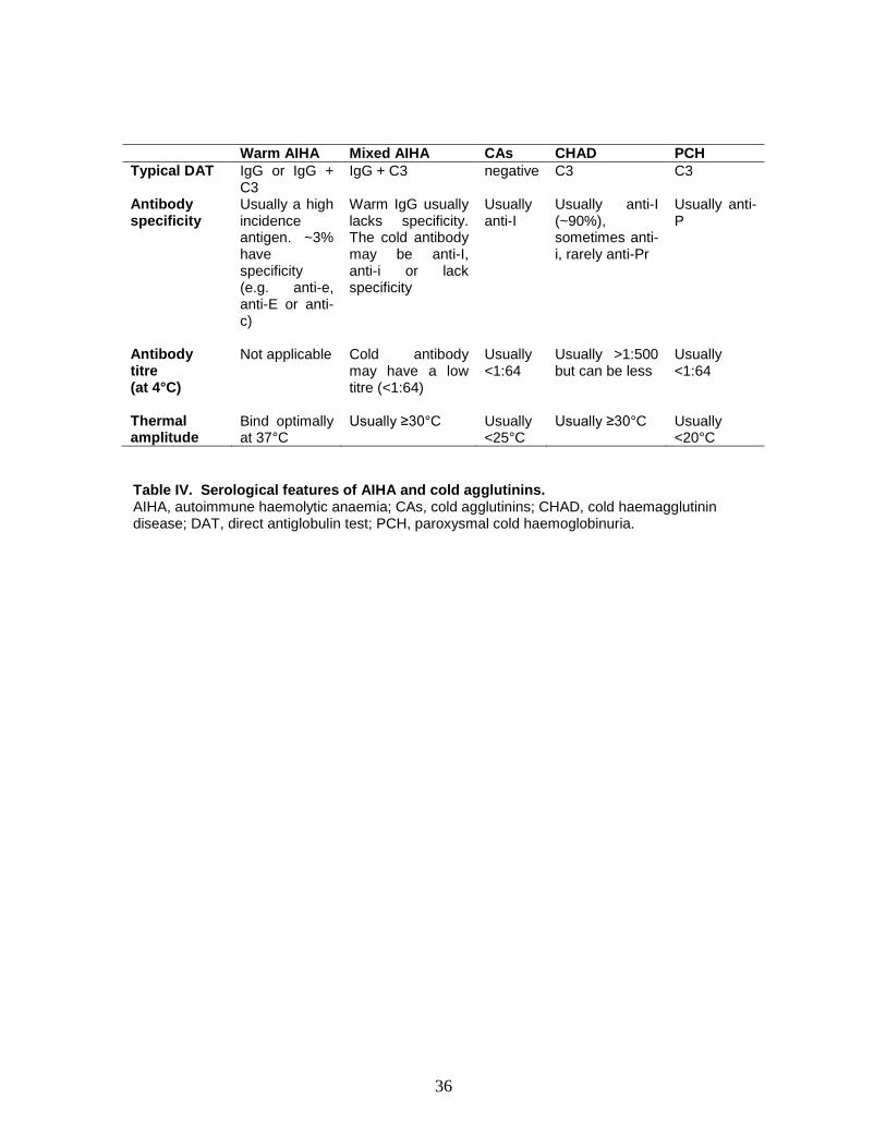
Warm AIHA Mixed AIHA CAs CHAD PCH
Typical DAT IgG or IgG + C3
IgG + C3 negative C3 C3
Antibody specificity
Usually a high incidence antigen. ~3% have specificity (e.g. anti-e, anti-E or anti-c)
Warm IgG usually lacks specificity. The cold antibody may be anti-I, anti-i or lack specificity
Usually anti-I
Usually anti-I (~90%), sometimes anti-i, rarely anti-Pr
Usually anti-P
Antibody titre (at 4°C)
Not applicable Cold antibody may have a low titre (<1:64)
Usually <1:64
Usually >1:500 but can be less
Usually <1:64
Thermal amplitude
Bind optimally at 37°C
Usually ≥30°C Usually <25°C
Usually ≥30°C Usually <20°C
Table IV. Serological features of AIHA and cold agglutinins. AIHA, autoimmune haemolytic anaemia; CAs, cold agglutinins; CHAD, cold haemagglutinin disease; DAT, direct antiglobulin test; PCH, paroxysmal cold haemoglobinuria.

37
negative
positive
positive
negative
yes no
negative
positive
Warm AIHA Cold haemagglutinin
disease
DAT: IgG only
DAggT** screen at RT
DAT: C3 ± IgG
Mixed AIHA Paroxysmal cold
haemoglobinuria
• Age <18 years
• Haemoglobinuria
• Cold associated haemolysis
• Atypical
serological features
Donath‒Landsteiner
Antibody titre
DAT: C3 +
IgG
Titre≥64 Titre <64
Insignificant cold
agglutinin DAT: C3
Further clinical* ± serological† assessment
negative
Evidence of haemolytic
anaemia (HA)
Consider alternative
causes of HA (table II).
If cause unclear, repeat
DAT using column
agglutination method including monospecific
reagents to IgG, M, A and
C3
DAT-negative AIHA
negative
positive
Clinical assessment
AIHA
Alternative causes for HA
and a positive DAT
HTR HDN
DIIHA
Post transplantation
ABO mismatch
PLS Post IVIg
Unrelated cause of HA with
an incidental +ve DAT
Direct antiglobulin test
(DAT)
No alternative cause for
HA and a positive DAT Red cell eluate
Figure 1. Diagnostic pathway for AIHA. AIHA, autoimmune haemolytic anaemia; CHAD, cold haemagglutinin disease; DAggT, direct agglutination test; DAT, direct antiglobulin test; DIIHA, drug-induced immune haemolytic anaemia; HA, haemolytic anaemia; HDN, haemolytic disease of the newborn; HTR, haemolytic transfusion reaction; IVIg, intravenous immunoglobulin; PLS, passenger lymphocyte syndrome; RT, room temperature. * The final diagnosis of CHAD or mixed AIHA is based on the overall clinical picture, including supportive serological findings. † For example the thermal amplitude. ** Saline suspended red cells and patient’s serum at room temperature for 30-60 minutes.
Re-assess
alternatives

38
Yes
If relapse No
No response
If
progression
Cold haemagglutinin
Disease Mixed AIHA Paroxysmal cold
haemoglobinuria Warm AIHA
Supportive care*
Steroids if severe
or persistent
disease
Consider
treatment as per
warm AIHA Symptomatic
anaemia
Severe
circulatory
symptoms
Transfusion
dependence
Mild chronic
anaemia
Supportive
care*
1) Rituximab
or
2) Consider adding fludarabine if clonal
disorder
- Prednisolone
1 mg/kg/day (adult),
1‒2 mg/kg/day (paediatric)
- Folic acid
- Ca2+/Vit D - Risk assess for
PPI, bisphosphonate
and LMWH
Rituximab
3rd line options
- Azathioprine
- Ciclosporin - Danazol
- Mycophenolate
mofetil - Splenectomy
Response ≤d 21
Reduce
prednisolone e.g.
in adults to 20-30 mg over 4-6
weeks, then by 5
mg every month
Figure 2. Therapeutic pathway for primary AIHA Ca2+/Vit D, Calcium/Vitamin D bone prophylaxis; ≤d 21, within 21 days; FBC, full blood count; LMWH, low molecular weight heparin; PPI, proton pump inhibitor. *keep warm, avoid active cooling, folic acid, monitor FBC +/- transfusion

39
References
Ahn,Y.S. (1990) Efficacy of danazol in hematologic disorders. Acta Haematol., 84, 122-
129.
Ahn,Y.S., Harrington,W.J., Mylvaganam,R., Ayub,J., & Pall,L.M. (1985) Danazol
therapy for autoimmune hemolytic anemia. Ann Intern.Med, 102, 298-301.
Akpek,G., McAneny,D., & Weintraub,L. (1999) Comparative response to splenectomy in
Coombs-positive autoimmune hemolytic anemia with or without associated
disease. American Journal of Hematology, 61, 98-102.
Aladjidi,N., Leverger,G., Leblanc,T., Picat,M.Q., Michel,G., Bertrand,Y., Bader-
Meunier,B., Robert,A., Nelken,B., Gandemer,V., Savel,H., Stephan,J.L.,
Fouyssac,F., Jeanpetit,J., Thomas,C., Rohrlich,P., Baruchel,A., Fischer,A.,
Chene,G., Perel,Y., & Centre de Reference National des Cytopenies Auto-
immunes de l'Enfant (CEREVANCE) (2011) New insights into childhood
autoimmune hemolytic anemia: a French national observational study of 265
children. Haematologica, 96, 655-663.
Allgood,J.W. & Chaplin,H., Jr. (1967) Idiopathic acquired autoimmune hemolytic
anemia. A review of forty-seven cases treated from 1955 through 1965. Am.J
Med, 43, 254-273.
Barcellini,W., Fattizzo,B., Zaninoni,A., Radice,T., Nichele,I., Di,B.E., Lunghi,M.,
Tassinari,C., Alfinito,F., Ferrari,A., Leporace,A.P., Niscola,P., Carpenedo,M.,
Boschetti,C., Revelli,N., Villa,M.A., Consonni,D., Scaramucci,L., De,F.P.,
Tagariello,G., Gaidano,G., Rodeghiero,F., Cortelezzi,A., & Zanella,A. (2014)
Clinical heterogeneity and predictors of outcome in primary autoimmune
hemolytic anemia: a GIMEMA study of 308 patients. Blood, 124, 2930-2936.
Barcellini,W., Zaja,F., Zaninoni,A., Imperiali,F.G., Battista,M.L., Di,B.E., Fattizzo,B.,
Consonni,D., Cortelezzi,A., Fanin,R., & Zanella,A. (2012) Low-dose rituximab in
adult patients with idiopathic autoimmune hemolytic anemia: clinical efficacy and
biologic studies. Blood, 119, 3691-3697.
Bay,A., Yilmaz,N., Nalbantoglu,O., Yilmaz,C., Etlik,O., & Faik,O.A. (2007) Multiple
brain abscesses in a child with autoimmune hemolytic anemia. Pediatric Blood &
Cancer, 49, 1034-1036.
Berentsen,S. (2011) How I manage cold agglutinin disease. [Review]. British Journal of
Haematology, 153, 309-317.
Berentsen,S., Randen,U., Vagan,A.M., Hjorth-Hansen,H., Vik,A., Dalgaard,J.,
Jacobsen,E.M., Thoresen,A.S., Beiske,K., & Tjonnfjord,G.E. (2010) High

40
response rate and durable remissions following fludarabine and rituximab
combination therapy for chronic cold agglutinin disease. Blood, 116, 3180-3184.
Berentsen,S. & Tjonnfjord,G.E. (2012) Diagnosis and treatment of cold agglutinin
mediated autoimmune hemolytic anemia. Blood Rev, 26, 107-115.
Berentsen,S., Tjonnfjord,G.E., Brudevold,R., Gjertsen,B.T., Langholm,R., Lokkevik,E.,
Sorbo,J.H., & Ulvestad,E. (2001) Favourable response to therapy with the anti-
CD20 monoclonal antibody rituximab in primary chronic cold agglutinin disease.
British Journal of Haematology, 115, 79-83.
Berentsen,S., Ulvestad,E., Gjertsen,B.T., Hjorth-Hansen,H., Langholm,R., Knutsen,H.,
Ghanima,W., Shammas,F.V., & Tjonnfjord,G.E. (2004) Rituximab for primary
chronic cold agglutinin disease: a prospective study of 37 courses of therapy in 27
patients. Blood, 103, 2925-2928.
Berentsen,S., Ulvestad,E., Langholm,R., Beiske,K., Hjorth-Hansen,H., Ghanima,W.,
Sorbo,J.H., & Tjonnfjord,G.E. (2006) Primary chronic cold agglutinin disease: a
population based clinical study of 86 patients. Haematologica, 91, 460-466.
Birgens,H., Frederiksen,H., Hasselbalch,H.C., Rasmussen,I.H., Nielsen,O.J., Kjeldsen,L.,
Larsen,H., Mourits-Andersen,T., Plesner,T., Ronnov-Jessen,D., Vestergaard,H.,
Klausen,T.W., & Schollkopf,C. (2013) A phase III randomized trial comparing
glucocorticoid monotherapy versus glucocorticoid and rituximab in patients with
autoimmune haemolytic anaemia. Br.J Haematol., 163, 393-399.
Bracken,C.A., Gurkowski,M.A., Naples,J.J., Smith,H., Steinmann,A., Samuel,J.,
Strickler,F.R., VanDenburgh,J., Sheikh,F., & Lumb,P. (1993) Case 6--1993.
Cardiopulmonary bypass in two patients with previously undetected cold
agglutinins. Journal of Cardiothoracic & Vascular Anesthesia, 7, 743-749.
Buchanan,G.R., Boxer,L.A., & Nathan,D.G. (1976) The acute and transient nature of
idiopathic immune hemolytic anemia in childhood. Journal of Pediatrics, 88,
780-783.
Bussone,G., Ribeiro,E., Dechartres,A., Viallard,J.F., Bonnotte,B., Fain,O., Godeau,B., &
Michel,M. (2009) Efficacy and safety of rituximab in adults' warm antibody
autoimmune haemolytic anemia: retrospective analysis of 27 cases. American
Journal of Hematology, 84, 153-157.
Carson,K.R., Beckwith,L.G., & Mehta,J. (2010) Successful treatment of IgM-mediated
autoimmune hemolytic anemia with bortezomib. Blood, 115, 915.
Carson,K.R., Evens,A.M., Richey,E.A., Habermann,T.M., Focosi,D., Seymour,J.F.,
Laubach,J., Bawn,S.D., Gordon,L.I., Winter,J.N., Furman,R.R., Vose,J.M.,
Zelenetz,A.D., Mamtani,R., Raisch,D.W., Dorshimer,G.W., Rosen,S.T., Muro,K.,
Gottardi-Littell,N.R., Talley,R.L., Sartor,O., Green,D., Major,E.O., &
Bennett,C.L. (2009) Progressive multifocal leukoencephalopathy after rituximab

41
therapy in HIV-negative patients: a report of 57 cases from the Research on
Adverse Drug Events and Reports project. Blood, 113, 4834-4840.
Chakravarty,E.F., Murray,E.R., Kelman,A., & Farmer,P. (2011) Pregnancy outcomes
after maternal exposure to rituximab. Blood, 117, 1499-1506.
CHERTKOW,G. & Dacie,J.V. (1956) Results of splenectomy in auto-immune
haemolytic anaemia. Br.J Haematol., 2, 237-249.
D'Arena,G., Califano,C., Annunziata,M., Tartarone,A., Capalbo,S., Villani,O.,
Amendola,G., Pietrantuono,G., Ferrara,F., Pinto,A., Musto,P., D'Arco,A.M., &
Cascavilla,N. (2007) Rituximab for warm-type idiopathic autoimmune hemolytic
anemia: a retrospective study of 11 adult patients. European Journal of
Haematology, 79, 53-58.
Dausset,J. & COLOMBANI,J. (1959) The serology and the prognosis of 128 cases of
autoimmune hemolytic anemia. Blood, 14, 1280-1301.
Davies,J.M., Lewis,M.P., Wimperis,J., Rafi,I., Ladhani,S., & Bolton-Maggs,P.H. (2011)
Review of guidelines for the prevention and treatment of infection in patients with
an absent or dysfunctional spleen: prepared on behalf of the British Committee for
Standards in Haematology by a working party of the Haemato-Oncology task
force. Br.J Haematol., 155, 308-317.
Dussadee,K., Taka,O., Thedsawad,A., & Wanachiwanawin,W. (2010) Incidence and risk
factors of relapses in idiopathic autoimmune hemolytic anemia. J Med
Assoc.Thai., 93 Suppl 1, S165-S170.
Everett,C.M., Dhillon,H., Samarasinghe,D., Berry,L., Warwick,S., & Turner,B. (2006) A
case of cerebral nocardiosis following brief immunosuppression. Eur.J Neurol.,
13, 431-432.
Fayek,M.H., Saad,A.A., Eissa,D.G., Tawfik,L.M., & Kamal,G. (2012) Role of gel test
and flow cytometry in diagnosis of Coombs' negative autoimmune haemolytic
anaemia. Int J Lab Hematol., 34, 311-319.
Fischer,G.D., Claypoole,V., & Collard,C.D. (1997) Increased pressures in the retrograde
blood cardioplegia line: an unusual presentation of cold agglutinins during
cardiopulmonary bypass. Anesthesia & Analgesia, 84, 454-456.
Flores,G., Cunningham-Rundles,C., Newland,A.C., & Bussel,J.B. (1993) Efficacy of
intravenous immunoglobulin in the treatment of autoimmune hemolytic anemia:
results in 73 patients. American Journal of Hematology, 44, 237-242.
Gehrs,B.C. & Friedberg,R.C. (2002) Autoimmune hemolytic anemia. American Journal
of Hematology, 69, 258-271.

42
Gottstein,R. & Cooke,R.W. (2003) Systematic review of intravenous immunoglobulin in
haemolytic disease of the newborn. Arch Dis.Child Fetal Neonatal Ed, 88, F6-10.
Grant,I.R., Parsons,S.W., Johnstone,J.M., & Wood,J.K. (1988) Elective splenectomy in
haematological disorders. Annals of the Royal College of Surgeons of England,
70, 29-33.
Grossman,J.M., Gordon,R., Ranganath,V.K., Deal,C., Caplan,L., Chen,W., Curtis,J.R.,
Furst,D.E., McMahon,M., Patkar,N.M., Volkmann,E., & Saag,K.G. (2010)
American College of Rheumatology 2010 recommendations for the prevention
and treatment of glucocorticoid-induced osteoporosis. Arthritis Care Res
(Hoboken.), 62, 1515-1526.
Gueli,A., Gottardi,D., Hu,H., Ricca,I., De,C.A., & Tarella,C. (2013) Efficacy of
rituximab-bendamustine in cold agglutinin haemolytic anaemia refractory to
previous chemo-immunotherapy: a case report. Blood Transfus., 11, 311-314.
Habibi,B., Homberg,J.C., Schaison,G., & Salmon,C. (1974) Autoimmune hemolytic
anemia in children. A review of 80 cases. American Journal of Medicine, 56, 61-
69.
Hansen,K.E., Wilson,H.A., Zapalowski,C., Fink,H.A., Minisola,S., & Adler,R.A. (2011)
Uncertainties in the prevention and treatment of glucocorticoid-induced
osteoporosis. J Bone Miner.Res, 26, 1989-1996.
Heisel,M.A. & Ortega,J.A. (1983) Factors influencing prognosis in childhood
autoimmune hemolytic anemia. American Journal of Pediatric
Hematology/Oncology, 5, 147-152.
Hendrick,A.M. (2003) Auto-immune haemolytic anaemia--a high-risk disorder for
thromboembolism?. Hematology, 8, 53-56.
Issaragrisil,S. & Kruatrachue,M. (1983) An association of pregnancy and autoimmune
haemolytic anaemia. Scandinavian Journal of Haematology, 31, 63-68.
Issitt,P.D. (1985) Serological Diagnosis and Characterization of the Causative
Autoantibodies. Immune Hemolytic Anemias (ed. by H. Chaplin, Jr.), pp. 1-47.
Churchill Livingstone.
Jain,M.D., Cabrerizo-Sanchez,R., Karkouti,K., Yau,T., Pendergrast,J.M., & Cserti-
Gazdewich,C.M. (2013) Seek and you shall find--but then what do you do? Cold
agglutinins in cardiopulmonary bypass and a single-center experience with cold
agglutinin screening before cardiac surgery. Transfus.Med Rev, 27, 65-73.
Klein,N.P., Ray,P., Carpenter,D., Hansen,J., Lewis,E., Fireman,B., Black,S., Galindo,C.,
Schmidt,J., & Baxter,R. (2010) Rates of autoimmune diseases in Kaiser
Permanente for use in vaccine adverse event safety studies. Vaccine, 28, 1062-
1068.

43
Lecouffe-Desprets,M., Neel,A., Graveleau,J., Leux,C., Perrin,F., Visomblain,B.,
Artifoni,M., Masseau,A., Connault,J., Pottier,P., Agard,C., & Hamidou,M. (2015)
Venous thromboembolism related to warm autoimmune hemolytic anemia: a
case-control study. Autoimmun.Rev, 14, 1023-1028.
Lekamwasam,S., Adachi,J.D., Agnusdei,D., Bilezikian,J., Boonen,S., Borgstrom,F.,
Cooper,C., Diez,P.A., Eastell,R., Hofbauer,L.C., Kanis,J.A., Langdahl,B.L.,
Lesnyak,O., Lorenc,R., McCloskey,E., Messina,O.D., Napoli,N., Obermayer-
Pietsch,B., Ralston,S.H., Sambrook,P.N., Silverman,S., Sosa,M., Stepan,J.,
Suppan,G., Wahl,D.A., & Compston,J.E. (2012) A framework for the
development of guidelines for the management of glucocorticoid-induced
osteoporosis. Osteoporos.Int, 23, 2257-2276.
Ly,B. & Albrechtsen,D. (1981) Therapeutic splenectomy in hematologic disorders.
Effects and complications in 221 adult patients. Acta Med Scand, 209, 21-29.
Maggiore,G., Sciveres,M., Fabre,M., Gori,L., Pacifico,L., Resti,M., Choulot,J.J.,
Jacquemin,E., & Bernard,O. (2011) Giant cell hepatitis with autoimmune
hemolytic anemia in early childhood: long-term outcome in 16 children. Journal
of Pediatrics, 159, 127-132.
Manoharan,A. (1987) Danazol therapy in patients with immune cytopenias. Australian &
New Zealand Journal of Medicine, 17, 613-614.
Maung,S.W., Leahy,M., O'Leary,H.M., Khan,I., Cahill,M.R., Gilligan,O., Murphy,P.,
McPherson,S., Jackson,F., Ryan,M., Hennessy,B., McHugh,J., Goodyer,M.,
Bacon,L., O'Gorman,P., Nee,A., O'Dwyer,M., Enright,H., Saunders,J., &
O'Keeffe,D. (2013) A multi-centre retrospective study of rituximab use in the
treatment of relapsed or resistant warm autoimmune haemolytic anaemia. Br.J
Haematol., 163, 118-122.
Miano,M., Scalzone,M., Perri,K., Palmisani,E., Caviglia,I., Micalizzi,C., Svahn,J.,
Calvillo,M., Banov,L., Terranova,P., Lanza,T., Dufour,C., & Fioredda,F. (2015)
Mycophenolate mofetil and Sirolimus as second or further line treatment in
children with chronic refractory Primitive or Secondary Autoimmune Cytopenias:
a single centre experience. Br.J Haematol., 171, 247-253.
Milkins,C., Berryman,J., Cantwell,C., Elliott,C., Haggas,R., Jones,J., Rowley,M.,
Williams,M., & Win,N. (2013) Guidelines for pre-transfusion compatibility
procedures in blood transfusion laboratories. British Committee for Standards in
Haematology. Transfus.Med, 23, 3-35.
Mohren,M., Markmann,I., Dworschak,U., Franke,A., Maas,C., Mewes,S., Weiss,G., &
Jentsch-Ullrich,K. (2004) Thromboembolic complications after splenectomy for
hematologic diseases. American Journal of Hematology, 76, 143-147.

44
Moyo,V.M., Smith,D., Brodsky,I., Crilley,P., Jones,R.J., & Brodsky,R.A. (2002) High-
dose cyclophosphamide for refractory autoimmune hemolytic anemia. Blood, 100,
704-706.
Naithani,R., Agrawal,N., Mahapatra,M., Kumar,R., Pati,H.P., & Choudhry,V.P. (2007)
Autoimmune hemolytic anemia in children. Pediatric Hematology & Oncology,
24, 309-315.
National Institute for Clinical Excellence. Guidance on cancer services: improving
outcomes in haematological cancer. http://guidance.nice.org.uk/CSGHO . 2003.
National Institute for Clinical Excellence. 12-5-2014.
Ref Type: Electronic Citation
Packman,C.H. (2008) Hemolytic anemia due to warm autoantibodies. Blood Reviews, 22,
17-31.
Passweg,J.R. & Rabusin,M. (2008) Hematopoetic stem cell transplantation for immune
thrombocytopenia and other refractory autoimmune cytopenias. Autoimmunity,
41, 660-665.
Petz,L.D. & Garratty,G. (1980) Acquired Immune Hemolytic Anemias, First edn,
Churchill Livingstone Inc.
Petz,L.D. (2004) Review: evaluation of patients with immune hemolysis.
Immunohematology, 20, 167-176.
Petz,L.D. (2008) Cold antibody autoimmune hemolytic anemias. Blood Reviews, 22, 1-
15.
Pirofsky,B. (1975) Immune haemolytic disease: the autoimmune haemolytic anaemias.
Clin Haematol., 4, 167-180.
Porter,T.F., Silver,R.M., Jackson,G.M., Branch,D.W., & Scott,J.R. (1997) Intravenous
immune globulin in the management of severe Rh D hemolytic disease. Obstet
Gynecol.Surv., 52, 193-197.
Pretlove,S.J., Fox,C.E., Khan,K.S., & Kilby,M.D. (2009) Noninvasive methods of
detecting fetal anaemia: a systematic review and meta-analysis. BJOG, 116, 1558-
1567.
Provan,D., Butler,T., Evangelista,M.L., Amadori,S., Newland,A.C., & Stasi,R. (2007)
Activity and safety profile of low-dose rituximab for the treatment of autoimmune
cytopenias in adults. Haematologica, 92, 1695-1698.
Public Health England. Immunisation against infectious disease. Salisbury, D and
Ramsey, M. https://www.gov.uk/government/collections/immunisation-against-
infectious-disease-the-green-book . 3-18-2014.
Ref Type: Electronic Citation

45
Reynaud,Q., Durieu,I., Dutertre,M., Ledochowski,S., Durupt,S., Michallet,A.S., Vital-
Durand,D., & Lega,J.C. (2015) Efficacy and safety of rituximab in auto-immune
hemolytic anemia: A meta-analysis of 21 studies. Autoimmun.Rev, 14, 304-313.
Rivero,S.J., Alger,M., & arcon-Segovia,D. (1979) Splenectomy for hemocytopenia in
systemic lupus erythematosus. A controlled appraisal. Arch Intern.Med, 139, 773-
776.
Rizzoli,R., Adachi,J.D., Cooper,C., Dere,W., Devogelaer,J.P., ez-Perez,A., Kanis,J.A.,
Laslop,A., Mitlak,B., Papapoulos,S., Ralston,S., Reiter,S., Werhya,G., &
Reginster,J.Y. (2012) Management of glucocorticoid-induced osteoporosis.
Calcif.Tissue Int, 91, 225-243.
Roche Products Ltd. Summary of Product Characteristics: Mabthera.
https://www.medicines.org.uk/emc/ . 1-4-2014.
Ref Type: Electronic Citation
Roth,A., Huttmann,A., Rother,R.P., Duhrsen,U., & Philipp,T. (2009) Long-term efficacy
of the complement inhibitor eculizumab in cold agglutinin disease. Blood, 113,
3885-3886.
Roumier,M., Loustau,V., Guillaud,C., Languille,L., Mahevas,M., Khellaf,M., Limal,N.,
Noizat-Pirenne,F., Godeau,B., & Michel,M. (2014) Characteristics and outcome
of warm autoimmune hemolytic anemia in adults: New insights based on a single-
center experience with 60 patients. Am.J Hematol., 89, E150-E155.
Sachs,U.J., Roder,L., Santoso,S., & Bein,G. (2006) Does a negative direct antiglobulin
test exclude warm autoimmune haemolytic anaemia? A prospective study of 504
cases. British Journal of Haematology, 132, 655-656.
Schollkopf,C., Kjeldsen,L., Bjerrum,O.W., Mourits-Andersen,H.T., Nielsen,J.L.,
Christensen,B.E., Jensen,B.A., Pedersen,B.B., Taaning,E.B., Klausen,T.W., &
Birgens,H. (2006) Rituximab in chronic cold agglutinin disease: a prospective
study of 20 patients. Leukemia & lymphoma, 47, 253-260.
Shulman,I.A., Branch,D.R., Nelson,J.M., Thompson,J.C., Saxena,S., & Petz,L.D. (1985)
Autoimmune hemolytic anemia with both cold and warm autoantibodies. JAMA,
253, 1746-1748.
Sokol,R.J., Booker,D.J., & Stamps,R. (1999) Erythropoiesis: Paroxysmal Cold
Haemoglobinuria: A Clinico-Pathological Study of Patients with a Positive
Donath-Landsteiner Test. Hematology, 4, 137-164.
Sokol,R.J., Hewitt,S., & Stamps,B.K. (1982) Erythrocyte autoantibodies, autoimmune
haemolysis and pregnancy. Vox Sanguinis, 43, 169-176.
Sokol,R.J., Hewitt,S., & Stamps,B.K. (1983) Autoimmune hemolysis: mixed warm and
cold antibody type. Acta Haematologica, 69, 266-274.
46
Sokol,R.J., Hewitt,S., Stamps,B.K., & Hitchen,P.A. (1984) Autoimmune haemolysis in
childhood and adolescence. Acta Haematologica, 72, 245-257.
Swiecicki,P.L., Hegerova,L.T., & Gertz,M.A. (2013) Cold agglutinin disease. Blood,
122, 1114-1121.
Szczepiorkowski,Z.M., Winters,J.L., Bandarenko,N., Kim,H.C., Linenberger,M.L.,
Marques,M.B., Sarode,R., Schwartz,J., Weinstein,R., & Shaz,B.H. (2010)
Guidelines on the use of therapeutic apheresis in clinical practice--evidence-based
approach from the Apheresis Applications Committee of the American Society
for Apheresis. J Clin Apher., 25, 83-177.
Thabet,A.F. & Faisal,M. (2014) Pulse cyclophosphamide therapy in refractory warm
autoimmune hemolytic anemia: a new perspective. Indian J Hematol.Blood
Transfus., 30, 313-318.
Thomsen,R.W., Schoonen,W.M., Farkas,D.K., Riis,A., Fryzek,J.P., & Sorensen,H.T.
(2010) Risk of venous thromboembolism in splenectomized patients compared
with the general population and appendectomized patients: a 10-year nationwide
cohort study. J Thromb.Haemost., 8, 1413-1416.
van't,R.M., Burger,J.W., van Muiswinkel,J.M., Kazemier,G., Schipperus,M.R., &
Bonjer,H.J. (2000) Diagnosis and treatment of portal vein thrombosis following
splenectomy. Br.J Surg, 87, 1229-1233.
Von,B.H. (2003) Plasmapheresis in immune hematology: review of clinical outcome data
with respect to evidence-based medicine and clinical experience.
Ther.Apher.Dial., 7, 127-140.
Ware,R.E. & Rosse,W.F. (1998) Auto-immune hemolytic anaemia. Nathan and Oski's
Hematology of Infancy and Childhood (ed. by D. G. Nathan, S. H. Orkin, & F. A.
Oski), p. 509. W B Saunders Co.
Weinstein,R.S. (2011) Clinical practice. Glucocorticoid-induced bone disease. N Engl J
Med, 365, 62-70.
Wimperis,J., Lunn,M., Jones,A., Herriot,R., Wood,P., O'Shaughnessy,D., & Qualie,M.
Clinical guidelines for immunoglobulin use.
https://www.gov.uk/government/publications/clinical-guidelines-for-
immunoglobulin-use-second-edition-update [Second]. 1-8-2011. 6-1-2013.
Ref Type: Electronic Citation
Worlledge,S.M., Brain,M.C., Cooper,A.C., Hobbs,J.R., & Dacie,J.V. (1968)
Immmunosuppressive drugs in the treatment of autoimmune haemolytic anaemia.
Proceedings of the Royal Society of Medicine, 61, 1312-1315.

47
Supplementary appendix 1: systematic review methodology for the 2016 guideline
on diagnosis and management of primary autoimmune haemolytic anaemia
The first systematic review of Medline and Embase was from 1960 to 24/01/2012 (search
strategy is outlined below). The search was limited to English language peer
reviewed publications and conference abstracts were not systematically reviewed.
All 4316 Medline and 6454 Embase titles were reviewed for relevance. The
DARE database was searched in August 2012 using the MeSH descriptor terms
anaemia, autoimmune and haemolytic. The COCHRANE database of systematic
reviews was also searched in August 2012. Some key studies published prior to
1960 were also been reviewed. The main search was also supplemented by
searches of Pubmed on specific topics including pregnancy and the prevention of
glucocorticoid induced osteoporosis.
The systematic review of Medline and Embase was updated 15/11/2015 using the same
search strategy. The DARE database and the COCHRANE database of
systematic reviews were also searched but no new documents identified.
Medline search (24/01/2012)
1. exp Anemia, Hemolytic, Autoimmune/
2. limit 1 to (english language and yr="1960 -Current")
3. autoimmune h?emolytic an?emia.mp. [mp=title, abstract, original title, name of
substance word, subject heading word, protocol supplementary concept, rare
disease supplementary concept, unique identifier]
4. limit 3 to (english language and yr="1960 -Current")
5. (autoimmune adj3 an?emia).mp. [mp=title, abstract, original title, name of substance
word, subject heading word, protocol supplementary concept, rare disease
supplementary concept, unique identifier]
6. limit 5 to (english language and yr="1960 -Current")
7. cold agglutinin?.mp. [mp=title, abstract, original title, name of substance word, subject
heading word, protocol supplementary concept, rare disease supplementary
concept, unique identifier]
8. limit 7 to (english language and yr="1960 -Current")
9. 2 or 4 or 6 or 8 (number of documents retrieved = 4316)
10. limit 9 to "all child (0 to 18 years)" (number of documents retrieved = 960)
Embase search (24/01/2012)
1. autoimmune h?emolytic an?emia.mp. [mp=title, abstract, subject headings, heading
word, drug trade name, original title, device manufacturer, drug manufacturer,
device trade name, keyword]
2. limit 1 to (english language and yr="1960 -Current")
3. (autoimmune adj3 an?emia).mp. [mp=title, abstract, subject headings, heading word,
drug trade name, original title, device manufacturer, drug manufacturer, device
trade name, keyword]

48
4. limit 3 to (english language and yr="1960 -Current")
5. cold agglutinin?.mp. [mp=title, abstract, subject headings, heading word, drug trade
name, original title, device manufacturer, drug manufacturer, device trade name,
keyword]
6. limit 5 to (english language and yr="1960 -Current")
7. exp autoimmune hemolytic anemia/
8. limit 7 to (english language and yr="1960 -Current")
9. 2 or 4 or 6 or 8 (number of documents retrieved = 6454)
10. limit 9 to child (number of documents retrieved = 542)