the cytoskeleton coordinates the early events of b-cell...
TRANSCRIPT

The Cytoskeleton Coordinates the Early Eventsof B-cell Activation
Naomi E. Harwood and Facundo D. Batista
Lymphocyte Interaction Laboratory, Cancer Research UK London Research Institute, Lincoln’s Inn FieldsLaboratories, London WC2A 3LY, United Kingdom
Correspondence: [email protected]
B cells contribute to protective adaptive immune responses through generation of antibodiesand long-lived memory cells, following engagement of the B-cell receptor (BCR) withspecific antigen. Recent imaging investigations have offered novel insights into the ensuingmolecular and cellular events underlying B-cell activation. Following engagement withantigen, BCR microclusters form and act as sites of active signaling through the recruitmentof intracellular signaling molecules and adaptors. Signaling through these “microsignalo-somes” is propagated and enhanced through B-cell spreading in a CD19-dependentmanner. Subsequently, the mature immunological synapse is formed, and functions as a plat-form forantigen internalization, enabling the antigen presentation to helper T cells required formaximal B-cell activation. In this review, we discuss the emerging and critical role for the cyto-skeleton in the coordination and regulation of these molecular events during B-cell activation.
An individual is continually bombarded bypotentially disease-causing agents. However,
under normal circumstances, the action of theimmune system ensures that these encountersrelatively infrequently result in the developmentof symptomatic illness. The immune system canbe broadly divided into two component parts,the innate and the adaptive branches. The innatebranch mediates rapid inflammatory responsesfollowing the recognition of motifs typicallyassociated with pathogens through a collectionof pattern recognition receptors. In contrast,adaptive immune responses tend to emerge afew days after initial infection and show fourcentral characteristics: memory, specificity, di-versity, and self-nonself discrimination. In spiteof these two divisions, the effective elimination
of pathogens most often requires intricatecollaboration between the innate and adaptiveimmune responses.
Small white blood cells, known as lympho-cytes, are the fundamental participants media-ting adaptive immune responses. Lymphocytesoriginate from hematopoietic stem cells in thebone marrow and although a subset migrateto the thymus to form T cells, others remainin the bone marrow to complete their develop-ment into B cells (Halin et al. 2005). Maturelymphocytes circulate throughout the bodyand are often localized in secondary lymphoidorgans such as the lymph nodes and the spleen,which are specialized sites for lymphocyte acti-vation (Junt et al. 2008). T cells are responsiblefor cell-mediated immunity and are commonly
Editors: Lawrence E. Samelson and Andrey S. Shaw
Additional Perspectives on Immunoreceptor Signaling available at www.cshperspectives.org
Copyright # 2011 Cold Spring Harbor Laboratory Press; all rights reserved; doi: 10.1101/cshperspect.a002360
Cite this article as Cold Spring Harb Perspect Biol 2011;3:a002360
1
on June 5, 2018 - Published by Cold Spring Harbor Laboratory Press http://cshperspectives.cshlp.org/Downloaded from

classified into CD4þ helper and CD8þ cytotoxiccells according to their expression of surfacecoreceptors. On the other hand, B cells mediatehumoral immunity through the secretion of anti-bodies that recognize and neutralize invadingpathogens.
To become activated to produce antibodies,B cells must first recognize specific antigenthrough the B-cell receptor (BCR). This specificbindingevent initiates intracellularsignaling lead-ing to altered gene expression, reorganization ofthe B-cell cytoskeleton, and antigen internaliza-tion. Importantly, BCR-mediated internalizationtargets antigen to endosomes containing newlysynthesized major histocompatibility complex(MHC) (Aluvihare et al. 1997; Amigorena et al.1994), such that processed antigen can be pre-sented to CD4þ T cells, thereby recruiting helpto facilitate maximal B-cell activation (Lanzavec-chia 1985; Rock et al. 1984). Activated B cells caneither rapidly mediate the secretion of lowaffinityantibodies (MacLennan et al. 2003), or can enterinto a specialized structure known as a germinalcenter (GC) to undergo affinity maturation, pro-ducing plasma cells capable of high-affinity anti-body production and long-lasting memory cells(MacLennan 1994; Rajewsky 1996).
The molecular events underlying BCR-me-diated signaling have historically been charac-terized using standard biochemical analysismethods in vitro. As such, the BCR has beenidentified as comprising of a membrane im-munoglobulin (mIg) responsible for bindingextracellular antigen, in complex with an Iga/b sheath containing immunoreceptor tyrosineactivation motifs (ITAMs) in the intracellulardomains (Reth 1989). Cross-linking of the BCRby multivalent antigen triggers phosphoryla-tion of the ITAMs through Src family kinasessuch as Lyn and Syk. This early phosphorylationleads to the recruitment of intracellular effectorsincluding PLCg2, Vav, Btk, and PI3K, and adap-tors including Blnk and Grb2, to form a multi-component assembly known as the signalosome(Dal Porto et al. 2004; DeFranco 1997; Kurosaki2002; Scharenberg et al. 2007). Cellular readoutsof the coordinated activity of the signalosomeinclude calcium signaling and activation oftranscription factors such as NF-kB. Although
these classical strategies have provided an essen-tial foundation of intracellular mediators in-volved in signaling downstream of the BCR,they contribute little insight into the spatiotem-poral dynamics and organization of molecularevents within the cell (Treanor and Batista2007).
To address the questions of how and wheremolecular events occur within the cell, imag-ing-based strategies have been developed andapplied to visualize directly molecular eventsunderlying B-cell activation. Importantly, theseapproaches not only allow verification of mo-lecular pathways identified previously, but alsoshed light on the spatiotemporal dynamics ofthese pathways within the cell. In addition,imaging approaches have led to the identifica-tion of unexpected and novel roles for moleculessuch as CD19 in mounting B-cell responsesto antigen. An essential tenet that has beenrevealed by these imaging studies is the impor-tance of molecular segregation as a means ofcoordinating and regulating interactions withinthe cell. Indeed, a pivotal role for the cytoskel-eton as a means of mediating segregation isemerging, offering a versatile mechanism forthe regulation of numerous molecular interac-tions. The importance of the cytoskeleton is evi-denced by the recent observation that mutationsin various cytoskeleton regulators are associatedwith the development of antibody deficiencysyndromes in humans (Conley et al. 2009). Inthis review, we discuss the contribution thatrecent imaging investigations have made to ourunderstanding of the process and regulation ofB-cell activation. In particular, we highlightthe central role for the cytoskeleton in the orga-nization and control of molecular events duringB-cell activation.
BCR DISTRIBUTION IN THE RESTING B CELLIS SHAPED BY THE CYTOSKELETON
As activation is initiated following cross-linkingof the BCR by antigen, it is important to charac-terize the distribution and dynamics of the BCRin the resting state to fully appreciate the mo-lecular events regulating the initiation of B-cellactivation. In view of the fact that multivalent,
N.E. Harwood and F.D. Batista
2 Cite this article as Cold Spring Harb Perspect Biol 2011;3:a002360
on June 5, 2018 - Published by Cold Spring Harbor Laboratory Press http://cshperspectives.cshlp.org/Downloaded from

but not monovalent, soluble antigens triggeractivation (S. Minguet, M. Reth, W. Schamel,pers. comm.; Tolar et al. 2009), it was widelyassumed that the BCR exists as a monomer inresting B cells. However, as native gel electropho-resis showed that BCRs are able to interact withone another, it was suggested that BCRs formoligomers on the surface of the resting B cell(Schamel and Reth 2000). On the contrary, a flu-orescence resonance energy transfer approachwas unable to detect an interaction between indi-vidual BCRs in the membrane, indicating thatBCRs are monomeric in the absence of antigen(Tolar et al. 2005). Thus, the precise state of theBCR in the membrane before antigen stimulationremains controversial.
Recently, we have sought to visualize thedynamics of BCR on the surface of the restingB cells using Dual-View total internal reflectionmicroscopy (TIRFM) (Treanor et al. 2010) (Fig.1A). Using this method, we were able to tracksingle particles of BCR in the resting B-cell
membrane. We observed that BCRs were notable to diffuse freely in the plane of the mem-brane and instead show a range of diffusionbehaviors, with a proportion of receptorsmuch less mobile (diffusion coefficient lessthan ,0.01 um2s21). This restriction in diffu-sion is contrary to the predictions of the originalfluid-mosaic model proposed by Singer andNicholson (1972); however, it is in line withmore recent investigations tracking single mole-cules of transmembrane proteins in fibroblasts(Kusumi et al. 2005; Sako and Kusumi 1994;Simson et al. 1995). Indeed, these observationsled the authors to suggest that the cell mem-brane contains discrete confinement zonesaround 30–700 nm in diameter that restrictlong-range diffusion between adjacent zones.This is known as the “picket-fence” model, inwhich selected transmembrane proteins act aspickets defining confinement zones and areattached to the underlying cytoskeleton fence(Fujiwara et al. 2002). Furthermore, it has
Laser beam
CCD camera
Objective lens
Beam splitters
Coverslip & specimen
Analysis computer
A B
BCR
Actin-ezrin network
Mobile receptor track
Immobile receptor track
Figure 1. Dual-View TIRFM for simultaneous tracking of BCR alongside the actin cytoskeleton. (A) Schematicrepresentation of the setup of the Dual-View TIRFM. The total internal reflection of incoming laser beam (pur-ple) generates evanescent waves capable of exciting flurophores in close proximity to the sample interface. Thetwo colored emission beams (green and red) are passed through beam splitters, detected simultaneously by aCCD camera and overlaid in an analysis computer. (B) Snapshot of a movie collected tracking single moleculesof BCR (red, anti-BCR Fab) and actin (green, LifeAct a marker of filamentous actin). BCRs located in actin-richareas are more restricted in their diffusion (yellow tracks in schematic zoom), whereas BCRs located inactin-poor areas tend to be less restricted in their diffusion (blue tracks in schematic zoom).
Cytoskeleton Regulates B-cell Activation
Cite this article as Cold Spring Harb Perspect Biol 2011;3:a002360 3
on June 5, 2018 - Published by Cold Spring Harbor Laboratory Press http://cshperspectives.cshlp.org/Downloaded from

been suggested that lipid microdomains and/orprotein islands may contribute to the observedrestriction in diffusion of transmembrane pro-teins (Lillemeier et al. 2006; Simons and Ikonen1997). Interestingly, we have used our method-ology to simultaneously visualize the BCRalongside components of the cytoskeleton, andshowed that slow-moving or immobile BCRswere often co-incident with regions with highdensity of an ezrin-defined actin network (Trea-nor et al. 2010) (Fig. 1B). Indeed, disruption ofthe cytoskeleton network through pharmaco-logical agents results in intracellular signalingsimilar to that observed following antigenstimulation, likely as a result of BCR cluster for-mation. Thus, it appears that the cytoskeletonplays an important role in regulating the distri-bution and dynamics of the BCR in the restingB cell.
RECOGNITION OF ANTIGEN TRIGGERSB-CELL ACTIVATION BY AN UNKNOWNMECHANISM
The BCR can recognize and respond to nativeantigens both in soluble or membrane-boundforms, though the latter have a lower thresholdand thus represent a more effective means oftriggering B-cell activation (Batista et al. 2001;Batista and Neuberger 1998, 2000). Althoughit has been known for some time that dendriticcells (Huang et al. 2005; Wykes et al. 1998), fol-licular dendritic cells (Chen et al. 1978; Mandelet al. 1980; Tew et al. 1980), and macrophages(Koppel et al. 2005) can present intact antigento B cells in vitro, very recent multiphotonmicroscopy investigations have verified thephysiological significance of antigen on the sur-face of presenting cells in mediating B-cell acti-vation in vivo (Carrasco and Batista 2007; Juntet al. 2007; Phan et al. 2007; Phan et al. 2009;Qi et al. 2006; Suzuki et al. 2009). In view ofthe predominance of membrane-bound anti-gen in initiating activation, the original hypoth-eses developed to describe BCR triggering inresponse to soluble antigen stimulation mustbe re-examined. Indeed, it has been observedthat monovalent antigen attached to the surfaceof a presenting cell, unlike monovalent soluble
antigen, can trigger B-cell activation (Tolar et al.2009). Furthermore, the additional constraintsthat are imposed on the B cell to recognize anti-gen immobilized on the surface of a presentingcell must be considered. As T cells absolutelyrequire the recognition of antigen in complexwith MHC on the surface of a presenting cell,models developed to describe the initiation ofT-cell activation may prove useful in the deriva-tion of a general description of activation of Blymphocytes.
As the modules responsible for ligand bind-ing and intracellular signaling in immunorecep-tors such as the BCR and TCR are separated,the molecular mechanism by which the externalbinding of antigen is communicated across themembrane is not immediately obvious. Indeed,this has been the subject of a number of inves-tigations and still remains controversial. Twomost likely models have emerged to explainthe triggering of activation in T cells (Fig. 2).The first of these is perhaps the more simpleconceptually and proposes that the binding ofantigen to the extracellular region is commu-nicated to the intracellular signaling regionthrough conformational change(s) in the inter-vening domains (Kuhns et al. 2006; Schamelet al. 2006) (Fig. 2A). Although high-resolutionstatic crystallographic data have not providedevidence in support of this view (reviewed inGarcia et al. 1999; Hennecke and Wiley 2001;Rudolph et al. 2006; van der Merwe 2001), anumber of recent studies have identified confor-mational changes in the CD31 domain of theTCR following ligation such that CD31 ITAMmotifs are exposed to activating Src-family kin-ases and binding of intracellular effectors (Gilet al. 2002; Xu et al. 2008). The second modelis known as kinetic-segregation and explainsthe initiation of T-cell triggering through thealterations in the membrane distribution of in-hibitory phosphatases, such as CD45 (Choud-huri et al. 2005; Davis and van der Merwe2006) (Fig. 2B). This model assumes that, beforeantigen stimulation, the TCR is at equilibriumsuch that it is equally likely to be phosphory-lated or dephosphorylated. However, on contactwith an antigen-presenting cell, small adhesionmolecules such as CD2 on the T-cell surface
N.E. Harwood and F.D. Batista
4 Cite this article as Cold Spring Harb Perspect Biol 2011;3:a002360
on June 5, 2018 - Published by Cold Spring Harbor Laboratory Press http://cshperspectives.cshlp.org/Downloaded from

establish close-contact zones that exclude morebulky cell-surface molecules, including CD45.Given that the extracellular domain of theTCR is relatively small, its diffusion into andout of the close contact zone is not restricted.Accordingly, TCR within the close contact zoneis more likely to be phosphorylated because ofthe size-dependent exclusion of CD45. Whenthe TCR residing in the close contact zone spe-cifically recognizes antigen on the surface ofthe presenting cell, it will be held in this zone,increasing the lifetime of phosphorylated TCR,such that triggering occurs. There are a numberof lines of experimental evidence supporting
this viewpoint, including the importance of thebalance of phosphorylation (Secrist et al. 1993)and the size-dependence of various extracellulardomains for triggering T-cell activation (Chou-dhuri et al. 2005; Irles et al. 2003). Taken together,it seems likely that some features of both the con-formational change and the kinetic-segregationmodels participate during the initiation of TCRtriggering.
Can these two models prove useful in thedescription of analogous processes during B-cellactivation? At this stage, there is no high-resolu-tion structural data that supports a rapid con-formational change to initiate triggering of the
TCR
Peptide loaded MHC
CD58-CD2adhesion pair
CD45(inhibitory bulkyphosphatase)Lck(activatorykinase)
Nck
Recruitment of intracellular effectors & adaptors
Conformational change
P
Close contact zone
Kinetic segregation
Conformationalchange
APC
T cell
Lck
Lck
Lck
Lck
Lck
Lck
Lck
Lck
Lck
Lck
Lck
Lck
APC
APC
APC
T cell
T cell
T cell
APC
T cell
APC
T cell
A B
P
P
Recruitment of intracellular effectors & adaptors
Lck
Figure 2. Two models of T-cell triggering. (A) The conformational change model states that extracellular pMHCbinding to the TCR triggers intracellular conformational changes in the associated CD3 complex, opening upbinding sites for the intracellular adaptor Nck. Subsequently, the “opened” TCR recruits activatory kinases(such as Lck) to phosphorylate the TCR, leading to assembly of the signalosome. (B) The kinetic segregationmodel postulates that, in the resting state, TCR is equally likely to be phosphorylated by activatory kinases ordephosphorylated inhibitory phosphatases. On proximity of an antigen-presenting cell, small adhesion mole-cules such as CD2 and CD58 establish a “close contact zone” that excludes bulky surface molecules such asCD45, but permits diffusion of TCR and Lck. TCR in the “close contact zone” is more likely to be found in aphosphorylated state, and the half-life of phosphorylated TCR in this region is increased following recognitionof specific pMHC, triggering the assembly of the signalosome and T-cell activation.
Cytoskeleton Regulates B-cell Activation
Cite this article as Cold Spring Harb Perspect Biol 2011;3:a002360 5
on June 5, 2018 - Published by Cold Spring Harbor Laboratory Press http://cshperspectives.cshlp.org/Downloaded from

BCR following antigen binding; however, thisdoes not necessarily eliminate the possibilitythat conformational changes may play a rolein the initiation of BCR triggering. Indeed,quantitative FRET imaging has revealed confor-mational changes in the cytoplasmic domainsof the BCR complex that are dependent onLyn recruitment and activity, and thus areassumed to occur later on during BCR signaling(Tolar et al. 2005). In terms of the kinetic-segregation model, it is clear that the balanceof phosphorylation is important for the initia-tion of B-cell activation (Rolli et al. 2002). Inter-estingly, although CD45 is the predominant cell-surface phosphatase in T cells (Mustelin et al.1989), B cells express significant amounts ofanother phosphatase CD148 (Zhu et al. 2008).Thus, though CD45-deficient B cells are not sig-nificantly impaired in their B-cell activation(Byth et al. 1996; Depoil et al. 2008; Kishiharaet al. 1993), it is possible that the bulkier CD148may play a redundant role in size-dependentexclusion during B-cell triggering. As such, itwill be necessary to investigate B cells deficientin CD45 and CD148 to assess the validity of thekinetic-segregation model to describe the initia-tion of BCR triggering. Future imaging investi-gations will provide insight into the dynamicdistribution of various molecules in the B-cellmembrane, and thus shed light on the precisemechanism underlying BCR triggering.
BCR MICROCLUSTERS FORM AND MEDIATEINTRACELLULAR SIGNALING AFTERANTIGEN STIMULATION
Although imaging methods have not yet pro-vided a definitive molecular mechanism under-lying the initiation of BCR triggering, they haveproved incredibly informative in the character-ization of early events of B-cell activation. Cur-rently, the earliest observable event associatedwith successful antigen stimulation is the forma-tion of microclusters of BCR in the membrane(Fig. 3). BCR microclusters have been visu-alized using TIRFM and time-lapse wide-fieldfluorescence microscopy following settling ofB cells on planar lipid bilayers containing anti-gen (Depoil et al. 2008; Fleire et al. 2006). These
microclusters are formed of approximately 50to 500 BCR molecules and can be comprisedof either or both of the BCR istoypes expressedby primary B cells, IgM, and IgD (Depoil et al.2008).
Interestingly, the formation of BCR micro-clusters was not impaired in Lyn-deficientB cells (Weber et al. 2008), indicating that thisprocess is not dependent on signaling throughthe BCR. In contrast, the formation of BCRmicroclusters is absolutely dependent on alter-ations in the underlying cytoskeleton (Depoilet al. 2008). In line with this, we have veryrecently observed that the ezrin-actin networkunderlying the B-cell membrane is reorga-nized following antigen stimulation, such thatdefined “corrals” form around BCR micro-clusters (B. Treanor and F.D. Batista, unpubl.data). Furthermore, as these corrals restrict BCRdiffusion, it seems likely that the cytoskeletonnetwork may contribute to the stability andintegrity of BCR microclusters. Thus, it couldbe that BCR microclusters form as a result ofdiffusion trapping following restriction of dif-fusion by the B-cell cytoskeleton. In addition,we expect that other factors play a role in for-mation of BCR microclusters. In line withthis, Pierce and colleagues observed that themembrane-proximal Cm4 domains of IgMhave the propensity to oligomerize, suggestingthat these domains play an important role inmicrocluster formation (Tolar et al. 2009). Theauthors do, however, note that in order to medi-ate this function, the IgM molecules must firstbe confined within the membrane, potentiallythrough the action of the B-cell cytoskeleton.
The assembly of BCR microclusters was ob-served to occur on a timescale coincident withthe initiation of calcium signaling, suggestingthat they may play a role in mediating intra-cellular signaling. In line with this, early obser-vations indicated that microclusters of antigenoften were coincident with staining for phos-photyrosine (Fleire et al. 2006) and excludedinhibitory phosphatases such as CD45 (Depoilet al. 2008). We used an approach involving theexpression of fluorescently labeled intracellulareffectors in B cells and observed the impact ofsettling on antigen-containing lipid bilayers by
N.E. Harwood and F.D. Batista
6 Cite this article as Cold Spring Harb Perspect Biol 2011;3:a002360
on June 5, 2018 - Published by Cold Spring Harbor Laboratory Press http://cshperspectives.cshlp.org/Downloaded from
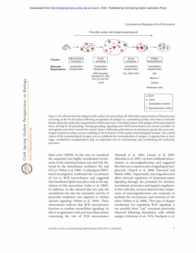
three-color TIRFM. In this way, we visualizedthe sequential and highly coordinated recruit-ment of the initiating kinases Lyn and Syk, fol-lowed by the downstream mediators Vav andPLCg2 (Weber et al. 2008). A subsequent FRET-based investigation confirmed the recruitmentof Lyn to BCR microclusters and suggestedthat membrane lipids may play a role in the reg-ulation of this association (Sohn et al. 2008).In addition, we also showed that not only therecruitment but also the enzymatic activity ofparticular mediators was required to initiatecalcium signaling (Weber et al. 2008). Theseobservations indicate that BCR microclustersfunction to mediate intracellular signaling. Asthis is in agreement with previous observationsconcerning the role of TCR microclusters
(Bunnell et al. 2002; Campi et al. 2005;Yokosuka et al. 2005), we have redefined micro-clusters as microsignalosomes and suggestedthat they are a common unit of signaling in lym-phocytes (Depoil et al. 2008; Harwood andBatista 2008). Importantly, microsignalosomesallow intricate regulation of immunoreceptorsignaling through the potential for dynamicrecruitment of positive and negative regulators.In line with this, we have observed that compo-nents of microsignalosomes can cooperate tofacilitate the recruitment and retention of theother (Weber et al. 2008). This type of elegantmechanism for regulating BCR signaling isnot possible from “cap” structures previouslyobserved following stimulation with solubleantigen (Schreiner et al. 1976; Stackpole et al.
Cytoskeletonreorganization
Cytoskeletonreorganization
Cytoskeletonreorganization
Cytoskeletonreorganization
Process Antigeninternalization
B-cellcontraction
B-cellspreading
Microclusterformation
MolecularRequirements
BCR signalingincluding Lyn, Syk,PLC γ2 and Vav
CD19
Vav, Grb2, Cbl ? Syk
Myosin II
Clathrin
Membrane rafts
Time after contact with antigen-presenting cell
BCR
CD19
Cytoskeleton network
Signaling active cluster
Figure 3. B-cell activation by antigen on the surface of a presenting cell. Schematic representation of the processesoccurring at the B-cell surface following recognition of antigen on a presenting surface and what is currentlyknown about the molecular requirements of these processes. On initial contact with antigen, BCR microclustersform, driving B-cell spreading. During spreading, signaling active BCR microclusters are moved (possibly viareterograde actin flow) toward the central cluster. Following achievement of maximum spread, the more pro-longed contraction phase occurs, resulting in the formation of the mature immunological synapse. The centralcluster of the immunological synapse acts as a platform for internalization of antigen. It appears that at everystage, cytoskeleton reorganizations play an important role in orchestrating and coordinating the molecularprocesses.
Cytoskeleton Regulates B-cell Activation
Cite this article as Cold Spring Harb Perspect Biol 2011;3:a002360 7
on June 5, 2018 - Published by Cold Spring Harbor Laboratory Press http://cshperspectives.cshlp.org/Downloaded from

1974; Unanue et al. 1972). Thus, given that thepredominant form of antigen-mediating acti-vation in vivo is membrane-bound, B cells canvery precisely regulate the extent of activationaccording to the context in which the antigenis presented.
SPREADING PROPAGATESMICROSIGNALOSOMES NECESSARYFOR B-CELL ACTIVATION
Although microsignalosomes are required formediating BCR signaling their formation alone,is not sufficient to elicit B-cell activation. Inaddition, it is necessary for the B-cell to amplifythe number of microsignalosomes such that thethreshold for antigen stimulation is exceeded(Weber et al. 2008). The B cell achieves thisamplification by spreading out its membraneacross the presenting cell, allowing contactwith greater amounts of antigen on the surface(Fleire et al. 2006) (Fig. 3). As BCR engagementof antigen at the leading edge drives the spread-ing response, the extent of spreading dependson the affinity and density of antigen on thepresenting surface and determines how muchantigen the B cell accumulates for presenta-tion to helper T cells (Fleire et al. 2006). Asspreading is abrogated in the presence of in-hibitors of actin polymerization and Src-familykinases, this cellular response absolutely re-quires intracellular signaling and cytoskeletonreorganization.
The signaling events downstream of theBCR important for mediating spreading haverecently been investigated (Weber et al. 2008)through a screen of an existing panel of mutantsin a chicken B-cell line (Shinohara and Kurosaki2006). The DT40 B-cell line shows a high rate ofhomologous recombination, enabling the rapidgeneration of B cells deficient in a particularmediator (Kurosaki 1999). In addition, thisstrategy has the advantage of allowing investiga-tion of molecules whose deletion is embryoni-cally lethal in a knockout animal system. Itwas observed that Lyn and Syk were absolutelyrequired for the initiation of B-cell spreadingin response to antigen-containing membranes
(Weber et al. 2008). Furthermore, although Bcells deficient in Vav and PLCg2 were severelyimpaired in mediating spreading, it is clear thatthese two mediators play a critical role in thepropagation of the cellular response. B cells defi-cient in Btk and Blnk were markedly impaired inspreading, suggesting that the activation andretention of PLCg2 is important during spread-ing. In contrast, B cells lacking all three IP3 recep-tors were fullycompetent inmediatingspreading,a somewhat surprising observation given thecalcium-dependence of the analogous processin T cells (Bunnell et al. 2001).
Although the key intracellular mediatorsinvolved in B-cell spreading have been estab-lished, the molecular mechanisms underlyingthe necessary cytoskeleton rearrangements cur-rently remain unclear. However, as Vav is knownto be an important regulator of the cytoskeletonand has been established as critical for propaga-tion of B-cell spreading (Weber et al. 2008), thisoffers insight into potential mechanisms forreorganization of the cytoskeleton. Alongsideits role as a molecular adaptor, Vav functionsas a guanine nucleotide exchange factor (GEF)to activate RhoGTPases such as Cdc42 andRac that are involved in the regulation of cyto-skeleton reorganizations (Jaffe and Hall 2005).In line with this, the GEF activity of Vav hasbeen shown to be essential during B-cell spread-ing (Weber et al. 2008). In addition, in T cells,Vav has been implicated as a regulator of ezrindephosphorylation in response to TCR stimu-lation (Faure et al. 2004). This is of particularinterest as dephosphorylation of ezrin leads torelease of the association between the plasmamembrane and the actin cytoskeleton, facilitat-ing the formation of cell conjugates. Interest-ingly, a rapid and global dephosphorylationof ezrin has been observed following stimula-tion of B cells with soluble antigen (Guptaet al. 2006). In conjunction with Vav, a numberof other intracellular B-cell effectors have beenimplicated in the regulation of cytoskeletonreorganizations following stimulation of theBCR. These include the leukocyte-specifichomolog of cortactin HS1 (Gomez et al. 2006;Hao et al. 2004; Yamanashi et al. 1997), cofilin
N.E. Harwood and F.D. Batista
8 Cite this article as Cold Spring Harb Perspect Biol 2011;3:a002360
on June 5, 2018 - Published by Cold Spring Harbor Laboratory Press http://cshperspectives.cshlp.org/Downloaded from
(DesMarais et al. 2005; Yonezawa et al. 1991),DOCK8 (Randall et al. 2009), and B-lympho-cyte adaptor molecule of 32kDa (Bam32)(Fournier et al. 2003; Han et al. 2003; Marshallet al. 2000). Bam32 represents a particularlyinteresting candidate as it is recruited to theplasma membrane and phosphorylated follow-ing BCR ligation (Marshall et al. 2000), andB cells deficient in Bam32 are impaired inactin polymerization and antigen internaliza-tion (Niiro et al. 2004). Given the importanceof the spreading response in shaping the out-come of B-cell activation, detailed investiga-tions of these and other mediators will beinvaluable in deriving a complete descriptionof the molecular mechanism communicatingBCR ligation with cytoskeleton reorganizations.
CD19 IS ESSENTIAL FOR B-CELLSPREADING AND ACTIVATION
During a dissection of the requirements forB-cell spreading, we uncovered a novel andunexpected role for the B-cell coreceptorCD19 (Depoil et al. 2008). Until this discovery,CD19 was most commonly known as a compo-nent of the CD21-CD19-CD81-leu13 complexresponsible for enhanced BCR signaling inresponse to antigen coated with complementfragments (Fearon and Carroll 2000). In thesecircumstances, the ligation of CD21 (also knownas complement receptor 2) triggers CD19 torecruit intracellular effectors that facilitate sig-naling through the BCR. However, the obser-vation that CD19-deficient mice show moreseverely impaired B-cell responses to antigenthan mice lacking CD21 (Ahearn et al. 1996;Engel et al. 1995; Rickert et al. 1995) suggeststhat CD19 performs an alternative role inde-pendent of CD21 during B-cell activation.In line with this, we observed that B cellslacking CD19 were unable to initiate calciumsignaling and thus could not mediate spreadingand activation in response to antigen on thesurface of presenting cells (Depoil et al. 2008).Interestingly, and in agreement with previousinvestigations, we observed that CD19-deficientB cells respond to soluble antigen in a similar
manner to wild-type B cells (Depoil et al. 2008;Fujimoto et al. 1999; Sato et al. 1997). Theseobservations nicely complement recent intravi-tal imaging investigations, demonstrating thecentral role of antigen bound to presenting cellsas the predominant form triggering B-cell acti-vation in vivo (Carrasco and Batista 2007; Juntet al. 2007; Phan et al. 2007; Phan et al. 2009;Qi et al. 2006; Suzuki et al. 2009).
Though the molecular mechanism by whichCD19 mediates this essential role in B-cellspreading has not yet been fully elucidated,imaging strategies have allowed us to gainsome insight into this process. The cytoplasmicdomain of CD19 contains several motifs thatcan mediate the recruitment of numerous in-tracellular effectors, including PI3K and Vav (Liet al. 1997; Tuveson et al. 1993). Furthermore,it has been suggested that CD19 forms oligo-mers in the membrane (Brooks et al. 2004),potentially allowing cooperation and prolongedretention of recruited molecules. Thus, it seemsplausible to suggest that CD19 mediates therecruitment and stabilization of additionalintracellular effectors in the microsignalosometo facilitate signaling through the BCR. Insupport of this mechanism, it was observedthat CD19 becomes transiently associatedwith BCR microclusters following stimulationwith membrane-bound antigen (Depoil et al.2008). More recently, we have used Dual-ViewTIRFM to visualize single particles of CD19moving between signaling BCR microclustersfollowing antigen stimulation (D. Depoil andF.D. Batista, unpubl. data). In addition, weobserved that mediators recruited throughCD19, such as PI3K, are laterally segregatedfrom those recruited solely through the BCRin the B-cell membrane. Thus, at this stage, itappears that CD19 plays a role in B cells similarto that of LAT in T cells (Zhang and Samelson2000), providing the boost to BCR signalingnecessary to stimulate cytoskeleton reorganiza-tion. Interestingly, mutations in CD19 andother potential mediators of cytoskeleton reor-ganization have been associated with antibodydeficiency syndromes in humans (Conley et al.2009).
Cytoskeleton Regulates B-cell Activation
Cite this article as Cold Spring Harb Perspect Biol 2011;3:a002360 9
on June 5, 2018 - Published by Cold Spring Harbor Laboratory Press http://cshperspectives.cshlp.org/Downloaded from

B-CELL CONTRACTION RESULTSIN FORMATION OF THEIMMUNOLOGICAL SYNAPSE
B cells attain maximum spread a few minutesafter initial contact with an antigen-presentingcell and subsequently the B-cell membraneundergoes a more prolonged contraction phase(Fleire et al. 2006) (Fig. 3). The mechanismunderlying the initiation of B-cell contractionremains unclear; however, a genetic screen toidentify mediators involved in the spreadingresponse revealed that B cells deficient in Vav,Grb2, or Cbl showed impaired contraction(Weber et al. 2008; T. Schnyder and F.D. Batista,unpubl. data). B-cell contraction results in thedramatic reorganization of the membraneforming a structure known as the immunologi-cal synapse (IS) (Batista et al. 2001; Carrascoet al. 2004). The IS was originally identified inCD4þ T cells (Grakoui et al. 1999; Krummelet al. 2000; Monks et al. 1998) and has sincebecome recognized as a common feature associ-ated with lymphocyte activation (Davis et al.1999; Potter et al. 2001; Stinchcombe et al.2001). The IS comprises a cluster of immunor-eceptors in a central supramolecular activationcluster (cSMAC) surrounded by a ring of adhe-sion molecules, such as the integrins LFA-1 andVLA-4, in the peripheral SMAC (pSMAC)(Monks et al. 1998). It is thought that duringspreading, clusters of signaling immunorecep-tors associated with the underlying actin cytos-keleton are passively translocated to the centerof contact via retrograde actin flow to assemblethe cSMAC (Kaizuka et al. 2007). Interestingly,this would provide an elegant mechanism toexplain the segregation of molecules withinthe IS according to their particular associationwith components of the cytoskeleton. In addi-tion, recent data suggested that in T cells, thistransportation is also mediated through anactive process involving the actin-based molec-ular motor myosin IIA (Ilani et al. 2009). How-ever, the contribution of the two mechanisms oralternatives during contraction and formationof the IS in B cells remains to be established.
Though imaging was used to visualize thestructure more than a decade ago, the function
of the IS has remained controversial. It was orig-inally put forward that given the concentrationof immunoreceptors in the cSMAC, this wouldlikely represent the site for active signaling.However, this assumption must be revisitedgiven that intracellular calcium signaling isobserved more rapidly than the formation ofthe mature IS, and in view of the more recentobservation that microclusters of immunore-ceptors actively signal the cell periphery duringspreading (Bunnell et al. 2002; Campi et al.2005; Depoil et al. 2008; Weber et al. 2008; Yoko-suka et al. 2005). In line with this, a recent in-vestigation tracking microcluster movement inT cells showed that slowing the movement ofmicroclusters toward the cSMAC prolongs sig-naling through the TCR (Nguyen et al. 2008).Thus, these observations support the proposalthat the cSMAC forms a platform for internal-ization of immunoreceptors and attenuationof signaling (Lee et al. 2002). However, it seemsthat the division of labor between componentsof the IS may not be absolute since under certainconditions, such as stimulation with weak ago-nists, signaling in the cSMAC can be observed(Cemerski et al. 2008).
The role of the IS in antigen internaliza-tion is of particular significance in B cells, asthis is known to be critical for the recruitmentof CD4þ T-helper cells required for maximalB-cell activation (Batista et al. 2001). It is clearthat the BCR functions not only to mediateintracellular signaling, but also to target antigento the correct compartment for intracellularprocessing (Aluvihare et al. 1997; Amigorenaet al. 1994). Indeed, it has been shown thatuptake through the BCR represents a moreefficient means to accumulate antigen for pre-sentation to T cells compared with fluid-phaseendocytosis (Aluvihare et al. 1997; Chenget al. 1999). BCR engagement initiates the re-organization of intracellular compartments toform a central multivesicular compartmentenriched in MHC-II, antigen, and accessorymolecules necessary for processing (Boes et al.2004; Drake et al. 1999; Lankar et al. 2002;Siemasko and Clark 2001; Siemasko et al.1998; Vascotto et al. 2007). Though the precisemolecular requirements for the BCR-mediated
N.E. Harwood and F.D. Batista
10 Cite this article as Cold Spring Harb Perspect Biol 2011;3:a002360
on June 5, 2018 - Published by Cold Spring Harbor Laboratory Press http://cshperspectives.cshlp.org/Downloaded from

internalization of antigen are not yet clear,actin-depolymerizing agents disrupt antigeninternalization, establishing a critical role forthe actin cytoskeleton during this step of B-cell activation (Barois et al. 1998; Brown andSong 2001). As B cells deficient in Syk showaltered intracellular trafficking and impairedpresentation of antigen to CD4þ T helper cells,it has been suggested that Syk plays a key role inregulating interactions between intracellularvesicles and actin filaments (Le Roux et al.2007). In addition, the actin-based motor pro-tein myosin II has been shown to be requiredfor antigen processing and presentation (Vas-cotto et al. 2007). It has been put forward thatmost efficient BCR-mediated antigen internal-ization occurs when the actin cytoskeletoncooperates with clathrin- and membrane-raft-mediated pathways (Stoddart et al. 2005).Although it is clear that the IS effectively coor-dinates the processes enabling antigen internal-ization, future imaging investigations will bevital to establish the molecular mechanismsunderlying antigen internalization, presenta-tion, and thus B-cell activation.
CONCLUDING REMARKS
It is evident from our discussion of the mole-cular events of B-cell activation that the actincytoskeleton plays a key role at each stage,including: regulating the distribution of BCRin the resting cell membrane; mediating theassembly and stability of BCR microclusters;underlying the cellular spreading and contrac-tion responses; and finally coordinating antigeninternalization and processing. Thus, the B celluses the actin cytoskeleton as a framework tosuccessfully manage numerous molecular inter-actions. Interestingly, the cytoskeleton structureprovides a scaffold to establish polarization ofintracellular effectors and compartments, suchas has been observed before cell division in Tcells (Chang et al. 2007). This is a particularlyintriguing issue in lymphocyte biology, as thistype of polarization can lead to asymmetric seg-regation of components after cell division,potentially allowing generation of two daughtercells with different effector functions.
ACKNOWLEDGMENTS
Cancer Research UK supports research in theLymphocyte Interaction Laboratory. We wouldlike to thank members of the Lymphocyte Inter-action Laboratory for critical reading of themanuscript particularly B. Treanor for provid-ing the imaging data shown in Figure 1B.
REFERENCES
Ahearn J, Fischer M, Croix D, Goerg S, Ma M, Xia J, Zhou X,Howard R, Rothstein T, Carroll M. 1996. Disruption ofthe Cr2 locus results in a reduction in B-1a cells and inan impaired B cell response to T-dependent antigen.Immunity 4: 251–262.
Aluvihare VR, Khamlichi AA, Williams GT, Adorini L, Neu-berger MS. 1997. Acceleration of intracellular targetingof antigen by the B-cell antigen receptor: Importancedepends on the nature of the antigen-antibody interac-tion. EMBO J 16: 3553–3562.
Amigorena S, Drake JR, Webster P, Mellman I. 1994. Transi-ent accumulation of new class II MHC molecules in anovel endocytic compartment in B lymphocytes. Nature369: 113–120.
Barois N, Forquet F, Davoust J. 1998. Actin microfilamentscontrol the MHC class II antigen presentation pathwayin B cells. J Cell Sci 111: 1791–1800.
Batista F, Neuberger M. 1998. Affinity dependence of the Bcell response to antigen: A threshold, a ceiling, and theimportance of off-rate. Immunity 8: 751–759.
Batista F, Neuberger M. 2000. B cells extract and presentimmobilized antigen: Implications for affinity discrimi-nation. EMBO J 19: 513–520.
Batista F, Iber D, Neuberger M. 2001. B cells acquire antigenfrom target cells after synapse formation. Nature 411:489–494.
Boes M, Cuvillier A, Ploegh H. 2004. Membrane specializa-tions and endosome maturation in dendritic cells and Bcells. Trends Cell Biol 14: 175–183.
Brooks S, Kirkham P, Freeberg L, Carter R. 2004. Binding ofcytoplasmic proteins to the CD19 intracellular domain ishigh affinity, competitive, and multimeric. J Immunol172: 7556–7564.
Brown BK, Song W. 2001. The actin cytoskeleton is requiredfor the trafficking of the B cell antigen receptor to the lateendosomes. Traffic 2: 414–427.
Bunnell S, Hong D, Kardon J, Yamazaki T, McGlade C, BarrV, Samelson L. 2002. T cell receptor ligation induces theformation of dynamically regulated signaling assemblies.J Cell Biol 158: 1263–1275.
Bunnell S, Kapoor V, Trible R, Zhang W, Samelson L. 2001.Dynamic actin polymerization drives T cell receptor-induced spreading: A role for the signal transductionadaptor LAT. Immunity 14: 315–329.
Byth K, Conroy L, Howlett S, Smith A, May J, Alexander D,Holmes N. 1996. CD45-null transgenic mice reveal apositive regulatory role for CD45 in early thymocyte
Cytoskeleton Regulates B-cell Activation
Cite this article as Cold Spring Harb Perspect Biol 2011;3:a002360 11
on June 5, 2018 - Published by Cold Spring Harbor Laboratory Press http://cshperspectives.cshlp.org/Downloaded from

development, in the selection of CD4þCD8þ thymo-cytes, and B cell maturation. J Exp Med 183: 1707–1718.
Cemerski S, Das J, Giurisato E, Markiewicz MA, Allen PM,Chakraborty AK, Shaw AS. 2008. The balance between Tcell receptor signaling and degradation at the center of theimmunological synapse is determined by antigen quality.Immunity 29: 414–422.
Campi G, Varma R, Dustin M. 2005. Actin and agonistMHC-peptide complex-dependent T cell receptormicroclusters as scaffolds for signaling. J Exp Med 202:1031–1036.
Carrasco Y, Batista F. 2007. B cells acquire particulate antigenin a macrophage-rich area at the boundary between thefollicle and the subcapsular sinus of the lymph node.Immunity 27: 160–171.
Carrasco Y, Fleire S, Cameron T, Dustin M, Batista F. 2004.LFA-1/ICAM-1 interaction lowers the threshold of Bcell activation by facilitating B cell adhesion and synapseformation. Immunity 20: 589–599.
Chang JT, Palanivel VR, Kinjyo I, Schambach F, IntlekoferAM, Banerjee A, Longworth SA, Vinup KE, Mrass P,Oliaro J, et al. 2007. Asymmetric T lymphocyte divisionin the initiation of adaptive immune responses. Science315: 1687–1691.
Chen L, Frank A, Adams J, Steinman R. 1978. Distributionof horseradish peroxidase (HRP)-anti-HRP immunecomplexes in mouse spleen with special reference to fol-licular dendritic cells. J Cell Biol 79: 184–199.
Cheng PC, Steele CR, Gu L, Song W, Pierce SK. 1999. MHCclass II antigen processing in B cells: accelerated intracel-lular targeting of antigens. J Immunol 162: 7171–7180.
Choudhuri K, Wiseman D, Brown M, Gould K, van derMerwe P. 2005. T-cell receptor triggering is criticallydependent on the dimensions of its peptide-MHCligand. Nature 436: 578–582.
Conley ME, Dobbs AK, Farmer DM, Kilic S, Paris K, Grigor-iadou S, Coustan-Smith E, Howard V, Campana D. 2009.Primary B cell immunodeficiencies: comparisons andcontrasts. Annu Rev Immunol 27: 199–227.
Dal Porto J, Gauld S, Merrell K, Mills D, Pugh-Bernard A,Cambier J. 2004. B cell antigen receptor signaling 101.Mol Immunol 41: 599–613.
Davis S, van der Merwe P. 2006. The kinetic-segregationmodel: TCR triggering and beyond. Nat Immunol 7:803–809.
Davis D, Chiu I, Fassett M, Cohen G, Mandelboim O, Stro-minger J. 1999. The human natural killer cell immunesynapse. Proc Natl Acad Sci 96: 15062–15067.
DeFranco A. 1997. The complexity of signaling pathwaysactivated by the BCR. Curr Opin Immunol 9: 296–308.
Depoil D, Fleire S, Treanor BL, Weber M, Harwood NE,Marchbank KL, Tybulewicz VL, Batista FD. 2008. CD19is essential for B cell activation by promoting B cellreceptor-antigen microcluster formation in response tomembrane-bound ligand. Nat Immunol 9: 63–72.
DesMarais V, Ghosh M, Eddy R, Condeelis J. 2005. Cofilintakes the lead. J Cell Sci 118: 19–26.
Drake JR, Lewis TA, Condon KB, Mitchell RN, Webster P.1999. Involvement of MIIC-like late endosomes in Bcell receptor-mediated antigen processing in murine Bcells. J Immunol 162: 1150–1155.
Engel P, Zhou L, Ord D, Sato S, Koller B, Tedder T. 1995.Abnormal B lymphocyte development, activation, anddifferentiation in mice that lack or overexpress theCD19 signal transduction molecule. Immunity 3: 39–50.
Faure S, Salazar-Fontana L, Semichon M, Tybulewicz V, Bis-muth G, Trautmann A, Germain R, Delon J. 2004. ERMproteins regulate cytoskeleton relaxation promoting Tcell-APC conjugation. Nat Immunol 5: 272–279.
Fearon D, Carroll M. 2000. Regulation of B lymphocyteresponses to foreign and self-antigens by the CD19/CD21 complex. Annu Rev Immunol 18: 393–422.
Fleire S, Goldman J, Carrasco Y, Weber M, Bray D, Batista F.2006. B cell ligand discrimination through a spreadingand contraction response. Science 312: 738–741.
Fournier E, Isakoff S, Ko K, Cardinale C, Inghirami G, Li Z,Curotto de Lafaille M, Skolnik E. 2003. The B cell SH2/PH domain-containing adaptor Bam32/DAPP1 is re-quired for T cell-independent II antigen responses. CurrBiol 13: 1858–1866.
Fujimoto M, Bradney A, Poe J, Steeber D, Tedder T. 1999.Modulation of B lymphocyte antigen receptor signaltransduction by a CD19/CD22 regulatory loop. Immun-ity 11: 191–200.
Fujiwara T, Ritchie K, Murakoshi H, Jacobson K, Kusumi A.2002. Phospholipids undergo hop diffusion in compart-mentalized cell membrane. J Cell Biol 157: 1071–1081.
Garcia KC, Teyton L, Wilson IA. 1999. Structural basis of Tcell recognition. Annu Rev Immunol 17: 369–397.
Gil D, Schamel WW, Montoya M, Sanchez-Madrid F, Alar-con B. 2002. Recruitment of Nck by CD3 epsilon reveals aligand-induced conformational change essential for Tcell receptor signaling and synapse formation. Cell 109:901–912.
Gomez T, McCarney S, Carrizosa E, Labno C, Comiskey E,Nolz J, Zhu P, Freedman B, Clark M, Rawlings D, et al.2006. HS1 functions as an essential actin-regulatoryadaptor protein at the immune synapse. Immunity 24:741–752.
Grakoui A, Bromley S, Sumen C, Davis M, Shaw A, Allen P,Dustin M. 1999. The immunological synapse: A molecu-lar machine controlling T cell activation. Science 285:221–227.
Gupta N, Wollscheid B, Watts J, Scheer B, Aebersold R,DeFranco A. 2006. Quantitative proteomic analysis ofB cell lipid rafts reveals that ezrin regulates antigenreceptor-mediated lipid raft dynamics. Nat Immunol 7:625–633.
Halin C, Rodrigo Mora J, Sumen C, von Andrian U. 2005. Invivo imaging of lymphocyte trafficking. Annu Rev CellDev Biol 21: 581–603.
Han A, Saijo K, Mecklenbrauker I, Tarakhovsky A, Nussenz-weig M. 2003. Bam32 links the B cell receptor to ERK andJNK and mediates B cell proliferation but not survival.Immunity 19: 621–632.
Hao J, Carey G, Zhan X. 2004. Syk-mediated tyrosine phos-phorylation is required for the association of hemato-poietic lineage cell-specific protein 1 with lipid raftsand B cell antigen receptor signalosome complex. J BiolChem 279: 33413–33420.
N.E. Harwood and F.D. Batista
12 Cite this article as Cold Spring Harb Perspect Biol 2011;3:a002360
on June 5, 2018 - Published by Cold Spring Harbor Laboratory Press http://cshperspectives.cshlp.org/Downloaded from

Harwood NE, Batista FD. 2008. New insights into the earlymolecular events underlying B cell activation. Immunity28: 609–619.
Hennecke J, Wiley DC. 2001. T cell receptor-MHC interac-tions up close. Cell 104: 1–4.
Huang N, Han S, Hwang I, Kehrl J. 2005. B cells productivelyengage soluble antigen-pulsed dendritic cells: Visualiza-tion of live-cell dynamics of B cell-dendritic cell interac-tions. J Immunol 175: 7125–7134.
Ilani T, Vasiliver-Shamis G, Vardhana S, Bretscher A, DustinML. 2009. T cell antigen receptor signaling and immuno-logical synapse stability require myosin IIA. Nat Immunol10: 531–539.
Irles C, Symons A, Michel F, Bakker T, van der Merwe P,Acuto O. 2003. CD45 ectodomain controls interactionwith GEMs and Lck activity for optimal TCR signaling.Nat Immunol 4: 189–197.
Jaffe A, Hall A. 2005. Rho GTPases: Biochemistry and biol-ogy. Annu Rev Cell Dev Biol 21: 247–269.
Junt T, Scandella E, Ludewig B. 2008. Form follows function:Lymphoid tissue microarchitecture in antimicrobialimmune defence. Nat Rev Immunol 8: 764–775.
Junt T, Moseman E, Iannacone M, Massberg S, Lang P, BoesM, Fink K, Henrickson S, Shayakhmetov D, Di Paolo N,et al. 2007. Subcapsular sinus macrophages in lymphnodes clear lymph-borne viruses and present them toantiviral B cells. Nature 450: 110–114.
Kaizuka Y, Douglass AD, Varma R, Dustin ML, Vale RD.2007. Mechanisms for segregating T cell receptor andadhesion molecules during immunological synapse for-mation in Jurkat T cells. Proc Natl Acad Sci 104: 20296–20301.
Kishihara K, Penninger J, Wallace V, Kundig T, Kawai K,Wakeham A, Timms E, Pfeffer K, Ohashi P, Thomas M.1993. Normal B lymphocyte development but impairedT cell maturation in CD45-exon6 protein tyrosinephosphatase-deficient mice. Cell 74: 143–156.
Koppel E, Wieland C, van den Berg V, Litjens M, Florquin S,van Kooyk Y, van der Poll T, Geijtenbeek T. 2005. Spe-cific ICAM-3 grabbing nonintegrin-related 1 (SIGNR1)expressed by marginal zone macrophages is essentialfor defense against pulmonary Streptococcus pneumoniaeinfection. Eur J Immunol 35: 2962–2969.
Krummel M, Sjaastad M, Wulfing C, Davis M. 2000. Differ-ential clustering of CD4 and CD3z during T cell recogni-tion. Science 289: 1349–1352.
Kuhns MS, Davis MM, Garcia KC. 2006. Deconstructing theform and function of the TCR/CD3 complex. Immunity24: 133–139.
Kurosaki T. 1999. Genetic analysis of B cell antigen receptorsignaling. Annu Rev Immunol 17: 555–592.
Kurosaki T. 2002. Regulation of B-cell signal transduction byadaptor proteins. Nat Rev Immunol 2: 354–363.
Kusumi A, Ike H, Nakada C, Murase K, Fujiwara T. 2005.Single-molecule tracking of membrane molecules:Plasma membrane compartmentalization and dynamicassembly of raft-philic signaling molecules. Semin Immu-nol 17: 3–21.
Lankar D, Vincent-Schneider H, Briken V, Yokozeki T,Raposo G, Bonnerot C. 2002. Dynamics of major histo-compatibility complex class II compartments during B
cell receptor-mediated cell activation. J Exp Med 195:461–472.
Lanzavecchia A. 1985. Antigen-specific interaction betweenT and B cells. Nature 314: 537–539.
Le Roux D, Lankar D, Yuseff M-I, Vascotto F, Yokozeki T,Faure-Andre G, Mougneau E, Glaichenhaus N, ManouryB, Bonnerot C, et al. 2007. Syk-dependent actin dynamicsregulate endocytic trafficking and processing of antigensinternalized through the B-cell receptor. Mol Biol Cell 18:3451–3462.
Lee K, Holdorf A, Dustin M, Chan A, Allen P, Shaw A. 2002.T cell receptor signaling precedes immunological synapseformation. Science 295: 1539–1542.
Li X, Sandoval D, Freeberg L, Carter R. 1997. Role of CD19tyrosine 391 in synergistic activation of B lymphocytes bycoligation of CD19 and membrane Ig. J Immunol 158:5649–5657.
Lillemeier BF, Pfeiffer JR, Surviladze Z, Wilson BS, DavisMM. 2006. Plasma membrane-associated proteins areclustered into islands attached to the cytoskeleton. ProcNatl Acad Sci 103: 18992–18997.
MacLennan I. 1994. Germinal centers. Annu Rev Immunol12: 117–139.
MacLennan IC, Toellner KM, Cunningham AF, Serre K, SzeDM, Zuniga E, Cook MC, Vinuesa CG. 2003. Extrafollic-ular antibody responses. Immunol Rev 194: 8–18.
Mandel T, Phipps R, Abbot A, Tew J. 1980. The folliculardendritic cell: long term antigen retention duringimmunity. Immunol Rev 53: 29–59.
Marshall A, Niiro H, Lerner C, Yun T, Thomas S, Disteche C,Clark E. 2000. A novel B lymphocyte-associated adaptorprotein, Bam32, regulates antigen receptor signalingdownstream of phosphatidylinositol 3-kinase. J Exp Med191: 1319–1332.
Monks C, Freiberg B, Kupfer H, Sciaky N, Kupfer A. 1998.Three-dimensional segregation of supramolecular acti-vation clusters in T cells. Nature 395: 82–86.
Mustelin T, Coggeshall K, Altman A. 1989. Rapid activationof the T-cell tyrosine protein kinase pp56lck by the CD45phosphotyrosine phosphatase. Proc Natl Acad Sci 86:6302–6306.
Nguyen K, Sylvain NR, Bunnell SC. 2008. T cell costimula-tion via the integrin VLA-4 inhibits the actin-dependentcentralization of signaling microclusters containing theadaptor SLP-76. Immunity 28: 810–821.
Niiro H, Allam A, Stoddart A, Brodsky F, Marshall A, ClarkE. 2004. The B lymphocyte adaptor molecule of 32 kilo-daltons (Bam32) regulates B cell antigen receptor inter-nalization. J Immunol 173: 5601–5609.
Phan T, Grigorova I, Okada T, Cyster J. 2007. Subcapsularencounter and complement-dependent transport of im-mune complexes by lymph node B cells. Nat Immunol 8:992–1000.
Phan TG, Green JA, Gray EE, Xu Y, Cyster JG. 2009. Immunecomplex relay by subcapsular sinus macrophages andnoncognate B cells drives antibody affinity maturation.Nat Immunol 10: 786–793.
Potter T, Grebe K, Freiberg B, Kupfer A. 2001. Formationof supramolecular activation clusters on fresh ex vivoCD8þ T cells after engagement of the T cell antigen
Cytoskeleton Regulates B-cell Activation
Cite this article as Cold Spring Harb Perspect Biol 2011;3:a002360 13
on June 5, 2018 - Published by Cold Spring Harbor Laboratory Press http://cshperspectives.cshlp.org/Downloaded from
receptor and CD8 by antigen-presenting cells. Proc NatlAcad Sci 98: 12624–12629.
Qi H, Egen JG, Huang AY, Germain RN. 2006. Extrafollicu-lar activation of lymph node B cells by antigen-bearingdendritic cells. Science 312: 1672–1676.
Rajewsky K. 1996. Clonal selection and learning in the anti-body system. Nature 381: 751–758.
Randall K, Lambe T, Johnson A, Kucharska E, DomaschenzH, Whittle B, Tze L, Enders A, Crockford T, Bouriez-Jones T, et al. 2009. Dock8 mutations cripple B cellimmunological synapses, germinal centers and long-lived antibody production. Nat Immunol 10: 1283–1291.
Reth M. 1989. Antigen receptor tail clue. Nature 338: 383–384.
Rickert R, Rajewsky K, Roes J. 1995. Impairment of T-cell-dependent B-cell responses and B-1 cell develop-ment in CD19-deficient mice. Nature 376: 352–355.
Rock K, Benacerraf B, Abbas A. 1984. Antigen presentationby hapten-specific B lymphocytes. I. Role of surfaceimmunoglobulin receptors. J Exp Med 160: 1102–1113.
Rolli V, Gallwitz M, Wossning T, Flemming A, Schamel W,Zurn C, Reth M. 2002. Amplification of B cell antigenreceptor signaling by a Syk/ITAM positive feedbackloop. Mol Cell 10: 1057–1069.
Rudolph MG, Stanfield RL, Wilson IA. 2006. How TCRsbind MHCs, peptides, and coreceptors. Annu Rev Immu-nol 24: 419–466.
Sako Y, Kusumi A. 1994. Compartmentalized structure ofthe plasma membrane for receptor movements asrevealed by a nanometer-level motion analysis. J CellBiol 125: 1251–1264.
Sato S, Miller A, Howard M, Tedder T. 1997. Regulation of Blymphocyte development and activation by the CD19/CD21/CD81/Leu 13 complex requires the cytoplasmicdomain of CD19. J Immunol 159: 3278–3287.
Schamel W, Reth M. 2000. Monomeric and oligomeric com-plexes of the B cell antigen receptor. Immunity 13: 5–14.
Schamel WW, Risueno RM, Minguet S, Ortız AR, Alarcon B.2006. A conformation- and avidity-based proofreadingmechanism for the TCR-CD3 complex. Trends Immunol27: 176–182.
Scharenberg A, Humphries L, Rawlings D. 2007. Calciumsignalling and cell-fate choice in B cells. Nat Rev Immunol7: 778–789.
Schreiner GF, Braun J, Unanue ER. 1976. Spontaneous redis-tribution of surface immunoglobulin in the motile Blymphocyte. J Exp Med 144: 1683–1688.
Secrist JP, Burns LA, Karnitz L, Koretzky GA, Abraham RT.1993. Stimulatory effects of the protein tyrosine phos-phatase inhibitor, pervanadate, on T-cell activationevents. J Biol Chem 268: 5886–5893.
Shinohara H, Kurosaki T. 2006. Genetic analysis of B cell sig-naling. Subcell Biochem 40: 145–187.
Siemasko K, Clark MR. 2001. The control and facilitation ofMHC class II antigen processing by the BCR. Curr OpinImmunol 13: 32–36.
Siemasko K, Eisfelder BJ, Williamson E, Kabak S, Clark MR.1998. Cutting edge: Signals from the B lymphocyte anti-gen receptor regulate MHC class II containing late endo-somes. J Immunol 160: 5203–5208.
Simons K, Ikonen E. 1997. Functional rafts in cell mem-branes. Nature 387: 569–572.
Simson R, Sheets ED, Jacobson K. 1995. Detection of tem-porary lateral confinement of membrane proteins usingsingle-particle tracking analysis. Biophys J 69: 989–993.
Singer SJ, Nicolson GL. 1972. The fluid mosaic model of thestructure of cell membranes. Science 175: 720–731.
Sohn H, Pierce S, Tzeng S. 2008. Live cell imaging revealsthat the inhibitory Fc{gamma}RIIB destabilizes B cellreceptor membrane-lipid interactions and blocksimmune synapse formation. J Immunol 180: 793–799.
Stackpole CW, Jacobson JB, Lardis MP. 1974. Two distincttypes of capping of surface receptors on mouse lymphoidcells. Nature 248: 232–234.
Stinchcombe J, Bossi G, Booth S, Griffiths G. 2001. Theimmunological synapse of CTL contains a secretorydomain and membrane bridges. Immunity 15: 751–761.
Stoddart A, Jackson AP, Brodsky FM. 2005. Plasticity of Bcell receptor internalization upon conditional depletionof clathrin. Mol Biol Cell 16: 2339–2348.
Suzuki K, Grigorova I, Phan TG, Kelly LM, Cyster JG. 2009.Visualizing B cell capture of cognate antigen from follic-ular dendritic cells. J Exp Med 206: 1485–1493.
Tew JG, Phipps RP, Mandel TE. 1980. The maintenance andregulation of the humoral immune response: Persist-ing antigen and the role of follicular antigen-bindingdendritic cells as accessory cells. Immunol Rev 53: 175–201.
Tolar P, Sohn H, Pierce S. 2005. The initiation of antigen-induced B cell antigen receptor signaling viewed in livingcells by fluorescence resonance energy transfer. NatImmunol 6: 1168–1176.
Tolar P, Hanna J, Krueger PD, Pierce SK. 2009. The constantregion of the membrane immunoglobulin mediates Bcell-receptor clustering and signaling in response tomembrane antigens. Immunity 30: 44–55.
Treanor B, Batista F. 2007. Mechanistic insight into lympho-cyte activation through quantitative imaging and theo-retical modelling. Curr Opin Immunol 19: 476–483.
Treanor B, Depoil D, Gonzalez-Granja A, Barral P, Weber M,Dushek O, Bruckbauer A, Batista FD. 2010. The mem-brane skeleton controls diffusion dynamics and signalingthrough the B cell receptor. Immunity 32: 187–199.
Tuveson D, Carter R, Soltoff S, Fearon D. 1993. CD19 of Bcells as a surrogate kinase insert region to bind phospha-tidylinositol 3-kinase. Science 260: 986–989.
Unanue ER, Perkins WD, Karnovsky MJ. 1972. Ligand-induced movement of lymphocyte membrane macro-molecules. I. Analysis by immunofluorescence and ultra-structural radioautography. J Exp Med 136: 885–906.
van der Merwe PA. 2001. The TCR triggering puzzle. Imm-unity 14: 665–668.
Vascotto F, Lankar D, Faure-Andre G, Vargas P, Diaz J, LeRoux D, Yuseff M-I, Sibarita J-B, Boes M, Raposo G, etal. 2007. The actin-based motor protein myosin II regu-lates MHC class II trafficking and BCR-driven antigenpresentation. J Cell Biol 176: 1007–1019.
Weber M, Treanor B, Depoil D, Shinohara H, Harwood NE,Hikida M, Kurosaki T, Batista FD. 2008. PhospholipaseC-g2 and Vav cooperate within signaling microclusters
N.E. Harwood and F.D. Batista
14 Cite this article as Cold Spring Harb Perspect Biol 2011;3:a002360
on June 5, 2018 - Published by Cold Spring Harbor Laboratory Press http://cshperspectives.cshlp.org/Downloaded from

to propagate B cell spreading in response to membrane-bound antigen. J Exp Med 205: 853–868.
Wykes M, Pombo A, Jenkins C, MacPherson G. 1998. Den-dritic cells interact directly with naive B lymphocytes totransfer antigen and initiate class switching in a primaryT-dependent response. J Immunol 161: 1313–1319.
Xu C, Gagnon E, Call ME, Schnell JR, Schwieters CD, Car-man CV, Chou JJ, Wucherpfennig KW. 2008. Regulationof T cell receptor activation by dynamic membrane bind-ing of the CD3epsilon cytoplasmic tyrosine-based motif.Cell 135: 702–713.
Yamanashi Y, Fukuda T, Nishizumi H, Inazu T, Higashi K,Kitamura D, Ishida T, Yamamura H, Watanabe T, Yama-moto T. 1997. Role of tyrosine phosphorylation of HS1in B cell antigen receptor-mediated apoptosis. J ExpMed 185: 1387–1392.
Yokosuka T, Sakata-Sogawa K, Kobayashi W, Hiroshima M,Hashimoto-Tane A, Tokunaga M, Dustin M, Saito T.2005. Newly generated T cell receptor microclusters ini-tiate and sustain T cell activation by recruitment ofZap70 and SLP-76. Nat Immunol 6: 1253–1262.
Yonezawa N, Homma Y, Yahara I, Sakai H, Nishida E. 1991.A short sequence responsible for both phosphoinositidebinding and actin binding activities of cofilin. J Biol Chem266: 17218–17221.
Zhang W, Samelson L. 2000. The role of membrane-associ-ated adaptors in T cell receptor signalling. Semin Immu-nol 12: 35–41.
Zhu J, Brdicka T, Katsumoto T, Lin J, Weiss A. 2008. Struc-turally distinct phosphatases CD45 and CD148 both reg-ulate B cell and macrophage immunoreceptor signaling.Immunity 28: 183–196.
Cytoskeleton Regulates B-cell Activation
Cite this article as Cold Spring Harb Perspect Biol 2011;3:a002360 15
on June 5, 2018 - Published by Cold Spring Harbor Laboratory Press http://cshperspectives.cshlp.org/Downloaded from

November 3, 20102011; doi: 10.1101/cshperspect.a002360 originally published onlineCold Spring Harb Perspect Biol
Naomi E. Harwood and Facundo D. Batista The Cytoskeleton Coordinates the Early Events of B-cell Activation
Subject Collection Immunoreceptor Signaling
Serine/Threonine KinasesThe Coordination of T-cell Function by
David Finlay and Doreen Cantrellof T Cell ActivationPerspectives for Computer Modeling in the Study
Altan-BonnetJesse Coward, Ronald N. Germain and Grégoire
ReceptorITAM-mediated Signaling by the T-Cell Antigen
Paul E. Love and Sandra M. Hayes Initiation of Signalinginto Receptor Assembly, Ligand Recognition, and Structural Biology of the T-cell Receptor: Insights
Call, et al.Kai W. Wucherpfennig, Etienne Gagnon, Melissa J.
Protein SLP-76Hematopoietic Cell Lineages by the Adaptor Coordination of Receptor Signaling in Multiple
Martha S. Jordan and Gary A. KoretzkySignaling Cross TalkInhibitory Pathways in Innate Immune Cells: Src-family and Syk Kinases in Activating and
Clifford A. Lowell
B-cell ActivationThe Cytoskeleton Coordinates the Early Events of
Naomi E. Harwood and Facundo D. BatistaCoordination, and ChoreographyThe LAT Story: A Tale of Cooperativity,
Sherman, et al.Lakshmi Balagopalan, Nathan P. Coussens, Eilon
An Enigmatic Tail of CD28 SignalingJonathan S. Boomer and Jonathan M. Green CARMA1, BCL10, and MALT1
B viaκAntigen Receptor Signaling to NF-
Pelzer, et al.Margot Thome, Jean Enno Charton, Christiane
MeshworksMediation of T-Cell Activation by Actin
Peter Beemiller and Matthew F. KrummelInitiation of B-Cell Receptor SignalingIt's All About Change: The Antigen-driven
Wanli Liu, Hae Won Sohn, Pavel Tolar, et al.
Kinase, ItkT-Cell Signaling Regulated by the Tec Family
Joseph, et al.Amy H. Andreotti, Pamela L. Schwartzberg, Raji E.
ZAP-70: An Essential Kinase in T-cell Signaling
Au-Yeung, et al.Haopeng Wang, Theresa A. Kadlecek, Byron B.
FunctionLipid Signaling in T-Cell Development and
Yina H. Huang and Karsten SauerImmunological SynapseUnderstanding the Structure and Function of the
S. ShawMichael L. Dustin, Arup K. Chakraborty and Andrey
http://cshperspectives.cshlp.org/cgi/collection/ For additional articles in this collection, see
Copyright © 2011 Cold Spring Harbor Laboratory Press; all rights reserved
on June 5, 2018 - Published by Cold Spring Harbor Laboratory Press http://cshperspectives.cshlp.org/Downloaded from