the coxsackievirus and adenovirus receptor (car) undergoes
TRANSCRIPT

The Coxsackievirus and Adenovirus Receptor (CAR)Undergoes Ectodomain Shedding and RegulatedIntramembrane Proteolysis (RIP)Nadia Houri, Kuo-Cheng Huang, Josephine Nalbantoglu*
Department of Neurology and Neurosurgery and Montreal Neurological Institute, McGill University, Montreal, Quebec, Canada
Abstract
The Coxsackievirus and Adenovirus Receptor (CAR) is a cell adhesion molecule originally characterized as a virusreceptor but subsequently shown to be involved in physiological processes such as neuronal and heart development,epithelial tight junction integrity, and tumour suppression. Proteolysis of cell adhesion molecules and a wide variety ofother cell surface proteins serves as a mechanism for protein turnover and, in some cases, cell signaling.Metalloproteases such as A Disintegrin and Metalloprotease (ADAM) family members cleave cell surface receptors torelease their substrates’ ectodomains, while the presenilin/ɣ-secretase complex mediates regulated intramembraneproteolysis (RIP), releasing intracellular domain fragments from the plasma membrane. In the case of somesubstrates such as Notch and amyloid precursor protein (APP), the released intracellular domains enter the nucleusto modulate gene expression. We report that CAR ectodomain is constitutively shed from glioma cells and developingneurons, and is also shed when cells are treated with the phorbol ester phorbol 12-myristate 13-acetate (PMA) andthe calcium ionophore ionomycin. We identified ADAM10 as a sheddase of CAR using assays involving shRNAknockdown and rescue, overexpression of wild-type ADAM10 and inhibition of ADAM10 activity by addition of itsprodomain. In vitro peptide cleavage, mass spectrometry and mutagenesis revealed the amino acids M224 to L227of CAR as the site of ADAM10-mediated ectodomain cleavage. CAR also undergoes RIP by the presenilin/γ-secretase complex, and the intracellular domain of CAR enters the nucleus. Ectodomain shedding is a prerequisitefor RIP of CAR. Thus, CAR belongs to the increasing list of cell surface molecules that undergo ectodomain sheddingand that are substrates for ɣ-secretase-mediated RIP.
Citation: Houri N, Huang K-C, Nalbantoglu J (2013) The Coxsackievirus and Adenovirus Receptor (CAR) Undergoes Ectodomain Shedding andRegulated Intramembrane Proteolysis (RIP). PLoS ONE 8(8): e73296. doi:10.1371/journal.pone.0073296
Editor: Jean Kanellopoulos, University Paris Sud, France
Received April 15, 2013; Accepted July 19, 2013; Published August 28, 2013
Copyright: © 2013 Houri et al. This is an open-access article distributed under the terms of the Creative Commons Attribution License, which permitsunrestricted use, distribution, and reproduction in any medium, provided the original author and source are credited.
Funding: The authors thank the Canadian Institutes for Health Research (CIHR) and the Natural Sciences and Engineering Research Council for financialsupport. NH was supported by a Principal's Graduate Fellowship (McGill University) and a studentship from the CIHR Strategic Training Program inNeuroinflammation. K-CH was supported by a studentship from the Fonds de la recherche en santé du Québec. The funders had no role in study design,data collection and analysis, decision to publish, or preparation of the manuscript.
Competing interests: The authors have declared that no competing interests exist.
* E-mail: [email protected]
Introduction
The Coxsackievirus and Adenovirus Receptor (CAR) is a celladhesion molecule of the Immunoglobulin (Ig) superfamily[1,2]. As its name suggests, CAR is the attachment site ofCoxsackie B viruses as well as some adenovirus serotypes[3–5]. Although much of the research on CAR has been withinthe context of adenovirus-mediated gene therapy, recentstudies have described a wide range of physiological andpathophysiological roles.
CAR is highly conserved, especially in its C-terminus [2], andit is expressed in a variety of mammalian and non-mammalianspecies such as D. rerio and X. laevis. It is highly expressed inthe developing nervous system, particularly in neuronal growthcones [6,7], and it mediates neurite extension by binding to the
extracellular matrix protein fibronectin [8]. As a component ofepithelial cell tight junctions, CAR participates in forming abarrier to paracellular flow of macromolecules, binding to tightjunction proteins such as zonula occludens-1 (ZO-1) [9] andmulti-PDZ domain protein-1 (MUPP-1) [10]. Although CARexpression is high in the developing heart and skeletal muscle,it is barely detectable by adulthood, becoming restricted toskeletal muscle neuromuscular junctions and cardiacintercalated discs [11,12]. CAR expression is critical for normalcardiac development, as its gene deletion in mice beforeembryonic day 11 results in cardiac abnormalities andembryonic lethality [13–15]. As well, CAR deletion in adulthoodleads to multiple organ phenotypes including impairment ofcardiac atrioventricular connection [16,17], atrophy of exocrine
PLOS ONE | www.plosone.org 1 August 2013 | Volume 8 | Issue 8 | e73296

pancreas, enlarged intestines, and an increase in the numberof thymocytes in the thymus [18].
In the immune system, CAR facilitates migration ofneutrophils across epithelial cell tight junctions and endothelialcells during inflammatory episodes via its interaction withjunctional adhesion molecule-like protein (JAML) [19,20].Furthermore, epithelial CAR interacts with JAML in resident γδT cells in the skin to promote effective γδ T cell response tochanges in epithelial integrity [21]. Interestingly, CAR isdownregulated in several cancers, and it inhibits the growth orinvasion of some of these cancer cell lines when its expressionis restored, such as in bladder cancer [22] and gliomas [23,24].Therefore, CAR’s levels are tightly controlled and it functions incell adhesion, migration and regulation of growth.
A wide variety of cell surface receptors are known to undergoproteolysis. One group of enzymes that cleave and release theectodomains of cell surface proteins into the cellularenvironment is the A Disintegrin and Metalloprotease (ADAM)family of transmembrane and secreted metalloproteases.ADAMs act on diverse substrates including cytokines, cytokinereceptors, cell adhesion molecules and growth factor receptors,with effects on a multitude of functions such as spermmaturation and sperm-egg adhesion, cell migration, axonguidance and cell fate determination in the nervous system[25]. Cell adhesion molecules that are shed by ADAMs includeL1 [26], close homolog of L1 (CHL1) [27] and N-cadherin [28].
The γ-secretase complex participates in another type ofproteolysis, regulated intramembrane proteolysis (RIP), whichreleases substrates’ intracellular domains into the cytosol.Intracellular domain (ICD) products of RIP are then degradedor participate in cell signaling [29]. Familial mutations in thePSEN1 and PSEN2 genes that encode presenilin (PS), thecatalytic component of the γ-secretase complex, causeautosomal-dominant inherited Alzheimer’s disease [30].Amyloid precursor protein (APP) is cleaved by PS/γ-secretase,and the resulting intracellular domain is rapidly degraded [29],although it has also been suggested to act as a transcriptionalactivator [31]. RIP of Notch produces an intracellular fragmentthat translocates to the nucleus and regulates transcription ofNotch-responsive genes such as HES [32].
Given that many cell surface receptors, including celladhesion molecules, undergo proteolysis, we wondered if thatis the case for CAR. Here, we report that CAR is shed in aconstitutive fashion as well as via activation of the proteinkinase C (PKC) and calcium pathways. ADAM10 is a majorsheddase of CAR ectodomain, and the site of ADAM10cleavage on CAR is in the area of amino acids 224-227. CARis also processed by γ-secretase, generating a 14 kDaintracellular domain fragment. Ectodomain shedding of CARprecedes its RIP. Finally, free CAR intracellular domain entersthe nucleus as has been shown for a number of RIPsubstrates.
Materials and Methods
Ethics statementThis study was carried out in strict accordance with the
Animal Care and Use Program Guidelines of McGill University.
The protocol was approved by the Animal Care Committee ofthe Montreal Neurological Institute, McGill University (PermitNumber: 2005-4971). Animals were sacrificed by CO2 andcervical dislocation, and all efforts were made to minimizesuffering.
ChemicalsPhorbol 12-myristate 13-acetate (PMA), O-phenanthroline,
ionomycin and trichloroacetic acid (TCA) were from Sigma.GM6001, GM6001 negative control, Compound E (γ-SecretaseInhibitor XXI), recombinant human TIMP-1, TIMP-2 andTIMP-3, TAPI-1, Pepstatin, E-64, Leupeptin, DAPT, Gö 6983,and MG132 were from Calbiochem. Epoxomicin was fromBiovision (Cedarlane). B-27 and N2 supplements were fromInvitrogen. Purified prodomain of ADAM10 was a kind gift fromDr. Marcia Moss (Biozyme, Inc.).
AntibodiesThe production, purification and characterization of the rabbit
polyclonal antibodies 2239 and 2240 raised against CAR N-terminal extracellular domain have been previously described[11]. The rabbit polyclonal antibody RP291 raised against theC-terminal intracellular domain of human CAR (46 kDa isoform)cross-reacts with the murine homolog mCAR1 [12,33], and wasa kind gift from Dr. Kerstin Sollerbrant (Karolinska Institute andUniversity Hospital, Stockholm, Sweden). Rabbit polyclonalantibody raised against ADAM10 was from AnaSpec, Inc.Mouse monoclonal antibody raised against the V5 tag wasfrom Invitrogen. Horseradish peroxidase (HRP)-conjugatedanti-glyceraldehyde -3-phosphate dehydrogenase (GAPDH)antibody was from Abcam. Goat or swine anti-mouse and anti-rabbit HRP-conjugated secondary antibodies were from Pierceand Dako. Goat anti-mouse Alexa Fluor 555 secondaryantibody was from Molecular Probes.
shRNA knockdownFive different small hairpin ribonucleic acid (shRNA)
sequences (TRC library) in a lentiviral vector (pLKO.1)targeting human ADAM10 and control shRNA targetingenhanced green fluorescent protein (eGFP) were purchasedfrom Open Biosystems. Production of lentiviruses, infection andstable selection of cells with 2 μg/ml puromycin were performedaccording to the RNAi Consortium (TRC library) guidelines.Cell lines were maintained with 2 μg/ml puromycin. The level ofknockdown in cells was initially assayed by real-timepolymerase chain reaction (PCR) of reverse-transcribed RNA,normalizing over GAPDH. Only shRNA sequences thatsufficiently knocked down adam10 expression without affectingadam17 levels were considered specific. Experiments weresubsequently performed using the anti-ADAM10 hairpinsequenceCCGGGCAGTATTACTTATGGGAATTCTCGAGAATTCCCATAAGTAATACTGCTTTTT (thereafter referred to as “6676”) or asecond hairpin anti-ADAM10 shRNA,CCGGGCTGTGCAGATCATTCAGTATCTCGAGATACTGAATGATCTGCACAGCTTTTT (referred to as “6675”).
Proteolysis of the Virus Receptor CAR
PLOS ONE | www.plosone.org 2 August 2013 | Volume 8 | Issue 8 | e73296

PlasmidsMurine CAR (isoform 1) cloned in pcDNA3 plasmid has been
previously described [11]. All point mutations or deletions wereperformed with the QuikChange II XL site-directedmutagenesis kit (Strategene), per manufacturer’s guidelines.Four mutants of CAR were generated using pcDNA3-mCAR1as template: MLAA (in which amino acids M224 and L225 weremutated to alanine residues), RLAA (in which amino acidsR226 and L227 were mutated to alanine residues), MLRLAAAA(in which M224, L225, R226 and L227 were mutated to alanineresidues) and Δ221-232 (in which amino acids 221-232 weredeleted).
The plasmid pcDNA 3.1/V5-His6x B (Invitrogen) was a kindgift from Dr. Alyson Fournier (McGill University). To generatethe CAR-V5 construct (full-length murine CAR isoform 1 with aV5/His6x tag at the C-terminus), CAR insert in pcDNA3plasmid was amplified with Phusion High Fidelity DNApolymerase (Finnzymes Inc.), including the Kozak sequenceand excluding the stop codon. Blunt-end cloning of the PCRproduct was performed with EcoRV-digested pcDNA 3.1/V5-His. To obtain a construct of the intracellular domain of CARwith C-terminal V5-His6x tags (herein referred to as CAR ICD-V5), full-length CAR in pcDNA3.1 V5-His plasmid was used astemplate with the QuikChange II XL site-directed mutagenesiskit (Stratagene). Amino acids 2-260 inclusive were deleted fromCAR-V5 to generate ICD-V5 using the QuikChange II XL site-directed mutagenesis kit (Stratagene).
Human ADAM10 cDNA in pCR4-TOPO plasmid (clone ID8991969) was purchased from Open Biosystems and clonedinto pcDNA3.1 plasmid using the NotI and PmeI sites. TheshRNA-resistant mutant was generated from this construct bydeleting nucleotides 2319-2325 using the QuikChange II XLsite-directed mutagenesis kit (Strategene), per manufacturer’sguidelines.
The plasmid pWPI with IRES-GFP (Addgene ID # 12254)was a kind gift of Dr. Didier Trono. Full-length CAR with C-terminal V5/6xHis tag and stop codon was amplified by PCRusing CAR-V5 (pcDNA3.1) plasmid as template. The PCRproduct was introduced into the pWPI vector at the PmeI sitevia blunt-end ligation.
All constructs were verified by sequencing at the Plateformede séquençage et de génotypage des génomes, Centre derecherche du CHUL (Québec, Canada).
Cell lines and culture conditionsThe human embryonic kidney (HEK) 293, human glioma
U251N and human glioma U87-MG cell lines were obtainedfrom the American Type Culture Collection (ATCC) (Rockville,MD). The murine embryonic fibroblast (MEF) cell line knockoutfor PS 1 and 2 (herein referred to as “MEF PS 1/2 KO”) andwild-type MEF cell line were kind gifts from Dr. Bart deStrooper, K.U. Leuven, Belgium [34,35]. Cells were maintainedin an incubator at 37°C with 5% CO2 using Dulbecco’s ModifiedEagle Medium (DMEM) supplemented with 10% heat-inactivated fetal bovine serum (FBS) and antibiotic cocktail(100 units of penicillin/ml, 100 μg of streptomycin/ml).
The generation of U87-MG polyclonal cell populations over-expressing CAR via a retroviral vector has been previously
described [11,24]. Briefly, retroviruses carrying the sequencefor full-length murine CAR (isoform 1) were produced.Retroviruses carrying empty vector with the neomycinresistance gene were used as control. The supernatants fromthe producer cell lines were used for infection of U87-MG cells,which were then selected for 10 days with G418 (600 μg/ml).Clones were pooled together to generate bulk populationsstably expressing CAR (U87 CAR) or control cells (U87 LNCX).
U87-MG, U251N and HEK 293 cells were transfected withwild-type or mutant CAR constructs using FuGENE 6 (Roche)or TransIT-2020 (Mirus) per manufacturers’ guidelines. Cellswere selected for 7-10 days with 600 μg/ml G418 and thenmaintained with 200 μg/ml G418. Clones were pooled togetherduring generation of stable cell lines in order to minimizeclonal-specific effects. For some experiments that involvedtransient transfection of U87-MG and U251N cells with DNA,the aforementioned transfection reagents were also used.
MEF cells were infected with lentivirus for expression of V5-tagged CAR (in pWPI vector) per RNAi Consortium (TRClibrary) guidelines. Cells were lysed 3 days post-infection foranalysis via SDS-PAGE and Western blot.
Culture of murine embryonic hippocampal neuronsCultures were prepared as previously described [7]. Timed
pregnant mice (Charles River Laboratories) at 17 daysgestation were sacrificed by CO2 and cervical dislocation, andembryos were sacrificed by decapitation. Hippocampus pairswere dissected from brains of embryos and collected into 4.5ml of ice-chilled Hank’s Balanced Salt Solution (HBSS)supplemented with 1.0 mM of sodium pyruvate, penicillin (100U/ml), and streptomycin sulfate (100 μg/ml). 0.5 ml of 2.5%trypsin was added (for a final concentration of 0.25% trypsin)and incubated at 37°C for 15 minutes, after which 0.5 ml ofheat-inactivated FBS was added to the trypsinized samples.The samples were then triturated several times using a fire-polished glass Pasteur pipette. Dissociated individual cellswere then separated from undissociated tissue debris byfiltering the triturated sample through a cell strainer (70 μm)into a 15ml tube, and cells were pelleted by centrifugation for 3minutes at 200 x g at room temperature. The cell pellet wasthen gently resuspended in Neurobasal medium supplementedwith B-27 supplement, N2 supplement, 0.5 mM L-glutamine,100 U/ml penicillin, and 100 μg/ml streptomycin sulfate.
Preparation and collection of conditioned mediaFor PMA-induced, ionomycin-induced, or constitutive
shedding of CAR, 2.5 x 10⁵–5 x 10⁵ cells were seeded,respectively, per well of a 6-well plate. The following day, cellswere washed and incubated in 1 ml of opti-MEM, either for 3-4hours with PMA (10-6 M final concentration; dimethyl sulfoxide(DMSO) vehicle as control) or for 30 minutes with ionomycin(1.5 μM final concentration; DMSO vehicle as control). Forconstitutive shedding, cells were incubated for 16-24 hours inopti-MEM. At the end of the experiment, conditioned mediawere transferred into chilled microtubes. Cells were lysed withbuffer containing 60 mM Tris-HCl pH 6.8, 4% sodium dodecylsulfate (SDS), 10% glycerol and protease inhibitors (RocheComplete EDTA-free protease inhibitor cocktail), and heated at
Proteolysis of the Virus Receptor CAR
PLOS ONE | www.plosone.org 3 August 2013 | Volume 8 | Issue 8 | e73296

95°C. The collected conditioned media were cleared of celldebris. The trichloroacetic acid (TCA) protein precipitationmethod was adapted with minor modifications from the protocoldeveloped by the Bjorkman group (Howard Hughes MedicalInstitute, California Institute of Technology). Per 1 ml ofconditioned media, 5 μg of bovine serum albumin (BSA) wasadded, followed by 250 μl of chilled 100% TCA (drop-wise).Samples were incubated overnight at 4°C and then pelleted athigh speed in a microcentrifuge at 4°C, followed by two washeswith ice-cold acetone. The acetone was evaporated by heatingthe samples briefly in a 60°C heating block. Protein pelletswere solubilized in Laemmli/2-mercaptoethanol buffer.
SDS-PAGE and Western blotsProtein concentrations of cell lysates were determined using
the bicinchoninic acid (BCA) assay kit from PierceBiotechnology, Inc. Samples were loaded on 13% SDS-PAGEgel, and electrophoresis was performed under reducingconditions. Precision Plus Protein Dual Color Standards(BioRad) and MagicMark XP (Invitrogen) were used tovisualize band sizes on SDS-PAGE gel and Western blot,respectively. Proteins were transferred to polyvinylidenefluoride (PVDF) membranes (Immobilon P, Thermo Scientific)at 0.3 amps for 1 hour in a mini-transblot apparatus (BioRad).Transfer efficiency was verified with Ponceau staining, andmembranes were then blocked in 10% (w/v) skim milk in Tris-buffered saline (TBS) containing 0.1% Tween-20 (TBS-T).Primary antibodies were diluted with 5% skim milk in TBS-Tand applied to membranes with gentle shaking overnight at4°C. After extensive washes with TBS-T, HRP-conjugatedsecondary antibody was applied with gentle shaking tomembranes for 1 hour at room temperature. The membraneswere extensively washed with TBS-T, and SuperSignal WestFemto substrate (Pierce Biotechnology, Inc.) was applied permanufacturer’s guidelines. Chemiluminescence signal wasdetected with a cooled charge-coupled device (CCD) cameraattached to an imaging capturing system (Gene-Gnome,Syngene) or on film (Denville HyBlot CL). Quantification ofband intensities was performed using Syngene software orImageJ. Western blots shown are representative of at least 3independent experiments each.
Calf intestinal phosphatase (CIP) treatmentsCalf intestinal alkaline phosphatase (CIP) was purchased
from New England Biolabs. U87 CAR cells were treated with 1µM PMA (vs. DMSO vehicle) for 4 hours to stimulate CARectodomain shedding. Cells were lysed with lysis buffer andproteins were quantified using BCA protein assays aspreviously described. 20 µg of protein per sample were treatedwith 40 units of CIP (vs. H2O) for 1 hour at 37°C, followed bysample analysis via SDS-PAGE and Western blot.
Reverse transcription of total RNA and real-time PCRTotal RNA was harvested from cells using the RNeasy Mini
kit and QiaShredder (both from Qiagen), followed by reversetranscription with M-MLV reverse transcriptase (Invitrogen)according to the manufacturers’ guidelines. cDNA sampleswere diluted to fall within the standard curves, and samples
were run in triplicate using an ABI Prism 7000 SequenceDetection System and SYBR Green PCR Core reagents(Applied Biosystems, Inc.) per manufacturer’s guidelines.Analyses were performed using the 7000 System software(Applied Biosystems, Inc.) and data were normalized overGAPDH transcript levels. Primers were designed using PrimerExpress 2.0 software (Applied Biosystems, Inc.) and thesequences used were as follows: human GAPDH-forward: 5’-CATCAATGACCCCTTCATTGAC -3’; human GAPDH-reverse:5’-CGCCCCACTTGATTTTGGA-3’; human ADAM10-forward:5’-GCGGCCCCGAGAGAGTTA-3’; human ADAM10-reverse:5’-AGGAAGAACCAAGGCAAAAGC-3’; human ADAM17-forward: 5’-GGATACATGCTCTTAGAAAATTCACTATTG-3’;and human ADAM17-reverse: 5’-GCAACCTCAGCCTCTCCAAGT-3’. Different concentrationswere tested for each pair of primers, and the optimalconcentration for each pair was found to be 900 nM. Primerswere synthesized by Alpha DNA (Montreal, Canada).
In vitro enzymatic cleavage and mass spectrometryA peptide with sequence VGSDQCMLRLDVVPPSNRAG
representing amino acids 218-237 of murine CAR isoform 1(mCAR1) was synthesized by GenScript, Inc. Recombinanthuman ADAM10 was purchased from R & D Systems, Inc. Thebuffer used for in vitro digestion contained 25 mM Tris, 2.5 μMZnCl2 and 0.005% Brij-35 detergent at pH 9.0. 10 μg of peptidewas digested with 2 μg recombinant enzyme in 10 μl finalvolume at 37°C for 4 or 16 hours. The following controls werealso included: peptide only or recombinant ADAM10 only (16hours digestion at 37°C for each), and peptide plusrecombinant ADAM10 at 0 hours incubation (prepared on iceand stopped immediately). 20% trifluoroacetic acid (TFA) wasused to stop each reaction with a final pH of 3. Samples wereanalyzed at the Genome Quebec Proteomics Platform(Montreal, Canada) via matrix-assisted laser desorption/ionization mass spectrometry (MALDI-MS) and tandem massspectrometry (MS/MS).
Cell surface biotinylationCells were seeded at a density of 106 cells per 10 cm dish.
The next day, cells were washed with phosphate-bufferedsaline (PBS) twice at 4°C and incubated with 7.5 mg of EZ-LinkSulfo NHS-LC-Biotin (Pierce Biotechnology) for 30 minutes at4°C. The reaction was quenched by washing cells twice with 1mM glycine, followed by two washes with PBS, all for 10minutes each at 4°C. Cells were then harvested with modifiedRIPA buffer [20 mM HEPES pH8, 150 mM NaCl, 0.1% SDS,1% Triton X-100, 1% sodium deoxycholate and proteaseinhibitor cocktail (Roche)], incubated on ice for 10 minutes,cleared by high speed centrifugation, and incubated withstreptavidin beads (Pierce Biotechnology) for 2 hours at 4°Cwith gentle rotation. Streptavidin beads were washed 3 timeswith modified RIPA buffer, eluted with Laemmli/2-mercaptoethanol buffer, and boiled at 95°C for 5 minutes.
ImmunofluorescenceCells were seeded on 22x22 mm glass square coverslips
(grade 1.5) in 6-well plates and transfected the following day.
Proteolysis of the Virus Receptor CAR
PLOS ONE | www.plosone.org 4 August 2013 | Volume 8 | Issue 8 | e73296

Immunofluorescence experiments were performed 24-48 hourspost-transfection. Cells were washed with PBS, fixed using 4%paraformaldehyde for 15 minutes at room temperature, andwashed again with PBS. Cells were permeabilized using 0.1%Triton X-100 in PBS for 10 minutes at room temperature,followed by washing the samples with PBS. Samples wereincubated for 1 hour at room temperature in blocking buffer(PBS with 10% goat serum). Primary antibody incubation usinganti-V5 antibody at 1:500 dilution in blocking buffer wasperformed for 1 hour at room temperature, followed by a PBSwash and a 30 minute incubation at room temperature withgoat anti-mouse Alexa Fluor 555 secondary antibody (1:1000dilution in blocking buffer). Nuclei were stained for 5 minutesusing DRAQ5 (Cell Signaling Technology) diluted 1:3000 inPBS. Coverslips were mounted on slides using ProLong GoldAntifade Reagent (Molecular Probes).
Confocal microscopyImages of samples were acquired using a Zeiss LSM 510
Meta laser scanning confocal microscope with a Plan-Apochromat 63x/1.4 oil DIC objective (Cell Imaging andAnalysis Network, McGill University). Multitrack mode wasused with dual excitation (633 nm for DRAQ5 and 543 nm forAlexa Fluor 555) and emission (LP 650 nm for DRAQ5 and BP560-615 nm for Alexa Fluor 555) filter sets.
Statistical analysesAll values reported are expressed as mean ± standard error
of the mean (SEM). Statistical significance was assessed byStudent’s t-test or by one-way analysis of variance (ANOVA) asstated in the figure legends using GraphPad Prism softwareversion 3.0. Statistical significance was defined as p < 0.05.
Results
CAR sheds its ectodomainTo investigate the possibility that CAR ectodomain (ECD) is
released from cells into the extracellular environment, wecollected conditioned media from a human glioma cell line U87-MG stably expressing CAR (U87 CAR), and performedWestern blot analyses using rabbit polyclonal antibodies raisedagainst the extracellular domain [11]. CAR (approximately 50kDa in molecular weight) was detected from lysates of U87CAR cells (Figure 1A), while no CAR was detected from thecontrol, vector-infected cell line (U87 LNCX). A fragment ofCAR with a lower molecular weight (approximately 32 kDa)was detected from conditioned media of U87 CAR cells; themolecular weight of this fragment corresponds to the expectedsize of CAR ECD (Figure 1A). Furthermore, this 32 kDafragment was not recognized by RP291 antibody raised againstCAR’s intracellular domain [12,33] (Figure S1), confirming thatthis shed fragment is indeed CAR ECD. Shedding was notlimited to exogenous expression in stable cell lines, as afragment of the same size was also detected from conditionedmedia of murine embryonic hippocampal neurons whichexpress CAR endogenously (Figure 1B) [6,7].
ECD shedding of cell surface proteins can be controlled bycell signaling pathways, with regulated stimulation of ECDshedding occurring within a relatively short period of timecompared to constitutive shedding. For example, the calciumionophore ionomycin can induce rapid cell surface proteinshedding [36] through stimulation of ADAM10 [37,38]. As seenin Figure 2A, treatment of U87 CAR cells with ionomycin for 30minutes upregulated CAR ECD shedding. Phorbol esters suchas PMA activate the PKC pathway [39] and also induce
Figure 1. An extracellular fragment of CAR is detectedfrom conditioned media of human glioma U87-MG cellsand mouse embryonic hippocampal neurons. (A) AWestern blot of conditioned media and cell lysates from U87LNCX (control) and U87 CAR cells using the anti-CAR N-terminus antibody 2240. The shed extracellular fragmentdetected from conditioned media migrates at approximately 32kDa, while full-length CAR detected from cell lysates migratesat approximately 50 kDa. (B) A Western blot of conditionedmedia from embryonic hippocampal neurons (3 days in vitro),using the anti-CAR N-terminus antibody 2240, revealed thepresence of a 32 kDa fragment, similarly to the U87-MG CAR-expressing cell line. Also shown is a Western blot of full-lengthCAR detected from neuronal lysate using the anti-CAR C-terminus antibody RP291.doi: 10.1371/journal.pone.0073296.g001
Proteolysis of the Virus Receptor CAR
PLOS ONE | www.plosone.org 5 August 2013 | Volume 8 | Issue 8 | e73296

shedding of cell surface proteins [40]. Shedding of substratesvia treatment with low concentrations of PMA for 1 hour or lessis generally mediated by ADAM17 [37,41]. In the case of U87CAR cells treated with 25 ng/ml of PMA, substantial levels ofCAR ECD were detected from conditioned media only after 2hours of treatment, suggesting that ADAM17 may not beinvolved in CAR ECD shedding (Figure 2B). PMA-inducedshedding of CAR ECD was inhibited by the PKC inhibitor Gö6983 in a dose-dependent manner (Figure 2C). Thus, CARshedding is regulated by signaling pathways.
During the course of these studies, we noticed that PMAtreatment was accompanied by the appearance of highermolecular weight bands of CAR in cell lysates (Figure 2C), aphenomenon that was not observed with constitutive orionomycin-mediated ECD shedding. We hypothesized thatPMA treatment of U87 CAR cells leads to a post-translationalmodification of the full-length receptor in the form ofphosphorylation. Indeed, treatment of lysates with calf intestinalphosphatase (CIP) abolished the appearance of these bands(Figure S2). This modification of CAR was dependent on PKCactivity (Figure 2C).
To determine which protease family mediates the ECDshedding of CAR, various protease inhibitors were used inconjunction with PMA treatment of U87 CAR cells. The broad-spectrum metalloprotease inhibitors O-phenathroline andTAPI-1 inhibited PMA-stimulated CAR ECD shedding, but theaspartyl protease inhibitor pepstatin, the cysteine proteaseinhibitor E64, and the cysteine/serine protease inhibitorleupeptin did not (Figure 3A). GM6001, another broad-spectrum metalloprotease inhibitor, inhibited PMA-stimulatedCAR ECD shedding (Figure 3B) as well as constitutiveshedding (data not shown). Thus, metalloproteases, and notother classes of proteases, are required for constitutive andPMA-induced ECD shedding of CAR.
To further confirm the involvement of metalloproteases, U87CAR cells were treated with physiological tissue inhibitors ofmetalloproteases (TIMPs), which inhibit ADAMs and matrixmetalloproteinases (MMPs) [42]. While TIMP1 and TIMP3completely blocked PMA-stimulated shedding of CAR, TIMP2did not have an effect (Figure 3C). TIMP1 inhibits ADAM10,and TIMP3 inhibits ADAM10, ADAM12, ADAM17, ADAM28and ADAM33 [25]. Given that ionomycin treatment stimulatedCAR ECD shedding (Figure 2A), that ADAM10 in generalmediates ionomycin-induced shedding of substrates [37,38],and that ADAM17 is likely not involved in CAR ECD shedding(Figure 2B), we chose to further investigate ADAM10 as acandidate sheddase of CAR.
ADAM10 mediates shedding of CAR ECDTransient overexpression of wild-type ADAM10 in human
glioma U251N cells stably expressing CAR (U251N CAR)increased shedding of CAR ECD (Figure 4A). Purifiedrecombinant prodomain of ADAM10 inhibits ADAM10 activitywhen added to cell culture media [43]. Addition of ADAM10prodomain markedly decreased constitutive shedding of CAR(Figure 4B). These results suggest that the metalloproteaseADAM10 is involved in CAR ECD shedding.
Figure 2. CAR shedding is stimulated by the calciumionophore ionomycin and the phorbol ester PMA. (A) U87CAR cells were treated with the calcium ionophore ionomycinat the indicated concentrations for 30 minutes. Ionomycintreatment stimulated the ECD shedding of CAR. (B) PMAtreatment (25 ng/ml) of U87 CAR cells did not trigger CAR ECDshedding within one hour. At this concentration, 4 hours ofPMA treatment led to robust CAR ECD shedding, but remainedlower than constitutive shedding over 16 hours. Volumes ofconditioned media loaded on SDS-PAGE were adjustedaccording to lysate protein concentrations. (C) The proteinkinase C (PKC) inhibitor Gö 6983 decreased PMA-stimulatedshedding of CAR ECD into conditioned media in a dose-dependent manner. PMA was used at a final concentration of 1μM, and Gö 6983 at the following concentrations: + = 1 nM; ++= 10 nM, and +++ = 100 nM. For the Western blots shown inthese panels, the anti-CAR N-terminus antibodies 2239 or2240 were used.doi: 10.1371/journal.pone.0073296.g002
Proteolysis of the Virus Receptor CAR
PLOS ONE | www.plosone.org 6 August 2013 | Volume 8 | Issue 8 | e73296

Figure 3. CAR shedding is mediated bymetalloproteases. (A) U87 CAR cells plated on poly-L-lysinecoated plates were pre-incubated for 45 minutes with a varietyof protease inhibitors (10 µM pepstatin A, 10 µM leupeptin, 10µM E64, 250 µM O-phenanthroline, 25 µM TAPI-1) followed by3 hours of treatment with 1 µM of PMA. None of the treatmentswere toxic to the cells under these conditions andconcentrations of inhibitors. CAR ECD released intoconditioned media was detected via Western blot using anti-CAR N-term. antibody 2240. The broad-spectrummetalloprotease inhibitors TAPI-1 and O-phenathrolinedecreased PMA-stimulated CAR ECD shedding, while theaspartyl protease inhibitor pepstatin, the cysteine proteaseinhibitor E64, and the cysteine/serine protease inhibitorleupeptin had no effect. Also shown are Western blots of full-length CAR from the corresponding cell lysates (anti-CAR C-term. antibody RP291). (B) U87 CAR cells were treated withPMA (1 µM) or DMSO vehicle, in the presence of 25 µM of thebroad spectrum metalloprotease inhibitor GM6001 or itsnegative control. GM6001, but not its negative control, inhibitedPMA-stimulated shedding of CAR ECD. (C) U87 CAR cellswere incubated for 3 hours with 1 µM PMA along with 10 µg/mlof TIMPs 1, 2 or 3. TIMP1 and TIMP3, but not TIMP2,decreased PMA-mediated ECD shedding of CAR, suggestingthat ADAM10 may be a sheddase. For the Western blotsshown in these panels, the anti-CAR N-terminus antibodies2239 or 2240 were used.doi: 10.1371/journal.pone.0073296.g003
To further confirm ADAM10’s role in CAR shedding, we usedshRNA to knock down ADAM10 expression in U87 CAR cells.Stable cell lines were generated for anti-eGFP shRNA (used ascontrol) or anti-ADAM10 shRNA (sequences #6675 or #6676).The shRNA sequences targeting ADAM10 successfullyknocked down ADAM10 mRNA levels (Figure S3) and,subsequently, ADAM10 protein levels (Figure 5A). The shRNAstable cell lines were then used to study constitutive andregulated CAR ECD shedding. Knockdown of ADAM10 in U87CAR cells significantly decreased levels of constitutively shedCAR by 40% (Figure 5B). Similar results were obtained withthe ADAM10 shRNA sequence #6675 (data not shown). PMA-mediated shedding of CAR also significantly decreased (by41%) with knockdown of ADAM10 (Figure 5C), although itshould be noted that the concentration and length of time used
Figure 4. ADAM10 is involved in CAR ECD shedding. (A)U251N cells stably expressing CAR were transfected withempty plasmid (mock) or ADAM10 plasmid. 24 hours aftertransfection, cells were washed and incubated in opti-MEM for24 hours, and conditioned media and cell lysates wereanalyzed by Western blots using anti-CAR N-terminus antibody(2239). Overexpression of ADAM10 increased constitutiveCAR ECD shedding. (B) U87 CAR cells were treated with 10μM purified ADAM10 prodomain (versus an equivalent volumeof buffer as a control), and conditioned media and cell lysateswere collected as previously described. A Western blot for CARextracellular domain (2240 antibody) shows that the prodomainof ADAM10, which inhibits ADAM10 activity, decreased CARECD shedding.doi: 10.1371/journal.pone.0073296.g004
Proteolysis of the Virus Receptor CAR
PLOS ONE | www.plosone.org 7 August 2013 | Volume 8 | Issue 8 | e73296

for the PMA treatments is considered to be chronic with a widerange of pleiotropic cellular effects; thus at these conditions,PMA activation of ADAM10 may be non-specific. Ionomycin-induced shedding of CAR ECD (following treatment of U87CAR cells at 1.5 µM for 30 minutes) was also found to beADAM10-dependent (Figure 5D). These data confirm thatADAM10 mediates constitutive, ionomycin-stimulated, andchronic PMA-stimulated ECD shedding of CAR.
For additional validation of ADAM10’s role in CAR ECDshedding, we generated a mutant ADAM10 construct resistantto shRNA #6676 by deleting nucleotides 2319-2325 (the targetsequence of shRNA #6676, lying outside the coding sequenceof human ADAM10). U251N CAR cells stably expressingcontrol shRNA or ADAM10 shRNA were transiently transfectedwith either the shRNA-resistant ADAM10 plasmid or emptyplasmid. The shRNA-resistant ADAM10 mutant partiallyrescued the ADAM10 shRNA-mediated decrease in CARshedding (Figure 6). Thus, these results provide furtherevidence for ADAM10’s role in CAR ECD shedding.
Mapping the site of cleavage within CAR’ s ECDAs ADAMs do not have consensus cleavage sites, in vitro
experiments were performed using recombinant humanADAM10 to map the putative cleavage site(s) within CAR’sECD. A peptide consisting of the 20 amino acid residuesupstream of the transmembrane domain of murine CAR(isoform 1) having the sequenceVGSDQCMLRLDVVPPSNRAG was digested in vitro withrecombinant ADAM10 at 37°C for 4 or 16 hours. The followingcontrols were also included: peptide only or enzyme only (16hours of incubation at 37°C each) and peptide with enzyme for0 hours. Samples were analyzed by MALDI-MS, and uniquepeaks at 1008 m/z and 1393 m/z were found that did notappear in the three control samples (Figure S4). MS/MSfragmentation was performed to deduce the amino acid identityof the fragments. The 1008 m/z peak corresponded to thepeptide fragment VGSDQCMLR, whereas the 1393 m/z peakcorresponded to the peptide fragment LRLDVVPPSNRAG.
Figure 7A illustrates the putative cleavage sites asdetermined from the mass spectrometry results: between M224and L225, and between R226 and L227. To verify the cleavagesites, we generated CAR constructs in which these pairs ofamino acids were mutated to alanines and the mutants namedML AA and RLAA, respectively. A third mutant (Δ221-232) wasgenerated in which 12 amino acids (amino acids 221-232inclusive), comprising most of the peptide sequence used forthe in vitro digestion experiments, were deleted. Wild-type CARand the mutants MLAA, RLAA, and Δ221-232 were stablyexpressed in U251N cells.
The MLAA and RLAA point mutants displayed abrogatedECD shedding, although not completely or consistently oversubsequent passages of the stable cell lines (Figure S5A).Therefore, a third mutant of CAR was generated in whichamino acids M224 to L227, inclusive, were changed to alanineresidues (MLRLAAAA). This mutant was found to becompletely defective in ECD shedding (Figure S5B), and thiseffect was consistent over further passages of these stable celllines. Similar results were obtained in U87-MG cells (data not
shown). Although we expected the deletion mutant Δ221-232not to shed since it lacked most of the amino acid sequenceused in the in vitro digestion experiments, this mutant’s ECDshed robustly, generating a slightly larger fragment than that ofwild-type CAR (Figure S5A and Figure S5B). As the deletedregion contains one of the cysteines that participate in adisulfide bridge [44], the structure of Δ221-232 is presumablydisrupted, possibly exposing additional cleavage sites.
However, these mutations may alter the trafficking of CAR inglioma cells as cell surface biotinylation experiments revealedthat the mutants were expressed at very low levels on the cellsurface compared to wild-type CAR (Figure S5C and FigureS5D). On the other hand, when we performed similar studieson HEK 293 cells, which express CAR endogenously, stabletransfection of wild-type CAR, MLRLAAAA and Δ221-232revealed that the two mutants are expressed on the cellsurface similarly to wild-type CAR and that the MLRLAAAA isindeed defective in ECD shedding (Figure 7B). Taken together,these results indicate that amino acids M224-L227 areimportant for cleavage of CAR ECD.
CAR undergoes RIP mediated by the γ-secretasecomplex
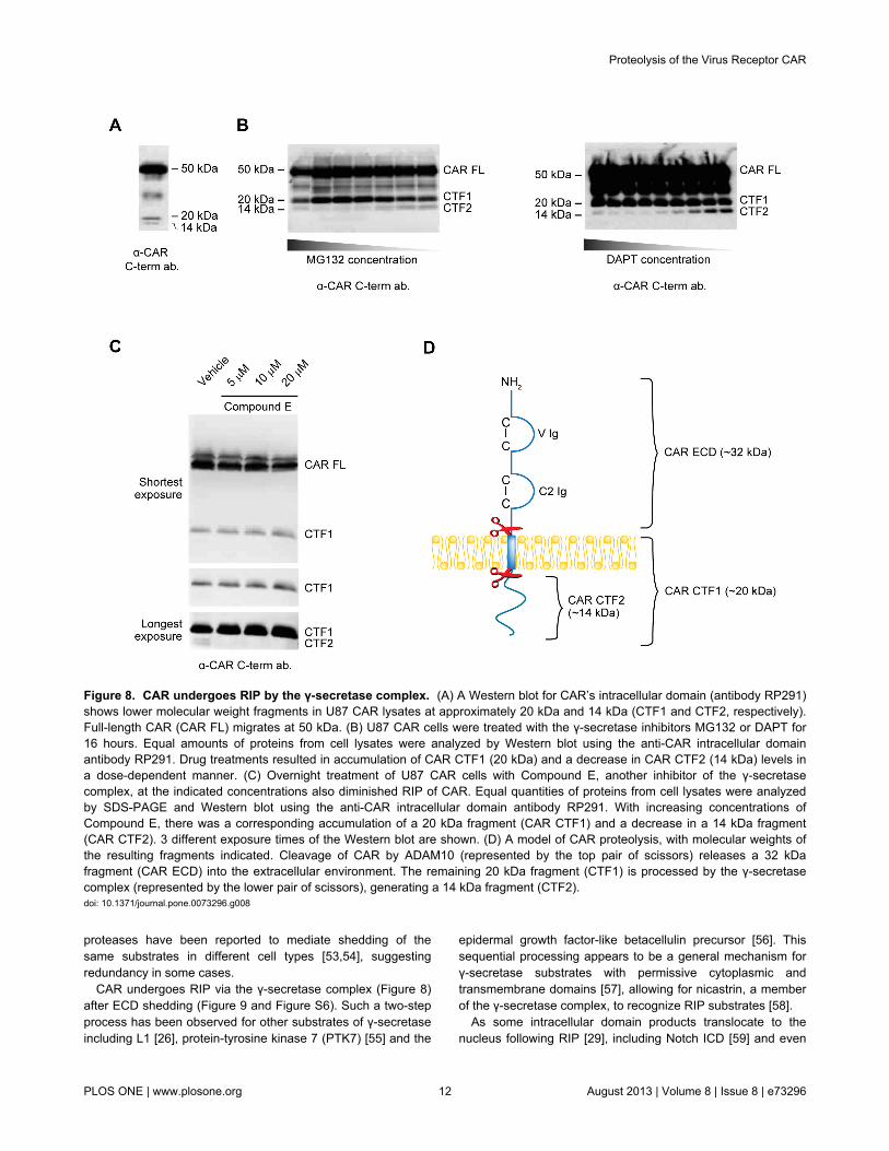
Often, cell surface proteins that undergo ECD shedding arealso cleaved by the γ-secretase complex, resulting in therelease of their intracellular domains into the cytosol. Toinvestigate if smaller fragments are generated from CAR, theanti-CAR intracellular domain antibody RP291 was used inWestern blots of U87 CAR cell lysates. Besides the full-lengthform of CAR (CAR FL), two C-terminal fragments (CTF1 andCTF2) of approximately 20 kDa and 14 kDa, respectively, weredetected from cell lysates (Figure 8A).
We hypothesized that CTF1 is the remaining part of CARafter its ECD is shed, and that CTF2 is the product of thesubsequent RIP of CTF1. If so, then inhibition of regulatedintramembrane proteases such as the γ-secretase complexshould lead to a decrease in CTF2 levels. Indeed, we foundthat treatment with the γ-secretase inhibitors MG132 and DAPTdecreased CTF2 in a dose-dependent manner, and that thisphenomenon was accompanied by an accumulation of CTF1levels (Figure 8B). Similar results were obtained with a third γ-secretase inhibitor, Compound E (Figure 8C). The products ofproteolytic cleavage of CAR and their molecular weights areillustrated in Figure 8D.
The results in Figure 8 were additionally verified by stablyexpressing a CAR construct with a C-terminal V5 tag (CAR-V5)in U87-MG and U251N cells. Western blots of lysates fromthese cell lines using an anti-V5 antibody revealed thepresence of CAR FL as well as two lower molecular weightspecies, CAR CTF1 (20 kDa) and CAR CTF2 (14 kDa) (Figure9A), similar to fragments of intracellular CAR detected with theRP291 antibody (Figure 8A). When CAR-V5-expressing U251Ncells were treated with MG132 to inhibit γ-secretase, CTF1levels accumulated while CTF2 levels decreased (Figure 9B),similar to results obtained using the anti-CAR intracellulardomain antibody RP291 (Figure 8B). Thus, in both U87-MGand U251N glioma cells, V5-tagged CAR undergoes RIP likeuntagged CAR.
Proteolysis of the Virus Receptor CAR
PLOS ONE | www.plosone.org 8 August 2013 | Volume 8 | Issue 8 | e73296

Figure 5. shRNA knockdown of ADAM10 decreases constitutive, PMA-mediated and ionomycin-mediated CAR ECDshedding. (A) A Western blot for ADAM10 using cell lysates of U87 CAR cells containing either control shRNA or ADAM10 (#6676)shRNA, in biological duplicates. Equal amounts of proteins were loaded on SDS-PAGE gel. Anti-GAPDH antibody was used as aloading control. Quantification of mean ADAM10 band intensities normalized over GAPDH revealed a decrease of approximately60% with anti-ADAM10 shRNA compared to control shRNA. (B) A constitutive shedding experiment was performed using U87 CARstable cell lines containing either control shRNA or anti-ADAM10 shRNA (#6676). Conditioned media and cell lysates were collectedafter 24 hours of incubation of cells in opti-MEM, and Western blotting was done using the anti-CAR N-terminus antibody 2239. Withanti-ADAM10 shRNA, there was a significant decrease (40%) in the levels of shed CAR. Results from 4 independent experimentsperformed in duplicates were quantified (unpaired t-test; p=0.0004 (***)). (C) U87 CAR cells containing either control shRNA orADAM10 shRNA (#6676) were treated with 1 µM PMA. Conditioned media and cell lysates were collected after 3 hours, andWestern blots were performed using the anti-CAR N-terminus antibody 2239. With shRNA knockdown of ADAM10, there was asignificant decrease of 41% in levels of shed CAR compared to control shRNA. Results from 3 independent experiments performedin duplicates were quantified (one-way ANOVA with Tukey’s multiple comparison test; *** = p < 0.001). (D) U87 CAR cellscontaining either control shRNA or ADAM10 shRNA (#6675) were treated with 1.5 µM ionomycin (vs. DMSO vehicle). Conditionedmedia and cell lysates were collected after 30 minutes of treatment, and Western blots were performed using the anti-CAR N-terminus antibody 2240. With shRNA knockdown of ADAM10, there was a significant decrease of 52% in levels of shed CAR withionomycin treatment compared to control shRNA. Results from 3 independent experiments (n=3 per group) were quantified (one-way ANOVA with Bonferroni’s multiple comparison test; * = p < 0.05, ** = p < 0.01, *** = p < 0.001).doi: 10.1371/journal.pone.0073296.g005
Proteolysis of the Virus Receptor CAR
PLOS ONE | www.plosone.org 9 August 2013 | Volume 8 | Issue 8 | e73296

As PS is the catalytic subunit of the γ-secretase complex[45], we hypothesized that lack of PS expression would resultin absence of the 14 kDa CAR CTF2 fragment. MEF wild-type(WT) and MEF PS 1 and 2 knockout (PS 1/2 KO) cells wereinfected with a lentiviral vector expressing CAR with a C-terminal V5 tag. Lysates were analyzed by Western blot usingantibody raised against V5. Although CAR CTF2 was readilydetected in MEF WT cells, it was absent in cells null for PS 1
Figure 6. Partial rescue of shRNA-mediated loss of CARshedding using an shRNA-resistant mutant ofADAM10. (A) shRNA stable cell lines of U251N CAR cellswere transfected with either empty plasmid (mock) or anshRNA-resistant construct of ADAM10. 18-24 hours post-transfection, cells were washed and incubated in opti-MEM.Conditioned media and cell lysates were collected 24 hourslater and analyzed by Western blot for CAR extracellulardomain (anti-CAR antibody 2239). The shRNA-resistantADAM10 mutant partially rescued CAR shedding in theADAM10 (6676) shRNA cell line, compared to transfecting thiscell line with empty plasmid (third and fourth Western blotbands from the left). (B) The band intensities of shed CARdetected by Western blot were quantified from 4 independentexperiments (One-way ANOVA with Newman-Keuls multiplecomparison test; * = p < 0.05, ** = p < 0.01, *** = p < 0.001).doi: 10.1371/journal.pone.0073296.g006
and 2 (Figure 9C). Thus, the PS/γ-secretase complex isrequired for RIP of CAR, resulting in generation of a 14 kDafragment, CTF2.
To determine whether or not RIP of CAR is contingent uponits ECD shedding, U87 CAR-V5 cells were treated for 4 hourswith 25 µM of the metalloprotease inhibitor GM6001 or itsnegative control. Inhibition of CAR ECD shedding wasaccompanied by a decrease in levels of both CTF1 and CTF2(Figure S6). For further confirmation, ADAM10 was knockeddown in U87 CAR-V5 cells (ADAM10 shRNA #6675), withcontrol shRNA for comparison. The ADAM10 shRNA cell linehad reduced amounts of the 20 kDa CTF1 fragment, asexpected (Figure 9D), since a decrease in ECD shedding isexpected to be accompanied by lower CTF1 levels.Importantly, levels of the 14 kDa CAR CTF2 also decreased.These data indicate that RIP of CAR is dependent on ECDshedding of the full-length receptor.
CAR intracellular domain enters the nucleusFinally, as RIP of some cell surface proteins produces
intracellular fragments that enter the nucleus, we investigated ifthat is the case for CAR. Nuclear immunoreactivity wasobserved with transient expression of full-length CAR-V5 in 293cells (data not shown); however, the appearance of thesespeckles was rare. We generated a construct expressing theintracellular domain (amino acids 261-365 of murine CARisoform 1) tagged at the C-terminus with V5, named CAR ICD-V5. U87-MG cells were transiently transfected with emptyplasmid, full-length CAR-V5 or CAR ICD-V5, andimmunofluorescence experiments were performed. Imageswere acquired using a confocal microscope. The ICD wasreadily detected in nuclei of U87 cells (Figure 10 and FigureS7). Similar results were obtained from experiments using293A cells (data not shown). Thus, the intracellular domain ofCAR is capable of nuclear entry.
We hypothesized that CAR CTF2 is not readily detectable innuclei due to rapid proteasomal degradation of this fragment.Indeed, treatment of U87 CAR-V5 cells with the proteasomeinhibitor epoxomicin increased levels of CTF2 and the ratio ofCTF2 to CTF1 (Figure S8A and Figure S8B). Levels of freeCAR ICD also accumulated in the presence of the proteasomeinhibitor MG132 (Figure S8C). However, despite theimprovement of CTF2 levels with epoxomicin, any nuclearCTF2 remained below detection level in epoxomicin-treatedU87 CAR-V5 cells (data not shown).
Discussion
A wide variety of cell surface proteins, including celladhesion molecules, shed their ectodomains and also undergoRIP [25,29]. Here, we report that the cell adhesion moleculeand virus receptor CAR is also subject to these processingevents. CAR ECD shed from glioma cells ectopicallyexpressing CAR as well as from developing neurons withendogenous CAR expression (Figure 1). In addition to thisconstitutive shedding, CAR ECD shed in a regulated fashionwhen cells were treated with the phorbol ester PMA (underchronic conditions) or with the calcium ionophore ionomycin
Proteolysis of the Virus Receptor CAR
PLOS ONE | www.plosone.org 10 August 2013 | Volume 8 | Issue 8 | e73296

(Figure 2), indicating an involvement of the PKC and calciumpathways, respectively, as has been demonstrated for agrowing number of other cell surface proteins. The PKC andcalcium signaling pathways are involved in cell migration,neuronal growth cone function and neurite outgrowth ofdeveloping neurons [46–49]. Regulation of CAR ECD sheddingby these signaling pathways may thus have implications forCAR’s role as a cell adhesion molecule [6,8].
Using various approaches, we identified ADAM10 as themetalloprotease mediating constitutive and ionomycin-inducedshedding of CAR, as well as shedding induced by highconcentrations of PMA, from human glioma cells (Figure 3,Figure 4, Figure 5 and Figure 6). As ADAMs do not haveconsensus cleavage sites on substrates, we performed in vitropeptide digestion assays and mass spectrometry to determinethe site of ADAM10 cleavage on CAR (Figure S4). The mutantMLRL AAAA stably expressed in HEK 293 cells did not shed itsECD into conditioned media (Figure 7B), indicating that aminoacids 224-227 are important for CAR ECD shedding.
Interestingly, deletion of a larger area consisting of amino acids221-232 resulted in a mutant that readily shed its ECD (Figure7B), perhaps due to a change in protein conformation thatallowed the mutant receptor to be processed by ADAM10 orsome other protease. While the mutants were expressedsimilarly to wild-type CAR at the cell surface in HEK 293 cells(Figure 7B), they were greatly diminished in expression at thesurface of U251N cells (Figure S5D). The differences in cellsurface expression levels of the mutants in HEK 293 andU251N cells may be due to differences in post-translationalmodification or protein folding.
Although ADAM10 is clearly a sheddase of CAR in oursystem, it is possible that other proteases, whether from theADAM family or other metalloprotease families, are alsocapable of cleaving CAR’s ECD. For example, the celladhesion molecule L1 is shed by both ADAM10 and ADAM17although via different stimuli [26], and the hyaluronan receptorCD44 is shed by ADAM10, ADAM17 [50] and the matrixmetalloproteinases MMP-9 [51] and MMP-14 [52]. Different
Figure 7. Confirmation of the area of ECD cleavage on CAR in HEK 293 cells. (A) A schematic showing putative ADAM10cleavage sites on CAR’s extracellular domain (arrows), as obtained from in vitro peptide digestion and mass spectrometry (FigureS4). (B) HEK 293 stable cell populations were generated to express wild-type CAR, the mutant MLRLAAAA or the mutantΔ221-232. The MLRLAAAA mutant did not shed into conditioned media of HEK 293 cells, while the Δ221-232 mutant shed its ECDsimilarly to wild-type CAR. Cell surface biotinylation experiments revealed that wild-type CAR and the two mutants are expressedsimilarly on the cell surface. Note that the low level of endogenous CAR in HEK 293 was detected after enrichment of cell surfacebiotinylated proteins (mock lane). Western blotting was performed using the anti-CAR N-terminus antibody 2240.doi: 10.1371/journal.pone.0073296.g007
Proteolysis of the Virus Receptor CAR
PLOS ONE | www.plosone.org 11 August 2013 | Volume 8 | Issue 8 | e73296
proteases have been reported to mediate shedding of thesame substrates in different cell types [53,54], suggestingredundancy in some cases.
CAR undergoes RIP via the γ-secretase complex (Figure 8)after ECD shedding (Figure 9 and Figure S6). Such a two-stepprocess has been observed for other substrates of γ-secretaseincluding L1 [26], protein-tyrosine kinase 7 (PTK7) [55] and the
epidermal growth factor-like betacellulin precursor [56]. Thissequential processing appears to be a general mechanism forγ-secretase substrates with permissive cytoplasmic andtransmembrane domains [57], allowing for nicastrin, a memberof the γ-secretase complex, to recognize RIP substrates [58].
As some intracellular domain products translocate to thenucleus following RIP [29], including Notch ICD [59] and even
Figure 8. CAR undergoes RIP by the γ-secretase complex. (A) A Western blot for CAR’s intracellular domain (antibody RP291)shows lower molecular weight fragments in U87 CAR lysates at approximately 20 kDa and 14 kDa (CTF1 and CTF2, respectively).Full-length CAR (CAR FL) migrates at 50 kDa. (B) U87 CAR cells were treated with the γ-secretase inhibitors MG132 or DAPT for16 hours. Equal amounts of proteins from cell lysates were analyzed by Western blot using the anti-CAR intracellular domainantibody RP291. Drug treatments resulted in accumulation of CAR CTF1 (20 kDa) and a decrease in CAR CTF2 (14 kDa) levels ina dose-dependent manner. (C) Overnight treatment of U87 CAR cells with Compound E, another inhibitor of the γ-secretasecomplex, at the indicated concentrations also diminished RIP of CAR. Equal quantities of proteins from cell lysates were analyzedby SDS-PAGE and Western blot using the anti-CAR intracellular domain antibody RP291. With increasing concentrations ofCompound E, there was a corresponding accumulation of a 20 kDa fragment (CAR CTF1) and a decrease in a 14 kDa fragment(CAR CTF2). 3 different exposure times of the Western blot are shown. (D) A model of CAR proteolysis, with molecular weights ofthe resulting fragments indicated. Cleavage of CAR by ADAM10 (represented by the top pair of scissors) releases a 32 kDafragment (CAR ECD) into the extracellular environment. The remaining 20 kDa fragment (CTF1) is processed by the γ-secretasecomplex (represented by the lower pair of scissors), generating a 14 kDa fragment (CTF2).doi: 10.1371/journal.pone.0073296.g008
Proteolysis of the Virus Receptor CAR
PLOS ONE | www.plosone.org 12 August 2013 | Volume 8 | Issue 8 | e73296

Figure 9. Generation of CAR CTF1 precedes CTF2 production. (A) Stable cell lines of U87-MG and U251N expressing eitherthe V5 tag alone (mock) or CAR with a C-terminal V5 tag were generated. Equal amounts of cell lysates were analyzed by SDS-PAGE and Western blot using a mouse monoclonal antibody raised against the V5 tag. Full-length CAR, CTF1 and CTF2 weredetected similarly to lysates of U87 CAR cells probed with anti-CAR C-term. antibody RP291 (Figure 8A). (B) U251N V5 and U251NCAR-V5 cells were treated overnight with MG132 (25 µM) or DMSO vehicle control. Equal amounts of proteins from cell lysateswere used for anti-V5 Western blots. In the case of the MG132-treated cells, CTF1 levels accumulated while CTF2 nearlydisappeared, similar to previous experiments with the U87 CAR cell line (Figure 8B). (C) MEF wild-type (MEF WT) or PS 1- and 2-knockout MEF cells (MEF PS1/2 KO) were infected with lentivirus to express full-length CAR with a C-terminal V5 tag. Cells werelysed 3 days post-infection and lysates were analyzed by Western blot using antibody raised against the V5 tag. MEF WT cells, butnot MEF PS1/2 KO cells, contained CAR CTF2, indicating that presenilin is required for generation of the 14 kDa CTF2 fragment ofCAR. (D) Verification of knockdown in ADAM10 expression in U87 CAR-V5 cells using ADAM10 shRNA (#6675); shown are anti-ADAM10 and anti-GAPDH Western blots. Lysates were also analyzed by Western blot using antibody raised against the V5 tag.The ADAM10 shRNA stable cell line had a decreased CAR CTF1 level, as expected. CAR CTF2 levels also decreased, indicatingthat shedding is a prerequisite for RIP of CAR.doi: 10.1371/journal.pone.0073296.g009
Proteolysis of the Virus Receptor CAR
PLOS ONE | www.plosone.org 13 August 2013 | Volume 8 | Issue 8 | e73296

the ICD of ADAM10 [60], we investigated if that is the case forCAR. Indeed, transient expression of CAR ICD revealed thatthis portion of CAR enters the nucleus (Figure 10 and FigureS7). On the other hand, nuclear entry of CAR CTF2 generatedvia RIP was difficult to detect (data not shown), possibly due toits degradation by the proteasome (Figure S8). Degradation ofRIP-generated intracellular domain fragments has beenreported for other γ-secretase substrates such as Notch,syndecan-3, nectin-1α, p75, deleted in colorectal cancer (DCC)and members of the APP family [61–64]. However, RIPproducts, such as the Notch ICD, can nevertheless impartcellular effects at amounts below detection levels [65]. Anotherpossible reason for the difficulty in detecting CAR CTF2 in thenucleus may be due to retention of the fragment at the plasmamembrane via palmitoylation. Cysteine palmitoylation of CARat amino acids 259 and 260 is required for proper cell surfacetargeting of the receptor [66]. These cysteines are the first twoamino acids in CAR’s cytoplasmic domain. Since RIP of CARlikely occurs upstream of these residues within thetransmembrane domain, perhaps the resulting CTF2 fragmentremains tethered to the plasma membrane by palmitoylation.
As palmitoylation is a dynamic and reversible modification[67,68], in this scenario a fraction of CTF2 fragments may bedepalmitoylated and freed from the plasma membrane to enterthe nucleus.
While ECD shedding and RIP may be mechanisms for CARdegradation and protein turnover, it is possible that theseprocessing events can modulate physiological functions ofCAR including its role as a cell adhesion molecule in thedeveloping brain [6]. Shedding of the cell adhesion moleculeneuroligin antagonizes spine formation of neuronal dendrites[69], while metalloprotease activity is required for neuriteoutgrowth mediated by L1 [26] and CHL1 [27]. Proteolysis ofneural cell adhesion molecule (NCAM) in hippocampal neuronspromotes their outgrowth [70,71], but NCAM proteolysis andaddition of soluble NCAM ECD inhibits outgrowth of corticalneurons [72,73]. CAR mediates outgrowth of developingneurons [8], so its shedding may regulate this function as hasbeen described for other neuronal cell adhesion molecules. It isalso possible that CAR ICD, generated from RIP, may beinvolved in regulating neurite outgrowth. For example, RIP ofthe neurotrophin p75 receptor promotes neurite outgrowth,
Figure 10. CAR’s intracellular domain (ICD) enters the nucleus. Immunofluorescence and confocal microscopy imagesshowing the presence of CAR ICD in nuclei of U87-MG cells. U87-MG cells were transiently transfected with empty pcDNA3.1V5/His plasmid, full-length CAR-V5 plasmid or with CAR ICD-V5 plasmid. Immunofluorescence staining was performed 24-48 hourspost-transfection using anti-V5 tag antibody and Alexa Fluor 555 secondary antibody (red). Nuclei were stained with DRAQ5 (blue).Images were acquired with a confocal microscope (63x oil objective). Images are representative of at least 3 independentexperiments. Scale bars: 5 µm.doi: 10.1371/journal.pone.0073296.g010
Proteolysis of the Virus Receptor CAR
PLOS ONE | www.plosone.org 14 August 2013 | Volume 8 | Issue 8 | e73296

branching and number in dorsal root ganglion neurons,relieving inhibition by myelin-derived ligands [74], andpromotes neurite outgrowth of tropomyosin receptor kinasefamily member A (TrkA)-expressing neurons in response tonerve growth factor (NGF) [75].
In polarized epithelial cells, CAR participates in the formationof tight junctions, where its expression is associated with lowercell permeability [9,76]. It also localizes to cell-cell contacts innon-polarized epithelial cells [9]. ADAM10 has been reported tolocalize mainly in adherens junctions, and to a limited extent intight junctions, of polarized epithelial cells, and its propertargeting to adherens junctions promotes cell migration in awound healing assay [77]. Perhaps ADAM10 or othermetalloproteases can access CAR in tight junctions and shedits ECD, possibly downregulating CAR-mediated tight junctionintegrity.
Finally, proteolysis may play a role in regulation of CAR’sfunction as a virus receptor. One can imagine that in a situationwhere proteolysis of CAR at the cell surface is upregulated, forexample, via the PKC or calcium signaling pathways or byincreased activity of ADAM10, host cells would be renderedless susceptible to infection by Coxsackievirus and adenovirusserotypes that require binding to CAR’s extracellular domain.
In conclusion, the characterization of shedding and RIP ofCAR presented in this work promote our understanding of thecell adhesion molecule and virus receptor CAR. Future workwill reveal what roles, if any, are played by these proteolyticcleavages in the function of CAR.
Supporting Information
Figure S1. A fragment of CAR consisting of part of itsextracellular domain is shed into media of U87 cells andcannot be detected with an anti-C terminus antibody. A 32kDa fragment of CAR was released into conditioned media ofU87 CAR cells upon 4 hours of treatment with 1 µM PMA. Thisfragment was recognized by anti-CAR extracellular domainantibody 2240 (A), but not by antibody RP291 raised againstCAR intracellular domain (B).(TIF)
Figure S2. PMA treatment leads to CAR phosphorylation.U87 CAR cells were treated for 4 hours with 1 µM PMA vs.DMSO vehicle, and cell lysates were collected. Calf intestinalphosphatase (CIP) treatment of lysates abolished theappearance of the higher molecular weight species of full-length CAR obtained with PMA treatment, indicating that PMAcauses a post-translational modification of full-length CAR inthe form of phosphorylation. Western blotting was performedwith the anti-CAR N-terminus antibody 2240.(TIF)
Figure S3. Real-time quantitative PCR for verification ofknockdown of ADAM10 mRNA levels. U87 CAR stable celllines infected with lentivirus containing control (anti-eGFP)shRNA or anti-ADAM10 (#6675 or #6676) shRNA weregenerated. RNA was isolated from these cells, followed byreverse transcription to cDNA and real-time PCR in triplicates
to quantify ADAM10, ADAM17 and GAPDH expression levels.The two anti-ADAM10 shRNA sequences #6675 and #6676successfully knocked down mRNA levels of ADAM10compared to control shRNA without affecting expression levelsof the related family member ADAM17.(TIF)
Figure S4. Mapping the sites of ECD cleavage on CAR. A20-amino acid peptide (VGSDQCMLRLDVVPPSNRAG)representing the juxtamembrane region in CAR ECD wasdigested with recombinant human ADAM10 at 37°C for 4 or 16hours, along with 3 controls (recombinant ADAM10 only, 16hours; peptide only, 16 hours; peptide and recombinantADAM10; 0 hours). Samples were analyzed by MALDI-MS.Two unique peaks (shaded grey) at (A) 1008 m/z and (B) 1393m/z were found that were not present in the 3 controls. Furtheranalysis was done with MS/MS in order to deduce the identitiesof the amino acids in each peptide fragment. These resultsrepresent 2 independent experiments.(TIF)
Figure S5. Characterization of CAR ECD mutants inhuman glioma U251N cells. (A) Stable U251N cell lines ofmock (empty vector), wild-type CAR, and 3 mutants (MLAA, RLAA and Δ221-232) were generated. Constitutive shedding ofCAR and the mutants was assayed. Mutating pairs of aminoacids to alanine (MLAA and RLAA) led to a decrease in CARECD shedding. However, this inhibition was reversed insubsequent cell passages. Deletion of 12 amino acids(Δ221-232) containing the potential area of ECD cleavageresulted in a mutant that still shed. (B) A mutant CAR wasgenerated in which amino acids 224-227 were changed toalanine residues (MLRL AAAA), and was stably expressed inU251N cells. Shedding of this mutant was completelyabrogated. Cell surface biotinylation experiments (panels Cand D) revealed that all the mutants were expressed at muchlower levels at the surface of U251N cells compared to wild-type CAR.(TIF)
Figure S6. GM6001 treatment results in a decrease in CARCTF1 and CTF2 levels. U87 cells stably expressing CAR witha C-terminal V5 tag (CAR-V5) were treated with 25 µM of themetalloprotease inhibitor GM6001 or its negative control for 4hours. Conditioned media and lysates were collected asdescribed, and Western blotting was performed with the anti-CAR N-terminus antibody 2240 (for conditioned media) andanti-V5 tag antibody (for lysates). GM6001 treatment abrogatedCAR ECD shedding as expected. There was a small decreasein levels of both CAR CTF1 and CTF2 with GM6001 treatment.(TIF)
Figure S7. Z stack images of a U87 cell transientlyexpressing CAR ICD. Confocal microscopy Z stack imageswere acquired of a U87 cell transiently expressing V5-taggedCAR ICD (red = anti-V5). Shown are 20 slices representing atotal thickness of 6.59 µm. Scale bar: 5 µm.(TIF)
Proteolysis of the Virus Receptor CAR
PLOS ONE | www.plosone.org 15 August 2013 | Volume 8 | Issue 8 | e73296

Figure S8. CAR ICD is subject to proteasomaldegradation. (A) U87 CAR-V5 cells were treated for 16 hourswith the proteasome inhibitor epoxomicin (1 µM or 5 µM) vs.DMSO vehicle. Shown is a representative Western blotperformed using antibody raised against the V5 tag. (B) CTF1and CTF2 band intensities were quantified from Western blots,and ratios of CTF2/CTF1 were calculated. The graphrepresents mean CTF2/CTF1 ratios obtained from 3independent experiments (n=3 per group). One-way ANOVAwith Bonferroni post-test, * = p < 0.05. (C) U87 cells transientlyexpressing V5-tagged CAR ICD were treated overnight with theproteasome inhibitor MG132 (25 µM) or DMSO vehicle control.Samples were analyzed by Western blotting for GAPDH andthe V5 tag. Treatment with MG132 led to an accumulation ofCAR ICD levels.
(TIF)
Acknowledgements
We wish to thank the following individuals for generouslysharing materials: Dr. Marcia Moss (Biozyme, Inc.) for purifiedADAM10 prodomain, Dr. Bart de Strooper (K.U. Leuven) for thepresenilin knockout MEF cell line, Dr. Peter Forsyth (Universityof Calgary) for the U251N cell line, Dr. Kerstin Sollerbrant(Karolinska Institute) for RP291 antibody, and Dr. AlysonFournier (McGill University) for the pcDNA3.1 V5/His plasmid.
Author Contributions
Conceived and designed the experiments: K-CH NH JN.Performed the experiments: K-CH NH. Analyzed the data: K-CH NH JN. Wrote the manuscript: K-CH NH JN.
References
1. Carson SD (2001) Receptor for the group B coxsackieviruses andadenoviruses: CAR. Rev Med Virol 11: 219-226. doi:10.1002/rmv.318.PubMed: 11479928.
2. Coyne CB, Bergelson JM (2005) CAR: a virus receptor within the tightjunction. Adv Drug Deliv Rev 57: 869-882. doi:10.1016/j.addr.2005.01.007. PubMed: 15820557.
3. Bergelson JM, Cunningham JA, Droguett G, Kurt-Jones EA, Krithivas Aet al. (1997) Isolation of a common receptor for Coxsackie B virusesand adenoviruses 2 and 5. Science 275: 1320-1323. doi:10.1126/science.275.5304.1320. PubMed: 9036860.
4. Tomko RP, Xu R, Philipson L (1997) HCAR and MCAR: the human andmouse cellular receptors for subgroup C adenoviruses and group Bcoxsackieviruses. Proc Natl Acad Sci U S A 94: 3352-3356. doi:10.1073/pnas.94.7.3352. PubMed: 9096397.
5. Carson SD, Chapman NN, Tracy SM (1997) Purification of the putativecoxsackievirus B receptor from HeLa cells. Biochem Biophys ResCommun 233: 325-328. doi:10.1006/bbrc.1997.6449. PubMed:9144533.
6. Honda T, Saitoh H, Masuko M, Katagiri-Abe T, Tominaga K et al.(2000) The coxsackievirus-adenovirus receptor protein as a celladhesion molecule in the developing mouse brain. Brain Res Mol BrainRes 77: 19-28. doi:10.1016/S0169-328X(00)00036-X. PubMed:10814828.
7. Huang KC, Yasruel Z, Guérin C, Holland PC, Nalbantoglu J (2007)Interaction of the Coxsackie and adenovirus receptor (CAR) with thecytoskeleton: binding to actin. FEBS Lett 581: 2702-2708. doi:10.1016/j.febslet.2007.05.019. PubMed: 17531226.
8. Patzke C, Max KE, Behlke J, Schreiber J, Schmidt H et al. (2010) Thecoxsackievirus-adenovirus receptor reveals complex homophilic andheterophilic interactions on neural cells. J Neurosci 30: 2897-2910. doi:10.1523/JNEUROSCI.5725-09.2010. PubMed: 20181587.
9. Cohen CJ, Shieh JT, Pickles RJ, Okegawa T, Hsieh JT et al. (2001)The coxsackievirus and adenovirus receptor is a transmembranecomponent of the tight junction. Proc Natl Acad Sci U S A 98:15191-15196. doi:10.1073/pnas.261452898. PubMed: 11734628.
10. Coyne CB, Voelker T, Pichla SL, Bergelson JM (2004) Thecoxsackievirus and adenovirus receptor interacts with the multi-PDZdomain protein-1 (MUPP-1) within the tight junction. J Biol Chem 279:48079-48084. doi:10.1074/jbc.M409061200. PubMed: 15364909.
11. Nalbantoglu J, Pari G, Karpati G, Holland PC (1999) Expression of theprimary coxsackie and adenovirus receptor is downregulated duringskeletal muscle maturation and limits the efficacy of adenovirus-mediated gene delivery to muscle cells. Hum Gene Ther 10:1009-1019. doi:10.1089/10430349950018409. PubMed: 10223734.
12. Shaw CA, Holland PC, Sinnreich M, Allen C, Sollerbrant K et al. (2004)Isoform-specific expression of the Coxsackie and adenovirus receptor(CAR) in neuromuscular junction and cardiac intercalated discs. BMCCell Biol 5: 42. doi:10.1186/1471-2121-5-42. PubMed: 15533241.
13. Asher DR, Cerny AM, Weiler SR, Horner JW, Keeler ML et al. (2005)Coxsackievirus and adenovirus receptor is essential for cardiomyocytedevelopment. Genesis 42: 77-85. doi:10.1002/gene.20127. PubMed:15864812.
14. Dorner AA, Wegmann F, Butz S, Wolburg-Buchholz K, Wolburg H et al.(2005) Coxsackievirus-adenovirus receptor (CAR) is essential for earlyembryonic cardiac development. J Cell Sci 118: 3509-3521. doi:10.1242/jcs.02476. PubMed: 16079292.
15. Chen JW, Zhou B, Yu QC, Shin SJ, Jiao K et al. (2006) Cardiomyocyte-specific deletion of the coxsackievirus and adenovirus receptor resultsin hyperplasia of the embryonic left ventricle and abnormalities ofsinuatrial valves. Circ Res 98: 923-930. doi:10.1161/01.RES.0000218041.41932.e3. PubMed: 16543498.
16. Lim BK, Xiong D, Dorner A, Youn TJ, Yung A et al. (2008)Coxsackievirus and adenovirus receptor (CAR) mediatesatrioventricular-node function and connexin 45 localization in themurine heart. J Clin Invest 118: 2758-2770. doi:10.1172/JCI34777.PubMed: 18636119.
17. Lisewski U, Shi Y, Wrackmeyer U, Fischer R, Chen C et al. (2008) Thetight junction protein CAR regulates cardiac conduction and cell-cellcommunication. J Exp Med 205: 2369-2379. doi:10.1084/jem.20080897. PubMed: 18794341.
18. Pazirandeh A, Sultana T, Mirza M, Rozell B, Hultenby K et al. (2011)Multiple phenotypes in adult mice following inactivation of theCoxsackievirus and Adenovirus Receptor (Car) gene. PLOS ONE 6:e20203. doi:10.1371/journal.pone.0020203. PubMed: 21674029.
19. Zen K, Liu Y, McCall IC, Wu T, Lee W et al. (2005) Neutrophil migrationacross tight junctions is mediated by adhesive interactions betweenepithelial coxsackie and adenovirus receptor and a junctional adhesionmolecule-like protein on neutrophils. Mol Biol Cell 16: 2694-2703. doi:10.1091/mbc.E05-01-0036. PubMed: 15800062.
20. Guo YL, Bai R, Chen CX, Liu DQ, Liu Y et al. (2009) Role of junctionaladhesion molecule-like protein in mediating monocyte transendothelialmigration. Arterioscler Thromb Vasc Biol 29: 75-83. doi:10.1161/ATVBAHA.108.177717. PubMed: 18948633.
21. Witherden DA, Verdino P, Rieder SE, Garijo O, Mills RE et al. (2010)The junctional adhesion molecule JAML is a costimulatory receptor forepithelial gammadelta T cell activation. Science 329: 1205-1210. doi:10.1126/science.1192698. PubMed: 20813954.
22. Okegawa T, Pong RC, Li Y, Bergelson JM, Sagalowsky AI et al. (2001)The mechanism of the growth-inhibitory effect of coxsackie andadenovirus receptor (CAR) on human bladder cancer: a functionalanalysis of car protein structure. Cancer Res 61: 6592-6600. PubMed:11522659.
23. Kim M, Sumerel LA, Belousova N, Lyons GR, Carey DE et al. (2003)The coxsackievirus and adenovirus receptor acts as a tumoursuppressor in malignant glioma cells. Br J Cancer 88: 1411-1416. doi:10.1038/sj.bjc.6600932. PubMed: 12778071.
24. Huang KC, Altinoz M, Wosik K, Larochelle N, Koty Z et al. (2005)Impact of the coxsackie and adenovirus receptor (CAR) on glioma cellgrowth and invasion: requirement for the C-terminal domain. Int JCancer 113: 738-745. doi:10.1002/ijc.20623. PubMed: 15499626.
25. Edwards DR, Handsley MM, Pennington CJ (2008) The ADAMmetalloproteinases. Mol Aspects Med 29: 258-289. doi:10.1016/j.mam.2008.08.001. PubMed: 18762209.
Proteolysis of the Virus Receptor CAR
PLOS ONE | www.plosone.org 16 August 2013 | Volume 8 | Issue 8 | e73296
26. Maretzky T, Schulte M, Ludwig A, Rose-John S, Blobel C et al. (2005)L1 is sequentially processed by two differently activatedmetalloproteases and presenilin/gamma-secretase and regulatesneural cell adhesion, cell migration, and neurite outgrowth. Mol Cell Biol25: 9040-9053. doi:10.1128/MCB.25.20.9040-9053.2005. PubMed:16199880.
27. Naus S, Richter M, Wildeboer D, Moss M, Schachner M et al. (2004)Ectodomain shedding of the neural recognition molecule CHL1 by themetalloprotease-disintegrin ADAM8 promotes neurite outgrowth andsuppresses neuronal cell death. J Biol Chem 279: 16083-16090. doi:10.1074/jbc.M400560200. PubMed: 14761956.
28. Reiss K, Maretzky T, Ludwig A, Tousseyn T, de Strooper B et al.(2005) ADAM10 cleavage of N-cadherin and regulation of cell-celladhesion and beta-catenin nuclear signalling. EMBO J 24: 742-752.doi:10.1038/sj.emboj.7600548. PubMed: 15692570.
29. Lichtenthaler SF, Haass C, Steiner H (2011) Regulated intramembraneproteolysis--lessons from amyloid precursor protein processing. JNeurochem 117: 779-796. doi:10.1111/j.1471-4159.2011.07248.x.PubMed: 21413990.
30. Wakabayashi T, De Strooper B (2008) Presenilins: members of thegamma-secretase quartets, but part-time soloists too. Physiol(Bethesda) 23: 194-204. doi:10.1152/physiol.00009.2008. PubMed:18697993.
31. Cao X, Südhof TC (2001) A transcriptionally [correction oftranscriptively] active complex of APP with Fe65 and histoneacetyltransferase Tip60. Science 293: 115-120. doi:10.1126/science.1058783. PubMed: 11441186.
32. Weinmaster G (2000) Notch signal transduction: a real rip and more.Curr Opin Genet Dev 10: 363-369. doi:10.1016/S0959-437X(00)00097-6. PubMed: 10889061.
33. Sollerbrant K, Raschperger E, Mirza M, Engstrom U, Philipson L et al.(2003) The Coxsackievirus and adenovirus receptor (CAR) forms acomplex with the PDZ domain-containing protein ligand-of-numbprotein-X (LNX). J Biol Chem 278: 7439-7444. doi:10.1074/jbc.M205927200. PubMed: 12468544.
34. Herreman A, Hartmann D, Annaert W, Saftig P, Craessaerts K et al.(1999) Presenilin 2 deficiency causes a mild pulmonary phenotype andno changes in amyloid precursor protein processing but enhances theembryonic lethal phenotype of presenilin 1 deficiency. Proc Natl AcadSci U S A 96: 11872-11877. doi:10.1073/pnas.96.21.11872. PubMed:10518543.
35. Herreman A, Van Gassen G, Bentahir M, Nyabi O, Craessaerts K et al.(2003) gamma-Secretase activity requires the presenilin-dependenttrafficking of nicastrin through the Golgi apparatus but not its complexglycosylation. J Cell Sci 116: 1127-1136. doi:10.1242/jcs.00292.PubMed: 12584255.
36. Yee NS, Langen H, Besmer P (1993) Mechanism of kit ligand, phorbolester, and calcium-induced down-regulation of c-kit receptors in mastcells. J Biol Chem 268: 14189-14201. PubMed: 7686152.
37. Horiuchi K, Le Gall S, Schulte M, Yamaguchi T, Reiss K et al. (2007)Substrate selectivity of epidermal growth factor-receptor ligandsheddases and their regulation by phorbol esters and calcium influx.Mol Biol Cell 18: 176-188. PubMed: 17079736.
38. Le Gall, Bobé P, Reiss K, Horiuchi K, Niu XD et al. (2009) ADAMs 10and 17 represent differentially regulated components of a generalshedding machinery for membrane proteins such as transforminggrowth factor alpha, L-selectin, and tumor necrosis factor alpha. MolBiol Cell 20: 1785-1794. doi:10.1091/mbc.E08-11-1135. PubMed:19158376.
39. Castagna M, Takai Y, Kaibuchi K, Sano K, Kikkawa U et al. (1982)Direct activation of calcium-activated, phospholipid-dependent proteinkinase by tumor-promoting phorbol esters. J Biol Chem 257:7847-7851. PubMed: 7085651.
40. Arribas J, Borroto A (2002) Protein ectodomain shedding. Chem Rev102: 4627-4638. doi:10.1021/cr010202t. PubMed: 12475204.
41. Sahin U, Weskamp G, Kelly K, Zhou HM, Higashiyama S et al. (2004)Distinct roles for ADAM10 and ADAM17 in ectodomain shedding of sixEGFR ligands. J Cell Biol 164: 769-779. doi:10.1083/jcb.200307137.PubMed: 14993236.
42. Murphy G (2011) Tissue inhibitors of metalloproteinases. Genome Biol12: 233. doi:10.1186/gb-2011-12-11-233. PubMed: 22078297.
43. Moss ML, Bomar M, Liu Q, Sage H, Dempsey P et al. (2007) TheADAM10 prodomain is a specific inhibitor of ADAM10 proteolyticactivity and inhibits cellular shedding events. J Biol Chem 282:35712-35721. doi:10.1074/jbc.M703231200. PubMed: 17895248.
44. Jiang S, Caffrey M (2007) Solution structure of the coxsackievirus andadenovirus receptor domain 2. Protein Sci 16: 539-542. doi:10.1110/ps.062643507. PubMed: 17322536.
45. Jorissen E, De Strooper B (2010) Gamma-secretase and theintramembrane proteolysis of Notch. Curr Top Dev Biol 92: 201-230.doi:10.1016/S0070-2153(10)92006-1. PubMed: 20816396.
46. Kiryushko D, Berezin V, Bock E (2004) Regulators of neurite outgrowth:role of cell adhesion molecules. Ann N Y Acad Sci 1014: 140-154. doi:10.1196/annals.1294.015. PubMed: 15153429.
47. Bolsover SR (2005) Calcium signalling in growth cone migration. CellCalcium 37: 395-402. doi:10.1016/j.ceca.2005.01.007. PubMed:15820386.
48. Larsson C (2006) Protein kinase C and the regulation of the actincytoskeleton. Cell Signal 18: 276-284. doi:10.1016/j.cellsig.2005.07.010. PubMed: 16109477.
49. Rosse C, Linch M, Kermorgant S, Cameron AJ, Boeckeler K et al.(2010) PKC and the control of localized signal dynamics. Nat Rev MolCell Biol 11: 103-112. doi:10.1038/nrm2847. PubMed: 20094051.
50. Nagano O, Murakami D, Hartmann D, De Strooper B, Saftig P et al.(2004) Cell-matrix interaction via CD44 is independently regulated bydifferent metalloproteinases activated in response to extracellularCa(2+) influx and PKC activation. J Cell Biol 165: 893-902. doi:10.1083/jcb.200310024. PubMed: 15197174.
51. Chetty C, Vanamala SK, Gondi CS, Dinh DH, Gujrati M et al. (2012)MMP-9 induces CD44 cleavage and CD44 mediated cell migration inglioblastoma xenograft cells. Cell Signal 24: 549-559. doi:10.1016/j.cellsig.2011.10.008. PubMed: 22024282.
52. Kajita M, Itoh Y, Chiba T, Mori H, Okada A et al. (2001) Membrane-type1 matrix metalloproteinase cleaves CD44 and promotes cell migration.J Cell Biol 153: 893-904. doi:10.1083/jcb.153.5.893. PubMed:11381077.
53. Murphy G (2010) Fell-Muir Lecture: Metalloproteinases: from demolitionsquad to master regulators. Int J Exp Pathol 91: 303-313. doi:10.1111/j.1365-2613.2010.00736.x. PubMed: 20666850.
54. Tousseyn T, Jorissen E, Reiss K, Hartmann D (2006) (Make) stick andcut loose--disintegrin metalloproteases in development and disease.Birth Defects Res C Embryo TODAY 78: 24-46. doi:10.1002/bdrc.20066. PubMed: 16622847.
55. Na HW, Shin WS, Ludwig A, Lee ST (2012) The cytosolic domain ofprotein-tyrosine kinase 7 (PTK7), generated from sequential cleavageby a disintegrin and metalloprotease 17 (ADAM17) and gamma-secretase, enhances cell proliferation and migration in colon cancercells. J Biol Chem 287: 25001-25009. doi:10.1074/jbc.M112.348904.PubMed: 22665490.
56. Stoeck A, Shang L, Dempsey PJ (2010) Sequential and gamma-secretase-dependent processing of the betacellulin precursorgenerates a palmitoylated intracellular-domain fragment that inhibitscell growth. J Cell Sci 123: 2319-2331. doi:10.1242/jcs.060830.PubMed: 20530572.
57. Hemming ML, Elias JE, Gygi SP, Selkoe DJ (2008) Proteomic profilingof gamma-secretase substrates and mapping of substraterequirements. PLOS Biol 6: e257. doi:10.1371/journal.pbio.0060257.PubMed: 18942891.
58. Shah S, Lee SF, Tabuchi K, Hao YH, Yu C et al. (2005) Nicastrinfunctions as a gamma-secretase-substrate receptor. Cell 122: 435-447.doi:10.1016/j.cell.2005.05.022. PubMed: 16096062.
59. Kopan R, Nye JS, Weintraub H (1994) The intracellular domain ofmouse Notch: a constitutively activated repressor of myogenesisdirected at the basic helix-loop-helix region of MyoD. Development 120:2385-2396. PubMed: 7956819.
60. Tousseyn T, Thathiah A, Jorissen E, Raemaekers T, Konietzko U et al.(2009) ADAM10, the rate-limiting protease of regulated intramembraneproteolysis of Notch and other proteins, is processed by ADAMS-9,ADAMS-15, and the gamma-secretase. J Biol Chem 284:11738-11747. PubMed: 19213735.
61. Walsh DM, Fadeeva JV, LaVoie MJ, Paliga K, Eggert S et al. (2003)gamma-Secretase cleavage and binding to FE65 regulate the nucleartranslocation of the intracellular C-terminal domain (ICD) of the APPfamily of proteins. Biochemistry 42: 6664-6673. doi:10.1021/bi027375c.PubMed: 12779321.
62. Tagami S, Okochi M, Yanagida K, Ikuta A, Fukumori A et al. (2008)Regulation of Notch signaling by dynamic changes in the precision ofS3 cleavage of Notch-1. Mol Cell Biol 28: 165-176. doi:10.1128/MCB.00863-07. PubMed: 17967888.
63. Fryer CJ, White JB, Jones KA (2004) Mastermind recruits CycC:CDK8to phosphorylate the Notch ICD and coordinate activation with turnover.Mol Cell 16: 509-520. doi:10.1016/j.molcel.2004.10.014. PubMed:15546612.
64. Kopan R, Ilagan MX (2004) Gamma-secretase: proteasome of themembrane? Nat Rev Mol Cell Biol 5: 499-504. doi:10.1038/nrg1380.PubMed: 15173829.
Proteolysis of the Virus Receptor CAR
PLOS ONE | www.plosone.org 17 August 2013 | Volume 8 | Issue 8 | e73296

65. Schroeter EH, Kisslinger JA, Kopan R (1998) Notch-1 signallingrequires ligand-induced proteolytic release of intracellular domain.Nature 393: 382-386. doi:10.1038/30756. PubMed: 9620803.
66. van’t Hof W, Crystal RG (2002) Fatty acid modification of thecoxsackievirus and adenovirus receptor. J Virol 76: 6382-6386. doi:10.1128/JVI.76.12.6382-6386.2002. PubMed: 12021372.
67. Salaun C, Greaves J, Chamberlain LH (2010) The intracellular dynamicof protein palmitoylation. J Cell Biol 191: 1229-1238. doi:10.1083/jcb.201008160. PubMed: 21187327.
68. Iwanaga T, Tsutsumi R, Noritake J, Fukata Y, Fukata M (2009)Dynamic protein palmitoylation in cellular signaling. Prog Lipid Res 48:117-127. doi:10.1016/j.plipres.2009.02.001. PubMed: 19233228.
69. Suzuki K, Hayashi Y, Nakahara S, Kumazaki H, Prox J et al. (2012)Activity-dependent proteolytic cleavage of neuroligin-1. Neuron 76:410-422. doi:10.1016/j.neuron.2012.10.003. PubMed: 23083742.
70. Kalus I, Bormann U, Mzoughi M, Schachner M, Kleene R (2006)Proteolytic cleavage of the neural cell adhesion molecule by ADAM17/TACE is involved in neurite outgrowth. J Neurochem 98: 78-88. doi:10.1111/j.1471-4159.2006.03847.x. PubMed: 16805798.
71. Hübschmann MV, Skladchikova G, Bock E, Berezin V (2005) Neuralcell adhesion molecule function is regulated by metalloproteinase-mediated ectodomain release. J Neurosci Res 80: 826-837. doi:10.1002/jnr.20530. PubMed: 15884014.
72. Brennaman LH, Maness PF (2008) Developmental regulation ofGABAergic interneuron branching and synaptic development in theprefrontal cortex by soluble neural cell adhesion molecule. Mol Cell
Neurosci 37: 781-793. doi:10.1016/j.mcn.2008.01.006. PubMed:18289872.
73. Hinkle CL, Diestel S, Lieberman J, Maness PF (2006) Metalloprotease-induced ectodomain shedding of neural cell adhesion molecule(NCAM). J Neurobiol 66: 1378-1395. doi:10.1002/neu.20257. PubMed:16967505.
74. Ahmed Z, Mazibrada G, Seabright RJ, Dent RG, Berry M et al. (2006)TACE-induced cleavage of NgR and p75NTR in dorsal root ganglioncultures disinhibits outgrowth and promotes branching of neurites in thepresence of inhibitory CNS myelin. FASEB J 20: 1939-1941. doi:10.1096/fj.05-5339fje. PubMed: 16849393.
75. Matusica D, Skeldal S, Sykes AM, Palstra N, Sharma A et al. (2013) Anintracellular domain fragment of the p75 neurotrophin receptor(p75(NTR)) enhances tropomyosin receptor kinase A (TrkA) receptorfunction. J Biol Chem 288: 11144-11154. doi:10.1074/jbc.M112.436469. PubMed: 23471969.
76. Raschperger E, Thyberg J, Pettersson S, Philipson L, Fuxe J et al.(2006) The coxsackie- and adenovirus receptor (CAR) is an in vivomarker for epithelial tight junctions, with a potential role in regulatingpermeability and tissue homeostasis. Exp Cell Res 312: 1566-1580.doi:10.1016/j.yexcr.2006.01.025. PubMed: 16542650.
77. Wild-Bode C, Fellerer K, Kugler J, Haass C, Capell A (2006) Abasolateral sorting signal directs ADAM10 to adherens junctions and isrequired for its function in cell migration. J Biol Chem 281:23824-23829. doi:10.1074/jbc.M601542200. PubMed: 16777847.
Proteolysis of the Virus Receptor CAR
PLOS ONE | www.plosone.org 18 August 2013 | Volume 8 | Issue 8 | e73296