the construction of the great pyramids
DESCRIPTION
Egypt pyramidTRANSCRIPT

University of Ljubljana
Faculty of Mathematics and Physics
The surprising truth behind the
construction of the Great Pyramids
Samo Ko²mrlj
Mentor:
dr. Rudolf Podgornik
February 17, 2009

Abstract
How the Great Pyramids of Giza were built has remained a mys-tery. The seminar deals with the hypothesis that some parts of thepyramids were cast in situ using granular limestone aggregate and analkali alumino-silicate based binder [1]. The hard evidence for thisidea is provided by comparing limestone samples from pyramids andsamples from nearby sites using scanning and transmission electronmicroscopy. The pyramid samples contain microconstituents with sig-ni�cant amounts of Si in combination with elements, such as Ca andMg, in ratios that do not exist in any of the potential limestone sources.The microscopic structure of the pyramid samples also suggests thatthe material from parts of pyramids is a man made precipitate ratherthan natural stone.
1

Contents
1 Introduction 3
2 Experimental procedure 6
3 Research methods in materials science 6
3.1 Electron microscopy . . . . . . . . . . . . . . . . . . . . . . . 63.1.1 Scanning electron microscopy (SEM) . . . . . . . . . . 73.1.2 Transmission electron microscopy (TEM) . . . . . . . 8
3.2 Other methods . . . . . . . . . . . . . . . . . . . . . . . . . . 9
4 Results 9
4.1 SEM . . . . . . . . . . . . . . . . . . . . . . . . . . . . . . . . 94.2 TEM . . . . . . . . . . . . . . . . . . . . . . . . . . . . . . . . 13
5 Discussion 14
5.1 Ubiquity of Si between calcite particles . . . . . . . . . . . . . 145.2 Presence of moisture . . . . . . . . . . . . . . . . . . . . . . . 145.3 "Common sense" evidence . . . . . . . . . . . . . . . . . . . . 15
6 Conclusion 15
2

1 Introduction
How the Great Pyramids of Egypt were built has remained an enduringmistery. Attempts to �t the historical and physical evidence into a coherentwhole have all failed. The prevailing model of construction is one in whichblocks of limestone were cut in local quarries, cut to shape using coppertools, transported to the pyramid site and then hauled up ramps and putin place using wedges and levers. This "carve and hoist" hypothesis, basedon accepted models of Egyptian life of the time, has a number of problems,biggest of those are:
1. Khufu's pyramid contains about 2.3 million blocks, averaging 2.5 tonseach, with average dimensions of 1.3m×1.3m×0.7m. Some of theseblocks are placed in tiers whose edges closely conform to the pyramidalenvelope, although the tiers vary from 0.5m to 1.25m in thickness withabrupt changes in the thickness of proximate tiers, which implies highprecision surveying, management and craftsmanship in forming andplacing these massive blocks.
3

(a)
(b)
Figure 1: (a) Edge of a gash in pyramid, made with gunpowder in 19thcentury by an overly eager explorer. The outer casing blocks mach perfectly,implying they were cast while core blocks appear carved. (b) Seam betweentwo outer casing blocks. The seam is neither straight nor smooth yet thetwo neighbouring blocks �t perfectly. (picture taken from [9])
4

2. Casing blocks which covered the pyramids closely correspond with eachothers shape on all contact surfaces. The currently remaining casingand backing blocks �t as close as 0.05mm across their entire adjacentvertical faces in some areas as well as their �at outer surfaces' angleto produce the precise outer slope of the pyramid. The backing stoneswere frequently shaped to �t exactly to the shape of the rear face of thecasing block This would make the work on pyramid with just the cutstones practically impossible with technology of that time. Even moreunnecessary considering the fact that those areas would be coveredfor eternity and that the construction of the pyramids needed to bereasonably fast to be �nished before the pharaoh died.
In the 1980's, Davidovits [2] proposed an alternative theory. This the-ory proposes that the pyramid blocks were cast in situ, with a wet mix oflimestone particles and a binder put into molds, which in time hardened intoconcrete with the macroscopic appearance and properties of native lime-stone. According to Davidovits, the concrete is made by mixing kaolinitic(clay-like) limestone with lime, plant/wood ash and water. The water sep-arates the clay from the limestone and the basic solution, resulting fromthe lime/ash dissolves the alumino-silicates. With time the alumino-silicatesreact with the alkali hydroxide to form sodium/potassium poly-silico-oxo-aluminates which function as a binder.The egyptologists agree that while the main bulk of pyramid core blockswere made from Giza limestone, the outer and inner casings were made froma much �ner grained limestone. The comparison of casing samples withsamples from di�erent sites in the vicinity of the pyramids showed that thecasing samples are distinct enough from samples from nearby sites to ruleout the possibility that the casing stones originated from there.
5
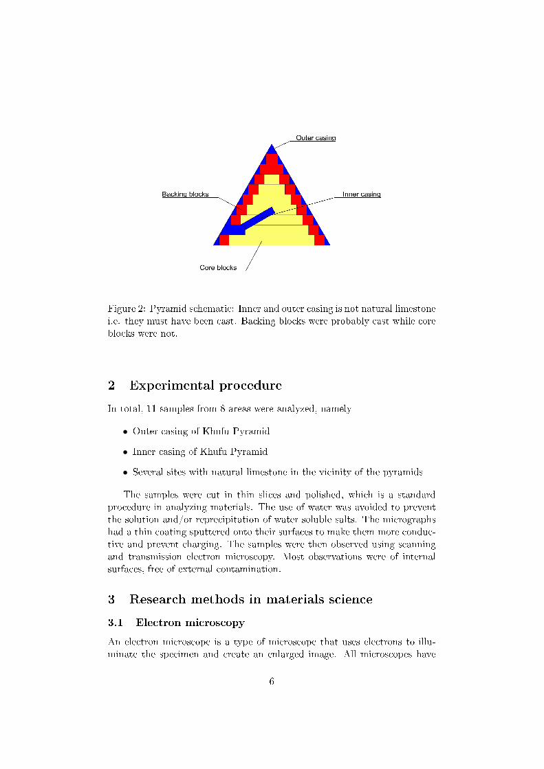
Figure 2: Pyramid schematic: Inner and outer casing is not natural limestonei.e. they must have been cast. Backing blocks were probably cast while coreblocks were not.
2 Experimental procedure
In total, 11 samples from 8 areas were analyzed, namely
• Outer casing of Khufu Pyramid
• Inner casing of Khufu Pyramid
• Several sites with natural limestone in the vicinity of the pyramids
The samples were cut in thin slices and polished, which is a standardprocedure in analyzing materials. The use of water was avoided to preventthe solution and/or reprecipitation of water soluble salts. The micrographshad a thin coating sputtered onto their surfaces to make them more conduc-tive and prevent charging. The samples were then observed using scanningand transmission electron microscopy. Most observations were of internalsurfaces, free of external contamination.
3 Research methods in materials science
3.1 Electron microscopy
An electron microscope is a type of microscope that uses electrons to illu-minate the specimen and create an enlarged image. All microscopes have
6

resolution limit due to the wavelength of particles they use. For example, op-tical microscopes use visible light with wavelengths between 400 and 700nm.The best resolving powers of high quality microscopes are thus about 200nanometers. The electron microscopes use electrons with energies of fewthousand eV. For example, electron with energy of 3600 eV has a (relativis-tic) wavelength
λe ≈h√
2m0E(1 + E2m0c2
)= 0.02nm
which would theoretically result in resolution of 0.01nm. However, construc-tion details decrease resolution of the microscope, which reduces resolvingpower of a standard electron microscope to about 1 nm. [3]
3.1.1 Scanning electron microscopy (SEM)
Scanning electron microscopy is a type of electron microscopy which utilizesa beam of high energy electrons to image the sample surface. The electronsinteract with atoms in the sample, producing signals that contain informa-tion about the sample surface topography, composition and other properties.Structure of a scanning electron microscope is shown in �gure 3:
Figure 3: Scanning electron microscope (picture taken from [4])
The electron gun thermionically emits electrons with energies up to 40keV. The electrons are focused by condenser lenses to a spot about 0.4nmto 5nm in diameter. This primary beam then passes through scan coils,which de�ect the beam so that it scans in a raster fashion over the area ofsample surface. When the primary electron beam interacts with the sample,the electrons lose energy by random scattering and absorption in specimen,extending from about 100nm to 5µm into the sample. This depth depends
7

on the electron energy, the atomic number of the specimen and the densityof the specimen. The energy exchange between the electrons and sample re-sults in the re�ection of electrons by elastic scattering, emission of secondaryelectrons by inelastic scattering and the emission of electromagnetic radia-tion. Each of those can be detected by specialized detectors. Particularlysigni�cant to sample analysis in our case were the backscattered electrons,providing composition data on samples. Also important are electrons re-�ected by elastic scattering, which provide data on the topography of thesamples. [4]
3.1.2 Transmission electron microscopy (TEM)
Figure 4: Transmission electron microscope (picture taken from [11])
Transmission electron microscopy is a type of electron microscopy wherethe electron beam is transmitted through an ultra thin specimen, interactingwith specimen as they pass through. Simply put, the transmission electronmicroscopy operates on the same basic principles as the light microscopebut uses electrons instead of light. At high resolutions, however, one mustalso consider the di�raction, which can provide us with a tool to analyze thecrystalline structure of the specimen. [5]
8

Figure 5: Typical di�raction pattern of a crystalline specimen (picture takenfrom [12])
3.2 Other methods
It is worth noting that materials research employs several other techniquesthat do not include electron microscopy. The materials can be tested fortheir mechanical properties (elastic and shear modulus, Poission's ratio etc.)thermal properties, electrical and magnetic properties etc. Common materi-als research techniques also include resonance (nuclear magnetic resonance,information on atomic and chemical structure of materials), X-rays (X-rayspectroscopy - information on elemental composition, X-ray scattering - in-formation on microstructure), ion beams (elemental composition, impuritydistribution, analysis of trace elements, high sensitivity measurements oflight elements) and others. Unfortunately, in-depth explanation of thesemethods is beyond the scope of this seminar. [5, 6, 7, 8]
4 Results
4.1 SEM
(A) Natural Stone: In all natural samples, microstructural analysis in theSEM indicated that the predominant phase was a porous calcite (CaCO3)that contained halite (mineral form of NaCl), silica, that is, a phase whereinthe O/Si ratio was 2:1 and the concentration of every other element was<0.05%. All samples also contained an organic substance, rich in C andO, found in various shapes, mostly at grain boundaries This substance wasalso found in the form of a thin coating that covered many of the phasesidenti�ed in the natural limestone, such as calcite, halite and CaCl2.In short: all samples of natural stone contained calcite, silica, halite, anorganic substance plus some smaller amounts of other impurities, all in ir-regular structures, which is a typical limestone structure.
9

(B) Outer Casing: In the outer casing microstructure, at least six di�erentmicroconstituents and/or phases, labeled M, G, D, T, Q and O were identi-�ed. Region Q is most likely calcite, region O is reminiscent of the organicphases observed in the natural limestone. Region D is most likely dolomite.Higher magni�cation SEM micrographs of region M (Figs. 3 (b) and (c))indicate a complex microstructure. In many locations, small rhomboedriccrystals roughly 2 µm in diameter are visible. Based on their morphologyit is reasonable to assume the cubes are single crystals. The chemistry ofthese particles shows they contain Si, apparently in solid solution in dolomite(region D). The importance of this observation is that Si is not known todissolve in dolomite in nature. The most intriguing, however, is the regionT. This thin strip contains very little Ca, is mostly considerably hydrated(lot of OH groups) and is not known to exist in calcite naturally.
10

Figure 6: Backscattered scanning electron microscopy micrographs of outercasing sample at low magni�cation (a), higher magni�cation of region Mshowing small cuboid particles (b) even higher magni�cation of region R1(c). Structure in (b) and (c) is very complex and unlike natural stone.(picture taken from [9])
11

Ca Mg Si Al O C
1 5 8 36 2 25 252 8 20 28 2 41 13 2 11 18 0.0 54 154 <1.0 27 61 2 8.5 2
Table 1: Elemental analysis of outer casing samples. The analysis revealsthat , in addition to well crystallized calcite and dolomite regions, regionscontaining Mg, Si, O and sometimes Ca were also found. [1]
(C) Inner Casing: The microstructure of the inner casing is characterizedby a matrix phase and two distinct microconstituents. The matrix phase iscomprised of exceptionally pure CaCO3, while the microconstituents includea great deal of S and Si and lack C and Ca at the same time. Figure 7 showsmicrograph of a region of inner casing sample together with elemental mapsof the region. The regions containing Ca are clearly visible, surrounded byregions rich in S and Si, but poor in Ca.
Figure 7: Scanning electron microscopy micrographs and elemental maps ofbulk of inner casing sample showing (a) secondary and (b) backscatteredimages, rest of images represent elemental maps. (picture taken from [9])
12

1 2 3 4 5 6 7 8 9 10 11
C 2.5 6.3 2.5 2.3 4.2 4.2 2.9 19.4 20.6 3.7 3.6O 65.8 65.3 64.8 62.8 69.8 61.4 65.4 60.5 58.7 63.6 64.5Na 1.4 0.7 1.4 1.0 0.8 1.1 1.4 0.3 0.6 0.9 0.9Si 2.4 2.9 2.4 7.9 8.1 3.4 6.3 0.5 0.2 31.0 29.3P 0.9 0.6 0.9 0.8 0.7 1.1 0.9 0.4 0.4 0.3 0.6S 14.2 13.0 14.2 12.5 8.6 14.7 12.0 0.3 0.3 0.3 0.5Ca 13.9 11.2 13.9 12.7 7.7 14.0 11.1 18.9 19.0 0.1 0.2
Table 2: Elemental analysis of inner casing samples regions, similar to thosein Fig. 7. In addition to areas with 1:2 ratio of Si:O (columns 10 and 11,areas with almost pure SiO2) and areas with Ca:C:O ratio of 1:1:3 (calciteCaCO3 region, columns 8 and 9), both areas containing almost no otherelements, there are also regions with abundance of S, Si, Na and P (columns1-7) [1]
4.2 TEM
The TEM scans have shown that the outer casing samples were either amor-phic or nanocrystalline, which corresponds to relatively rapid precipitation.
Figure 8: Selected area di�raction of select outer casing samples con�rmingthat they were either amorphous (a) or nanocrystalline (b), (c) shows TEMmicrograph of a typical region examined (picture taken from [1])
13

5 Discussion
5.1 Ubiquity of Si between calcite particles
According to Davidovits, the pyramid blocks are made of calcite aggregatesheld together with a silica based binding phase. If this theory is correct, Sishould be ubiquitous in the "grain boundary" areas, i.e., the areas betweenthe calcite and other aggregates. Based on data, the ubiquity of Si is clear, itwas found in T, M and R regions in outer casing samples, and together withS and Ca or P in the inner casing sample. Most of these regions also appearto be hydrated to some extent. As Si is a common geologic element, it is notsurprising that it is found everywhere, but its presence in combination withelements and structures that have not been yet observed in nature certainlyis. This implies the pyramid material to be aggregate with silica bindingphase. However, probably the most compelling evidence that Si is in solidsolution in calcite or dolomite are the small cuboid precipitates shown inFig. 3 (b). From their size and morphology, it is quite clear they are singlephase, i.e., with Si in solid solution.
5.2 Presence of moisture
Most of the casing samples appear to be hydrated. However, it is importantto emphasize again that neither calcite nor dolomite is known to form hy-drates in nature, and that only the "calcite" in the grain boundary areas ishydrated, neither the aggregates in the samples nor the natural limestonescontained any water of hydration. In short, the only part of the blocks thatwas hydrated was the binding material. Supporting the hydration is the factthat when the Great Pyramid was opened in 820 AD, the interior chamberswere reported to be encrusted with salt, which is consistent with damp andporous rock. Even today, the pyramids are not dry. For example, the elec-tromagnetic sounder experiment of 1974, intending to map the interior ofthe pyramids, failed because of high moisture of the pyramid stone. A sim-ple explanation would be that various ions found in the microconstituentswere at some time present in solution and precipitated or reacted togetherrelatively fast to form the glue necessary to fabricate the synthetic stone.In this scheme, some of the added water would end up bound in the stoneas observed. Under such conditions, the resulting microconstituents wouldtend to be amorphous and/or nanocrystalline, which corresponds with TEMobservations.
14

5.3 "Common sense" evidence
Careful examination of the visible pyramid blocks on the Giza plateau sug-gests that most, especially in the core, have been carved. Some, near thesurface, including the casing, appear to be cast.
Figure 9: Gash in Snefru Pyramid. Snefru Pyramid was built few yearsbefore the Great Pyramid probably utilizing same construction techniques.Inthis picture you can clearly see the (rough) carved interior and smooth outercasing (picture taken from [9])
6 Conclusion
The simplest explanation for the presence of many di�erent microconstituents,some of which appear to possess chemical compositions and morphologies notfound in the natural stone, is that the various ions were in solution and pre-cipitated relatively fast. This makes a very impressive implication that theblocks from pyramids were created by ancient egyptians and have enduredthrough millenia, while the best concrete that our civilization is currentlycapable to produce, only lasts for about 150 years. Apparently the ancientegyptians were much better at materials science and chemistry as was the-orized before. Supporting this theory is the recent casting of blocks closelyresembling the ones in pyramids [10]
However, one of this seminar's primary intentions was also to illustratehow far physics can reach in helping provide scientists in material sciencewith tools for their analysis, who, when equipped with such tools can inturn help archeologists uncover some more fascinating data about our past.
15
Furthermore, results acquired in the research of the pyramidal material couldlead us to making concrete that is much more durable than the best knownconcrete while reducing the pollution from the production by as much as90%.[10]
References
[1] M. W. Barsoum and A. Ganguly Microstructural Evidence of Reconsti-tuted Limestone Blocks in the Great Pyramids of Egypt J. Am. Ceram.
Soc, 2006.
[2] J. Davidovits X-Rays Analysis and X-Rays Di�raction of Casing Stonesfrom the Pyramids of Egypt, and the Limestone of the Associated Quar-ries Science in Egyptology Symposia, 1984
[3] Electron Microscopy at Die Eidgenössische Technische Hochschule Zürichhttp://www.microscopy.ethz.ch/, 16th Feb. 2009
[4] Boston Museum of Science http://www.mos.org/, 16th Feb. 2009
[5] National Physics Laboratory http://www.npl.co.uk/, 16th Feb. 2009
[6] Ion Beam Materials Laboratory http://www.lanl.gov/mst/ibml/
index.shtml, 16th Feb. 2009
[7] X-Ray Scattering Group, Condensed Matter Physics and Materials Sci-ence group at Brookhaven National Laboratory http://www.bnl.gov/
cmpmsd/xray, 16th Feb. 2009
[8] Surface and Materials Science at Stanford University http://
www-group.slac.stanford.edu/sms, 16th Feb. 2009
[9] The Great Pyramids of Giza: Evidence for Cast Blocks http://www.
materials.drexel.edu/pyramids/PyramidPresentation_Lores.pdf,16th Feb. 2009
[10] Davidovits, J. Ils ont bâti les pyramides Jean-Cyrille Godefroy, Paris,2002
[11] Encyclopaedia Britannica http://media-2.web.britannica.com/,16th Feb. 2009
[12] University of Liverpool Materials and Chemistry Group http://www.
matter.org.uk/, 16th Feb. 2009
16