the conserved fanconi anemia nuclease fan1 and the sumo e3
TRANSCRIPT
The conserved Fanconi anemia nuclease Fan1 and the SUMO E3 ligase Pli1 act in two novel Pso2-independent pathways of DNA interstrand crosslink repair in yeast
Article (Published Version)
http://sro.sussex.ac.uk
Fontebasso, Y, Etheridge, T J, Oliver, A W, Murray, J M and Carr, A M (2013) The conserved Fanconi anemia nuclease Fan1 and the SUMO E3 ligase Pli1 act in two novel Pso2-independent pathways of DNA interstrand crosslink repair in yeast. DNA Repair, 12 (12). pp. 1011-1023. ISSN 15687864
This version is available from Sussex Research Online: http://sro.sussex.ac.uk/49894/
This document is made available in accordance with publisher policies and may differ from the published version or from the version of record. If you wish to cite this item you are advised to consult the publisher’s version. Please see the URL above for details on accessing the published version.
Copyright and reuse: Sussex Research Online is a digital repository of the research output of the University.
Copyright and all moral rights to the version of the paper presented here belong to the individual author(s) and/or other copyright owners. To the extent reasonable and practicable, the material made available in SRO has been checked for eligibility before being made available.
Copies of full text items generally can be reproduced, displayed or performed and given to third parties in any format or medium for personal research or study, educational, or not-for-profit purposes without prior permission or charge, provided that the authors, title and full bibliographic details are credited, a hyperlink and/or URL is given for the original metadata page and the content is not changed in any way.
DNA Repair 12 (2013) 1011– 1023
Contents lists available at ScienceDirect
DNA Repair
jo ur nal home p age: www.elsev ier .com/ locate /dnarepai r
The conserved Fanconi anemia nuclease Fan1 and the SUMO E3 ligasePli1 act in two novel Pso2-independent pathways of DNA interstrandcrosslink repair in yeast
Y. Fontebasso a,b, T.J. Etheridge a, A.W. Oliver a, J.M. Murray a, A.M. Carr a,∗
a Genome Damage and Stability Centre, University of Sussex, Brighton, East Sussex BN1 9RQ, UKb Breakthrough Breast Cancer Research Centre, The Institute of Cancer Research, 237 Fulham Road, London SW3 6JB, UK
a r t i c l e i n f o
Article history:
Received 9 May 2013
Received in revised form 1 October 2013
Accepted 7 October 2013
Available online 2 November 2013
Keywords:
ICL
Genetic screen
Synthetic array
Epistasis
Schizosaccharomyces pombe
Cisplatin
a b s t r a c t
DNA interstrand cross-links (ICLs) represent a physical barrier to the progression of cellular machinery
involved in DNA metabolism. Thus, this type of adduct represents a serious threat to genomic stability and
as such, several DNA repair pathways have evolved in both higher and lower eukaryotes to identify this
type of damage and restore the integrity of the genetic material. Human cells possess a specialized ICL-
repair system, the Fanconi anemia (FA) pathway. Conversely yeasts rely on the concerted action of several
DNA repair systems. Recent work in higher eukaryotes identified and characterized a novel conserved
FA component, FAN1 (Fanconi anemia-associated nuclease 1, or FANCD2/FANCI-associated nuclease 1).
In this study, we characterize Fan1 in the yeast Schizosaccharomyces pombe. Using standard genetics, we
demonstrate that Fan1 is a key component of a previously unidentified ICL-resolution pathway. Using
high-throughput synthetic genetic arrays, we also demonstrate the existence of a third pathway of ICL
repair, dependent on the SUMO E3 ligase Pli1. Finally, using sequence-threaded homology models, we
predict and validate key residues essential for Fan1 activity in ICL repair.
© 2013 Published by Elsevier B.V.
1. Introduction
Interstrand cross-links (ICLs) represent a particularly insidious
threat to genomic stability. These adducts create covalent bonds
linking the two DNA strands in a duplex, generating an abnor-
mal structure that poses a physical obstacle to the progression
of cellular machinery like DNA replisomes [1,2]. The mechanisms
underlying the response to ICLs in unicellular organisms depend
on components involved in many of the major DNA repair path-
ways: nucleotide excision repair (NER), base excision repair (BER),
mismatch repair (MMR), post-replication repair (PRR, comprising
translesion synthesis, TLS) and homologous recombination (HR)
[2]. Conversely, only a few proteins have been identified as spe-
cific to the response to ICLs. In Saccharomyces cerevisiae, Pso2/Snm1
has been identified as a key player in the response to interstrand
cross-linking agents [3–5]. A role for Snm1/Pso2 has been postu-
lated where its exonuclease activity resects the DNA flanking the
ICL to facilitate TLS or homologous recombination [6,7]. Although
little is known about the resolution of DNA ICLs in the fission yeast
Schizosaccharomyces pombe, the corresponding Pso2 nuclease has
∗ Corresponding author. Tel.: +44-1273-678122; fax: +44-1273-678121.
E-mail address: [email protected] (A.M. Carr).
been similarly shown to be required for normal resistance to ICL-
inducing agents [8].
In higher eukaryotes, multiple DNA repair pathways are also
involved in the resolution of ICLs, albeit the existence of the special-
ized Fanconi anemia (FA) pathway [9] marks a significant difference
compared to the yeasts. The current model for the involvement
of the FA pathway in ICL repair is as follows: the ICL is recog-
nized by FANCM-FAAP24 bound to the recently discovered MHF
complex [9–11]. FANCM-FAAP24-MHF recruits a downstream E3
ubiquitin ligase complex known as the “FA core complex”, which
in turn monoubiquitinates FANCD2 and FANCI on chromatin [9,12].
FANCD2-FANCI then recruits further downstream factors and inter-
acts with HR and TLS proteins, finally facilitating HR-dependent ICL
repair [9]. It is also proposed that a parallel crosstalk with S-phase
checkpoint proteins mediates and coordinates ICL repair with other
DNA damage response mechanisms [9].
Recent work in higher eukaryotes identified and characterized
FAN1 (Fanconi anemia-associated nuclease 1, or FANCD2/FANCI-
associated nuclease 1) [13–18]. Human FAN1 colocalizes to
ICL-induced foci with and dependently on monoubiquitinated
FANCD2, suggesting a role with the FA pathway. Defects in homol-
ogous recombination in FAN1-depleted cells suggest that this
protein is involved in the HR processes linked to ICL repair. As
DSB resection is not impaired in the absence of FAN1 and RAD51
foci persist in FAN1-depleted cells, it has been proposed that FAN1
1568-7864/$ – see front matter © 2013 Published by Elsevier B.V.
http://dx.doi.org/10.1016/j.dnarep.2013.10.003
1012 Y. Fontebasso et al. / DNA Repair 12 (2013) 1011– 1023
may be required for late stages of HR-dependent repair [14,15]. A
homolog of FAN1 is present in the fission yeast S. pombe, but not in
the budding yeast S. cerevisiae. Thus, the appearance of FAN1 earlier
than the FA core complex-dependent pathway on the evolutionary
scale suggests that the role of this component is either functionally
distinct from the canonical FA pathway of higher eukaryotes or is
regulated by this pathway. For this reason, the study of Fan1 in S.
pombe has the potential for revealing mechanisms of ICL repair in
higher eukaryotes which act in parallel with, or are controlled by,
the FA pathway.
In the present study, we investigate the function of S. pombe Fan1
(the gene is named fan1 following the work discussed above) using
standard and high-throughput genetics. We demonstrate that Fan1
is a novel component of a Pso2-independent ICL resolution pathway
and genetically dissect these two pathways to assign epistatic rela-
tionships with known components of DNA damage repair pathways
involved in ICL repair. Using high-throughput synthetic genetic
arrays to explore genetic relationships in the response to ICL-
inducing agents, we identify the existence of an additional ICL
resolution pathway dependent on the SUMO E3 ligase Pli1. Finally,
we identify key Fan1 residues necessary for its activity.
2. Material and methods
2.1. DNA damaging agents
UV irradiation was performed with a Stratagene® Stratalinker®
using the settings (J/m2) indicated. Other drugs used, all from
SIGMA®: methyl methanesulfonate (MMS), cat. no. 129925; cis-
platin (cis-platinum(II)diammine dichloride, product no. P4394;
mitomycin C (MMC), cat. no. M0503; HN1 (2-chloro-N,N-
dimethylethylamine hydrochloride), product no. 24362; HN2
(mechlorethamine hydrochloride), product no. 122564; cyclohex-
imide, product no. C7698 (100 mg/l from a 100 mg/ml stock in
DMSO).
2.2. Strains
A list of all the strains used in this study is provided in sup-
plementary Table 4. Details of the strain construction for specific
mutants are given below.
2.3. fan1-d strain construction
Two independently-derived fan1-d mutants (both fan1::kanMX;
kanMX confers resistance to the drug geneticin, or G418) have
been analysed in parallel in this study. The first mutant (3909)
was kindly donated by Professor Paul Nurse; the second mutant
(14152) is derived from the Bioneer® S. pombe deletion mutant
library (http://pombe.bioneer.co.kr/). The two strains were ver-
ified by Southern blot. To allow a flexible and rapid series of
genetic crosses between different deletion mutants, the original
kanMX deletion cassettes in the 3909 and 14152 strains were
replaced with a natMX6 deletion cassette, which confers resis-
tance to nourseothricin [19]. The natMX6 null mutants derived
from 3909 and 14152 were named 3909N and 14152N, respec-
tively. These new mutants showed the same sensitivity to the
drugs used in this study as the original 3909 and 14152 strains
(data not shown). The two independently derived mutants always
showed consistent sensitivity to the drugs tested. The 3909/3909N
and 14152/14152N strains were used both in parallel for nearly all
the experiments conducted, although in the interest of space only
one of the two mutant is usually presented in the figures of this
study. fan1 mutants were created employing site-directed muta-
genesis using a Stratagene QuikChange® kit as described in [20] and
Recombinase-Mediated Cassette Exchange (RMCE) as described in
[21].
2.4. Spontaneous mutation rate assays
Single colonies were isolated on YEA from individual streaks.
Eleven colonies from each strain were grown in 5 ml YE in individ-
ual tubes. Samples were incubated at 30 ◦C for 48 h to stationary
phase. Cultures were serially diluted as follows: 10 �l saturated cul-
ture in 1 ml H2O; 10 �l of this dilution into 1 ml H2O. A 50 �l of this
dilution were plated on YE-Agar (YEA) plates. A 50 �l of saturated
culture were plated on YE-5-FOA (5-Fluoroorotic acid; Melford®
F5001) plates (0.1% final concentration). Plates were incubated for
3–4 days at 30 ◦C. Spontaneous mutation rates were calculated by
the Lea-Coulson method of the median (Rosche and Foster, 2000;
Foster, 2006).
2.5. In vivo survival assays: Spot tests
Strains were inoculated in 5 ml YE and grown at 30 ◦C o/n. 107
cells from each logarithmically growing culture were harvested and
resuspended in 1 ml water. Four serial 1/10 dilutions were pre-
pared from each culture. A 10 ul were spotted onto YEA plates added
with increasing doses of DNA damaging agents. All the spots were
deposited in duplicates on the same plate to guarantee an inter-
nal control. Plates were incubated at 30 ◦C for 3 days. Images were
acquired with a Syngene® Ingenius® apparatus.
2.6. In vivo survival assays: Survival curves
A 2 × 10⁸ cells grown to exponential phase were centrifuged and
washed with PBS. Pellets were resuspended in 10 ml and split into
five 2 ml aliquots in 15 ml tubes. Each drug dilution was inoculated
into the 2 ml aliquoted cultures and tubes incubated at 30 ◦C with
shaking for 1 h. Approximately 200 cells were plated onto YEA and
grown at 30 ◦C for 3–4 days.
2.7. Automated Screening of the Bioneer deletion library
Note: all the parameters of the programs indicated below are
detailed in the supplementary section. A loopful of query mutant
(Q) was inoculated from a fresh patch into 15 ml YE + NAT and
grown for at least 6 h. The above culture was poured into an empty
PlusPlate® (“Q bath”). Once thawed, library plates were replicated
onto YEA PlusPlates®: four liquid 96-well plates combined onto
one YEA PlusPlate® (384 spots) [PROGRAM 1, TWICE PER ARRAY].
A 384 agar plate was build using the Q bath as a source (“Q YEA
PlusPlates®”) [PROGRAM 2, TWICE PER ARRAY]. Cells were grown
for 2–3 days at 30 ◦C (or until colonies are grown to satisfac-
tory size). Each library was replicated to fresh YEA PlusPlates® (“L
YEA PlusPlates®”) [PROGRAM 3]. Mating: colonies were combined
from the L and Q YEA PlusPlates® onto ELN PlusPlates® [PRO-
GRAM 4, RUN TWICE PER ARRAY]. ELN PlusPlates® were incubated
at 25 ◦C for 4 days. YEA PlusPlates® were incubated at 30 ◦C for
3 days: pictures were taken approximately every 12 h to moni-
tor the fitness of the single mutants. Spore germination: colonies
were replicated from ELN PlusPlates® to YEA PlusPlates® [PRO-
GRAM 5] and incubated at 30 ◦C for 3 days. Selection 1: colonies
were replicated from YEA PlusPlates® to YE + GC PlusPlates® [PRO-
GRAM 5] and incubated at 30 ◦C for 2–3 days (or until colonies
have grown to satisfactory size). Selection 2: cells were replicated
from YE + GC PlusPlates® to YE + GNC PlusPlates® [PROGRAM 3]
and incubate at 30 ◦C for 1–3 days. Pictures to assess the fitness
of double mutants were taken at this stage every approximately
12 h.

Y. Fontebasso et al. / DNA Repair 12 (2013) 1011– 1023 1013
Table 1
Fan1 is not involved in the suppression of spontaneous mutation rate. Spontaneous forward mutation rate of fan1-d mutants in cdc6+ and cdc6-L591M backgrounds. Data from
three independent experiments. For each strain, 11 colonies were grown to saturation at 30 ◦C for 48 h. Fluctuation analysis was performed as described in Section 2.
Mutation rate/cell division
Experiment 1 Experiment 2 Experiment 3 Average Standard error Fold elev.
cdc6+ 8.09E − 10 5.27E − 10 5.65E − 10 6.33E − 10 8.85E − 11 1
cdc6+ fan1-d (3909) 9.98E − 10 8.45E − 10 9.17E − 10 9.20E − 10 4.42E − 11 1
cdc6+ fan1-d (14152) 6.49E − 10 5.74E − 10 5.02E − 10 5.75E − 10 4.24E − 11 1
cdc6-L591M 2.08E − 06 1.08E − 05 2.82E − 06 5.25E − 06 2.81E − 06 1
cdc6-L591M fan1-d (3909) 2.55E − 06 6.35E − 06 3.37E − 06 4.09E − 06 1.16E − 06 1
cdc6-L591M fan1-d (14152) 1.90E − 06 2.93E − 06 2.99E − 06 2.61E − 06 3.54E − 07 0
For the assessment of resistance to DNA damaging agents,
cells were replicated from YE + GNC PlusPlates® to YE PlusPlates®
added with different concentrations of chosen DNA damaging
agents [PROGRAM 3]. Plates were incubated at 30 ◦C for 2-4 days
and pictures were taken approximately every 12 h. Pictures were
taken using a Syngene® Ingenius® apparatus. Software used for
colony size analysis: HT Colony Grid Analyser 1.1.0/1.1.7, Adobe®
Photoshop® CS5 Extended, Microsoft® Excel® 2007/2010.
3. Results
3.1. The Fanconi anemia—Associated nuclease Fan1 is not
involved in the suppression of DNA spontaneous mutation rate
Human FAN1 (also known as KIAA1018) has been shown to
interact with MMR components such as MLH1, PMS1 and PMS2
[13,14,22]. Thus, we set out to test whether a similar scenario holds
true for SpFan1, and whether this protein could be involved in
the mismatch repair pathway. As we were unable to detect direct
physical interactions of SpFan1 with other MMR components (data
not shown), we performed a forward mutation assay in order to
determine the rate of spontaneous mutation in fan1-deleted cells.
In this system, the readout is the switch from uracil autotrophy
to uracil heterotrophy. The estimated mutation rate during DNA
replication in eukaryotic cells is lower than 1 mutation every 109
bases [23], which would be undetectable by our current mutation
assays. In S. cerevisiae, a mutation in the catalytic subunit of poly-
merase delta (Pol3-L615M) leads to a 7-fold increased spontaneous
mutation rate with no measurable changes in other phenotypes
monitored [24]. In our study, the background spontaneous muta-
tion rate was therefore increased to detectable levels by using a
strain harbouring the corresponding mutation in polymerase delta,
Cdc6 (cdc6-L591M) [25].
In a cdc6-L591M background, the mutation rate is increased
to approximately 1 in 106 (Table 1; consistent with [25]). How-
ever, no significant increase in this spontaneous mutation rate was
observed following concomitant deletion of fan1. These data argued
against a direct involvement of SpFan1 in the MMR pathway.
3.2. Fan1 is a component of a Pso2-independent interstrand
crosslink repair pathway
In order to determine whether SpFan1 could be involved in
other pathways of DNA repair, we performed in vivo survival
assays to assign genetic interactions between SpFan1 and known
components of characterized repair pathways involved in the ICL
response. To assess the response of fan1-d mutants to a vari-
ety of DNA lesions, the two SpFan1 deletion mutants 3909 and
14152 were initially back-crossed twice to a wild-type strain
and five independent G418-resistant colonies were isolated and
tested under increasing concentrations of various DNA damaging
agents. All the fan1 deletion isolates showed wild-type sensitivity
to UV, camptothecin (CPT), methyl methanesulfonate (MMS) and
hydroxyurea (HU) (data not shown). However, a subtle but repro-
ducible sensitivity was observed when fan1-d cells were exposed
to cis-platinum diammine-dichloride (cisplatin, CDDP) and mito-
mycin C (MMC). These drugs belong to a family of DNA damaging
agents that induce covalent DNA interstrand cross-links [2]. The
mild sensitivity towards ICL-inducing agents suggested that SpFan1
is implicated in ICL repair, but that its role overlaps with the func-
tion of other components of the DNA repair machinery.
To test this, the original 3909 and 14152 fan1 null mutants
were crossed with a series of deletion mutants of genes reported
to be involved in the ICL resolution pathway, either in S. pombe
or in the budding yeast S. cerevisiae. In S. pombe, the nuclease
Pso2 (also known as Snm1 in cerevisiae) has been shown to be
specifically required for normal resistance to ICL-inducing agents
[8]. When exposed to increasing doses of cisplatin and MMC,
the fan1-d pso2-d double mutant showed a dramatic reduction
in viability compared to the corresponding single mutants or
the wild-type (wt) control strain (Fig. 1A, left panel). In order
to confirm that SpFan1 is specifically involved in ICL repair, cell
survival assays were repeated for fan1-d and pso2-d using bis(2-
chloroethyl)methylamine (HN2, mechloretamine), an agent shown
to generate a higher proportion of DNA interstrand cross-links com-
pared to cisplatin [2]. When exposed to increasing concentrations
of HN2, fan1-deleted cells showed a marked decrease in viabil-
ity only when combined with pso2 deletion (Fig. 1A, right panel).
As a further control, the same experiment was conducted in the
presence of HN1 (2-dimethylaminoethylchloride hydrochloride), a
mono-functional nitrogen mustard which does not form ICLs [26].
None of the strains treated with this agent, including the double
mutant pso2-d fan1-d, showed any sensitivity to this agent (data
not shown).
Taken together, these data confirm that SpFan1 is a novel com-
ponent of the DNA repair pathway that specifically acts to repair
cross-links linking covalently the two strands of a DNA molecule,
and that SpFan1 and SpPso2 act in parallel pathways or subpath-
ways.
3.3. The NER nuclease Rad13 is involved only in the
pso2-dependent ICL repair pathway
ICL repair mechanisms in lower and higher eukaryotes have
proven to be elusive due to the intersection of different DNA repair
pathways. In S. cerevisiae, components of the nucleotide excision
repair (NER), post-replication repair (PRR) and homologous recom-
bination (HR) pathways have all been implicated in the resolution
of interstrand cross-links [2]. To test whether Fan1-dependent ICL
repair intersects with these pathways, a series of double and triple
mutants were created and tested for sensitivity to cisplatin.
rhp18, the fission yeast gene encoding the homolog of S.
cerevisiae Rad18 involved in post-replication repair[2], displayed
hypersensitivity to cisplatin to concentrations as low as 50 �M
(supplementary Fig. 1). The combination of the fan1 and rhp18
mutations did not display increased sensitivity to cisplatin,
1014 Y. Fontebasso et al. / DNA Repair 12 (2013) 1011– 1023
whereas a mild but reproducible increase in sensitivity was
observed when pso2-d was combined with rhp18-d (supplemen-
tary Fig. 1). This indicates that Rhp18 is involved in the resolution
of ICL adducts in a step that is common to the Fan1 pathway. The
deletion of the nuclease Exo1 displayed no significant sensitivity
to ICL-inducing agents, and only a marginal increased sensitivity in
combination with pso2-d, fan1-d (double mutants) or with pso2-d
fan1-d (triple mutant) (data not shown).
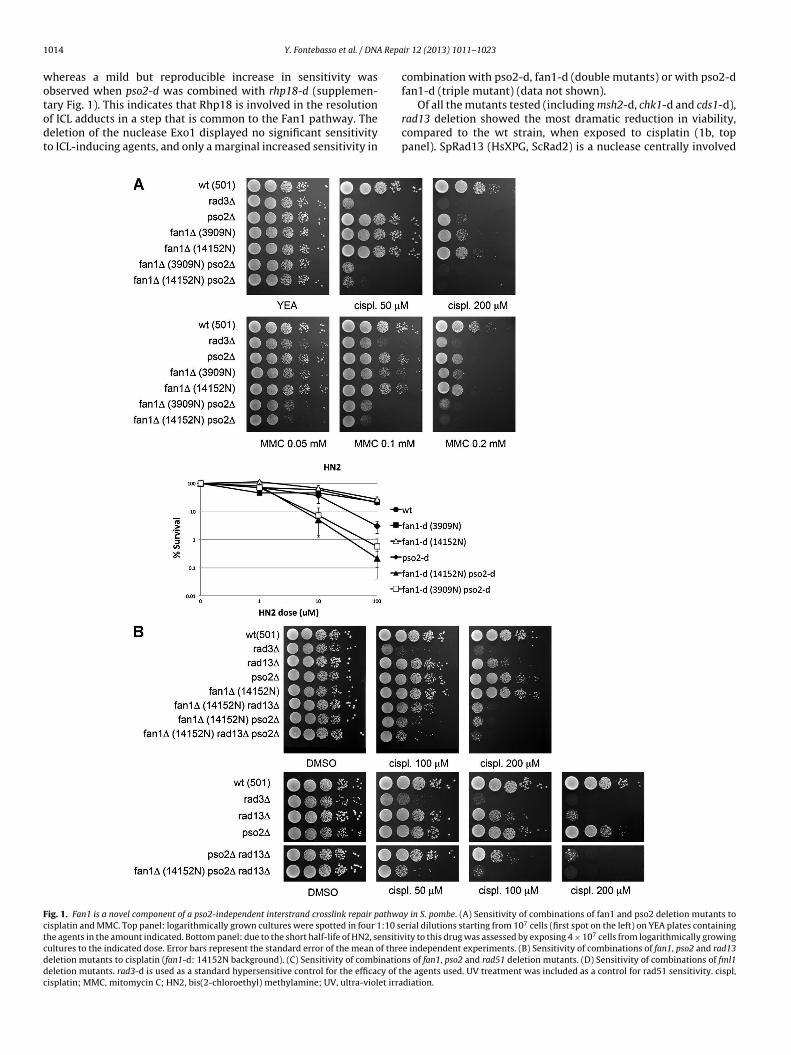
Of all the mutants tested (including msh2-d, chk1-d and cds1-d),
rad13 deletion showed the most dramatic reduction in viability,
compared to the wt strain, when exposed to cisplatin (1b, top
panel). SpRad13 (HsXPG, ScRad2) is a nuclease centrally involved
Fig. 1. Fan1 is a novel component of a pso2-independent interstrand crosslink repair pathway in S. pombe. (A) Sensitivity of combinations of fan1 and pso2 deletion mutants to
cisplatin and MMC. Top panel: logarithmically grown cultures were spotted in four 1:10 serial dilutions starting from 107 cells (first spot on the left) on YEA plates containing
the agents in the amount indicated. Bottom panel: due to the short half-life of HN2, sensitivity to this drug was assessed by exposing 4 × 10⁷ cells from logarithmically growing
cultures to the indicated dose. Error bars represent the standard error of the mean of three independent experiments. (B) Sensitivity of combinations of fan1, pso2 and rad13
deletion mutants to cisplatin (fan1-d: 14152N background). (C) Sensitivity of combinations of fan1, pso2 and rad51 deletion mutants. (D) Sensitivity of combinations of fml1
deletion mutants. rad3-d is used as a standard hypersensitive control for the efficacy of the agents used. UV treatment was included as a control for rad51 sensitivity. cispl,
cisplatin; MMC, mitomycin C; HN2, bis(2-chloroethyl) methylamine; UV, ultra-violet irradiation.
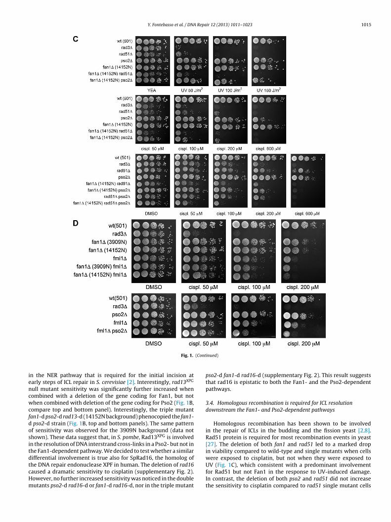
Y. Fontebasso et al. / DNA Repair 12 (2013) 1011– 1023 1015
Fig. 1. (Continued)
in the NER pathway that is required for the initial incision at
early steps of ICL repair in S. cerevisiae [2]. Interestingly, rad13XPG
null mutant sensitivity was significantly further increased when
combined with a deletion of the gene coding for Fan1, but not
when combined with deletion of the gene coding for Pso2 (Fig. 1B,
compare top and bottom panel). Interestingly, the triple mutant
fan1-d pso2-d rad13-d (14152N background) phenocopied the fan1-
d pso2-d strain (Fig. 1B, top and bottom panels). The same pattern
of sensitivity was observed for the 3909N background (data not
shown). These data suggest that, in S. pombe, Rad13XPG is involved
in the resolution of DNA interstrand cross-links in a Pso2- but not in
the Fan1-dependent pathway. We decided to test whether a similar
differential involvement is true also for SpRad16, the homolog of
the DNA repair endonuclease XPF in human. The deletion of rad16
caused a dramatic sensitivity to cisplatin (supplementary Fig. 2).
However, no further increased sensitivity was noticed in the double
mutants pso2-d rad16-d or fan1-d rad16-d, nor in the triple mutant
pso2-d fan1-d rad16-d (supplementary Fig. 2). This result suggests
that rad16 is epistatic to both the Fan1- and the Pso2-dependent
pathways.
3.4. Homologous recombination is required for ICL resolution
downstream the Fan1- and Pso2-dependent pathways
Homologous recombination has been shown to be involved
in the repair of ICLs in the budding and the fission yeast [2,8].
Rad51 protein is required for most recombination events in yeast
[27]. The deletion of both fan1 and rad51 led to a marked drop
in viability compared to wild-type and single mutants when cells
were exposed to cisplatin, but not when they were exposed to
UV (Fig. 1C), which consistent with a predominant involvement
for Rad51 but not Fan1 in the response to UV-induced damage.
In contrast, the deletion of both pso2 and rad51 did not increase
the sensitivity to cisplatin compared to rad51 single mutant cells
1016 Y. Fontebasso et al. / DNA Repair 12 (2013) 1011– 1023
(Fig. 1C, bottom panel). Interestingly, the triple deletion of the genes
coding for Fan1, Rad51 and Pso2 resulted in the most dramatic
decrease in viability compared to all the combinations of mutants
tested (Fig. 1C, bottom panel). These data suggest a crucial role for
Rad51 in the resolution of ICLs outside the Pso2 and Fan1 pathways.
The notable difference in sensitivity between the combinations of
fan1-d rad51-d and pso2-d rad51-d double mutants further sug-
gests differential extents for the involvement of Rad51-dependent
processes in the Pso2 and Fan1 pathways of ICL resolution.
3.5. The conserved Fanconi anemia component Fml1 acts in a
Pso2-independent ICL resolution pathway
Fml1 is the S. pombe homolog of the human FANCM heli-
case/translocase, a component of the Fanconi anemia pathway
[28,29]. Fml1 has been previously shown to be required for wild-
type resistance to interstrand cross-linking agents such as cisplatin
[30]. Recent work on the homolog Mph1 in S. cerevisiae indicates
that Mph1 and Pso2 act in independent pathways of ICL resolu-
tion upon exposure to HN2 [31]. To test whether the same scenario
holds true in S. pombe, we created combined double mutants of
fml1-d and pso2-d or fan1-d and assessed the sensitivity of these
mutants to cisplatin. Whereas the combination of fml1-d and fan1-
d did not increase the sensitivity to the drug compared to the single
mutants, the concomitant deletion of fml1 and pso2 showed a more
accentuated sensitivity (Fig. 3D). This data suggests that, in paral-
lel with the situation in the budding yeast, the conserved Fanconi
anemia component Fml1 and the nuclease Pso2 act on independent
pathways in response to resolution of DNA interstrand adducts.
3.6. The nuclease and the SAP DNA binding domain are required
for Fan1 activity
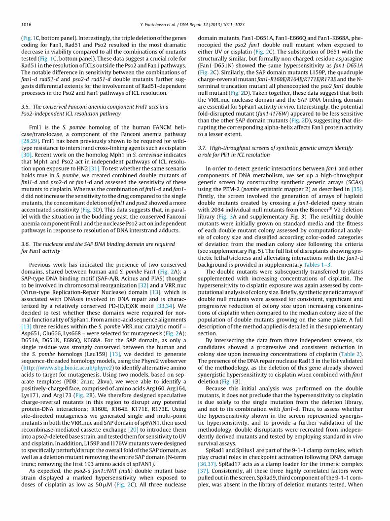
Previous work has indicated the presence of two conserved
domains, shared between human and S. pombe Fan1 (Fig. 2A): a
SAP-type DNA binding motif (SAF-A/B, Acinus and PIAS) thought
to be involved in chromosomal reorganization [32] and a VRR nuc
(Virus-type Replication-Repair Nuclease) domain [13], which is
associated with DNAses involved in DNA repair and is charac-
terized by a relatively conserved PD-(D/E)XK motif [33,34]. We
decided to test whether these domains were required for nor-
mal functionality of SpFan1. From amino-acid sequence alignments
[13] three residues within the S. pombe VRR nuc catalytic motif –
Asp651, Glu666, Lys668 – were selected for mutagenesis (Fig. 2A);
D651A, D651N, E686Q, K668A. For the SAP domain, as only a
single residue was strongly conserved between the human and
the S. pombe homologs (Leu159) [13], we decided to generate
sequence-threaded homology models, using the Phyre2 webserver
(http://www.sbg.bio.ic.ac.uk/phyre2) to identify alternative amino
acids to target for mutagenesis. Using two models, based on sep-
arate templates (PDB: 2rnn; 2kvu), we were able to identify a
positively-charged face, comprised of amino acids Arg160, Arg164,
Lys171, and Arg173 (Fig. 2B). We therefore designed speculative
charge-reversal mutants in this region to disrupt any potential
protein-DNA interactions; R160E, R164E, K171E, R173E. Using
site-directed mutagenesis we generated single and multi-point
mutants in both the VRR nuc and SAP domain of spFAN1, then used
recombinase-mediated cassette exchange [20] to introduce them
into a pso2-deleted base strain, and tested them for sensitivity to UV
and cisplatin. In addition, L159P and I176W mutants were designed
to specifically perturb/disrupt the overall fold of the SAP domain, as
well as a deletion mutant removing the entire SAP domain (N-term
trunc; removing the first 193 amino acids of spFAN1).
As expected, the pso2-d fan1::NAT (null) double mutant base
strain displayed a marked hypersensitivity when exposed to
doses of cisplatin as low as 50 �M (Fig. 2C). All three nuclease
domain mutants, Fan1-D651A, Fan1-E666Q and Fan1-K668A, phe-
nocopied the pso2 fan1 double null mutant when exposed to
either UV or cisplatin (Fig. 2C). The substitution of D651 with the
structurally similar, but formally non-charged, residue asparagine
(Fan1-D651N) showed the same hypersensitivity as fan1-D651A
(Fig. 2C). Similarly, the SAP domain mutants L159P, the quadruple
charge-reversal mutant fan1-R160E/R164E/K171E/R173E and the N-
terminal truncation mutant all phenocopied the pso2 fan1 double
null mutant (Fig. 2D). Taken together, these data suggest that both
the VRR nuc nuclease domain and the SAP DNA binding domain
are essential for SpFan1 activity in vivo. Interestingly, the potential
fold-disrupted mutant (fan1-I176W) appeared to be less sensitive
than the other SAP domain mutants (Fig. 2D), suggesting that dis-
rupting the corresponding alpha-helix affects Fan1 protein activity
to a lesser extent.
3.7. High-throughput screens of synthetic genetic arrays identify
a role for Pli1 in ICL resolution
In order to detect genetic interactions between fan1 and other
components of DNA metabolism, we set up a high-throughput
genetic screen by constructing synthetic genetic arrays (SGAs)
using the PEM-2 (pombe epistatic mapper 2) as described in [35].
Firstly, the screen involved the generation of arrays of haploid
double mutants created by crossing a fan1-deleted query strain
with 2034 individual null mutants from the Bioneer® V2 deletion
library (Fig. 3A and supplementary Fig. 3). The resulting double
mutants were initially grown on standard media and the fitness
of each double mutant colony assessed by computational analy-
sis of colony size and classified according color-coded categories
of deviation from the median colony size following the criteria
(see supplementary Fig. 5). The full list of disruptants showing syn-
thetic lethal/sickness and alleviating interactions with the fan1-d
background is provided in supplementary Tables 1–3.
The double mutants were subsequently transferred to plates
supplemented with increasing concentrations of cisplatin. The
hypersensitivity to cisplatin exposure was again assessed by com-
putational analysis of colony size. Briefly, synthetic genetic arrays of
double null mutants were assessed for consistent, significant and
progressive reduction of colony size upon increasing concentra-
tions of cisplatin when compared to the median colony size of the
population of double mutants growing on the same plate. A full
description of the method applied is detailed in the supplementary
section.
By intersecting the data from three independent screens, six
candidates showed a progressive and consistent reduction in
colony size upon increasing concentrations of cisplatin (Table 2).
The presence of the DNA repair nuclease Rad13 in the list validated
of the methodology, as the deletion of this gene already showed
synergistic hypersensitivity to cisplatin when combined with fan1
deletion (Fig. 1B).
Because this initial analysis was performed on the double
mutants, it does not preclude that the hypersensitivity to cisplatin
is due solely to the single mutation from the deletion library,
and not to its combination with fan1-d. Thus, to assess whether
the hypersensitivity shown in the screen represented synergis-
tic hypersensitivity, and to provide a further validation of the
methodology, double disruptants were recreated from indepen-
dently derived mutants and tested by employing standard in vivo
survival assays.
SpRad1 and SpHus1 are part of the 9-1-1 clamp complex, which
play crucial roles in checkpoint activation following DNA damage
[36,37]. SpRad17 acts as a clamp loader for the trimeric complex
[37]. Consistently, all these three highly correlated factors were
pulled out in the screen. SpRad9, third component of the 9-1-1 com-
plex, was absent in the library of deletion mutants tested. When

Y. Fontebasso et al. / DNA Repair 12 (2013) 1011– 1023 1017
Table 2
Double mutants (background: fan1-d mutant) that showed progressive increased sensitivity to cisplatin in all the three independent screens. Gene IDs, Bioneer® plate
reference, gene names and descriptions are extracted from the strain list provided with the Bioneer® deletion mutant haploid set.
Gene ID Bioneer® plate Ref. Gene name Description
SPAC1687.05 V2-05-A01 pli1 SUMO E3 ligase Pli1
SPAC1952.07 V2-05-B05 rad1 Checkpoint clamp complex protein Rad1
SPBC3E7.08c V2-13-C04 rad13 DNA repair nuclease Rad13
SPAC20G4.04c V2-19-C02 hus1 Checkpoint clamp complex protein Hus1
SPAC9E9.14 V2-28-D07 vps24 Vacuolar sorting protein Vps24
SPAC14C4.13 V2-30-G05 rad17 RFC related checkpoint protein Rad17
Fig. 2. The nuclease and the SAP domains of Fan1 are required for wild-type resistance to cisplatin. (A) Amino acid sequence alignment between HsFAN1 and SpFan1. Manually
annotated ClustalW2 alignment (http://www.ebi.ac.uk/Tools/clustalw2/index.html). The boxed regions indicate the conserved PD-(D/E)-XK nuclease motif [33]. Asterisks
indicate the residues mutated in our study (Leu159, Asp651, Glu666, Lys668 in the S. pombe homolog). These, plus additional mutants are listed in Fig. 3B (inset table). (B)
Phyre2 sequence-threaded models of the spFAN1 SAP domain. Molecular ‘cartoon’ representations of the structural models based on PDB templates 2rnn and 2kvu. Key
amino acids are additionally show in stick representation. The extent, quality and detail of each model is indicated by the inset amino acid sequence alignment and associated
Phyre2 summary table. (C) Sensitivity of point mutations in the conserved residues of the nuclease domain to cisplatin and UV. A pso2-d background was used in order to
compare the effect of the mutations to the hypersensitive double mutant fan1-d pso2-d. Logarithmically grown cultures were spotted in four 1:10 serial dilutions starting
from 107 cells (first spot on the left) on YEA plates containing the agents in the amount indicated. rad3-d is used as a standard hypersensitive control for the efficacy of the
agents used. UV, ultra-violet irradiation; cispl, cisplatin; q. mutant, fan1-R160E R164E K171E R173E; N-term trunc, N-terminal truncation mutant d. Sensitivity of point and
truncation mutants in the SAP domain to cisplatin. As described under (C).

1018 Y. Fontebasso et al. / DNA Repair 12 (2013) 1011– 1023
Fig. 2. (Continued)
tested for sensitivity to cisplatin, both the fan1-d mutants 3909N
and 14152N showed a strong hypersensitivity when combined with
rad1-d, hus1-d or rad17-d (Fig. 3B). However, the single mutants
were similarly highly sensitive, indicating an epistatic interaction
between these checkpoint components and fan1. To determine
whether the same occurs for the third component of the 9-1-1
complex SpRad9, independently derived double mutants fan1-d
rad9-d were constructed and tested by in vivo survival assays. Con-
sistently with a common role as part of the 9-1-1 heterotrimer,
rad9-d phenocopied hus1-d and rad1-d, either as a single mutant
or in combination with fan1-d (Fig. 3B).
Intriguingly, fan1-d pli1-d was also pulled out as a hypersensi-
tive double deletion mutant. Pli1 is a SUMO (small ubiquitin-related
modifier) E3 ligase that has been associated with DNA repair,
although its roles have not yet been fully elucidated [38]. When
tested using in vivo survival assays, independently constructed
fan1-d pli1-d mutants (3909 or 14152 background) showed hyper-
sensitivity to cisplatin compared to the wild-type, fan1-d and pli1-d
strains (Fig. 3C). This increased sensitivity is dramatic following
exposure to cisplatin and absent upon UV irradiation, indicating
that the two proteins are required in response to the formation of
a significant amount of DNA interstrand cross-links.
Taken together, these findings confirm that the application of
the computational analysis of colony size to the high-throughput
screen for sensitivity to cisplatin is an effective methodology,
as it facilitated the identification of the involvement of the
Table 3
Double mutants (background: pso2-d mutant) that showed progressive increased sensitivity to cisplatin in two independent screens. Gene IDs, Bioneer® plate reference,
gene names and descriptions are extracted from the strain list provided with the Bioneer® deletion mutant haploid set.
Gene ID Bioneer® plate Ref. Gene name Description
SPAC24B11.12c V2-05-D11 P-type ATPase
SPBC3E7.08c V2-13-C04 rad13 DNA repair nuclease Rad13
SPAC11E3.04c V2-16-E10 ubc13 Ubiquitin conjugating enzyme Ubc13
SPAC20G4.04c V2-19-C02 hus1 Checkpoint clamp complex protein Hus1
SPCC23B6.05c V2-27-B11 ssb3 DNA replication factor A subunit Ssb3
SPAC4D7.06c V2-27-E12 Siroheme synthase
SPAC14C4.13 V2-30-G05 rad17 RFC related checkpoint protein Rad17

Y. Fontebasso et al. / DNA Repair 12 (2013) 1011– 1023 1019
1020 Y. Fontebasso et al. / DNA Repair 12 (2013) 1011– 1023
SUMO E3 ligase Pli1 in the resolution of DNA interstrand cross-
links.
3.8. Further exploration of genetic relationships in the
Pso2-independent ICL repair pathway
As our high-throughput computational approach proved to be
effective in identifying new factors acting in a parallel pathway with
Fan1 in response to ICL exposure, we adopted the same approach to
identify potential genetic interactions triggered by cisplatin expo-
sure in the absence of the pso2-dependent ICL responses. Similarly
to the methodology described above, we assessed the reduction of
colony size in haploid double mutants generated by crossing a pso2-
deleted query mutant with a series of deletion mutants included in
the Bioneer® V2 deletion library. A selection of candidates, pre-
sented in Table 3, showed progressive dramatic sensitivity in two
independent screens as a consequence of exposure to increasing
concentrations of cisplatin.
Interestingly, the 9-1-1 protein Hus1 and the clamp loader
Rad17 were again identified as hypersensitive mutants, confirming
the importance of these components in response to ICL. Similarly
to the screen with the fan1-deleted query mutant, we also pulled
out Rad1 in one replica of the screen, but as the sensitivity was not
evident in the second replica, this candidate was not included in
the final list. The reason for the lack of significant sensitivity to cis-
platin in the second replica is unknown. However, as our previous
experiments showed clearly that rad1 null mutant is hypersensi-
tive to cisplatin, we classify this as experimental noise, likely due
to cross-contamination with other strains, or due to the rise and
over-growth of a cisplatin-resistant strain within the colony. All
the mutants pulled out as hits in the pso2-deleted cisplatin screen
were subsequently re-made using the single mutants present in the
deletion library. A further independent test for cisplatin sensitivity
validated the results obtained from the screen (supplementary Fig.
4). Interestingly, all the null mutants identified in this branch of the
screen were not epistatic with Pso2 (supplementary Fig. 4).
4. Discussion
The data presented in this study substantiate a conserved role
for FAN1 in the resolution of interstrand cross-links across eukary-
otes. The prospective role for SpFan1 in the resolution of this type
of adducts was confirmed not only by the sensitivity of the null
mutant to a series of ICL-inducing agents, but also by the dramatic
increase in sensitivity to the same agents when the deletion of fan1
and pso2 were combined (Fig. 1A).
4.1. Genetic dependencies in the novel SpFan1-dependent
pathway of ICL resolution
As the nuclease SpPso2 was previously identified as a key com-
ponent of the ICL response in S. pombe [8], our results suggested that
SpFan1 is a key component of a novel pathway or sub-pathway of
ICL repair, acting in parallel with the one dependent on SpPso2. The
initial systematic genetic analysis with other double and triple dele-
tion mutants of candidate genes identified only one other dramatic
increased combined sensitivity to interstrand cross-linkers: the
fan1-d rad13-d double mutant (Fig. 1B). SpRad13 (homolog of
Rad2Sc and XPGHs) is a core nuclease involved in the double inci-
sion step of the nucleotide excision repair pathway, 3′ to the lesion
[39]. The finding that the combination of pso2-d and rad13-d did
not lead to increased sensitivity to cross-linkers (Fig. 1B) places
this nuclease uniquely in the Pso2-dependent pathway of ICL res-
olution.
Consistently with other studies in eukaryotes, the E3 ubiqui-
tin ligase Rhp18 was found to be required for wild-type resistance
to interstrand cross-links (supplementary Fig. 1 and [8,40–42].
However, only the combined deletion rhp18-d pso2-d showed
increased sensitivity to cisplatin compared to the most sensitive
single mutant (supplementary Fig. 1), suggesting that Rhp18 is
required for the Fan1- and not for the Pso2-dependent pathway.
In this context, the involvement of SpRhp18 in ICL repair might
echo what has been proposed in S. cerevisiae, where Rad18 would
be implicated in controlling DNA synthesis at late stages of ICL
processing in conjunction with Rad6. However, further work is
needed to support this hypothesis.
A fourth gene deletion found to confer sensitivity to cisplatin
was rad51-d, coding for the homolog of the recombination protein
Rad51 [43,44]. Interestingly, but not unexpectedly, the deletion of
rad51 showed increased sensitivity following exposure to cisplatin
when combined with either fan1 or pso2 deletion, compared to the
single mutants (Fig. 1C). Rad51 has been already implicated in ICL
repair in the fission yeast [45]. The data presented in this study
suggests that Rad51 would be involved in both the Fan1- and Pso2-
dependent pathways (Fig. 1C). In particular, the hypersensitivity of
rad51-d seems to be more dramatic in combination with fan1-d,
suggesting that the Fan1 pathway would rely on Rad51-dependent
homologous recombination to a lesser extent when compared to
the Pso2 pathway. It is also interesting to note that the triple dele-
tion strain fan1-d pso2-d rad51-d appears to be even more sensitive
compared to any of the cognate strains (Fig. 1C). This observation
might suggest that Rad51 has additional functions in ICL response
that are independent of Fan1 and Pso2. Alternatively, it may reflect
the fact that the agents used do not exclusively induce ICLs.
Finally, the systematic genetic analysis led to the discovery of
the epistatic relationship between Fan1 and Fml1, the FANCM heli-
case/translocase homolog in S. pombe. To our knowledge, prior to
this study Fml1 was the only conserved component of the FA path-
way in the fission yeast. Thus, following the work presented here,
the epistasis with Fan1 in ICL resolution suggests that these two
enzymes may represent a prototypical FA pathway in S. pombe.
4.2. The molecular function of Fan1 in ICL resolution
Very limited assumptions can be made about the function of
SpFan1 in this novel pathway of ICL repair. Data from the anal-
ysis of SpFan1 point mutants lead to the conclusion that at least
three key residues in the VRR nuc nuclease domain are required
for the function of the protein in the ICL response: D651, E666 and
K668 (Fig. 2C). In human cells, point mutations in the corresponding
residues D960, E975, K977 compromise Fan1 exo- and endonucle-
olytic activities [13,15,17]. Although biochemical studies with S.
Fig. 3. The PEM-2 screen identifies a novel Pli1-dependent pathway of ICL repair. (A). Schematic representing the marker selection process throughout the PEM-2 high-throughput
screen. The PEM-2 (Pombe Epistatic Mapper—2) approach is based on recessive resistance to the drug cycloheximide. Step1 (blue panel): construction of the fan1::natMX6
query mutant. Step 2 (green panel): screen of the Bioneer® deletion mutant library. Mating and selection procedures ensure the maintenance of the three markers NATR , G418R
and cyhR (at the native locus), conferring to the final double deletion mutant resistance to nourseothricin, geneticin and cycloheximide, respectively. See supplementary
section and [35] for further details. (B). Sensitivity to cisplatin of the combination of mutants hus1, rad1 and rad17 with fan1. Logarithmically grown cultures were spotted in
four 1:10 serial dilutions starting from 107 cells (first spot on the left) on YEA plates containing the agents in the amount indicated. rad3-d is used as a standard hypersensitive
control for the efficacy of the agents used. The double mutants tested in this and the above experiments were derived from independently constructed single deletion
mutants. fan1-d: 3909 and 14152 backgrounds. UV, ultra-violet irradiation; cispl, cisplatin. (C) Sensitivity to UV and cisplatin of the combination of pli1 and fan1 null mutants.
As described under (B). (For interpretation of the references to color in this figure legend, the reader is referred to the web version of this article.)

Y. Fontebasso et al. / DNA Repair 12 (2013) 1011– 1023 1021
Fig. 4. Pli1 acts on a pathway of ICL repair distinct from the fan1- and the pso2-dependent systems. (A) Sensitivity of pli1-deleted mutants combined with deletions of fan1
and pso2. fan1-d: 3909 background. Logarithmically grown cultures were spotted in four 1:10 serial dilutions starting from 107 cells (first spot on the left) on YEA plates
containing the agents in the amount indicated. rad3-d is used as a standard hypersensitive control for the efficacy of the agents used. Abbreviations used: UV, ultra-violet
irradiation; cispl, cisplatin. (B) Proposed schematic of ICL resolution in S. pombe. The components of the various DNA repair pathways are shown in the relevant boxes, as
assigned from the genetic analysis presented in this study. Left panel: possible roles for Fan1 in the Fan1-dependent resolution pathway. For simplicity, only the double fork
model of ICL resolution [48] is shown.
pombe Fan1 have not been performed, it is reasonable to speculate
that SpFan1 acts in the ICL resolution pathway as a nuclease. The
depletion of the conserved SAP domain in Fan1 (L159P, quadru-
ple mutant and N-term truncation mutant) significantly affects its
function. This would be consistent with a role for the conserved SAP
domain in mediating contact with the damaged substrate DNA, as
has been proposed for other proteins possessing this domain [32].
From the limited data available thus far, it is not possible
to assign a specific function to Fan1 in processing ICL lesions.
However, it is interesting to note that the nuclease Rad13XPG has
been found to be non-epistatic with Fan1 and epistatic with Pso2
(Fig. 1B). It is tempting to speculate that another nuclease may be
needed in the Fan1 pathway to cover the role exerted by Rad13XPG
in the Pso2 pathway. Rad13XPG (homolog of ScRad2/HsXPG) is a cru-
cial component of the nucleotide excision repair pathway, involved
in the endonucleolytic incision 3′ to the adduct [39]. Consistently
with its role in NER, it has been proposed that, in mammalian cells.
XPG would be involved in the unhooking step of the ICL pathway (3′

1022 Y. Fontebasso et al. / DNA Repair 12 (2013) 1011– 1023
of the lesion), although the finding that XPG-depleted cells are only
mildly sensitive to ICL agents suggests that other nucleases, such
as MUS81-EME1, may also play a prominent, potentially redun-
dant, role [46,47]. It is possible that in S. pombe, as well as in higher
eukaryotes, multiple nucleases are involved in the endonucleolytic
unhooking step of ICL resolution. In the light of the biochemical
studies with mammalian FAN1 [13–15], it can be suggested that
SpFan1 may be implicated in this reaction, either 3′ or 5′ to the
ICL. It would be interesting to test the in vitro and in vivo require-
ments for various nucleases that may be predicted to be involved at
this stage in the fission yeast, including Mus81/Eme1, the Rad16XPF,
Rad13XPG and Fan1 itself.
The biochemical data for human FAN1 indicates that this
enzyme may be additionally involved in other stages of ICL repair.
Firstly, its exonuclease activity might be required in the trimming of
the unhooked ICL. Secondly and more importantly, the significant
defects shown for FAN1- depleted cells at late stages of homologous
repair indicate that this nuclease might be predominantly involved
in the processing of recombination intermediates generated by
treatments with DNA cross-linkers [14,15]. The data presented in
this study do not allow further conclusion on a similar role for
SpFan1.
4.3. The role of SUMOylation in the DNA interstrand cross-link
pathway
An interesting outcome of the cisplatin high-throughput screen
was the identification of the increased sensitivity of the combined
fan1-d pli1-d mutant compared to the parental single mutants
(Table 2 and Fig. 3C). SpPli1 is a ligase involved in the post-
translational conjugation of small proteins named SUMO (small
ubiquitin-related modifiers). Although the exact significance of this
conjugation (SUMOylation) is still debated, it is clear that this class
of reversible modifications plays a widespread and important role
in the regulation of eukaryotic biological processes including DNA
repair (reviewed in [38]). In the context of this study, the hypersen-
sitivity of the fan1-d pli1-d mutant to cisplatin highlights a crucial
involvement of SUMOylation in an ICL resolution pathway distinct
from the one in which SpFan1 is implicated. The additional epista-
sis analysis presented in Fig. 4A indicates that this Pli1-dependent
ICL resolution pathway is likely defining an additional, third way of
addressing this type of adduct in S. pombe. Our study thus demon-
strates an unprecedented role for SUMOylation in the resolution
of interstrand cross-links in S. pombe which might be conserved in
higher eukaryotes.
4.4. Multiple pathways or sub-pathways of ICL resolution in
Schizosaccharomyces pombe
Based on the data presented here, it is possible to delineate the
participation of some of the components of the DNA repair machin-
ery in the resolution of interstrand cross-links in the fission yeast S.
pombe. A schematic is presented in Fig. 4B, where the known Pso2
pathway of ICL resolution is paralleled by the newly identified Fan1
and Pli1 pathways.
Fig. 4B (left panel) shows the possible molecular roles for Fan1
in the ICL resolution pathway, as discussed in the previous section.
5. Conclusions
This study profited from the use of the fission yeast S. pombe as
a model organism to investigate the role of novel components act-
ing in response to DNA interstrand cross-link formation, one of the
most insidious threats posed to genomic stability. DNA interstrand
cross-linking agents are amongst the most widely used treatments
of a wide range of cancers. Studies in mammalian systems stem-
ming from the outcome of the present work may thus ultimately
translate to an increased efficacy of the current clinical options,
for instance by targeting parallel ICL repair pathways in ICL repair-
deficient tumours to selectively aggravate the cytotoxicity of the
current oncological treatments.
Work individual contributions
Conception and design: Carr AM, Murray JM, Fontebasso Y.
Experimental execution: Fontebasso Y (standard genetic assays
and setup of high-throughput genetic screens), Etheridge TJ (site-
directed mutagenesis and generation of mutant strains), Oliver AW
(generation of sequence-threaded homology models and design of
mutants).
Writing, review, and/or revision of the manuscript: Fontebasso
Y (writing), Carr AM, Oliver AW (review/revision)
Conflict of interest statement
None.
Acknowledgments
We are grateful to Professor Paul Nurse, Dr Tim Humphrey, Pro-
fessor Matthew Whitby and Dr Felicity Watts for kindly providing
yeast strains. We thank Dr Sean Collins for helpful inputs on the
analysis of the high-throughput data. We thank Marieke Aarts for
useful comments on the manuscript. This work was supported by
grants from the Medical Research Council (G1100074) and ERC
(268788-SMI-DDR).
Appendix A. Supplementary data
Supplementary material related to this article can be
found, in the online version, at http://dx.doi.org/10.1016/
j.dnarep.2013.10.003.
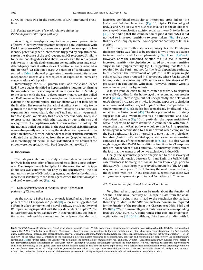
References
[1] P.J. McHugh, V.J. Spanswick, J.A. Hartley, Repair of DNA interstrand crosslinks:molecular mechanisms and clinical relevance, Lancet Oncol. 2 (2001) 483–490.
[2] P. Lehoczky, P.J. McHugh, M. Chovanec, DNA interstrand cross-link repair inSaccharomyces cerevisiae, FEMS Microbiol. Rev. 31 (2007) 109–133.
[3] J.A. Henriques, E. Moustacchi, Isolation and characterization of pso mutantssensitive to photo-addition of psoralen derivatives in Saccharomyces cerevisiae,Genetics 95 (1980) 273–288.
[4] A. Ruhland, M. Kircher, F. Wilborn, M. Brendel, A yeast mutant specificallysensitive to bifunctional alkylation, Mutat. Res. 91 (1981) 457–462.
[5] C. Cassier-Chauvat, E. Moustacchi, Allelism between pso1-1 and rev3-1mutants and between pso2-1 and snm1 mutants in Saccharomyces cerevisiae,Curr. Genet. 13 (1988) 37–40.
[6] X. Li, J. Hejna, R.E. Moses, The yeast Snm1 protein is a DNA 5′-exonuclease, DNARepair (Amst.) 4 (2005) 163–170.
[7] P.J. McHugh, S. Sarkar, DNA interstrand cross-link repair in the cell cycle: acritical role for polymerase zeta in G1 phase, Cell Cycle 5 (2006) 1044–1047.
[8] S. Lambert, S.J. Mason, L.J. Barber, J.A. Hartley, J.A. Pearce, A.M. Carr, et al., Sch-
izosaccharomyces pombe checkpoint response to DNA interstrand cross-links,Mol. Cell. Biol. 23 (2003) 4728–4737.
[9] Y. Kee, A.D. D’Andrea, Expanded roles of the Fanconi anemia pathway in pre-serving genomic stability, Genes Dev. 24 (2010) 1680–1694.
[10] T.R. Singh, D. Saro, A.M. Ali, X.-F. Zheng, C. Du, M.W. Killen, et al., MHF1-MHF2,a histone-fold-containing protein complex, participates in the Fanconi anemiapathway via FANCM, Mol. Cell 37 (2010) 879–886.
[11] Z. Yan, M. Delannoy, C. Ling, D. Daee, F. Osman, P.A. Muniandy, et al., Ahistone-fold complex and FANCM form a conserved DNA-remodeling complexto maintain genome stability, Mol. Cell 37 (2010) 865–878.
[12] A.F. Alpi, K.J. Patel, Monoubiquitylation in the Fanconi anemia DNA damageresponse pathway, DNA Repair (Amst.) 8 (2009) 430–435.
[13] A. Smogorzewska, R. Desetty, T.T. Saito, M. Schlabach, F.P. Lach, M.E. Sowa,et al., A genetic screen identifies FAN1, a Fanconi anemia-associated nucleasenecessary for DNA interstrand crosslink repair, Mol. Cell 39 (2010) 36–47.
Y. Fontebasso et al. / DNA Repair 12 (2013) 1011– 1023 1023
[14] C. MacKay, A.-C. Déclais, C. Lundin, A. Agostinho, A.J. Deans, T.J. MacArtney,et al., Identification of KIAA1018/FAN1, a DNA repair nuclease recruited to DNAdamage by monoubiquitinated FANCD2, Cell 142 (2010) 65–76.
[15] K. Kratz, B. Schöpf, S. Kaden, A. Sendoel, R. Eberhard, C. Lademann, et al.,Deficiency of FANCD2-associated nuclease KIAA1018/FAN1 sensitizes cells tointerstrand crosslinking agents, Cell 142 (2010) 77–88.
[16] K. Yoshikiyo, K. Kratz, K. Hirota, K. Nishihara, M. Takata, H. Kurumizaka, et al.,KIAA1018/FAN1 nuclease protects cells against genomic instability induced byinterstrand cross-linking agents, PNAS (2010).
[17] T. Liu, G. Ghosal, J. Yuan, J. Chen, J. Huang, FAN1 acts with FANCI-FANCD2 topromote DNA interstrand cross-link repair, Science 329 (2010) 693–696.
[18] R.D. Shereda, Y. Machida, Y.J. Machida, Human KIAA1018/FAN1 localizes tostalled replication forks via its ubiquitin-binding domain, Cell Cycle 9 (2010)3977–3983.
[19] P. Hentges, B. Van Driessche, L. Tafforeau, J. Vandenhaute, A.M. Carr, Threenovel antibiotic marker cassettes for gene disruption and marker switching inSchizosaccharomyces pombe, Yeast 22 (2005) 1013–1019.
[20] L. Zheng, U. Baumann, J.-L. Reymond, An efficient one-step site-directedand site-saturation mutagenesis protocol, Nucleic Acids Res. 32 (2004)e115.
[21] A.T. Watson, V. Garcia, N. Bone, A.M. Carr, J. Armstrong, Gene tagging andgene replacement using recombinase-mediated cassette exchange in Schizo-
saccharomyces pombe, Gene 407 (2008) 63–74.[22] E. Cannavo, B. Gerrits, G. Marra, R. Schlapbach, J. Jiricny, Characterization of the
interactome of the human MutL homologues MLH1, PMS1, and PMS2, J. Biol.Chem. 282 (2007) 2976–2986.
[23] S.D. McCulloch, T.A. Kunkel, The fidelity of DNA synthesis by eukaryotic replica-tive and translesion synthesis polymerases, Cell Res. 18 (2008) 148–161.
[24] R.N. Venkatesan, J.J. Hsu, N.A. Lawrence, B.D. Preston, L.A. Loeb, Mutator phen-otypes caused by substitution at a conserved motif A residue in eukaryotic DNApolymerase delta, J. Biol. Chem. 281 (2006) 4486–4494.
[25] I. Miyabe, T.A. Kunkel, A.M. Carr, The major roles of DNA polymerases epsilonand delta at the eukaryotic replication fork are evolutionarily conserved, PLoSGenet. 7 (2011) e1002407.
[26] P.J. McHugh, W.R. Sones, J.A. Hartley, Repair of intermediate structures pro-duced at DNA interstrand cross-links in Saccharomyces cerevisiae, Mol. Cell.Biol. 20 (2000) 3425–3433.
[27] F. Paques, J.E. Haber, Multiple pathways of recombination induced by double-strand breaks in Saccharomyces cerevisiae, Microbiol. Mol. Biol. Rev. 63 (1999)349–404.
[28] A.R. Meetei, A.L. Medhurst, C. Ling, Y. Xue, T.R. Singh, P. Bier, et al., A humanortholog of archaeal DNA repair protein Hef is defective in Fanconi anemiacomplementation group M, Nat. Genet. 37 (2005) 958–963.
[29] G. Mosedale, W. Niedzwiedz, A. Alpi, F. Perrina, J.B. Pereira-Leal, M. Johnson,et al., The vertebrate Hef ortholog is a component of the Fanconi anemia tumor-suppressor pathway, Nat. Struct. Mol. Biol. 12 (2005) 763–771.
[30] W. Sun, S. Nandi, F. Osman, J.S. Ahn, J. Jakovleska, A. Lorenz, et al., The FANCMortholog Fml1 promotes recombination at stalled replication forks and lim-its crossing over during DNA double-strand break repair, Mol. Cell 32 (2008)118–128.
[31] T.A. Ward, Z. Dudásová, S. Sarkar, M.R. Bhide, D. Vlasáková, M. Chovanec, et al.,Components of a Fanconi-like pathway control Pso2-independent DNA inter-strand crosslink repair in yeast, PLoS Genet. 8 (2012) e1002884.
[32] L. Aravind, E.V. Koonin, S.A.P.- a putative DNA-binding motif involved in chro-mosomal organization, Trends Biochem. Sci. 25 (2000) 112–114.
[33] L.N. Kinch, K. Ginalski, L. Rychlewski, N.V. Grishin, Identification of novel restric-tion endonuclease-like fold families among hypothetical proteins, NucleicAcids Res. 33 (2005) 3598–3605.
[34] L.M. Iyer, M.M. Babu, L. Aravind, The HIRAN domain and recruitment of chro-matin remodeling and repair activities to damaged DNA, Cell Cycle 5 (2006)775–782.
[35] A. Roguev, M. Wiren, J.S. Weissman, N.J. Krogan, High-throughput genetic inter-action mapping in the fission yeast Schizosaccharomyces pombe, Nat. Methods4 (2007) 861–866.
[36] T. Caspari, M. Dahlen, G. Kanter-Smoler, H.D. Lindsay, K. Hofmann, K.Papadimitriou, et al., Characterization of Schizosaccharomyces pombe Hus1: aPCNA-related protein that associates with Rad1 and Rad9, Mol. Cell. Biol. 20(2000) 1254–1262.
[37] E.R. Parrilla-Castellar, S.J.H. Arlander, L. Karnitz, Dial 9-1-1 for DNA damage:the Rad9-Hus1-Rad1 (9-1-1) clamp complex, DNA Repair (Amst.) 3 (2004)1009–1014.
[38] S. Bergink, S. Jentsch, Principles of ubiquitin and SUMO modifications in DNArepair, Nature 458 (2009) 461–467.
[39] A. O’Donovan, A.A. Davies, J.G. Moggs, S.C. West, R.D. Wood, XPG endonucleasemakes the 3′ incision in human DNA nucleotide excision repair, Nature 371(1994) 432–435.
[40] H.I. Wu, J.A. Brown, M.J. Dorie, L. Lazzeroni, J.M. Brown, Genome-wide iden-tification of genes conferring resistance to the anticancer agents cisplatin,oxaliplatin, and mitomycin C, Cancer Res. 64 (2004) 3940–3948.
[41] S. Tateishi, H. Niwa, J.-I. Miyazaki, S. Fujimoto, H. Inoue, M. Yamaizumi,Enhanced genomic instability and defective postreplication repair in RAD18knockout mouse embryonic stem cells, Mol. Cell. Biol. 23 (2003) 474–481.
[42] K. Nojima, H. Hochegger, A. Saberi, T. Fukushima, K. Kikuchi, M. Yoshimura,et al., Multiple repair pathways mediate tolerance to chemotherapeutic cross-linking agents in vertebrate cells, Cancer Res. 65 (2005) 11704–11711.
[43] P. Sung, Catalysis of ATP-dependent homologous DNA pairing and strandexchange by yeast RAD51 protein, Science 265 (1994) 1241–1243.
[44] E. Namsaraev, P. Berg, Characterization of strand exchange activity of yeastRad51 protein, Mol. Cell. Biol. 17 (1997) 5359–5368.
[45] S. Lambert, A. Watson, D.M. Sheedy, B. Martin, A.M. Carr, Gross chromosomalrearrangements and elevated recombination at an inducible site-specific repli-cation fork barrier, Cell 121 (2005) 689–702.
[46] I.U. De Silva, P.J. McHugh, P.H. Clingen, J.A. Hartley, Defects in interstrand cross-link uncoupling do not account for the extreme sensitivity of ERCC1 and XPFcells to cisplatin, Nucleic Acids Res. 30 (2002) 3848–3856.
[47] R.D. Wood, Mammalian nucleotide excision repair proteins and interstrandcrosslink repair, Environ. Mol. Mutagen. 51 (2010) 520–526.
[48] M. Räschle, P. Knipscheer, M. Enoiu, T. Angelov, J. Sun, J.D. Griffith, T.E.Ellenberger, O.D. Schärer, J.C. Walter, Mechanism of replication-coupled DNAinterstrand crosslink repair, Cell 134 (6) (2008) 969–980.