the complex spatio-temporal regulation of the drosophilamyoblast ... · the complex spatio-temporal...
TRANSCRIPT

The Complex Spatio-Temporal Regulation of theDrosophila Myoblast Attractant Gene duf/kirreK. G. Guruharsha1,2¤, Mar Ruiz-Gomez3, H. A. Ranganath2, Rahul Siddharthan4, K. VijayRaghavan1*
1 National Centre for Biological Sciences, Tata Institute of Fundamental Research, Bangalore, India, 2 Department of Studies in Zoology, University of Mysore,
Manasagangothri, Mysore, India, 3 Centro de Biologia Molecular Severo Ochoa, CSIC and UAM, Cantoblanco, Madrid, Spain, 4 Institute of Mathematical Sciences, CIT
Campus, Taramani, Chennai, India
Abstract
A key early player in the regulation of myoblast fusion is the gene dumbfounded (duf, also known as kirre). Duf must beexpressed, and function, in founder cells (FCs). A fixed number of FCs are chosen from a pool of equivalent myoblasts andserve to attract fusion-competent myoblasts (FCMs) to fuse with them to form a multinucleate muscle-fibre. The spatial andtemporal regulation of duf expression and function are important and play a deciding role in choice of fibre number,location and perhaps size. We have used a combination of bioinformatics and functional enhancer deletion approaches tounderstand the regulation of duf. By transgenic enhancer-reporter deletion analysis of the duf regulatory region, we foundthat several distinct enhancer modules regulate duf expression in specific muscle founders of the embryo and the adult. Inaddition to existing bioinformatics tools, we used a new program for analysis of regulatory sequence, PhyloGibbs-MP,whose development was largely motivated by the requirements of this work. The results complement our deletion analysisby identifying transcription factors whose predicted binding regions match with our deletion constructs. Experimentalevidence for the relevance of some of these TF binding sites comes from available ChIP-on-chip from the literature, andfrom our analysis of localization of myogenic transcription factors with duf enhancer reporter gene expression. Our resultsdemonstrate the complex regulation in each founder cell of a gene that is expressed in all founder cells. They provideevidence for transcriptional control—both activation and repression—as an important player in the regulation of myoblastfusion. The set of enhancer constructs generated will be valuable in identifying novel trans-acting factor-binding sites andchromatin regulation during myoblast fusion in Drosophila. Our results and the bioinformatics tools developed provide abasis for the study of the transcriptional regulation of other complex genes.
Citation: Guruharsha KG, Ruiz-Gomez M, Ranganath HA, Siddharthan R, VijayRaghavan K (2009) The Complex Spatio-Temporal Regulation of the DrosophilaMyoblast Attractant Gene duf/kirre. PLoS ONE 4(9): e6960. doi:10.1371/journal.pone.0006960
Editor: Laszlo Tora, Institute of Genetics and Molecular and Cellular Biology, France
Received March 27, 2009; Accepted June 9, 2009; Published September 9, 2009
Copyright: � 2009 Guruharsha et al. This is an open-access article distributed under the terms of the Creative Commons Attribution License, which permitsunrestricted use, distribution, and reproduction in any medium, provided the original author and source are credited.
Funding: Wellcome Trust, UK (NLO- 056727/Z/99/B), the Indo- French Centre for Research (CEFIPRA 3203-1), the Department of Biotechnology, India and thePRISM project at IMSc. Council for Scientific and Industrial Research fellowship to K.G.G. The funders had no role in study design, data collection and analysis,decision to publish, or preparation of the manuscript.
Competing Interests: The authors have declared that no competing interests exist.
* E-mail: [email protected]
¤ Current address: Department of Cell Biology, Harvard Medical School, Boston, Massachusetts, United States of America
Introduction
Multinucleate muscle fibres form by the regulated fusion of
myoblasts. Muscles of different shapes and sizes are made as a
result of coordinated myoblast fusion and morphogenesis. This
process is perhaps best studied in the embryonic muscles of the
fruitfly Drosophila melanogaster (reviewed in [1]). Within myogenic
domains, myoblasts are separated into founder cells (FCs) and
fusion-competent myoblasts (FCMs) by Notch (N) mediated lateral
inhibition and other signaling pathways [2,3]. FCs seed the
formation of muscles by attracting FCMs to fuse and form multi-
nucleate fibres in a defined pattern. The mutual recognition of
FCs and FCMs is mediated by a group of transmembrane
proteins belonging to the immunoglobulin superfamily. One of
these, Dumbfounded (Duf, also called Kirre), marks the surface
of FCs [4] and another, Sticks and stones (Sns) the complemen-
tary subset of FCMs [5]. When examined by mRNA in situ
hybridization or reporter-gene expression, sns and duf are
expressed transiently in FCs and FCMs respectively and are
turned off soon after the fusion process is complete [4,5]. This
suggests that duf and sns are subject to strong transcriptional
regulation.
In the Drosophila adult and invertebrates, muscles consist of
many myotubes bundled together to form a contractile element.
Drosophila adult muscle precursor cells segregate as sister cells from
embryonic founders and give rise to all adult muscles in the pupa
[6,7]. These adult myoblasts maintain twi expression, proliferate
during larval life and remain associated with imaginal discs in the
thorax and neurons in the abdomen [8]. Specific myoblast groups
are chosen to give rise to different muscles under the influence of
signaling molecules and transcription factors. Apterous (Ap) and
Cut (Ct) are important for direct flight muscles development [9];
Vestigial (Vg) and Cut (Ct), regulated by Wingless (Wg), are
responsible for indirect flight muscle development [10]. Unlike in
the embryo, Notch-mediated lateral inhibition is not involved in
founder cell specification during adult thoracic myogenesis [11].
However, as in the embryo [12,13], components of the Fibroblast
growth factor (FGF) pathway mediates founder cell choice [14].
This results in a precise pattern of founder cells for each multi-
fibre array of adult abdominal muscles. Expression of myoblast
PLoS ONE | www.plosone.org 1 September 2009 | Volume 4 | Issue 9 | e6960

fusion genes is transient and tightly regulated in adult founder
analogs too [14].
The size of the muscle fibre is probably dependent upon the
number of fusion events [15]. The duration and level of Duf/Kirre
on the FC membrane along with other fusion proteins, especially
Rolling pebbles 7 (Rols7; also known as Antisocial), [16,17]
appears to regulate this mechanism. Duf has been shown to be a
rate-limiting factor in myoblast fusion during embryonic myogen-
esis [18]. Characterization of enhancer sequences of duf is
therefore important to understand the transcriptional machinery
that recognizes a FC. This will also allow us to understand the role
of different factors responsible for transcriptional control of duf in
different FCs and thereby development of muscle pattern.
Bioinformatics tools can predict possible transcription factor
(TF) binding sites, either by comparing with previously identified
sites for known TFs, or ab initio by looking for short inexact
repeated patterns or ‘‘motifs’’. Enhancers and cis-regulatory
modules can be predicted by clustering predicted binding sites,
an approach taken by programs such as Stubb [19,20], eCis-
Analyst [21,22] and Cluster-Buster [23]. Recently, with the
availability of sequence information from related species including
twelve Drosophila genomes [24], new approaches have been
developed to make use of orthologous sequence from related
species to specific region of interest. A simple approach is
‘‘phylogenetic footprinting’’ [25,26,27], which confines searches
to sequences that are highly conserved across species, using the
assumption that such regions are more likely to be functional.
However, it is also known that gene regulation evolves significantly
even among closely related species, and binding sites that are
known to be functional in one species disappear or are replaced by
new sites in other species (for example see [28]). Therefore, some
newer programs, including PhyloGibbs [29] (a motif-finder) and
Stubb [19,20] (a module-prediction program), both of which we
have used in this study, analyse both conserved and non-conserved
sequences but modify scoring to take into account phylogenetic
relationship between species. However, these approaches cannot
by themselves give a sense of the temporal or spatial aspects of
gene regulation. To be effective, they must be combined with prior
experimental information about the transcriptional regulation and
spatio- temporal expression pattern of the genes of interest. For
example, a recent [30] study made use of known spatio- temporal
concentrations of transcription factors to predict the expression
levels of cis-regulatory modules in the segmentation network.
Unfortunately such prior data is a luxury often unavailable.
We have used a combination of bioinformatics and functional
enhancer deletion approaches to understand the regulation of duf.
In addition to existing bioinformatics tools, we used a new
program for analysis of regulatory sequence, PhyloGibbs-MP,
described in a recent paper [31], whose development was largely
motivated by the requirements of this work. Studies with Stubb
[19,20] using published consensus sequences for mesoderm
relevant factors found evidence of a modular structure of
enhancers both upstream of the gene and in its intron. Deletion
analysis of the duf regulatory region using reporter constructs
reveals specific aspects of duf regulation during Drosophila
myogenesis. We find that several distinct enhancer modules
regulate duf expression in specific muscle founders of the embryo
and the adult. While embryonic enhancers are proximal, adult-
specific enhancers are located more distal to the duf start site.
These results merited a further, detailed study of the 10 kb region
upstream of duf. We made a list of 44 position weight matrices for
transcription factors relevant to mesoderm development, of which
38 were constructed using either the FlyReg database for DNAse I
footprints [32] or recent data from bacteria-one-hybrid systems
[33] and 6 were taken from the literature. We rejected 13 as not
being specific enough or not showing significant predictions in
preliminary runs. We used the remaining 31 matrices with two
programs, Stubb [19,20] and PhyloGibbs-MP [31], to predict cis-
regulatory modules as well as binding sites for individual
transcription factors. The results complement our deletion analysis
by identifying transcription factors whose predicted binding
regions match with our deletion constructs. Most predicted sites
are conserved in other Drosophila species, suggesting functional
importance. Experimental evidence for the relevance of some of
these TF binding sites comes from both specific and global ChIP-
on-chip analysis from the literature, using key mesodermal
regulators [34,35,36]. Though bioinformatic predictive tools and
ChIP-on-chip approaches are unable, by themselves, to predict the
full spatio-temporal behaviour of gene regulation, we demonstrate
their utility when combined with our experimental information.
Given the many conserved aspects of myogenic regulation
between flies and vertebrates, recently underscored by the
demonstration of the role of kirre in zebrafish [37], our results
are likely to be of broad value.
Results
Identification of duf Enhancer RegionPreliminary studies of the 40 kb region upstream of duf and of
the first intron (29 kb) were made using Matinspector Professio-
nalH [38]. MatInspector is a tool for transcription factor binding
site analysis by Genomatix which utilizes its own transcription
factor knowledge base (MatBase) to locate transcription factor
binding sites in sequences of any length. Additional binding sites
information for nuclear effectors of important signaling pathways
and mesoderm specific factors from published work that were not
available in Matinspector ProfessionalH were integrated into our
search for cis-regulatory elements regulating duf expression in FCs.
The consensus sequences (and source) for these factors are
described in Materials and Methods.
Analysis of this sequence revealed presence of many strong
binding sites for nuclear effectors of different intercellular signaling
pathways. Some clustering of binding sites was seen in the 10 kb
region immediately upstream of duf. The arrangement of these
putative binding sites in duf upstream region is shown in Figure 1A
for select factors. Distinct PREs (Polycomb group Response
Elements) and TREs (Trithorax Response Elements) are also
found in this region (Figure 1A). The list of factors and their
putative binding sites are listed in Supplementary Datasheet S1.
The putative binding sites were compared using sequence
similarity between Drosophila melanogaster and Drosophila pseudoobscura
genomes. A significant number of the putative binding sites for
signaling pathway effectors and transcription factors with
mesodermal role and early patterning genes map within or in
the vicinity of conserved sequence stretches (vertical blue bars
Figure 1A). The results are tabulated for all TF binding sites in
Supplementary Datasheet S2 and summarized for key signaling
pathway effectors and mesodermal factors in Figure 1A.
To characterize the regulatory potential of putative duf enhancer
sequences, chosen genomic sequences were amplified by the
polymerase chain reaction (PCR) from the duf 59 region. Fragments
representing progressive deletions from both distal and proximal
ends were amplified and cloned as EcoRI - BamHI fragments into
pCaSpeR AUG bGal [39], or pPTGal [40]. Transgenic flies were
generated from these constructs producing either lacZ (blue bars in
Figure 1B) or Gal4 lines (red bars in Figure 1B) as described in
Materials and Methods. The expression patterns of these duf
enhancer constructs were analyzed for reporter expression during
Regulation of a Myoblast Gene
PLoS ONE | www.plosone.org 2 September 2009 | Volume 4 | Issue 9 | e6960

Figure 1. Consensus binding sites in duf enhancer sequences and deletion analysis of duf genomic region. A. Occurrence of bindingsites for nuclear effectors of signaling pathways and mesodermal factors in the 10 kb sequence 59 to duf is diagrammatically shown. Publishedconsensus binding sequences for Ets, Su(H), Ci, dTCF, Mad, Brk, Twi, Tin, Mef2 are shown. Downward pointing arrow indicates binding on + strandand upward pointing arrow indicates binding on –strand. Sequences that are well conserved in D. pseudoobscura are shown as blue vertical bars.Several putative binding sites for GAGA factor encoded by the Trithorax-like gene (Trl) characteristic of TREs (Trithorax Response Elements) arepresent within 3.8 kb from the duf start site (red verticle bars) but no sites further upstream in the 10 kb region. Similarly, putative binding sites forPHO (pleiohomeotic) and PHO-like, polycomp group proteins (PcG) that bind to PREs (Polycomb group Response Elements) are found between28.0 kb to 29.3 kb region (green vertical bars). B. Schematic of constructs generated to characterize the regulatory potential of putative dufenhancer sequences during Drosophila embryonic and adult myogenesis. Putative enhnacer fragments with deletions from both distal (blue bars)and proximal ends (red bars) of the duf 59 region were PCR amplified and cloned as EcoRI - BamHI fragments into pCaSpeR AUG bGal, or pPTGal.Transgenic flies were generated from these constructs producing either lacZ (blue bars) or Gal4 lines (red bars). duf 28.220.6 was cloned intoZGLpWW vector (magenta bar).doi:10.1371/journal.pone.0006960.g001
Regulation of a Myoblast Gene
PLoS ONE | www.plosone.org 3 September 2009 | Volume 4 | Issue 9 | e6960

embryonic and adult myogenesis. Additional duf enhancer-deletion
reporter constructs for further analysis (results not discussed) are in
Supplementary data Figure S1.
Modular Enhancers Regulate duf Expression duringEmbryonic Myogenesis
The dynamics of wildtype duf expression during embryonic
muscle development has been characterized using mRNA in-situ
hybridizations [4] and by the use of rP298 lacZ, [41] a P- nuclear
lacZ insertion into the duf locus, which reproduces duf -like
reporter- expression in all founder cells during embryonic [4] and
adult myogenesis [11]. The expression pattern of different duf
upstream reporter constructs were compared with rP298 lacZ
(Figure 2, A and A9).
The smallest fragment close to the start site, duf 21.0 kb lacZ, is
capable of driving mesodermal expression in the developing
embryo during stages 12–14 (Figure 2, B and B9). The reporter
expression is seen in many somatic FCs in the abdomen. The
pattern appears to be slightly diffuse. This reporter also ectopically
marks a ventral cluster of cells in all the three thoracic segments
(arrows, Figure 2, B and B9). This is in the region where adult
imaginal myoblasts reside. No expression is seen in the developing
visceral mesoderm. In a slightly larger fragment of the enhancer,
duf 21.5 kb lacZ, the ectopic expression in the ventral thoracic
region seen in duf 21.0 kb lacZ is completely repressed (Figure 2, C
and C9). duf 21.5 kb lacZ also shows strong expression in ventral
and dorsal clusters of somatic FCs in the abdominal segments
(arrowheads, Figure 2, C and C9). Weak expression is also seen in
the lateral cluster of FCs of the abdomen. There is no expression
in any of the visceral muscles. duf 22.4 kb lacZ is expressed very
weakly in all somatic founder myoblasts of the abdomen at stage
12–14 (Figure 2, D and D9). There is no expression in any of the
visceral muscles. duf 23.0 kb lacZ is specifically expressed in
longitudinal visceral muscle FCs that originate from the caudal
mesoderm (arrow, Figure 2E). These FCs migrate from the
posterior end over the developing embryonic viscera (gut) during
stage 12. They fuse with the remaining FCMs in the gut region
and align longitudinally along the entire length of the embryonic
gut by stage 14 (arrow, Figure 2E9). duf 23.0 kb lacZ expression is
also seen specifically in 3 FCs in the abdomen (arrowheads,
Figure 2, E and E9). No expression is seen in circular visceral FCs
or in any other somatic myoblasts. In comparison, duf 23.8 kb lacZ
shows very specific expression in a large subset of somatic muscle
FCs of the thorax and the abdomen. Expression is absent from
circular and longitudinal visceral FCs. duf 24.6 kb lacZ (Supple-
mentary data Figure S2) and duf 25.1 kb lacZ (Figure 2, G and G9)
shows strong expression in a larger number of somatic muscle FCs
and is again absent in visceral mesoderm founders. duf 25.35 kb
lacZ and duf 25.5 kb lacZ (data not shown) also show strong
expression in somatic muscle FCs very similar to duf 25.1 kb lacZ
(Figure 2, G and G9) and very weak expression in the visceral
mesoderm. Thus, from 23.8 to 25.5 kb, there is no change in the
muscle type except that the number of somatic myoblasts
expressing the reporter is increased. Preliminary analysis of the
duf 26.4 kb fragment indicates that it is expressed weakly in
somatic muscles and ectopically in the trachea. duf 27.2 kb lacZ
reporter expression is seen in both somatic and visceral muscles as
well as garland cells where wildtype duf is known to be expressed
[4], but embryonic muscle expression is very weak compared to
other reporter constructs (Supplementary data Figure S2).
Deletions from the proximal end i.e. enhancer constructs without
sequences close to the start site, show a different pattern of reporter
expression. duf 28.2 20.6 kb (Figure 2, H and H9), duf 27.9 23.8 kb
(Supplementary data Figure S2) and duf 29.8 23.8 kb (Figure 2, I
and I9) fragments are specifically expressed in FCs of the developing
mid-gut circular visceral muscles and also in a subset of somatic
muscle FCs of the abdomen at embryonic stage 12–13. At stage 14,
expression is clearly seen in the circular visceral muscles in a ribbon-
like arrangement following fusion with neighboring FCMs
(Figure 2H–I9, ). The expression is completely restricted to circular
visceral founder cells and completely excluded from the longitudinal
visceral muscle founders in the gut. The entire length of the putative
enhancer duf 29.8 kb lacZ recapitulates the complete wildtype duf
expression pattern in all the embryonic somatic as well as both types
of visceral muscle founder cells (Figure 2, J and J9). Preliminary
analysis of some of the smaller proximal and distal enhancer
deletion reporter constructs, for example duf 24.623.0 kb Gal4
showed nonspecific and ectopic reporter expression in the epidermis
(Supplementary data Figure S2) while duf 22.421.5 kb Gal4 had no
detectable mesodermal expression pattern (data not shown). From
this expression analysis it is clear that elements in the 10 kb region
59 of duf are capable of driving reporter expression in different
subsets of muscle FCs. This also indicates that there are independent
modules for duf expression in different muscles. These modules are
not noticeably overlapping, eliciting expression in a different subset
of muscles with every addition of a few hundred base-pairs of the
enhancer. Each additional fragment in the 59 represses the
expression seen in a smaller proximal fragment and directs
expression in different domains of duf expression. All the necessary
elements for this complex spatio-temporal regulation of duf
expression in all embryonic muscle FCs appears to be located in
the 10 kb region 59 of duf coding region.
Duf Enhancer Modules Mark Different Subsets ofEmbryonic Muscle Founder Cells
The expression pattern seen with duf upstream-lacZ reporters
appears to be in specific subsets of FCs in different constructs. We
double labeled enhancer-reporter constructs with mesodermal and
founder- cell markers to verify this. Expression patterns of the duf
enhancer lines were also confirmed by co-localization with duf Gal4
driven UAS-GFP (Supplementary data Figure S3). To examine if
any duf upstream enhancer construct ectopically expresses in Twi
positive adult muscle precursors or imaginal myoblasts in the
embryo (see Figure 3D), stage 12–15 embryos of enhancer
transgenic lines were double labeled with antibodies against b-Gal
and Twi (Figure 3, E–I). duf 21.0 kb lacZ shows strong expression in
the ventral thoracic region, where the imaginal disc primordia
reside (Figure 3E). The expression pattern of the reporter appears to
colocalize in this region with some Twi positive cells adhering to the
imaginal discs (arrows Figure 3E). The reporter also ectopically
marks some ectodermal cells (i.e. non-mesodermal cells) of the
ventral imaginal disc primordia (arrowheads Figure 3E). duf 21.0 kb
lacZ does not express in Twi positive adult muscle precursors of the
abdomen. No other construct shows expression in any of the Twi
positive adult precursors or imaginal myoblasts in the embryo. This
indicates that the regulatory elements located 21.0 kb region
immediately upstream of duf are sufficient to promote reporter
expression in most of the embryonic mesoderm including the
ventral thoracic region similar to Twi expression domain. Elements
present further upstream as in duf 21.5 kb lacZ have repressor
elements that suppress expression in ventral thoracic region but at
the same time also promote the expression of the reporter in the
dorsal and ventral subset of somatic FCs recapitulating a part of
wildtype duf expression.
Additionally, co-localization was done with the duf enhancer
reporter constructs with other known FC markers such as Kruppel
(Kr), Vg and even-skipped (Eve). These transcription factors mark
different somatic muscles as follows: Kr is expressed in DA1, DO1,
Regulation of a Myoblast Gene
PLoS ONE | www.plosone.org 4 September 2009 | Volume 4 | Issue 9 | e6960

Figure 2. Modular enhancers regulate duf expression during embryonic myogenesis. Confocal projections of different duf enhancerreporter expression assayed using anti b gal staining in Drosophila embryos. Level of reporter expression in muscles is represented by intensity ofgreen colour in the cartoon to the right. Wildtype duf expression is visualized using rP298 lacZ (A and A9). duf 21.0 kb lacZ is expressed in few somaticFCs (arrowheads in B and B9) and ectopically in ventral thoracic region (arrows in B and B9), which is repressed in duf 21.5 kb lacZ (arrows in C and C9).duf 21.5 kb lacZ is expressed in ventral and dorsal abdominal somatic FCs (arrowheads in C and C9) and weakly in lateral somatic FCs. duf 22.4 kblacZ is expressed very weakly in somatic FCs (arrows in D and D9). duf 23.0 kb lacZ is specifically expressed in longitudinal visceral FCs (arrow in E)that align longitudinally around the gut following fusion with FCMs (arrow in E9). It is also expressed in 3 somatic FCs (arrowheads in E and E9) but notin circular visceral muscles. duf 23.8 kb lacZ and duf 25.1 kb lacZ are expressed in a large subset of somatic FCs (arrows in F–G9) but not in visceralmesoderm. duf 28.2 20.6 kb Gal4 and duf 29.823.8 kb Gal4 marks the circular visceral muscle FCs (arrows in H–I9) and few ventral somatic muscleFCs (arrowheads in H–I9). duf 29.8 kb lacZ recapitulates the complete wildtype duf expression and marks all the embryonic muscle FCs. All embryosare lateral view with anterior to the left and dorsal to the top. Scale bar = 50 microns.doi:10.1371/journal.pone.0006960.g002
Regulation of a Myoblast Gene
PLoS ONE | www.plosone.org 5 September 2009 | Volume 4 | Issue 9 | e6960

LL1, LT2, LT4, VL3, VA2, VO2 and VO4 somatic FCs
(Figure 3B). Vg is expressed in DA1-3, LL1, VL1, VL2, VL3,
VL4 somatic muscle FCs and Eve is expressed only in DA1 muscle
founder (Figure 3B) and a subset of pericardial cells. Colocaliza-
tion with Kr (Figure 3, J–O), Eve (Figure 3, P–W) and Vg (data not
shown), reveal that the expression pattern of the reporter
constructs is in specific founders. Co-localizations with duf Gal4
. UAS-GFP (Supplementary data Figure S3) and Kr were also
very useful in identifying specific muscles that were marked by
different duf enhancer constructs. duf 21.5 kb lacZ is expressed in
DA1 and DO1, and very weakly in other Kr positive FCs. duf
23.0 kb lacZ is expressed only in DA1 among all Kr expressing
FCs. duf 23.8 kb lacZ is expressed strongly in 6 Kr positive somatic
FCs but very weakly in LT2, LT4 and VO2 somatic FCs. In duf
23.8 kb Gal4 no expression was detected in LT2, LT4 and VO2
somatic FCs but there was strong expression in other Kr positive
somatic FCs. duf 24.6 kb lacZ (data not shown) and duf 25.1 kb
lacZ are expressed weakly in LT2, not detectable in VA2 but
strongly in all other Kr positive somatic FCs. duf 28.220.6 kb
Gal4 is expressed weakly in VO4 but in none of the other Kr
positive FCs. rP298 lacZ was used as positive control for
comparison. Colocalization of all duf enhancer constructs with
Eve also revealed that none of the enhancer reporters tested
expressed ectopically in pericardial cells (Figure 3, P–W). Eve is
expressed in DA1 somatic FC and subset of pericardial cells in the
dorsal mesoderm. duf 21.0 kb lacZ (Figure 3P) is expressed the
dorsal row of cells which are not pericardial cells. No colocaliza-
tion is detected in pericardial cells. duf 21.5 kb, 23.0 kb, 23.8 kb,
24.6 kb and 25.5 kb enhancer lacZ lines colocalize with Eve
expressing DA1 somatic FC but not with pericardial cells (Figure 3,
Q–U). duf 28.220.6 kb Gal4 is not expressed in Eve positive DA1
or pericardial cells (Figure 3V).
Interestingly, in two cases, duf enhancer reporters are expressed
in domains where wildtype duf expression is not known. These
Figure 3. duf enhancer reporters are expressed in specific muscle founder cells. Cartoon representation of Drosophila embryo depictingdifferent muscles (A) and Kr expression (in B) and Eve expressing DA1 (in C). (D) Brown box region (in A) is enlarged to show Twi expressing adultmuscle precursors (AMPs, adapted from [74]). The same region is discussed in (E). (F–W) Confocal projections of stage 14 embryos double labeledwith antibodies against b Gal (in green, E–W) and either Twi (in red; E–I), Kr (in magenta; J–O) or Eve (in blue; P–W) corresponding to cyan box regionin (A). Wildtype duf expression is seen using rP298 lacZ (I, O and W). Ectopic expression of duf 21.0 kb lacZ (arrowheads in E) is ventral to Twiexpressing AMPs (arrows in E). All duf enhancer reporters do not colocalize with Twi in the abdomen (arrows in F–I). Longitudinal visceral muscles areseen in duf 23.0 kb lacZ (arrowheads in G). Non-specific staining of trachea (asterisk in E, F and H). duf 21.5 kb lacZ colocalizes with Kr positive FCs(arrow J). duf 23.0 kb lacZ is expressed in longitudinal visceral muscles (arrowhead) and colocalizes with Kr only in DA1 (arrow in K). duf 23.8 kb lacZ(L) and duf 25.1 kb lacZ (M) colocalize with all Kr positive somatic FCs. duf 28.220.6 kb Gal4 expressed in circular visceral muscles (arrowhead in N)do not colocalize with Kr. Kr expression in CNS is marked by asterisk. (P) duf 21.0 kb lacZ is not expressed in DA1 or pericardial cells. duf 21.5 kb,23.0 kb, 23.8 kb, 24.6 kb and 25.5 kb colocalize with Eve in DA1 (arrows) but not with pericardial cells (Q–U). duf 28.220.6 kb Gal4 is not expressedin DA1 (arrows) or pericardial cells (V). Eve expression in the CNS (arrowheads Q–U). Scale Bar = 50 microns.doi:10.1371/journal.pone.0006960.g003
Regulation of a Myoblast Gene
PLoS ONE | www.plosone.org 6 September 2009 | Volume 4 | Issue 9 | e6960

expression patterns are described in Supplementary data Figure
S4. By stage 16, duf 22.4 kb lacZ is also expressed ectopically in a
large subset of the developing cardioblasts, those that express seven-
up (svp) but not tin. Wildtype duf is not expressed in the developing
cardioblasts and loss of duf function does not affect the formation
of the embryonic heart [4]. The cardioblast expression seen in duf
22.4 kb lacZ is completely repressed in a slightly larger duf
enhancer fragment: duf 23.0 kb lacZ. Similarly, the reporter
expression in duf 23.0 kb lacZ is also seen in the embryonic central
nervous system in a large subset of neuroblasts as compared to
wildtype duf at stage 16. All the larger duf enhancer constructs do
not show ectopic reporter expression in the ventral thoracic
segments, cardioblasts or embryonic central nervous system. This
suggests the presence of repressor elements that would restrict duf
expression specifically to different founder cells.
Distal Enhancers Regulate duf Expression During AdultMyogenesis
Adult muscle founder-specific expression of all the reporters was
assayed in imaginal myoblasts associated with the wing imaginal
discs (data not shown) and during pupal myogenesis for all the
different muscle subtypes in the thorax, and Dorsal, Ventral and
Lateral muscles in the abdomen. Expression was compared by
colocalization with mouse monoclonal antibody 22C10, which
marks neurons and the abdominal founder myoblasts very clearly
[11]. rP298 lacZ or duf Gal4-UAS lacZ was used for wildtype
comparison (Figure 4).
Analysis of duf enhancer deletion constructs during different
stages of adult myogenesis reveals several interesting aspects of duf
regulation in the adult. All smaller duf enhancer constructs up to
3.8 kb show no, or comparable to background, reporter expression
in both thoracic as well as abdominal muscle founder cells of the
adult. duf 23.8 kb lacZ shows weak expression in lateral abdominal
muscles (Figure 4C0). duf 25.1 kb is clearly expressed in lateral
abdominal muscles but not in dorsal abdominal muscles
(Figure 4D0). This expression is not seen in a slightly larger
fragment i.e. duf 25.5 kb lacZ (data not shown) which indicates that
region around 23.0 25.1 kb upstream of duf has a module for
expression in lateral abdominal muscles. None of the constructs up
to those with 25.5 kb of 59 sequence are expressed in dorsal
abdominal muscles or in any of the thoracic muscles. duf
28.220.6 Gal4 is expressed strongly in a subset of dorsal as well
as lateral abdominal muscles (Figure 4, E9 and E0). Expression is
also seen in one kind of indirect flight muscle of the thorax –the
Dorso-Ventral Muscles (DVMs) (Figure 4E). There is some
variation in the expression pattern of this particular construct.
The duf 29.8 23.8 kb Gal4 is specifically expressed in all adult
muscle founders. It marks the founder larval templates [11,42] of
the Dorsal Longitudinal Muscles (DLMs) in the thorax (Figure 4F)
and dorsal, lateral and ventral abdominal founders [14] (Figure 4,
F9 and F0). The larger duf 29.8 kb lacZ shows expression in all the
developing adult muscle founders (Figure 4, G–G0) and also marks
several epidermal cells ectopically.
The results show that enhancer fragments close to the duf
transcription start site are very important to drive expression in
specific muscles of the embryo and deletion of these elements
appears to enhance expression specifically in the adult muscles.
Taken together, these results merited further computational
analysis of this region.
Computational Study of the duf EnhancerThe complex and non cumulative expression pattern observed
in duf enhancer reporter constructs justified a more detailed study
that we describe below, where we made use of Stubb and
PhyloGibbs-MP [31], an updated version of the motif-finder
PhyloGibbs [29] that is capable of module prediction either ab
initio or using prior position weight matrices (PWMs). First, we
made a list of high-quality PWMs for factors of known importance
in mesoderm development, using sequences from PWMs, using
the Flyreg database of DNAse I footprints [32], binding sequences
found in recent bacteria-one-hybrid system (B1H) studies [33,43],
and weight matrices from the literature. In all, 44 matrices were
generated in this way, including six from the literature, 11 from
the B1H data Noyes et al. [33,43], and the remainder from the
Flyreg database of DNAse I footprints [32]. The details are
described in Materials and Methods. These high quality PWMs
and their associated sequence logos are in Supplementary data
Figure S5. A subset of 31 matrices were eventually used, the
remainder being poor-quality (not specific enough) or not making
significant predictions in preliminary runs.
Next, we used the module prediction program Stubb [19,20]
with these PWMs to predict enhancers upstream of and in the
intron of duf. Finally, we used the same PWMs and PhyloGibbs-
MP to confirm the prediction of the enhancers as well as to predict
individual binding sites for transcription factors.
Predicting Enhancers with StubbWe used the 31 PWMs described above with Stubb [19,20] to
determine upstream and intronic regions of interest. Unlike naive
methods based on clustering predicted sites, Stubb incorporates
competition between factors, carefully handling the situation
where multiple factors may compete for the same binding
sequence; it calculates a ‘‘partition function’’ that takes account
of all possible ways of ‘‘parsing’’ a sequence into regulatory sites
and ‘‘background’’, and then uses this to predict ‘‘binding
energies’’ for each factor at each site. It also calculates an overall
‘‘free energy’’ function that serves to indicate likely locations of
CRMs.
We ran Stubb on the 30 kb upstream region of duf, and also on
its 29 kb intron, using a window size of 1000 bp and a shift of
100 bp. The free-energy profile shows significant enhancer
structure: the first 10 kb upstream have significant binding free
energies for these factors, but there also occur peaks at about 12 kb
215 kb upstream and 25 kb upstream (Figure 5A), and in the
intron (Figure 5B).
Apart from global free energies, Stubb predicts binding affinities
for individual factors at individual sites, taking competition with
other factors into account. Some factors show numerous low-
affinity sites, usually attributable to a poorly-defined input weight
matrix. Some factors, however, seem to show distinct clustering in
certain parts of the 10 kb region. A discussion of binding site
predictions for individual factors is deferred to the next subsection.
Predicting Enhancers and Regulatory Sites withPhyloGibbs-MP
PhyloGibbs is a motif-finder with the ability to incorporate
orthologous sequence from closely-related species that may have
significant non-functional conservation. It reports predictions with
significance estimates that are posterior probabilities, obtained
from extended sampling, that the predictions are binding sites
given the prior assumptions. We showed [29] that these
significance estimates are reliable in synthetic data, and given
some reasonable assumptions on gene regulation in yeast
(Saccharomyces cerevisiae) and the state of present experimental
knowledge, the significance estimates are probably very accurate
in experimental systems too.
PhyloGibbs-MP is an update to PhyloGibbs, which, among
other new features, has the ability to localise predictions to small
Regulation of a Myoblast Gene
PLoS ONE | www.plosone.org 7 September 2009 | Volume 4 | Issue 9 | e6960

Figure 4. Expression of duf enhancers in developing Drosophila adult muscles. (A) Diagrammatic representation of 28 hr APF (afterpuparium formation) pupa depicting developing muscles of the thorax and the abdomen. Dorsal Longitudinal Muscles (DLMs; in green) Dorso-Ventral Muscles (DVMs; in yellow) make up the indirect flight muscles (IFMs) of the thorax. Abdominal dorsal muscles (red) and lateral muscles (blue)are shown (B–G0). Confocal projections of 28 (61) hr APF pupae from duf enhancer lacZ lines double labelled with anti-b-gal (green) and m22C10(red). Pupal thoracic DLMs (six fibres, B–G), abdominal dorsal muscles (of a2 hemisegment, B9–G9) and lateral muscles (B0–G0) are shown. rP298 lacZexpression is seen in the nuclei of six DLM fibers (B) and duf Gal4 driven lacZ is seen in of dorsal (B9) and lateral muscle FCs that are also marked bym22C10 (B0). duf 23.8 kb lacZ expression is weak in DLMs (C) and dorsal muscles (C9) but stronger in lateral abdominal muscles (C0). duf 25.1 kb lacZis expressed in lateral muscles (D0) but not in DLMs (D) or dorsal muscles (D9). 28.2 to 20.6 kb Gal4 drives expression in DVMs. DVM II is seen here(arrowhead in E). Specific expression is seen in dorsal muscles (E9) and lateral muscles (E0). Some amount of variation is seen in the expression of thisGal4. duf 29.823.8 kb Gal4 is specifically expressed in DLMs (arrows in F). It also drives expression in dorsal (F9) and lateral muscles (F0). duf 29.8 kblacZ is expressed in DLMs (G) and DVMs (data not shown) and in lateral muscles (G0) but less specifically in the dorsal muscles (G9). Backgroundexpression is also seen in FCM population and attachment fibers (asterisk in G). Anterior to the top, dorsal midline to the right (B–G and B0–G0); andanterior to the right, dorsal midline to the top (B9–G9). Scale bar = 50 mm.doi:10.1371/journal.pone.0006960.g004
Regulation of a Myoblast Gene
PLoS ONE | www.plosone.org 8 September 2009 | Volume 4 | Issue 9 | e6960
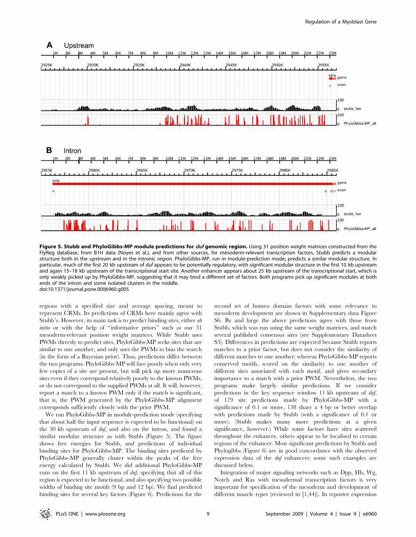
regions with a specified size and average spacing, meant to
represent CRMs. Its predictions of CRMs here mainly agree with
Stubb9s. However, its main task is to predict binding sites, either ab
initio or with the help of ‘‘informative priors’’ such as our 31
mesoderm-relevant position weight matrices. While Stubb uses
PWMs directly to predict sites, PhyloGibbs-MP seeks sites that are
similar to one another, and only uses the PWMs to bias the search
(in the form of a Bayesian prior). Thus, predictions differ between
the two programs: PhyloGibbs-MP will fare poorly when only very
few copies of a site are present, but will pick up more numerous
sites even if they correspond relatively poorly to the known PWMs,
or do not correspond to the supplied PWMs at all. It will, however,
report a match to a known PWM only if the match is significant,
that is, the PWM generated by the PhyloGibbs-MP alignment
corresponds sufficiently closely with the prior PWM.
We ran PhyloGibbs-MP in module-prediction mode (specifying
that about half the input sequence is expected to be functional) on
the 30 kb upstream of duf, and also on the intron, and found a
similar modular structure as with Stubb (Figure 5). The figure
shows free energies for Stubb, and predictions of individual
binding sites for PhyloGibbs-MP. The binding sites predicted by
PhyloGibbs-MP generally cluster within the peaks of the free
energy calculated by Stubb. We did additional PhyloGibbs-MP
runs on the first 11 kb upstream of duf, specifying that all of this
region is expected to be functional, and also specifying two possible
widths of binding site motifs (9 bp and 12 bp). We find predicted
binding sites for several key factors (Figure 6). Predictions for the
second set of homeo domain factors with some relevance to
mesoderm development are shown in Supplementary data Figure
S6. By and large the above predictions agree with those from
Stubb, which was run using the same weight matrices, and match
several published consensus sites (see Supplementary Datasheet
S3). Differences in predictions are expected because Stubb reports
matches to a prior factor, but does not consider the similarity of
different matches to one another; whereas PhyloGibbs-MP reports
conserved motifs, scored on the similarity to one another of
different sites associated with each motif, and gives secondary
importance to a match with a prior PWM. Nevertheless, the two
programs make largely similar predictions. If we consider
predictions in the key sequence window 11 kb upstream of duf,
of 179 site predictions made by PhyloGibbs-MP with a
significance of 0.1 or more, 138 share a 4 bp or better overlap
with predictions made by Stubb (with a significance of 0.1 or
more). (Stubb makes many more predictions at a given
significance, however.) While some factors have sites scattered
throughout the enhancer, others appear to be localised to certain
regions of the enhancer. Most significant predictions by Stubb and
Phylogibbs (Figure 6) are in good concordance with the observed
expression data of the duf enhancers: some such examples are
discussed below.
Integration of major signaling networks such as Dpp, Hh, Wg,
Notch and Ras with mesodermal transcription factors is very
important for specification of the mesoderm and development of
different muscle types (reviewed in [1,44]). In reporter expression
Figure 5. Stubb and PhyloGibbs-MP module predictions for duf genomic region. Using 31 position weight matrices constructed from theFlyReg database, from B1H data (Noyes et al.), and from other sources, for mesoderm-relevant transcription factors, Stubb predicts a modularstructure both in the upstream and in the intronic region. PhyloGibbs-MP, run in module-prediction mode, predicts a similar modular structure. Inparticular, much of the first 20 kb upstream of duf appears to be potentially regulatory, with significant modular structure in the first 10 kb upstreamand again 15–18 kb upstream of the transcriptional start site. Another enhancer appears about 25 kb upstream of the transcriptional start, which isonly weakly picked up by PhyloGibbs-MP, suggesting that it may bind a different set of factors. Both programs pick up significant modules at bothends of the intron and some isolated clusters in the middle.doi:10.1371/journal.pone.0006960.g005
Regulation of a Myoblast Gene
PLoS ONE | www.plosone.org 9 September 2009 | Volume 4 | Issue 9 | e6960

Figure 6. PhyloGibbs-MP and Stubb results for 11 kb duf upstream. Predictions of individual binding sites, for various factors of interest,from Stubb and PhyloGibbs-MP along with different duf enhancer reporter deletion constructs. Stubb predicts a binding affinity of a known factorusing its previously characterised position weight matrix, while PhyloGibbs-MP predicts sites that are similar to one another, but allows its search tobe biased via ‘‘informative prior’’ weight matrices. For both programs, position weight matrices for 31 mesoderm-relevant factors, as discussed in thetext, were used as priors. These are shown in the left column of sequence logos, marked ‘‘Prior WM’’. PhyloGibbs-MP in addition reports a posteriorWM for all motifs it finds, which is a base-count of all sites reported in each motif, weighted by their significance. Sequence logos for these are in theright column, ‘‘Posterior WM’’. Stubb reports no posterior WM. PhyloGibbs-MP9s posterior WMs are, in general, similar to the prior WMs, at least intheir core features. Moreover, several positions in each PWM have high information scores, indicating a high degree of similarity among the predictedsites. These two facts encourage confidence in the quality of PhyloGibbs-MP9s site predictions as well as in the associations with the prior WMs.Nevertheless, as with any bioinformatics program, some false positives are expected, and several genuine sites may have been missed. The sequencelogos were made with Weblogo 2.8 [75]. The predictions were plotted with our genome visualisation tool (S. Acharya and R. Siddharthan,unpublished).doi:10.1371/journal.pone.0006960.g006
Regulation of a Myoblast Gene
PLoS ONE | www.plosone.org 10 September 2009 | Volume 4 | Issue 9 | e6960

studies, the proximal enhancers show very diverse expression in
different muscle types of the Drosophila embryo. This includes
dorsal and ventral group of somatic founders, ectopic expression in
cardioblasts, longitudinal visceral founders and large subset of
somatic founders. No significant expression is detected in any of
the adult muscles in constructs up to duf 23.8 kb lacZ. In this
region, among the signaling pathway effectors, strong Pnt binding
sites are predicted by both PhyloGibbs and Stubb Weak Su(H) and
Pan (dTCF) sites are predicted by Stubb. Phylogibbs predicts
strong Mad binding sites and few Brk binding sites in the proximal
constructs. Both programs predict couple of Ci binding sites in the
23.0 kb construct (Figure 6).
Several clusters of binding sites are also predicted for TFs that
are important for mesoderm development (Figure 6). Twi, which is
critical for mesoderm specification, has two clusters predicted by
both programs in constructs 21.0 and 21.5 kb and another in
constructs 23.0 kb and 23.8 kb. Similar structure is also seen for
Drosophila zinc finger transcription CF2-II, a myogenic marker
downstream of MEF2 during muscle development. The somatic-
visceral subdivision of the embryonic mesoderm is initiated by Dl
gradient thresholds [45]. Several Dl binding sites are predicted
between 22.0 to 24.0 kb. dMef2 being another master regulator
of myogenic differentiation also has clusters predicted in constructs
21.0 kb (both PhyloGibbs and Stubb), 21.5 kb and 22.4 kb
(PhyloGibbs). These predictions agree very well with pan
mesodermal expression seen in smaller constructs (duf 21.0 kb
lacZ) that gets restricted more specifically to somatic muscles FCs
in duf 23.8 kb lacZ.
Bin and NK2 group of HD factors are known to be critical for
visceral mesoderm development. Stubb and PhyloGibbs predicts
clusters of binding sites for bin and NK2 in constructs 21.5, 22.4
and 23.0 kb and just one site in 23.8 kb lacZ (Figure 6). This
agrees very well with the predominant visceral muscle expression
of 22.4 kb and 23.0 kb lacZ constructs (Figure 2). Additionally,
Stubb and PhyloGibbs predicts clusters of binding sites for bin and
NK2 in 27.8 to 29.8 kb region. duf 29.823.8 kb and 8.220.6 kb
enhancers constructs are also expressed specifically in circular
visceral muscles and few somatic muscles of the embryo.
PhyloGibbs also predicts one twi site and cluster of dMef2 sites
in this region.
More distal duf enhancer constructs (25.5 kb to 29.8 kb lacZ)
drive expression in adult specific muscle FC cells. Stubb predicts
two clusters of Ecdysone-induced protein 74EF (Eip74EF) binding
sites in duf enhancer. One cluster is in constructs 25.5 kb and
26.4 kb and a second cluster is constructs 28.6 kb and 29.8 kb.
Stubb also predicts another Pnt, Pan, Mad and Ci cluster between
25.5 kb to 28.6 kb region. Stubb predicts ladybird group binding
sites in 5.5 kb lacZ. dMef2 cluster is predicted by both programs in
the 5.5 kb construct and another cluster in the distal region
between 27.2 kb to 29.5 kb (by PhyloGibbs). This region shows
distinct enhancer activity during adult myogenesis. STAT binding
sites are found in two clusters, one cluster in proximal enhancers,
which show embryonic expression and second cluster in distal
enhancers, which show adult expression (Supplementary data
Figure S6). Antp and Abd-B group factors have fewer sites in
proximal enhancers and several sites predominantly in the distal
enhancers (Supplementary data Figure S6).
Additionally, ChIP data are available for the following factors:
Dl, Twi, and Sna [36]; Mef2 [34]; Twi, Mef2, Tin, and Dl [35];
Bap and Bin [46]; and Trl [47], and we compared our predictions
with these data. Much of these data are for early embryonic
development stages and suggest no significant binding near duf;
however, we find striking agreement with our predictions with
published data for Trl [45] and Twi [34] and with more recent
data for Tin, Mef2, Bap and Bin [unpublished data from the
Furlong Laboratory, EMBL, Heidelberg; E. Furlong, personal
communication; see Figure 7]. In the case of Dl [35], no significant
predictions are made by the authors, but a plot of lower-
significance predictions from the supplementary data of the same
paper agrees well with our predictions. Similarly, while Twi
binding is predicted via ChIP in [34], there are no significant
predictions in [35]; but again lower-significance predictions from
the supplementary data agree with our predictions as well as with
the ChIP results from [34]. Finally, the published ChIP predictions
for Trl [47] support our predictions; a re-analysis of the raw tiling
array data using MAT [48], with a specified p-value of 0.05, results
in a larger region that strikingly overlaps our predicted sites. These
data along with expression domains of duf enhancer modules are
shown in Figure 7.
Discussion
Gene Regulation in Drosophila Muscle Founder CellsA gene which is expressed in all cells of one category—duf in
muscle FCs, for example—can be proposed to be regulated by a
relatively simple mechanism, which is a consequence of the
specification of that broad cell type. In fact, from earlier studies on
mesodermal enhancers of eve, a network of factors - dTCF, Mad,
Pnt, Twi and Tin - have been shown to positively and negatively
regulate the specification of muscle FCs and also FCMs [13].
Further, by using specific genetic perturbations, several genes with
localized expression in FCs and FCMs were identified [49].
Philippakis et al. [50] attempted to identify enhancers that
contained matches to five transcription factor binding site motifs–
dTCF/Mad/Pnt/Twi/Tin—as generalized regulators of FC gene
expression, which identified a heartbroken (hbr) enhancer that drove
expression in dorsal FCs- indicating that the FC gene regulation
was not exclusively regulated by these five TFs. But with a smaller
and more general subset of TFs i.e. Pnt, Twi, and Tin, four new
enhancers that drive FC like expression were identified [50]. We
suspect that FC gene regulation is more complex than previously
proposed. Additional mechanisms and combinations of dTCF/
Mad/Pnt/Twi/Tin along with other, currently unknown, motifs
may regulate FC gene expression. Our results from the analysis of
duf regulation during embryonic and adult myogenesis also reflects
this complexity. They demonstrate the complex regulation of a
gene that is expressed in all founder cells. Spatio-temporally
regulated activation/repression of specific duf enhancer modules in
distinct muscle types suggests an elegant mechanism of generating
muscle diversity by transcriptional control of a key player in
myoblast fusion.
Functional and Experimental Evidence for duf EnhancersThe smallest enhancer fragment, duf 21.0 kb, close to the
transcriptional start site has basic elements that accurately identify
mesodermal lineage (Twi, see also Figure 7) and allows expression
in some of the somatic muscles. In slightly larger constructs (for
example: duf 21.5 kb, 23.0 kb and 23.8 kb), the reporter
expression is very well restricted to specific muscle types. Several
ChIP-on-chip results indicate Twi binds between 22.4 kb to
23.8 kb of the duf enhancer (Figure 7). In this region, we also find
sites for Pnt, Mad and DMef2 that match the published consensus.
ChIP-on-chip studies (Figure 7) indicate these Dmef2 sites are
occupied during early mesodermal development. DMef-2 is a
downstream target for twi, its early expression pattern modulates
as the mesoderm organizes into cell groupings with distinct fates.
DMef2 is expressed in the segregating primordia as well as the
differentiated cells of the somatic, visceral and heart musculature.
Regulation of a Myoblast Gene
PLoS ONE | www.plosone.org 11 September 2009 | Volume 4 | Issue 9 | e6960

We also find Nk2 group (Tin, bap) sites in the 21.5 to 22.4 kb
using Stubb. When this fragment (duf 22.4 to 21.5 kb) was
analyzed independently, it was found to be insufficient to drive any
reporter expression. Interestingly, duf 22.4 kb lacZ is expressed
strongly in cardioblasts and very weakly in all somatic muscles,
while the smaller duf 21.5 kb construct is expressed in large subset
of somatic muscle FCs.
RTK signalling and Twi are critical for the somatic muscle fate
[1]. ChIP-on-chip [34,35] results show that binding of Twi (23 kb
) and Dmef2 (23 to 24 kb region) during early mesodermal
development. These results corroborate our bioinformatic predic-
tions and enhancer deletion studies. In addition, we predict Dmef2
binding sites in the 25 to 25.5 kb region.duf 23.8 kb lacZ and duf
25.1 kb lacZ are expressed strongly in majority of the somatic
muscle FCs but not in any of the gut muscles. duf 25.1 kb lacZ is
also expressed clearly in adult lateral abdominal muscles which is
not seen in duf 25.3 kb lacZ and duf 25.5 kb lacZ.
Enhancer Elements for Embryonic Gut and Body WallMuscles
Body wall (somatic) muscles provide the force for the peristaltic
locomotion of the larva while the gut (visceral) muscles provide the
peristaltic force for movement of food during digestion. Longitu-
dinal and circular muscles of the midgut as well as the visceral
muscles of the foregut and hindgut arise from different primordia
and follow diverse developmental pathways [51]. In contrast to
most other larval tissues that are histolyzed during metamorphosis,
the visceral musculature persists through metamorphosis. This
Figure 7. Comparison of PhyloGibbs-MP and Stubb predictions with ChIP data. Comparison of PhyloGibbs-MP and Stubb predictions withChIP data where available in the literature. Expression pattern of different duf enhancer reporter deletion constructs are also shown. The graphs‘‘dl_chip_zeitlinger’’ and ‘‘twi_chip_zeitlinger’’ are plots of regions with reported MedianOfRatios .1.0 (logs of ratios are plotted) from the fileMoRs.xls in the supporting data of Zeitlinger et al. While the original authors report no significant binding by the factors dorsal or twist in this region,these less significant predictions show correlation with our predicted binding sites, and in the case of twist, with ChIP data from elsewhere. The graph‘‘twi_chip_sandmann’’ is from the supporting data of Sandmann et al. The graph ‘‘Trl_chip_Lee’’ is from Lee et al [47]. The graph ‘‘Trl_chip_Lee_MAT’’is a reanalysis of the same data from Lee et al, using the program MAT [48], as described in the text. The graphs labelled ‘‘_furlong’’ are unpublisheddata from the Furlong Laboratory, EMBL, Heidelberg (E Furlong, personal communication). In most cases, ChIP predictions corroborate significantbinding region predictions via our computational approaches. It should also be noted that most of these ChIP experiments were done over earlydevelopment time courses, and therefore may not adequately reflect binding during myoblast development.doi:10.1371/journal.pone.0006960.g007
Regulation of a Myoblast Gene
PLoS ONE | www.plosone.org 12 September 2009 | Volume 4 | Issue 9 | e6960

might be an important aspect, as in our deletion studies we find duf
enhancer fragments (29.8 to 23.8 kb, 28.6 to 20.6 kb) express
strongly in visceral muscles of the embryo and also in the
persisitent larval muscles, which are FCs of the DLMs, at the onset
of adult development.
Visceral mesoderm development is abnormal in shn mutants: shn
mediates action of Dpp on mesodermal cells by inducing bap [52].
Phylogibbs finds couple weak Shn binding sites in this region
(Supplementary data Figure S6). A stronger Shn binding sites are
predicted further upstream by Stubb and Phylogibbs. Stubb
predicts NK2 (which includes bap) binding sites in 21.5 kb to
22.4 kb constructs. PhyloGibbs predicts two weak binding sites in
22.4 kb and 23.0 kb constructs. New ChIP-on-Chip experiments
(Furlong lab, personal communication) suggest that Bap binds in
this region of duf enhancer in mesodermal cells from stage 6–8 of
embryonic development (Figure 7). duf 23.0 kb lacZ is expressed
strongly in Longitudinal Visceral Muscle FCs (Figure 2), that
originate from the caudal mesoderm [53] but not in circular
visceral FCs that arise from the midgut [54]. Similarly, several Bin
binding sites are predicted by both Stubb and PhyloGibbs between
21.0 to 23.0 kb (Figure 6). bin is important for maintaining the
distinction between visceral and somatic mesoderm and its activity
is essential for differentiation of the visceral mesoderm into midgut
musculature [55].
Deletions from the proximal end such as duf 27.923.8 kb Gal4,
duf 29.823.8 kb Gal4 and duf 28.220.6 kb Gal4 all show
consistent and strong expression patterns in circular visceral
muscle FCs and very few somatic FCs in the embryo. Many
visceral mesoderm factor binding sites are predicted in this region.
For example, several NK2 group sites are predicted by Stubb and
few weak sites are predicted by PhyloGibbs between 29.8 to
23.8 kb region. Again several Bin sites are predicted by Stubb and
PhyloGibbs between 29.8 to 27.8 kb. Similarly, two clusters of
byn sites are predicted by Stubb (Supplementary data Figure S6)
between 21.0 to 23.0 kb and 25.5 to 27.2 kb.
Thus, the results from our bioinformatics analysis are in very
good agreement with the transgenic deletion studies, ChIP-on-
chip data and the existing literature.
Enhancer Elements for Embryonic and Adult MyogenesisDrosophila uses its genome to make two distinct developmental
body plans: the larva and the adult. Duf is critical for myoblast
fusion during both embryonic [4] and adult myogenesis [11,14].
Embryonic muscles of Drosophila are single fibres whereas adult
muscles are bundles of muscle fibres - similar to those of
vertebrates [11,14,56,57]. We find specific enhancer elements
repsonsible for duf expression during adult myogenesis. duf
29.823.8 kb Gal4 and duf 28.2 20.6 kb Gal4 are expressed
strongly in all the adult muscle FC analogs during adult
myogenesis. Removal of enhancer elements close to the transcrip-
tion start site appears to promote expression in circular visceral
muscles of the embryo and all adult muscles.
Ecdysone-induced protein 74EF (Eip74EF), has putative
binding sites in the region .5 kb from the start site. PhyloGibbs
predictes dMef2 sites between 27.2 to 29.5 kb (Figure 6). Several
signaling pathway effectors also have distinct set of binding sites
occuring in the proximal and distal regions of the enhancer
(Figure 6). Several homeodomain factors (for example: Antp and
Abd-B) also have several binding sites, predicted by both Stubb
and PhyloGibbs predominantly in the distal region of the
enhancer (Supplementary data Figure S6).
Addtionally, putative binding sites for GAGA factor encoded by
the Trithorax-like gene (Trl) characteristic of TREs (Trithorax
Response Elements) are found in 24.0 kb from the duf start site
(Figure 1). Sites further upstream are predicted by Stubb and
PhyloGibbs, but the strongest sites occur below 24.0 kb (Figure 6).
This region shows good embryonic expression but no expression in
adult muscles. Published ChIP predictions for Trl [47] agree well
with our predictions in this region. A re-analysis of the raw tiling
array data from Lee et al [47] using MAT [48] and a lower
threshold, results in a larger region that overlaps a significant
region of our predictions (Figure 7).
Similarly, putative binding sites for PHO (pleiohomeotic) and
PHO-like polycomb group proteins (PcG) that bind to PREs
(Polycomb group Response Elements) are found between 28.0 kb
to 29.3 kb region (Figure 1). ChIP-on-chip experiments for TREs
and PREs by Schwartz et al [58] find GAF (Trl) binding upstream
of duf, but only very little binding of PHO, in Sg4 tissue culture
cells. However, data from similar experiments using Drosophila
embryos suggest that PHOL binds upstream of the duf promoter.
PcG protein binding at duf has not been detected, but a binding
peak for ASH1 is usually associated with PcG target genes when
they are derepressed, has been found (Vincenzo Pirrotta, personal
communication).
PREs and TREs in duf enhancer region appears restricted to
specific regions. TRE/PRE mediated silencing may be responsible
for switching between embryonic and adult specific enhancers by
restricting access of TF to chromatin in appropriate tissue and
time points. Experiments designed to test the presence or absence
of each of these factors during embryonic or adult myogenesis
would help answer this question more accurately.
Taken together, this study has uncovered a complex regulatory
mechanism operating to control important myoblast fusion gene
such as duf during Drosophila myogenesis. A set of enhancer
constructs generated for this study will be valuable reagents in
identifying important trans-acting factor-binding sites and chro-
matin regulation during myoblast fusion. We describe several
contexts where the bioinformatics predictive tools and ChIP-on-
chip approaches have great value and others where, clearly, more
information is needed before predictive tools can be applied.
Materials and Methods
DNA constructsSpecific primers were designed to PCR upstream fragments of
duf with BamHI or EcoRI overhangs to assist cloning. Details of the
primers used and genomic location they cover are tabulated in
Supplementary data Table S1. PCR products were cloned either
into pCaSpeR AUG bGal [39], or pPTGal [40]. Sequence
confirmed clones were used to generate transgenic flies. Reporter
gene expression was assayed in a minimum of 5 independent
insertion lines for every construct.
Fly stocksThe following fly strains were used: UAS lacZ and UAS GFP
stocks were from Bloomington Stock centre. rP298 (duf) lacZ [4,41]
and duf Gal4 [16] with P insertion into the duf locus faithfully
reproduces the wildtype duf expression pattern in muscle FCs. All
transgenic duf enhancer-reporter deletion lines described were
generated for this study.
ImmunohistochemistryAnti-b-galactosidase antibodies raised in Rabbit (Molecular
Probes) or in mouse (The Developmental Studies Hybridoma
Bank) were used at a dilution of 1:5000 and 1:50 respectively.
Anti-Twist antibody (Siegfried Roth, University of Koln), was used
pre-adsorbed at a dilution of 1:500 to late stage (15–16) embryos
and then used. Anti-Tinman antibody (Manfred Frasch) was used
Regulation of a Myoblast Gene
PLoS ONE | www.plosone.org 13 September 2009 | Volume 4 | Issue 9 | e6960

at a dilution at 1:200, Anti-Evenskipped (Nipam Patel) at 1:10,
Anti Kr at 1:500, Mouse 22C10 (The Developmental Studies
Hybridoma Bank) at 1:75 and Anti GFP (Molecular Probes) at
1:500. Secondary antibodies conjugated to Alexa Fluor dyes-
Alexa 488 and Alexa 568 (from Molecular Probes, Eugene, OR)
was used. Fluorescent preparations were scanned using the
confocal microscopes (MRC-1024, BioRad Laboratories, Hercu-
les, CA) or Ziess LSM 510Meta (Carl Zeiss GmbH). Raw images
were analyzed using Confocal Assistant (version 4.02) or Ziess
Image Viewer (version 3,2,0,70. Carl Zeiss GmbH) and processed
in AdobeH PhotoshopH CS3 version 10.0.
BioinformaticsFor initial screen of duf genomic region for potential TF binding
sites, Matinspector ProfessionalH [38] was used. Additional binding
sites information for nuclear effectors of important signaling
pathways and some mesoderm specific factors from original
published work were also used. The consensus sequences for a
combination of signaling pathways such as E-twenty six (Ets)
[NSYGGAWRY] [13] downstream of RTK pathway, Mothers
against Dpp (Mad) [GCCGNCGC] [59] an activator of Dpp
pathway, Cubitus interruptus (Ci) [TGGGWGGTC] [60] of Hh
pathway, and Brinker (Brk) [TGGCGYY] [61,62] a transcriptional
repressor of Dpp and Wg pathways were used. All these factors,
except Brk, have been shown to have important roles in early
mesodermal development, cell fate specification, founder selection
and muscle differentiation [13,49]. Similarly, consensus binding
sites for important mesodermal factors such as Twi [CACATGT]
[13], Tin [TYAAGTG] [63] and [TCAAGTGG] [64], Mef2
[YTAWWWWTAR] [65], CF2 [RTATATRTA] [66,67] and
PDP1 [RTTTWAYGTAAY] [68] were integrated into our search
for cis-regulatory elements regulating duf expression in FCs.
Position Weight matrices44 weight matrices for transcription factors, or groups of related
homeodomain factors, were chosen based on their known relevance
to mesoderm development (as given by their flybase annotations or
literature references) and availability of position weight matrices or
binding site data. For the factors bin, brk, byn, dl, en, E(spl), Mad,
pan, sna, Su(H), twi, br-Z1, br-Z2, br-Z3, br-Z4, Cf2-II, Eip74EF,
exd, ey, gsb, gsb-n, Med, prd, srp, toy, sd, Trl, matrices were
constructed from DNAse-I footprints available for D. melanogaster in
the Flyreg database [32], and orthologous sequence from D.
simulans, D. yakuba, D. erecta, D. ananassae, D. pseudoobscura, using
PhyloGibbs-MP as described in [31]. For Kr, kni and ttk, matrices
were constructed from binding sequences reported in bacteria-one-
hybrid (B1H) experiments from [43]. For eight groups of
homeodomain factors, namely Abd-B group (Abd-B, Cad), Antp
group (Antp, AbdA, Ubx, Dfd, Zen), Ap group (Ap), Bcd group
(Bcd, Ct), En group (En, Dr), Ladybird group (Eve, Ftz), NK1 group
(Bsh, Slou, Dll), and NK2 group (Tin, Bap, Vnd), matrices (one
matrix per group) were constructed from B1H data in [33]. Raw
sequences for multiple factors (as listed above) were aligned for each
group, since their binding motifs are extremely similar and probably
not distinguishable via our bioinformatic tools. Matrices from other
sources were STAT [69] (from the Transfac 7 database [70]), shn1
and shn2 (the two binding domains of shn, from Dai et al. [71]), Ci
(constructed from the consensus sequence TGGGTGGTC, with
consensus bases given a weight of 0.85 and the remainder
distributed uniformly), Mef2 (from binding sequences from Elgar
et al. [72]) and Pnt (from Halfon et al. [73]). Sequence logos for all
these weight matrices are in Supplementary data Figure S5. Only
the first 31 matrices listed there were actually used. The raw weight
matrix files are included in Supplementary Datasets S1, S2, S3.
Enhancer prediction: StubbStubb [19,20] version 2.1 was run on the 30 kb region upstream
of duf, and in the approximately 29 kb intron, using these 31 input
weight matrices. In release 4 coordinates, the upstream region
selected was 2926233–2956233 and the intron was 2956475–
2985408, on chromosome X. Orthologous sequence from D.
pseudoobscura was selected (using the alignments cited above), and
Stubb was run on these sequences in multi-sequence mode. (As
part of the process of running Stubb, these sequences were re-
aligned with LAGAN.) The raw input and output files and exact
command lines are in Supplementary Dataset S1.
Enhancer prediction: PhyloGibbs-MPPhyloGibbs-MP was run, with these prior weight matrices and
in ‘‘module-finding’’ mode, on the same upstream and intronic
regions. The upstream region used was 2926000–2956000, and
the intron region 2956475–2985408, on chromosome X. Ortho-
logous sequence was taken from D. pseudoobscura, D. yakuba, D.
simulans (again, from the alignments of Eisen et al. cited above),
and re-aligned with Sigma. (We used only four species, including
melanogaster, because including more species slows down the
performance of PhyloGibbs-MP without noticeably improving
results.) In this run, PhyloGibbs-MP searched for motifs in 500bp
contiguous regions, separated on average by 500 bp (thus
expecting that about half each region is occupied by cis-regulatory
modules), and within each such region, on each sequence, the
probability of a given site being a binding site (for any factor) is
0.005. Upto 55 motifs are sought (of which up to 31 may be
associated with the prior PWMs), and each 500 bp module may
contain up to 40 motifs. The raw input and output files and exact
command lines are in Supplementary Dataset S2.
Analysis of 11 kb upstream region: PhyloGibbs-MPThe region 2945000–2956000 (about 11 kb upstream of duf) was
examined in detail. Sequence preparation was as described above,
but PhyloGibbs-MP was run treating all of the sequence as
potentially regulatory (the specified ‘‘module dimension’’ being the
entire length of the sequence). Also, after preliminary runs revealed
poor or non-specific predictions for many factors, the list of input
factors was trimmed to the following: bin, byn, E(spl), toy, kni, shn1,
shn2, STAT, Ci, dmef, brk, dl, Mad, pan, Su(H), twi, Cf2-II,
Eip74EF, exd, Kr, Med, prd, sd, Trl, Pnt, Abd-B group, Antp
group, Ap group, Bcd group, Ladybird group, NK2 group. These
were separated into two groups, one with consensus motifs 8 bp
long or shorter, and the other with longer consensus motifs, because
PhyloGibbs-MP cannot handle differing lengths of motifs and, while
robust to small variations, will show inferior performance with
highly varying motif sizes. The raw input and output files and exact
command lines are in Supplementary Dataset S3.
Supporting Information
Datasheet S1 MatInspector results for potential TF binding sites
in 10 kb duf upstream region. This table lists MatInspector results
for potential binding sites for different TFs in 10 kb duf upstream
region. Also listed are occurrences of potential TF binding sites that
match the published consensus binding sites for signalling and
mesodermal factors (shown in bold). The arrangement of these
putative binding sites found in duf upstream region along with primer
binding sites used for cloning different duf enhancer constructs are
shown. Putative GAGA binding sites characteristic of TREs and
PHO and PHO-like binding sites PREs are also highlighted.
Found at: doi:10.1371/journal.pone.0006960.s001 (0.07 MB
XLS)
Regulation of a Myoblast Gene
PLoS ONE | www.plosone.org 14 September 2009 | Volume 4 | Issue 9 | e6960

Datasheet S2 Conserved TF binding sites in the 210 kb duf
upstream sequence between Drosophila melanogaster and Drosophila
pseudoobscura. This table lists putative binding sites in the 10 kb
upstream of duf from Drosophila melanogaster compared with that of
Drosophila pseudoobscura. The + or 2 sign in the parenthesis indicating
the location of the binding site on the coding or non-coding strand
in this sequence respectively. Primer binding sites used for cloning
are shown in bold green. Light blue and pale green numbers
indicate the stretch of sequence identical between the two genomes.
Sites highlighted in red (signaling pathways) and green (transcription
factors) are well conserved. Those in orange are weakly conserved;
those in light yellow are conserved in position/location and those
shown in white are unique to D. melanogaster.
Found at: doi:10.1371/journal.pone.0006960.s002 (0.11 MB
XLS)
Datasheet S3 Predictions from Stubb and PhyloGibbs-MP that
match several published consensus sites. This table shows
comparison of different predictions of putative TF binding sites
using Stubb, PhyloGibbs-MP, and MatInspector ProfessionalHand published consensus sequences in 210 kb duf enhancer
region. Matching predictions are highlighted along with the
sequence and distance from duf start site and primers used for
cloning the enhancer fragments.
Found at: doi:10.1371/journal.pone.0006960.s003 (0.05 MB
XLS)
Figure S1 Additional duf enhancer reporter deletion constructs.
Additional set of enhancer deletion constructs available for
studying transcriptional regulation of duf. Preliminary analysis
has been carried out on some of the constructs. duf 20.6 to 6.0 kb
and duf 6.0 to 8.5 kb (magenta bars) covering part of the first intron
sequence were tested and they are not expressed in any muscles.
duf 214 to 215 kb Gal4 showed no expression in mesoderm/
muscles. This small fragment has ectopic expression in epidermal
cells that appears to be apodemes (muscle attachment sites within
the epidermis). Others constructs are available as sequence verified
plasmids for further detailed analysis of duf enhancer region.
Found at: doi:10.1371/journal.pone.0006960.s004 (0.20 MB TIF)
Figure S2 Expression pattern of additional duf enhancer
reporter deletion constructs. Lateral view confocal images of
embryos from different duf enhancer reporter lines showing
reporter expression during important embryonic myogenesis.
Reporter expression was assayed by using antibodies against
bGal. Gal4 transgenic lines were crossed with UAS-lacZ. A. Stage
16 duf Gal4 embryo showing expression in all the somatic (body
wall) muscles. In comparison, duf 23.8 kb Gal4 reporter expression
(B) is seen not in all, but a large subset of somatic muscles. duf
23.8 kb Gal4 shows clear expression in easily identifiable subset of
somatic muscle FCs at stage 13 (shown in C). In comparison, in
lacZ version of the same construct (duf 23.8 kb lacZ shown in D),
reporter expression appears diffuse in some somatic FCs. Whereas,
in a slightly larger construct duf 24.6 kb lacZ (shown in C), the
reporter expression is more stronger in majority of the somatic
muscle FCs. duf 27.923.8 kb Gal4 reporter expression is
specifically seen in circular visceral muscle FCs (arrow in F and
G) and few somatic FCs (white arrow heads G and H). Ectopic
expression is also seen in some other cells at later stages (red arrow
heads in G and H). duf 27.2 kb lacZ is expressed in all the somatic
muscle FCs (arrow heads in I and J) and also in circular visceral
muscles (arrows in J) revealed by optical slices closer to the centre
of the embryo. Reporter expression of duf 27.2 kb lacZ is weak
compared to other reporter constructs. duf 26.4 kb lacZ (K-M)
shows ectopic expression in the epidermis and trachea (red arrow
heads in L and M) and very weak expression in the somatic
muscles. duf 24.623.0 kb Gal4 (N and O) shows ectopic reporter
expression the epidermis. Scale bar = 50 microns.
Found at: doi:10.1371/journal.pone.0006960.s005 (1.90 MB TIF)
Figure S3 duf enhancer lacZ reporter expression colocalizes with
duf Gal4; UAS GFP in stage 14 embryos. Confocal projections of
different duf enhancer lacZ reporter construct with duf Gal4; UAS
GFP in stage 14 embryos. The embryos are double labeled with
antibodies against b galactosidase (in red) to show reporter
expression and GFP (in green) to report wildtype duf expression in
muscle FCs driven by duf Gal4. The size of the construct is
indicated on panel showing colocalization (in yellow). Expression
of different duf enhancer lacZ constructs is seen in specific founders
that give rise to different muscles of the embryo. Expression in the
visceral muscles is obscured by the overlying somatic muscles.
Complete colocalization with duf Gal4 driven GFP is seen in
somatic muscles of duf 25.3 kb lacZ embryos. Lateral view
embryos with anterior is to the left and dorsal to the top. Scale
Bar = 100 microns.
Found at: doi:10.1371/journal.pone.0006960.s006 (2.53 MB TIF)
Figure S4 Descripton of ectopic expression observed in duf
22.4 kb lacZ and duf 23.0 kb lacZ. Confocal projections of duf
enhancer lacZ constructs showing ectopic reporter expression in
cardioblasts and neuroblasts. Top panels are stage 16 dorsal view
of duf Gal4; UAS lacZ and duf 22.4 kb lacZ embryos double labeled
with antibodies against b galactosidase (green) and Tin (magenta)
to mark the cardioblasts that form the heart. Bottom panels are
stage 16 ventral view of duf 23.0 kb lacZ and rP298 (duf) lacZ
embryos double labeled with antibodies against b galactosidase
(green) and Eve (red) to mark large subset of neuroblasts. Box
region is ,125 microns covering A2-A5 abdominal segments in
the top panel, and A2-A6 in the bottom panel, magnified to show
details in individual channels. duf 22.4 kb lacZ is strongly
expressed in two cells that do not express Tinman and weakly in
other in cardioblasts (arrows in D – F). Wildtype duf expression in
duf Gal4; UAS lacZ is not detected in developing cardioblasts (arrow
in B). Wildtype duf is expressed in CNS midline by stage 16 (small
arrows in H). duf 23.0 kb lacZ is also expressed in the CNS but in a
larger subset of neuroblasts (K) when compared to wildtype duf
(H). (All images- anterior to the left).
Found at: doi:10.1371/journal.pone.0006960.s007 (3.69 MB TIF)
Figure S5 Sequence logos corresponding to position weight
matrices generated by PhyloGibbs-MP. Sequence logos corre-
sponding to position weight matrices generated by PhyloGibbs-
MP from FlyReg DNAse I footprints database [32] or recent data
from bacteria-one-hybrid systems [33]. In addition, logos
corresponding to five factors from the literature are included. A
subset of 31 PWMs was eventually used, the remainder (13 PWMs)
being poor-quality (not specific enough) or not making significant
predictions in preliminary runs. The sequence logos were made
with Weblogo 2.8 [75]. These PWMs were used as prior
information for the PhyloGibbs-MP and Stubb runs reported in
Figures 5 and 6.
Found at: doi:10.1371/journal.pone.0006960.s008 (0.18 MB
PDF)
Figure S6 Additional PhyloGibbs-MP and Stubb Results for
11 kb duf Upstream. Predictions of individual binding sites for
various factors from Stubb and PhyloGibbs-MP in the duf 11 kb
region. For both programs, weight matrices for 31 mesoderm-
relevant factors, as discussed in the text, were used as priors. The
first set of high priority factors are shown and discussed in Fig. 6.
The second set of homeo domain factors with some relevance to
mesoderm development are shown here. PWMs for several
Regulation of a Myoblast Gene
PLoS ONE | www.plosone.org 15 September 2009 | Volume 4 | Issue 9 | e6960

homeodomain factors have very similar core structures so there is
significant competition between predictions for these factors. The
sequence logos were made with Weblogo 2.8 [75]. The predictions
were plotted with our genome visualization tool (S. Acharya and
R. Siddharthan, unpublished).
Found at: doi:10.1371/journal.pone.0006960.s009 (0.82 MB TIF)
Table S1 Details of the primers designed to amplify different duf
upstream genomic fragments. Specific primers were designed to
PCR different upstream fragments of duf. Most of the forward
primers (F) have EcoRI (E) and reverse primers (R) have either
BamHI (B) or EcoRI as the ectopic restriction site, which were
included in the 59 end of the primers as overhangs to assist in
cloning. An additional five to eight bases were added after the
restriction site to the primer for improved digestion by the
restriction enzymes. The primer matches the target sequence
exactly 39 to the restriction site (shown as a slash). Distance (in base
pairs) from duf transcription start site is indicated as start and end.
Found at: doi:10.1371/journal.pone.0006960.s010 (0.04 MB
DOC)
Dataset S1 The raw input and output files of enhancer
predictions using Stubb. Stubb version 2.1 was run on the 30 kb
region upstream of duf and approximately 29 kb intron, using 45
input weight matrices. In release 4 coordinates, the upstream
region selected was 2926233–2956233 and the intron was
2956475–2985408, on chromosome X. Orthologous sequence
from D. pseudoobscura was selected. As part of the process of running
Stubb, these sequences were re-aligned with LAGAN and Stubb
was run on these sequences in multi-sequence mode. The raw
input and output files are in Supplementary Data S11.
Found at: doi:10.1371/journal.pone.0006960.s011 (4.59 MB ZIP)
Dataset S2 The raw input and output files of enhancer
predictions using PhyloGibbs-MP. PhyloGibbs-MP was run, with
these prior weight matrices and in ‘‘module-finding’’ mode, on the
same upstream and intronic regions. The upstream region used
was 2926000–2956000, and the intron region 2956475–2985408,
on chromosome X. Orthologous sequence was taken from D.
pseudoobscura, D. yakuba, D. simulans and re-aligned with Sigma. The
raw output files are in Supplementary Data S12.
Found at: doi:10.1371/journal.pone.0006960.s012 (0.12 MB ZIP)
Dataset S3 The raw output of the analysis of 11 kb upstream
region using PhyloGibbs-MP. The region 2945000–2956000 (about
11 kb upstream of duf) was examined in detail. Sequence preparation
was as described above, but PhyloGibbs-MP was run treating 2/3 of
the sequence, rather than half the sequence, as potentially regulatory
(the specified ‘‘module dimension’’ being the entire length of the
sequence). The raw output is in Supplementary data 13.
Found at: doi:10.1371/journal.pone.0006960.s013 (0.06 MB ZIP)
Acknowledgments
We thank Dr. Eileen Furlong for sharing the ChIP-on-chip data for
mesodermal factors at the duf/kirre locus and Dr. Vincenzo Pirrotta for
sharing ChIP-on-chip data for PREs and TREs in duf/kirre region. We
thank the Bloomington stock centre and the Drosophila community for
generously providing fly strains, antibodies and other reagents. Special
thanks to the NCBS Central Imaging and Flow Facility for microscopy and
imaging.
Author Contributions
Conceived and designed the experiments: KGG MRG HAR RS KV.
Performed the experiments: KGG RS. Analyzed the data: KGG HAR RS
KV. Contributed reagents/materials/analysis tools: KGG MRG HAR RS.
Wrote the paper: KGG RS KV.
References
1. Furlong EE (2004) Integrating transcriptional and signalling networks duringmuscle development. Curr Opin Genet Dev 14: 343–350.
2. Carmena A, Bate M, Jimenez F (1995) Lethal of scute, a proneural gene,
participates in the specification of muscle progenitors during Drosophilaembryogenesis. Genes Dev 9: 2373–2383.
3. Carmena A, Murugasu-Oei B, Menon D, Jimenez F, Chia W (1998) Inscuteable
and numb mediate asymmetric muscle progenitor cell divisions duringDrosophila myogenesis. Genes Dev 12: 304–315.
4. Ruiz-Gomez M, Coutts N, Price A, Taylor MV, Bate M (2000) Drosophila
dumbfounded: a myoblast attractant essential for fusion. Cell 102: 189–198.
5. Bour BA, Chakravarti M, West JM, Abmayr SM (2000) Drosophila SNS, a
member of the immunoglobulin superfamily that is essential for myoblast fusion.Genes Dev 14: 1498–1511.
6. Lawrence PA (1982) Cell lineage of the thoracic muscles of Drosophila. Cell 29:
493–503.
7. Anant S, Roy S, VijayRaghavan K (1998) Twist and Notch negatively regulate
adult muscle differentiation in Drosophila. Development 125: 1361–1369.
8. Currie DA, Bate M (1991) The development of adult abdominal muscles inDrosophila: myoblasts express twist and are associated with nerves. Develop-
ment 113: 91–102.
9. Ghazi A, Anant S, VijayRaghavan K (2000) Apterous mediates development ofdirect flight muscles autonomously and indirect flight muscles through epidermal
cues. Development 127: 5309–5318.
10. Sudarsan V, Anant S, Guptan P, VijayRaghavan K, Skaer H (2001) Myoblast
diversification and ectodermal signaling in Drosophila. Dev Cell 1: 829–839.
11. Dutta D, Anant S, Ruiz-Gomez M, Bate M, VijayRaghavan K (2004) Foundermyoblasts and fibre number during adult myogenesis in Drosophila. Develop-
ment 131: 3761–3772.
12. Carmena A, Gisselbrecht S, Harrison J, Jimenez F, Michelson AM (1998)Combinatorial signaling codes for the progressive determination of cell fates in
the Drosophila embryonic mesoderm. Genes Dev 12: 3910–3922.
13. Halfon MS, Carmena A, Gisselbrecht S, Sackerson CM, Jimenez F, et al. (2000)
Ras pathway specificity is determined by the integration of multiple signal-
activated and tissue-restricted transcription factors. Cell 103: 63–74.
14. Dutta D, Shaw S, Maqbool T, Pandya H, Vijayraghavan K (2005) Drosophila
Heartless acts with Heartbroken/Dof in muscle founder differentiation. PLoS
Biol 3: e337.
15. Taylor MV (2000) Muscle development: molecules of myoblast fusion. Curr Biol10: R646–648.
16. Menon SD, Chia W (2001) Drosophila rolling pebbles: a multidomain protein
required for myoblast fusion that recruits D-Titin in response to the myoblastattractant Dumbfounded. Dev Cell 1: 691–703.
17. Rau A, Buttgereit D, Holz A, Fetter R, Doberstein SK, et al. (2001) rolling
pebbles (rols) is required in Drosophila muscle precursors for recruitment ofmyoblasts for fusion. Development 128: 5061–5073.
18. Menon SD, Osman Z, Chenchill K, Chia W (2005) A positive feedback loop
between Dumbfounded and Rolling pebbles leads to myotube enlargement inDrosophila. J Cell Biol 169: 909–920.
19. Sinha S, Schroeder MD, Unnerstall U, Gaul U, Siggia ED (2004) Cross-speciescomparison significantly improves genome-wide prediction of cis-regulatory
modules in Drosophila. BMC Bioinformatics 5: 129.
20. Sinha S, van Nimwegen E, Siggia ED (2003) A probabilistic method to detectregulatory modules. Bioinformatics 19 Suppl 1: i292–301.
21. Berman BP, Pfeiffer BD, Laverty TR, Salzberg SL, Rubin GM, et al. (2004)
Computational identification of developmental enhancers: conservation andfunction of transcription factor binding-site clusters in Drosophila melanogaster
and Drosophila pseudoobscura. Genome Biol 5: R61.
22. Berman BP, Nibu Y, Pfeiffer BD, Tomancak P, Celniker SE, et al. (2002)
Exploiting transcription factor binding site clustering to identify cis-regulatory
modules involved in pattern formation in the Drosophila genome. Proc NatlAcad Sci U S A 99: 757–762.
23. Frith MC, Li MC, Weng Z (2003) Cluster-Buster: Finding dense clusters ofmotifs in DNA sequences. Nucleic Acids Res 31: 3666–3668.
24. Clark AG, Eisen MB, Smith DR, Bergman CM, Oliver B, et al. (2007) Evolution
of genes and genomes on the Drosophila phylogeny. Nature 450: 203–218.
25. Blanchette M, Schwikowski B, Tompa M (2002) Algorithms for phylogenetic
footprinting. J Comput Biol 9: 211–223.
26. Blanchette M, Tompa M (2002) Discovery of regulatory elements by acomputational method for phylogenetic footprinting. Genome Res 12: 739–748.
27. Lenhard B, Sandelin A, Mendoza L, Engstrom P, Jareborg N, et al. (2003)
Identification of conserved regulatory elements by comparative genome analysis.J Biol 2: 13.
28. Ludwig MZ, Palsson A, Alekseeva E, Bergman CM, Nathan J, et al. (2005)
Functional evolution of a cis-regulatory module. PLoS Biol 3: e93.
Regulation of a Myoblast Gene
PLoS ONE | www.plosone.org 16 September 2009 | Volume 4 | Issue 9 | e6960

29. Siddharthan R, Siggia ED, van Nimwegen E (2005) PhyloGibbs: a Gibbs
sampling motif finder that incorporates phylogeny. PLoS Comput Biol 1: e67.30. Segal E, Raveh-Sadka T, Schroeder M, Unnerstall U, Gaul U (2008) Predicting
expression patterns from regulatory sequence in Drosophila segmentation.
Nature 451: 535–540.31. Siddharthan R (2008) PhyloGibbs-MP: Module prediction and discriminative
motif-finding by Gibbs sampling. PLoS Comp Biol (In Press).32. Bergman CM, Carlson JW, Celniker SE (2005) Drosophila DNase I footprint
database: a systematic genome annotation of transcription factor binding sites in
the fruitfly, Drosophila melanogaster. Bioinformatics 21: 1747–1749.33. Noyes MB, Meng X, Wakabayashi A, Sinha S, Brodsky MH, et al. (2008) A
systematic characterization of factors that regulate Drosophila segmentation viaa bacterial one-hybrid system. Nucl Acids Res 36: 2547–2560.
34. Sandmann T, Jensen LJ, Jakobsen JS, Karzynski MM, Eichenlaub MP, et al.(2006) A temporal map of transcription factor activity: mef2 directly regulates
target genes at all stages of muscle development. Dev Cell 10: 797–807.
35. Sandmann T, Girardot C, Brehme M, Tongprasit W, Stolc V, et al. (2007) Acore transcriptional network for early mesoderm development in Drosophila
melanogaster. Genes Dev 21: 436–449.36. Zeitlinger J, Zinzen RP, Stark A, Kellis M, Zhang H, et al. (2007) Whole-
genome ChIP-chip analysis of Dorsal, Twist, and Snail suggests integration of
diverse patterning processes in the Drosophila embryo. Genes Dev 21: 385–390.37. Srinivas BP, Woo J, Leong WY, Roy S (2007) A conserved molecular pathway
mediates myoblast fusion in insects and vertebrates. Nat Genet 39: 781–786.38. Quandt K, Frech K, Karas H, Wingender E, Werner T (1995) MatInd and
MatInspector: new fast and versatile tools for detection of consensus matches innucleotide sequence data. Nucleic Acids Res 23: 4878–4884.
39. Thummel CS, Boulet AM, Lipshitz HD (1988) Vectors for Drosophila P-
element-mediated transformation and tissue culture transfection. Gene 74:445–456.
40. Sharma Y, Cheung U, Larsen EW, Eberl DF (2002) PPTGAL, a convenientGal4 P-element vector for testing expression of enhancer fragments in
drosophila. Genesis 34: 115–118.
41. Nose A, Isshiki T, Takeichi M (1998) Regional specification of muscleprogenitors in Drosophila: the role of the msh homeobox gene. Development
125: 215–223.42. Fernandes J, Bate M, Vijayraghavan K (1991) Development of the indirect flight
muscles of Drosophila. Development 113: 67–77.43. Noyes MB, Christensen RG, Wakabayashi A, Stormo GD, Brodsky MH, et al.
(2008) Analysis of Homeodomain Specificities Allows the Family-wide Prediction
of Preferred Recognition Sites. Cell 133: 1277–1289.44. Riechmann V, Rehorn KP, Reuter R, Leptin M (1998) The genetic control of
the distinction between fat body and gonadal mesoderm in Drosophila.Development 125: 713–723.
45. Maggert K, Levine M, Frasch M (1995) The somatic-visceral subdivision of the
embryonic mesoderm is initiated by dorsal gradient thresholds in Drosophila.Development 121: 2107–2116.
46. Jakobsen JS, Braun M, Astorga J, Gustafson EH, Sandmann T, et al. (2007)Temporal ChIP-on-chip reveals Biniou as a universal regulator of the visceral
muscle transcriptional network. Genes Dev 21: 2448–2460.47. Lee C, Li X, Hechmer A, Eisen M, Biggin MD, et al. (2008) NELF and GAGA
factor are linked to promoter-proximal pausing at many genes in Drosophila.
Mol Cell Biol 28: 3290–3300.48. Johnson WE, Li W, Meyer CA, Gottardo R, Carroll JS, et al. (2006) Model-
based analysis of tiling-arrays for ChIP-chip. Proceedings of the NationalAcademy of Sciences of the United States of America 103: 12457–12462.
49. Estrada B, Choe SE, Gisselbrecht SS, Michaud S, Raj L, et al. (2006) An
integrated strategy for analyzing the unique developmental programs of differentmyoblast subtypes. PLoS Genet 2: e16.
50. Philippakis AA, Busser BW, Gisselbrecht SS, He FS, Estrada B, et al. (2006)Expression-guided in silico evaluation of candidate cis regulatory codes for
Drosophila muscle founder cells. PLoS Comput Biol 2: e53.
51. Klapper R, Stute C, Schomaker O, Strasser T, Janning W, et al. (2002) Theformation of syncytia within the visceral musculature of the Drosophila midgut is
dependent on duf, sns and mbc. Mech Dev 110: 85–96.52. Staehling-Hampton K, Laughon AS, Hoffmann FM (1995) A Drosophila
protein related to the human zinc finger transcription factor PRDII/MBPI/HIV-EP1 is required for dpp signaling. Development 121: 3393–3403.
53. Georgias C, Wasser M, Hinz U (1997) A basic-helix-loop-helix protein expressed
in precursors of Drosophila longitudinal visceral muscles. Mech Dev 69:
115–124.
54. Martin BS, Ruiz-Gomez M, Landgraf M, Bate M (2001) A distinct set of
founders and fusion-competent myoblasts make visceral muscles in the
Drosophila embryo. Development 128: 3331–3338.
55. Zaffran S, Kuchler A, Lee HH, Frasch M (2001) biniou (FoxF), a central
component in a regulatory network controlling visceral mesoderm development
and midgut morphogenesis in Drosophila. Genes Dev 15: 2900–2915.
56. Soler C, Daczewska M, Da Ponte JP, Dastugue B, Jagla K (2004) Coordinated
development of muscles and tendons of the Drosophila leg. Development 131:
6041–6051.
57. Maqbool T, Soler C, Jagla T, Daczewska M, Lodha N, et al. (2006) Shaping leg
muscles in Drosophila: role of ladybird, a conserved regulator of appendicular
myogenesis. PLoS ONE 1: e122.
58. Schwartz YB, Kahn TG, Nix DA, Li XY, Bourgon R, et al. (2006) Genome-
wide analysis of Polycomb targets in Drosophila melanogaster. Nat Genet 38:
700–705.
59. Kim J, Johnson K, Chen HJ, Carroll S, Laughon A (1997) Drosophila Mad
binds to DNA and directly mediates activation of vestigial by Decapentaplegic.
Nature 388: 304–308.
60. Von Ohlen T, Lessing D, Nusse R, Hooper JE (1997) Hedgehog signaling
regulates transcription through cubitus interruptus, a sequence-specific DNA
binding protein. Proc Natl Acad Sci U S A 94: 2404–2409.
61. Kirkpatrick H, Johnson K, Laughon A (2001) Repression of dpp targets by
binding of brinker to mad sites. J Biol Chem 276: 18216–18222.
62. Saller E, Bienz M (2001) Direct competition between Brinker and DrosophilaMad in Dpp target gene transcription. EMBO Rep 2: 298–305.
63. Chen CY, Schwartz RJ (1995) Identification of novel DNA binding targets and
regulatory domains of a murine tinman homeodomain factor, nkx-2.5. J BiolChem 270: 15628–15633.
64. Gajewski K, Kim Y, Choi CY, Schulz RA (1998) Combinatorial control of
Drosophila mef2 gene expression in cardiac and somatic muscle cell lineages.Dev Genes Evol 208: 382–392.
65. Andres V, Cervera M, Mahdavi V (1995) Determination of the consensus
binding site for MEF2 expressed in muscle and brain reveals tissue-specificsequence constraints. J Biol Chem 270: 23246–23249.
66. Bagni C, Bray S, Gogos JA, Kafatos FC, Hsu T (2002) The Drosophila zinc
finger transcription factor CF2 is a myogenic marker downstream of MEF2during muscle development. Mech Dev 117: 265–268.
67. Gogos JA, Jin J, Wan H, Kokkinidis M, Kafatos FC (1996) Recognition of
diverse sequences by class I zinc fingers: asymmetries and indirect effects onspecificity in the interaction between CF2II and A+T-rich elements. Proc Natl
Acad Sci U S A 93: 2159–2164.
68. Reddy KL, Wohlwill A, Dzitoeva S, Lin MH, Holbrook S, et al. (2000) TheDrosophila PAR domain protein 1 (Pdp1) gene encodes multiple differentially
expressed mRNAs and proteins through the use of multiple enhancers andpromoters. Dev Biol 224: 401–414.
69. Yan R, Small S, Desplan C, Dearolf CR, Darnell JE, Jr. (1996) Identification of
a Stat gene that functions in Drosophila development. Cell 84: 421–430.
70. Matys V, Kel-Margoulis OV, Fricke E, Liebich I, Land S, et al. (2006)TRANSFAC and its module TRANSCompel: transcriptional gene regulation in
eukaryotes. Nucleic Acids Res 34: D108–110.
71. Dai H, Hogan C, Gopalakrishnan B, Torres-Vazquez J, Nguyen M, et al. (2000)The zinc finger protein schnurri acts as a Smad partner in mediating the
transcriptional response to decapentaplegic. Dev Biol 227: 373–387.
72. Elgar SJ, Han J, Taylor MV (2008) mef2 activity levels differentially affect geneexpression during Drosophila muscle development. Proc Natl Acad Sci U S A
105: 918–923.
73. Halfon MS, Grad Y, Church GM, Michelson AM (2002) Computation-baseddiscovery of related transcriptional regulatory modules and motifs using an
experimentally validated combinatorial model. Genome Res 12: 1019–1028.
74. Hartenstein V (1993) Atlas of drosophila development. Plainview, N.Y. (10Skyline Dr., Plainview 11803–2500): Cold Spring Harbor Laboratory Press, v,
57.
75. Crooks GE, Hon G, Chandonia JM, Brenner SE (2004) WebLogo: a sequencelogo generator. Genome Res 14: 1188–1190.
Regulation of a Myoblast Gene
PLoS ONE | www.plosone.org 17 September 2009 | Volume 4 | Issue 9 | e6960