the comparative morphology of the pectoral free rays in
TRANSCRIPT

Loyola University ChicagoLoyola eCommons
Master's Theses Theses and Dissertations
2013
The Comparative Morphology of the Pectoral FreeRays in Scorpaenoid Fishes (perciformes:Scorpaenoidea))Jeremy Peter HarrisLoyola University Chicago
This Thesis is brought to you for free and open access by the Theses and Dissertations at Loyola eCommons. It has been accepted for inclusion inMaster's Theses by an authorized administrator of Loyola eCommons. For more information, please contact [email protected].
This work is licensed under a Creative Commons Attribution-Noncommercial-No Derivative Works 3.0 License.Copyright © 2013 Jeremy Peter Harris
Recommended CitationHarris, Jeremy Peter, "The Comparative Morphology of the Pectoral Free Rays in Scorpaenoid Fishes (perciformes: Scorpaenoidea))"(2013). Master's Theses. Paper 1457.http://ecommons.luc.edu/luc_theses/1457

LOYOLA UNIVERSITY CHICAGO
THE COMPARATIVE MORPHOLOGY OF THE PECTORAL FREE RAYS IN SCORPAENOID
FISHES (PERCIFORMES: SCORPAENOIDEA)
A THESIS SUBMITTED TO
THE FACULTY OF THE GRADUATE SCHOOL
IN CANDIDACY FOR THE DEGREE OF
MASTER OF SCIENCE
PROGRAM IN BIOLOGY
BY
JEREMY HARRIS
CHICAGO, ILLINOIS
MAY 2013

Copyright Jeremy Harris 2013 All Rights Reserved

iii
ACKNOWLEDGEMENTS
I would thank to thank the many people who helped to make this thesis possible.
In particular, the members of my committee have each provided wonderful advice and
encouragement throughout this process. My advisor, Dr. Terry Grande has helped
shepherd me through this long process and has helped refine and focus my original
ideas. Similarly, Dr. Leo Smith has made his tremendous expertise in scorpaenoid fishes
available to me from the outset. Drs. Martin Berg and Sushma Reddy have enriched my
experience at Loyola both as teachers and as part of my committee. I also wish to thank
all my lab mates for their constant support and camaraderie. I am especially grateful to
the ever-present Dr. Calvin Borden, who helped me through many of the day to day
roadblocks that trouble scientific research.
I would also like to thank the staff of the museums from around the country that
agreed to provide the specimens that are the foundation of this work. These include the
University of Kansas, the Field Museum, the American Museum, the Museum of
Comparative Zoology, the Bishop Museum, and the Smithsonian. An extra debt is owed
to Dr. Jeff Daniels of the Smithsonian, whose kindness and generosity allowed for
inclusion of many rare specimens.

iv
TABLE OF CONTENTS
ACKNOWLEDGEMENTS iii
LIST OF TABLES v LIST OF FIGURES vi CHAPTER ONE: AN OVERVIEW OF SEAROBINS, SCORPAENOIDEA, AND PECTORAL FINS 1 CHAPTER TWO: THE PECTORAL FIN MORPHOLOGY OF SEAROBINS 30 CHAPTER THREE: THE COMPARATIVE MORPHOLOGY OF THE FREE RAYS IN SCORPAENOIDEA 62 CHAPTER FOUR: SUMMARY, REMARKS, AND CONCLUSIONS 108 REFERENCE LIST 122 VITA 127

v
LIST OF TABLES
Table 1. Distribution of Free Rays in Scorpaenoidea Table 2. Summary of Taxa Sampled Table 3. Description of Landmarks Table 4. Geometric Morphometric Landmarks Table 5. Principal Component Loadings for Triglidae Table 6. Discriminant Function Analysis of Triglid Free Rays Table 7. Distribution of Free Rays in Scorpaenoidea Table 8. Geometric Morphometric Landmarks Table 9. Principle Component Loadings for Scorpaenoid Free Rays Table 10. Discriminant Function Analysis of Scorpaenoid Families Table 11. Discriminant Function Analysis of Scorpaenoid Free Rays Table 12. Summary of Scorpaenoid Free Ray Musculature
15
20
25
37
54
55
65
76
97
100
100
110

vi
LIST OF FIGURES
Figure 1. The Northern Searobin, Prionotus carolinus Figure 2. Pectoral Fin, from Neogobius melanostoma Figure 3. Structure of Teleost Fin Rays, from N. melanostoma Figure 4. Lateral Pectoral Fin Musculature, from N. melanostoma Figure 5. Medial Pectoral Fin Musculature, from N. melanostoma Figure 6. Phylogeny of the Scorpaenoidea, from Imamura (2004) Figure 7. Landmarks Used in Geometric Morphometric Analysis Figure 8. The Mexican Searobin, Prionotus alatus Figure 9. Landmarks Used in Geometric Morphometric Analysis Figure 10. Triglid Lateral Superficial Muscles Figure 11. Lateral Free Ray Muscles Figure 12. Triglid Abductor Profundus Figure 13. Triglid Arrector Ventralis Figure 14. Triglid Medial Superficial Muscles Figure 15. Subdivisions of the Adductor Superficialis Figure 16. Alpha Subunit of the Adductor Profundus Figure 17. Beta Subunit of the Adductor Profundus Figure 18. Triglid Arrector Dorsalis
2
6
8
10
11
17
25
32
37
40
41
42
43
45
46
47
48
49

vii
Figure 19. Skeletal Elements of the Triglid Pectoral Girdle Figure 20. Principal Component Analysis of Triglid Free Rays Figure 21. Phylogeny of the Scorpaenoidea, from Imamura (2004) Figure 22. Landmarks Used in Geometric Morphometric Analysis Figure 23. Comparison of Scorpaenoid Lateral Pectoral Muscles Figure 24. Comparison of Scorpaenoid Medial Musculature Figure 25. Comparison of the Subdivisions of the Adductor Superficialis Figure 26. Comparison of the Alpha Subunit of the Adductor Profundus Figure 27. Comparison of the Beta Subunit of the Adductor Profundus Figure 28. Comparison of the Subdivisions of the Adductor Profundus Figure 29. Comparison of the Free Ray Bases in Scorpaenoids Figure 30. Comparison of Free and Fin Ray Bases Figure 31. Principal Component Analysis of Scorpaenoid Families Figure 32. Deformation Grids for Scorpaenoid Families Figure 33. Comparison of the Subdivisions of the Adductor Superficiailis Figure 34. Accessory Spinal Lobes in Triglidae and Peristediidae Figure 35. Phylogenetic Distribution of Muscle Subdivisions in Scorpaenoidea
50
55
67
76
79
82
84
86
88
90
93
95
98
99
112
115
118
1
CHAPTER ONE
AN OVERVIEW OF SEAROBINS, SCORPAENOIDEA, AND PECTORAL FINS
Searobins and Pectoral Free Rays
Searobins (Triglidae) are a group of benthic marine teleosts found at moderate to
shallow depths throughout much of the world. The family, recognized since Linnaeus
(1758), currently contains 121 species in 9 genera. In general, they can be found in all
temperate and tropical waters, but the greatest diversity of species is found in the Indo-
Pacific and tropical waters of the Atlantic. Most species reach an adult size of around 25-
35 cm, although some larger species may reach up to 70 cm (Nelson 2006). Almost all
species are benthic and most share a flattened ventral surface, a rigid, armored head,
and shovel-shaped rostrum. They possess numerous spikes and ridges on their head,
operculum, and cleithrum, but unlike their relatives, the scorpionfishes, their dorsal fin
spines are not known to be venomous. Many searobins are brightly colored and tend to
have elaborate fins, especially the pectoral fins.
Most triglid species appear to be generalist predators with the bulk of their diet
consisting of various crustaceans, with a smaller though significant portion coming from
smaller fish (Manderson et al. 1999, Byron & Link 2010). Their primary habitats are sand
beds and deep reefs where they forage across the bottom. Fisheries research has
suggested that searobins are important predators of juvenile cod and flounder, and

2
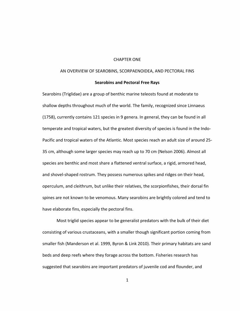
Figure 1. The Northern Searobin, Prionotus carolinus
Northern Searobin, Prionotus carolinus. Note the presence of three free rays at the anterior end of the pectoral fin. Modified from Fishery Bulletin of the Fish and Wildlife Service vol. 53.
laboratory trials have demonstrated that they are highly proficient at locating cryptic
prey buried in sand beds (Scharf et al 2006). There is also a growing body of research
that suggests predation from searobins may have a significant impact on fish stocks in
the Northern Atlantic (Floeter et al. 2005). In Europe, as traditional fish stocks continue
to decline, there has been some interest in commercial fishing for triglids.
The common name “searobin” derives from their large pectoral fins and their
habit of holding them extended while swimming. With fins extended and occasionally
flapping against the body, the movement is reminiscent of a bird gliding along in flight.
The pectoral fins themselves are often vividly colored and can be used in various
interactions with conspecifics or as a method of confusing and intimidating potential
predators. (Anorim et al 2004) The large, rounded pectoral fin is also thought to provide
a strong initial boost at the start of swimming locomotion (Gosline 1994). However, the

3
most interesting modification to the pectoral fins in searobins is the set of “free rays,”
consisting of a group of three rays located along the anterio-ventral edge of the pectoral
fin (Fig. 1). They appear as several finger-like projections separate from the rest of the
fin but still attached to the pectoral girdle. Early in development, the free rays (hereafter
FRs) separate from the rest of the pectoral fin and lose the fin membrane (Morill 1895).
The FRs can operate independently of one another, have an increased range of motion
compared to the main pectoral fin, and they are able to actively flex the distal end of
each ray. Although similar in general structure to the rays of the fin, several studies have
suggested the presence of derived musculature and nervous system modifications in the
FRs (Imamura 1996, Finger 2000). The FRs are associated with several behaviors that
interact directly with the sandy substrate that is their preferred habitat, including
sensing prey (Finger 2000), walking locomotion (Renous et al 2000), and digging
(Manderson et al 1999). However, no clear picture that integrates these behaviors has
been advanced and their structure has been only partially described (Tiedemann 1816,
Imamura 1996).
The sensory function of the triglid FRs has received considerable study. In
searobins, the distal tips of the FRs are covered with dense clusters of solitary
chemoreceptor cells (SCCs) (Finger 2000). These cells appear to be tuned to detect
protein signals of the sort that are indicative of potential prey. Interestingly, these cells
are not homologous to taste buds and are not part of the olfactory system, but instead
are innervated by the third spinal nerve. Signals from the FRs are processed by a series
of specialized lobes dorsal to the spinal cord before being passed to and integrated with

4
the rest of the brain (Herrick 1907, Finger 2000). Behavioral tests have also
demonstrated that triglids are capable of following scent trails using only their FRs
(Bardach and Case 1965). Taken together, there is a firm consensus that much of their
function is sensory.
A second set of studies has accumulated ample research supporting the
potential use of FRs in locomotion. Researchers have found that the FRs move in a
coordinated gait consistent with a walking behavior (Renous et al. 2000). In a lab setting,
triglids would settle on the substrate, partially adduct their fins, and then commence to
move slowly along the bottom. During this behavior, the fish would repeat a 1 – 3 – 2
pattern of FR movement. Later it was demonstrated that during this behavior, the
substrate exerts a normal force on each free ray, thereby supporting the fish’s weight
(Jamon et al, 2007). Using an aquarium lined with a force conductive gel, the force
exerted by the FRs was found to be opposite the fish’s direction of travel and hence
consistent with locomotion. This force was relatively small, but not implausible given
the slow speed of movement and buoyancy of the fish. The specific utility of this
behavior is not understood, but is thought to aid in foraging for prey, possibly by
minimizing disruptions in the water while also maximizing exposure to chemical signals
from the substrate.
Lastly, during a set of feeding experiments, searobins have been observed using
their FRs to uncover buried prey and flush them from hiding (Manderson et al. 1999).
During this behavior, searobins were observed to settle over top of a buried juvenile
flounder and then disturb the substrate with repeated movements of their FRs. Once
5
uncovered or flushed from hiding, the vulnerable flounder are chased and consumed. In
a controlled setting, searobins were able to locate and consume 90% of prey buried in
sand beds with most attacks preceded by digging or walking using the free rays (Scharf
et al. 2006). These studies concluded that the ability of searobins to detect cryptic prey
makes them an important obstacle to Atlantic nurseries.
Overview of Pectoral Fin Morphology
The FRs appear to be modified from several rays of the pectoral fin. While the
specialization of the FRs is somewhat unique, many fish clades have highly modified
pectoral fins and fin structure is a major source of teleost diversity. For explanatory
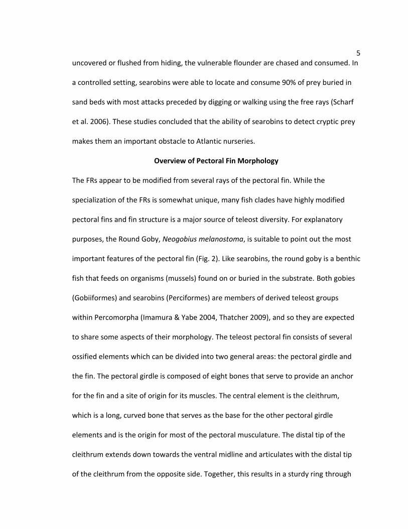
purposes, the Round Goby, Neogobius melanostoma, is suitable to point out the most
important features of the pectoral fin (Fig. 2). Like searobins, the round goby is a benthic
fish that feeds on organisms (mussels) found on or buried in the substrate. Both gobies
(Gobiiformes) and searobins (Perciformes) are members of derived teleost groups
within Percomorpha (Imamura & Yabe 2004, Thatcher 2009), and so they are expected
to share some aspects of their morphology. The teleost pectoral fin consists of several
ossified elements which can be divided into two general areas: the pectoral girdle and
the fin. The pectoral girdle is composed of eight bones that serve to provide an anchor
for the fin and a site of origin for its muscles. The central element is the cleithrum,
which is a long, curved bone that serves as the base for the other pectoral girdle
elements and is the origin for most of the pectoral musculature. The distal tip of the
cleithrum extends down towards the ventral midline and articulates with the distal tip
of the cleithrum from the opposite side. Together, this results in a sturdy ring through

6
Figure 2. Pectoral Fin, from Neogobius melanostoma
Left lateral view of N. melanostoma (LUC 77.90 mm) pectoral girdle, dissected, cleared, and stained. The tight fusion of the scapula with the cleithrum is somewhat atypical, and is not representative of searobins or scorpaenoids in general. Abbr. Cl = Cleithrum, SCl = Supracleithrum, Sca = Scapula, Cor = Coracoid, R = Radials, Fn = Fin Ray.
which force from the fins may be applied to the body as a whole. Dorsally, the
supracleithrum connects the cleithrum to the post-temporal bone of the skull, providing
additional stability. Ventrally, the scapula connects to the proximal end of the cleithrum
while the coracoid connects to the distal end. These bones provide additional space for
musculature and support the radial bones, sometimes called actinosts. In N.
Melanostoma (Fig. 2), the scapula is fused with the cleithrum, which is somewhat
different from the condition in searobins. Attached to the coracoid and scapula and
cleithrum are the four radial bones. In most teleosts, these small bones are usually

7
barred or hourglass shaped. However, gobies have large square shaped radials, a
condition shared with searobins and some other benthic fishes. Each radial bone is
embedded in a flexible fibro-cartilage pad. This provides a flexible site of attachment for
the individual rays that comprise the fin. Finally, the postcleithra are a variable set of
bones extending ventrally from the medial side of the cleithrum. Although the
postcleithra are often an informative character for systematists, their functional
significance is unknown. Furthermore, they tend to obscure other details and have been
removed from all specimens prior to drawing or photography.
In actinopterygian fishes, the fin itself is made up of a series of rays or
lepidotrichia, so named because they are thought to have evolved from modified scales.
Each lepidotrichium is composed of two hemitrichia (Fig 3, A). There is a lateral
hemitrichium that receives insertions from the lateral muscles and a medial
hemitrichium that receives insertions from the medial muscles. Each hemitrichium can
be divided into three sections. The most proximal segment is typically larger in diameter
and may have a variety of processes near its base for muscle attachment (Fig. 3, B). Of
particular interest is the posterior process, which is the insertion site for the adductor
profundus muscle, and the internal process, which articulates with the fibrocartilage
pad and anchors the ray to the pectoral girdle. Extending from the hemitrichium base is
a middle portion that is unsegmented and usually lacks processes or other features
(Geerlink 1987). The majority of each hemitrichium consists of a series of semi-
cylindrical segments linked end to end, which provide a balance of rigidity and flexibility.
The hemitrichia are tightly bound by elastic fibers, but not fused, and there is a

8
Figure 3. Structure of Teleost Fin Rays, from N. melanostoma
Photograph of a cleared and stained fin ray from N. melanostoma (LUC 77.90 mm). The medial direction is towards the top and proximal direction is to the left. (A) A broad view of an entire lepidotrichium shows the hemitrichia separate at the base, combined in middle, and then branching distally. (B) Magnified view of the proximal end of the lepidotrichium showing the considerable variation between the lateral and medial hemitrichia and a clearer view of processes at the base of the medial hemitrichium. Abbv: MH= medial hemitrichium, LH = lateral hemitrichium, post. pr. = posterior process, int. pr. = internal process.
noticeable gap near the base that allows them to shift relative to each other. At the
distal end, the fin rays are typically branched with the hemitrichia fanning apart to
provide a large surface for the fin membrane to stretch across. The individual rays of the
pectoral fin are traditionally numbered beginning with the marginal ray. This ray usually
is the leading edge of the fin and thus controls a variety of hydrodynamic factors. Its

9
shape is usually specialized to aid this role and it will receive additional muscle
attachments. In the pectoral fin, it is not uncommon for the lateral and medial
hemitrichia to be significantly different within an individual, but the hemitrichia on a
given side are generally similar, except the marginal ray (i.e. 3rd medial ≠ 3rd lateral, but
3rd medial = 10th medial).
Each lepidotrichium, except the marginal ray, receives inputs from four muscles,
and these will be an important topic in this work (Winterbottom 1974). The two lateral
muscles are abductors and serve to rotate the fin away from the body (Fig. 4). The two
medial muscles are adductors and serve to pull the fin back against the body (Fig. 5).
The lateral muscles insert to the lateral hemitrichium and the medial muscles insert to
the medial hemitrichium. The most lateral muscle is the abductor superficialis (ABS). It
originates from the cleithrum and runs ventrally to insert near the base of each lateral
hemitrichium. Next is the abductor profundus (ABP) which originates from the distal
cleithrum and coracoid and runs posteriorly underneath the ABS to insert near the base
of each lateral hemitrichium. On the medial side (Fig. 5), the adductor superficialis (ADS)
originates from the proximal cleithrum and runs ventrally to insert on each medial
hemitrichium, somewhat distal from its base. The adductor profundus (ADP) originates
from the distal cleithrum and runs posteriorly beneath (lateral to) the ADS. It inserts
near the base of each medial hemitrichium on the posterior process. The specific
structure of these muscles may vary by taxa, with some groups having more or less
muscle mass, different angles of insertion, longer or shorter tendons, or loss of a
particular muscle insertion to some lepidotrichia.

10
Figure 4. Lateral Pectoral Fin Musculature, from N. melanostoma
Diagram of N. melanostoma (LUC 77.90 mm), left pectoral girdle, lateral view. The muscles of the lateral side are dominated by the ABS and ABP. The small arrector ventralis is obscured underneath the ABS. ABBV: Cl = cleithrum, SCl = supracleithrum, Cor = coracoid Fn = fin ray, ABS = abductor superficialis, ABP = abductor profundus. Scale bar = 2mm.
The pectoral fin has a variety of other intrinsic muscles that bear mentioning but
are of less importance to this study. The marginal ray receives two additional muscles,
the arrectors. The arrector ventralis (ARRV) originates from the lateral side of the
cleithrum and inserts to the lateral side of the medial hemitrichium of the marginal ray.
The arrector dorsalis (ARRD) originates from the medial side of the cleithrum and
inserts to the medial side of the medial hemitrichium of the marginal ray. Together, they
11
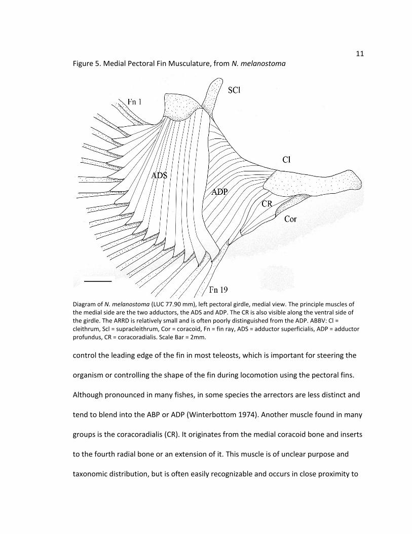
Figure 5. Medial Pectoral Fin Musculature, from N. melanostoma
Diagram of N. melanostoma (LUC 77.90 mm), left pectoral girdle, medial view. The principle muscles of the medial side are the two adductors, the ADS and ADP. The CR is also visible along the ventral side of the girdle. The ARRD is relatively small and is often poorly distinguished from the ADP. ABBV: Cl = cleithrum, Scl = supracleithrum, Cor = coracoid, Fn = fin ray, ADS = adductor superficialis, ADP = adductor profundus, CR = coracoradialis. Scale Bar = 2mm.
control the leading edge of the fin in most teleosts, which is important for steering the
organism or controlling the shape of the fin during locomotion using the pectoral fins.
Although pronounced in many fishes, in some species the arrectors are less distinct and
tend to blend into the ABP or ADP (Winterbottom 1974). Another muscle found in many
groups is the coracoradialis (CR). It originates from the medial coracoid bone and inserts
to the fourth radial bone or an extension of it. This muscle is of unclear purpose and
taxonomic distribution, but is often easily recognizable and occurs in close proximity to

12
the ADP. Although other pectoral muscles have been identified in certain taxa, none are
found in searobins.
The segmented nature of the lepidotrichia of actinopterygian fishes
differentiates them from the swimming appendages used by other aquatic vertebrates.
Unlike the rigid fins of sharks or the flippers of marine mammals, the lepidotrichia allow
significant bending along their length. Furthermore, this bending appears to be under
the active control of the fish. This is accomplished by a shift in the relative position of
the bases of each hemitrichium. Thus, a shift in the base of the medial hemitrichium
results in bending of the ray in the medial direction (Geerlink 1987). This shift is
mediated by the pectoral muscles and is not merely the result of elasticity in the ray
itself. Ray bending is not uniform and is governed by a complex set of factors including
the size of the segments and the specific composition of the fibers joining the rays
(Alben et al. 2006). The outcome of these factors is that small displacements of the ray
bases may produce significant curvature of the ray and exert a large force against their
environment. The ability to bend their rays gives them improved control over the shape
of the fin surface, which presumably leads to improved swimming performance over a
variety of conditions. Although the diversity of swimming modes in fishes has been well
studied, variation in the properties of fin rays is almost entirely unknown. Almost all
functional work on fin ray properties has been carried out on pelagic fishes, with most
of that focused on the caudal fin (McCutchen 1970, Geerlink 1987). Also, these
functional models have presumed perfect symmetry of the lepidotrichia, which is
mathematically convenient but frequently not true (Taft 2009). Work in a variety of

13
fishes and closer attention to variation in the morphology of the lepidotrichia will be
necessary to further advance our understanding.
Superfamily Scorpaenoidea
To better understand the function and evolution of the pectoral fin, it is advantageous
to study a group with a diversity of ecological modes and fin morphologies. The
superfamily Scorpaenoidea, which includes searobins, is well suited to this purpose.
Almost all scorpaenoids are benthic predators and unlike most pelagic fishes, they rely
less on swift or agile locomotion to capture prey and avoid predators. They possess a
variety of modifications such as venom, camouflage, and ambush behaviors, which have
removed the necessity of maximizing swimming performance and have allowed greater
diversification of the fins (Gosline 1994). Additionally, benthic fishes often use their fins
to interact with the substrate in a variety of ways such as perching, digging, and
occasionally walking. This extensive variation produces a sort of natural experiment in
pectoral fin design, and understanding the structure of scorpaenoid pectoral fins will aid
researchers in drawing inferences about other groups.
Free Rays are not restricted to searobins, but occur in five families of
scorpaenoid fishes accounting for over 200 species (Table 1). The Triglidae, with over
121 species, a wide distribution, and no venom, are merely the best studied. Related to
the Triglidae are the Peristediidae, or armored searobins. This group contains
approximately 36 species all having two FRs and is found in deeper water throughout
much of the globe. The Hoplichthyidae are known as ghost flatheads, and consist of only
12 species, each with three FRs. They are found in deep (500+ m) water in the Indian

14
and Pacific oceans. The Apistidae, sometimes called waspfishes, is a small family of
three species bearing a single FR. They are found in shallow water in the Indo-Pacific.
Last is the family Synanceiidae, the stonefishes, containing about 35 species found
throughout the Indo-Pacific. Synanceids show a variety of pectoral fin forms which
correspond to different genera: Minous has a single FR and eleven species, Inimicus has
two FRs and ten species, Choridactylus has three FRs and four species. These three
genera are called by a variety of names such as stingfishes, devilfishes, and ghouls. The
term stonefish is usually reserved for the remaining ten species belonging to a set of five
genera that lack FRs (Ishida 1994, Nelson 2006). Of these, the genus Synanceia has five
species and is the most widespread and most venomous. The genus Erosa has two
species, and the genera Leptosynanceia, Pseudosynanceia, and Trachicephalus are
monotypic. Each of these genera is found in the Indian and Pacific oceans, and tends to
occupy muddy or sandy bottom areas, though many are found near coral reefs as well.
Although externally similar, there is reason to suspect significant variations in the
osteology and musculature of the FRs among these families.
The ecology of fishes with FRs varies considerably at the family level. As
mentioned above, Triglidae has been the best studied, and most of its species are
midsized benthic predators that actively forage sandy areas for crustaceans and small
fish. Less is known about the Peristediidae, but morphological similarities may suggest
that they are ecologically similar to triglids. The Hoplichthyidae is generally found too
deep for direct observation by divers or for transfer into an aquarium, and there are
little data with which to infer behavior or lifestyle. Superficially, they resemble the

15
Table 1. Distribution of Free Rays in Scorpaenoids
Summary of scorpaenoid fishes possessing FRs. Behavioral observations for Triglidae from Bardach & Case(1965), Manderson et al (1999), Renous et al. (2000), all others from Gosline (1994).
peristediids, in that both are covered by an extensive set of dermal armor. The Apistidae
are found in sandy environments similar to the triglids, but are thought to use their
single FR to confuse or corral prey, rather than for sensory or locomotion purposes
(Gosline 1994). Likewise, few scientific studies exist for the Synanceiidae, but they
arehighly valued in the aquarium trade and well known to divers and medical doctors
for their extremely potent venom. In the wild, they are ambush predators and are
known to wait half-buried in the sand. The pectoral fins and FRs are reported to help
them rapidly and accurately spring from hiding when attacking prey (Eschmeyer et al.
1979a & 1979b, Grobecker 1983).
Phylogenetic Relationships of Scorpaenoid Fishes
To understand the evolution of the scorpaenoid pectoral fin, it would be useful to know
if and how the families with FRs are related. Our understanding of the phylogeny of
these fishes is undergoing rapid changes. In the past, researchers placed these families
Taxon Species # Free Ray # Free Ray Behavior
Triglidae 121 3 Digging, Tracking,
Locomotion
Peristediidae 36 2 No data
Hoplichthyidae 12 3 No data
Apistidae 3 1 Corral/Confuse Prey
Synanceiidae Minous 11 1 Striking
Synanceiidae Inimicus 10 2 Striking
Synanceiidae Choridactylus 4 3 Striking
Synanceiidae All Other Genera
10 0 N/A

16
in the now defunct order Scorpaeniformes, which contained over 1400 species. Leading
researchers working independently have produced strong evidence that
Scorpaeniformes is not a monophyletic group (Imamura & Yabe 2002, Smith & Wheeler
2004, Smith & Craig 2007). Based on morphology, Imamura and Yabe (2002) concluded
that this order actually consisted of two unrelated lineages, which by virtue of a shared
benthic lifestyle have converged in a number of aspects. The Scorpaenoidea he
proposed contains roughly 700 species and has 4 synapomorphies favoring its validity.
The picture painted by molecular studies is much more complex and includes a variety
of serranids, percids, and other taxa mixed among the traditional scorpaeniform
assemblage (Smith & Craig 2007). While this represents an important improvement, the
interrelationships of many families of scorpaenoid fishes are still poorly supported and
the systematics of this group remains contentious. While the details of the two
phylogenies differ quite substantially, they agree on the major points of relevance to
this particular study, specifically that the species possessing FRs belong to at least two
separate clades. Hence, the phylogeny of Imamura (2004) will be used as a working
hypothesis as it includes a better sampling of taxa with FRs and fewer groups of outside
interest.
While the overall phylogeny is uncertain, there is strong evidence that each of
the five families with FRs is independently monophyletic. This study will use the
phylogeny of Imamura (2004) as a starting point for examining the evolution of the FRs.
This phylogeny is based on morphological characters and is useful in that it is the only
published phylogeny that includes all five of the families of interest in this study. This
17
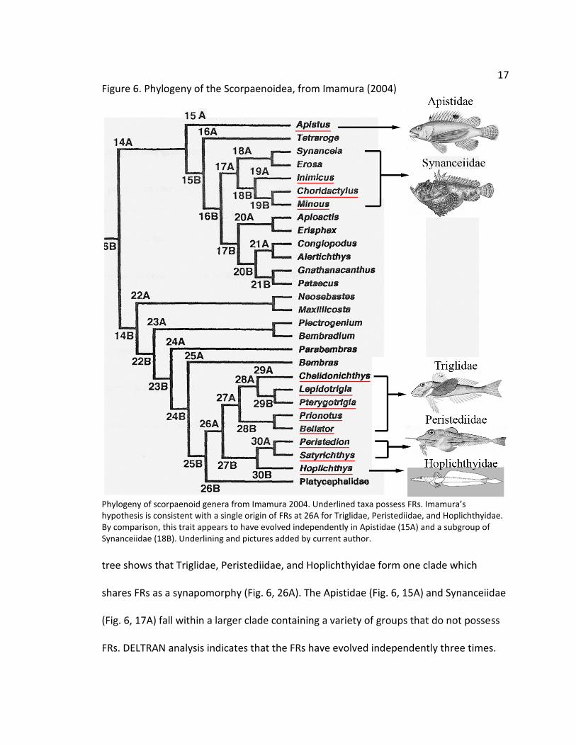
Figure 6. Phylogeny of the Scorpaenoidea, from Imamura (2004)
Phylogeny of scorpaenoid genera from Imamura 2004. Underlined taxa possess FRs. Imamura’s hypothesis is consistent with a single origin of FRs at 26A for Triglidae, Peristediidae, and Hoplichthyidae. By comparison, this trait appears to have evolved independently in Apistidae (15A) and a subgroup of Synanceiidae (18B). Underlining and pictures added by current author.
tree shows that Triglidae, Peristediidae, and Hoplichthyidae form one clade which
shares FRs as a synapomorphy (Fig. 6, 26A). The Apistidae (Fig. 6, 15A) and Synanceiidae
(Fig. 6, 17A) fall within a larger clade containing a variety of groups that do not possess
FRs. DELTRAN analysis indicates that the FRs have evolved independently three times.
18
This raises the possibility that there are significant morphological differences in each
family that have not been recognized and may provide new information regarding
evolution in these groups.
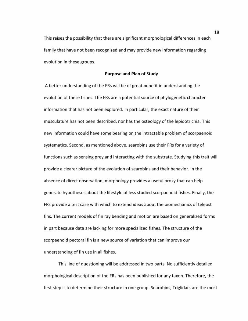
Purpose and Plan of Study
A better understanding of the FRs will be of great benefit in understanding the
evolution of these fishes. The FRs are a potential source of phylogenetic character
information that has not been explored. In particular, the exact nature of their
musculature has not been described, nor has the osteology of the lepidotrichia. This
new information could have some bearing on the intractable problem of scorpaenoid
systematics. Second, as mentioned above, searobins use their FRs for a variety of
functions such as sensing prey and interacting with the substrate. Studying this trait will
provide a clearer picture of the evolution of searobins and their behavior. In the
absence of direct observation, morphology provides a useful proxy that can help
generate hypotheses about the lifestyle of less studied scorpaenoid fishes. Finally, the
FRs provide a test case with which to extend ideas about the biomechanics of teleost
fins. The current models of fin ray bending and motion are based on generalized forms
in part because data are lacking for more specialized fishes. The structure of the
scorpaenoid pectoral fin is a new source of variation that can improve our
understanding of fin use in all fishes.
This line of questioning will be addressed in two parts. No sufficiently detailed
morphological description of the FRs has been published for any taxon. Therefore, the
first step is to determine their structure in one group. Searobins, Triglidae, are the most

19
logical choice with which to begin because they are the largest, most diverse, and most
widespread family. Furthermore, because they have been the subject of more research
than the other families, it is easier to place any findings into context. While the FRs are
part of the pectoral fin, they have not been compared to those of traditional fin rays.
Also, it has been reported that the muscles serving the FRs are arranged into bundles
distinct from the fin musculature (Imamura 1996), but no deeper exploration has been
attempted. Given the many new functions associated with FRs, new muscle
specializations seem probable. Lastly, the research in searobins has been focused on a
few species easily available in the northern Atlantic. A sample including all triglid genera
will indicate whether these findings can be generalized to the entire family, or if distinct
morphs are present below the family level. This work will provide a basis for future
investigations of pectoral fin morphology in scorpaenoids.
The second step is to generalize the findings in searobins to other scorpaenoid
fishes. After an adequate picture of the FRs in Triglidae is complete, the other groups
can be compared and evaluated. The FRs are assumed to be modified to the pectoral fin
in all cases, so some similarity should be expected in their form and function. However,
the multiple evolutionary origins could also lead to significant variation. Once the FRs of
all five groups are characterized, differences in their anatomy can be interpreted with
respect to other variables, such as habitat or feeding mode, which will provide a broader
picture of FR function and pectoral fin evolution in these groups. Findings are
considered with respect to current phylogenetic hypotheses and the potential impact of
new morphological data is considered.

20
Overview of Materials and Methods
In order to address these questions, specimens were first gathered from public
museums. Material consisted primarily of alcohol preserved specimens and cleared and
stained specimens. Efforts were made to borrow a broad taxonomic sample of
scorpaenoids with free rays, though many specimens are quite rare and were not
available for loan or examination. Table 2 summarizes the specimens obtained. For
Triglidae, Peristediidae, and Hoplichthyidae, this study used one or two species as a
reference for detecting intraspecific variation. The reference species were chosen
according to the availability of at least ten specimens for dissection, following the
method of Kesner (1994). For other species, dissections of two or three specimens are
generally sufficient. Due to limited availability, a reference species including 10
specimens could not be drawn from Apistidae or Synanceiidae. However, examinations
Table 2. Summary of Taxa Sampled
Taxon Total Genera Total Species Genera
Sampled
Species
Sampled
Triglidae 9 125 9 22
Peristediidae 4 36 2 6
Hoplichthyidae 1 12 1 1
Apistidae 3 3 1 1
Minous 1 12 1 3
Inimicus 1 11 1 2
Choridactylus 1 4 0 0
Other
Synanceiids 6 10 1 2

21
of the reference species in the other families displayed almost zero variation below the
family level, so it seems reasonable to include Apistidae and Synanceiidae despite this
limitation. In summary, 113 scorpaenoid specimens were examined.
Additionally, several non-scorpaenoid fishes were used as a general reference
for pectoral girdle anatomy. These were unaccessioned specimens drawn from the
Loyola University of Chicago teaching collection, and are listed below.
Cichlidae
Herichthys nigrofasciatus: 2 specimens (SL: 75.90 mm & 79.40 mm), LUC (2 alcohol)
Cottidae
Cottus cognatus: 3 specimens (SL: 41.65 – 46.60 mm), LUC (3 alcohol)
Cottus bairdii: 1 specimen (SL: 78.55 mm), LUC (1 C&S)
Gobiidae
Neogobius melanostoma: 1 specimen (SL: 77.90 mm), LUC (1 alcohol)
Once obtained, specimens were measured for standard length and then
prepared for either dissection or clearing and staining. In order to observe the muscle
structure, the left pectoral fin was dissected from the fish body, except in the reference
species where bilateral dissections were performed (Kesner 1994). This was
accomplished by severing all extrinsic muscles and then carefully separating the
supracleithrum from the post-temporal bone and any connections between the
cleithrum or coracoid and the pelvic bones. Dissected pectoral fins were briefly
immersed in Alcian blue to stain for cartilage. This step was also useful in highlighting
any fascial tissue around the muscles, which aided their examination. After this, the
22
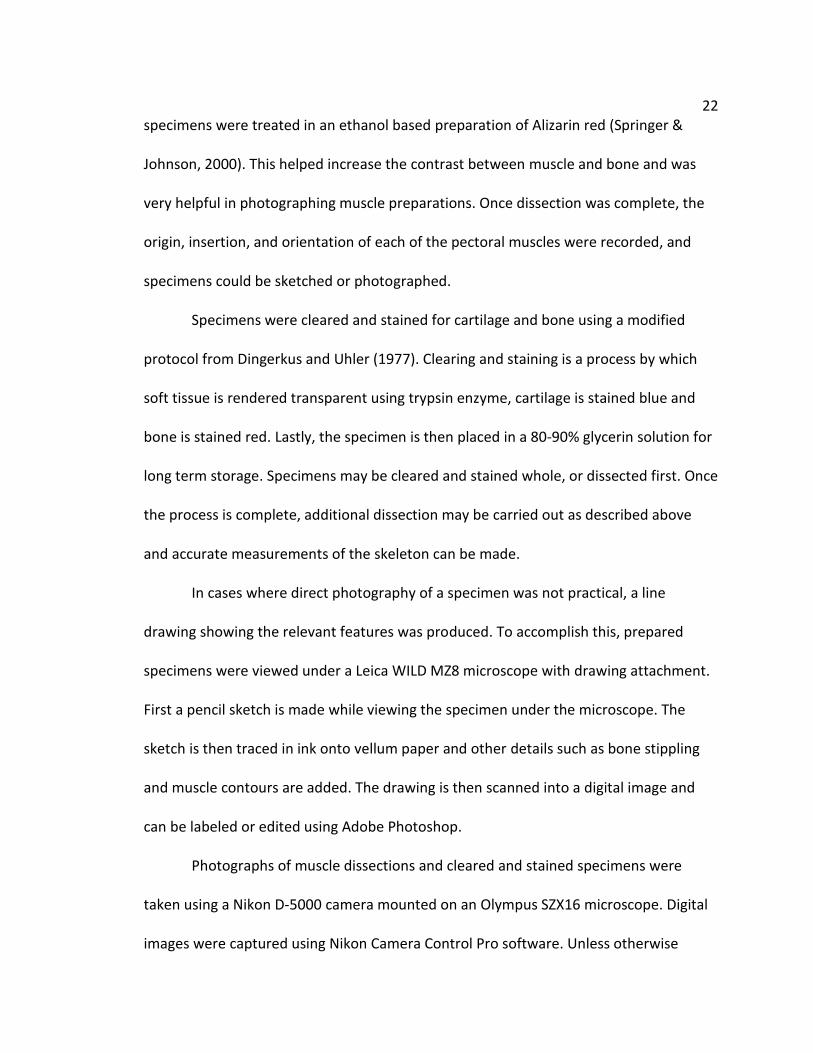
specimens were treated in an ethanol based preparation of Alizarin red (Springer &
Johnson, 2000). This helped increase the contrast between muscle and bone and was
very helpful in photographing muscle preparations. Once dissection was complete, the
origin, insertion, and orientation of each of the pectoral muscles were recorded, and
specimens could be sketched or photographed.
Specimens were cleared and stained for cartilage and bone using a modified
protocol from Dingerkus and Uhler (1977). Clearing and staining is a process by which
soft tissue is rendered transparent using trypsin enzyme, cartilage is stained blue and
bone is stained red. Lastly, the specimen is then placed in a 80-90% glycerin solution for
long term storage. Specimens may be cleared and stained whole, or dissected first. Once
the process is complete, additional dissection may be carried out as described above
and accurate measurements of the skeleton can be made.
In cases where direct photography of a specimen was not practical, a line
drawing showing the relevant features was produced. To accomplish this, prepared
specimens were viewed under a Leica WILD MZ8 microscope with drawing attachment.
First a pencil sketch is made while viewing the specimen under the microscope. The
sketch is then traced in ink onto vellum paper and other details such as bone stippling
and muscle contours are added. The drawing is then scanned into a digital image and
can be labeled or edited using Adobe Photoshop.
Photographs of muscle dissections and cleared and stained specimens were
taken using a Nikon D-5000 camera mounted on an Olympus SZX16 microscope. Digital
images were captured using Nikon Camera Control Pro software. Unless otherwise

23
stated, all photographic images contained herein are composites of 4-8 separate images
that have been focus stacked using the program Combine ZP on default settings (Hadley
2009). When viewing three dimensional objects at high magnification, it is usually
impossible to focus on objects at different depths simultaneously. This method removes
the depth of field restriction by combining several photographs that have been
individually focused into one digital image. Additional photo editing was performed
using Adobe Photoshop CS2.
Statistical Analysis and Morphometrics
Preliminary analysis suggested that the shape of the medial hemitrichium is especially
important to the morphology of the FRs. Comparisons of the morphology of two
organisms can be investigated by using geometric morphometrics. This method removes
variation based on size and orientation and allows for statistical analysis of differences
in shape (Zelditch et al. 2004). The general procedure consists of four steps: 1)
preparation and photography of the specimens, 2) assigning landmarks that describe
the shape, 3) alignment and superimposition of the specimens, and 4) statistical analysis
of the results. The medial hemitrichium is well suited to geometric morphometric
analysis in that it is largely two dimensional, has a relatively simple shape, and possesses
several homologous points that can be consistently applied across taxa.
Medial hemitrichia of specimens were prepared from dissected left pectoral
girdles by gently pulling each FR away from the pectoral girdle and using fine dissecting
scissors to cut away the fibrocartilage. Muscles connections to the FR were also severed,
though a small slip of muscle was sometimes left to aid landmark recognition in the next

24
section. Once each FR was removed from the pectoral girdle, it was separated into its
component hemitrichia using a scalpel. The total length of each hemitrichium was
measured using calipers, and then it was photographed using the same camera and
microscope as above. However, since the hemitrichia are very flat, a single image
allowed sufficient resolution and the focus stacking technique was unnecessary. Overall,
204 hemitrichia from 84 specimens were included in the morphometric portion of this
study. A number of hemitrichia were excluded from the study because of damage or
poor preservation.
Landmarks were added on each photograph using the TPS line of morphometric
programs (Rohlf 2010). The TPS line of software converts photos with landmarks into a
matrix of digital coordinates in a format used by most morphometric analysis software.
Ideally, landmarks are chosen because they represent presumably homologous points.
Unlike the homology of bones, this type of landmark is often identified via the
intersection of two or more important features on a structure (i.e. muscle insertions,
processes, intersecting ridges or sutures). A second type of landmark, called a “semi-
landmark” carries important information about the overall shape of an object but does
not have a strong argument in favor of homology (i.e. points located along a curve or
surface, points opposite of other landmarks) (Zelditch 2004). In all cases, it is necessary
that landmarks can be applied consistently across all taxa in the study. This study uses
six homologous landmarks based on the position of muscle insertions and bone
processes and three relevant semi-landmarks that describe the shape of the main shaft
of the hemitrichium, which lacks identifiable homologous points (Fig. 7 & Table 3).
25
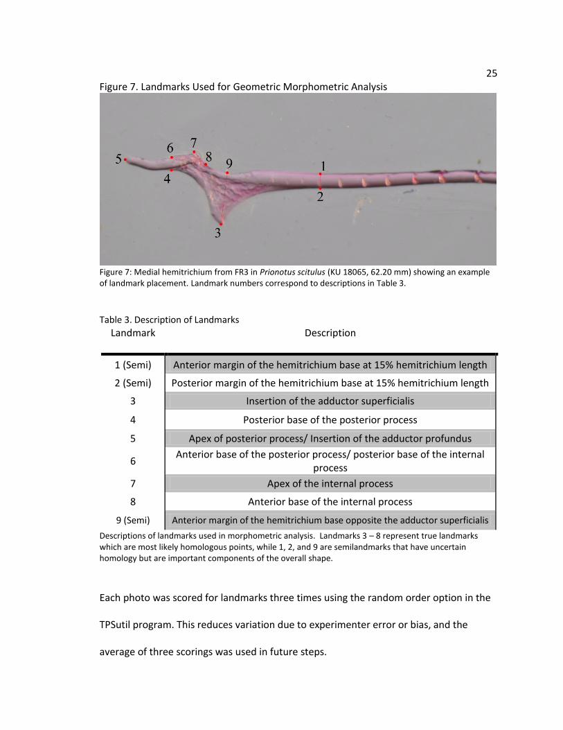
Figure 7. Landmarks Used for Geometric Morphometric Analysis
Figure 7: Medial hemitrichium from FR3 in Prionotus scitulus (KU 18065, 62.20 mm) showing an example of landmark placement. Landmark numbers correspond to descriptions in Table 3.
Table 3. Description of Landmarks
Landmark Description
1 (Semi) Anterior margin of the hemitrichium base at 15% hemitrichium length
2 (Semi) Posterior margin of the hemitrichium base at 15% hemitrichium length
3 Insertion of the adductor superficialis
4 Posterior base of the posterior process
5 Apex of posterior process/ Insertion of the adductor profundus
6 Anterior base of the posterior process/ posterior base of the internal
process
7 Apex of the internal process
8 Anterior base of the internal process
9 (Semi) Anterior margin of the hemitrichium base opposite the adductor superficialis
Descriptions of landmarks used in morphometric analysis. Landmarks 3 – 8 represent true landmarks which are most likely homologous points, while 1, 2, and 9 are semilandmarks that have uncertain homology but are important components of the overall shape.
Each photo was scored for landmarks three times using the random order option in the
TPSutil program. This reduces variation due to experimenter error or bias, and the
average of three scorings was used in future steps.

26
Once landmark coordinates were obtained, they were entered into the program
MorphoJ for additional analysis (Kleinburg 2011). This program is capable of performing
a variety of functions on landmark data including generating graphs and performing
statistical tests. First, coordinates from all groups are superimposed using a full
Procrustes fit algorithm, which removes any variation associated with size, orientation,
or position. The Procrustes fitted coordinates shape analysis can procede in a variety of
ways. Principal component analysis (PCA) is a common method of visualizing
multidimensional information. This process begins by producing a covariance matrix
describing the variation in the data. Next, the axis or vector is computed which contains
the greatest variation of the data; this is the first principal component (PC1). Each
observation can then be scored according to this axis. The next greatest source of
variation is included in the second axis (PC2), and so on. These data are usually
presented as a scatterplot of the scores from the first few PCs, and is useful for
exploring trends or grouping within the data. However, PCA is not a hypothesis test and
cannot be used to draw inferences about the similarity or differences between groups.
For this, a second process call discriminant function analysis (DFA) is used. This process
is similar in that it reduces variation in a data set to a series of vectors or axes, but
where PCA chooses axes that maximize the total variation of the data set, DFA
maximizes the variation between a set of experimentally defined groups, such as
different families or genera. This procedure computes a number called the Mahalanobis
distance, which is a measure of the separation of two multivariate means. This number
is then used to calculate Hotelling’s T2 statistic, which is a standard method of

27
comparing multi-dimensional means in two groups and is roughly analogous to the
common Student’s T test for one dimensional data. The null hypothesis for these tests
is no difference in the average shape between groups. Discriminant function analysis is
generally quite robust in dealing with deviations from normality and sampling
differences in the data set, but can only test two groups at a time. If many groups are
present in the data set, DFA can be conducted in a pairwise manner, and a Bonferroni
correction is applied when deciding whether or not to reject the null hypothesis.
Preview of Chapters
This first chapter has endeavored to provide a suitable background for this study overall.
It covers the general ecology, function, behavior, and systematics of scorpaenoid fishes
that have informed this study. I have provided a general review of pectoral girdle
anatomy, and an overview of the methods to be used in future chapters. This
background is revisited and expanded upon in later chapters as needed. Chapter’s two
and three are constructed as a series of manuscripts intended for publication at a later
date, and so each begins with introduction and methods sections specific to that
chapter.
Chapter two covers an in depth analysis of the pectoral anatomy and
morphology of searobins (Triglidae). It begins with a review of some general information
pertinent to this group and restates the methods used in this portion of the study. The
results cover a very detailed look at each muscle group in the pectoral fin with an eye
towards comparing the musculature of the FRs to the musculature of the fin. The
important components of the FR system are identified and new myological and

28
osteological features are presented. A morphometric analysis of the FRs of triglids is also
presented and explained. Discussion of these findings attempts to correlate reports of
behavior and function with the structures observed in this study.
Chapter three continues this line of work by extending the techniques of chapter
two to a broader swathe of scorpaenoid fishes. It begins by detailing the taxonomic
distribution of FRs across five families of scorpaenoid fishes and offers relevant
background information for each. The methodology of chapter three is similar to
chapter two, and the anatomy and morphology of each family is described in depth.
However, the primary focus is a comparative analysis of FRs across taxa in an effort to
identify and understand any variations in form or function. Morphometric analysis is
offered for this expanded data set and serves to emphasize the considerable differences
in the construction of FRs in each family. Discussion highlights the most important
variations observed in the FRs, and compares each family to the others to show how
ecological and behavioral differences correspond to structural differences in this trait.
Chapter four concludes this project by offering a summary of the data obtained
and offering further insight into the structure, function, and evolution of the FR
apparatus in scorpaenoids. Themes from both chapters are drawn upon, and a number
of larger patterns are identified and explored. In a few cases, specific inferences are
made as to the evolution of some components of the FRs and the phylogenetic
distribution of FR characters is considered. In particular, the implications of FRs on our
understanding of fin function are discussed and further research is recommended.
Finally, the evolution of the pectoral girdle is considered in light of the ecology of these

29
organisms and the possible adaptive significance of the FRs in benthic habitats is
considered.
30
CHAPTER TWO
THE PECTORAL FIN MORPHOLOGY OF SEAROBINS
Abstract
The teleost family Triglidae, commonly called searobins, is a group of benthic marine
fishes found over sandy bottoms in all temperate and tropical oceans. The common
name “searobins” derives from their large pectoral fins and their habit of holding them
extended while swimming. These pectoral fins have been implicated in a variety of
behaviors and functions including locomotion, display, and foraging. Many of these
functions are accomplished through the use of modified pectoral fin rays or “free rays.”
Despite evidence of unique nervous, osteological, and muscular traits, the morphology
of the free rays has received little attention from researchers. An examination of
representatives from all nine triglid genera reveals major differences between the free
rays and unmodified fin rays in terms of osteology and musculature. The osteology of
the free and fin rays are described and compared, including a novel set of processes
extending from the base of the free ray lepidotrichia. Subdivisions of the adductor
muscles are described in detail including insertions to the above mentioned set of
processes. Morphometric analysis of the shape of the free rays found significant
morphological differences in the basal segments of the medial hemitrichia of the free

31
rays based on position. However, the same analysis failed to show a significant
difference based on species or genus identity. The morphology of triglid pectoral fins is
considered in light of observed behavior and possible functional roles are considered for
novel musculoskeletal modifications.
Introduction
Benthic fishes display a wide variety of modifications of their paired fins that assist in
various interactions with the substrate. Some examples include the fused pelvic fins of
sculpins (Webb 1989), the walking fins of handfishes and anglerfishes (Laurenson et al
2004), and the hooked fins of blennies (Brandstätter et al 1990). However, many of
these traits have not received sufficient study and their basic structure and anatomy is
largely unknown. Searobins (Triglidae) are one such example. The family Triglidae
contains approximately 121 species in nine genera and is found in temperate and
tropical waters across most of the world (Nelson 2006). They are generalist predators
who forage for crustaceans and small fish living on or buried in sandy substrates (Byron
& Link 2010).
The common name “searobin” derives from their large pectoral fins which are
held extended while swimming. These pectoral fins have been implicated in a variety of
behaviors and functions including locomotion (Renous et al 2000, Jamon et al 2007),
display (Anorim 1994), and foraging (Manderson et al 1999). As such, there have been a
number of interesting modifications to the anatomy of the triglid pectoral girdle that
accommodate these behaviors. One striking feature of triglid pectoral fin is the set of
“free rays.” The free rays (hereafter FRs) are a group of three rays located along the
32
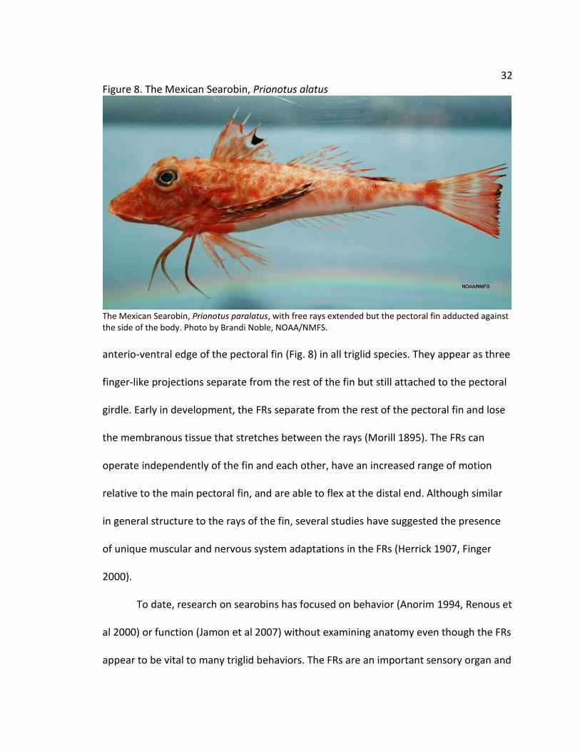
Figure 8. The Mexican Searobin, Prionotus alatus
The Mexican Searobin, Prionotus paralatus, with free rays extended but the pectoral fin adducted against the side of the body. Photo by Brandi Noble, NOAA/NMFS.
anterio-ventral edge of the pectoral fin (Fig. 8) in all triglid species. They appear as three
finger-like projections separate from the rest of the fin but still attached to the pectoral
girdle. Early in development, the FRs separate from the rest of the pectoral fin and lose
the membranous tissue that stretches between the rays (Morill 1895). The FRs can
operate independently of the fin and each other, have an increased range of motion
relative to the main pectoral fin, and are able to flex at the distal end. Although similar
in general structure to the rays of the fin, several studies have suggested the presence
of unique muscular and nervous system adaptations in the FRs (Herrick 1907, Finger
2000).
To date, research on searobins has focused on behavior (Anorim 1994, Renous et
al 2000) or function (Jamon et al 2007) without examining anatomy even though the FRs
appear to be vital to many triglid behaviors. The FRs are an important sensory organ and

33 are capable of responding to protein cues and following scent trails (Bardach & Case
1965, Finger 2000). Once prey is located, the FRs may be used to physically dig in the
substrate and flush its prey from hiding (Manderson et al 1999). Meanwhile, other
studies have proposed a possible role in locomotion, noting that the free rays move in a
coordinated gait (Renous et al 2000) and exert a normal force against the substrate
(Jamon et al 2007).
Several works have noticed modifications to the muscles of the pectoral fin such
as division of the adductors serving the FRs (Imamura 1996, 2004). However, these
observations have usually been a side note within a larger investigation of scorpaenoid
systematic relationships and have not been explored in depth. This study expands upon
these initial findings by providing an in depth description and analysis of the FRs in
Triglidae. First, the musculature and osteology of the FRs are described for a
representative triglid, Prionotus carolinus. This species is well-studied and easily
available, which makes it an appropriate starting point for anatomical studies. Second,
these findings are extended to the entire genus Prionotus as well as the other eight
genera, thus providing an estimate of the amount of variation among triglid species and
genera. A landmark based morphometric test is used to explore morphological
variations among FRs based on position and genus identity. Last, these findings are
considered in light of current evidence about the unique functional properties of the FRs
and their effect on triglid behavior and ecology.

34 Materials and Methods
Materials Examined
Specimens for this study were borrowed from institutional collections such as
the Field Museum of Natural History (FMNH), the American Museum of Natural History
(AMNH), the Smithsonian (USNM), the Museum of Comparative Zoology (MCZ), the
University of Kansas (KU), and the Bernice Pauahi Bishop Museum (BPBM). Material
consisted of alcohol preserved and cleared and stained specimens. First, 12 specimens
of Prionotus carolinus were examined to establish a reference for the basic
characteristics of triglid pectoral fin structure, and to establish the extent of variation
amongst individual specimens (Kesner 1994). Following this, two or three specimens of
all species were examined to search for variation in the musculature or osteology. This
study includes representatives of all nine genera. Muscles were identified based on
Winterbottom (1974) and follow his abbreviations and terminology.
Specimens:
Aspitrigla cuculus: 2 specimens (SL: 110.45 mm & 118.60 mm), MCZ 64304 ( 1 alcohol &
1 C&S).
Bellator militaris: 4 specimens (SL: 64.15 – 104.35 mm), FMNH 45617 (3 alcohol); KU
13219 (1 C&S).
Bellator xenisma: 1 specimen (SL: 51.40 mm), AMNH 236341 (1 alcohol).
Chelidonichthys capensis: 2 specimens (SL: 196.00 mm & 203.25 mm), USNM 325764 (2
alcohol).

35 Chelidonichthys lucerna: 2 specimens (SL: 65.70 mm & 105.00 mm), USNM 289662 (1
alcohol & 1 C&S)
Eutrigla gurnardus: 5 specimens (SL: 154.40 – 198.20 mm), FMNH 33168, 33170, 33172,
33174, 33175 (5 alcohol).
Lepidotrigla brachyoptera: 1 specimen (SL: 91.70 mm), KU 27892 (1 C&S).
Lepidotrigla mulhalli: 2 specimens (SL: 104.75 mm & 112.00 mm), USNM 393322 (1
alcohol & 1 C&S).
Prionotus alatus: 3 specimens (SL: 132.75 – 135.20 mm), FMNH46593 (3 alcohol).
Prionotus carolinus: 12 specimens (SL: 55.00 – 135.20 mm), FMNH 17951-37960 (9
alcohol & 1 C&S); KU 13208 (1 C&S); KU 14059 (1 alcohol).
Prionotus ophryas: 3 specimens (SL: 82.65 – 127.45 mm), FMNH 88719 (2 alcohol & 1
C&S).
Prionotus paralatus: 3 specimens (SL: 107.80 – 127.95 mm), FMNH 67497 (3 alcohol).
Prionotus punctatus: 3 specimens (SL: 86.20 – 97.20 mm), FMNH 67538 (2 alcohol & 1
C&S).
Prionotus roseus: 1 specimen (SL: 101.70 mm), KU 13226 (1 alcohol).
Prionotus scitulus: 1 specimen (SL: 62.20 mm), KU 18065 (1 C&S).
Prionotus stearnsi: 4 specimens (SL: 76.35 – 95.35 mm), FMNH 64054 (3 alcohol); FMNH
66579 (1 C&S).
Prionotus tribulus: 2 specimens (SL: 51.00 mm & 82.95 mm), KU 17083 (1 C&S); KU
22961 (1 alcohol).
36 Pterygotrigla arabica: 2 specimens (SL: 52.50 mm & 114.60 mm), USNM 356390 (1
alcohol & 1 C&S).
Pterygotrigla megalops: 2 specimens (31.30 mm & 76.05), USNM 393319 (2 alcohol).
Trigla lyra: 2 specimens (160.25 mm & 170.90 mm), USNM 201776 (2 alcohol).
Trigloporus lastoviza: 2 specimens (137.50 mm & 160.10 mm), USNM 201770 (2
alcohol).
Specimen Preparation
Standard lengths of all specimens were measured prior to any preparation. To
observe the muscle structure, the left pectoral girdle was first removed from the fish
body. Dissected pectoral girdles were briefly immersed in Alcian blue to stain for
cartilage. After this, the specimens were treated in an ethanol based preparation of
Alizarin red (Springer & Johnson 2000) to improve the contrast between muscle,
connective tissue, and bone. Following the examination of the muscles, bones were
examined after clearing and staining the pectoral girdle using the technique from
Dingerkus and Uhler (1977) with modified staining times and concentrations.
Drawing and Photography
Line drawings and diagrams were made using Leica WILD MZ8 microscope with
drawing attachment. Photographs of muscle dissections and cleared and stained
specimens were taken using a Nikon D-5000 camera mounted on an Olympus SZX16
microscope. Digital images were captured using Nikon Camera Control Pro software.
Unless otherwise stated, all photographic images contained herein are composites of 4-
8 separate images that have been focus stacked using the program Combine ZP (Hadley
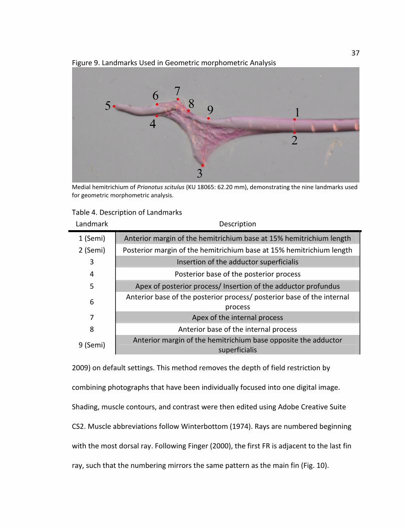
37 Figure 9. Landmarks Used in Geometric morphometric Analysis
Medial hemitrichium of Prionotus scitulus (KU 18065: 62.20 mm), demonstrating the nine landmarks used for geometric morphometric analysis.
Table 4. Description of Landmarks
Landmark Description
1 (Semi) Anterior margin of the hemitrichium base at 15% hemitrichium length
2 (Semi) Posterior margin of the hemitrichium base at 15% hemitrichium length
3 Insertion of the adductor superficialis
4 Posterior base of the posterior process
5 Apex of posterior process/ Insertion of the adductor profundus
6 Anterior base of the posterior process/ posterior base of the internal
process
7 Apex of the internal process
8 Anterior base of the internal process
9 (Semi) Anterior margin of the hemitrichium base opposite the adductor
superficialis
2009) on default settings. This method removes the depth of field restriction by
combining photographs that have been individually focused into one digital image.
Shading, muscle contours, and contrast were then edited using Adobe Creative Suite
CS2. Muscle abbreviations follow Winterbottom (1974). Rays are numbered beginning
with the most dorsal ray. Following Finger (2000), the first FR is adjacent to the last fin
ray, such that the numbering mirrors the same pattern as the main fin (Fig. 10).

38 Morphometrics
To assess possible shape differences within Triglidae, all three of the left FRs
were removed from prepared specimens and separated into lateral and medial
hemitrichia using a scalpel and fine scissors. Each hemitrichium was measured
separately using calipers and a single digital photo was taken of its base. These
hemitrichia are approximately two dimensional and make suitable subjects for
geometric analysis. The TPS series of morphometric programs (Rohlf 2010) was used to
place 9 landmarks on each photo (Fig. 9). Landmarks were chosen according to the
criteria listed in Zelditch et al. (2004), and include six homologous landmarks and three
semilandmarks chosen to describe areas lacking distinct processes or muscle insertions.
The specific landmarks are explained in Table 4. Each image was scored for landmarks
three times using the random order option, and the average coordinates were used for
further tests. These data were then entered into the program MorphoJ (Kleinburg
2011), and a Procrustes fitting algorithm was used to align the specimens and remove
non-shape variation. Principal component analysis (PCA) was conducted to visualize any
potential patterns within the data. The hypothesis of no shape difference was tested
using discriminant function analysis (DFA). This procedure was used because it is robust
with respect to deviations from normality and differences in within groups variances.
Discriminant function analysis computes Mahalanobis and Procrustes distance
measurements, which are then used to calculate Hotelling’s T2 statistic and evaluate
each hypothesis. Pairwise comparisons were followed by a standard Bonferroni
correction to control procedure-wide error rates.

39 Results
Muscle Structure Overview
In most teleosts, the pectoral fin is served by a series of six muscles, two
abductors, two adductors, and two arrectors. The two abductors are located on the
lateral pectoral girdle and act together to rotate the fin away from the body. In contrast,
the adductors are located on the medial pectoral girdle and rotate the fin back against
the body. One arrector is located on either side of the pectoral girdle and these muscles
control the leading edge of the fin. As such, they attach to only the 1st fin ray or
“marginal ray.” Each muscle inserts to the lepidotrichia, or fin rays. Each fin ray, or
lepidotrichium, is comprised of two smaller hemitrichia, one lateral and one medial.
In general, triglids show two trends relating to their musculature. First, we
observe that the muscle bundles serving the FRs are often separate from the muscle
bundles serving the fin and from each other (Fig. 10). This gives the impression of four
distinct bundles for each abductor and adductor: one large mass for the pectoral fin,
and three smaller bundles serving the individual FRs. Second, the adductor musculature
of the FRs has become subdivided into multiple bundles that differ in both their origin
and insertion. In these cases, each bundle is given a greek letter subscript. The
subdivision being closest in appearance to the traditional muscle in terms of origin,
insertion, and fiber direction is given the α subscript, whereas the other more novel
subdivision is β. The specifics of each muscle are described in depth below. Last, very
little variation in the structure of the pectoral girdle occurs within the family Triglidae. In
particular, the arrangement of the muscles of both the fin and FRs is constant

40 Figure 10. Triglid Lateral Superficial Muscles
Lateral view of the left pectoral girdle of Prionotus carolinus (FMNH 37953: 135.20 mm). The ABS is a series of vertically oriented fibers, whereas the ABP is located medial to the ABS and is horizontally oriented, and is only partially visible in this view. In both muscles, the bundles serving the FRs are distinctly separated from those serving the fin. ABBV: Cl = cleithrum, SCl = supracleithrum, FR = free ray, Fn = fin ray, ABS = abductor superficialis, ABP = abductor profundus, STH = sternohyoidius. Scale = 2mm.
throughout the nine triglid genera. Although the genus Prionotus was used as a
reference and for most photographs and drawings, all these results can be generalized
to other genera and species as well.

41 Figure 11. Lateral Free Ray Muscles
Lateral view of the left pectoral girdle of P. carolinus (FMNH 37953: 135.20 mm). The ABS bundle serving FR3 is divided into two parts that share an insertion, but have different origins. The bundles of the ABP serving the free rays can be seen behind the ABS. ABBV: Cl = cleithrum, FR = free ray, ABS = abductor superficialis, ABP = abductor profundus, STH = sternohyoidius. Scale = 2mm.
Abductor Superficialis (ABS)
The abductor superficialis (ABS) is the first muscle encountered on the lateral
side of the pectoral fin. Its origin in most teleosts is on the cleithrum and it inserts to the
lateral hemitrichium of each ray. A similar situation is observed in the pectoral fin of all
searobin species. Its origin is along the lateral edge of the proximal part of the cleithrum
and its fibers run ventrally until they meet the lateral hemitrichium for all fin rays except
the marginal ray (Fig. 10). Insertion is via a very short tendon that connects directly to

42 Figure 12. Triglid Abductor Profundus
Lateral view of left pectoral girdle of P. carolinus (FMNH 37953: 135.20 mm) with the ABS removed. With the superficial muscles removed, the bundles of the ABP are clearly visible. ABBV: Cl = cleithrum, R = radial, FR = free ray, ABP = abductor profundus. Scale = 2mm.
the base of the ray. In the FRs, the origin and insertion of this muscle is the same as in
the fin. However, the fin rays are served by one large mass, but each bundle of the ABS
serving the FRs is distinct and well separated (Fig. 11). These muscle bundles are also
much larger than in the fin rays. Additionally, the bundle serving FR3 is split into two
sections, with different fiber orientations. This arrangement of muscles serving FR3 was
found in all species and genera examined.
Abductor Profundus (ABP)
The abductor profundus is a large muscle located medial to the ABS. In most
teleost fishes, this muscle originates from the distal end of the cleithrum, runs
underneath the ABS, and inserts to the lateral hemitrichium of each fin ray. A similar

43 Figure 13. Triglid Arrector Ventralis
Lateral view of the left pectoral girdle of P. carolinus (FMNH 37953: 135.20 mm) with ABS removed. The ARRV is relatively small and inserts to the medial hemitrichium, whereas the ABP appears larger and inserts to the lateral hemitrichium. ABBV: Cl = cleithrum, Sca = scapula, R = radial, Fn = fin ray, ABP = abductor profundus, ARRV = arrector ventralis. Scale = 2mm.
state is observed in all triglids, although the origin was found to be from the coracoid
bone as well as the cleithrum. Insertion is via a short tendon that connects to a small
ridge near the base of the ray. As in the ABS, the fin rays are served by one
undifferentiated muscle mass, but the FRs are served by three large, well separated
bundles (Fig. 12).
Arrector Ventralis (ARRV)
The arrector ventralis (ARRV) is located medial to the ABS and dorsal to the ABP
in most teleost fishes. All triglids show the generalized condition of this muscle. Its origin
44 is from the posterior-lateral cleithrum, and it inserts via a single bundle to the medial
hemitrichium of the marginal ray (Fig. 13). Insertion is to a large process at the base of
the ray. This muscle serves only the marginal ray and no others.
Adductor Superficialis (ADS)
The adductor superficialis (ADS) is the first muscle encountered in medial view
(Fig. 14). In teleosts, it originates from the medial side of the cleithrum, and inserts to
each ray. In triglids, the origin is from the medial side of the proximal end of the
cleithrum. Fibers run ventrally to insert to the medial hemitrichium of each fin ray,
except the marginal ray. The insertion generally occurs somewhat distal from the base
of each ray. As in the lateral musculature, the ADS bundles serving the fin appear as one
large mass, while the bundles serving the FRs are well separated.
In all triglids, the ADS bundles serving the free rays are subdivided into two
sections each with a distinct architecture (Fig. 15A). These subdivisions have distinct
origins and insertions, and are therefore designated ADSα and ADSβ. The ADSα is similar
in form to the bundles serving the fin rays. It originates from the medial cleithrum and
runs ventrally to insert to the medial hemitrichium of each free ray. Insertion is to the
central part of the ray a considerable distance from the base, and usually occurs without
a noticeable tendon. ADSβ originates from the medial cleithrum, slightly posterior and
lateral to the ADSα. However, it runs not just in a ventral, but also an anterior-medial
direction, thus while its origin is posterior-lateral to the ADSα, its insertion is anterior-
medial to the ADSα. The fibers for ADSβ fuse into a long tendon for about 40-50% of its
length, before inserting on a pronounced triangle shaped process on the medial

45 Figure 14. Triglid Medial Superficial Muscles
Medial view of the left pectoral girdle of P. carolinus (FMNH 37959: 89.15 mm). The ADS and ADP are clearly visible, but the bundles serving the free rays are divided into two separate subunits, each with its own origin and insertion. ABBV: Cl = cleithrum, SCL = supracleithrum, Cor = coracoid , FR= free ray, Fn = fin ray, ADS = adductor superficialis, ADP = adductor profundus, CR = coracoradialis. Scale = 2mm.
hemitrichium of each FR (Fig. 15B). This process is distal from the base of the ray, and
next to the point of insertion of the ADSα.
Adductor Profundus (ADP)
The ADP is usually a well-developed muscle located lateral to the ADS (Fig. 16).

46 Figure 15. Subdivisions of the Adductor Superficialis
Medial view of the left pectoral girdle of P. carolinus, showing (A) (FMNH 37959: 89.15 mm) a line drawing of the ADS bundles of the FRs, and (B) (FMNH 37951: 95.10 mm) a magnified view of their insertion. Black arrows indicate ADSα bundles and white arrows indicate ADSβ bundles. The mass of the ADS muscle serving the fin has been dissected away to better reveal the origin and insertion of the ADS bundles of the FRs. The ADSα bundles are similar to those of the fin, but the ADSβ demonstrates several innovations, such as insertion to a large process and possessing a long tendon. Note in panel B that the ADSβ bundles originate posterior to the ADSα, but it cuts across to insert more anteriorly. ABBV: Cl = cleithrum, FR = free ray, ADS = adductor superficialis, ADP = adductor profundus. Scale bar = 2mm.
Its origin is from the anterior-medial edge of the cleithrum and the coracoid, and its
insertion is to the posterior process at the base of the medial hemitrichium of each fin
ray. A similar condition exists in the triglid pectoral fin, with the addition that some
fibers originate from the enlarged radial bones as well. As usual, these fibers run
posteriorly to insert on the posterior process of each fin ray via a short tendon, except
the marginal ray.
As with the other muscles, the ADP bundles serving the FRs are well separated
from each other and the bundles of the main fin. These bundles are also split into ADPα
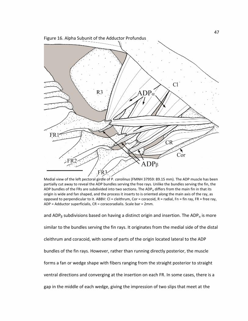
47 Figure 16. Alpha Subunit of the Adductor Profundus
Medial view of the left pectoral girdle of P. carolinus (FMNH 37959: 89.15 mm). The ADP muscle has been partially cut away to reveal the ADP bundles serving the free rays. Unlike the bundles serving the fin, the ADP bundles of the FRs are subdivided into two sections. The ADPα differs from the main fin in that its origin is wide and fan shaped, and the process it inserts to is oriented along the main axis of the ray, as opposed to perpendicular to it. ABBV: Cl = cleithrum, Cor = coracoid, R = radial, Fn = fin ray, FR = free ray, ADP = Adductor superficialis, CR = coracoradialis. Scale bar = 2mm.
and ADPβ subdivisions based on having a distinct origin and insertion. The ADPα is more
similar to the bundles serving the fin rays. It originates from the medial side of the distal
cleithrum and coracoid, with some of parts of the origin located lateral to the ADP
bundles of the fin rays. However, rather than running directly posterior, the muscle
forms a fan or wedge shape with fibers ranging from the straight posterior to straight
ventral directions and converging at the insertion on each FR. In some cases, there is a
gap in the middle of each wedge, giving the impression of two slips that meet at the

48 Figure 17. Beta Subunit of the Adductor Profundus
Medial view of the left pectoral girdle of P. carolinus (FMNH 37959: 89.15 mm). The deepest layer of muscle is the ADPβ, which can only be seen clearly once all other muscle layers have been dissected. This muscle originates mainly from the radial bones and inserts to a small process or knob near the base of each ray. ABBV: Cl = cleithrum, Cor = coracoid, FR = Free ray, ADP = adductor profundus, CR = coracoradialis. Scale bar = 2mm.
insertion, but this was not found to be a consistent pattern across all triglids surveyed.
The insertion is to the posterior process on the base of each hemitrichium.
The ADPβ is small bundle located on the medial side, lateral to the ADPα (Fig. 17).
Its origin covers parts of the medial surface of the coracoid, cleithrum, and radials 3 and
4. Its fibers run the short length ventrally and medially to insert via a short, wide tendon
to the internal process at the base of the medial hemitrichium. As the internal process is
also the point where the rays articulate with the fibrocartilage of the pectoral girdle, the

49 Figure 18. Triglid Arrector Dorsalis
Medial View of the left pectoral girdle of P. carolinus (FMNH 37953: 135.20 mm). The ADS has been removed to show the ARRD and the insertion of the ADP muscles. This muscle inserts only to Fn1, the marginal ray. The distinction between the ADP and ARRD is muted in many specimens, with many muscle fibers appearing to overlap. ABBV: Cl = cleithrum, Sca = scapula, Fn = fin ray, ADP = adductor profundus, ARRD = arrector dorsalis. Scale = 2mm.
insertion of this muscle is often difficult to distinguish amongst the copious amounts of
connective tissue.
Arrector Dorsalis (ARRD)
In most teleost fishes, the ARRD originates from the medial cleithrum, dorsal to
the ADP. The ARRD inserts to a process on the base of the medial hemitrichium of the
marginal ray. Like the ARRV, this muscle serves only the marginal ray (Fig. 18). In all
triglids, the origin and insertion follow the above pattern, though it is often difficult to
separate from the ADP.

50 Figure 19. Skeletal Elements of the Triglid Pectoral Girdle
Cleared and stained specimens showing the osteology of the triglid pectoral fin. (A) Lateral view of the left pectoral girdle of Pterygotrigla Arabica (USNM356390: 52.50 mm). Note that the larger FRs are located more anterior and ventral that those of the main fin, and that R1 is much reduced compared to the other radial bones. (B)This Prionotus scitulus (KU 18065: 62.20 mm) fin ray lacks the elaborate processes observed in the FRs (C) of the same specimen. Also note that segmentation begins in the FRs and before it begins in the fin rays. Abbv: Cl = cleithrum, SCl = supracleithrum, Sca = scapula, Cor = Coracoid, R1 – 4 = Radials 1 – 4, Fn = fin ray, MH = medial hemitrichium, LH = lateral hemitrichium, Post = posterior process, Int = Internal process, ADS Pr = ADS process.
Osteology
The pectoral girdle of triglids shows several general patterns not seen in other
scorpaenoid fishes. First, the cleithrum and coracoid extend a considerable distance
anterior and the fin itself is diagonally oriented in an anterio-ventral direction (Fig. 19).
As a result, the ventral most fin and free rays are the most anterior. Second, the radials
are much enlarged and form a sort of shelf between the scapula and coracoid which
provides a large surface for muscle attachment as observed above (Fig.19 A). Unlike
some scorpaenoids, the cleithrum does not articulate with an extension of the pelvis. In
51 all specimens, one thin post-cleithrum is present.
Relatively little variation was observed across triglid genera or species. In most
teleosts, the scapula features a large foramen (Fig. 19 A) which allows nerves and blood
vessels to serve the lateral part of the fin musculature. However, it was noticed that
members of the genus Prionotus have their scapular foramen between the cleithrum
and scapula, as opposed to the standard arrangement. The scapula in Prionotus is “U”
shaped and articulates with the cleithrum so as to form an opening. In all the other
triglid genera examined, the scapula has the more standard condition described above.
Another source of variation occurs in Radial 1, which may be either large or small. In the
first case, the radial extends between Radial 2 and the scapula, whereas in other genera,
radial 1 is reduced and radial 2 and the scapula articulate directly (Fig. 19 A). Prionotus
and Bellator were observed to have a larger first radial, but all other genera had smaller
first radial bones. These observations were consistent with previous studies of triglid
osteology (Richards & Jones 2000).
Fin rays, or lepidotrichia, are each composed of two hemitrichia, which articulate
with a sturdy fibrocartilage pad extending from and surrounding the radial bones. Each
ray or lepidotrichium is composed of two hemitrichia. Each hemitrichium consists of a
proximal unsegmented base, and followed distally by a flexible series of many joined
segments. The lateral hemitrichium receives insertions from the abductor muscles (ABS
& ABP). The lateral hemitrichia of the fin contains a large knob that serves as the point
of articulation with the radial, and a neck or ridge just distal to the base for the insertion
of the ABP. In the fin rays, sometimes a small ridge is seen at the insertion site of the

52 ABS. The medial hemitrichia receive insertions from the adductor musculature (ADS &
ADP). The base of the medial hemitrichium usually expands into two processes. The
posterior process protrudes at a perpendicular angle from the ray and is the insertion
site of the ADP. The size of this process is variable, with the more ventrally located fin
rays tending to have larger processes. The small internal process is located opposite the
posterior process and it connects to the fibrocartilage and anchors the hemitrichia to
the pectoral girdle. No process is observed at the insertion of the ADS in the fin rays.
All triglid specimens possessed three FRs along the anterio-ventral border of
their pectoral fin. Like the fin rays, they are held in place by a sturdy fibrocartilage pad
extending from the radials. In all cases, the first free ray is associated with radial 3 and
the others with radial 4, and generally a small notch is visible in the bone around the
articulation point (Fig. 19B). By comparison, with the fin rays, the FRs are uniformly
larger in width, though not necessarily length. There is no tissue connecting the FRs
except very close to the base. Additionally, there is more space between the lateral and
medial hemitrichia in the FRs than in the fin rays. This allows greater relative motion of
the hemitrichium bases, which is associated with increased bending. Interestingly, the
FRs are capable of bending only in the medial direction, or towards the adductors, and
they resist movement in the opposite direction. In all triglid specimens, the
unsegmented basal portion of the hemitrichia was shorter in the FRs than the fin rays.
Additionally, the medial hemitrichia of the free rays is distinguished by pronounced
segmentation beginning rather close to the base of the ray, while segmentation in the
lateral hemitrichia appears more gradually. The shapes of the lateral hemitrichia of the
53 FRs are similar to what is seen in the fin rays. However the medial hemitrichia differ
substantially in the shape of the two traditional processes, and the presence of a third
process not found in fin rays. Like in the entire ADS in the fin rays, the ADSα inserts
directly to the main shaft of the bone, near the distal base of the large triangle shaped
process. In contrast, the ADSβ inserts to the apex of this process (Fig. 19C). Instead of
perpendicular, the posterior process in the free rays is in line with the long axis of the
rays, and is often long and sometimes curved. The ADPα subdivision inserts to the apex
of this process. The length and curvature of this process vary slightly among the free
rays, with FR3 usually being longest and most sharply curved. Last, the internal process
is serves two roles in the FRs. It is the site of articulation with the cartilage of the
pectoral girdle, but is also the insertion site of the ADPβ.
Morphometrics
Principal components analysis of geometric morphometric data from the medial
hemitrichia of the free rays was conducted to search for possible patterns in the data.
Analysis of the 9 landmarks reveals that the first two PCs account for 74.9 % of the total
variance in shape. Principal compoenent one is associated with the movement of
landmark 5 in the proximal direction, the movement of landmark 7 out away from
landmarks 6 & 8, and a reduction in the space between landmarks 1 & 9 and 2 & 3. This
implies a long posterior process, a wide, pointed internal process, and a distally placed
process at the insertion of the ADS. Principal component two is associated with the
distal movement of landmark 5, the distal movement of landmark 8, and the proximal
and outward movement of landmark 3.

54 Table 5. Principal Component Loadings of Triglid Free Rays
PC1 PC2 PC1 PC2
X1 -0.39434 -0.04925 X6 -0.01071 -0.01247
Y1 0.117647 0.344681 Y6 0.039109 0.030532
X2 -0.32706 0.067792 X7 0.148049 0.099492
Y2 -0.11276 0.07716 Y7 0.104947 0.035859
X3 0.41513 -0.31473 X8 0.278531 0.250478
Y3 -0.31735 -0.68945 Y8 0.083574 0.077102
X4 -0.00969 0.11824 X9 0.335661 -0.37694
Y4 -0.06705 -0.00219 Y9 0.064281 0.091057
X5 -0.43558 0.217389
Y5 0.087593 0.035254
Principal component loadings for the first two PCs computed during the geometric morphometric analysis. Together, these two axes explain 74.9 % of the total shape variation in the medial hemitrichia of the FRs.
Respectively, these changes suggest a short posterior process, a wide but shallow
internal process, and a proximally placed process for insertion of the ADS with a
pronounced apex. Examination of these data with respect to genera failed to uncover
any meaningful patterns in the shapes of the FR hemitrichia across genera. Interestingly
though, to the extent that the genera do differ at all they differ with respect to PC2. By
contrast, examining the data with respect to FR number reveals a pronounced pattern in
the shapes of the FRs. Data points obtained from the more posterior FR1 have a lower
score on PC1, indicating the characteristics described above. In contrast, those from the
more anterior FR3 have higher scores on PC1 indicating the opposite. Scores for FR2 are
intermediate between these extremes.

55 Figure 20. Principal Component Analysis of Triglid Free Rays
Scatterplot of PC scores obtained from medial hemitrichia specimens. The data points have been colored with respect to FR #, and ellipses represent a 90% equal frequency area. This plot suggests that FR morphology may vary with respect to its position on the body. FR1 is most posterior, and FR3 most anterior.
Table 6. Discriminant Function Analysis of Triglid Free Rays
FR Comparison T2 P
1---2 117.7312 <.0001
1---3 266.1349 <.0001
2---3 197.0791 <.0001
Discriminant factor analysis of medial hemitrichium shape with respect to free ray #, and α=.017. All three FRs were well separated and have a significantly different shape.
Almost no variation is observed on PC2 with respect to FR number. Discriminant
function analysis shows that these differences are statistically significant.This test
confirms that each FR has its own distinct morphology based upon free ray number and
56 related to its relative position on the pectoral girdle. This also suggests that the previous
attempt to discriminate according to genus was likely confounded by the underlying
variation based on FR position. The above analyses were run again with the data
segregated by FR number to control for this possible influence. However, these analyses
also failed to distinguish triglid genera at a statistically significant level.
Discussion
Although several studies have investigated triglid behavior (Manderson 1999, Anorim
2006), or phylogeny (Richards 2002, Imamura 1996, 2004), this is the first study to
examine the fine details of pectoral fin structure in all genera of Triglidae. A survey of
the literature of searobin behavior reveals three factors that separate the motion of the
free rays from the fin. First, they are under individual motor control and can be moved
while the pectoral fin is at rest. Additionally, Renous et al. (2000) reported the ability of
the FRs to move independently of each other, such that one FR may be abducted or
adducted when the adjacent free ray moves in the opposite position. In life, these fish
use a cyclical gait that conveys the appearance of walking on the substrate. Second, free
rays have a somewhat larger range of motion than the pectoral fin. When fully
abducted, the FRs extend forward to the lateral margins of the mouth. In FR3, this
requires almost 180° rotation from its resting position. The other FRs are somewhat
more limited. Third, triglids are capable of intentionally bending the distal ends of their
FRs and holding them in this position. Because the distal tips of the rays are the primary
point of contact with the substrate, this flexibility is likely important to behaviors that

57 probe sandy area for chemical traces of prey, or possibly in generating forces during
walking locomotion.
The ability of searobins to move their FRs independently is consistent with the
more or less complete separation of their musculature from that serving the fin rays.
Both abductors (Fig. 11 & 12) and adductors (Fig. 15, 16, & 17) are divided into clear
bundles serving a single free ray with no overlapping of the fibers or tendons. It is less
clear what differences allow the FRs their increased range of motion. No major
subdivisions of the abductor musculature were found, and the lateral hemitrichia of the
FRs have no novel processes or other osteological features. Free Ray 3 has the widest
range of motion and is different from the other free rays in having a second slip of the
ABS muscle that runs at a more horizontal angle (Fig. 11). Searobins are already known
to considerably abduct their main pectoral fin during threat displays and conspecific
interactions (Anorim 2004). It is possible that this adjustment to the ABS fiber
orientation is sufficient to account for the range of motion observed in living specimens.
Bending in teleost fin rays is accomplished by forces exerted at the base of each
hemitrichium which displace one side relative to the other. This displacement causes a
tension that is transmitted along the many segments of the ray. Thus, forces exerted on
the base of the medial hemitrichium can cause the distal portion of the ray to bend
towards the body (Geerlink 1987). Considering that the free rays only bend in the
medial direction, this motion is expected to be mediated by one of the adductors,
presumably the ADPα & ADPβ. These muscles insert close to the base of the medial
hemitrichium and appear to be oriented in such a way as to transmit forces along the

58 long axis of the bone. In particular, the unique ADPβ is positioned to apply considerable
forces directly to the base of the medial hemitrichium. It inserts to the internal process,
which is also the site of articulation with the cartilage of the pectoral girdle. One
interesting possibility is that this muscle provides tension to the base of the ray, helping
to hold it in place or pull it back during bending. Holding the base of the ray steady is
crucial to the production of forces at the distal end of the ray, as determined by
mathematical models (Alben et al. 2007). The osteology of the free rays also seems to
be designed to promote bending in the medial direction. Segmentation of the medial
hemitrichium occurs very close to the base, a trait that is associated with increased
flexibility in mathematical models (Alben et al 2007). The fact that segmentation
appears to begin at a more distal point on the lateral hemitrichium may be partly
responsible for the unidirectional bending of the ray.
The majority of novel muscle groupings and osteological features are on the
medial side of the pectoral girdle. This suggests that the additional mobility acquired by
the triglid FRs are primarily mediated by the adductor musculature acting on the medial
hemitrichia. The ADSα of FRs does not insert to a distinct process, and is overall quite
similar in form to the ADS seen in fin rays. This suggests a similar function in adducting
the FRs towards the body. In contrast the ADSβ inserts to a novel process or ridge on the
medial hemitrichia. Additionally, the posterior process to which the ADPα inserts has
been modified from its usual form in the fin rays. These changes suggest that adduction
is different and more complex in the FRs. One plausible explanation for these variations
is the increased range of motion. When fully abducted, the angle of insertion of the
59 ADSα may no longer be favorable for moving the ray. In this situation, the ADSβ may
provide the additional leverage necessary to return to the FRs to an adducted position.
Similarly, the ADPα inserts to a process at the proximal base of the medial hemitrichium.
Its fibers form a wide arc that could potentially exert force over a range of angles.
Tests of mean shape showed that each FR in Triglidae has its own distinct
morphology. The range of motion is different for each ray, and this may account for
some of the difference in shape. Another possible explanation relates to force
production. Jamon et al. (2007) observed that the force exerted by searobins against the
substrate was largest in FR1 (Jamon’s FR3), and smaller in the others. Principal
components analysis indicates that this free ray is associated with a large posterior
process for the ADPα, and a more distally located insertion of the ADSβ. The
specialization of the FRs into distinct functional roles fits with several other studies of
pectoral structure. Thorsen & Westneat (2005) demonstrated that in some labriform
swimmers, the abductor musculature serves only a distinct subset of the fin rays.
Similarly, Taft (2009) showed that the bending properties the fin rays map to distinct
regions of the fin in mottled sculpin, Cottus bairdi.
In this study, hemitrichium shape was not significantly different with respect to
genus. Morphometric analysis of Prionotus found that this genus occupied a large area
of the component space and tended to overlap other genera. However, this may be a
result of uneven sampling. In this study, the genus Prionotus comprised over half of all
specimens. In contrast, the genera Pterygotrigla and Lepidotrigla comprise over half of
all triglid species, but are not well represented in many museum collections and were

60 not available for testing. DFA is somewhat sensitive to small sample sizes and uneven
sampling of categories (James & McCulloch 1990), both of which urge caution in
drawing generalizations from the present data. As a result, of the aforementioned
limitations, it cannot be determined whether this implies that Prionotus is more variable
or is an artifact of the sampling design. On the other hand, past studies of triglids have
shown that they occupy similar environments, specifically sandy areas at shallow to
moderate depth. A study of several species of Atlantic searobins (primarily Prionotus)
found that species with overlapping distributions often occupied the same trophic level,
but focused on different prey sizes and types (Ross 1977). The overall similarity of the
free rays across taxa may reflect shared ecological roles, with selection acting to
reinforce the observed pectoral fin anatomy.
Despite the fundamental importance of fin lepidotrichia to locomotion in ray-
finned fishes, there have been few studies focused on the morphology of the fin rays. As
such, the meaning of numerous morphological variations to the fin rays are not well
understood. This is particularly true of many benthic fishes, which use their fins to
interact with the substrate in addition to the water. In past, studies of fin ray bending
have been conducted on pelagic fish (Geerlink 1979, 1987). Benthic fishes often have
fins suited to interacting with the substrate that make it problematic to extrapolate
findings from pelagic species. Studies of pelagic fishes have also tended to focus on the
caudal fin, which generally possesses symmetric hemitrichia, thought to allow equal
bending strength in either direction. The pronounced asymmetry of the FRs presents a
challenge to our understanding of fin ray mechanics and demonstrates that studies

61 focused on pelagic locomotion miss a broad swathe of fish diversity. A more
comprehensive model will require future work on the bending properties of fin rays to
include representatives from benthic taxa.
In summary, the impressive mobility of the free rays is accompanied by
significant modifications to the pectoral girdle. The many novel functions of the
searobin free rays are a result of adaptation to the medial side of the pectoral girdle.
The ADS and ADP muscles serving the FRs are subdivided into two bundles each with a
distinct architecture that is absent from the fin rays. Furthermore, the medial
hemitrichia of the FRs have been extensively modified. The base of each medial
hemitrichium has a unique process for the ADS that is absent in the fins, and
modifications to the posterior and internal processes. By contrast, the lateral
musculature and lateral hemitrichia are changed only slightly from their condition in the
fin rays. Also, geometric morphometric analysis revealed that the FRs are not identical
but have consistent differences in the shapes of their hemitrichia. Each FR appears to be
adapted to a particular regimen of forces and movements, fitting a growing literature of
regionalization in the fins of benthic fishes. Last, the striking morphological variation
found in triglid free rays suggest that the fine structure of fin lepidotrichia may be an
untapped resource for comparing groups with specialized fins. Extending this approach
in future studies will improve our understanding of the evolution of other scorpaenoids
and benthic fishes in general.

62
CHAPTER THREE
THE COMPARATIVE MORPHOLOGY OF THE FREE RAYS IN SCORPAENOIDEA
Abstract
The Scorpaenoidea is a group of over 700 overwhelmingly marine fish species. The
majority of these species are benthic and many have enlarged or elaborate pectoral fins.
Of these, approximately 200 species in five families possess a set of one or more free
pectoral rays. This trait is thought to allow specialized interactions with the substrate
that supports unique sensory and locomotive behaviors. This study provides the first in
depth survey of pectoral fin ray and free ray morphology in scorpaenoid fishes.
Representatives from all five families are examined revealing significant variation in free
ray structure at the family and sometimes genus level. In most cases, free rays are
accompanied by novel muscle configurations and modifications to the bone structure of
the lepidotrichia. Geometric morphometric analysis of the shape of the free ray
hemitrichia finds significant differences among families and with respect to free ray
number. The functional and behavioral consequences of this morphology are considered
for each group. Overall, these data point to at least two divergent patterns in free ray
and pectoral fin evolution in scorpaenoids. This observation coupled with behavioral
observations suggests that this trait has evolved multiple times and has taken on unique
specialized roles in each clade. This study shows that the pectoral fin of scorpaenoid

63
fishes contain undocumented variation in morphology with potential impacts on our
understanding of the systematics, behavior, and ecology of these fishes.
Introduction
The Scorpaenoidea (sensu Imamura 2004), is a perciform superfamily consisting of over
700 species. In general, these fishes are known for their benthic habitat, predatory
habits, elaborate fins, and venomous spines. They are almost exclusively marine with
only a few brackish species, and may be found in all temperate and tropical seas. The
greatest diversity is found in the Indo-Pacific region and many species are regular
inhabitants of coral reefs (Nelson 2006). Several species such as the pacific rockfishes
(Sebastidae) are regularly harvested for commercial purposes, and the venom from
scorpionfishes has been suggested as a possible source for new bioactive
pharmaceuticals (Smith & Wheeler 2006). In recent years, great attention has been paid
to the growing significance of the lionfish Pterois volitans as an invasive species in
Atlantic and Caribbean waters.
The majority of scorpaenoids occupy benthic habitats, often living on or partially
buried in the substrate. As such, many of these fishes show a suite of adaptations
designed to interact with the substrate. Ventrally flattened bodies and camouflage are
quite common, but perhaps most interesting are changes to the paired fins. Although
almost all are carnivorous, few scorpaenoids rely on swift or agile locomotion to capture
prey. Instead, most scorpaenoids obtain food through ambush tactics, confusing
displays, or the ability to sense and pursue buried prey. With a reduced emphasis on
swimming locomotion, scorpaenoid pectoral fins show a variety of adaptions that

64
function in signaling, cornering prey, and interacting with the substrate (Gosline 1994).
This makes scorpaenoid fishes an excellent group in which to study variation in pectoral
fin morphology. In particular, substrate interactions comprise an interesting and often
overlooked set of behaviors that likely have a significant morphological component.
Some reported substrate interactions may include a variety of behaviors such as digging,
searching for prey, perching, and pushing off the bottom. A specific example of a trait
that appears adapted to substrate interactions are free pectoral rays. Free pectoral rays
(free rays) are a set of one to three fin lepidotrichia occurring along the ventral edge of
the pectoral girdle that lack a membrane connecting them to the remaining rays.
The Searobins (Triglidae) are the most numerous and best studied fishes
possessing free rays. Several studies on different species have suggested that the free
rays (hereafter “FRs”) are functionally and morphologically distinct from the fin rays,
and may possess novel sensory (Bardach & Case 1965, Finger 2000) and locomotive
functions (Renous et al 2000, Jamon et al 2007). The previous chapter posited that
three characteristics distinguish the FRs from fin rays in Triglidae: 1) independence from
the main pectoral fin, 2) increased range of motion, and 3) the ability to distally flex
their lepidotrichia. This study identified new subdivisions of the adductor superficialis
and adductor profundus muscles associated only with the FRs. These muscles, which
insert to the medial hemitrichium of each FR, are accompanied by a set of bony
processes that are also unique to the FRs. Lastly, geometric analysis of the FRs found
consistent differences in the shape of the processes on the medial hemitrichium
between the three FRs.
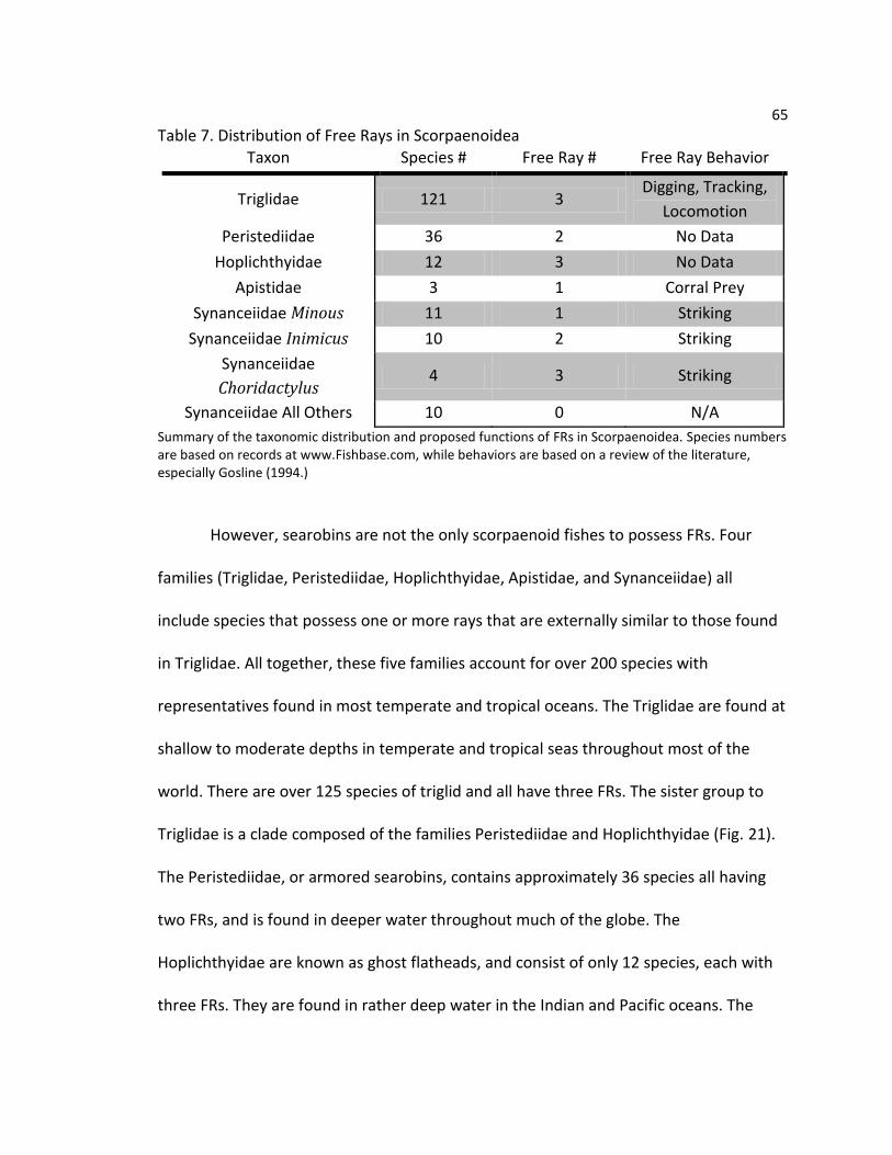
65
Table 7. Distribution of Free Rays in Scorpaenoidea Taxon Species # Free Ray # Free Ray Behavior
Triglidae 121 3 Digging, Tracking,
Locomotion
Peristediidae 36 2 No Data
Hoplichthyidae 12 3 No Data
Apistidae 3 1 Corral Prey
Synanceiidae Minous 11 1 Striking
Synanceiidae Inimicus 10 2 Striking
Synanceiidae
Choridactylus 4 3 Striking
Synanceiidae All Others 10 0 N/A
Summary of the taxonomic distribution and proposed functions of FRs in Scorpaenoidea. Species numbers are based on records at www.Fishbase.com, while behaviors are based on a review of the literature, especially Gosline (1994.)
However, searobins are not the only scorpaenoid fishes to possess FRs. Four
families (Triglidae, Peristediidae, Hoplichthyidae, Apistidae, and Synanceiidae) all
include species that possess one or more rays that are externally similar to those found
in Triglidae. All together, these five families account for over 200 species with
representatives found in most temperate and tropical oceans. The Triglidae are found at
shallow to moderate depths in temperate and tropical seas throughout most of the
world. There are over 125 species of triglid and all have three FRs. The sister group to
Triglidae is a clade composed of the families Peristediidae and Hoplichthyidae (Fig. 21).
The Peristediidae, or armored searobins, contains approximately 36 species all having
two FRs, and is found in deeper water throughout much of the globe. The
Hoplichthyidae are known as ghost flatheads, and consist of only 12 species, each with
three FRs. They are found in rather deep water in the Indian and Pacific oceans. The
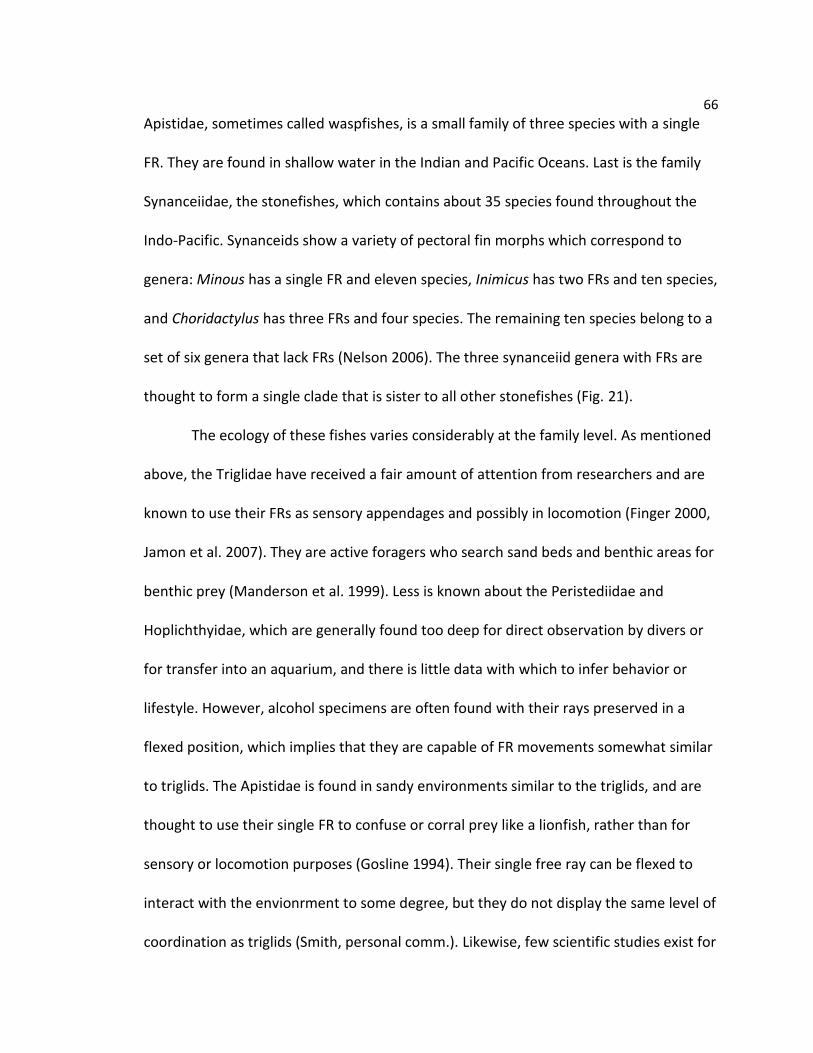
66
Apistidae, sometimes called waspfishes, is a small family of three species with a single
FR. They are found in shallow water in the Indian and Pacific Oceans. Last is the family
Synanceiidae, the stonefishes, which contains about 35 species found throughout the
Indo-Pacific. Synanceids show a variety of pectoral fin morphs which correspond to
genera: Minous has a single FR and eleven species, Inimicus has two FRs and ten species,
and Choridactylus has three FRs and four species. The remaining ten species belong to a
set of six genera that lack FRs (Nelson 2006). The three synanceiid genera with FRs are
thought to form a single clade that is sister to all other stonefishes (Fig. 21).
The ecology of these fishes varies considerably at the family level. As mentioned
above, the Triglidae have received a fair amount of attention from researchers and are
known to use their FRs as sensory appendages and possibly in locomotion (Finger 2000,
Jamon et al. 2007). They are active foragers who search sand beds and benthic areas for
benthic prey (Manderson et al. 1999). Less is known about the Peristediidae and
Hoplichthyidae, which are generally found too deep for direct observation by divers or
for transfer into an aquarium, and there is little data with which to infer behavior or
lifestyle. However, alcohol specimens are often found with their rays preserved in a
flexed position, which implies that they are capable of FR movements somewhat similar
to triglids. The Apistidae is found in sandy environments similar to the triglids, and are
thought to use their single FR to confuse or corral prey like a lionfish, rather than for
sensory or locomotion purposes (Gosline 1994). Their single free ray can be flexed to
interact with the envionrment to some degree, but they do not display the same level of
coordination as triglids (Smith, personal comm.). Likewise, few scientific studies exist for
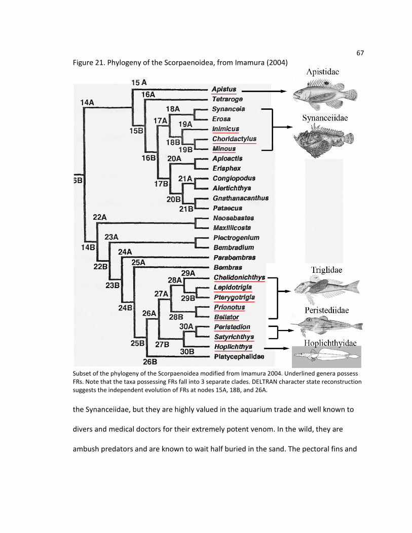
67
Figure 21. Phylogeny of the Scorpaenoidea, from Imamura (2004)
Subset of the phylogeny of the Scorpaenoidea modified from Imamura 2004. Underlined genera possess FRs. Note that the taxa possessing FRs fall into 3 separate clades. DELTRAN character state reconstruction suggests the independent evolution of FRs at nodes 15A, 18B, and 26A.
the Synanceiidae, but they are highly valued in the aquarium trade and well known to
divers and medical doctors for their extremely potent venom. In the wild, they are
ambush predators and are known to wait half buried in the sand. The pectoral fins and

68
FRs are reported to help stonefishes rapidly spring from hiding when attacking prey
(Grobecker 1983, Gosline 1994).
Based on morphological data, Imamura’s (2004) phylogeny of the Scorpaenoidea
hypothesizes that FRs have evolved separately on three occasions. The first is at the last
common ancestor of Hoplichthyidae, Peristediidae, and Triglidae, (Fig. 21, Node 26A).
All of the fish in this clade possess two or three FRs, as well as other synapomorphies
relating to the pelvis and cranium. While Imamura considers the Hoplichthyidae and
Peristediidae to be sister groups, other authors have theorized that the Triglidae and
Peristediidae are more closely related. Molecular evidence also finds Triglidae and
Peristediidae to be sister groups (Smith & Wheeler 2004, Smith & Craig 2007). Free rays
evolve again in the Apistidae, (Fig. 21, Node 15A), which have a single FR. Some previous
authors have suggested a relationship between the Apistidae and Triglidae based on the
presence of FRs and fusions of the infraorbital bones, (Washington et al. 1984), but
most recent analyses have rejected this hypothesis (Ishida 1994, Imamura 1996, 2004).
Thus far, no molecular systematic data have been published for the Apistidae. The third
appearance occurs within the Synanceiidae (Fig. 21, Node 18B). This clade includes the
genera Minous, Inimicus, and Choridactylus, all of which have FRs, as opposed to other
synanceiids in which they are absent. Imamura’s tree is in agreement with other studies,
which consistently place the stonefishes amongst a diverse group of scorpaenoids
including aploactids and congiopodids (Ishida 1994, Smith & Wheeler 2004, Smith &
Craig 2007).

69
Given the diversity of evolutionary origins and ecological roles, it is reasonable to
question to what extent the FRs differ in their morphology. Alterations to the structure
of fin lepidotrichia have been associated with changes between a benthic vs. pelagic
mode of life (Taft 2011). Additionally, in some sculpins, only the most ventral fin rays
contact the substrate, and these rays have osteological characteristics that indicate
increased flexibility compared to the rest of the fin (Taft 2009). Previous work in triglids
shows a specific set of modifications to the medial hemitrichia of the FRs, associated
with novel subdivisions of the adductor musculature. One might hypothesize that the
Triglidae, Peristediidae, and Hoplichthyidae, have a similar pattern of muscle insertion
and bone structure. These families share a common evolutionary history and possibly
the use of FRs in a foraging. In contrast, we might expect new variations to be found in
the more distantly related Synanceiidae, perhaps related to their importance when
striking prey. By the same token, the phylogenetic position and reported behaviors in
Apistidae suggest that they may share little in common with the other groups.
Thus, the primary question of this study is whether and to what extent the
muscle anatomy of the pectoral fin and the morphology of the FR lepidotrichia in these
groups vary according to taxon. The current study will address this question by
undertaking a full comparative description of scorpaenoids possessing FRs. This study
will focus on three areas. First, dissection and examination of specimens from each
family (or synanceiid genus) is used to characterize the orientation, origin, and insertion
of the pectoral fin muscles. Special attention is paid to the muscles inserting to the FRs
and any novel subdivisions are identified and described. Second, an examination of the
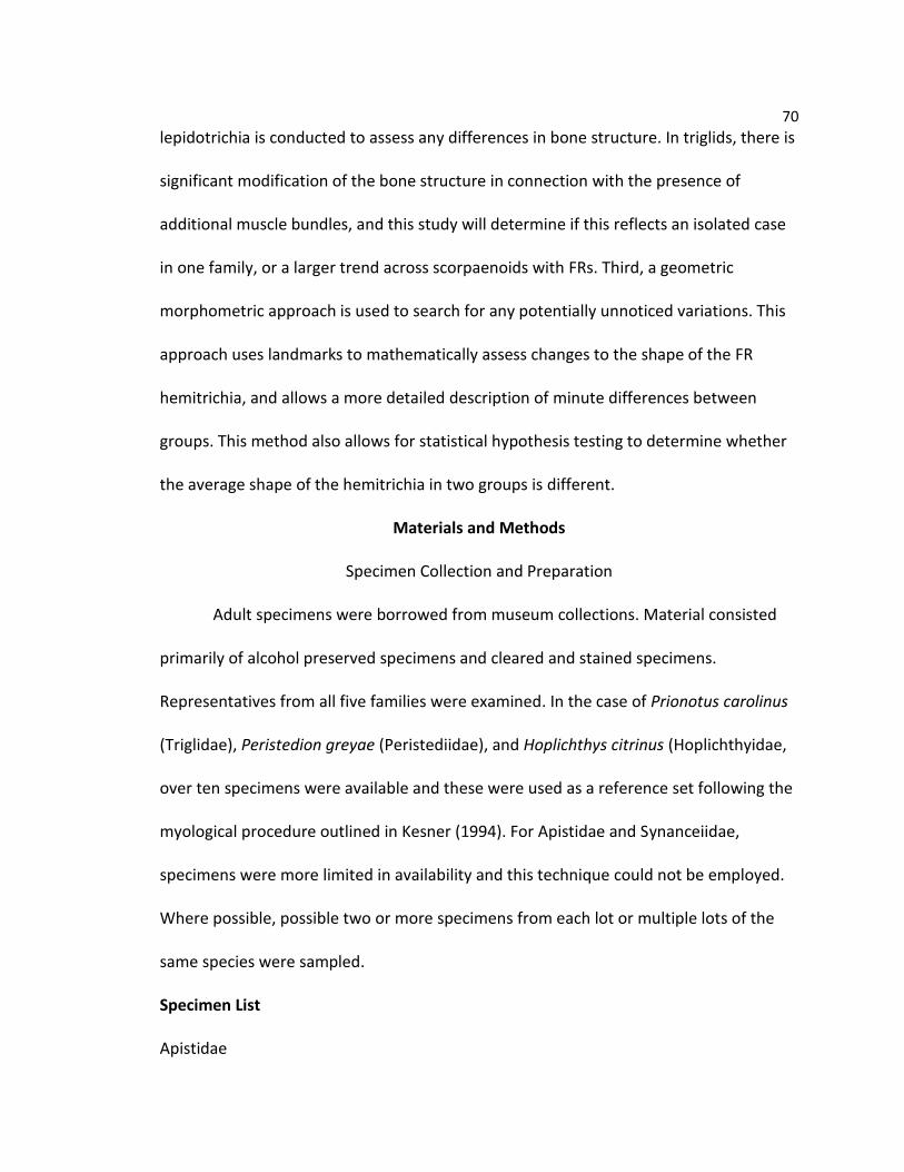
70
lepidotrichia is conducted to assess any differences in bone structure. In triglids, there is
significant modification of the bone structure in connection with the presence of
additional muscle bundles, and this study will determine if this reflects an isolated case
in one family, or a larger trend across scorpaenoids with FRs. Third, a geometric
morphometric approach is used to search for any potentially unnoticed variations. This
approach uses landmarks to mathematically assess changes to the shape of the FR
hemitrichia, and allows a more detailed description of minute differences between
groups. This method also allows for statistical hypothesis testing to determine whether
the average shape of the hemitrichia in two groups is different.
Materials and Methods
Specimen Collection and Preparation
Adult specimens were borrowed from museum collections. Material consisted
primarily of alcohol preserved specimens and cleared and stained specimens.
Representatives from all five families were examined. In the case of Prionotus carolinus
(Triglidae), Peristedion greyae (Peristediidae), and Hoplichthys citrinus (Hoplichthyidae,
over ten specimens were available and these were used as a reference set following the
myological procedure outlined in Kesner (1994). For Apistidae and Synanceiidae,
specimens were more limited in availability and this technique could not be employed.
Where possible, possible two or more specimens from each lot or multiple lots of the
same species were sampled.
Specimen List
Apistidae

71
Apistus carinatus: 2 specimens, (SL: 98.20 mm & 115.40 mm), USNM 200291 (1 alcohol
& 1 C&S).
Hoplichthyidae
Hoplichthys citrinus: 12 specimens, (SL: 106.15 – 179.90 mm), BPBM 23673 (5 alcohol);
BPBM 24984 (5 alcohol); USNM 388705 (1 alcohol & 1 C&S).
Peristediidae
Peristedion ecuadorensis: 6 specimens, (SL: 109.35 – 141.70 mm), FMNH 66521 (4
alcohol & 2 C&S).
Peristedion gracile: 1 specimen, (SL: 131.65 mm), AMNH 38418 (1 alcohol).
Peristedion greyae: 10 specimens (SL: 41.90 – 146.80 mm), FMNH 45638 (9 alcohol & 1
C&S).
Peristedion miniatum: 9 specimens (SL: 107.80 – 230.15 mm) FMNH45640 (2 alcohol);
FMNH 66552 (6 alcohol); KU 26976 (1 alcohol).
Satyrichthys engyceros: 6 specimens (SL: 77.75 – 150.75 mm), BPBM 23708 (5 alcohol).
Synanceiidae
Inimicus didactylus: 1 specimen (SL 80.40 mm), USNM 136458 (1 alcohol with left
pectoral girdle C&S).
Inimicus japonicus: 1 specimen (SL: 188.40 mm), USNM 71982, (1 alcohol).
Minous coccineus: 1 specimen (SL: 59.65 mm), USNM 218452 (1 alcohol).
Minous monodactylus: 1 specimen (SL: 83.65 mm), USNM 26247 (1 alcohol).
Minous pictus: 2 specimen (SL: 78.50 mm &110.85 mm), USNM 272492 (1 alcohol with
left pectoral girdle C&S); USNM 272704 (1 alcohol).

72
Synanceia horrida: 1 specimen (SL: 65.60 mm), USNM 174014 (1 alcohol with left
pectoral girdle C&S).
Synanceia verrucosa: 1 specimen (SL: 177.85 mm), USNM 52320 (1 alcohol).
Triglidae
Aspitrigla cuculus: 2 specimens (SL: 110.45 mm & 118.60 mm), MCZ 64304 ( 1 alcohol &
1 C&S).
Bellator militaris: 4 specimens (SL: 64.15 – 104.35 mm), FMNH 45617 (3 alcohol); KU
13219 (1 C&S).
Bellator xenisma: 1 specimen (SL: 51.40 mm), AMNH 236341 (1 alcohol).
Chelidonichthys capensis: 2 specimens (SL: 196.00 mm & 203.25 mm), USNM 325764 (2
alcohol).
Chelidonichthys lucerna: 2 specimens (SL: 65.70 mm & 105.00 mm), USNM 289662 (1
alcohol & 1 C&S)
Eutrigla gurnardus: 5 specimens (SL: 154.40 – 198.20 mm), FMNH 33168, 33170, 33172,
33174, 33175 (5 alcohol).
Lepidotrigla brachyoptera: 1 specimen (SL: 91.70 mm), KU 27892 (1 C&S).
Lepidotrigla mulhalli: 2 specimens (SL: 104.75 mm & 112.00 mm), USNM 393322 (1
alcohol & 1 C&S).
Prionotus alatus: 3 specimens (SL: 132.75 – 135.20 mm), FMNH46593 (3 alcohol).
Prionotus carolinus: 12 specimens (SL: 55.00 – 135.20 mm), FMNH 17951-37960 (9
alcohol & 1 C&S); KU 13208 (1 C&S); KU 14059 (1 alcohol).

73
Prionotus ophryas: 3 specimens (SL: 82.65 – 127.45 mm), FMNH 88719 (2 alcohol & 1
C&S).
Prionotus paralatus: 3 specimens (SL: 107.80 – 127.95 mm), FMNH 67497 (3 alcohol).
Prionotus punctatus: 3 specimens (SL: 86.20 – 97.20 mm), FMNH 67538 (2 alcohol & 1
C&S).
Prionotus roseus: 1 specimen (SL: 101.70 mm), KU 13226 (1 alcohol).
Prionotus scitulus: 1 specimen (SL: 62.20 mm), KU 18065 (1 C&S).
Prionotus stearnsi: 4 specimens (SL: 76.35 – 95.35 mm), FMNH 64054 (3 alcohol); FMNH
66579 (1 C&S).
Prionotus tribulus: 2 specimens (SL: 51.00 mm & 82.95 mm), KU 17083 (1 C&S); KU
22961 (1 alcohol).
Pterygotrigla arabica: 2 specimens (SL: 52.50 mm & 114.60 mm), USNM 356390 (1
alcohol & 1 C&S).
Pterygotrigla megalops: 2 specimens (31.30 mm & 76.05), USNM 393319 (2 alcohol).
Trigla lyra: 2 specimens (160.25 mm & 170.90 mm), USNM 201776 (2 alcohol).
Trigloporus lastoviza: 2 specimens (137.50 mm & 160.10 mm), USNM 201770 (2
alcohol).
Specimen Preparation
Prior to dissection, standard length was measured using calipers in all specimens.
Osteological specimens were prepared using the clearing and staining technique of
Dingerkus and Uhler (1977), with staining times and reagent concentrations modified
slightly based on the size of specimens. In order to observe the muscle structure, the

74
left pectoral fin was dissected from the fish body. This was accomplished by severing all
extrinsic muscles and then carefully separating the supracleithrum from the post-
temporal bone and any connections between the cleithra and the pelvic bones.
Dissected pectoral fins were briefly immersed in Alcian blue to stain for cartilage. This
step was also useful in highlighting any fascial tissue around the muscles, which aided
their examination. After this, the specimens were treated in an ethanol based
preparation of Alizarin red (Springer & Johnson 2000). This helped increase the contrast
between muscle and bone and was very helpful in observing muscle preparations.
Line drawings and diagrams were made using Leica WILD MZ8 microscope with
drawing attachment. Photographs of muscle dissections and cleared and stained
specimens were taken using a Nikon D-5000 camera mounted on an Olympus SZX16
microscope. Digital images were captured using Nikon Camera Control Pro software.
Photographic images contained herein are composites of 4-8 separate images that have
been focus stacked using the program Combine ZP (Hadley 2009) on default settings.
This method removes the depth of field restriction by combining photographs that have
been individually focused into one digital image. Additional photo editing was
performed using Adobe Creative Suite CS2. Muscle abbreviations follow Winterbottom
(1974). Rays are numbered beginning with the most dorsal ray, and fin rays and FRs are
numbered separately.
Morphometrics
To statistically test the extent of shape differences in the FR lepidotrichia
between different families, a geometric morphometric approach was used. Following

75
the steps above, FR lepidotrichia were dissected from the left fin of each specimen in
the study. Each lepidotrichium was then carefully separated into its lateral and medial
hemitrichia using a fine scalpel. Preliminary work suggested that similar to triglids, the
most pronounced variation in shape was confined to the medial hemitrichium, and thus
this work does not include an analysis of the lateral hemitrichium. The length of each
medial hemitrichium was measured using calipers and a single photograph of the
proximal base region was taken. Hemitrichia were positioned horizontally with the
proximal head to the right and rotated such that the insertion of the adductor
superficialis was visible at the lower margin of the bone. This position was chosen
because reveals almost all observed variation in the hemitrichia in a single plane, and
could be consistently aligned in all specimens. In summary, 204 medial hemitrichia from
84 specimens were used in the morphometric portion of this study.
To measure the shape of the hemitrichia, six landmarks and 3 semi-landmarks
were chosen according to the criteria described in Table 8. Landmarks 3-8 were chosen
to fit the criterion of homologous points as described in Zelditch et al (2004), while
landmarks 1, 2, & 9 were chosen to provide additional coverage of the overall shape in
areas where no clear homologous landmarks exist, but that can still be consistently
applied across all the taxa in this study (Zelditch et al 2004). Table 8 summarizes the
landmarks used for this study and Figure 22 shows an example. Landmark coordinates
were obtained from these pictures using the program tpsdig (Rohlf 2010). Each image
was scored for landmarks three times using the random order option, and the average
coordinates were used for further tests. Coordinates were superimposed using a full

76
Table 8. Geometric Morphometric Landmarks
Landmark Description
1 (Semi) Anterior margin of the hemitrichium base at 15% hemitrichium length
2 (Semi) Posterior margin of the hemitrichium base at 15% hemitrichium length
3 Insertion of the adductor superficialis
4 Posterior base of the posterior process
5 Apex of posterior process/ Insertion of the adductor profundus
6 Anterior base of the posterior process/ posterior base of the internal
process
7 Apex of the internal process
8 Anterior base of the internal process
9 (Semi) Anterior margin of the hemitrichium base opposite the adductor
superficialis Description of the landmarks and semi-landmarks used in this study.
Figure 22. Landmarks Used in Geometric Morphometric Analysis
Example of landmark placement on one of the medial hemitrichia of a triglid FR, from Prionotus scitulus(KU 18065: 62.20 mm).
Procrustes fit algorithm in the program MorphoJ (Kleinburg2011), using the align by
principal axes option. This procedure removes information that is independent of shape,
such as overall size and rotation, allowing specimens to be directly compared on a single
scale. Principal component analysis (PCA) was conducted on the Procrustes fitted
coordinates to visualize any potential patterns within the data. Lastly, discriminant

77
function analysis was conducted to test the null hypothesis of no shape difference with
respect to family taxon, and relative position of FRs, i.e. FR1 vs. FR2. This procedure was
used for dual reasons. First, this allows for the detection of differences between each
group, and second, it is robust to factors such as uneven sampling and non-normality.
Like PCA, it relies on eigenvector analysis to identify patterns of variation. However,
where PCA chooses vectors that maximize total variation, DFA seeks to maximize the
variation between identified groups. Thus it can provide a statistically testable method
of distinguishing whether the average shape of an element changes between two
groups. Since multiple pairwise tests were conducted in each analysis, a Bonferroni
correction was applied to control procedure-wise error rates.
Results
Muscle Structure Overview
In most teleosts, the pectoral fin is served by a series of six muscles, two
abductors, two adductors, and two arrectors. The two abductors are located on the
lateral pectoral girdle and act together to rotate the fin away from the body. In contrast,
the adductors are located on the medial pectoral girdle and rotate the fin back against
the body. One arrector is located on either side of the pectoral girdle and these muscles
control the leading edge of the fin. As such, they attach to only the 1st fin ray or
“marginal ray.” Each muscle inserts to the lepidotrichia, or fin rays. The number of fin
rays is highly variable and is often species or even population specific. By contrast, is the
number of free rays is not variable, and is defined at the family or genus level. Each ray,
or lepidotrichium, is comprised of two smaller hemitrichia, one lateral and one medial.

78
Each hemitrichium has a base with several processes for muscle attachment, and a
flexible series of small segments which allow for bending.
Abductor Superficialis (ABS)
The ABS is the first muscle encountered on the lateral side of the pectoral girdle.
It originates from the cleithrum and its fibers run in a mostly vertical direction to insert
to the lateral hemitrichium. This muscle does not serve the marginal ray in any of the
taxa in this study. It was observed that the bundles of the ABS inserting to the FRs were
generally well separated, especially in Triglidae, Peristediidae, and Synanceiidae (Fig. 23
B, C, F). In these cases, each bundle is distinct at both its origin and insertion from the
main body of the muscle. This gives the impression of several discrete bundles inserting
to the FRs and a large mass of united bundles inserting to the fin rays. This separation is
present to a lesser extent in Hoplichthyidae and Apistidae (Fig. 23 D & E), where the
bundles inserting to the FRs are not distinct at the origin, but are well separated at the
insertion. Except in the Apistidae, each bundle serving a FR is larger than any one bundle
serving the fin rays. Further variation is found in Peristediidae and Triglidae. In
Peristediidae (Fig. 23 C), each bundle takes the form of a fan-shaped wedge, with some
fibers originating more anteriorly and running horizontally, while others originate more
posteriorly and have a more vertical orientation. In Triglidae, a somewhat similar
pattern is seen is the ABS bundle of FR3 (Fig. 23 B). In this case, the bundle consists of
two sections separated by a space: one that has horizontally oriented fibers and one
with vertically oriented fibers. Apistidae (Fig. 23 E) differed from the other families, in
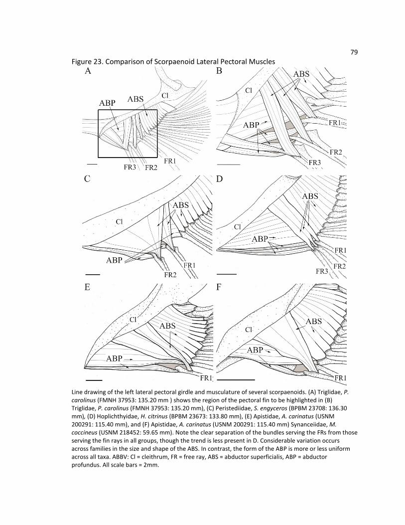
79
Figure 23. Comparison of Scorpaenoid Lateral Pectoral Muscles
Line drawing of the left lateral pectoral girdle and musculature of several scorpaenoids. (A) Triglidae, P. carolinus (FMNH 37953: 135.20 mm ) shows the region of the pectoral fin to be highlighted in (B) Triglidae, P. carolinus (FMNH 37953: 135.20 mm), (C) Peristediidae, S. engyceros (BPBM 23708: 136.30 mm), (D) Hoplichthyidae, H. citrinus (BPBM 23673: 133.80 mm), (E) Apistidae, A. carinatus (USNM 200291: 115.40 mm), and (F) Apistidae, A. carinatus (USNM 200291: 115.40 mm) Synanceiidae, M. coccineus (USNM 218452: 59.65 mm). Note the clear separation of the bundles serving the FRs from those serving the fin rays in all groups, though the trend is less present in D. Considerable variation occurs across families in the size and shape of the ABS. In contrast, the form of the ABP is more or less uniform across all taxa. ABBV: Cl = cleithrum, FR = free ray, ABS = abductor superficialis, ABP = abductor profundus. All scale bars = 2mm.
80
that its FR is served by a slender muscle bundle approximately half the size of the
bundles serving the fin rays.
Abductor Profundus (ABP)
The ABP is a large muscle that is located just medial to the ABS on the lateral side
of the pectoral girdle. It originates from the cleithrum and its fibers run posteriorly to
insert to the lateral hemitrichium. Figure 23 shows the origin and orientation of the ABP
in each taxa, though it is obscured in the Peristediidae by the large fan-shaped ABS. In
all taxa observed, the ABP bundles serving the FRs are well separated from those serving
the fin, similar to the condition of the ABS. Like the bundles serving from the fin, they
originate from near the anterior tip of the cleithrum, and can be seen running along the
ventral margin of the pectoral girdle to insert at the base of each FR. This pattern was
common to all taxa observed and no variations in the anatomy of this muscle were
noticed.
Arrector Ventralis (ARRV)
The ARRV originates from the dorsal-lateral part of the cleithrum, medial to the
ABS.It is composed of a single bundle which inserts to the medial hemitrichium of the
first (marginal) fin ray. In general, the ARRV is relatively small in the scorpaenoid species
examined in this study. The characteristics of this muscle were the same in all specimens
examined and no novel subdivisions or other interesting variations were noted. This
muscle is not associated with the free rays in any taxon in this study.

81
Adductor Superficialis (ADS)
The ADS is the most medial of the muscles originating from the pectoral girdle,
and is the first encountered in medial view (Fig. 24 A). The origin is from the dorsal part
of the medial face of the cleithrum, usually near the articulation with the
supracleithrum. The muscle fibers run vertically and insert to the shaft of the medial
hemitrichium either directly or via a small tendon. This bundles from this muscle insert
to all lepidotrichia, except the marginal ray. This muscle continues the pattern where
the bundles inserting to the FRs are well separated from each other and from the
bundles serving the fin. While several differences were noticed among the families
surveyed, no important variations were found at the genus or species level.
In Triglidae and Peristediidae, the ADS bundles inserting to the FRs are divided
into two distinct subunits, designated here as ADSα & ADSβ (Fig. 24 B &C). The ADSα is
similar to the form observed in the fin rays. It originates from the cleithrum, runs
ventrally to insert on the shaft of the medial hemitrichium, and is composed of fibers
without a noticeable tendon. By contrast, the ADSβ originates slightly posterior and
lateral to the ADSα. However, its fibers run diagonally across the medial side of the ADSα,
before fusing into a stout tendon. Insertion is to the apex of a large triangle-shaped
process that extends posteriorly from the medial hemitrichium (Fig. 25 A & B). Thus,
while the origin of the ADSβ is posterior to the ADSα, its insertion is slightly anterior.
Small variations are noticed between Triglidae and Peristediidae, in that peristediids
have a slightly smaller process associated with the ADSβ and the tendon is not as long

82
Figure 24. Comparison of Scorpaenoid Medial Musculature
Line Drawing of the left medial pectoral girdle and musculature of several scorpaenoid groups. (A) Triglidae, P. carolinus (FMNH 37959: 89.15 mm) shows the region of the pectoral fin to be highlighted in (B) Triglidae, P. carolinus (FMNH 37959: 89.15 mm), (C) Peristediidae, S. engyceros (BPBM 23708: 136.30 mm), (D) Hoplichthyidae, H. citrinus (BPBM 23673: 133.80 mm), (E) Apistidae, A. carinatus (USNM 200291: 115.40 mm), and (F) Synanceiidae, M. coccineus (USNM 218452: 59.65 mm). Notice the presence of multiple bundles of the ADS serving each FR in B and C. In D, E, and F, no evidence of subdivision of the ADS muscle is observed. ABBV: Cl = cleithrum, FR = free ray, ADS = adductor superficialis, ADP = adductor profundus. All scale bars = 2mm.

83
(Fig. 25 C & D). No subdivisions within the ADS muscle are observed in association with
the fin rays.
In Hoplichthyidae, no subdivisions of the muscle are detected. The each bundle
is roughly fan or wedge-shaped, with a wide origin (Fig. 24 D). The insertion is to the
shaft of the bone, via a small tendon. Fibers from the posterior part of the insertion fuse
into the tendon, whereas fibers from the anterior part insert pinnately onto the tendon
itself (Fig. 25 E & F). In some larger specimens, the insertion is associated with a very
small process, but more often, the muscle inserts directly to the shaft of the medial
hemitrichium.
In Apistidae, again, no subdivisions are noticed. The bundle serving the one FR
fuses into a single, long tendon for most of its length. The insertion is directly to the
shaft of the medial hemitrichium bone with no process in all specimens examined (Fig.
24 E).
In the Synanceiidae, the ADS muscle serving the FRs is a single unit without
subdivisions. The muscle is long and straplike, though fuses into a small tendon just
before its insertion (Fig. 24 F). It is dissimilar from Hoplichthyidae in that few if any of
the fibers insert pinnately onto the tendon itself, and instead they gradually unite
without changing their angle or orientation. In some cases, the insertion of this muscle
is to the apex of a small process. This process is similar in shape to that observed other
taxa, and larger than what is seen in Hoplichthyidae, but smaller than in Triglidae or
Peristediidae.

84
Figure 25. Comparison of the Subdivisions of the Adductor Superficialis
Panels showing a magnified view of the insertion of the ADS muscle with line drawings compared to photographs for (A & B) Triglidae, P. carolinus (FMNH37959: 89.15 mm & FMNH 37951: 95.10 mm ), (C & D) Peristediidae, S. engyceros (BPBM 23708: 136.30 mm), (E & F) Hoplichthyidae, H. citrinus (BPBM 23673: 133.80 mm & USNM 388708: 147.15 mm). A, B, C, and D all show subdivision of the ADS into fibrous ADSα bundles that insert to the lateral side of the medial hemitrichia, and more tendinous ADSβ bundles which insert to a raised process on the medial hemitrichia. By contrast, in E & F, the muscle fibers connect are pinnately arranged along the length of a tendon, resulting in a single point of insertion. ABBV: FR = free ray, ADS = adductor superficialis, ADP = adductor profundus. All scale bars = 2mm.

85
Adductor Profundus (ADP)
The ADP is a large muscle that is located lateral to the ADS. Its origin is from the
medial side of the anterior end of the cleithrum, often from within a shelf or ridge in the
bone, which is enclosed by a posterior-medial outgrowth of the distal cleithrum. Often,
some portion of this muscle can also be found originating from the corcoid or radial
bones, though these do not generally comprise a separate unit and fuse completely with
the remainder of the muscle. Muscle fibers run posteriorly to insert at the proximal
base of each medial hemitrichium.
Subdivisions of the ADP bundles inserting to the FRs were observed in all families
except Apistidae. These subdivisions are designated ADPα and ADPβ. The ADPα originates
from the cleithrum and posterior part of the coracoid. It is generally large and wedge-
shaped. It inserts to a modified posterior process from the proximal base of the medial
hemitrichium (Fig. 26). By contrast, the ADPβ is located lateral to the alpha subunit and
originates primarily from the coracoid, third radial, and fourth radial bones. Without
dissection, the ADPβ is usually obscured by the ADPα. It inserts to a modified portion of
the internal process of the medial hemitrichium.
In Triglidae & Peristediidae, the condition of both muscles is quite similar. In the
Peristediidae, the ADPα bundles are like a compact wedge (Fig. 26 C) with most fibers
originating from the same area. However, in Triglidae, the ADPα is sometimes seen as
two distinct sets of bundles that join at the insertion site, giving the impression of a fan
shape with its middle cut out (Fig. 26 B). When compared, these bundles appear to be
similar in form, except that the origin is spread over a wider area in Triglidae with the

86
Figure 26. Comparison of the Alpha Subunit of the Adductor Profundus
Line drawings showing the left medial pectoral girdle with the ADP partially dissected. (A) Triglidae, P. carolinus (FMNH 37959: 89.15 mm) shows the region of the pectoral fin to be highlighted in (B) Triglidae, P. carolinus (FMNH 37959: 89.15 mm), (C) Peristediidaem S. engyceros (BPBM 23708: 136.30 mm), (D) Hoplichthyidae, H. citrinus (BPBM 23673: 133.80 mm), (E) Apistidae, A. carinatus (USNM 200291: 115.40 mm), and (F) Synanceiidae, M. coccineus (USNM 218452: 59.65 mm). Except for E, all show subdivision of the ADP into two distinct units. The α bundles are more medially located and insert to the posterior process of the medial hemitrichium while the β bundles are more laterally located and insert to the internal process of the medial hemitrichium. ABBV: Cl= cleithrum, Cor = coracoid, FR = free ray, R = radial, ADS = adductor superficialis, ADP = adductor profundus, CR = coracoradialis. All Scale bars = 2 mm.

87
result that the fibers appear less dense and more separated. In both cases, the ADPα
bundles insert to a modified posterior process that extends in the proximal direction in
line with the long axis of the medial hemitrichium. For the ADPβ, the origin of the
bundles for the FRs are from the coracoid and radial 4 in Peristediidae (Fig. 27 C), with
the addition of radial 3 in Triglidae (Fig. 27 B). The insertion of this muscle is to the
internal process of the medial hemitrichium. Traditionally, the internal process is
embedded in the fibrocartilage plate that anchors each ray to the fin at large (Geerlink
1987), and the insertion site is surrounded by dense connective tissue (Fig. 28 A & B).
The process itself is small, usually no more than a small knob or ridge.
In Hoplichthyidae, all three FRs receive independent bundles of the ADP, but
only the bundle serving FR3 is subdivided into α and β subunits. The ADP bundles are
not as well separated as in Triglidae or Peristediidae, though they do form group distinct
from the ADP bundles serving the fin rays (Fig. 26 D). The insertion is to the posterior
process of the FRs. This process is less modified than is seen in Triglidae or Peristediidae
and extends in a somewhat more posterior than proximal direction. For FR3, the ADPα is
very similar to the ADP bundles in FR1 and FR2. The ADPβ originates mostly from radial
4, though sometimes from the coracoid as well. It inserts to a small internal process on
the medial hemitrichium of FR3 (Fig. 27 D). The insertion itself is partially tendinous, and
often covered in connective tissue (Fig. 28 C & D).
In the Apistidae, the ADP bundle inserting to the FR is not subdivided. This
bundle is well separated from the bundles of the main fin, and can be observed

88
Figure 27. Comparison of the Beta Subunit of the Adductor Profundus
Line drawings showing the left medial pectoral girdle with the ADP and ADPα partially dissected. (A) Triglidae, P. carolinus (FMNH 37959: 89.15 mm) shows the region of the pectoral fin to be highlighted in (B) Triglidae, P. carolinus (FMNH 37959: 89.15 mm), (C) Peristediidaem S. engyceros (BPBM 23708: 136.30 mm), (D) Hoplichthyidae, H. citrinus (BPBM 23673: 133.80 mm), (E) Apistidae, A. carinatus (USNM 200291: 115.40 mm), and (F) Synanceiidae, M. coccineus (USNM 218452: 59.65 mm). Note again the presence of a β subdivision of the ADP in all except E. Addditionally, note that D has the β subdivision only in connection with FR3, and not FR1 or FR2. ABBV: Cl= cleithrum, Cor = coracoid, FR = free ray, R = radial, ADP = adductor profundus, CR = coracoradialis. All Scale bars = 2 mm.

89
originating from the cleithrum and inserting to the FR (Fig. 26 E). The insertion is to a
somewhat reduced posterior process.
In Synanceiidae, the anatomy of the ADP muscle differs between the genera
examined here. The genus Minous has a single FR and Inimicus has two. In Minous, the
ADP bundle inserting on the FR is divided into two subunits. The ADPα is a large fan-
shaped muscle that originates from the coracoid, radial 3 and radial 4. Its insertion is to
a very large process that extends proximally along the axis of the bone, similar to the
state seen in Triglidae or Peristediidae (Fig. 26 F). The ADPβ is fan shaped and originates
from the corcoid, R3, and R4, lateral to the ADPα. This muscle inserts to a much enlarged
internal process that extends from the base of the way at a right angle (Fig. 27 F). The
muscle grades into a tendinous sheet near the insertion site, and unlike other taxa, it is
not usually obscured by the connective tissue that attaches the ray to the fibrocartilage
of the pectoral girdle (Fig. 28 E & F). In the second genus, Inimicus, the bundle to FR2 is
subdivided, but not the bundle inserting to FR1. In FR2, the ADPα and ADPβ are identical
to the condition seen in Minous. In FR1, the entire ADP bundle inserts to the posterior
process, which is angled towards the posterior direction (Fig. 30 F), as opposed to
extending along the long axis of the ray.
Arrector Dorsalis (ARRD)
The arrector dorsalis originates from the dorso-medial part of the cleithrum and
inserts to the medial hemitrichium of the first (marginal) fin ray. This muscle is generally
located lateral to the ADS and dorsal to the ADP. In most species, the separation

90
Figure 28. Comparison of the Subdivisions of the Adductor Profundus
Panels showing a magnified view of the ADP muscle and subdivisions with line drawing compared to photographs for(A & B) Peristediidae, S engyceros (BPBM 23708: 136.30 mm), (C & D) Hoplichthyidae, H citrinus (BPBM 23673: 133.80 mm), and (E & F) Synanceiidae, M. coccineus (USNM 218452: 59.65 mm). In the photos, the black arrow shows the α subdivision and the white arrow shows the β subdivision. ABBV: Cl= cleithrum, Cor = coracoid, FR = free ray, ADP = adductor profundus. All Scale bars = 2 mm.
between the ARRD and the ADP was poorly defined at the origin along the cleithrum.
Like the ARRV, no new subdivisions or other variations were noticed among the species
examined here, and this muscle is not associated with the FRs.

91
Osteology of Rays
Each ray or lepidotrichium is composed of two hemitrichia. Each hemitrichium is
composed of a solid basal segment that may have several processes for muscle insertion
and a flexible series of many small segments. Just as most of the variation in
musculature concerned the adductors on the medial side of the pectoral girdle, most of
the variation in osteology affects the medial hemitrichia. Traditionally, there are two
processes associated with the medial hemitrichium. The posterior process is the site of
attachment for the adductor profundus (ADP), while an internal process is the point of
articulation with the fibrocartilage plate that surrounds the radials (Geerlink 1989). It is
important to note that despite its name, this process points in the medial or slightly
anterior direction when the fin is at rest. In the scorpaenoid species examined here, this
process is posterior only once the fin has been abducted.
The lateral hemitrichium shows processes for both the ABS and ABP. Variation
appears to be localized to two areas. First is the amount of curvature of the proximal
head relative to the shaft of the bone near the insertion site of the ABS. Second is the
size of the shelf where the ABP inserts. In Triglidae, (Fig. 29A) the proximal head is fairly
curved and the shelf is relatively small compared to the other families in this study.
Peristediidae (Fig. 29 B) is similar in the first respect, but has a larger area for the
attachment of the ABP. Hoplichthyidae and Apistidae (Fig. 29 C & D) both have no or
very little curvature of the proximal head, but a large, wide shelf for the insertion of the
ABP. Last, Synanceiidae (Fig. 29 E) has both a strongly curved proximal head, and a
relatively large area for the ABP.

92
A much wider range of shapes and sizes is observed for the medial hemitrichium.
Moreover, the novel subdivisions of the ADS and ADP appear to be associated with
novel bone processes in some taxa. In other cases, particularly the ADPα, muscle
subdivisions are associated with significant alterations of the existing posterior process.
A novel subunit of the adductor superficialis (ADSβ) is found inserting to the FRs
in Triglidae and Peristediidae. The insertion of the ADSβ is always to the tip of a large
triangular process somewhat distal from the base of the medial hemitrichium (Fig. 29 A
& B). In these two families, the ADSα attaches to the side of this process and the main
shaft of bone, similar to the undivided ADS of other taxa. While a small process is
sometimes observed at the insertion of the ADS in Hoplichthyidae and Synanceiidae
(Fig. 29 C & E), it is always small in comparison to Triglidae or Peristediidae.
In contrast, subdivisions of the ADP bundles inserting to the FRs were observed
in four of the five families studied. In Triglidae, Peristediidae, and Synanceiidae, the
ADPα inserts to a heavily modified version of the posterior process. This process has
been rotated about 90° from its usual position until it is roughly in line with the long axis
of the medial hemitrichium. Within these three families, Triglidae can be distinguished
as having a thinner and sometimes curved posterior process, as opposed to the wider
and straight shapes seen in Peristediidae and Synanceiidae. A more traditional
positioning of this process is observed in Hoplichthyidae, where the posterior process is
not in line with the main shaft of the ray (Fig. 29 C). The posterior process in Apistidae is
very small, and the process itself appears to be directed in the opposite position (Fig. 29
D).

93
Figure 29. Comparison of the Free Ray Bases in Scorpaenoids
Panels showing the proximal head of cleared and stained FR lepidotrichia from (A) Triglidae, P. scitulus (KU 18065: 62.20 mm), (B) Peristediidae, P. greyae (FMNH 45638: 114.00 mm), (C) Hoplicthyidae, H. citrinus (USNM 388705: 147.15 mm), (D) Apistidae, A. carinatus (USNM 200291: 98.20 mm), (E) Synacneiidae, M. pictus (USNM 272492: 110.85 mm). In all cases, the lateral hemitrichium is shown above the medial hemitrichium. Black arrows indicate α subdivisions of the ADS and ADP while white arrows indicate the β subdivisions. Note the presence of large processes in association with the ADSβ in A & B, vs. the small or absent process associated with the ADS in C, D, and E. Similarly, the presence of an ADPβ subdivision is often associated with a process in A, B, C, & E. ABBV: ADS = adductor superficialis, ADP = adductor profundus.
For the ADPβ, many of the same patterns are observed. Again, Triglidae and
Peristediidae are very similar in the shape of the small internal process (Fig. 29 A & B). In
94
both these groups, the internal process is a small knob of bone which receives the
insertion of the ADPβ as well as articulates with the fibrocartilage around the radials. In
Hoplichthyidae, the internal process is a rounded knob that similarly receives both the
insertion and articulates with the fibrocartilage (Fig. 29 C). In the Synanceiidae, the
internal process is greatly enlarged, and the articulation and insertion points are
separated by some distance. The articulation with the fibrocartilage pad is at the base of
the large process, whereas the ADPβ muscle inserts to the apex and along its proximal
side (Fig. 29 E). The ADSβ is not found in Apistidae, and almost no internal process is
observed (Fig. 29 D). Instead, the bone seems to articulate into the cartilage of the
radials directly from the base of the medial hemitrichium.
In some taxa, the osteology of the FRs bears little resemblance to that of the fin
rays. In Triglidae and Peristediidae, the FRs appear to follow one very distinct pattern,
while the fin rays display a different structure (Fig. 30 A & B). Notably, the fin rays lack
the triangular process associated with ADSβ, and the posterior and internal processes at
the base of the hemitrichium are much reduced in size by comparison, giving the overall
impression of a less complex shape. By contrast, in Hoplichthyidae, the medial
hemitrichia of the FRs have a wider posterior process, but are otherwise similar in shape
to the medial hemitrichia of the fin rays (Fig. 30 C). Although no bundle of the ADPβ is
found on the posterior FRs, FR1 or FR2, the overall shape of the FRs is rather consistent.
In Apistidae, the proximal head of the FR is relatively simplified compared to the fin ray
(fig. 30 D). While the fin ray contains recognizable posterior and internal processes, the
FR appears to have these features reduced. In Synanceiidae, the amount of variation is

95
Figure 30. Comparison of the Free and Fin Ray Bases
Panels showing a spread of all FRs for each family vs. a representative fin ray in (A) Triglidae, P. scitulus (KU 18065: 62.20 mm), (B) Peristediidae, P. ecuadorensis (FMNH 66521: 109.35 mm), (C) Hoplichthyidae, H. citrinus (USNM 388705: 147.15 mm), (D) Apistidae, A. carinatus (USNM 200291: 98.20 mm), (E) Synanceiidae, M. pictus (USNM 272492: 110.85 mm), (F) Synanceiidae, I. didactylus (USNM 136458: 80.40 mm). The FRs may be similar to eachother but not the fin ray as in A, B, and E, or present a gradual transition as in C and F. Note that in F, FR2 receives a bundle of the ADPβ muscle, but FR1 does not. ABBV: FR = free rays, Fn = Fin Ray.
dependent on the genus being observed. In Minous, which has a single FR, the
differences between the FR and the fin rays are quite pronounced. The angle of the
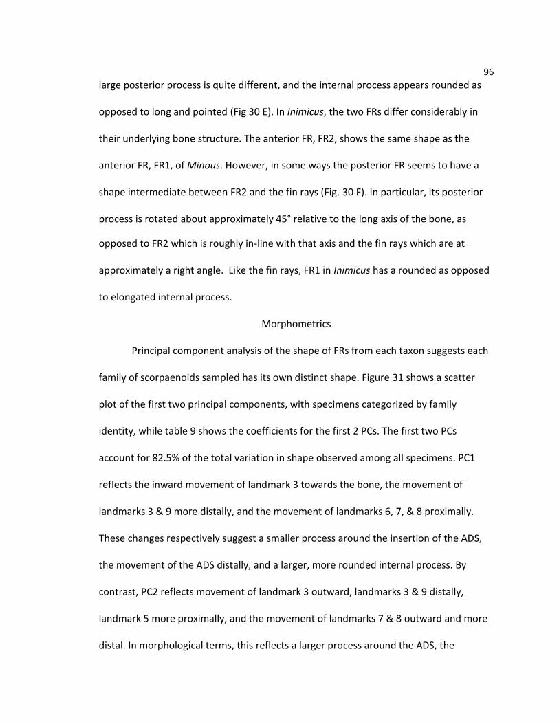
96
large posterior process is quite different, and the internal process appears rounded as
opposed to long and pointed (Fig 30 E). In Inimicus, the two FRs differ considerably in
their underlying bone structure. The anterior FR, FR2, shows the same shape as the
anterior FR, FR1, of Minous. However, in some ways the posterior FR seems to have a
shape intermediate between FR2 and the fin rays (Fig. 30 F). In particular, its posterior
process is rotated about approximately 45° relative to the long axis of the bone, as
opposed to FR2 which is roughly in-line with that axis and the fin rays which are at
approximately a right angle. Like the fin rays, FR1 in Inimicus has a rounded as opposed
to elongated internal process.
Morphometrics
Principal component analysis of the shape of FRs from each taxon suggests each
family of scorpaenoids sampled has its own distinct shape. Figure 31 shows a scatter
plot of the first two principal components, with specimens categorized by family
identity, while table 9 shows the coefficients for the first 2 PCs. The first two PCs
account for 82.5% of the total variation in shape observed among all specimens. PC1
reflects the inward movement of landmark 3 towards the bone, the movement of
landmarks 3 & 9 more distally, and the movement of landmarks 6, 7, & 8 proximally.
These changes respectively suggest a smaller process around the insertion of the ADS,
the movement of the ADS distally, and a larger, more rounded internal process. By
contrast, PC2 reflects movement of landmark 3 outward, landmarks 3 & 9 distally,
landmark 5 more proximally, and the movement of landmarks 7 & 8 outward and more
distal. In morphological terms, this reflects a larger process around the ADS, the

97
Table 9. Principle Component Loadings for Scorpaenoid Free Rays
Component PC1 PC2 Component PC1 PC2
x1 0.003887 -0.3932 x6 -0.08547 -0.05277
y1 -0.30765 0.114158 y6 -0.02124 0.042005
x2 -0.08355 -0.33612 x7 -0.3211 0.137423
y2 -0.2182 -0.05438 y7 0.128286 0.144439
x3 0.229134 0.487853 x8 -0.23681 0.186184
y3 0.637656 -0.30622 y8 0.044685 0.044408
x4 0.110605 -0.06045 x9 0.391958 0.395433
y4 -0.08895 -0.07141 y9 0.005311 0.011344
x5 -0.00865 -0.36435
y5 -0.17991 0.075647 Principal component coefficients for the first two PCs. The first 2 principal components represent 82.5 % of the total variation in shape.
placement of the process more distally, the elongation of the posterior process, and a
more distally located, protruding internal process.
Two families, the Triglidae and Peristediidae appear closer according to PCA than
any of the other groups (Fig. 31). Together, they are distinguished by low scores along
PC1, and widely variable scores on PC2. Together, this suggests a large, but somewhat
variable process around the insertion of the ADS, and the more proximal insertion of
this muscle. The variability around PC2 suggests that the size and shape of the internal
and posterior processes may vary by genus or species. By contrast, the Synanceiidae and
Hoplichthyidae both score highly along PC1, suggesting a small process around the ADS,
a more distal insertion of the ADS, and a slightly larger internal process. However they
differ with respect to PC2. Hoplichthyidae scores very low on PC2, which reflects an
even smaller process around the ADS, a small posterior process, and a very proximally
located internal process. Synanceiidae has high scores on both PC1 and 2, which
suggests a small to moderate process around the ADS, a very distally located insertion of
98
Figure 31. Principal Component Analysis of Scorpaenoid Families
Scatterplot of PCA scores from the geometric morphometric analysis. Ellipses represent a 90% equal frequency confidence interval for each group. Note that each family appears to occupy a distinct region of the component space, though Triglidae and Peristediidae are more similar than any other pair of groups.
the ADS, a somewhat large posterior process, and a strong increase in the size of the
internal process. Although, the Apistidae was included in this analysis, the small number
of samples limits any ability to draw conclusions about this family. This analysis strongly
suggests that there are meaningful, systematic differences in the shapes of
thehhemitrichia in each family.

99
Figure 32. Deformation Grids for Scorpaenoid Families
Panel showing the average shape of each family as a deformation from the overall average of all scorpaenoids in this study. A) Shows the overall average of all scorpaenoids in red, and red lines represent deformations from this average in B) Triglidae, C) Peristediidae, D) Hoplichthyidae, E) Apistidae, and F) Synanceiidae.
As an alternate method of describing these changes, an average shape was
calculated for each family from the Procrustes coordinates (Fig. 32 B-F). The average
shapes of the five individual families were then averaged again to produce a broad
composite of the average shape of a free ray hemitrichium (Fig. 32 A).These shapes can
then be compared to give a description of the shape change that characterizes each
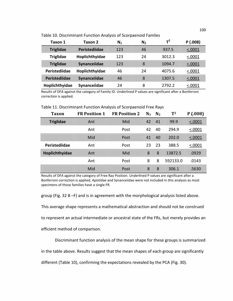
100
Table 10. Discriminant Function Analysis of Scorpaenoid Famlies
Results of DFA against the category of Family ID. Underlined P values are significant after a Bonferroni correction is applied.
Table 11. Discriminant Function Analysis of Scorpaenoid Free Rays
Taxon FR Position 1 FR Position 2 N1 N2 T2 P (.008)
Triglidae Ant Mid 42 41 99.9 <.0001
Ant Post 42 40 294.9 <.0001
Mid Post 41 40 202.0 <.0001
Peristediidae Ant Post 23 23 388.5 <.0001
Hoplichthyidae Ant Mid 8 8 13872.5 .0929
Ant Post 8 8 592133.0 .0143
Mid Post 8 8 306.1 .5630
Results of DFA against the category of Free Ray Position. Underlined P values are significant after a Bonferroni correction is applied. Apistidae and Synanceiidae were not included in this analysis as most specimens of those families have a single FR.
group (Fig. 32 B –F) and is in agreement with the morphological analysis listed above.
This average shape represents a mathematical abstraction and should not be construed
to represent an actual intermediate or ancestral state of the FRs, but merely provides an
efficient method of comparison.
Discriminant function analysis of the mean shape for these groups is summarized
in the table above. Results suggest that the mean shapes of each group are significantly
different (Table 10), confirming the expectations revealed by the PCA (Fig. 30).
Taxon 1 Taxon 2 N1 N2 T2 P (.008)
Triglidae Peristediidae 123 46 937.5 <.0001
Triglidae Hoplichthyidae 123 24 3012.3 <.0001
Triglidae Synanceiidae 123 8 1094.7 <.0001
Peristediidae Hoplichthyidae 46 24 4075.6 <.0001
Peristediidae Synanceiidae 46 8 1307.5 <.0001
Hoplichthyidae Synanceiidae 24 8 2792.2 <.0001

101
Additionally, the hypothesis of differences based on FR position is also tested for
Triglidae, Peristediidae, and Hoplichthyidae. These results are summarized in Table 11.
Interestingly, these results show that even within a given family, the FRs are not
identical in shape. Significant shape differences were found between the anterior and
posterior FRs in Triglidae and Peristediidae. For Hoplichthyidae, the tests suggests a
marginal difference between the anterior and posterior FRs, though not any other
combinations.
Discussion
Variation in Free Ray Morphology
This study is the first to examine the morphology of the FRs that includes
representatives of all five scorpaenoid families. From previous work in triglids, the FRs
are involved in a variety of behaviors such as locomotion and sensing prey, and have
three main characteristics. First, they are separate from the main fin and can move
independently of it. Second, they have a larger range of motion. Third, the distal ends of
FRs can be strongly flexed. In Triglidae, these abilities are thought to be mediated in part
by novel subdivisions of the adductor superficialis (ADS) and adductor profundus (ADP).
Furthermore, these muscles insert to processes on medial hemitrichium that appear to
be modified from their traditional form. A very similar pattern is observed in the family
Peristediidae. This group possesses all of the same muscle subdivisions and processes as
triglids, with only minor variations separating them. This is also evidenced by their
overall proximity in PCA using landmark data. Although Hoplichthyidae and
Synanceiidae appear to have these same capabilities, their underlying structure is quite

102
different. Neither of these groups shows the subdivision of the ADS muscle or the
enlarged process found in searobins. However, they do share a novel subdivision of the
ADP muscle, though in these families, it serves only the most anterior FR. The
stonefishes are further distinguished by a large internal process that is the insertion site
for the ADPβ. Lastly, the Apistidae appear to represent a simplified version of the FRs.
They lack both derived muscle subdivisions as well as any extensive modifications to the
bone structure of the hemitrichia.
This study was not able to correlate morphological observations to the behavior
or function of live specimens. Thus the exact function of the novel muscle subdivisions is
unknown. However, comparative observations of the five families under study can
provide some hints. For instance, the ADPβ is found in all the specimens examined
except Apistidae, a pattern that may suggest a function involving the distal bending of
the ray. Apistidae is the only scorpaenoid family that possesses FRs, but does not seem
to be able to distally flex them. By contrast, the other four families possess this muscle
subdivision and the ability to distally flex their rays. This correlation would seem to
suggest that the ADP is in some way responsible for this flexing behavior. However, this
picture is complicated by the condition of the posterior FRs in Hoplichthyidae and
Synanceiidae, which lack a bundle of the ADPβ but are able to flex. It should be noted
that all lepidotrichia are capable of some flexion, so the issue is one of degree. It is
possible that the ADPβ allows for increased or better controlled of flexion. The extent of
bending is difficult to quantify in preserved specimens, so this hypothesis would best be

103
tested by studying individual FR movements of a living specimen of Hoplichthys or
Inimicus. Until then, the purpose of the ADPβ cannot be stated with any confidence.
Another interesting pattern arises at the insertion of the ADPα on the posterior
process. Triglidae, Peristediidae, and Synanceiidae all have their posterior process
rotated in line with the long axis of the ray, as opposed to at a right angle as is observed
in most fin rays. This state would be convergent according to existing phylogenetic
hypotheses, which suggests a possible shared functional role. One interesting possibility
is that this modification is related to the increased range of motion observed in the FRs,
and perhaps provides a more favorable angle for pulling the abducted FRs back towards
the body. Because the FRs rotate through a large arc of almost 180°, no single angle of
insertion will allow a muscle to be effective at all points in a movement. Similarly, the
purpose of the ADSb is something of a mystery. This subdivision is only found in Triglidae
and Peristediidae. The prominent size of the process to which it attaches suggests that it
exerts considerable leverage on the ray. On the other hand, its absence in other groups
may indicate that it is not essential to the basic characteristics of FRs. Additional study
and a better understanding of ray adduction in general will be necessary to fill in the
gaps in this picture.
The variations in FR morphology may underlie potential differences in their
ecology and function. Given that the FRs are most likely evolved from fin rays, one
explanation for the structural diversity is that they have become specialized in different
directions. For instance, if the medial hemitrichia does not reflect real differences in
performance, it would be surprising to find that evolution has maintained such distinct

104
shapes associated with each family. Although its function is unknown, in Triglidae and
Peristediidae, the large process of the ADSβ and smaller size of the internal process
connected with ADPβ, may indicate the relative importance of a particular type of
substrate interaction. Triglids are known to probe the substrate for chemical cues of
their prey (Bardach & Case, 1965), so perhaps this combination is somehow beneficial to
this particular behavior. On other hand, Synanceiidae have the opposite condition, a
large internal process and a small or absent process for ADS. Possibly this reflects the
relative importance of the pectoral fin and FRs to providing force during attacks and
feeding (Grobecker 1983). The picture is less clear in Hoplichthyidae. Here the FRs do
not differ as much from the fin rays, and thus may represent something of an
intermediate or perhaps they support some unobserved behavior that differs from the
other groups. Interestingly, Apistidae, with its seemingly simplified FR, may actually
reflect a loss of some functions. Observations suggest that it is not used in either
swimming locomotion or substrate interactions. Hence, this simplicity may be consistent
with its use primarily as a prop for luring or corralling prey, which requires merely basic
abduction and adduction.
Overall, this study demonstrates the rich variety of ways in which the pectoral fin
lepidotrichia have been modified in the Scorpaenoidea. As the fine details of fin
lepidotrichia and musculature are not frequently studied, many of these modifications
have not been reported before now. This should be of particular interest to
biomechanical studies, which have tended to ignore benthic species, and have instead
focused on pelagic swimmers (Geerlink 1987, Alben et al 2006). These pelagic fishes

105
tend to have symmetric fins specialized to a single function. This work joins a growing
body of evidence that demonstrates increased asymmetry and the localization of
multiple functions to specific regions in the fins of benthic fishes (Taft 2009, 2011). A
more complete model of fin structure and function will require the increased inclusion
of benthic species in biomechanical studies. The FRs described in this study would be
useful as a sort of extreme case to test the limits of any model of ray bending and
pectoral fin muscle recruitment.
Finally, while the goal of this study was not to find systematic characters, the
wealth of family specific information gleaned from studying FRs deserves attention.
However, there is an immediate challenge to be overcome before these data can be
applied as systematic characters. Thus far, no detailed study of the homology of
individual fin rays has been attempted, and the standard method of ordering fin
elements may be inadequate. The traditional numbering system for pectoral fin rays
begins with the marginal ray. In most fishes, this ray bears a number of distinct features
that provide a stronger basis for homology, including the arrector muscles. This pattern
also reflects a developmental pattern with the generally more proximal elements of the
pectoral girdle forming first, and more distal fin rays appearing in a serial fashion (Davis
et al 2004). However, the total number of pectoral fin rays is usually too variable to
make detailed investigation of the homology of the more distal rays worthwhile.
In the scorpaenoids included here, the distal rays are morphologically distinct.
The tendency of the most distal ray to be more modified may suggest that these rays
should be numbered in the opposite order, with number counted in the opposite

106
direction. The synanceiids illustrate this problem quite clearly. In both Inimicus and
Minous, the anterior-most FR is modified with a novel muscle subdivision and large
internal process. As such, there is ground to propose that these modifications of the
anterior free ray are homologous in these taxa. However, by the traditional numbering
scheme, in Minous this is the 11th pectoral fin ray (Fn11) and the first free ray (FR1), but
in Inimicus it is the 12th pectoral fin ray (Fn12) and FR2. A similar pattern of distal
modifications to the pectoral fin rays has been observed in other studies of benthic
fishes (Taft 2011, Brandstätter et al 1990). The apparent localization of different
functions to different sub-regions within the pectoral fin implies that each sub-region is
under differential genetic control and may possess a distinct evolutionary history.
Future studies that utilize pectoral fin rays as more than a meristic character should
develop a numbering system that reflects this potential reality.
A second conundrum arises with respect to the pattern of gains and losses of FRs
within clades. Of particular note is the family Peristediidae. Species in Peristediidae
possess two FRs, but this family’s close relatives, Triglidae and Hoplichthyidae, possess
three distinct FRs. The question becomes whether FRs are added from the proximal or
distal end? This study cannot provide a full answer, but in Hoplichthyidae, the similarity
of FR3 with the fin rays (Fig 30 C) suggests the presence of a morphological gradient
with one extreme located at the distal most end. A similar circumstance is observed in
the more distant synanceiid genus Inimicus (Fig. 30F). Thus, the formation of free rays
would seem to proceed in a distal to proximal direction, with changes occurring at the
interface between the modified FRs and the main pectoral fin. Therefore, the presence

107
of two vs. three FRs in Peristediidae vs. Triglidae or Hoplichthyidae reflects a change in
the state along this intermediate boundary. This hypothesis could be tested in a number
of ways. An expanded morphometric study incorporating both fin and free rays may
reveal further patterns. Another promising line of evidence may lay in the nervous
innervation of the free rays. Triglidae is well known for a set of unique neuroanatomical
traits (Morill 1895, Finger 2000), and the FRs are innervated by unusually large segments
of the third spinal nerve. These nerves are generally large enough to be observed
without special preparation or particularly careful dissection. By contrast, other taxa
have much smaller nervous bundles in association with their free rays. A comparative
study of this trait could provide a parallel line of evidence for use in evaluating the
homology of pectoral fin rays.
Despite the need for additional work in the above areas, these data tend to
support a close link between the Triglidae and Peristediidae, based on both myological
and osteological findings. This is consistent with somewhat older views of scorpaenoid
phylogeny (Washington et al. 1984) as well as recent molecular studies (Smith &
Wheeler 2004). The FR lepidotrichia appear to be both very distinct and highly
specialized. Especially in benthic taxa which show numerous specializations to the fins,
this combination could prove highly useful to systematists. This project hints that
significant phylogenetic data may be hidden within the structure of teleost fin
lepidotrichia. Time and further study will be needed to determine if this is true of all
taxa, benthic taxa, or just a subset of scorpaenoids.

108
CHAPTER FOUR
SUMMARY, REMARKS, AND CONCLUSIONS
Recapitulation
This study is the first to date that examines the structure of the pectoral fin in all
five scorpaenoid families with free rays. This trait appears to have evolved multiple
times within the primarily benthic Scorpaenoidea in groups including the searobins
(Triglidae), armored searobins (Peristediidae), ghost flatheads (Hoplichthyidae),
waspfishes (Apistidae), and stonefishes (Synanceiidae). Free rays are a set of
lepidotrichia located on the pectoral girdle that are morphologically and functionally
separate from the fin itself. Particularly, they are able to act independently, have a
greater range of motion, and increased ability to bend distally. Most species use their
FRs for behaviors involving contact with the substrate such as digging, sensing buried
prey, and locomotion. Investigations of the morphology of this structure found novel
subdivisions of the adductor musculature and modifications to the shape of the FR
hemitrichia. Each family examined displayed its own unique suite of modifications,
consistent with the multiple proposed functions and different evolutionary origins.
These differences were statistically significant when measured using geometric
morphometrics. Much remains to be understood about the FRs, but this research

109
provides a foundation by which the behavioral, mechanical, and phylogenetic
significance of this trait can be investigated.
Patterns in Free Ray Musculature
The most prevalent pattern observed in relation to the FRs is the separation of the fin
musculature and FR musculature. The muscle bundles that serve each ray of the fin
generally appear to grade together into a common origin and are separate only at the
point of insertion. By contrast, the bundles of the pectoral musculature that serve the
FRs are clearly separate at both the origin and insertion. Moreover, these bundles are
separate not just from the fin, but from each other. For example, triglids, with three FRs,
have four independent bundles of the ABS muscle: one large muscle mass that consists
of the united bundles of all the fin rays and three separate masses each serving an
individual FR. This trend was found in all specimens examined and affected all of the
pectoral muscles serving the FRs, though it was somewhat less evident in the abductor
superficialis (ABS) and adductor superficialis (ADS) in Hoplichthyidae. This trend fits one
of the defining characteristics of the FRs, namely their ability to operate independently
of the main fin. This may also be important in allowing for their increased range of
motion.
A second pattern is the subdivision of the adductor musculature on the medial
side of the pectoral girdle. Of the five families examined, four possessed distinct
subdivisions of the adductor profundus (ADP) and two had subdivisions of the adductor
superficialis (ADS) as well. The distribution of novel muscle subdivisions is summarized
in Table 12. In contrast, no subdivisions were noted in the lateral, abductor
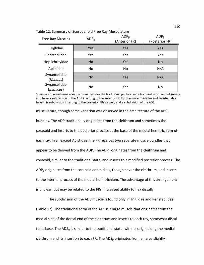
110 Table 12. Summary of Scorpaenoid Free Ray Musculature
Free Ray Muscles ADSβ ADPβ
(Anterior FR) ADPβ
(Posterior FR)
Triglidae Yes Yes Yes
Peristediidae Yes Yes Yes
Hoplichthyidae No Yes No
Apistidae No No N/A
Synanceiidae (Minous)
No Yes N/A
Synanceiidae (Inimicus)
No Yes No
Summary of novel muscle subdivisions. Besides the traditional pectoral muscles, most scorpaenoid groups also have a subdivision of the ADP inserting to the anterior FR. Furthermore, Triglidae and Peristediidae have this subdivision inserting to the posterior FRs as well, and a subdivision of the ADS.
musculature, though some variation was observed in the architecture of the ABS
bundles. The ADP traditionally originates from the cleithrum and sometimes the
coracoid and inserts to the posterior process at the base of the medial hemitrichium of
each ray. In all except Apistidae, the FR receives two separate muscle bundles that
appear to be derived from the ADP. The ADPα originates from the cleithrum and
coracoid, similar to the traditional state, and inserts to a modified posterior process. The
ADPβ originates from the coracoid and radials, though never the cleithrum, and inserts
to the internal process of the medial hemitrichium. The advantage of this arrangement
is unclear, but may be related to the FRs’ increased ability to flex distally.
The subdivision of the ADS muscle is found only in Triglidae and Peristediidae
(Table 12). The traditional form of the ADS is a large muscle that originates from the
medial side of the dorsal end of the cleithrum and inserts to each ray, somewhat distal
to its base. The ADSα is similar to the traditional state, with its origin along the medial
cleithrum and its insertion to each FR. The ADSβ originates from an area slightly

111 posterior and medial to the ADSα, and it inserts to a prominent process or ridge on each
FR, adjacent to the insertion of the ADSα. Additionally, the ADSβ fuses into a long tendon
that comprises approximately 40 - 50% of the muscle’s total length in Triglidae, or
approximately 25% in Peristediidae. The function of the muscle is unknown, but when
compared to Hoplichthyidae, an interesting pattern emerges. In Hoplichthyidae, the
posterior part of the ADS muscle fuses into a small tendon near the insertion point, with
nearby fibers attached pinnately to the tendon (Fig. 33 A & B). Thus, one sees a single
muscle bundle consisting of both a tendon and fibers, with no bony process at the
insertion site. In Peristediidae, the ADS is divided into two bundles, one with a tendon
which inserts to a process and one that is fibrous and inserts to the shaft of the
hemitrichium (Fig. 33 C & D). Last, in Triglidae, the ADS is again divided into two, but the
tendon of the ADSβ is even longer and the process at its insertion is even larger (Fig. 33 E
& F), while the ADSα is unchanged from the Peristediidae. These taxa are thought to be
closely related, with each group separated by a few minor changes. This raises the
possibility that the muscle arrangement in Hoplichthyidae represents the pleisiomorphic
state of the ADS muscle in this clade, with modifications occurring in the Peristediidae
and continuing in Triglidae.
Osteology and Morphometrics
Significant variation was also discovered in the shape of the proximal head of the medial
hemitrich bones. Of particular interest is that shape variations seem to group according
to the existence of derived muscle subdivisions. In Triglidae and Peristediidae, a large
ridge or process in found at the site of the insertion of the ADSβ. By contrast, in other
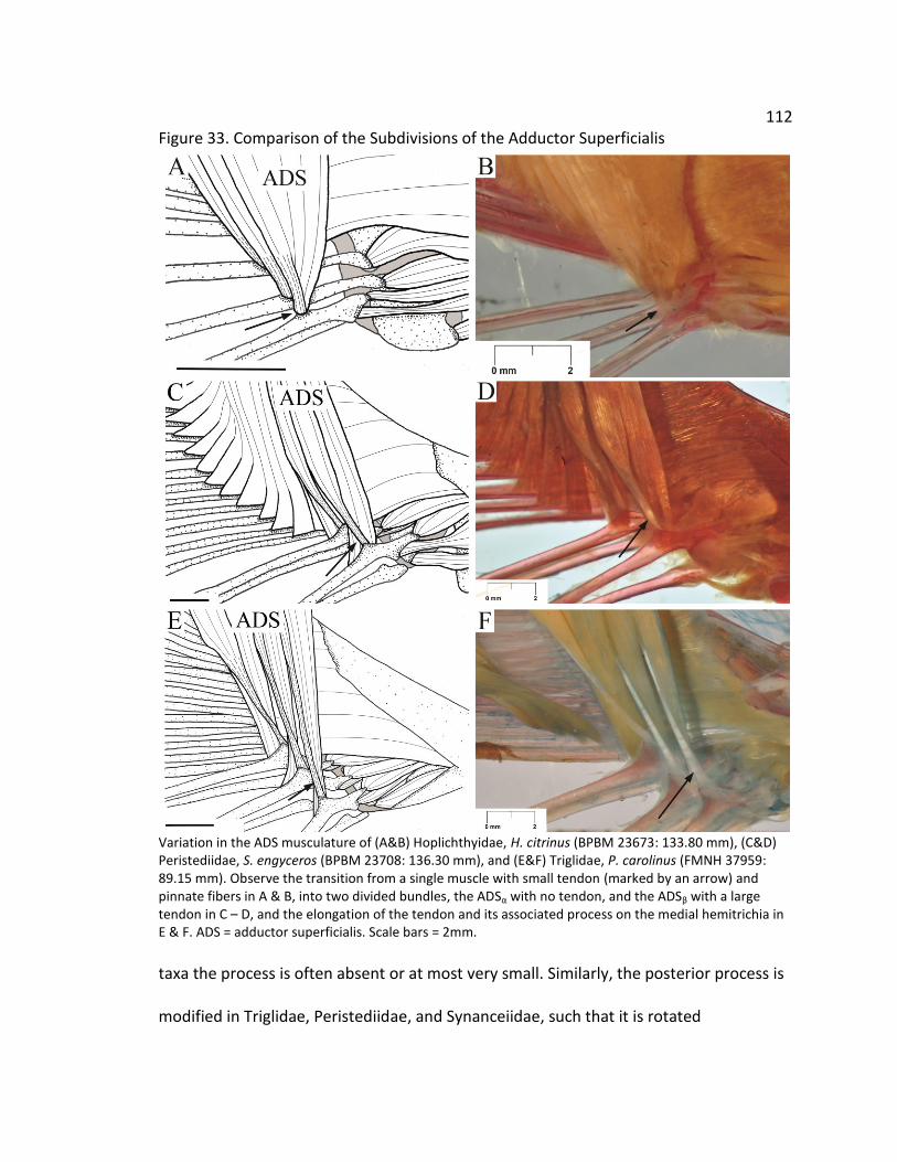
112 Figure 33. Comparison of the Subdivisions of the Adductor Superficialis
Variation in the ADS musculature of (A&B) Hoplichthyidae, H. citrinus (BPBM 23673: 133.80 mm), (C&D) Peristediidae, S. engyceros (BPBM 23708: 136.30 mm), and (E&F) Triglidae, P. carolinus (FMNH 37959: 89.15 mm). Observe the transition from a single muscle with small tendon (marked by an arrow) and pinnate fibers in A & B, into two divided bundles, the ADSα with no tendon, and the ADSβ with a large tendon in C – D, and the elongation of the tendon and its associated process on the medial hemitrichia in E & F. ADS = adductor superficialis. Scale bars = 2mm.
taxa the process is often absent or at most very small. Similarly, the posterior process is
modified in Triglidae, Peristediidae, and Synanceiidae, such that it is rotated

113 approximately 90° from its usual position. Lastly, in taxa with a subdivided ADP, the
ADPβ inserts to the internal process, which is not usually involved in muscle attachment.
This switch is most pronounced in the Synanceiidae, where the process is dramatically
enlarged. Given the prevalence of these changes, it seems reasonable to postulate that
they important to the function of the FRs, though the specifics have yet to be
determined. Another interesting finding is that the shapes of the medial hemitrichia are
family specific, and can be distinguished using geometric morphometrics. Tests of
average shape revealed that Triglidae, Peristediidae, Synanceiidae, and Hoplichthyidae
are each significantly different. Geometric morphometric studies are capable of
quantifying small changes in bone shape. In this study, the differences were quite large
and easily detected. This is significant in that it suggests that the FR hemitrichia have
been an important component in the evolution of this trait. Ray hemitrichia are largely
two dimensional and have several easily identified points that can be used as landmarks.
This makes them well suited to this type of analysis. If the same level of variation found
in scorpaenoids is present in other groups, then landmark based analysis of the ray
hemitrichia may provide an excellent method of comparing pectoral fin morphology for
teleost fishes in general.
Behavior
An improved understanding of pectoral fin rays will improve our understanding of the
ecology and behavior of scorpaenoids as a whole. Several studies have already shown
that the FRs are important to the behavior of triglids. These rays are covered in dense
chemosensory cells that allow searobins to detect prey buried in sandy substrates.

114 Searobins have also been observed using their FRs to “walk” across the bottom and dig
through the substrate, although some authors have debated the extent to which the FRs
may provide the impetus for actual locomotion. This study found that searobins possess
unique subdivisions of the adductor superficialis (ADS) and the adductor profundus
(ADP). Additionally, each of these subdivisions was accompanied by extensive
modification of the bone structure of the medial hemitrichia. While the exact purpose of
these additional structures is unclear, it seems consistent with a more complex set of
movements than is usually observed in the pectoral fin itself. However, there is one line
of comparative evidence not listed in earlier chapters that might bear upon this
question. Peristediidae and Triglidae share the same set of derived muscle bundles and
very similar hemitrichia shapes, which suggests relatively similar biomechanical function
of the FRs. However, species in Triglidae have long been known to possess an additional
specialization of the nervous system. Tiedemann (1816) first noticed that searobins have
an enlarged set of spinal nerves serving the FRs as well as a set of “accessory spinal
lobes” on the dorsal side of the spinal cord that correspond to each FR. Later work by
Finger (2000) confirmed that these lobes are responsible for conveying sensory input to
the rest of the brain. This is the first study to note that this same set of neuroanatomical
adaptations is not found in Peristediidae (Fig. 34 B), even though they appear to share
all the same osteological and myological characteristics as Triglidae (Fig. 34 A). Similarly,
this trait is not found in Hoplichthyidae or Apistidae either, though it could not be
assessed in the Synanceiidae due to specimen restrictions. Together, this may suggest
that the sensory function that is well documented in Triglidae may be a derived feature,

115 Figure 34. Accessory Spinal Lobes in Triglidae and Peristediidae
Dorsal view of the rostal spinal cord of (A) Triglidae, E. gurnardus (FMNH 33174: 163.25), and (B) Peristediidae, P. ecuadorensis (FMNH 66521: 141.70 mm). Although both have FRs, only species in Triglidae possess accessory spinal lobes. A shows a series of one minor and 3 major accessory lobes, whereas B shows none. Note that the 3
rd major accessory lobe may appear to be divided in two, but this is
a small furrow caused by a blood vessel (Finger 2000). ABBV: Cb = cerebellum, TecO = Optic tectum, ALM = Major accessory lobe, ALm = Minor accessory lobe.
and that the basal function of the FRs is in locomotion. A definitive test would look at
the histology of the FR epithelium in Peristediidae and Hoplichthyidae to test for the
presence of solitary chemoreceptor cells, the end organ for the chemosense in triglids.
By contrast, the free rays in Synanceiidae are believed to have a locomotive
function but no sensory function. One study from the reef stonefish, Synanceia
verrucosa, a related species with no FRs, still found that the pectoral fins were
important to positioning the body prior to strikes on food (Grobecker 1983). This
stonefish can strike with great speed, but is relatively inflexible and can hit only a
narrow sector directly above its mouth. This study found that the pectoral fins adjust
the tilt of the overall head and body, thus essentially targeting the highly protrusible
mouth. Aquarists and researchers have reported that synanceiids like Inimicus and
Choridactylus use their FRs in a similar manner, propping themselves up during feeding

116 or slowly shuffling across the bottom (Eschmeyer et al 1979, Gosline 1994). These fish
are largely piscivorous and obtain their prey from above, rather than below.
Ray Bending and Function
Although the presence of segmented fin lepidotrichia is common to all actinopterygians,
the properties of these fin rays have received less attention than other aspects of
movement such as feeding or swimming. Each lepidotrichium consists of two
hemitrichia, which I turn can be divided into a proximal solid head segment and a
flexible series of distal segments. Between the two halves is a dense collection of
collagen and other connective tissue binding them together. An interesting
consequence of this arrangement is that control of the flexible segments can be
achieved through relative displacement of the two proximal bases. Thus, muscles
inserting only to the base can nonetheless influence bending across the entire length of
the structure. Several studies of swimming in actinopterygians have found that active
control of bending in the normally flexible rays is required in order to efficiently push
against the water (Geerlink 1979, Alben et al 2007).
Some of the characteristics observed in the FRs have been associated with
increased bending force in mathematical models. Under a variety of conditions,
unbranched rays with fused hemitrichia are expected to increase the force produced by
ray bending (Alben et al 2007). Another factor concerns the ability of a ray to resist
bending, which is likely important in perching and locomotive behaviors involving
substrate contact. Performance at these tasks is predicted to be related to the ability to
maintain the base of each hemitrichium in a fixed position. In theory, this could be

117 caused either by rigid attachment of the rays to the pectoral girdle, or by increased
muscle mass used to resist bending. One flaw of the existing model is that it does not
explore the mechanical link by which the pectoral muscles produce forces, but rather
treat only the flexible segments of the rays. As such it is impossible to distinguish
between these scenarios at this time. In the current model (Alben et al. 2007), forces
are assumed to originate from each hemitrichium base in a uniform fashion. Hence, it is
not possible to infer precisely how the derived subdivisions of the adductors or the
processes found in taxa with FRs may affect ray bending. Another persistent problem
that has been recognized is the lack of data concerning variation in lepidotrichia
structure and bending function (Taft 2011). Almost all data are collected from a small
number of model species, all of which have pelagic habits. Future work in this field
should seek to understand how force inputs at the ray bases are translated into the
bending properties of the lepidotrichia. In coming to a general theory of this process, it
will also be necessary to include a variety of benthic fishes in the study, as there is
increasing evidence linking benthic habitat with increased morphological variation of
the pectoral fin lepidotrichia (Taft et al 2009, Taft 2011).
Phylogenetics
This study has found several new areas of morphological variation that may be of use to
phylogenetic reconstruction. In particular, the osteology of FR lepidotrichia may prove
useful for illuminating family relationships among scorpaenoids. The processes found on
the base of the medial hemitrichia are easily recognized under a microscope in almost
all specimens. In particular, the shape data obtained from Triglidae, Peristediidae, and

118 Figure 35. Phylogenetic Distribution of Muscle Subdivisions in Scorpaenoidea
Phylogeny of the Scorpaenoidea, modified from Imamura (2004). Underlined taxa possess FRs. Brackets summarize muscle data for each group. Notice that only the Triglidae and Peristediidae share a subdivided ADS muscles, even though they are not sister in this cladogram.
Hoplichthyidae may influence arrangement of these clades. Triglidae and Peristediidae
share one putative synapomorphy in the presence of a subdivided ADS (Fig. 35), and the
geometric morphometric data suggests the shapes of their medial hemitrichia are more
similar to each other than to Hoplichthyidae. While this study cannot evaluate the
validity of this hypothesis, a sister group relationship has been proposed for
Peristediidae and Triglidae in the past (Washington et al 1984, Smith & Wheeler 2004,

119 Smith & Craig 2007). The findings here lay the groundwork for the inclusion of FR
myology and hemitrichium data in future cladistics studies.
While the picture is less clear for the Synanceiidae and Apistidae, future work
might resolve this by establishing methods to compare FRs to fin rays, or even simply to
compare the fin rays of more distantly related taxa. At current, this study has examined
a number of forms with unique characters. In order for this information to be useful, the
patterns affecting the transition between more generalized vs. more specialized
lepidotrichia must be better understood. Sufficient numbers of stonefishes are difficult
to collect, and many species are quite rare. However, a comparison of the genera
Minous, Inimicus, and Choridactylus, (which have FRs) to others such as Synanceia or
Erosa (which do not) may be a useful first step (Fig. 35). This or a similar design using
known sister groups which exhibit suitable variation may provide for an understanding
of ray morphology that allows for meaningful comparisons among groups not included
in this study.
Scorpaenoids are one part of the problematic “percomorph bush” that appears
atop of the teleost tree, and its resolution is the subject of a massive collaborative effort
of systematists and taxonomists from around the world. While it is generally agreed that
future phylogenetic studies will need to include an ever increasing number of taxa, new
sources of character data will also be necessary. Very few studies have explored the
structure of fin lepidotrichia as a source of phylogenetic information, but this study
found a wealth of potential information in the specialized fins of scorpaenoids. It is
questionable whether this will be useful in pelagic species, which have usually been
120 reported to have a simple, symmetric shape. However, many benthic perciform groups
are known to have derived pectoral or pelvic fins that interact with the substrate,
including several large groups such as blennies, gobies, sculpins, snailfishes, and others.
Many of these groups already have established perching, clinging or digging functions
associated with their fins, but little has been published in the way of close examination
of their lepidotrichia. Based on this study, it seems reasonable to suspect that there are
unreported morphological variations in the ray lepidotrichia of these species as well.
This information can only aid future work in understanding the evolutionary
relationships of percamorph fishes.
Closing Remarks
The Scorpaenoidea, including the fish in this study, most likely evolved from pelagic
swimming ancestors. Morphological and molecular evidence seems to be converging on
the conclusion that sister group of the Scorpaenoidea is some combination of serranids
(sea basses) and other perciform groups (Imamura & Yabe 2002, Smith & Craig 2007).
Somewhere along the line, a transition to a benthic habitat occurred, and with it a
change in the requirements for swimming performance. The different requirements of
benthic habitats have led to numerous changes in the morphology of scorpaenoids.
Whereas pelagic fishes live in an open, 3D environment, benthic species tend to
maintain contact with the substrate, and live more two dimensional lives. Consistent
with the asymmetric environment, numerous scorpionfishes have developed fin
modifications to the lower edge of the fin that interact with the bottom. Simple
modifications often include stiffened fin rays to help support the fish’s weight or loss of
121 the fin webbing or membrane. Similar modifications have been reported in other
benthic species such as sculpins (Taft et al 2009) and blennies (Brandstätter et al 1990).
The FRs of the scorpaenoid pectoral fin represent an excellent case with which to
study the evolution of benthic fishes. These FRs represent a significant portion of the
pectoral girdle that has adapted to interact exclusively with the substrate as opposed to
water. As revealed in this study, they have a complex anatomy with several unique
modifications to both their musculature and osteology. These modifications allow for
the increased range of motion, independence, and bending that set them apart from
traditional fin lepidotrichia. The FRs appear to be adaptable, with some taxa using them
to dig through the substrate and others pushing off of it. They also likely have multiple
evolutionary origins, suggesting a shared response to their benthic habitat. Consistent
with these theories, this study found major variations that map to different scorpaenoid
taxa. Several patterns of musculature were found to be specific to different groups, and
the family identify of a specimen can be statistically recovered based on its hemitrichia
bones. Together, these findings suggest that a wealth of information is waiting to be
found in the anatomy of the pectoral fin and the morphology of the fin lepidotrichia. An
understanding of these adaptations will be relevant to understanding the function of
pectoral fins, the phylogeny of scorpaenoids, and the overall transition to a benthic
mode of life.

122
REFERENCE LIST
Alben S, Madden PG, Lauder GV. 2007. The mechanics of active fin-shape control in ray-
finned fishes. Journal of the Royal Society Interface 4:243-256.
Anorim MC, Stratoudakis Y, Hawkins AD. 2004. Sound production during competitive
feeding in grey gurnard. Journal of Fish Biology 65: 182-194.
Brandstätter R, Misof B, Pazmandi C, Wagner GP. 1990. Microanatomy of the pectoral
fin in blennies (Blenniini, Blennioidea, Teleostei). Journal of Fish Biology 37: 729-
743.
Bardach JE, Case J. 1965. Sensory capabilities of the modified fins of squirrel hake
(Urophycis chuss) and searobins (Prionotus carolinus and P. evolans). Copeia
1965: 194-206.
Byron CJ, Link JS. 2010. Stability in the feeding ecology of four demersal fish predatorsr
in the US Northeast Shelf Large marine ecosystem. Marine Ecology Progress
Series 406: 239-250.
Davis MC, Shubin NH, Force A. 2004. Pectoral fin and girdle development in basal
Actinopterygians Polyodon spathula and Acipenser transmontanus. Journal of
Morphology 262: 608 – 628.
Dingerkus G, Uhler L. 1977. “Enzyme clearing of alcian blue stained whole small
vertebrates for demonstration of cartilage.” Stain Technology 52:229-232.
Eschmeyer WN, Hallacher LE, Rama-Rao KV. 1979. The scorpionfish genus Minous
(Scorpaenidae, Minoinae) including a new species from the Indian ocean.
Proceedings of the California Academy of Sciences 41: 453-473.
Eschmeyer WN, Rama-Rao KV, Hallacher LE. 1979. Fishes of the scorpionfish subfamily
Choridactylinae from the Western Pacific and the indian Ocean. Proceedings of
the California Academy of Sciences 41: 475-500.
Finger TE. 2000. Ascending spinal systems in the fish, Prionotus carolinus. Journal of
Comparative Neurology 422:106-122. 2000.
123
Floeter J, Kempf A, Vinther M, Schrum C, Temming A. 2005. Grey gurnard (Eutrigla
gurnardus) in the North Sea: an emerging key predator? Canadian Journal of
Fisheries and Aquatic Sciences 62: 1853-1864.
Geerlink PJ. 1979. The anatomy of the pectoral fin in Sarotherodon niloticus Trewavas
(Cichlidae). Netherlands Journal of Zoology 29: 9-32.
Geerlink PJ, Videler JJ. 1987. The relation between structure and bending properties of
teleost fin rays. Netherlands Journal of Zoology 37: 59-80.
Gosline WA. 1994. Function and structure in the paired fins of scorpaeniform fishes.
Environmental Biology of Fishes 40: 219-226.
Grobecker DB. 1983. The “lie-in-wait” feeding mode of a cryptic teleost, Synanceia
verrucosa. Environmental Biology of Fishes 8: 191-202.
Hadley A. 2009. Combine ZP. GNU public license software. Available at
http://www.hadleyweb.pwp.blueyonder.co.uk/.
Herrick CJ. 1907. The tactile centers in the spinal cord and brain of the sea robin
Prionotus carolinus L. Jounral of Comparative Neurology and psychology 17: 307-
327.
Imamura H. 1996. Phylogeny of the family Platycephalidae and related taxa (Pisces:
Scorpaeniformes). Species Diversity 1:123-233.
Imamura H. 2004. Phylogenetic relationships and new classification of the superfamily
Scrpaenoidea (Actinopterygii: Perciformes). Species Diversity 9: 1-36.
Imamura H, Yabe M. 2002. Demise of the Scorpaeniformes (Actinopterygii:
Percomorpha): an alternative phylogenetic hypothesis. Bulletin of Fisheries
Science, Hokkaido University 53:107-128.
Ishida M. 1994. Phylogeny of the suborder Scorpaenoidei (Pisces: Scorpaeniformes).
Bulletin of the Nansei National Fisheries Research Institute 27: 1-112.
James FC, McCulloch CE. 1990. Multivariate analysis is ecology and systematics: panacea
or Pandora’s box? Annual Review of Ecology and Systematics 21: 129 – 166.
Jamon M, Renous S, Gasc JP, Bels V, Davenport J. 2007. Evidence of force exchanges
during the six-legged walking of the bottom-dwelling fish, Chelidonichthys
lucerna. Journal of Experimental Zoology 307:542-547.

124
Kesner MH. 1994. The impact of morphological variant on a cladistics hypothesis with an
example from a myological data set. Systematic Biology 43: 41-57.
Klingenberg CP. 2011. MorphoJ: an integrated software package for geometric
morphometrics. Molecular Ecology Resources 11: 353-357.
Laurenson CH, HudsonIR, Jones DOB, Priede IG. 2004. Deep water observations of
Lophius piscatorius in the north-eastern Atlantic by means of a remotely
operated vehicle. Journal of Fish Biology 65: 947-960.
Manderson JP, Phelan BA, Bedja AJ, Stehlik LL, Stoner AW. 1999. Predation by striped
searobin (Prionotus evolans, Triglidae) on young of the year winter flounder
(Pseudopleuronectes americanus, Walbaum): examining prey size selection and
prey choice using field observations and laboratory experiments. Journal of
Experimental Marine Biology and Ecology 242: 211-231.
McCutchen CW. 1970. The trout tail fin: a self-cambering hydrofoil. Journal of
Biomechanics 3: 271-281.
Morrill AD. 1895. The pectoral appendages of Prionotus and their innervation. Journal
of Morphology 11:177-192.
Nelson JS. 2006. Fishes of the World 4th ed. Hoboken, NJ: John Wiley & Sons Inc.
Renous S, Gasc JP, Bels V, Davenport J. 2000. Six-legged walking by a bottom-dwelling
fish. Journal Marine Biology 80:757-758.
Richards WJ, Jones DL. 2002. Preliminary classification of the gurnards (Triglidae:
Scorpaeniformes). Marine and Freshwater Research 53: 275-282.
Rohlf FJ. 2010. tpsDig, digitize landmarks and outlines, version 2.16. Department of
Ecology and Evolution, State University of New York at Stony Brook.
Ross ST. 1977. Patterns of Resource Partitioning in Searobins (Pisces:Triglidae). Copeia
1977: 561-571.
Scharf FS, Manderson JP, Fabrizio MC. 2006. The effects of seafloor habitat complexity
on survival of juvenile fishes: Species-specific interactions with structural refuge.
Journal of Experimental Marine Biology and Ecology 335: 167-176.
125
Smith WL, Craig MT. 2007. Casting the percomorph net widely: the importance of broad
taxonomic sampling in the search for the placement of serranid and percid
fishes. Copeia 2007: 35-55.
Smith WL, Wheeler WC. 2004. Polyphyly of the mail-cheeked fishes (Teleostei:
Scorpaeniformes): evidence from mitochondrial and nuclear sequence data.
Molecular Phylogenetics and Evolution 32:627-646.
Smith WL, Wheeler WC. 2006. Venom Evolution Widespread in Fishes: A Phylogenetic
Road Map for the Bioprospecting of Piscine Venoms. Journal of Heredity 97: 206-
217.
Springer VG, Johnson GD. 2000. Use and advantages of ethanol solution of alizarin red S
dye for staining bone in fishes. Copeia 2000: 300-301.
Taft NK. 2011. Functional Implications of Variation in Pectoral Fin ray Morphology
Between Fishes with Different Patterns of Pectoral Fin Use. Journal of
Morphology 272: 1144-1152.
Taft NK, Lauder GV, Madden PGA. 2008. Functional regionalization of the pectoral fin of
the benthic longhorn sculpin during station holding and swimming. Journal of
Zoology 276: 159-167.
Thatcher CE. 2009. Phylogeny of Gobioidei and placement within Acanthomorpha, with
a new classification and investigation of diversification and character evolution.
Copeia 2009: 93 – 104.
Thorsen DH, Westneat MW. 2005. Diversity of Pectoral Fin Structure and Function in
Fishes with Labriform Propulsion. Journal of Morphology 263: 133-150.
Tiedemann F. 1816. Von dem Hirn und den fingerformigen Forsätzen der triglen.
Deutsches Archiv für Physiologie 2: 103-110.
Washington BB, Eschmeyer WN, Howe KM. 1984. “Scorpaeniformes relationships.” In:
Moser HG, Richards WJ, Cohen DM, Fahay MP, Kendall Jr AW, Richardson SL
(eds). Ontogeny and Systematics of Fishes. American Society of Ichthyologists
and Herpetologists Special Publication 1. Allen Press, Lawrence, pp. 438-447.
Webb PW. 1989. Station-holding by 3 species of benthic fishes. Journal of Experimental
Biology 145: 303-320.

126
Winterbottom R. 1974. A descriptive synonymy of the Striated Muscles of the Teleostei.
Proceedings of the Academy of Natural Sciences of Philadelphia 125:225-317.
Zelditch ML, Swiderski DL, Sheets HD, Fink WL. 2004. Geometric morphometrics for
biologists: a primer. London, UK: Elevier academic Press.

127
VITA
Jeremy Harris was born in Philadelphia, Pennsylvania in 1983. From an early age
he displayed a great interest in Science. At the University of Chicago, he became
fascinated by the concept of evolution and its importance to the structure of biological
organisms. After graduating with a BA in 2005, Jeremy worked a number of jobs,
including serving as a paramedic on Chicago’s South Side. He resumed his education at
Loyola University of Chicago in 2009.
While a graduate student at Loyola, Jeremy continued his interest in
evolutionary processes by studying the morphology and function of fishes.
Some highlights from these years include presenting his work at the 2011 conference of
the American Society of Ichthyologists and Herpetologists. He also discovered a genuine
fondness for teaching while working as a TA and as instructor for several introductory
biology courses.
Jeremy currently resides in Seattle, Washington where he is earning his PhD and
still studying the evolution of fishes.