the coccidioidal complement fixation and …iai.asm.org/content/60/7/2588.full.pdfcomplement...
TRANSCRIPT
INFEcrION AND IMMUNITY, JUlY 1992, p. 2588-2592 Vol. 60, No. 70019-9567/92/072588-05$02.00/0Copyright © 1992, American Society for Microbiology
The Coccidioidal Complement Fixation and Immunodiffusion-Complement Fixation Antigen Is a Chitinase
SUZANNE M. JOHNSON AND DEMOSTHENES PAPPAGIANIS*
Department of Medical Microbiology and Immunology, School ofMedicine,University of California, Davis, California 95616
Received 27 January 1992/Accepted 7 April 1992
Culture filtrates and autolysates of Coccidioides immitis have provided suitable crude antigens for theserodiagnosis and prognosis of coccidioidomycosis. One of these, a heat-labile antigen which participates in theimmunodiffusion reaction corresponding to the complement fixation reaction (IDCF), has been characterized asa 110-kDa native protein that, when subjected to reducing conditions and heat, yields a 48-kDa component. Thepresent report provides serologic and biochemical evidence that this antigen is a chitinase. This chitinase, isolatedfrom 48-h culture filtrate of the spherule-endospore-phase C. immitis by affinity adsorption to chitin, formed a lineof identity with the IDCF reference antigen and participated in the complement fixation reaction with humanserum. It lost its enzymatic as well as antigenic activity when heated, but when not heated it retained its enzymaticactivity even when precipitated with coccidioidal antibody present in human serum. This chitinase represents asignificant serodiagnostic substance and may be important in the morphogenesis of C. immitis.
Coccidioides immitis, the etiologic agent of a systemicfungal disease of humans and other species, grows as ahyphal-arthroconidial form in nature and under usual labo-ratory conditions. In the host and under specialized labora-tory conditions, it grows as an endosporulating spherule.Following its inhalation or parenteral introduction, the in-fectious arthroconidium becomes rounded, sheds an outerwall layer, and enlarges. It becomes an immature spheruleundergoing cytoplasmic and nuclear division to produce themature spherule, which contains endospores. These endo-spores are then released by the rupture of the spherule,perhaps discharged through an ostiole (15). The liberatedendospores can enlarge into new endosporulating spherulesto repeat the cycle.Both spherule-endospore (SE) and hyphal growth phases
contain chitin, a linear polymer of ,B-(1-4)-linked N-acetyl-glucosamine, within the cell wall (10, 32). However, therelative chitin content within the wall varies with bothmorphological state and age (10, 32). Chitinase activitywithin the culture filtrate of SE-phase C. immitis has beendemonstrated and has been previously associated with athinning of the wall chitin layer (11). Chitinase, therefore,may be associated with weakening of the mature spherulewall prior to discharge of the contained endospores.We have recently isolated a protein from SE-phase culture
filtrate which exhibits chitinase activity. The present reportprovides some characterization of this protein, including itsuse as a serodiagnostic antigen in the complement fixation(CF) and immunodiffusion tests.
MATERUILS AND METHODSOrganism and culture conditions. The Silveira strain of C.
immitis (ATCC 28868) was grown in virtually synchronousSE-phase culture in modified Converse medium by themethod of Levine et al. (18). Briefly, small endosporesobtained by differential centrifugation (9) were used toinoculate flasks of medium that were then incubated at 37°Cwith shaking. Forty-eight hours after inoculation, the cul-
* Corresponding author.
tures were centrifuged at 400 x g for 15 min. The superna-tant was sterilized by passage through a 0.2-,um-pore-sizeNalge cellulose acetate filter unit (Nalgene, Rochester,N.Y.). Phenylmethylsulfonyl fluoride (Boehringer Mann-heim Biochemicals, Indianapolis, Ind.) was added to theculture filtrate to a final concentration of 1 mM to inhibitendogenous proteases, and thimerosal (Lilly, Indianapolis,Ind.) was added to a final concentration of 1:10,000 toprevent microbial contamination.Enzyme isolation. The crude filtrate was concentrated
200-fold by using an Amicon stirred pressure cell with aDiaflo YM-10 filter (Mr exclusion, 10,000; Amicon Division,W. R. Grace and Co., Danvers, Mass.). Affinity purificationof the chitinase was carried out by a modification of theaffinity adsorption-desorption procedure previously de-scribed for purification of chitinase from Serratia marces-cens (4). This procedure, described below, is based on thebinding of the soluble enzyme to the insoluble chitin sub-strate. The adsorbed chitinase is recovered by allowing theenzyme to digest the substrate, with subsequent release ofthe enzyme. Regenerated chitin (reacetylated chitosan) wasprepared from crab shell chitosan (Sigma Chemical Co., St.Louis, Mo.) as described by Cabib (3), by using unlabeledacetic anhydride. The chitin was added to the crude concen-trated culture filtrate at 3 mg (dry weight) per ml of filtrate,and the mixture was incubated overnight at 4°C to permitadsorption of the enzyme to the chitin substrate. The sus-pension was then centrifuged with refrigeration at 1,100 x g,and the supernatant was decanted. The pellet was washedtwice with 50 mM potassium phosphate buffer (pH 6.3),resuspended in an equal volume of the same buffer, andincubated overnight at 37°C. Residual chitin after the diges-tion by the chitinase was removed by centrifugation (1,100 xg, 15 min). The soluble reaction products were removed bypassing the supernatant through a Sephadex G-25 column(Pharmacia, Piscataway, N.J.).
Enzymatic activity. Enzymatic activity was measured semi-quantitatively with 4-methylumbelliferyl (4-Muf)-conjugatedsubstrates (Sigma Chemical Co.) (2). The substrates testedwere 4-Muf-N-acetyl-13-D-glucosaminide (4-Muf-GlcNAc), 4-Muf-p-D-N,N'-diacetylchitobioside (4-Muf-GlcNAc2), 4-Muf-
2588
on July 22, 2018 by guesthttp://iai.asm
.org/D
ownloaded from
C. IMMITIS CHITINASE 2589
,B-D-N,N',N'-triacetylchitotriose (4-Muf-GlcNAc3), 4-Muf-,B-D-glucoside (4-Muf-Glc), and 4-Muf-p-guanidinobenzoate (4-Muf-GB), a substrate for trypsinlike proteases. Stocksolutions, 50 mM, were prepared in dimethylformamide.Prior to use, the stock solution was diluted 1:100 (finalconcentration, 0.5 mM) in 50 mM phosphate buffer, pH 6.3.Stock and unused working solutions were stored at -20°C.A 10-,ul volume of the working substrate (0.5 mM) was addedto 10 ,ul of the enzyme in a microtiter plate and mixed bygentle tapping. The plate was incubated at 37°C for 10 min.Activity, demonstrated by the release of the free 4-Muf, wasobserved as a light blue fluorescence when viewed with a
UV transilluminator (Fotodyne, New Berlin, Wis.).Chitinase activity was similarly demonstrated in situ fol-
lowing immunodiffusion and gel electrophoresis. Immuno-diffusion plates containing precipitated antigen-antibodycomplexes were washed three times for 10 min with 50 mMphosphate buffer, pH 6.3, to remove diffused unreactedantigen and antibody as well as other extraneous serumcomponents. One milliliter of the 4-Muf-GlcNAc3 workingsubstrate was poured into the immunodiffusion plate andincubated at 37°C for 10 min. Fluorescence was observed asbefore. Likewise, following electrophoresis, the polyacryl-amide gel was soaked in 50mM phosphate buffer, pH 6.3, for5 min. A 10-ml volume of working substrate was poured overthe gel and incubated at 37°C for 10 min. Both gels andimmunodiffusion plates were photographed through a Wrat-ten no. 3 gelatin filter (Kodak, Rochester, N.Y.).
Protein estimation. The protein concentration was esti-mated by the method of Lowry et al. (19), with bovine serumalbumin as the standard.
Gel electrophoresis and blot analysis. Sodium dodecylsulfate-polyacrylamide gel electrophoresis (SDS-PAGE) wasperformed by using the discontinuous buffer system ofLaemmli (17) as previously described (33). Prior to loading,the samples were reduced by being boiled for 4 min in thepresence of mercaptoethanol. Proteins in their native statewere similarly electrophoresed, except that all buffers werefree of SDS and mercaptoethanol and the samples were notheated. Gels were stained with silver as described by Merrilet al. (21). The molecular weights of the relative mobilitymarkers (Bio-Rad, Richmond, Calif.) ranged from 200,000 to14,000. Western blot (immunoblot) analysis was performedby the method of Towbin et al. (31) with 100 mM 3-cyclo-hexylamino-1-propane sulfonic acid (CAPS) buffer, pH 11,in 10% methanol as previously described (1, 33). Blots wereblocked for 2 h at room temperature with 5% skim milk inphosphate-buffered saline (0.1 M phosphate-0.15 M NaCl,pH 7.2)-0.5% Tween 20 (BLOTTO). Pooled human serumcontaining coccidioidal antibodies detected by the immuno-diffusion tests corresponding to the tube precipitin andcomplement fixation tests (IDTP and IDCF, respectively), aswell as pooled normal serum, was used as the primaryantibody, diluted 1:50 in BLOTTO. Pooled normal serumconsisted of specimens negative by immunodiffusion forcoccidioidal antibodies after eightfold concentration. Goatanti-human immunoglobulin G (IgG) and anti-human IgMhorseradish peroxidase conjugates (Cappel Lab, Durham,N.C.) were used as the secondary antibodies, diluted 1:1,000in BLOTTO, and 3,3'-diaminobenzidine tetrahydrochloridewas used as the indicator dye as previously described (33).Prior to blocking, parallel strips containing molecular sizemarkers were stained with 0.1% Coomassie blue in 40%methanol-1% acetic acid and destained with 50% methanol(1).
Antigens. F171, a pooled mycelial-phase culture filtrate of
TABLE 1. Enzymatic activities of coccidioidal antigens
Fluorescence intensity with thefollowing substratea:
Antigen4-Muf- 4-Muf- 4-Muf- 4-Muf- 4-Muf-GIcNAc GlcNAc2 GlcNAc3 Glc GB
C. immitis chitinase - +++ ++MFS - ++ +Heated MFS -
Heated F171 -
SF77 - ++ + _Immuno-Mycologics - + + +
immunodiffusionantigen
a Abbreviations: 4-Muf, 4-methylumbelliferyl; GlcNAc, glucosaminide;GlcNAc2, diacetylchitobioside; GlcNAc3, triacetylchitotriose; Glc, glucoside;GB, guanidinobenzoate. Intensities were rated on a scale of - to + + +, with+ + + being the most brilliant.
22 strains of C. immitis, and MFS, a mixed filtrate of C.immitis Silveira prepared in 1971, were used for antigeniccomparison. F171, a previously characterized antigen, con-tains IDCF as well as IDTP activities and is routinely used inour laboratory to detect coccidioidal antibodies (24, 34).IDCF activity can be eliminated from the preparation byheating at 60°C for 30 min. In the present study, we havenoted that heating at 56°C for 30 min suffices to inactivate theantigen, as was noted previously for the CF antigen (23).Heated F171 is routinely used to differentially detect IgMprecipitins by IDTP (24). MFS also contains IDCF activityand is used in our laboratory to detect IgG by IDCF. Otherantigens utilized for enzymatic comparison were SF 77, aSilveira strain filtrate antigen also used in our laboratory todetect IgG, and Immuno-Mycologics Coccidioides immuno-diffusion antigen (courtesy of S. Bauman, Immuno-Myco-logics, Norman, Okla.), which contains both IDCF andIDTP activities.
Immunodiffusion. Antigenic reactivity was assessed bydouble immunodiffusion with pooled human sera which areutilized in diagnostic testing by IDTP and IDCF for coccid-ioidal antibodies corresponding to IgM and IgG, respectively(22, 27). The serum was allowed to prediffuse for 2 h beforethe addition of antigen (13).Complement fixation. Affinity-purified chitinase from C.
immitis was tested for complement-fixing ability by themodified Kolmer CF test of Smith et al. (28, 29). Theaffinity-purified chitinase was prepared at protein concentra-tions of 10, 20, 40, and 80 ,ug/ml, and a box titration wascarried out with complement-fixing serum from a humanwith coccidioidomycosis.
RESULTS
Affinity purification. Chitinase was removed from the 48-hculture filtrate by adsorption to insoluble chitin at 4°C andwas released by digestion of the chitin at 37°C. Approxi-mately 2.2 mg of isolated protein per liter of culture filtratewas recovered following affinity purification.Enzyme activity. Enzyme activity was detected by a semi-
quantitative assay using 4-Muf-conjugated substrates (2).Fractions were scored (-, +, + +, or + + +) according to thefluorescence intensities observed (Table 1). All antigenic
VOL. 60, 1992
on July 22, 2018 by guesthttp://iai.asm
.org/D
ownloaded from

2590 JOHNSON AND PAPPAGIANIS
92'-
4545o-- -~~~~~~~~~~~~
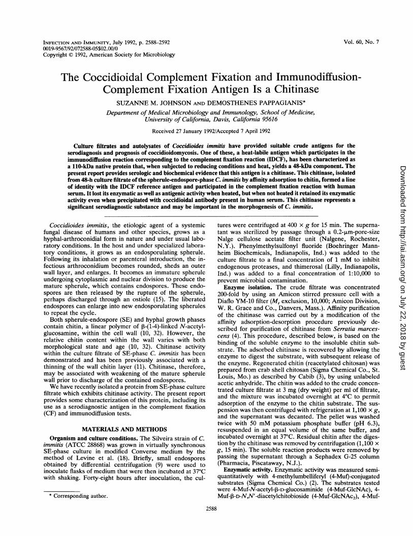
FIG. 1. Nonreduced, non-SDS PAGE of affinity-purified chiti-nase (12% gel). Lane A, silver stain; lane B, fluorescence of4-Muf-GlcNAc3 chitinase activity stain.
preparations known to contain IDCF activity, as well as thepurified chitinase, were capable of liberating free 4-Muf from4-Muf-GlcNAc2 and 4-Muf-GlcNAc3 substrates. No glucosi-dase or protease activity was observed. When previouslyheated, the coccidioidal antigens (MFS and F171) exhibitedno enzymatic activity. Chitinase activity against regeneratedchitin was inferred from the visual disappearance of chitinduring the last step of the purification.PAGE. PAGE of affinity-purified chitinase not exposed to
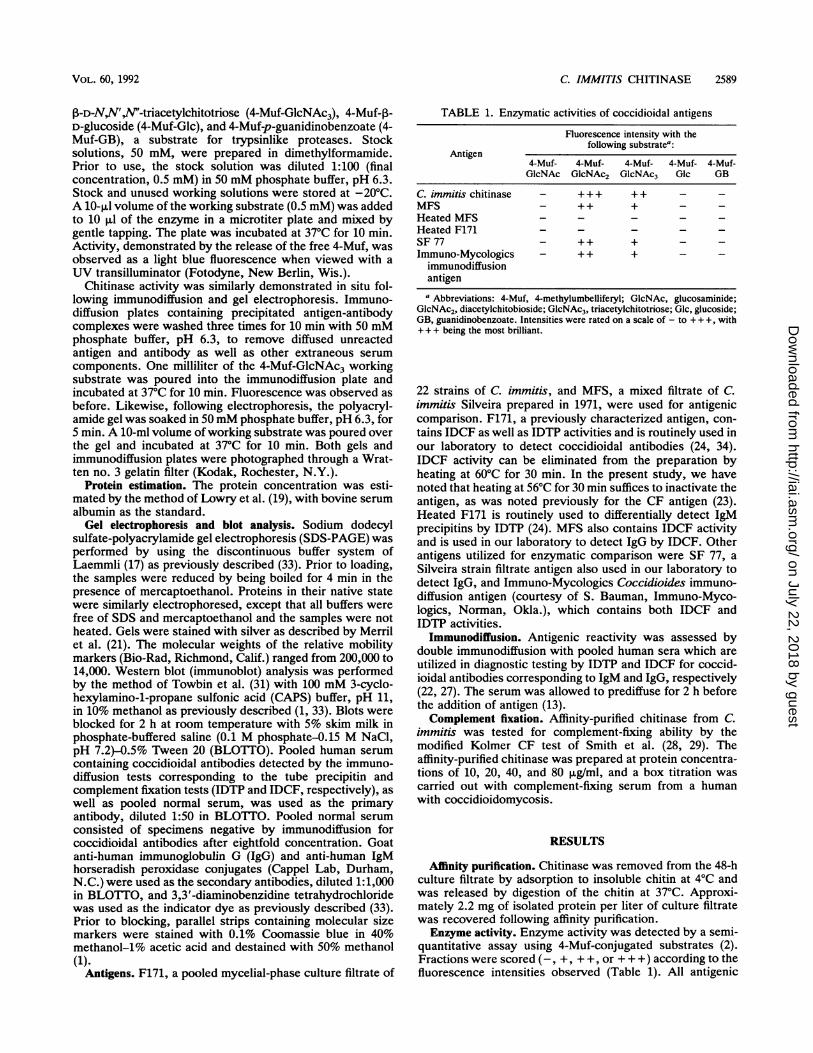
mercaptoethanol and SDS yielded a single diffuse band (Fig.1). This band contained chitinase activity, demonstrated insitu as fluorescence when 4-Muf-GlcNAc3 was overlaid andthe gel was briefly incubated. When the same affinity-purified chitinase was subjected to reducing SDS-PAGE, ityielded a doublet band with a mobility corresponding to amolecular weight of 48,000 (Fig. 2). It was not possible todemonstrate activity associated with either one or both ofthese bands, as enzymatic activity was destroyed by therequired boiling. Furthermore, activity was lost when thechitinase preparation was heated for 30 min at 56°C (asindicated above, this treatment also destroys the antigenicactivity in the IDCF test).
Following electrophoresis, the reduced, purified proteinwas transferred to a nitrocellulose membrane. The 48-kDa
- 92
45
- 31
f 22
141 2
FIG. 2. Reduced SDS-PAGE (12% gel). Lane 1, affinity-purifiedchitinase; lane 2, molecular size markers (in kilodaltons). The arrowindicates a 48-kDa doublet.
14s
v G)
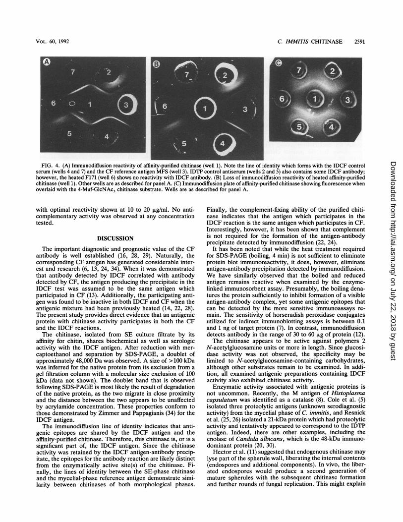
FIG. 3. SDS-PAGE immunoblot of affinity-purified chitinase(12% gel). Lanes: CF, CF control pooled human serum; PPT,precipitin control pooled human serum; NEG, negative pooledhuman serum; IgM, anti-IgM secondary antibody; IgG, anti-IgGsecondary antibody. Molecular size markers (in kilodaltons) are onthe left.
bands reacted intensely as a single unit when probed withpooled human serum positive for coccidioidal CF antibody,detected with peroxidase-labeled anti-IgG secondary anti-body (Fig. 3). This area also stained when probed withhuman serum positive for IDTP and anti-IgG secondaryantibody, indicating that CF antibodies were present in thatpooled serum also. Such antibodies were demonstrated inthis serum pool by IDCF (Fig. 4).
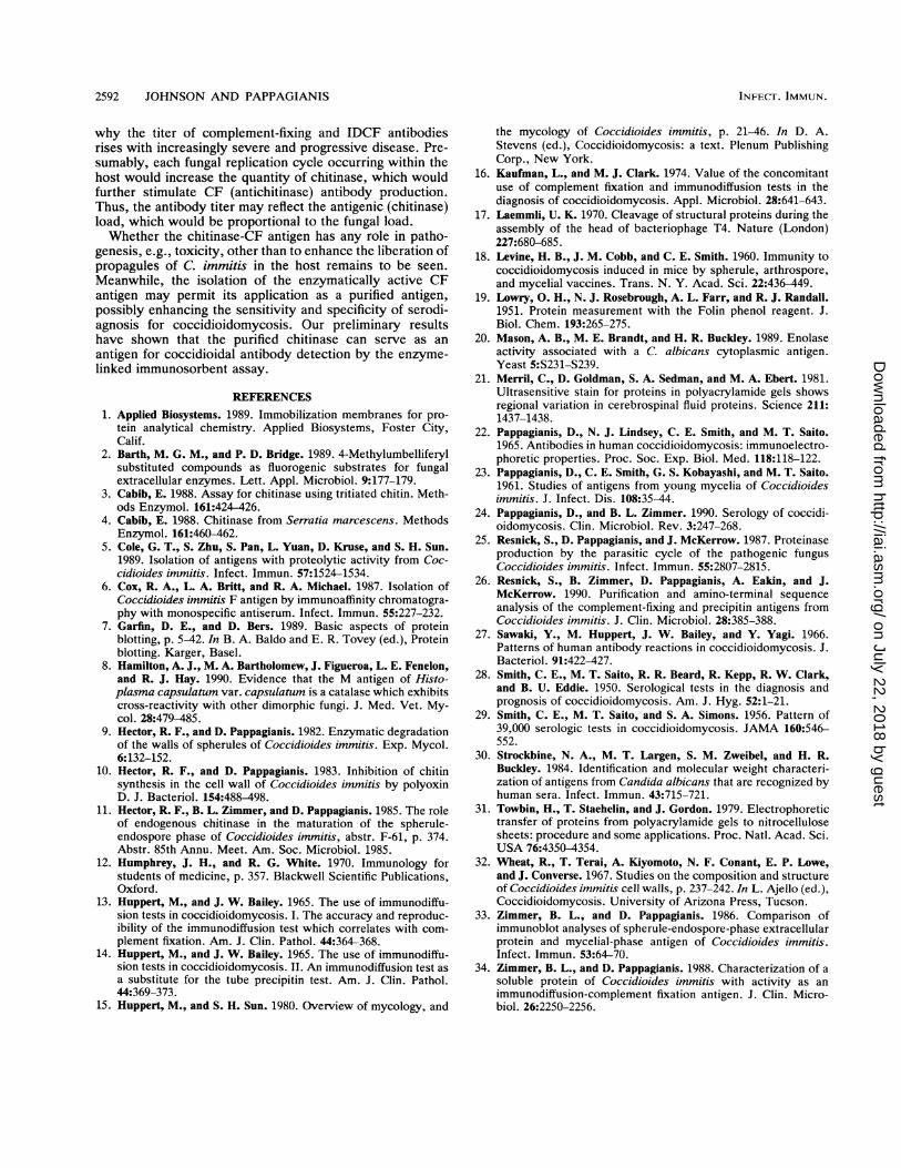
Serologic activity. Serologic activity was determined bydouble immunodiffusion and CF. Immunodiffusion producedan antigen-antibody reaction yielding a line of identity be-tween the affinity-purified chitinase and the IDCF referenceantigen MFS (Fig. 4A). This line of precipitation was notobserved when the purified chitinase was heated for 30 minat 56°C (Fig. 4B) or boiled for 4 min (not shown). This heattreatment had previously been observed to inactivate enzy-matic activity. Furthermore, chitinase activity, demon-strated with the 4-Muf-GlcNAc3 substrate, was associatedwith the precipitate line of identity produced between thereference IDCF antigen and the purified chitinase whenreacted with the IDCF-positive serum (Fig. 4C). No fluores-cence was associated with the IDTP line, and no fluores-cence was manifested by the chitinase-antibody precipitatein the absence of the 4-Muf-GlcNAc3 substrate.To ascertain whether the affinity-purified chitinase could
also serve as a complement-fixing antigen, the chitinase wassubstituted for the coccidioidin in a box titration CF test withhuman serum of known CF antibody titer. CF comparable tothat shown by our reference antigen occurred at chitinaseprotein concentrations of 10 to 80 pg/ml (the range tested),
INFECT. IMMUN.
_-1-~ , ",.
on July 22, 2018 by guesthttp://iai.asm
.org/D
ownloaded from
C. IMMITIS CHITINASE 2591
FIG. 4. (A) Immunodiffusion reactivity of affinity-purified chitinase (well 1). Note the line of identity which forms with the IDCF controlserum (wells 4 and 7) and the CF reference antigen MFS (well 3). IDTP control antiserum (wells 2 and 5) also contains some IDCF antibody;however, the heated F171 (well 6) shows no reactivity with IDCF antibody. (B) Loss of immunodiffusion reactivity of heated affinity-purifiedchitinase (well 1). Other wells are as described for panel A. (C) Immunodiffusion plate of affinity-purified chitinase showing fluorescence whenoverlaid with the 4-Muf-GlcNAc3 chitinase substrate. Wells are as described for panel A.
with optimal reactivity shown at 10 to 20 ,ug/ml. No anti-complementary activity was observed at any concentrationtested.
DISCUSSION
The important diagnostic and prognostic value of the CFantibody is well established (16, 28, 29). Naturally, thecorresponding CF antigen has generated considerable inter-est and research (6, 13, 24, 34). When it was demonstratedthat antibody detected by IDCF correlated with antibodydetected by CF, the antigen producing the precipitate in theIDCF test was assumed to be the same antigen whichparticipated in CF (13). Additionally, the participating anti-gen was found to be inactive in both IDCF and CF when theantigenic mixture had been previously heated (14, 22, 28).The present study provides direct evidence that an antigenicprotein with chitinase activity participates in both the CFand the IDCF reactions.The chitinase, isolated from SE culture filtrate by its
affinity for chitin, shares biochemical as well as serologicactivity with the IDCF antigen. After reduction with mer-captoethanol and separation by SDS-PAGE, a doublet ofapproximately 48,000 Da was observed. A size of > 100 kDawas inferred for the native protein from its exclusion from agel filtration column with a molecular size exclusion of 100kDa (data not shown). The doublet band that is observedfollowing SDS-PAGE is most likely the result of degradationof the native protein, as the two migrate in close proximityand the distance between the two appears to be unaffectedby acrylamide concentration. These properties conform tothose demonstrated by Zimmer and Pappagianis (34) for theIDCF antigen.The immunodiffusion line of identity indicates that anti-
genic epitopes are shared by the IDCF antigen and theaffinity-purified chitinase. Therefore, this chitinase is, or is asignificant part of, the IDCF antigen. Since the chitinaseactivity was retained by the IDCF antigen-antibody precip-itate, the epitopes for the antibody reaction are likely distinctfrom the enzymatically active site(s) of the chitinase. Fi-nally, the lines of identity between the SE-phase chitinaseand the mycelial-phase reference antigen demonstrate simi-larity between chitinases of both morphological phases.
Finally, the complement-fixing ability of the purified chiti-nase indicates that the antigen which participates in theIDCF reaction is the same antigen which participates in CF.Interestingly, however, it has been shown that complementis not required for the formation of the antigen-antibodyprecipitate detected by immunodiffusion (22, 24).
It has been noted that while the heat treatment requiredfor SDS-PAGE (boiling, 4 min) is not sufficient to eliminateprotein blot immunoreactivity, it does, however, eliminateantigen-antibody precipitation detected by immunodiffusion.We have similarly observed that the boiled and reducedantigen remains reactive when examined by the enzyme-linked immunosorbent assay. Presumably, the boiling dena-tures the protein sufficiently to inhibit formation of a visibleantigen-antibody complex, yet some antigenic epitopes thatcan be detected by the more sensitive immunoassays re-main. The sensitivity of horseradish peroxidase conjugatesutilized for indirect immunoblotting assays is between 0.1and 1 ng of target protein (7). In contrast, immunodiffusiondetects antibody in the range of 30 to 60 ,ug of protein (12).The chitinase appears to be active against polymers 2
N-acetylglucosamine units or more in length. Since glucosi-dase activity was not observed, the specificity may belimited to N-acetylglucosamine-containing carbohydrates,although other substrates remain to be examined. In addi-tion, all examined antigenic preparations containing IDCFactivity also exhibited chitinase activity.Enzymatic activity associated with antigenic proteins is
not uncommon. Recently, the M antigen of Histoplasmacapsulatum was identified as a catalase (8). Cole et al. (5)isolated three proteolytic antigens (unknown serodiagnosticactivity) from the mycelial phase of C. immitis, and Resnicket al. (25, 26) isolated a 21-kDa protein which had proteolyticactivity and tentatively appeared to correspond to the IDTPantigen. Indeed, there are other examples, including theenolase of Candida albicans, which is the 48-kDa immuno-dominant protein (20, 30).Hector et al. (11) suggested that endogenous chitinase may
lyse part of the spherule wall, liberating the internal contents(endospores and additional components). In vivo, the liber-ated endospores would produce a second generation ofmature spherules with the subsequent chitinase formationand further rounds of fungal replication. This might explain
VOL. 60, 1992
on July 22, 2018 by guesthttp://iai.asm
.org/D
ownloaded from
2592 JOHNSON AND PAPPAGIANIS
why the titer of complement-fixing and IDCF antibodiesrises with increasingly severe and progressive disease. Pre-sumably, each fungal replication cycle occurring within thehost would increase the quantity of chitinase, which wouldfurther stimulate CF (antichitinase) antibody production.Thus, the antibody titer may reflect the antigenic (chitinase)load, which would be proportional to the fungal load.Whether the chitinase-CF antigen has any role in patho-
genesis, e.g., toxicity, other than to enhance the liberation ofpropagules of C. immitis in the host remains to be seen.
Meanwhile, the isolation of the enzymatically active CFantigen may permit its application as a purified antigen,possibly enhancing the sensitivity and specificity of serodi-agnosis for coccidioidomycosis. Our preliminary resultshave shown that the purified chitinase can serve as an
antigen for coccidioidal antibody detection by the enzyme-linked immunosorbent assay.
REFERENCES1. Applied Biosystems. 1989. Immobilization membranes for pro-
tein analytical chemistry. Applied Biosystems, Foster City,Calif.
2. Barth, M. G. M., and P. D. Bridge. 1989. 4-Methylumbelliferylsubstituted compounds as fluorogenic substrates for fungalextracellular enzymes. Lett. Appl. Microbiol. 9:177-179.
3. Cabib, E. 1988. Assay for chitinase using tritiated chitin. Meth-ods Enzymol. 161:424-426.
4. Cabib, E. 1988. Chitinase from Serratia marcescens. MethodsEnzymol. 161:460-462.
5. Cole, G. T., S. Zhu, S. Pan, L. Yuan, D. Kruse, and S. H. Sun.1989. Isolation of antigens with proteolytic activity from Coc-cidioides immitis. Infect. Immun. 57:1524-1534.
6. Cox, R. A., L. A. Britt, and R. A. Michael. 1987. Isolation ofCoccidioides immitis F antigen by immunoaffinity chromatogra-phy with monospecific antiserum. Infect. Immun. 55:227-232.
7. Garfin, D. E., and D. Bers. 1989. Basic aspects of proteinblotting, p. 5-42. In B. A. Baldo and E. R. Tovey (ed.), Proteinblotting. Karger, Basel.
8. Hamilton, A. J., M. A. Bartholomew, J. Figueroa, L. E. Fenelon,and R. J. Hay. 1990. Evidence that the M antigen of Histo-plasma capsulatum var. capsulatum is a catalase which exhibitscross-reactivity with other dimorphic fungi. J. Med. Vet. My-col. 28:479-485.
9. Hector, R. F., and D. Pappagianis. 1982. Enzymatic degradationof the walls of spherules of Coccidioides immitis. Exp. Mycol.6:132-152.
10. Hector, R. F., and D. Pappagianis. 1983. Inhibition of chitinsynthesis in the cell wall of Coccidioides immitis by polyoxinD. J. Bacteriol. 154:488-498.
11. Hector, R. F., B. L. Zimmer, and D. Pappagianis. 1985. The roleof endogenous chitinase in the maturation of the spherule-endospore phase of Coccidioides immitis, abstr. F-61, p. 374.Abstr. 85th Annu. Meet. Am. Soc. Microbiol. 1985.
12. Humphrey, J. H., and R. G. White. 1970. Immunology forstudents of medicine, p. 357. Blackwell Scientific Publications,Oxford.
13. Huppert, M., and J. W. Bailey. 1965. The use of immunodiffu-sion tests in coccidioidomycosis. I. The accuracy and reproduc-ibility of the immunodiffusion test which correlates with com-
plement fixation. Am. J. Clin. Pathol. 44:364-368.14. Huppert, M., and J. W. Bailey. 1965. The use of immunodiffu-
sion tests in coccidioidomycosis. II. An immunodiffusion test asa substitute for the tube precipitin test. Am. J. Clin. Pathol.44:369-373.
15. Huppert, M., and S. H. Sun. 1980. Overview of mycology, and
the mycology of Coccidioides immitis, p. 21-46. In D. A.Stevens (ed.), Coccidioidomycosis: a text. Plenum PublishingCorp., New York.
16. Kaufman, L., and M. J. Clark. 1974. Value of the concomitantuse of complement fixation and immunodiffusion tests in thediagnosis of coccidioidomycosis. Appl. Microbiol. 28:641-643.
17. Laemmli, U. K. 1970. Cleavage of structural proteins during theassembly of the head of bacteriophage T4. Nature (London)227:680-685.
18. Levine, H. B., J. M. Cobb, and C. E. Smith. 1960. Immunity tococcidioidomycosis induced in mice by spherule, arthrospore,and mycelial vaccines. Trans. N. Y. Acad. Sci. 22:436-449.
19. Lowry, 0. H., N. J. Rosebrough, A. L. Farr, and R. J. Randall.1951. Protein measurement with the Folin phenol reagent. J.Biol. Chem. 193:265-275.
20. Mason, A. B., M. E. Brandt, and H. R. Buckley. 1989. Enolaseactivity associated with a C. albicans cytoplasmic antigen.Yeast 5:S231-S239.
21. Merril, C., D. Goldman, S. A. Sedman, and M. A. Ebert. 1981.Ultrasensitive stain for proteins in polyacrylamide gels showsregional variation in cerebrospinal fluid proteins. Science 211:1437-1438.
22. Pappagianis, D., N. J. Lindsey, C. E. Smith, and M. T. Saito.1965. Antibodies in human coccidioidomycosis: immunoelectro-phoretic properties. Proc. Soc. Exp. Biol. Med. 118:118-122.
23. Pappagianis, D., C. E. Smith, G. S. Kobayashi, and M. T. Saito.1961. Studies of antigens from young mycelia of Coccidioidesimmitis. J. Infect. Dis. 108:35-44.
24. Pappagianis, D., and B. L. Zimmer. 1990. Serology of coccidi-oidomycosis. Clin. Microbiol. Rev. 3:247-268.
25. Resnick, S., D. Pappagianis, and J. McKerrow. 1987. Proteinaseproduction by the parasitic cycle of the pathogenic fungusCoccidioides immitis. Infect. Immun. 55:2807-2815.
26. Resnick, S., B. Zimmer, D. Pappagianis, A. Eakin, and J.McKerrow. 1990. Purification and amino-terminal sequenceanalysis of the complement-fixing and precipitin antigens fromCoccidioides immitis. J. Clin. Microbiol. 28:385-388.
27. Sawaki, Y., M. Huppert, J. W. Bailey, and Y. Yagi. 1966.Patterns of human antibody reactions in coccidioidomycosis. J.Bacteriol. 91:422-427.
28. Smith, C. E., M. T. Saito, R. R. Beard, R. Kepp, R. W. Clark,and B. U. Eddie. 1950. Serological tests in the diagnosis andprognosis of coccidioidomycosis. Am. J. Hyg. 52:1-21.
29. Smith, C. E., M. T. Saito, and S. A. Simons. 1956. Pattern of39,000 serologic tests in coccidioidomycosis. JAMA 160:546-552.
30. Strockbine, N. A., M. T. Largen, S. M. Zweibel, and H. R.Buckley. 1984. Identification and molecular weight characteri-zation of antigens from Candida albicans that are recognized byhuman sera. Infect. Immun. 43:715-721.
31. Towbin, H., T. Staehelin, and J. Gordon. 1979. Electrophoretictransfer of proteins from polyacrylamide gels to nitrocellulosesheets: procedure and some applications. Proc. Natl. Acad. Sci.USA 76:4350-4354.
32. Wheat, R., T. Terai, A. Kiyomoto, N. F. Conant, E. P. Lowe,and J. Converse. 1967. Studies on the composition and structureof Coccidioides immitis cell walls, p. 237-242. In L. Ajello (ed.),Coccidioidomycosis. University of Arizona Press, Tucson.
33. Zimmer, B. L., and D. Pappagianis. 1986. Comparison ofimmunoblot analyses of spherule-endospore-phase extracellularprotein and mycelial-phase antigen of Coccidioides immitis.Infect. Immun. 53:64-70.
34. Zimmer, B. L., and D. Pappagianis. 1988. Characterization of asoluble protein of Coccidioides immitis with activity as animmunodiffusion-complement fixation antigen. J. Clin. Micro-biol. 26:2250-2256.
INFECT. IMMUN.
on July 22, 2018 by guesthttp://iai.asm
.org/D
ownloaded from