the citric acid cycle - 140.126.122.189140.126.122.189/upload/1052/b20202a201714113551.pdf · the...
TRANSCRIPT

The Citric Acid Cycle
Copyright © 2006 Pearson Prentice Hall, Inc.

Overview • Acetyl CoA is
– formed by the oxidative decarboxylation of pyruvate • with the release of CO2
– one of the key intermediates in the interconversion of small organic acids
• The citric acid cycle (CAC) begins with – The transfer of acetyl group
• from acetyl CoA
• to the four-carbon dicarboxylic acid oxaloacetate to
• form a new six-carbon tricarboxylic acid known as citrate
• Citrate can then be oxidized in a – seven-step pathway to
– regenerate oxaloacetate and
– release two molecules of CO2
• The pathway is known as – the citric acid cycle
– the tricarboxylic acid cycle (TCA cycle), or
– the Krebs cycle

Overview of the citric acid cycle

The citric acid cycle the hub of energy metabolism in eukaryotic cells
• The energy released in the cycle is largely
– conserved as reducing power in
– coenzyme NADH and
– reduced ubiquinone (QH2)
• NADH and QH2 are substrates in
– membrane-associated electron transport that
– lead to the formation of a proton gradient which
– drives synthesis of ATP

Conversion of Pyruvate to Acetyl CoA
• catalyzed by – the pyruvate dehydrogenase complex
– a large complex of enzymes and cofactors
– need coenzyme A (HS–CoA)
– Two electrons are transferred to NADH from pyruvate

The pyruvate dehydrogenase complex
• a multienzyme complex containing
– multiple copies of three distinct enzymatic
activities:
• pyruvate dehydrogenase (E1)
• dihydrolipoamide acetyltransferase (E2), and
• dihydrolipoamide dehydrogenase(E3)
• There are 5 steps in the oxidative
decarboxylation of pyruvate

1. Formation of a hydroxyethyl—TPP
intermediate and the release of CO2
The E1 component contains the prosthetic group thiamine
pyrophosphate (TPP, vitamin B1)
the carbanion or ylid form

2. Two-carbon hydroxyethyl group is transferred
to the lipoamide group of E2
oxidation of hydroxyethyl—TPP is
coupled to the reduction
of the disulfide of lipoamide

The lipoamide group consists of •lipoic acid covalently bound by an amide linkage
•to a lysine residue of an subunit E2
This particular coenzyme is only found in pyruvate dehydrogenase
and related enzymes.
A coenzyme

3. Transfer of the acetyl group to HS—CoA, forming acetyl CoA and leaving
the lipoamide in the reduced dithiol form

4. The reduced lipoamide of E2 must be
reoxidized in order to
regenerate the prosthetic group
FAD (also a coenzyme) is the prosthetic group of E3.

5. E3—FADH2 is reoxidized to FAD
• E3—FADH2 + NAD+ E3—FAD + NADH +
H+
• regenerates the original pyruvate
dehydrogenase complex,
– completing the catalytic cycle

The interplay of five coenzymes in the
pyruvate dehydrogenase complex
• Two are cosubstrates
– HS—CoA and NAD+
• Three are prosthetic groups
– TPP,
– lipoamide, and
– FAD
• illustrates the importance of coenzymes in metabolic reactions

Lipoamide prosthetic group acts as a
swinging arm that
visits the three active sites in the enzyme complex
Reactions of the pyruvate dehydrogenase complex
The swinging arm mechanism of lipoamide
• The various subunits of the complex are arranged in a
way
– to ensure that
• the product of one reaction
• does not diffuse into the medium but is
• immediately acted on by the next component
• This arrangement is called
– the
– swinging arm mechanism of lipoamide

Structural model of the pyruvate
dehydrogenase complex
The eukaryotic pyruvate dehydrogenase complex is the largest
multienzyme complex known.

The inner core:
•60 E2 enzymes
•in the shape of a pentagonal
Dodecahedron
•with one E2 trimer at each of the
20 vertices
A single trimer is outlined
by a yellow box.
The center of the pentagon
shape is indicated by the red
pentagon.

Cutaway view of
outer E1 enzymes (yellow)
E3 enzymes (red)
located in the space between the
E2 enzymes

Electron micrograph of pyruvate dehydrogenase
complexes from E. coli.

2-oxo acid dehydrogenase family
• A family of multi-enzyme complexes – Pyruvate dehydrogenase is a member
– Pyruvate is the smallest 2-oxo organic acid
• Two other 2-oxo (or a-keto) acid dehydrogenases – A citric acid cycle enzyme
a-ketoglutarate dehydrogenase
– branched-chain a-keto acid dehydrogenase, • used in amino acid metabolism
• catalyze essentially irreversible reactions in which – an organic acid is oxidized to CO2 and
– an energy-rich coenzyme A derivative is formed

In eukaryotic cells the enzymes of the citric acid cycle are located in
the mitochondria.
The eight reactions of the citric acid cycle

Transporting pyruvate into mitochondria
• In bacterial cells,
– pyruvate is converted to acetyl CoA in the cytosol
• In eukaryotic cells the pyruvate dehydrogenase complex is located in mitochondria
– enclosed by a double membrane
– Small molecules pass through the outer membrane via
• aqueous channels formed by transmembrane proteins called
• porins
– Pyruvate translocase
• a specific transport protein
• transports pyruvate in symport with H+
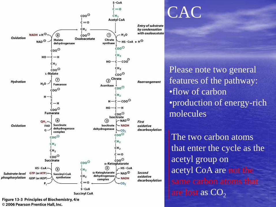
CAC
Please note two general
features of the pathway:
•flow of carbon
•production of energy-rich
molecules
The two carbon atoms
that enter the cycle as the
acetyl group on
acetyl CoA are not the
same carbon atoms that
are lost as CO2


The citric acid cycle is equivalent to the
•oxidation of an acetyl CoA molecule with release of 8 electrons
6 electrons are transferred to 3 NAD+ and
2 electrons to one ubiquinone (Q)

The oxidation of an acetyl CoA equivalent by the citric acid cycle
showing the valence electrons in the reactants and products.
CAC淨反應之價電子變化情形
Most of the time, electrons are released when double bonds
are formed.

Fates of the carbon atoms during
one turn of the citric acid cycle.
Carbon atoms from acetyl CoA (green) are
•uniformly distributed in the four-carbon
intermediates leading to oxaloacetate.

1. Citrate Synthase
Citrate is the first of two tricarboxylic acids in the cycle.
DGo’ = -31.5 kJ mol-1
The large negative free energy ensures that
•the reaction proceeds in the direction of citrate synthesis
•when the concentration of oxaloacetate is very low
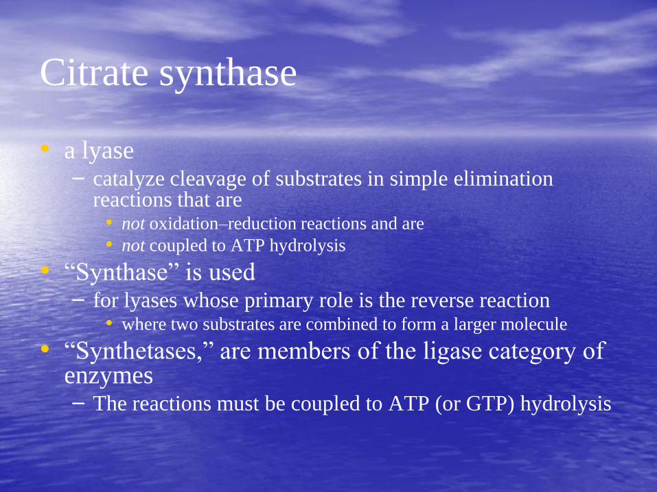
Citrate synthase
• a lyase – catalyze cleavage of substrates in simple elimination
reactions that are • not oxidation–reduction reactions and are
• not coupled to ATP hydrolysis
• “Synthase” is used – for lyases whose primary role is the reverse reaction
• where two substrates are combined to form a larger molecule
• “Synthetases,” are members of the ligase category of enzymes – The reactions must be coupled to ATP (or GTP) hydrolysis
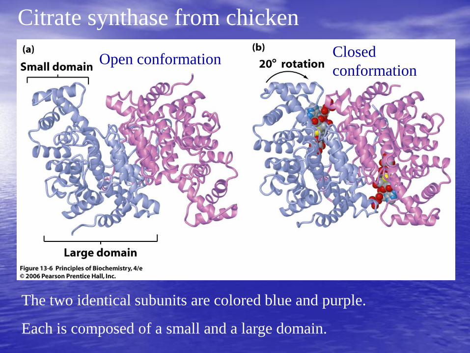
Citrate synthase from chicken
The two identical subunits are colored blue and purple.
Each is composed of a small and a large domain.
Open conformation Closed
conformation

2. Aconitase
A near-equilibrium conversion of citrate to isocitrate.
Citrate is a tertiary alcohol and thus
•cannot be oxidized directly to a keto acid
•The formation of a keto acid intermediate
•is required for the oxidative decarboxylation
a secondary alcohol

2 chiral centers at C2 and
C3
•There are four different
stereoisomers
•only one of
these is produced
•2R,3S-isocitrate
Structure of
2R,3S-isocitrate
C2
C3

Three-point Attachment of Prochiral
Substrates to Enzymes (Box 13.2)
• In the citrate-to-isocitrate reaction – a chiral molecule was produced from a non-chiral molecule
– But only one of the two possible forms of 2R,3S-isocitrate was produced
• Why formation of the double bond of cis-aconitate, and – Subsequent addition of water to form isocitrate, occurred
– only in the moiety contributed originally by oxaloacetate
– not in the group derived from acetyl CoA
• Remember that citrate is symmetric

Two forms of isocitrate. The green carbon atoms represent the
group originally derived from acetyl CoA.
The reaction was expected to yield two forms of isocitrate in equal
quantities.
Only this form
was produced.

Three-point Attachment of Prochiral Substrates to Enzymes
The substrate itself is symmetric.
However, when there are 3 binding sites, the way of binding is not
symmetric.

3. Isocitrate Dehydrogenase
catalyzes the oxidative decarboxylation of isocitrate to
Form a-ketoglutarate
(2-oxoglutarate)

4. The a-Ketoglutarate Dehydrogenase Complex
analogous to the reaction catalyzed by pyruvate dehydrogenase.
the reactants are an a-keto acid and HS–CoA and
the products are CO2 and a high-energy thioester compound.
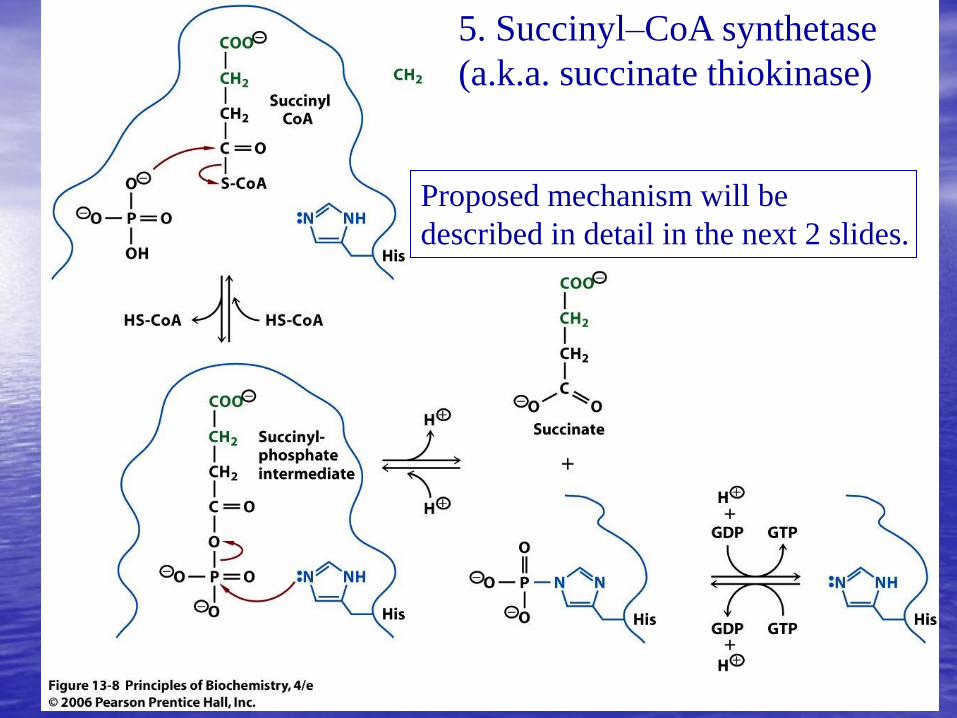
5. Succinyl–CoA synthetase
(a.k.a. succinate thiokinase)
Proposed mechanism will be
described in detail in the next 2 slides.

Phosphate displaces CoA,
forming the mixed acid anhydride
succinyl phosphate

1. Forming covalent phosphoenzyme intermediate
2. Transfering phosphoryl group to GDP (or ADP, depending on the
organism)

The overall stoichiometry of the succinyl–CoA
synthetase reaction
the enzyme is named for the reverse reaction where
succinyl CoA is synthesized from succinate
at the expense of GTP or ATP
It is called a synthetase because the reaction
combines two molecules and it is
coupled to the hydrolysis of a nucleoside triphosphate

6. Succinate Dehydrogenase Complex
catalyzes the oxidation of succinate to fumarate, forming
a carbon–carbon double bond with the
loss of two protons and two electrons
electrons are passed to a quinone
+ QH2
an essential cofactor,
covalently bound to
the enzyme

a competitive inhibitor of the
succinate dehydrogenase complex

7. Fumarase (fumarate hydratase)
near-equilibrium conversion of fumarate to malate through
the stereospecific trans addition of water
to the double bond of fumarate
Fumarate, like citrate, is a prochiral molecule.
product is L stereoisomer of the hydroxy acid malate

8. Malate Dehydrogenase
analogous to the reversible reaction catalyzed by lactate dehydrogenase
lactate dehydrogenase and
malate dehydrogenase share a common ancestor


BOX 13.3 Converting One Enzyme into Another
Lactate
dehydrogenase
Malate
dehydrogenase
low identity of amino
acid sequences
closely related in
three-dimensional
structure, evolved
from a common
ancestor
Conversion of
Gln-102 to Arg-102
positively charged
arginine forms an ion
pair with 3-
carboxylate group

Reduced Coenzymes Can Fuel
the Production of ATP
net reaction of the citric acid cycle
NADH and QH2 can be oxidized by the membrane associated
electron-transport chain that is
coupled to the production of ATP.

The complete oxidation of 1 molecule of acetyl CoA
by the citric acid cycle and subsequent reactions is
associated with the production of approximately 10 ATP

The catabolism of 1 glucose by glycolysis, the citric acid cycle, and
reoxidation of NADH and QH2
The complete oxidation of glucose produces up to 32 ATP.

Regulation of the Citric Acid Cycle
• The citric acid cycle occupies a central position in cellular metabolism
– The pathway is stringently controlled
• Regulation is mediated by
– allosteric modulators and by
– covalent modification of the citric acid cycle enzymes
– supply of acetyl CoA

Substrates activate the complex.
Accumulation of the products acetyl CoA and NADH decreases flux.
The activity of the pyruvate dehydrogenase complex controls the
supply of acetyl CoA.

Regulation of the mammalian pyruvate dehydrogenase complex by
covalent modification.
Dephosphorylation
Phosphorylation

Regulation of the CAC
• Three reactions are regulated
– citrate synthase,
– isocitrate dehydrogenase, and
– a-ketoglutarate dehydrogenase complex

The Citric Acid Cycle Isn’t Always a “Cycle”
• not exclusively a catabolic pathway for the oxidation of acetyl CoA
• It also plays a central role in metabolism at the intersection of several other pathways
• Some intermediates are important metabolic precursors in biosynthesis
• Some catabolic pathways produce citric acid cycle intermediates.

Routes leading to and
from the CAC.
Intermediates of the
CAC are precursors of
carbohydrates, lipids,
and amino acids, as
well as nucleotides and
porphyrins.
Reactions feeding into
the cycle replenish the
cycle intermediates.

The production of oxaloacetate by
pyruvate carboxylase
• The rate at which the citric acid cycle metabolizes acetyl CoA is extremely sensitive to
– changes in the concentrations of its intermediates
• Metabolites removed by entry into biosynthetic pathways must be replenished by
– anaplerotic (Greek for “filling up”) reactions
• An important regulated replenishment reaction is

The Glyoxylate Pathway
• bypasses some of the reactions of the CAC
• named after the two-carbon molecule glyoxylate
• There are only two reactions – In the first reaction,
• a six-carbon tricarboxylic acid (isocitrate) is split into
• a two-carbon molecule (glyoxylate) and
• a four-carbon dicarboxylic acid (succinate)
• catalyzed by isocitrate lyase
– In the second reaction, • the two-carbon glyoxylate molecule combines with
• a two-carbon acetyl CoA molecule
• to make a four-carbon dicarboxylic acid (malate)
• catalyzed by malate synthase

Glyoxylate pathway
The steps which produce CO2 are
bypassed.
When the pathway is functioning,
the acetyl carbon atoms of acetyl
CoA are converted to malate rather
than oxidized to CO2.

Why glyoxylate pathway?
• provides an anabolic alternative for the metabolism of acetyl CoA – leading to the formation of glucose
• Cells that contain glyoxylate pathway enzymes can – synthesize all their required carbohydrates
– from any substrate that is a precursor of acetyl CoA
• Yeast can grow on ethanol – oxidize ethanol to form acetyl CoA
• Similarly, many bacteria use the glyoxylate pathway to sustain growth on acetate

The glyoxylate pathway in plants
• especially active in oily seed plants
• stored seed oils (triacylglycerols) are
converted to carbohydrates that
– Sustain the plant during germination.

The net effect of the glyoxylate pathway
• can be regarded as a shunt within the CAC
• bypass around the CO2-producing reactions of the CAC
• net formation of a four-carbon molecule from two molecules of acetyl CoA supplies
– a precursor that can be converted to glucose

Regulation of the glyoxylate pathway
in bacteria
• often used to replenish citric acid cycle metabolites – that are diverted into a number of biosynthesis pathways
• The key regulated enzyme is isocitrate dehydrogenase – covalent modification
• Kinase-catalyzed phosphorylation of – a serine residue
– abolishes isocitrate dehydrogenase activity
• In the dephosphorylated form of – the serine residue forms
– a hydrogen bond with a carboxylate group of isocitrate
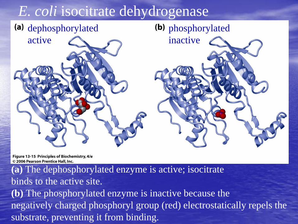
(a) The dephosphorylated enzyme is active; isocitrate
binds to the active site.
(b) The phosphorylated enzyme is inactive because the
negatively charged phosphoryl group (red) electrostatically repels the
substrate, preventing it from binding.
dephosphorylated
active
phosphorylated
inactive
E. coli isocitrate dehydrogenase

A bifunctional enzyme catalyzes both phosphorylation and
dephosphorylation
The two activities of the bifunctional enzyme are reciprocally
regulated allosterically by intermediates of glycolysis and the
citric acid cycle.
Regulation of E. coli isocitrate dehydrogenase
Regular CAC route

Evolution of the Citric Acid Cycle
• probably evolved from the more primitive forked pathway – found in many modern species of bacteria
• The evolution involved several of the pathway evolution mechanisms – discussed in Chapter 10
• There is evidence for – gene duplication,
– pathway extension,
– retro-evolution,
– pathway reversal, and
– enzyme theft

The left-hand side: a reductive
pathway leading to the synthesis of
succinate or a-ketoglutarate
The right-hand branch: an oxidative
pathway similar to the first few reactions
of the CAC
Forked pathway
in many bacteria