the channel issue1, 2008 - cook medical
TRANSCRIPT
issue 1, 2008
The ChannelA COOK NeWs PuBLiCATiON
Bigger isn’t always Better 2
extracting Bile duct stones 4
Performing circumferential emr 6
a leading endoscoPy center weathers a storm 8
what’s uP doc? 10
a “right there & then” comBination 10
Biliary anastomotic strictures 11
31st annual nysge 12
news from signea 14
attention gi nurses 16
Continued on page 3
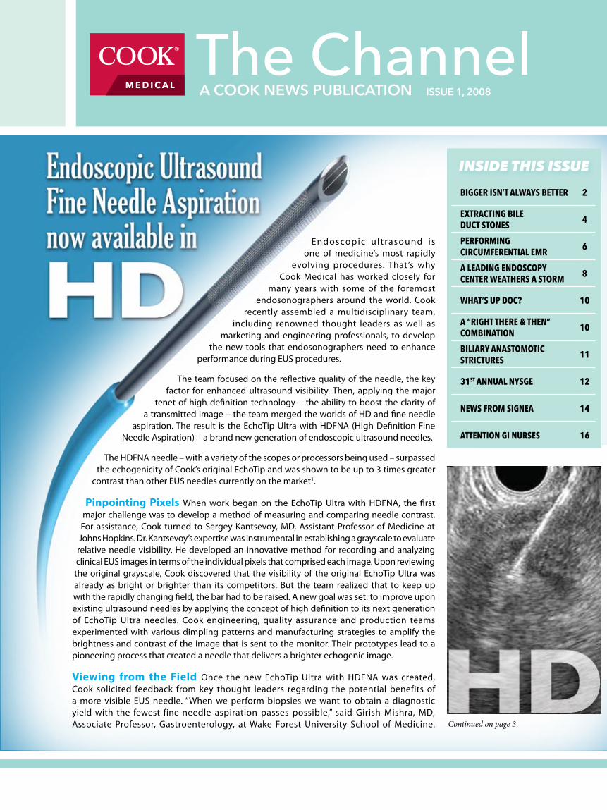
Endoscopic u l t rasound is one of medicine’s most rapidly
evolving procedures. That’s why Cook Medical has worked closely for
many years with some of the foremost endosonographers around the world. Cook
recently assembled a multidisciplinary team, including renowned thought leaders as well as
marketing and engineering professionals, to develop the new tools that endosonographers need to enhance
performance during EUS procedures.
The team focused on the reflective quality of the needle, the key factor for enhanced ultrasound visibility. Then, applying the major
tenet of high-definition technology – the ability to boost the clarity of a transmitted image – the team merged the worlds of HD and fine needle
aspiration. The result is the EchoTip Ultra with HDFNA (High Definition Fine Needle Aspiration) – a brand new generation of endoscopic ultrasound needles.
The HDFNA needle – with a variety of the scopes or processors being used – surpassed the echogenicity of Cook’s original EchoTip and was shown to be up to 3 times greater
contrast than other EUS needles currently on the market1.
Pinpointing Pixels When work began on the EchoTip Ultra with HDFNA, the first major challenge was to develop a method of measuring and comparing needle contrast. For assistance, Cook turned to Sergey Kantsevoy, MD, Assistant Professor of Medicine at Johns Hopkins. Dr. Kantsevoy’s expertise was instrumental in establishing a grayscale to evaluate relative needle visibility. He developed an innovative method for recording and analyzing clinical EUS images in terms of the individual pixels that comprised each image. Upon reviewing the original grayscale, Cook discovered that the visibility of the original EchoTip Ultra was already as bright or brighter than its competitors. But the team realized that to keep up with the rapidly changing field, the bar had to be raised. A new goal was set: to improve upon existing ultrasound needles by applying the concept of high definition to its next generation of EchoTip Ultra needles. Cook engineering, quality assurance and production teams experimented with various dimpling patterns and manufacturing strategies to amplify the brightness and contrast of the image that is sent to the monitor. Their prototypes lead to a pioneering process that created a needle that delivers a brighter echogenic image.
Viewing from the Field Once the new EchoTip Ultra with HDFNA was created, Cook solicited feedback from key thought leaders regarding the potential benefits of a more visible EUS needle. “When we perform biopsies we want to obtain a diagnostic yield with the fewest fine needle aspiration passes possible,” said Girish Mishra, MD, Associate Professor, Gastroenterology, at Wake Forest University School of Medicine.
inside this issue
www.cookmedical.com2
Dr. Armstrong
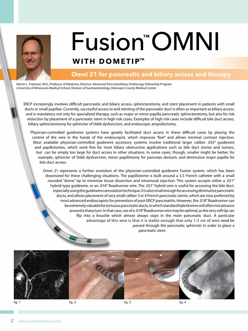
Martin L. Freeman, M.D., Professor of Medicine, Director, Advanced Pancreatobiliary Endoscopy Fellowship Program University of Minnesota Medical School, Division of Gastroenterology, Hennepin County Medical Center
Omni 21 for pancreatic and biliary access and therapy
fig. 1 fig. 2 fig. 3 fig. 4
ERCP increasingly involves difficult pancreatic and biliary access, sphincterotomy, and stent placement in patients with small ducts or small papillae. Currently, successful access to and stenting of the pancreatic duct is often as important as biliary access, and is mandatory not only for specialized therapy, such as major or minor papilla pancreatic sphincterotomy, but also for risk reduction by placement of a pancreatic stent in high-risk cases. Examples of high risk cases include difficult bile duct access, biliary sphincterotomy for sphincter of Oddi dysfunction, and endoscopic ampullectomy.
Physician-controlled guidewire systems have greatly facilitated duct access in these difficult cases by placing the control of the wire in the hands of the endoscopist, which improves “feel” and allows minimal contrast injection. Most available physician-controlled guidewire accessory systems involve traditional larger caliber .035” guidewire and papillotomes, which work fine for most biliary obstructive applications such as bile duct stones and tumors, but can be simply too large for duct access in other situations. In some cases, though, smaller might be better; for
example, sphincter of Oddi dysfunction, minor papillotomy for pancreas divisum, and diminutive major papilla for bile duct access.
Omni 21 represents a further evolution of the physician-controlled guidewire Fusion system, which has been downsized for these challenging situations. The papillotome is built around a 5.5 French catheter with a small
rounded “dome” tip to minimize tissue dissection and intramural injection. This system accepts either a .021” hybrid-type guidewire, or an .018” Roadrunner wire. The .021” hybrid wire is useful for accessing the bile duct,
especially using the guidewire cannulation technique. It is also small enough for accessing diminutive pancreatic ducts, and allows placement of very small caliber 3 or 4 French pancreatic stents, which are now preferred by
most advanced endoscopists for prevention of post-ERCP pancreatitis. However, the .018” Roadrunner can be extremely valuable for tortuous pancreatic ducts, in which standard hybrid wires will often not advance
around a sharp turn. In that case, use of a .018” Roadrunner wire may be optimal, as the very soft tip can flip into a knuckle which almost always stays in the main pancreatic duct. A particular
advantage of this wire is that it is stable enough that only 1-2 cm of wire need be passed through the pancreatic sphincter in order to place a
pancreatic stent.
3
The Channel
www.cookmedical.com
Randomized Clinical Trial
ISN’T ALWAYS BETTER:
An advantage of the Omni 21 papillotome system is that after it has been inserted deeply into the duct and access has been gained, the guidewire can be changed without removing the papillotome from the duct. With most other physician-controlled wire systems, once the catheter or papillotome has entered the duct, it is not possible to change guidewires.
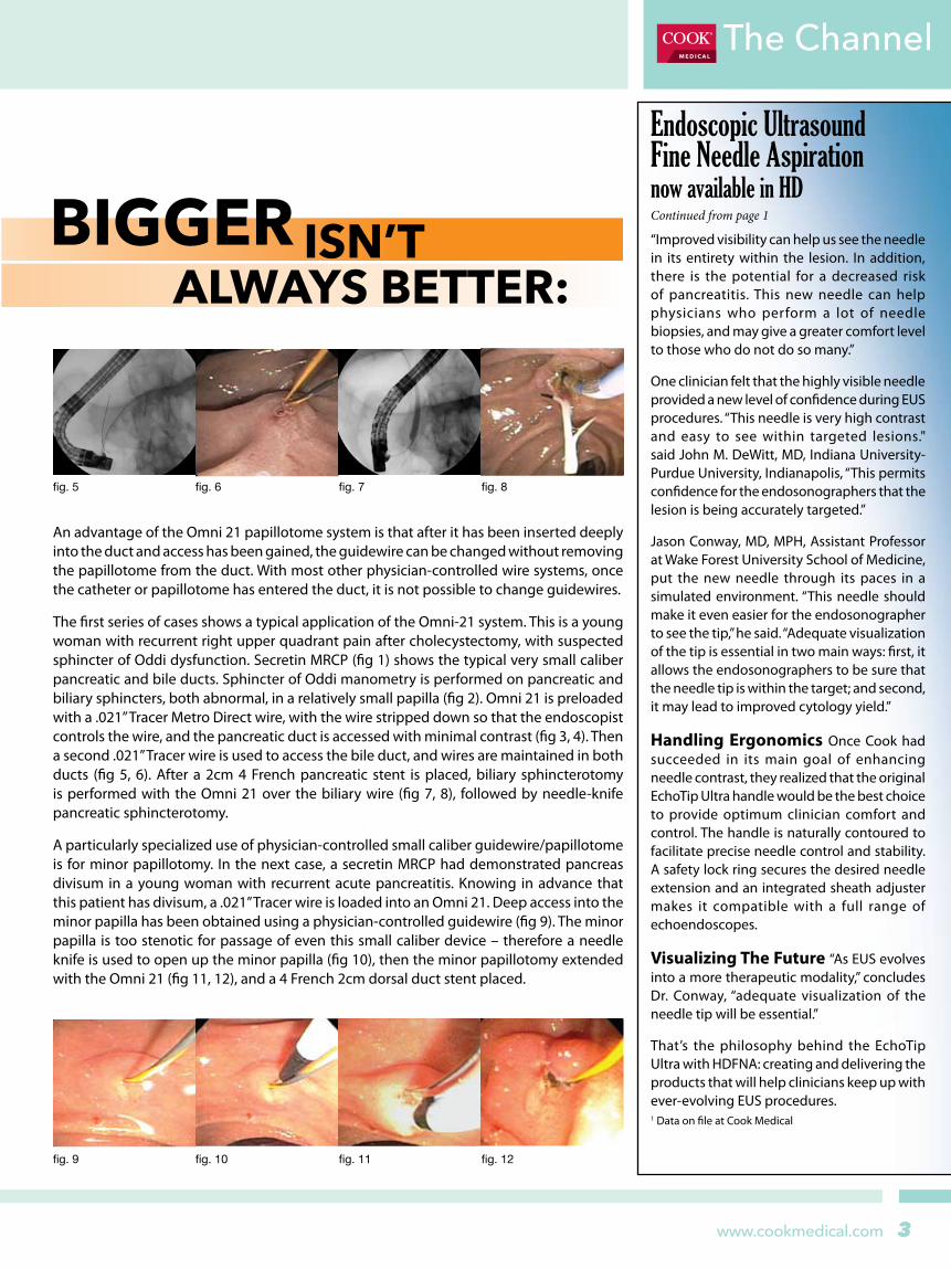
The first series of cases shows a typical application of the Omni-21 system. This is a young woman with recurrent right upper quadrant pain after cholecystectomy, with suspected sphincter of Oddi dysfunction. Secretin MRCP (fig 1) shows the typical very small caliber pancreatic and bile ducts. Sphincter of Oddi manometry is performed on pancreatic and biliary sphincters, both abnormal, in a relatively small papilla (fig 2). Omni 21 is preloaded with a .021” Tracer Metro Direct wire, with the wire stripped down so that the endoscopist controls the wire, and the pancreatic duct is accessed with minimal contrast (fig 3, 4). Then a second .021” Tracer wire is used to access the bile duct, and wires are maintained in both ducts (fig 5, 6). After a 2cm 4 French pancreatic stent is placed, biliary sphincterotomy is performed with the Omni 21 over the biliary wire (fig 7, 8), followed by needle-knife pancreatic sphincterotomy.
A particularly specialized use of physician-controlled small caliber guidewire/papillotome is for minor papillotomy. In the next case, a secretin MRCP had demonstrated pancreas divisum in a young woman with recurrent acute pancreatitis. Knowing in advance that this patient has divisum, a .021” Tracer wire is loaded into an Omni 21. Deep access into the minor papilla has been obtained using a physician-controlled guidewire (fig 9). The minor papilla is too stenotic for passage of even this small caliber device – therefore a needle knife is used to open up the minor papilla (fig 10), then the minor papillotomy extended with the Omni 21 (fig 11, 12), and a 4 French 2cm dorsal duct stent placed.
fig. 5
fig. 9
fig. 6
fig. 10
fig. 7
fig. 11
fig. 8
fig. 12
BIGGER “Improved visibility can help us see the needle in its entirety within the lesion. In addition, there is the potential for a decreased risk of pancreatitis. This new needle can help physicians who perform a lot of needle biopsies, and may give a greater comfort level to those who do not do so many.”
One clinician felt that the highly visible needle provided a new level of confidence during EUS procedures. “This needle is very high contrast and easy to see within targeted lesions." said John M. DeWitt, MD, Indiana University-Purdue University, Indianapolis, “This permits confidence for the endosonographers that the lesion is being accurately targeted.”
Jason Conway, MD, MPH, Assistant Professor at Wake Forest University School of Medicine, put the new needle through its paces in a simulated environment. “This needle should make it even easier for the endosonographer to see the tip,” he said. “Adequate visualization of the tip is essential in two main ways: first, it allows the endosonographers to be sure that the needle tip is within the target; and second, it may lead to improved cytology yield.”
Handling Ergonomics Once Cook had succeeded in its main goal of enhancing needle contrast, they realized that the original EchoTip Ultra handle would be the best choice to provide optimum clinician comfort and control. The handle is naturally contoured to facilitate precise needle control and stability. A safety lock ring secures the desired needle extension and an integrated sheath adjuster makes it compatible with a full range of echoendoscopes.
Visualizing The Future “As EUS evolves into a more therapeutic modality,” concludes Dr. Conway, “adequate visualization of the needle tip will be essential.”
That’s the philosophy behind the EchoTip Ultra with HDFNA: creating and delivering the products that will help clinicians keep up with ever-evolving EUS procedures.1 Data on file at Cook Medical
Endoscopic Ultrasound Fine Needle Aspirationnow available in HDContinued from page 1
www.cookmedical.com4
closed to secure the stone. At this step, if the stone is grasped too tightly, it may break into many fragments, which would make subsequent removal complicated and difficult. Therefore, careful attention should be paid in closing the basket gently to avoid breaking the stone.
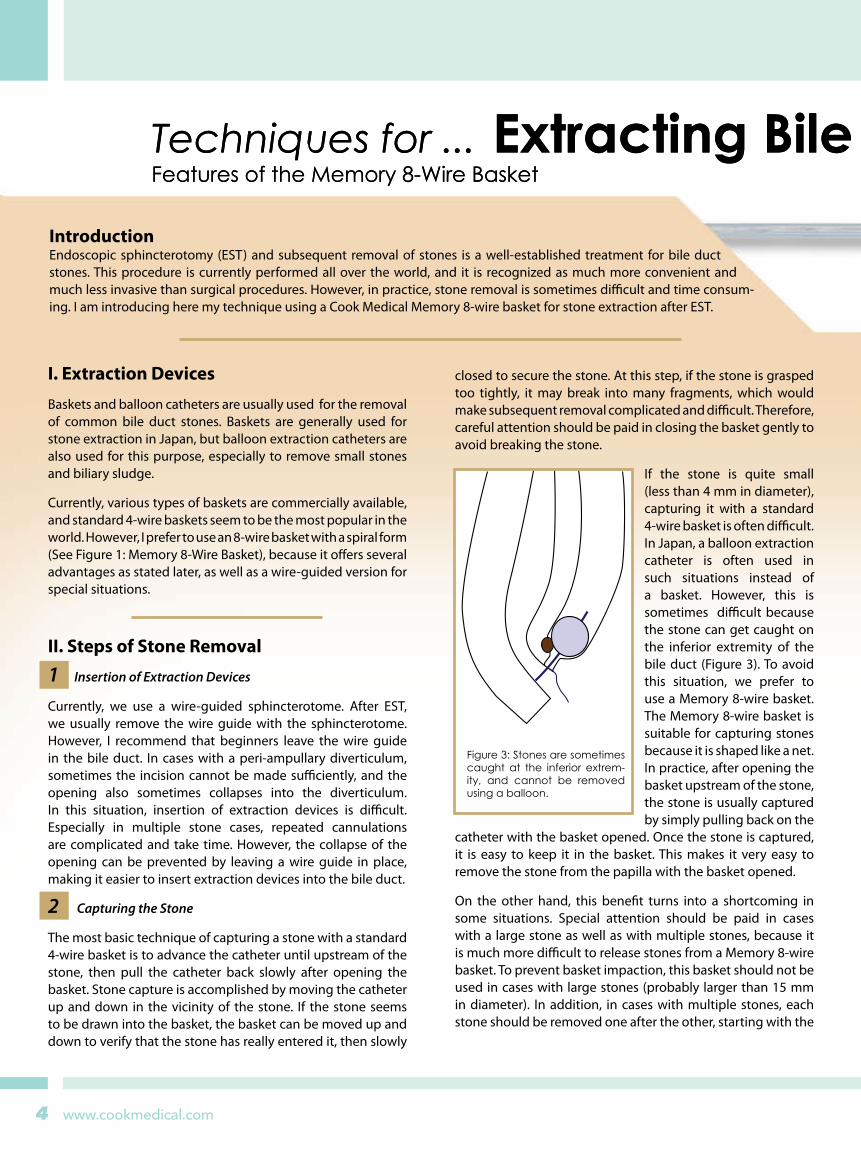
If the stone is quite small (less than 4 mm in diameter), capturing it with a standard 4-wire basket is often difficult. In Japan, a balloon extraction catheter is often used in such situations instead of a basket. However, this is sometimes difficult because the stone can get caught on the inferior extremity of the bile duct (Figure 3). To avoid this situation, we prefer to use a Memory 8-wire basket. The Memory 8-wire basket is suitable for capturing stones because it is shaped like a net. In practice, after opening the basket upstream of the stone, the stone is usually captured by simply pulling back on the
catheter with the basket opened. Once the stone is captured, it is easy to keep it in the basket. This makes it very easy to remove the stone from the papilla with the basket opened.
On the other hand, this benefit turns into a shortcoming in some situations. Special attention should be paid in cases with a large stone as well as with multiple stones, because it is much more difficult to release stones from a Memory 8-wire basket. To prevent basket impaction, this basket should not be used in cases with large stones (probably larger than 15 mm in diameter). In addition, in cases with multiple stones, each stone should be removed one after the other, starting with the
Techniques for ... Extracting Bile Duct StonesFeatures of the Memory 8-Wire Basket
IntroductionEndoscopic sphincterotomy (EST) and subsequent removal of stones is a well-established treatment for bile duct stones. This procedure is currently performed all over the world, and it is recognized as much more convenient and much less invasive than surgical procedures. However, in practice, stone removal is sometimes difficult and time consum-ing. I am introducing here my technique using a Cook Medical Memory 8-wire basket for stone extraction after EST.
Figure 3: Stones are sometimes caught at the inferior extrem-ity, and cannot be removed using a balloon.
I. Extraction Devices
Baskets and balloon catheters are usually used for the removal of common bile duct stones. Baskets are generally used for stone extraction in Japan, but balloon extraction catheters are also used for this purpose, especially to remove small stones and biliary sludge.
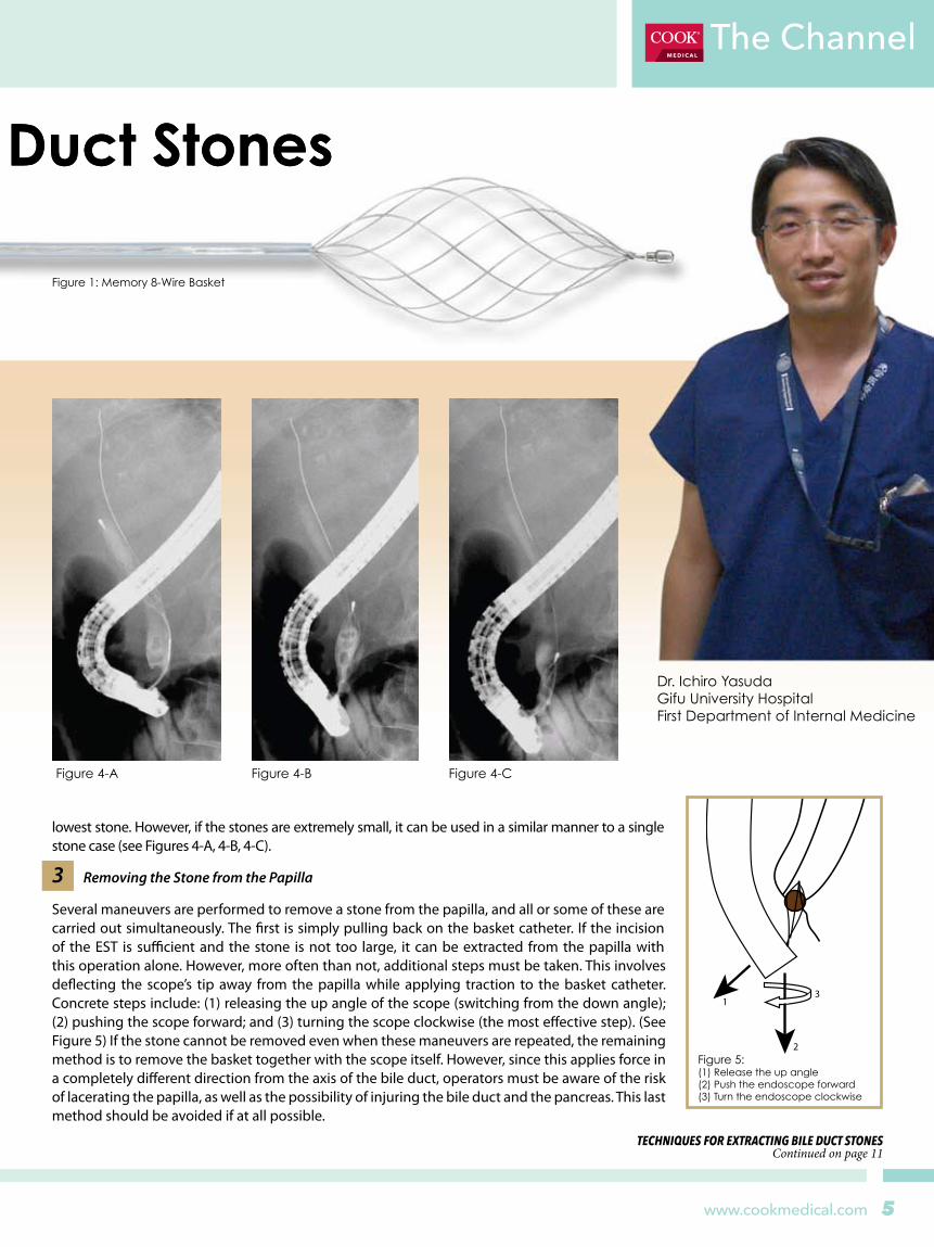
Currently, various types of baskets are commercially available, and standard 4-wire baskets seem to be the most popular in the world. However, I prefer to use an 8-wire basket with a spiral form (See Figure 1: Memory 8-Wire Basket), because it offers several advantages as stated later, as well as a wire-guided version for special situations.
II. Steps of Stone Removal
1 Insertion of Extraction Devices
Currently, we use a wire-guided sphincterotome. After EST, we usually remove the wire guide with the sphincterotome. However, I recommend that beginners leave the wire guide in the bile duct. In cases with a peri-ampullary diverticulum, sometimes the incision cannot be made sufficiently, and the opening also sometimes collapses into the diverticulum. In this situation, insertion of extraction devices is difficult. Especially in multiple stone cases, repeated cannulations are complicated and take time. However, the collapse of the opening can be prevented by leaving a wire guide in place, making it easier to insert extraction devices into the bile duct.
2 Capturing the Stone
The most basic technique of capturing a stone with a standard 4-wire basket is to advance the catheter until upstream of the stone, then pull the catheter back slowly after opening the basket. Stone capture is accomplished by moving the catheter up and down in the vicinity of the stone. If the stone seems to be drawn into the basket, the basket can be moved up and down to verify that the stone has really entered it, then slowly
Techniques for ... Extracting Bile Duct StonesFeatures of the Memory 8-Wire Basket
5
The Channel
www.cookmedical.com
Techniques for ... Extracting Bile Duct StonesFeatures of the Memory 8-Wire Basket
Dr. Ichiro YasudaGifu University HospitalFirst Department of Internal Medicine
Techniques for ... Extracting Bile Duct StonesFeatures of the Memory 8-Wire Basket
Figure 1: Memory 8-Wire Basket
Figure 4-A Figure 4-B Figure 4-C
lowest stone. However, if the stones are extremely small, it can be used in a similar manner to a single stone case (see Figures 4-A, 4-B, 4-C).
3 Removing the Stone from the Papilla
Several maneuvers are performed to remove a stone from the papilla, and all or some of these are carried out simultaneously. The first is simply pulling back on the basket catheter. If the incision of the EST is sufficient and the stone is not too large, it can be extracted from the papilla with this operation alone. However, more often than not, additional steps must be taken. This involves deflecting the scope’s tip away from the papilla while applying traction to the basket catheter. Concrete steps include: (1) releasing the up angle of the scope (switching from the down angle); (2) pushing the scope forward; and (3) turning the scope clockwise (the most effective step). (See Figure 5) If the stone cannot be removed even when these maneuvers are repeated, the remaining method is to remove the basket together with the scope itself. However, since this applies force in a completely different direction from the axis of the bile duct, operators must be aware of the risk of lacerating the papilla, as well as the possibility of injuring the bile duct and the pancreas. This last method should be avoided if at all possible.
TECHNIQUES FoR ExTRACTING BILE DUCT SToNES Continued on page 11
1
2
3
Figure 5: (1) Release the up angle(2) Push the endoscope forward(3) Turn the endoscope clockwiseJonathan Cohen, MD
Clinical Professor of Medicine New York University School of Medicine

www.cookmedical.com6
As Dr. Shou Jiang Tang read the patient file, he realized he and his colleagues faced an unusual and daunting challenge.
A 58-year-old male was referred for Barrett’s dysplasia. His past medical history included multiple neurological conditions, hypertension, rheumatoid arthritis, depression, bronchitis, Raynaud’s disease, Reiter’s syndrome, and seizure disorder. The patient was wheelchair bound. Three endoscopic sessions with biopsies demonstrated a long and circumferential segment of Barrett’s Esophagus (BE) of 14 cm with multi-focal high-grade dysplasia (HGD) and low-grade dysplasia (LGD) in the distal part of the BE. Two independent GI pathologists confirmed multifocal Barrett’s HGD.
The patient declined surgical resection after evaluation by two experienced cardiothoracic surgeons.
Only a few digestive disease centers in the world would—or could—address this complex set of demands. Fortunately for the patient, Dr. Tang, Assistant Professor of Internal Medicine at UT Southwestern Medical Center and Director of Endoscopy at Parkland Memorial Hospital, believed his team could achieve positive outcomes with endoscopic mucosal resection.
“After detailed discussion regarding non-surgical treatment options, including endoscopic radiofrequency ablation and photodynamic therapy, the patient chose complete and circumferential endoscopic mucosal resection,” recalls Dr. Tang. “The patient elected EMR even when we fully disclosed the significant risk of stricture formation. We planned two sessions of
EMR. The first session was for staging and therapeutic purpose, and the second session was for curative intent.
“With the availability of the Duette Multi-Band Mucosectomy device,” says Dr. Tang, "endoscopists are able to
perform EMR without pre-injection and repeated snare loading within the cap, with good endoscopic view, and requiring minimal
technical expertise. Although the Duette device is increasingly
being used for ablation of localized
lesions or short segment
BE, many endoscopists have doubts
about using Duette in removing long segments of BE.
We did not have those reservations.”
The UT Southwestern Division of Digestive and Liver Diseases is made up of nationally renowned clinicians, educators, and researchers. The division’s expertise in diagnosis, treatment, and endosurgical care is world-class. In addition to Barrett’s esophagus, the group has expertise in diseases such as hepatitis, cirrhosis and its complications, acute liver failure, gallstones, acid reflux disease (GERD), inflammatory bowel disease, gastrointestinal bleeding, pancreatitis, as well as gastrointestinal, esophageal and pancreatic cancer.
The division, part of the Department of Internal Medicine, has developed extremely active and robust clinical research programs, including in areas of acute liver failure (via the Acute Liver Failure Study Group, part of an eight-year national study sponsored by the NIH), drug induced liver injury (also NIH supported), primary biliary cirrhosis, gastrointestinal bleeding, and more.
The division’s physicians treat patients in the James. W. Aston Ambulatory Care Building, Parkland Memorial Hospital, UT Southwestern University Hospital and the Dallas Veterans Affairs Medical center. Dr. Tang credits much of his interest and expertise in the field to an early mentor, Dr. Norman Marcon, Director of the Therapeutic Endoscopy Training Program at St. Michael’s Hospital in Canada, one of the largest in North America. Dr. Tang also expresses appreciation to Dr. Don Rockey, Chief of the Division of Digestive and Liver Diseases at UT Southwestern, and to his colleagues Jayaprakash Sreenarasimhaiah, M.D., Assistant Professor of Internal Medicine, and Luis F. Lara, M.D., Assistant Professor of Internal Medicine.
Performing Circumferential EMR
at UT Southwestern Medical Center
World-class physicians & researchers
A choice: circumferential EMR

7
The Channel
www.cookmedical.com
During the first session of EMR, Dr. Tang circumferentially resected a 2-3 cm segment of BE with three Duette devices. Pathological examination of the EMR specimens revealed a mix of HGD and LGD with only mucosal involvement. Vertical margins were clear in all resected specimens.
“We performed the second session of EMR eight weeks later, after the patient recovered from an episode of pneumonia,” Dr. Tang reports. “One week prior to the second session of EMR, the patient developed an upper extremity deep venous thrombosis (DVT) that was related to an intravenous line placement, and the patient insisted on having the second session of EMR prior to anti-coagulation.”
During the second session of EMR, there was early squamous epithelization within the previously treated EMR territory in the distal esophagus. With ten Duette devices, a 10-11 cm BE was completely and circumferentially resected without wasting a single band during ligation and resection. Each band was placed on the BE immediately adjacent to the previously resected site and the EMR was performed in an upward and spiral fashion.
“In the end, a total of 68 bands were placed and the entire 12 cm of BE was completely resected without any hemorrhage or other immediate complications. There was not even a single miss or misfiring of the bands from the Duette device,” says Dr. Tang. “Every single band was used for mucosal resection.”
Ten days after the second session of EMR, anti-coagulation with Coumadin was started to treat the patient’s DVT, and a therapeutic range of INR was achieved. During the three-month EMR follow up, the patient denied any esophageal or chest discomfort and there was no GI bleeding since he has been on Coumadin.
During the follow-up endoscopy three months after the second EMR, there was no esophageal luminal narrowing, no apparent residual BE, ulceration, or any mass lesion seen. There was near complete squamous epithelialization within the mid-distal esophagus under white light and narrow band imaging.
“For Barrett’s HGD and mucosal cancer, endoscopic ablation such as EMR can offer a potential cure,” says Dr. Tang. “In patients with only mucosal involvement, the risk of lymph node metastasis is close to zero. In my opinion, compared with endoscopic ultrasound staging, EMR staging offers more precise assessment of the depth of tumor invasion into the submucosa.
“Our case,” concludes Dr. Tang, “demonstrates that complete and circumferential EMR can be conveniently performed in very long segment BE offering the advantages of: no pre-injection and repeated snare loading; good endoscopic view during the entire EMR procedure; contiguous mucosal resection without leaving Barrett’s islands between each resection; and it requires minimal technical expertise. There is a theoretical advantage that endoscopic mucosal resection with the Duette device can result in a lower incidence of residual Barrett’s Esophagus and recurrence due to complete resection.”
Resecting a 10-11 cm Barrett’s Esophagus
Positive results
Dr. Tang routinely performs circumferential EMR exclusively with Duette devices and
with good results. The bottom 3 images are from another patient during and after a 4cm
circumferential EMR with the Duette.

www.cookmedical.com8
We had patients staying in the hospital as boarders until the National Guard could escort them somewhere. Some patients stayed for a month or more.
“Fortunately, we had done a great deal of planning. We’ve experienced storms in the past that required evacuations. Each department had an emergency plan. Our procedures included setting up emergency teams from each specialty. I was on the endoscopy team.”
Even before New Orleans Mayor Ray Nagin declared a state of emergency and announced the city’s first ever mandatory evacuations, Dr. Etemad had made up his mind to remove his family from their home, and he drove them to safety in Houston.
On August 31, Dr. Etemad learned that Ochsner Medical Center had survived the storm relatively intact, and he returned to New Orleans to help the hospital and the community however he could.
The story of Ochsner Medical Center following Hurricane Katrina“A male patient in his mid 50s with obstructive jaundice was one of the first patients we saw following the storm. The major challenge was preparing for his ERCP. We had to be sure we had replacement stents, adequate staff, sufficient anesthesia and a safe place to dispose of hospital waste. But after the ERCP, I felt there was hope. It was like an ark needing to land.”
After Hurricane Katrina hit New Orleans in August 2005, hope was certainly a needed commodity at the Ochsner Medical Center. Nothing in Dr. Bob Etemad’s extensive training and experience prepared him for the conditions he would face in the next week, month, or year.
”I think that what we experienced here was unique for American physicians,” he said. “Within 24 to 48 hours we were confronted with a potential loss of all that was familiar.”
Normal conditions no longer existed
Handling family needs was a priority
Ochsner survives the storm
As he performed the ERCP procedure following the storm, Dr. Etemad fondly recalled the normal medical conditions from a few weeks prior. Normal conditions, however, no longer existed in New Orleans.
When Hurricane Katrina, a category 3 hurricane, made landfall on the northern Gulf coast, the resulting storm surge ravaged New Orleans and caused levee breaches that led to the costliest natural disaster and the worst engineering disaster in U.S. history. An estimated 80% of the city flooded with water up to 20 feet deep in some areas.
Ochsner Medical Center, however, was one of just three hospitals in the New Orleans area to remain open and to continue treating patients through Hurricane Katrina.
Dr. Etemad recalls: “Although a few of the operating rooms took on water from leaking roofs, the majority of the hospital survived without significant damage. One generator was lost, so we were without power for air conditioning in one part of the hospital or another for some time.
“After patients got better, there was often no place for them to go. Their homes were gone, and the hotels were all full.
9www.cookmedical.com
The ChannelThe Channel
He found most of the major roads traveling into and out of the city damaged. “There was lots of debris in the streets – nails, sheet metal, fallen tree branches and roof shingles were everywhere,” he says.
“The first week I was back at the hospital, I never once went outside,” he remembers. “Initially, the workload in our department was low. Patients didn’t have access into the city, and to the hospital. We saw urgent and emergent oncology cases, but we conducted only a few screenings. However, when word reached people that we had services, they began to arrive. Within four months, we were at 85% capacity. After six months, we were at full capacity.
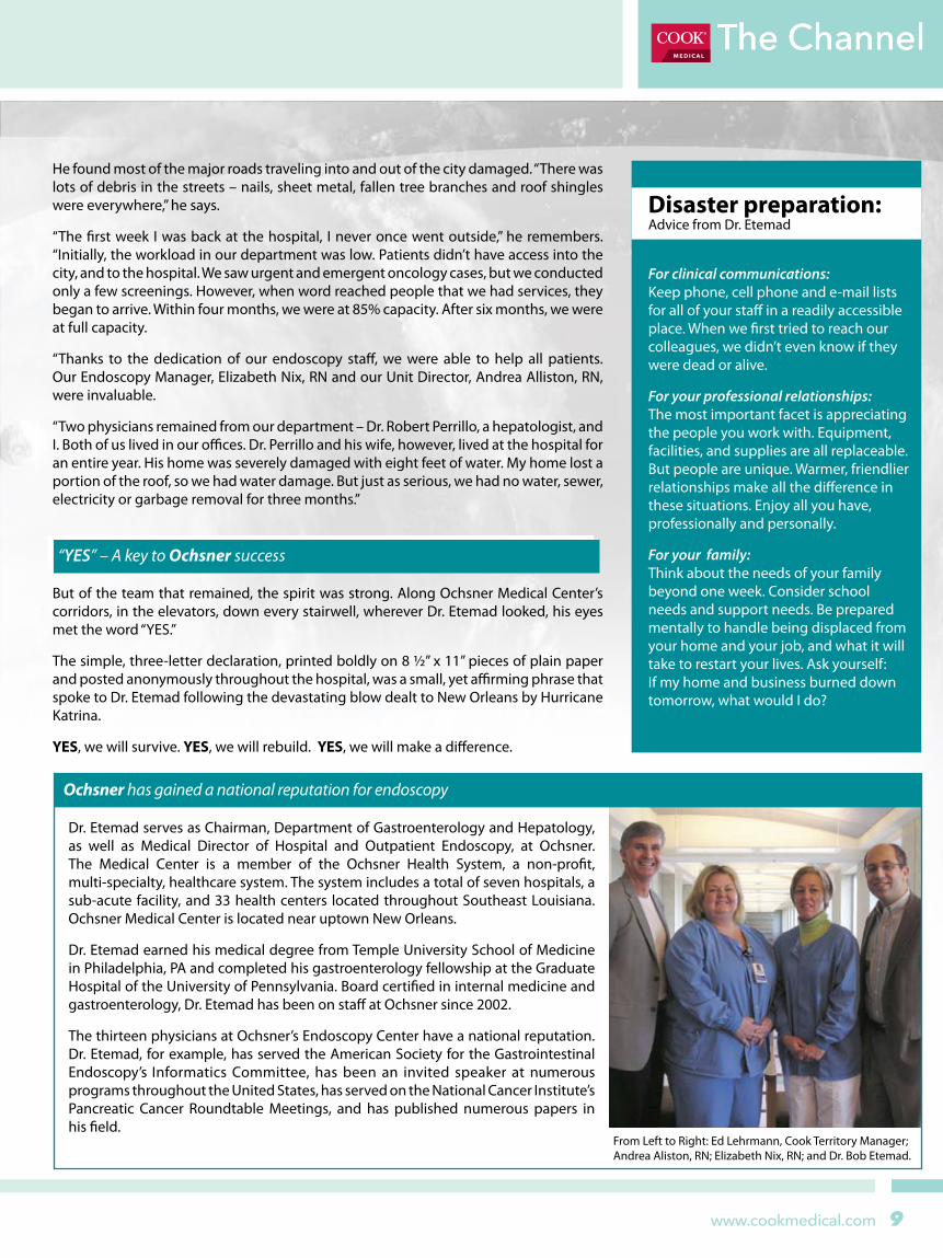
“Thanks to the dedication of our endoscopy staff, we were able to help all patients. Our Endoscopy Manager, Elizabeth Nix, RN and our Unit Director, Andrea Alliston, RN, were invaluable.
“Two physicians remained from our department – Dr. Robert Perrillo, a hepatologist, and I. Both of us lived in our offices. Dr. Perrillo and his wife, however, lived at the hospital for an entire year. His home was severely damaged with eight feet of water. My home lost a portion of the roof, so we had water damage. But just as serious, we had no water, sewer, electricity or garbage removal for three months.”
But of the team that remained, the spirit was strong. Along Ochsner Medical Center’s corridors, in the elevators, down every stairwell, wherever Dr. Etemad looked, his eyes met the word “YES.”
The simple, three-letter declaration, printed boldly on 8 ½” x 11” pieces of plain paper and posted anonymously throughout the hospital, was a small, yet affirming phrase that spoke to Dr. Etemad following the devastating blow dealt to New Orleans by Hurricane Katrina.
YES, we will survive. YES, we will rebuild. YES, we will make a difference.
“YES” – A key to Ochsner success
Disaster preparation: Advice from Dr. Etemad
For clinical communications: Keep phone, cell phone and e-mail lists for all of your staff in a readily accessible place. When we first tried to reach our colleagues, we didn’t even know if they were dead or alive.
For your professional relationships: The most important facet is appreciating the people you work with. Equipment, facilities, and supplies are all replaceable. But people are unique. Warmer, friendlier relationships make all the difference in these situations. Enjoy all you have, professionally and personally.
For your family: Think about the needs of your family beyond one week. Consider school needs and support needs. Be prepared mentally to handle being displaced from your home and your job, and what it will take to restart your lives. Ask yourself: If my home and business burned down tomorrow, what would I do?
From Left to Right: Ed Lehrmann, Cook Territory Manager; Andrea Aliston, RN; Elizabeth Nix, RN; and Dr. Bob Etemad.
Ochsner has gained a national reputation for endoscopy
Dr. Etemad serves as Chairman, Department of Gastroenterology and Hepatology, as well as Medical Director of Hospital and Outpatient Endoscopy, at Ochsner. The Medical Center is a member of the Ochsner Health System, a non-profit, multi-specialty, healthcare system. The system includes a total of seven hospitals, a sub-acute facility, and 33 health centers located throughout Southeast Louisiana. Ochsner Medical Center is located near uptown New Orleans.
Dr. Etemad earned his medical degree from Temple University School of Medicine in Philadelphia, PA and completed his gastroenterology fellowship at the Graduate Hospital of the University of Pennsylvania. Board certified in internal medicine and gastroenterology, Dr. Etemad has been on staff at Ochsner since 2002.
The thirteen physicians at Ochsner’s Endoscopy Center have a national reputation. Dr. Etemad, for example, has served the American Society for the Gastrointestinal Endoscopy’s Informatics Committee, has been an invited speaker at numerous programs throughout the United States, has served on the National Cancer Institute’s Pancreatic Cancer Roundtable Meetings, and has published numerous papers in his field.
www.cookmedical.com10
Approximately 5 million c o l o n o s c o p i e s a r e performed in the US every year, a large proportion of which are to diagnose the source of rectal bleeding. In the vast majority of cases, the source is identified as nothing more than internal hemorrhoids.
The simplest and most effective method of treating internal hemorrhoids is rubber band ligation. Prior to the Triview and ShortShot multiple hemorrhoidal ligator, physicians used conventional single-slot anoscopes and non-disposable single-shot ligators. This usually required 3 office visits, to ligate all 3 internal hemorrhoids. The introduction of the Triview anoscope and ShortShot multiple hemorrhoidal ligator simplifies the procedure of hemorrhoidal ligation and makes the procedure more convenient for the physician and patient alike.
Since most colonoscopies are performed to diagnose the source of rectal bleeding and internal hemorrhoids are the most common source, it makes clinical and economic sense to repair the source of the bleeding at the same time as the colonoscopy. This makes the diagnosis and treatment of hemorrhoidal bleeding a one-stop procedure. In addition, the patient is already sedated, is laying in the left lateral position and is under hemodynamic monitoring….what could be a better time to ligate the hemorrhoids than right there and then?
I have recently reported a series of 500 patients who underwent combined colonoscopy and 3-quadrant hemorrhoidal ligation using the Triview anoscope and ShortShot multiple hemorrhoidal ligator. Of the 500 patients treated over a 3 year period, 467 (or 93.4%) reported symptomatic relief from their hemorrhoids and required no further treatment. Thirty-three patients (6.6%), who had unusually large internal hemorrhoids, required repeat ligation. Eleven patients (2.2%) required hemorrhoidectomy for persistent symptoms. In the series of 500 patients, no patient experienced post ligation hemorrhage or post-ligation sepsis.
A sensible precaution before performing colonoscopy/ligation is to ensure the patient is not taking anticoagulants, does not have an immunosuppressant disorder such as untreated HIV/AIDS and has no concomitant disorder such as fissure, fistula or Crohn’s disease.
The new procedure of combined colonoscopy and multiple hemorrhoidal ligation is therefore a sensible and logical addition to routine colonoscopy. The procedure is safe, effective and more convenient for the patient and physician alike. In an era of cost-conscious healthcare delivery, it is also a cost–effective means of treating one of the most common GI complaints.
Welcome to a new section in The Channel where we present a clinical image and ask you to participate.
Jason D. Conway, MD, MPH Assistant Professor of
Medicine Wake Forest UniversityBaptist Medical Center
A 67 year old man had an MRI to evaluate liver lesions seen incidentally on a non-contrasted CT performed for kidney stones. The MRI revealed a 1.4 cm enhancing lesion in the tail of the pancreas with decreased signal centrally consistent with necrosis (see Figure 1). The patient was completely asymptomatic. Labs including LFT’s, amylase and lipase, and CA 19-9 were all normal. EUS revealed a 13.7 mm hypoechoic well defined mass in the tail of the pancreas with a central anechoic area immediately adjacent to the splenic vein (see Figure 2). EUS-guided FNA was performed using the 25 gauge Echotip Ultra Endoscopic Ultrasound needle. Cytology revealed monotonous-appearing cells containing nuclei with a “salt and pepper” appearance which were positive for CYKAE1/AE3, CD56, synaptophysin, and chromogranin.
What is the diagnosis?
*fig.1
*fig.2
To confirm your diagnosis, click on newsletter button on endoscopy homepage of www.cookmedical.com <http://www.cookmedical.com>
We welcome your participation in this column. Please contact Kevin Chmura at [email protected].
David N. Armstrong, M.D. FRCS FACS FASCRS Program Director, Georgia Colon and Rectal Surgical Clinic, Atlanta GA
11
The Channel
www.cookmedical.com
Techniques for ... Extracting Bile Duct StonesFeatures of the Memory 8-Wire Basket
4 Releasing the Captured Stone
In cases with multiple stones, it is necessary to insert the extraction device into the bile duct repeatedly to remove the stones, each time releasing the captured stone in the duodenum. Stones can be released relatively easily with a standard 4-wire basket by opening the basket completely and shaking the stones out by moving the catheter back and forth quickly. However, when a Memory 8-wire basket is used, it is difficult to release the stones using only this method. In such cases, one should press the tip of the basket against the duodenal wall beside the papilla with the basket fully opened. This maneuver opens the basket elliptically, with the basket wires coming away from the stone. As a result, the stone can be released easily by closing the basket in this position. (Figure 6).
Although more difficult, the same technique can be used to release a stone from a Memory 8- wire basket in the bile duct by pressing the basket’s tip against the bile duct wall.
ConclusionI have introduced the technique to remove bile duct stones using a Memory 8-wire basket. I hope that these techniques are helpful for beginners.
Figure 6: Press the basket against the duodenal wall and release the stone
We work in a referral center for liver transplantation (LT) in Northern Italy where diagnosis and treatment of biliary complications after LT is a frequent challenge. ERCP is now considered the first approach to confirm the presence of an anastomotic stricture and to proceed to its treatment which is carried out with a high rate of success. As recent data suggests, progressive increase of the number of plastic stents placed side-by-side across the stricture (multi-stenting) achieves a prolonged clinical remission.
In one case, a 52-year-old patient presented with itching and progressive impairment of cholestasis six months after LT for HCV cirrhosis performed at our center. An anastomotic biliary stricture was suggested by magnetic resonance cholangiography. During ERCP, a tight stricture was confirmed. A dilation with a 4-mm hydrostatic balloon catheter was performed and a single 10-French plastic stent placed across the anastomosis. In a few weeks, the itching resolved and the cholestasis markedly improved. Therefore, as it is our common practice, we
performed endoscopic multi-stenting therapy, in which we progressively increase the number of plastic stents placed side-by-side across the stricture every three months for one year. The Fusion System was used (Fig 1 and 2) and our patient was successfully treated with no recurrence of itching and cholestasis.
Endoscopic therapy consists of placing a single plastic stent across the anastomosis as the first step, and then multi-stenting for one year with stent exchanges at three-month intervals to prevent clogging. When we used the traditional 480-cm wire guide, we dilated the residual stricture first and placed more multiple stents thereafter, being limited by the maximal diameter of the balloon dilator available and by the need of multiple insertions of the catheter through the papilla and the strictured/angulated anastomosis, alongside stents already in place. The Fusion System has
changed our traditional approach and led to better clinical results in a shorter time and with minimal discomfort for the patient. With the Fusion System, the sequence of steps to increase the number of stents has changed. After removal of the occluded stents, we now 1) place first the new stents with the same number as the previous procedure less one, 2) dilate the residual stricture with the balloon dilator alongside the stents already in place across the anastomosis (Fig 1 and 3) add stents one-by-one according to maximal diameter of the CBD (Fig 2 and 3). These procedural changes were made possible by the technical characteristics of the Fusion System
which allows us to maintain a stable access to the biliary tree during all steps of the procedure and by the use of intraductal exchange achieving the placement of multiple stents with one short wire guide. We have used the Fusion System in more than twenty procedures. Only one dilatation per session was needed with little discomfort for the patient and no stent displacement. A higher number of stents, three to five, were placed at the time of final treatment and a wider patency of the biliary anastomosis was seen at one year after stent removal. The nurses on our staff have commented on the ease of use with this short wire guide system and leads to shortening of procedures.
fig.2
fig.1
fig.3
Improvement in multi-stenting of
after liver transplantation using Fusion Systembiliary anastomotic strictures
Dr. Paolo Cantù, MD; Prof. Roberto Penagini, MD Chair of Gastroenterology, Università degli Studi, U.O. Gastroenterologia 2
Dr. Cantú, MD Prof. Penagini, MD
Continued from page 5

www.cookmedical.com12
Techniques for ... Extracting Bile Duct StonesFeatures of the Memory 8-Wire BasketTechniques for ... Extracting Bile Duct StonesFeatures of the Memory 8-Wire Basket
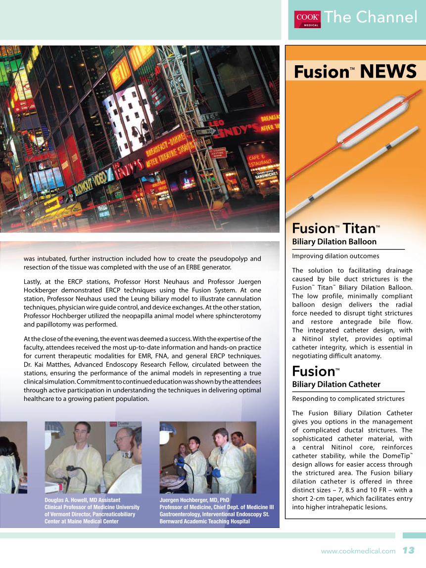
As pedestrians walked up and down Times Square preparing for the gift giving season, dedicated physicians and fellows attended the 31st Annual NYSGE New York Course: From the Podium to your Practice, held at the Marriott Marquis. The Advanced Therapeutic Endoscopy: EMR, EUS, and ERCP session was held Thursday, December 13, 2007. During this educational event, participants received valuable information focused on use of equipment and key techniques, as well as an interactive hands-on session with faculty using animal models to simulate the clinical setting. Upon completion of the course, participants received AMA continuing education credits.
The evening began with Dr. Charles Lightdale delivering a powerpoint presentation on the Duette, for use with endoscopic mucosal resection, followed by Dr. Michelle Kim, the course director, covering the different modalities for EUS sampling, and finally Professor Horst Neuhaus discussing ERCP techniques using the Fusion System. At the conclusion of the lectures, the attendees were divided into six groups
where invaluable one-on-one usage of the equipment, from the initial setup of the device to complete functional use was performed. The hands-on stations included two of each: EMR, EUS, and ERCP.
At the EUS stations, Dr. Kim and Dr. Lightdale demonstrated the ECHOTIP Ultra with HDFNA while using a Pentax linear ultrasound scope and Hitachi processor. The animal model provided true lesions that allowed the physician to perform FNA with the new ECHOTIP Ultra HD needle. With the HD technology, the physician sampled the targeted area with confidence and was successful in obtaining simulated cells.
The EMR station was represented by Dr. Jonathan Cohen and Dr. Doug Howell. For this session, the Duette, the DT-6-5F which is compatible with a 2.8 mm accessory channel, was used in conjunction with the Pentax gastroscope. Dr. Cohen and Dr. Howell began the hands-on training with initial set up, demonstrating device preparation. Once the animal model
Charles J. Lightdale, MDProfessor of Clinical MedicineNew York Presbyterian Medical CenterColumbia Campus
Michelle Kang Kim, MD, MScAssistant Professor of MedicineDirector, Endoscopic UltrasoundMount Sinai School of Medicine
Horst Neuhaus, MDProfessor of MedicineHead, Dpt. of Internal MedicineEvangelisches Krankenhaus Düsseldorf
Jonathan Cohen, MDClinical Professor of Medicine New York University School of Medicine
13
The Channel
www.cookmedical.com
Techniques for ... Extracting Bile Duct StonesFeatures of the Memory 8-Wire BasketTechniques for ... Extracting Bile Duct StonesFeatures of the Memory 8-Wire Basket
Douglas A. Howell, MD Assistant Clinical Professor of Medicine University of Vermont Director, Pancreaticobiliary Center at Maine Medical Center
Juergen Hochberger, MD, PhD Professor of Medicine, Chief Dept. of Medicine IIIGastroenterology, Interventional Endoscopy St. Bernward Academic Teaching Hospital
was intubated, further instruction included how to create the pseudopolyp and resection of the tissue was completed with the use of an ERBE generator.
Lastly, at the ERCP stations, Professor Horst Neuhaus and Professor Juergen Hockberger demonstrated ERCP techniques using the Fusion System. At one station, Professor Neuhaus used the Leung biliary model to illustrate cannulation techniques, physician wire guide control, and device exchanges. At the other station, Professor Hochberger utilized the neopapilla animal model where sphincterotomy and papillotomy was performed.
At the close of the evening, the event was deemed a success. With the expertise of the faculty, attendees received the most up-to-date information and hands-on practice for current therapeutic modalities for EMR, FNA, and general ERCP techniques. Dr. Kai Matthes, Advanced Endoscopy Research Fellow, circulated between the stations, ensuring the performance of the animal models in representing a true clinical simulation. Commitment to continued education was shown by the attendees through active participation in understanding the techniques in delivering optimal healthcare to a growing patient population.
Biliary Dilation Balloon
Improving dilation outcomes
The solution to facilitating drainage caused by bile duct strictures is the Fusion™ Titan™ Biliary Dilation Balloon. The low profile, minimally compliant balloon design delivers the radial force needed to disrupt tight strictures and restore antegrade bile flow. The integrated catheter design, with a Nitinol stylet, provides optimal catheter integrity, which is essential in negotiating difficult anatomy.
Biliary Dilation Catheter
Responding to complicated strictures
The Fusion Biliary Dilation Catheter gives you options in the management of complicated ductal strictures. The sophisticated catheter material, with a central Nitinol core, reinforces catheter stability, while the DomeTip™ design allows for easier access through the strictured area. The Fusion biliary dilation catheter is offered in three distinct sizes – 7, 8.5 and 10 FR – with a short 2-cm taper, which facilitates entry into higher intrahepatic lesions.
Fusion TM NEWS
Fusion TM Titan TM
Fusion TM

www.cookmedical.com14
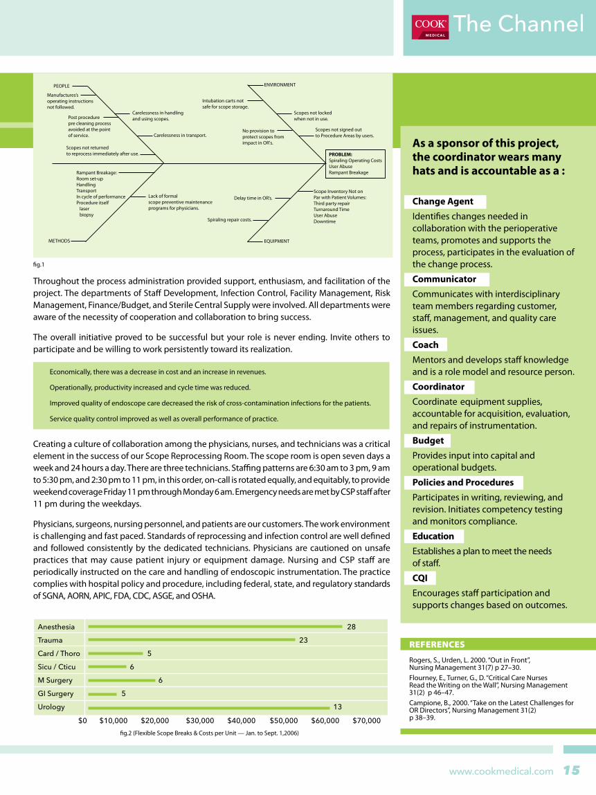
The operating rooms involved were: Urology, Cardiac, Thoracic, Otolaryngology, Pediatric, Gastroenterology, all of the Surgical ICU areas, M inor Surger y, and Shock Trauma OR and ICU’s. Involved teams from each of these perioperative areas met with the sponsor to brainstorm and gather data relevant to the specific issues. Cause and effect diagrams (fig. 1) were compiled to address the root causes hindering performance associated with these critical issues. The defined problem was identified. It was then decided which major systems and processes contributed to the problem. The most commonly used systems and processes are people, material, policy/methods, machine/technology, and environment. The team addressed the what, why, when, and where of the cause/effect. Discussion evolved looking for possible causes of the problem in each class. Typically there were 4-5 causes for each problem. These root causes were categorized and prioritized, selecting those with the greatest impact and verifying them with data.
A root cause analysis was completed and the following action goals were set to improve performance and reduce the risk of these problems reoccurring.
Establish policies for reprocessing and high level disinfection of endoscopes.
Prepare competency levels for the reprocessing of endoscopes, testing all involved staff for individual performance and compliance.
Centralize the reprocessing and storage of endoscopes into one area.
Define job descriptions, skill mix levels, and workload measures for dedicated personnel in the reprocessing area.
Establish a mentoring program for all perioperative staff to increase knowledge of endoscope anatomy, care, handling, and reprocessing.
Plan and initiate an effective quality performance improvement program with an emphasis on infection control and patient cross-contamination risks.
Prepare a formal proposal to present to our administration to activate the sponsor job description and carry out goals.
A centrally located storyboard was used to communicate information to the staff. The storyboard displays visually the project initiative, why the initiative was selected, and results of the root cause analysis. Actions that were taken and graphic evaluations (fig. 2) were also displayed and updated monthly. Specific problems within each perioperative area were pinpointed to validate their significance and to motivate staff to improve their performance.
N E W S F R O M
Society of International Gastroenterological Nurses and Endoscopy AssociatesSIGNEAN E W S F R O M
Improving Quality and Performance Practices When Using Endoscopes in Perioperative Areas: A Case Study
Bettie Jean Howard RN, CGRN, Scope Coordinator, Perioperative Services University of Maryland Medical Center, Baltimore, Maryland 21201, USA
ABSTRACT
Randomized Clinical Trial
Today’s health care environment relies on groups to reach its goals. Quality functioning of these groups improves organizational effectiveness. Quality Improvement tools are discussed. These tools help to identify causes of problems, evaluate the variability of performance, prioritize actions, visualize work processes, and assist the team identifying the root cause of the problem. The success of the team in accomplishing its goals and objectives depended on the collaboration and accomplishments of its members.
In response to the impact of health care reform and cross-country sentinel events, critical issues within the perioperative areas of the University of Maryland Medical Center (UMMC) were addressed. Analysis of these situations using endoscopic inventory data revealed delays in service, unreliable quality controlled reprocessing systems, a lack of knowledge among staff in handling and caring for endoscopes, and rampant breakage. As a result of my expertise and strong Society of Gastroenterology Nurses and Associates (SGNA) background, I was chosen to sponsor a project to address these issues. During an open dialog with the director of perioperative services, an initiative goal was agreed upon. The performance practices of the perioperative staff, reprocessing or using endoscopes, would be defined, modified, audited, communicated, and evaluated.
The process began by assessing each perioperative area and setting a direction for the project using the following guidelines:
What will be the proposed initiative?
What and who will be involved?
What technologies, if any, will be proposed?
What process will be changed with the implementation of the initiative?
What benefits will occur and what will the indicators be that show the initiative has been successful?
Will the initiatives influence other departments?
What can the sponsor of the initiative expect?
15
The Channel
www.cookmedical.com
PROBLEM:Spiraling Operating CostsUser AbuseRampant Breakage
Scope Inventory Not on Par with Patient Volumes:Third party repairTurnaround TimeUser AbuseDowntime
Delay time in OR’s.
Scopes not signed outto Procedure Areas by users.
Intubation carts not safe for scope storage.
Scopes not locked when not in use.
No provision to protect scopes from impact in OR's.
ENVIRONMENT
EQUIPMENT
Rampant Breakage:Room set-upHandlingTransportIn cycle of performanceProcedure itself laser biopsy
Spiraling repair costs.
PEOPLE
METHODS
Carelessness in transport.
Carelessness in handlingand using scopes.
Lack of formalscope preventive maintenanceprograms for physicians.
Manufactures’s operating instructionsnot followed.
Post procedurepre cleaning processavoided at the point of service.
Scopes not returned to reprocess immediately after use.
fig.1
fig.2 (Flexible Scope Breaks & Costs per Unit — Jan. to Sept. 1,2006)
As a sponsor of this project, the coordinator wears many hats and is accountable as a :
Change Agent
Identifies changes needed in collaboration with the perioperative teams, promotes and supports the process, participates in the evaluation of the change process.
Communicator
Communicates with interdisciplinary team members regarding customer, staff, management, and quality care issues.
Coach
Mentors and develops staff knowledge and is a role model and resource person.
Coordinator
Coordinate equipment supplies, accountable for acquisition, evaluation, and repairs of instrumentation.
Budget
Provides input into capital and operational budgets.
Policies and Procedures
Participates in writing, reviewing, and revision. Initiates competency testing and monitors compliance.
Education
Establishes a plan to meet the needs of staff.
CQI Encourages staff participation and supports changes based on outcomes.
REFERENCES
Rogers, S., Urden, L. 2000. “Out in Front”, Nursing Management 31(7) p 27–30.Flourney, E., Turner, G., D. “Critical Care Nurses Read the Writing on the Wall”, Nursing Management 31(2) p 46–47.Campione, B., 2000. “Take on the Latest Challenges for OR Directors”, Nursing Management 31(2) p 38–39.
Anesthesia 28
Trauma 23
Card / Thoro 5
Sicu / Cticu 6
M Surgery 6
GI Surgery 5
Urology 13
$0 $10,000 $20,000 $30,000 $40,000 $50,000 $60,000 $70,000
Throughout the process administration provided support, enthusiasm, and facilitation of the project. The departments of Staff Development, Infection Control, Facility Management, Risk Management, Finance/Budget, and Sterile Central Supply were involved. All departments were aware of the necessity of cooperation and collaboration to bring success.
The overall initiative proved to be successful but your role is never ending. Invite others to participate and be willing to work persistently toward its realization.
Economically, there was a decrease in cost and an increase in revenues.
Operationally, productivity increased and cycle time was reduced.
Improved quality of endoscope care decreased the risk of cross-contamination infections for the patients.
Service quality control improved as well as overall performance of practice.
Creating a culture of collaboration among the physicians, nurses, and technicians was a critical element in the success of our Scope Reprocessing Room. The scope room is open seven days a week and 24 hours a day. There are three technicians. Staffing patterns are 6:30 am to 3 pm, 9 am to 5:30 pm, and 2:30 pm to 11 pm, in this order, on-call is rotated equally, and equitably, to provide weekend coverage Friday 11 pm through Monday 6 am. Emergency needs are met by CSP staff after 11 pm during the weekdays.
Physicians, surgeons, nursing personnel, and patients are our customers. The work environment is challenging and fast paced. Standards of reprocessing and infection control are well defined and followed consistently by the dedicated technicians. Physicians are cautioned on unsafe practices that may cause patient injury or equipment damage. Nursing and CSP staff are periodically instructed on the care and handling of endoscopic instrumentation. The practice complies with hospital policy and procedure, including federal, state, and regulatory standards of SGNA, AORN, APIC, FDA, CDC, ASGE, and OSHA.
18804/010816 www.cookmedical.com
An official publication of Cook Endoscopy.
4900 Bethania Station Rd., Winston-Salem, NC 27105 P: 336-744-0157 F: 336-744-5785
If you would like to submit material for The Channel, please email us at [email protected]. We welcome your comments and suggestions.
upcoming 2008 events10th Symposium of Diagnostic & Therapeutic Endoscopy Dusseldorf, Germany Feb. 8-9
IU Hepato-Pancreato-Biliary Disease Scottsdale, AZ Feb. 11-13
Rocky Mountain Interventional Endoscopy Denver, CO Feb. 14-16
Chulalongkorn Hospital Workshop Bangkok, Thailand Feb. 19-21
Belgian Gastroenterology Week Antwerp, Belgium Feb. 21-23
Gastroenterology & Hepatology Linked Endoscopy Scottsdale, AZ Feb. 26-
Mar. 2
University Hospital Endoscopy 2008 Kuala Lumpur, Malaysia
Feb. 29- Mar. 2
Canadian DDW Quebec Feb. 29- Mar. 3
Journées Francophones de Pathologie Digestive - JFPD Paris, France Mar. 8-12
British Gastroenterology Week Birmingham, UK Mar. 11-13
Therapeutic Endoscopy-Brigham & Womens/Univ. Utah Park City, Utah Mar. 13-16
Endoscopy Unit Directors - Georgetown Washington, DC Mar. 30
Euro EUS Milano, Italy April 3-5
5th Gastroenterology & Endoscopy Congress Panama City, Panama April 3-5
DDW San Diego, CA May 18-21
SGNA Salt Lake City, UT May 23-25
Attention GI Nurses!As part of an ongoing
commitment to supporting your educational development and
expertise in your nursing practice, we are pleased to announce that the
American Board of Certification for Gastroenterology Nurses (ABCGN) has reviewed our educational activities and recognized them as offering GI specific content. This recognition is
being added to the accreditation sec-tion provided with the descriptions
of our educational activities.
Available activities include: Endoscopic Polypectomy
Options for Enteral FeedingMalignant Biliary Disease
Biliary Stone ManagementPrimary Sclerosing Cholangitis
If you are interested in any of these educational topics and
would like to schedule an activity presentation, please contact your
COOK Endoscopy territory manager for more information.
Continuing Nursing Education activities are sponsored by