the challenges of sequencing ffpe dna using ngs hazel ingram
TRANSCRIPT

The Challenges Of Sequencing FFPE DNA Using
NGS
Hazel Ingram

Problems with FFPE
Poor quality/fragmented DNA Fixation artefacts Insufficient tumour material Low tumour content Measuring Quality
• Nanodrop vs Qubit
• Fragmentation
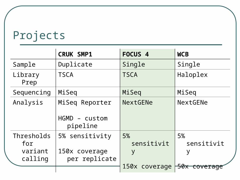
Projects
CRUK SMP1 FOCUS 4 WCB
Sample Duplicate Single Single
Library Prep TSCA TSCA Haloplex
Sequencing MiSeq MiSeq MiSeq
Analysis MiSeq Reporter NextGENe NextGENe
HGMD – custom pipeline
Thresholds for variant calling
5% sensitivity
150x coverage per replicate
5% sensitivity
150x coverage
5% sensitivity
50x coverage

Validation Determine acceptable coverage for regions
of interest
To match or increase sensitivity compared to original methods (pyro and sanger)
Low level of false positives
Must have at least 90% concordance

CRUK SMP1 Validation Overview Illumina validation:
• 42 samples sent:• 37/42 matched (88%)• 3/42 non concordant
• low coverage• 2/42 non concordant
• design issue In House validation:
• 45 samples• 43 matched (95%)• 2/45
• low coverage• Overall, between hubs 18 extra variants previously undetected in 124 samples
• Technological fail of pyro/sanger• Higher sensitivity of NGS
Large number of artefacts detected Failure rate: approx 10%
Artefacts
Low Coverage
Technological Design
Higher Sensitivity of NGS

Artefacts Duplicate Testing Solution = testing of each sample
in duplicate Sequencing artefacts only seen
in 1 replicate
True variants seen in both replicates Pipeline - only sequence variants
observed in both replicates retained for analysis
StrategyAverage number of variants per sample
Range in number of variants per sample
Singlicate testing 113 3-546
Duplicate testing 30 0-60
* Duplication used for CRUK panel only

Artefacts - Sanger Seq False Positive (deamination)
KIT c.1745G>A p.Trp582X – CRUK SMP
Mutation not detected by NGS (single testing: >4000x)
Mean coverage across panel: 4840x
KIT re-tested by Sanger…no mutation detected
C>T deamination artefact caused by formalin fixation
…duplicate testing solves this issue
UDG treatment prior to PCR may help removes uracil lesions by hydrolysing N-glycosidic bond

Minimum Coverage: CRUK & FOCUS4
Known variants became undetectable when coverage for region of interest <150x
150x minimum coverage cut-off implemented • For CRUK: <150x coverage in either replicate = failed
exon
• Avoided reporting false negative results
• Failure rates were still comparable to original testing

Design
CRUK (TSCA)• PTEN 27bp del missed
• Deletion removed probe binding site
FOCUS4 (TSCA)• NRAS c.182A>G p.Q61R
• Region had zero reads poor amplification
WCB (Haloplex)• PIK3CA ex9 c.1634A>G, p.E545G missed
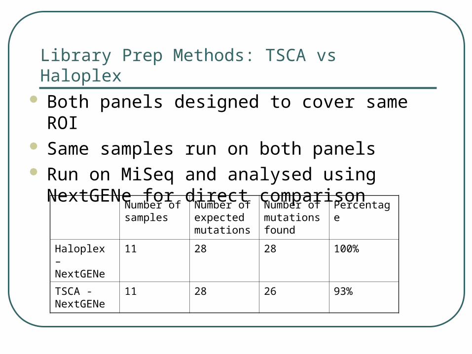
Library Prep Methods: TSCA vs Haloplex Both panels designed to cover same ROI Same samples run on both panels Run on MiSeq and analysed using NextGENe for
direct comparison
Number of samples
Number of expected mutations
Number of mutations found
Percentage
Haloplex – NextGENe
11 28 28 100%
TSCA - NextGENe
11 28 26 93%

TSCA vs Haloplex

Projects
CRUK SMP FOCUS 4 WCB
Sample Duplicate Single Single
Library Prep TSCA TSCA Haloplex
Sequencing MiSeq MiSeq MiSeq
Analysis MiSeq Reporter NextGENe NextGENe
HGMD – custom pipeline
Thresholds for variant calling
5% sensitivity
150x coverage per duplicate
5% sensitivity
150x coverage
5% sensitivity
50x coverage
Conclusions and Future Work DNA quality from FFPE tissue is a major
challenge Help interpretation by:
• Running in duplicate
• being careful with assay design and minimum coverage
Additional steps e.g. UDG treatment can help minimise deamination artefacts
Need a more robust NGS technology for low quality DNA from FFPE
• Alternative NGS platforms / providers
• Alternative enrichment methods e.g. target capture as alternative to PCR based

Acknowledgements Alex Stretton James Eden Helen Roberts Rachel Butler Matt Mort (HGMD) The All Wales Medical Genetics Service