the cellular level of organization - university of illinois · the cellular level of organization...
TRANSCRIPT
The original lecture presentation by Lee Ann Frederick (University of Texas at Arlington) has been extensively modified for use in MCB 244 & 246 by Drs. Kwast & Brown, University of Illinois at Champaign-Urbana (2016–2017)
Chapter 3The Cellular Level
of Organization
© 2015 Pearson Education, Inc.

3-1 An Introduction to Cells: Learning Outcomes
• Understand the basic anatomy & physiology (function) of the cell, including structure/function of the plasma membrane and cellular organelles (Please use the publisher’s resources to review basic cell biology concepts presented in sections 1 – 4 [pp. 64 – 87]).
• Understand the difference between osmolarity and tonicity.
• Understand and be able to describe the different types of membrane transport (passive vs. active; uniport vs. symport vs. antiport; endocytosis vs. exocytosis).
• Describe the overall function of cellular transport systems.2

3-1 An Introduction to Cells• Cell Theory
• Developed from Robert Hooke’s research• Cells are the building blocks of all plants and animals• All cells come from the division of preexisting cells• Cells are the smallest units that perform all vital physiological
functions• Each cell maintains homeostasis at the cellular level
• Two General Classes of Cells:
1. Germ (sex) cells a. Spermb. Oocyte
2. Somatic cells (soma = body) = All body cells except sex cells3

3-1 Defining the Cell’s BoundaryPlasma membrane (cell membrane) separates cytoplasm from the
extracellular fluid (interstitial fluid) • Cytoplasm = all materials within the cell except for the nucleus
• Cytosol = liquid• Intracellular structures collectively known as organelles
• Functions of the Plasma Membrane• Structural support
• Anchors cells and tissues
• Physical isolation• Barrier
• Regulates exchange with environment• Ions and nutrients enter• Wastes eliminated and cellular products released
• Monitors the environment• Extracellular fluid composition• Chemical signals
4
3-1 & 3-2 The Cytosol & Organelles• Cytosol (fluid)
• Dissolved materials: nutrients, ions, proteins, and wastes• High potassium/low sodium (Na+/K+-ATPase pump)• High protein• High carbohydrate/low amino acid and fat (the latter found primarily in
adipose tissue)
• Organelles• Membranous
• endoplasmic reticulum (ER), the Golgi apparatus, lysosomes, peroxisomes, and mitochondria
• Non-membranous• Includes the cytoskeleton, microvilli, centrioles, cilia, ribosomes, and
proteasomes5

The Anatomy of a Model Human Cell (Fig 3-1)
6
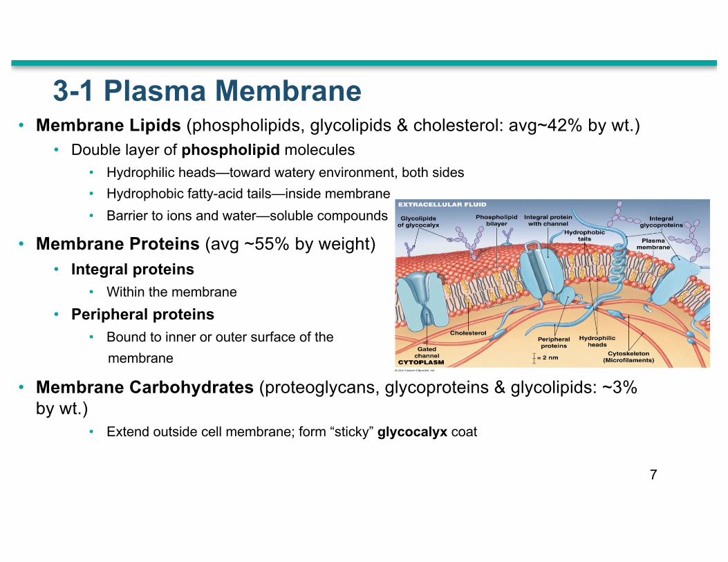
3-1 Plasma Membrane• Membrane Lipids (phospholipids, glycolipids & cholesterol: avg~42% by wt.)
• Double layer of phospholipid molecules• Hydrophilic heads—toward watery environment, both sides• Hydrophobic fatty-acid tails—inside membrane • Barrier to ions and water—soluble compounds
• Membrane Proteins (avg ~55% by weight)• Integral proteins
• Within the membrane • Peripheral proteins
• Bound to inner or outer surface of the membrane
• Membrane Carbohydrates (proteoglycans, glycoproteins & glycolipids: ~3% by wt.)
• Extend outside cell membrane; form “sticky” glycocalyx coat
7
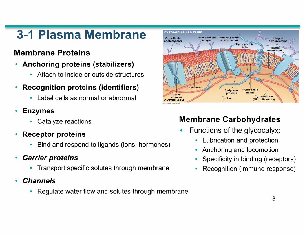
Membrane Carbohydrates• Functions of the glycocalyx:
• Lubrication and protection• Anchoring and locomotion• Specificity in binding (receptors)• Recognition (immune response)
3-1 Plasma MembraneMembrane Proteins• Anchoring proteins (stabilizers)
• Attach to inside or outside structures
• Recognition proteins (identifiers) • Label cells as normal or abnormal
• Enzymes • Catalyze reactions
• Receptor proteins• Bind and respond to ligands (ions, hormones)
• Carrier proteins • Transport specific solutes through membrane
• Channels• Regulate water flow and solutes through membrane
8
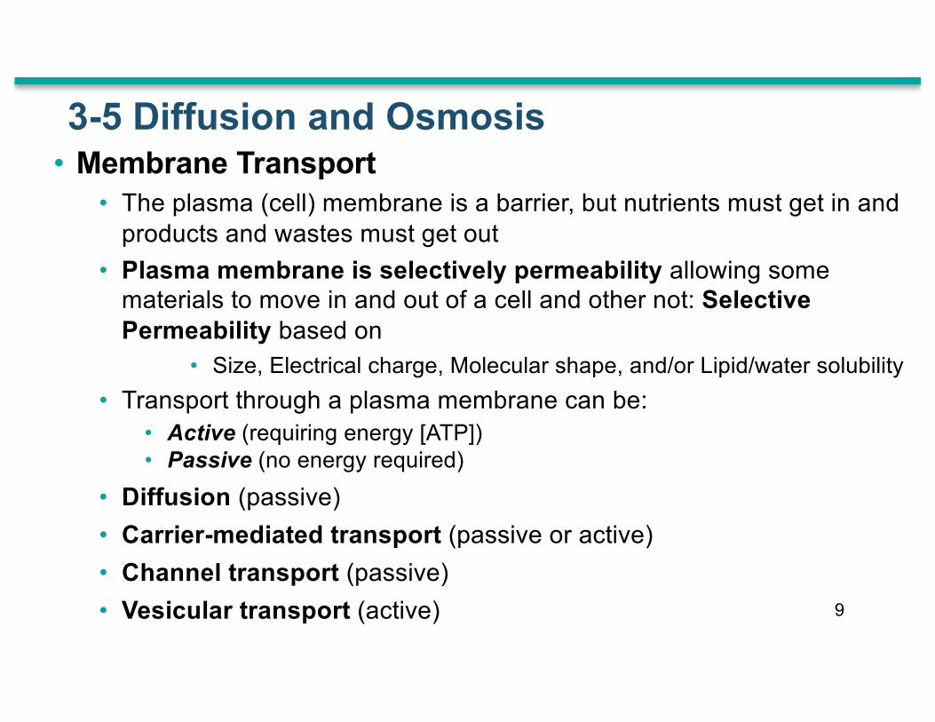
3-5 Diffusion and Osmosis• Membrane Transport
• The plasma (cell) membrane is a barrier, but nutrients must get in and products and wastes must get out
• Plasma membrane is selectively permeability allowing some materials to move in and out of a cell and other not: Selective Permeability based on
• Size, Electrical charge, Molecular shape, and/or Lipid/water solubility• Transport through a plasma membrane can be:
• Active (requiring energy [ATP])• Passive (no energy required)
• Diffusion (passive)• Carrier-mediated transport (passive or active)• Channel transport (passive)• Vesicular transport (active) 9
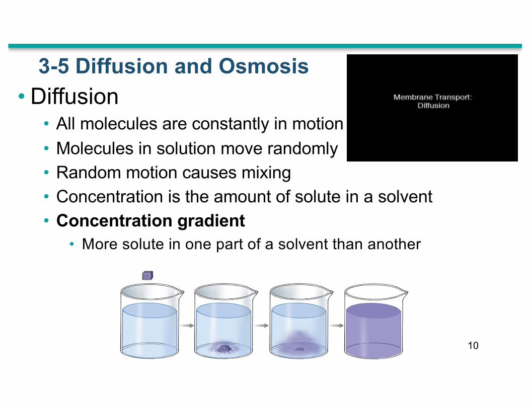
3-5 Diffusion and Osmosis• Diffusion
• All molecules are constantly in motion • Molecules in solution move randomly• Random motion causes mixing• Concentration is the amount of solute in a solvent• Concentration gradient
• More solute in one part of a solvent than another
10

3-5 Diffusion and Osmosis
• Factors Influencing Diffusion• Distance the particle has to move
• Molecule Size • Smaller is faster
• Temperature• More heat, faster motion
• Concentration Gradient• The difference between high and low concentrations
• Electrical Forces
• Opposites attract, like charges repel
11

3-5 Diffusion and OsmosisDiffusion across Plasma Membranes• Can be simple (through lipid bilayer) OR channel mediated
• Materials that diffuse by simple diffusion• Lipid-soluble compounds
(alcohols, fatty acids, and steroids)
• Dissolved gases (oxygen and carbon dioxide)
12
ethanolX

3-5 Diffusion across the Plasma membrane• Channel-mediated diffusion
• Works for small, water-soluble compounds and ions
• Factors influencing channel-mediated diffusion:• Size, • Charge, • Interaction
with the channel (leak)
Figure 3-15 Diffusion across the Plasma Membrane
13

3-5 Diffusion and Osmosis• Osmosis: A Special Case of Diffusion
• Osmosis is the diffusion of water—in this case—across the cell membrane
• More solute molecules, lower concentration of water molecules
• Membrane must be freely permeable to water, selectively permeable to solutes
• Water molecules diffuse across membrane toward solution with more solutes (less water)
• Volume increases on the side with more solutes 14

Figure 3-16 Osmosis
15
A
1
Watermolecules
2 3
Solutemolecules
Selectively permeable membrane
B
Volumedecreased
Originallevel
Volumeincreased
Volumesequal
Appliedforce
Two solutions containingdifferent solute concentrationsare separated by a selectivelypermeable membrane. Watermolecules (small blue dots)begin to cross the membranetoward solution B, the solutionwith the higher concentration of solutes (large pink dots)
At equilibrium, the soluteconcentrations on the two sidesof the membrane are equal. Thevolume of solution B hasincreased at the expense of thatof solution A.
Osmosis can be prevented byresisting the change in volume. Theosmotic pressure of solution B isequal to the amount of hydrostaticpressure required to stop theosmotic flow.

3-5 Osmolarity and Tonicity• Osmolarity is a measure of solute concentration (number of osmoles (Osm) of
solute per liter) • Osmolarity is distinct from molarity because it measures moles of solute particles
rather than moles of solute--distinction arises because some compounds can dissociate in solution whereas others cannot.
• Tonicity describes the osmotic effect of a solution on a cell (shrinks, swells, or no change)
• two fluids may have equal osmolarity but different tonicity (e.g., because the cell is selectively permeable to some but not all solutes)
• Isotonic (iso- = same, tonos = tension)• A solution that does not cause osmotic flow of water in or out of a cell
• Hypotonic (hypo- = below)• A solution that causes the cell to swell (for erythrocyte [RBC], if it burst = hemolysis)
• Hypertonic (hyper- = above) • A solution that causes the cell to shrink (called crenation for erythrocyte) 16

Figure 3-17 Osmotic Flow across a Plasma Membrane
17
Watermolecules
Isotonic solutiona
Solutemolecules
In an isotonic saline solution, noosmotic flow occurs, and the redblood cells appear normal.
SEM of a normal RBCin an isotonic solution
SEM of RBC in ahypotonic solution
SEM of crenated RBCsin a hypertonic solution
Hypotonic solutionb
In a hypotonic solution, the waterflows into the cell. The swelling maycontinue until the plasma membraneruptures, or lyses.
Hypertonic solutionc
In a hypertonic solution, watermoves out of the cell. The redblood cells shrivel and becomecrenated.
17

3-6 Carriers & Vesicles: Carrier-Mediated Transport• Carrier-Mediated Transport (by integral membrane proteins)
• Carry ions and organic substrates • Characteristics
• High Specificity • One transport protein, one set of substrates
• Saturation Limits• Max rate depends on number of transport proteins
• Regulation • By cofactors such as hormones
• Cotransport (by symport)• Two substances move in the same direction at the same time
• Countertransport (by antiport)• One substance moves in while another moves out 18

3 Classes of Carrier Proteins• Uniport
• Transport of a single molecule. • e.g., GLUT1-12 - glucose carriers
• Symport • Cotransport of two dissimilar solutes together (obligatorily coupled) • A gradient of one substrate--usually an ion--may drive uphill (active) transport of
a co-substrate (i.e., against a concentration gradient). • e.g, glucose-Na+ symport (SGLT1,2), in plasma membranes of some epithelial cells
• Antiport• “Exchange diffusion” where one solute is exchanged for another (also called
countertransport by your book). • Often exhibits "ping pong" kinetics –1st is transported and released causing a
conformational change in the protein that facilitates the transport of the 2nd. • e.g., mitochondrial ADP/ATP exchanger (inner membrane), Na+K+-ATPase
3-6 Carriers & Vesicles: Carrier Protein Classes
19

3-6 Carriers & Vesicles: Carrier-Mediated Transport• Facilitated Diffusion (Passive)
• Carrier proteins transport molecules too large to fit through channel proteins (glucose, amino acids)
• Molecule binds to receptor site on carrier protein• Protein changes shape, molecules pass through
Fig. 3-18A glucose uniport
20

• antiport carrier protein (countertransport)
•
3-6 Carriers & Vesicles: Carrier-Mediated Transport• Active Transport (primary or
secondary):• molecules move against a concentration
gradient• requires energy, such as ATP • ion pumps move ions (e.g., Na+, K+,
Ca2+, Mg2+) • exchange pump counter-transports two
ions at the same time
Primary Active Transport• For Na+/K+-ATPase, moves 3 Na+ out
and 2 K+ into the cell per ATP.• electrogenic as it results in an unequal
charge across the membrane
Figure 3–19 The Sodium–Potassium Exchange Pump (Na+/K+-ATPase)
21

3-6 Carriers & Vesicles: Carrier-Mediated TransportSecondary Active Transport• Transport mechanism itself does not directly require energy • Example: Glucose uptake via glucose-Na+ symport
• Na+ concentration gradient drives glucose transport• Na+/K+-ATPase pumps Na+ back out
Figure 3–20 Secondary Active Transport
22

3-6 Carriers and Vesicles: Vesicular Transport• Vesicular Transport (Bulk Transport)
• Materials move into or out of cell in vesicles• Endocytosis – the cell takes in macromolecules by forming new
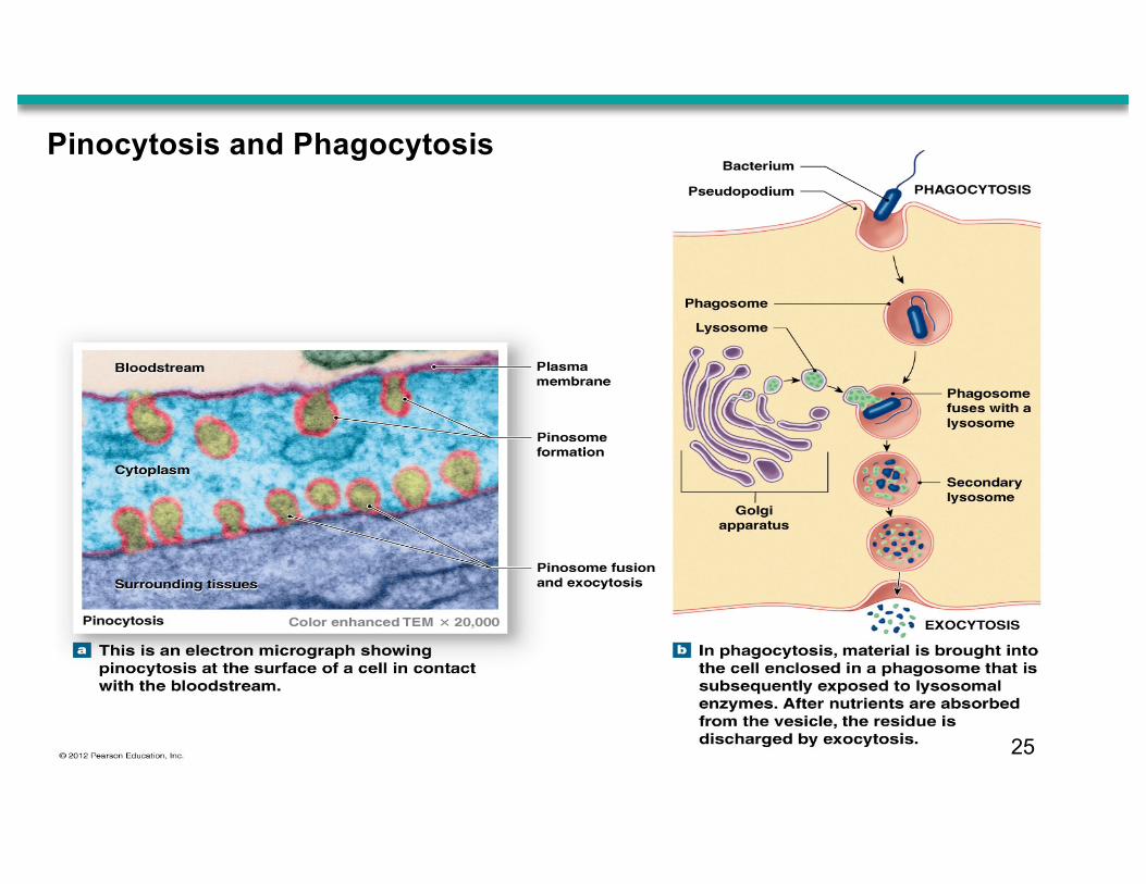
vesicles from the plasma membrane (3 types)1. Phagocytosis – “cell eating” – for solid substances
2. Pinocytosis - “cell drinking” – for dissolved substances
3. Receptor-Mediated (receptors = glycoproteins)
• Exocytosis - transport vesicles migrate to plasma membrane, fuse with it and release their contents
• Granules or droplets are released from the cell
23
EXTRACELLULAR FLUID
12
3
4
5
6
7
7
2
1
3
4
5
6
CYTOPLASM
Ligands bindingto receptors
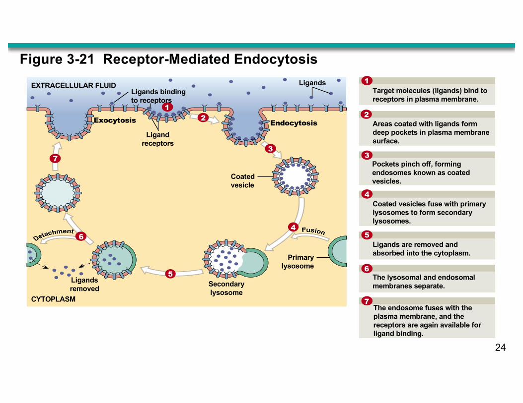
LigandsTarget molecules (ligands) bind toreceptors in plasma membrane.
Ligandreceptors
Exocytosis Endocytosis
Coatedvesicle
Primarylysosome
Secondarylysosome
Ligandsremoved
Areas coated with ligands formdeep pockets in plasma membranesurface.
Pockets pinch off, formingendosomes known as coatedvesicles.
Coated vesicles fuse with primarylysosomes to form secondarylysosomes.
Ligands are removed andabsorbed into the cytoplasm.
The lysosomal and endosomalmembranes separate.
The endosome fuses with theplasma membrane, and thereceptors are again available forligand binding.
Figure 3-21 Receptor-Mediated Endocytosis
24
25
Pinocytosis and Phagocytosis

Figure 3-22 Overview of Membrane Transport (Part 7 of 8).
Example:Cholesteroland iron ionsare transportedthis way.
Endocytosis Endocytosis is the packaging of extracellular materials into a vesicle for transport into the cell.
Extracellular fluid Target molecules
Receptorproteins Vesicle
containingtarget molecules
Cytoplasm
Pinosome
Pinocyticvesicleforming
Pseudopodiumextends to
surround object
Phagosome
Example:Once thevesicle is insidethe cytoplasm,water and smallmolecules enterthe cell acrossthe vesiclemembrane.
Example:Large particlesare broughtinto the cell bycytoplasmicextensions (calledpseudopodia)that engulf theparticle and pull it into the cell.
Cell
Cell
Receptor-MediatedEndocytosis
Pinocytosis Phagocytosis
In receptor-mediatedendocytosis, target molecules bind to receptor proteins on the membrane surface, triggering vesicle formation.
Substances Involved: Targetmolecules called ligands
Factors Affecting Rate: Number ofreceptors on the plasma membrane andthe concentration of target molecules
Substances Involved: Extracellular fluid, with dissolvedmolecules such as nutrients
Substances Involved: Bacteria,viruses, cellular debris, and otherforeign material
Factors Affecting Rate: Stimulusand mechanism not understood
Factors Affecting Rate: Presenceof pathogens and cellular debris
In pinocytosis, vesicles form atthe plasma membrane and bring fluids and small molecules into the cell. This process is often called “cell drinking.”
In phagocytosis, vesicles form atthe plasma membrane to bring solidparticles into the cell. This process isoften called “cell eating.”
Chapter 3 Summary Questions and Goals• Be able to describe the overall function of the cell’s major
organelles.
• Be able to describe the various mechanisms for transporting substances across the plasma membrane.
• Know the 3 types or classes of carrier proteins, the difference between passive and active transport (both primary and secondary).
• Know the difference between tonicity and osmolarity.• Be able to describe the 3 types of mechanisms by which
bulk substances enter the cell.27