the catalytic site atlas 2.0: cataloging catalytic sites and residues
TRANSCRIPT
The Catalytic Site Atlas 2.0: cataloging catalyticsites and residues identified in enzymesNicholas Furnham1,*, Gemma L. Holliday1, Tjaart A. P. de Beer1,
Julius O. B. Jacobsen1, William R. Pearson2 and Janet M. Thornton1
1European Molecular Biology Laboratory, European Bioinformatics Institute, Wellcome Trust Genome Campus,Hinxton, Cambridge CB10 1SD, UK and 2Department of Biochemistry and Molecular Genetics, University ofVirginia, 1300 Jefferson Park Ave., Charlottesville, VA 22908, USA
Received September 12, 2013; Revised November 7, 2013; Accepted November 8, 2013
ABSTRACT
Understanding which are the catalytic residues in anenzyme and what function they perform is crucial tomany biology studies, particularly those leadingto new therapeutics and enzyme design. Theoriginal version of the Catalytic Site Atlas (CSA)(http://www.ebi.ac.uk/thornton-srv/databases/CSA)published in 2004, which catalogs the residuesinvolved in enzyme catalysis in experimentallydetermined protein structures, had only 177curated entries and employed a simplisticapproach to expanding these annotations to hom-ologous enzyme structures. Here we present a newversion of the CSA (CSA 2.0), which greatly expandsthe number of both curated (968) and automaticallyannotated catalytic sites in enzyme structures,utilizing a new method for annotation transfer. Thecurated entries are used, along with the variation inresidue type from the sequence comparison, togenerate 3D templates of the catalytic sites, whichin turn can be used to find catalytic sites in newstructures. To ease the transfer of CSA annotationsto other resources a new ontology has been de-veloped: the Enzyme Mechanism Ontology, whichhas permitted the transfer of annotationsto Mechanism, Annotation and Classification inEnzymes (MACiE) and UniProt Knowledge Base(UniProtKB) resources. The CSA database schemahas been re-designed and both the CSA data andsearch capabilities are presented in a new modernweb interface.
INTRODUCTION
Enzymes represent �45% of the collective proteinproducts of all the genomes cataloged by resources suchas the UniProt Knowledge Base (UniProtKB) (1). As bio-logical catalysts they facilitate the many metabolicprocesses and pathways that are critical for life to existand have been the focus of studies by biologists andchemists for over 100 years. They are also some of theprincipal targets in pharmaceutical drug development,with many approved drugs acting to modify the actionof enzymes implicated in disease processes. In additionthey are often the focal point for biotechnology applica-tions. Detailed information on catalytic residues andenzyme active sites are essential for understanding the re-lationship between protein structure and functions, designof inhibitors and enzyme design.The Catalytic Site Atlas (CSA) (2) was established to
provide curated annotations of the small number of highlyconserved residues that are directly involved inundertaking the catalytic activity in enzymes whose struc-tures have been deposited in the Protein Data Bank (PDB)(3). These curated entries can in turn be used for inferringcatalytic residues in other enzyme structures throughhomology, using a simple PSIBlast method.The original resource contained 177 hand-annotated
entries and 2608 homologous entries, and covered �30%of all EC numbers found in PDB. We present here a newversion of the Catalytic Site Atlas—CSA 2.0. We havesignificantly increased the number of curated entries to968 and implement a new more sophisticated method fortransferring the annotations to homologous structuresincreasing the robustness of annotation transfer. The ex-pansion of curated entries also permits the addition of new
*To whom correspondence should be addressed. Tel: +44 1233 494631; Fax: +44 1223 494 496; Email: [email protected] addresses:Gemma L. Holiday, University of California, San Francisco, Box 2550, 1700 4th Street, San Francisco, CA 94143 – 2550, USA.Julius O. B. Jacobsen, Wellcome Trust Sanger Institute, Wellcome Trust Genome Campus, Hinxton, Cambridge CB10 1SD, UK.
Published online 6 December 2013 Nucleic Acids Research, 2014, Vol. 42, Database issue D485–D489doi:10.1093/nar/gkt1243
� The Author(s) 2013. Published by Oxford University Press.This is an Open Access article distributed under the terms of the Creative Commons Attribution License (http://creativecommons.org/licenses/by/3.0/), whichpermits unrestricted reuse, distribution, and reproduction in any medium, provided the original work is properly cited.
Downloaded from https://academic.oup.com/nar/article-abstract/42/D1/D485/1063115by gueston 09 February 2018

3D structural templates, which have been used in arevision of the Catalytic Site Search service. In additionthe database schema has been re-designed, integratingit into a sister database of enzyme mechanisms: theMechanism, Annotation and Classification in Enzymes(MACiE) database (4). We have also developed a newontology, the Enzyme Mechanism Ontology (EMO),permitting the integration of CSA information into bothMACiE and UniProtKB data structures and can be usedas a controlled vocabulary for describing aspects ofprotein sequence and structure with chemistry and mech-anistic terms across resources.
CSA CONTENT
The principle data held in the CSA are the protein residuesfrom experimentally determined atomic structures that aredefined as catalytic. Residues are designated as being cata-lytic by fulfilling any one of the following criteria:(i) Direct involvement in the catalytic mechanism;(ii) Alters the pKA of another residue or water moleculedirectly involved in the catalytic mechanism;(iii) Stabilization of a transition state or intermediate;and (iv) Activation of a substrate. Note that it does notinclude residues that are involved solely in ligand bindingand thus differs from other resources, such as UniProtKBannotations. Entries are made with respect to the de-posited PDB structure, with the potential to have manycatalytic sites within a single entry.Catalytic residue annotations are made either by
manual curation or through sequence comparison.Entries to be manually annotated are chosen from thePDB based on the quality of the structure and availableexperimental evidence of the reaction catalysed. Thisincludes details of the catalytic mechanism, also validatedby experimental data where possible. Annotators providea brief free-text description of the enzyme as well as amore detailed summary of the enzyme mechanism. Thereaction itself is also presented and marked up to showthe changes in molecular substructures and bond order/valence changes using an atom–atom matching algorithmimplemented in small molecule subgraph detector (SMSD)(5). For each residue in each catalytic site the functionalpart of the residue is recorded as well as its function andtarget described using a controlled vocabulary and a shortfree-text description of how the residue performs thefunction. Evidence tags provide a direct link to the litera-ture from which the annotations where derived. For eachcatalytic site a search can be performed returning all othercatalytic sites in the CSA that have the same catalyticresidues grouped by their E.C. numbers. In addition,hyperlinks to external resources, such as PDBSum (6)and IntEnz (7), are provided. Internal links to otherentries which share the same E.C. number (8) orsequence accession numbers or PDB identifiers aremade. A summary of the types of data shown for anentry is given in Figure 1.Developers involved in the prediction of proteins of
unknown function can use the extended number of curatedentries to train and test the methodologies being developed.
In addition individual users can access both curated andhomology derived entries to gain details of the catalyticresidues in a structure of interest, which has the potentialto be useful in design of further experiments. The user ex-perience has been enhanced using BioJS libraries (9) thatprovide a 3D viewing panel as well as a marked-upsequence viewer highlighting the catalytic residues.
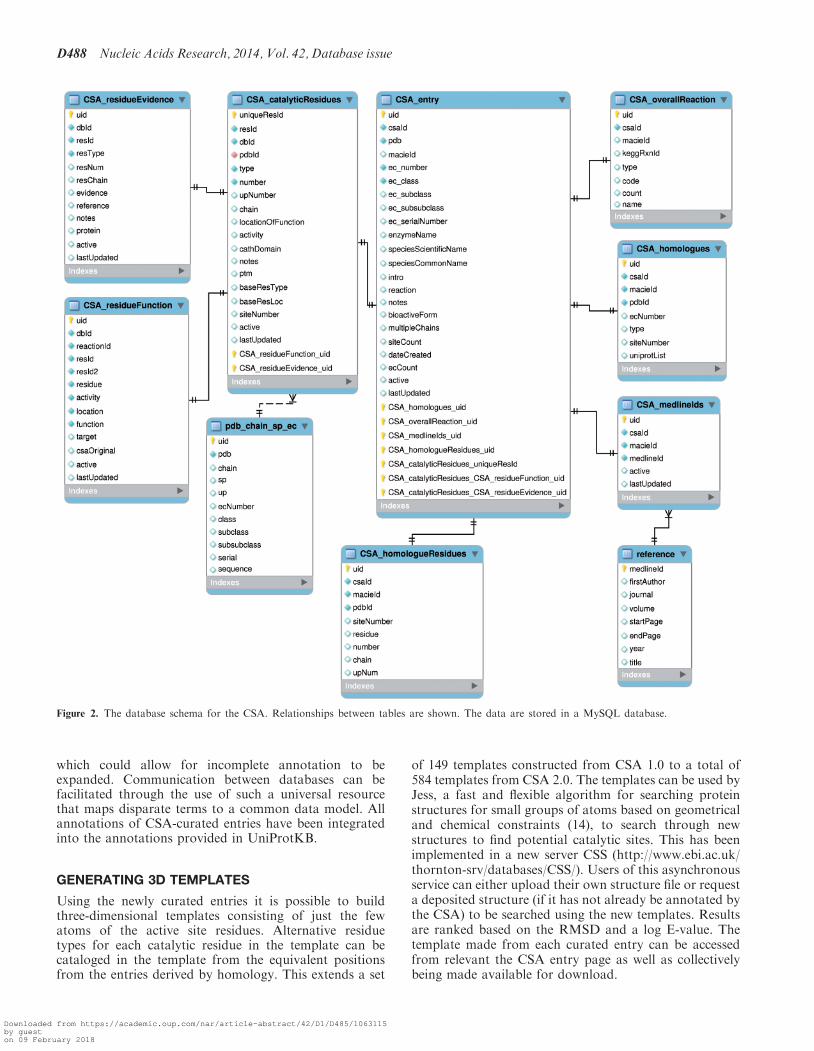
As part of a wider integration of resources, the CSA hasbeen merged with a sister database MACiE. The databaseschema for the CSA-related tables is shown in Figure 2.The CSA is designed as a relational database using atypical Linux, Apache, MySQL and PHP platform aidedby JavaScript utilizing the BioJS library.
INFERRING CATALYTIC RESIDUES THROUGHSEQUENCE COMPARISON
Entries are also annotated using an automated sequencecomparison method that utilizes the curated entries toinfer catalytic residues. 433 protein sequences from theMACIE enzyme mechanism database and the 911 se-quences unique to the CSA were extracted and labeledusing annotations for side-chain, main-chain, modified,reactant and spectator residues. CSA homologs in thePDB and reviewed section of UniProtKB were identifiedusing SSEARCH36 (10) with a statistical significancethreshold of E<10�6. SSEARCH alignments used the –V option to project the identity/conservative/non-conser-vative status of the aligned annotated functional residuesfrom the MACiE/CSA sequences to the homologoussequences in the PDB and SwissProt.
The entries derived by homology, in addition to thelinks to external data sources found in the curatedentries, also have an internal link to the manuallyannotated entries that was used to infer catalyticresidues. The CSA 2.0 provides a manually curatedresource of 968 enzyme structures and their catalyticsites including information on the functional part ofeach catalytic residue and its role in the enzyme mechan-ism. The use of sequence comparisons extends these an-notations to a further 32 216 structures annotated byhomology, providing a total of 34 096 annotated struc-tures out of possible 49 049 structures deposited in thePDB that are enzymatic. This greatly extends the 177curated entries and 2608 entries annotated by homologyin CSA 1.0. Additionally, the CSA 2.0 has entries for 1189E.C. numbers covering all the E.C. classification classesand subclasses and most sub-subclasses (Figure 3).
ENZYME MECHANISM ONTOLOGY
Although the CSA and MACiE resources have been de-veloped somewhat in tandem and thus share a commondata model, it is currently challenging to link these toenzyme annotations in resources such as UniProtKB dueto differences in the definitions of enzyme properties andthe vocabularies used in their description. Though descrip-tions and definitions of some of the information held in allthree databases are made in existing ontologies such asGO (11) and the ChEBI (12) ontology, marrying these
D486 Nucleic Acids Research, 2014, Vol. 42, Database issue
Downloaded from https://academic.oup.com/nar/article-abstract/42/D1/D485/1063115by gueston 09 February 2018

and applying them uniformly to all three databases provedfar from trivial.
The CSA and its sister database, MACiE, utilize acontrolled vocabulary, with MACiE possessing a moredetailed vocabulary as it focuses on enzymes in a muchgreater depth to include thorough descriptions ofthe chemical reaction steps performed. Likewise, thereviewed section of the UniProtKB (UniProtKB/Swiss-Prot) also captures enzyme-related data at a broaderprotein sequence level, including information on catalyticresidues. Annotations are made as both free text and usingan independently developed controlled vocabulary.
To address this we have developed the EMO whichbuilds upon the controlled vocabulary developed forMACiE and the CSA and will be submitted to the OBOFoundry (13). This vocabulary (see Supplementary
Material or http://purl.bioontology.org/ontology/EMO)was created to describe the active components of theenzyme’s reactions (cofactors, amino acids and cognateligands) and their roles in the reaction. EMO buildsupon this by formalizing key concepts, and the relation-ships between them, necessary to define enzymes and theirfunctions. This describes not only the general features ofan enzyme, including the E.C. number (catalytic activity),3D structure and cellular locations, but also allows for thedetailed annotation of the mechanism. This mechanisticdetail can be either at a gross level (overall reaction onlyas captured in the CSA), or the more detailed granularityof the steps and components required to effect the overallchemical transformation.EMO allows for many different resources to be drawn
together, even where annotations are only partially made,
CSA LITERATURRE entry for 1q6x
E.C. name choline O-acetyltransferase
Species Rattus norvegicus (Rat)
E.C. Number 2.3.1.6
CSA Homologues of 1q6x There are 24 Homologs
CSA Entries With UniProtID P32738
PDBe Entry 1q6x
PDBSum Entry 1q6x
MACiE Entry 1q6x
Show
Site 1
Options Antialias Rotation
Color:
Default
Style:
Surface:
Style:
Surface:
Cartoon
Su ace:None
Annotaated By Refference TTo The Literature - Site 1
Residue Chain NumberUniProtKB
NumberFunctional
PartFunction Target Description
His A 334 334 Side Chain Acid / Base
SubstrateActs as a general base, extracting a proton from the attacking hydroxyl group of choline. Later protonates the departing sulphydryl group of CoA.
Catalytic Sites for 1q6x
IntroductionCholine acetyltransferase synthesises the neurotransmitter acetylcholine from choline in neurones and other cell types. It catalyses the reversible transfer of an acetyl group between acetyl CoA and choline, and belongs to the choline/carnitine acyltranserase family which also includes enzymes involved in fatty acid metabolism.
MechanismHis 334 acts as a general base to remove the proton from the choline OH group as the oxygen attacks the carbonyl of acetyl CoA. The resulting tetrahedral oxyanion intermediate is stabilised by Ser 550. Collapse of the tetrahedral intermediate releases CoA which is protonated by the His 334. Tyr 95 and Pro 108 function to stabilise
Reaction
Literature Report
A B
C
D
Figure 1. Overview of data presented for a CSA-curated entry. Meta-data descriptors such as enzyme name and species as well as internal links tofind entries in the CSA that share properties along with links to external web resources are shown in a table (A). A 3D viewer (B) displays theenzyme structure, highlighting each of the catalytic sites (from a pull-down menu) in red. A free-text report of the overall reaction and mechanismare provided (C) with a reaction diagram marked up with groups conserved across the reaction and bond changes. (D) Shows the annotations heldfor each catalytic residues in each catalytic site.
Nucleic Acids Research, 2014, Vol. 42, Database issue D487
Downloaded from https://academic.oup.com/nar/article-abstract/42/D1/D485/1063115by gueston 09 February 2018
which could allow for incomplete annotation to beexpanded. Communication between databases can befacilitated through the use of such a universal resourcethat maps disparate terms to a common data model. Allannotations of CSA-curated entries have been integratedinto the annotations provided in UniProtKB.
GENERATING 3D TEMPLATES
Using the newly curated entries it is possible to buildthree-dimensional templates consisting of just the fewatoms of the active site residues. Alternative residuetypes for each catalytic residue in the template can becataloged in the template from the equivalent positionsfrom the entries derived by homology. This extends a set
of 149 templates constructed from CSA 1.0 to a total of584 templates from CSA 2.0. The templates can be used byJess, a fast and flexible algorithm for searching proteinstructures for small groups of atoms based on geometricaland chemical constraints (14), to search through newstructures to find potential catalytic sites. This has beenimplemented in a new server CSS (http://www.ebi.ac.uk/thornton-srv/databases/CSS/). Users of this asynchronousservice can either upload their own structure file or requesta deposited structure (if it has not already be annotated bythe CSA) to be searched using the new templates. Resultsare ranked based on the RMSD and a log E-value. Thetemplate made from each curated entry can be accessedfrom relevant the CSA entry page as well as collectivelybeing made available for download.
Figure 2. The database schema for the CSA. Relationships between tables are shown. The data are stored in a MySQL database.
D488 Nucleic Acids Research, 2014, Vol. 42, Database issue
Downloaded from https://academic.oup.com/nar/article-abstract/42/D1/D485/1063115by gueston 09 February 2018

CONCLUSIONS
CSA 2.0 provides a new modern interface to a much-extended manually curated dataset of residues involvedin enzyme catalytic sites and the functional role theyplay in the reaction. A new method for reliablyextrapolating the annotations and identification of cata-lytic residues to homologous structures has been imple-mented. In addition the curated entries can be used tobuild 3D templates of the catalytic sites, which in turncan be used to search new structures for catalytic site iden-tification using a revised CSS service. Furthermore a newontology has been developed to permit the transfer of an-notations relating to enzyme catalysis between resources.This has been used to include CSA annotations inUniProtKB and MACiE.
The database is available at http://www.ebi.ac.uk/thornton-srv/databases/CSA, while the CSS service canbe found at http://www.ebi.ac.uk/thornton-srv/databases/CSS. Both are compatible with most modern webbrowsers. All the data in the CSA is downloadable andfreely available to the academic community.
SUPPLEMENTARY DATA
Supplementary Data are available at NAR Online.
ACKNOWLEDGEMENTS
The authors would like to thank the efforts of the manyannotators who have contributed to the curated entries inthe CSA. We would also like to thank Dr Syed A.Rahman for supplying the marked-up reaction diagrams.
FUNDING
Wellcome Trust [081989/Z/07/A to N.F.]; EuropeanMolecular Biology Laboratory (EMBL) (to G.L.H. andvarious annotators); US Department of Energy Contract(in part) [DE-AC02-06CH11357 to T.A.P.deB.] as part ofthe Midwest Center for Structural Genomics (MCSG).Funding for open access charge: The Wellcome Trust.
Conflict of interest statement. None declared.
REFERENCES
1. Uniprot Consortium. (2013) Update on activities at the UniversalProtein Resource (UniProt) in 2013. Nucleic Acids Res., 41,D43–D47.
2. Porter,C.T., Bartlett,G.J. and Thornton,J.M. (2004) The CatalyticSite Atlas: a resource of catalytic sites and residues identified inenzymes using structural data. Nucleic Acids Res., 32,D129–D133.
3. Velankar,S., Alhroub,Y., Best,C., Caboche,S., Conroy,M.J.,Dana,J.M., Fernandez Montecelo,M.A., van Ginkel,G.,Golovin,A., Gore,S.P. et al. (2012) PDBe: Protein Data Bank inEurope. Nucleic Acids Res., 40, D445–D452.
4. Holliday,G.L., Andreini,C., Fischer,J.D., Rahman,S.A.,Almonacid,D.E., Williams,S.T. and Pearson,W.R. (2012) MACiE:exploring the diversity of biochemical reactions. Nucleic AcidsRes., 40, D783–D789.
5. Rahman,S., Bashton,M., Holliday,G., Schrader,R. andThornton,J. (2009) Small Molecule Subgraph Detector (SMSD)toolkit. J. Cheminform., 1, 12.
6. Laskowski,R.A. (2009) PDBsum new things. Nucleic Acids Res.,37, D355–D359.
7. Fleischmann,A., Darsow,M., Degtyarenko,K., Fleischmann,W.,Boyce,S., Axelsen,K.B., Bairoch,A., Schomburg,D., Tipton,K.F.and Apweiler,R. (2004) IntEnz, the integrated relational enzymedatabase. Nucleic Acids Res., 32, D434–D437.
8. International Union of Biochemistry and Molecular Biology,Nomenclature,C. and Webb,E.C. (1992) Enzyme Nomenclature1992 : recommendations of the Nomenclature Committee of theInternational Union of Biochemistry and Molecular Biology on theNomenclature and Classification of Enzymes/Prepared for NC-IUBMB by Edwin C. Webb. Published for the InternationalUnion of Biochemistry and Molecular Biology by AcademicPress, San Diego.
9. Gomez,J., Garcia,L.J., Salazar,G.A., Villaveces,J., Gore,S.,Garcia,A., Martin,M.J., Launay,G., Alcantara,R., Del-Toro,N.et al. (2013) BioJS: an open source JavaScript frameworkfor biological data visualization. Bioinformatics, 29,1103–1104.
10. Sierk,M.L. and Pearson,W.R. (2004) Sensitivity and selectivity inprotein structure comparison. Protein Sci., 13, 773–785.
11. Blake,J.A., Dolan,M., Drabkin,H., Hill,D.P., Li,N., Sitnikov,D.,Bridges,S., Burgess,S., Buza,T., McCarthy,F. et al. (2013) GeneOntology annotations and resources. Nucleic Acids Res., 41,D530–D535.
12. Hastings,J., de Matos,P., Dekker,A., Ennis,M., Harsha,B.,Kale,N., Muthukrishnan,V., Owen,G., Turner,S., Williams,M.et al. (2013) The ChEBI reference database and ontology forbiologically relevant chemistry: enhancements for 2013. NucleicAcids Res., 41, D456–D463.
13. Smith,B., Ashburner,M., Rosse,C., Bard,J., Bug,W., Ceusters,W.,Goldberg,L.J., Eilbeck,K., Ireland,A., Mungall,C.J. et al. (2007)The OBO Foundry: coordinated evolution of ontologies tosupport biomedical data integration. Nat. Biotechnol., 25,1251–1255.
14. Barker,J.A. and Thornton,J.M. (2003) An algorithm forconstraint-based structural template matching: application to3D templates with statistical analysis. Bioinformatics, 19,1644–1649.
Figure 3. E.C. coverage in the CSA. The Enzyme Commission classifi-cation of all E.C. codes classified by the Enzyme Commission rendered asa rooted tree. Each major class is labeled with (i) Oxidoreductases, (ii)Transferases, (iii) Hydrolases, (iv) Lyases, (v) Isomerases and (vi) Ligases.Each E.C. number in the CSA is colored red, with all major classes andsubclasses present and most of the sub-subclasses.
Nucleic Acids Research, 2014, Vol. 42, Database issue D489
Downloaded from https://academic.oup.com/nar/article-abstract/42/D1/D485/1063115by gueston 09 February 2018