the cardiovascular system “a muscular pump equipped with one- way valves and a system of large and...
TRANSCRIPT

The Cardiovascular SystemThe Cardiovascular System
““A muscular pump equipped with one-A muscular pump equipped with one-way valves and a system of large and way valves and a system of large and small plumbing tubes within which the small plumbing tubes within which the blood travels.”blood travels.”
= pump= pump
= plumbing tubes= plumbing tubes

Early development of the heart and Early development of the heart and circulatory systemcirculatory system
• 2 weeks post-conception-2 weeks post-conception-• 3 weeks-3 weeks-• 4 weeks-4 weeks-
• By week 8-By week 8-

During this period the fetal heart During this period the fetal heart is at great risk from agents that is at great risk from agents that may cause congenital defects.may cause congenital defects.
RUBELLARUBELLA
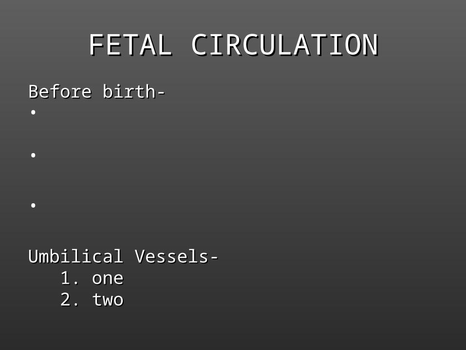
FETAL CIRCULATIONFETAL CIRCULATION
Before birth-Before birth-•
•
•
Umbilical Vessels-Umbilical Vessels- 1. one1. one 2. two2. two

To bypass the liverTo bypass the liver
Umbilical vein ductus venosus inferior vena cava right atrium ->
uum

To bypass the lungsTo bypass the lungs
Right Atrium Right Atrium Foramen Ovale Foramen Ovale Left Atrium Left Atrium Left Ventricle Left Ventricle Aorta Aorta Body Body (majority of blood)(majority of blood)
OrOr
Right Atrium Right Atrium Right Ventricle Right Ventricle Pulmonary Trunk Pulmonary Trunk Ductus Arteriosis Ductus Arteriosis Aorta Aorta BodyBody
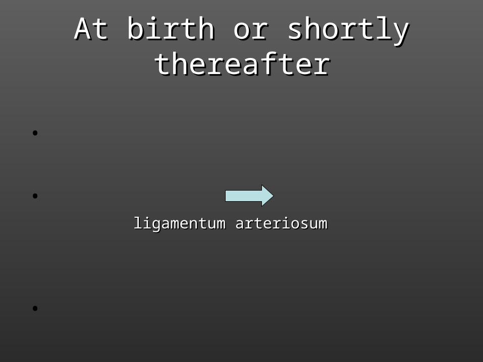
At birth or shortly thereafterAt birth or shortly thereafter
•
• ligamentum arteriosumligamentum arteriosum
•


Anatomy of the HeartAnatomy of the Heart
1.1. Location and SizeLocation and Size
•
•
•
• Pointed Pointed APEXAPEX - -
• BASEBASE - -

2. Coverings and wall of the heart2. Coverings and wall of the hearta. covered by double sac- a. covered by double sac- PERICARDIUMPERICARDIUM
Parietal Pericardium
(pericardial fluid)Visceral Pericardium (outer layer heart)MyocardiumEndocardium
b. Layers of Heart Wallb. Layers of Heart Wall

C. Chambers of the HeartC. Chambers of the Heart
• Two receiving chambers- Two receiving chambers- • Two discharging chambers (pumps)-Two discharging chambers (pumps)-• Right and left side separated by the – Right and left side separated by the – SEPTUMSEPTUM
a. upper septum =a. upper septum =b. lower septum =b. lower septum =
Atrium-Atrium-Right Ventricle-Right Ventricle-Left Ventricle-Left Ventricle-

Cardiac Muscles (myocardium)Cardiac Muscles (myocardium)

A SchematicA Schematic


• BLUE-pulmonary circuit RED- systemic circuitBLUE-pulmonary circuit RED- systemic circuit

Great Vessels of the HeartGreat Vessels of the Heart
Pulmonary Systemic
Circuit Circuit

Valves of the HeartValves of the Heart
• Located at entrance/exit ventriclesLocated at entrance/exit ventricles
• Are 1-way, prevent back flow of bloodAre 1-way, prevent back flow of blood
• Atrioventricular- Atrioventricular-
• Semilunar-Semilunar-


Lubb Vs. Dupp

Cardiac CirculationCardiac Circulation
• Only the _________________ is in direct Only the _________________ is in direct contact with the blood. Other heart tissue contact with the blood. Other heart tissue must have its own blood supplymust have its own blood supply
• CORONARY ARTERIESCORONARY ARTERIES
• CARDIAC VEINSCARDIAC VEINS
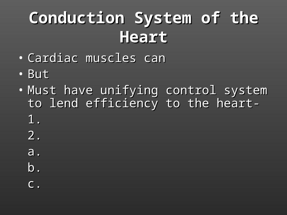
Conduction System of the HeartConduction System of the Heart
• Cardiac muscles canCardiac muscles can• ButBut• Must have unifying control system to lend Must have unifying control system to lend
efficiency to the heart-efficiency to the heart-1.1.2.2.
a.a.b.b.c.c.


SA node (SA node ( ))to theto the
Atrial myocardium (Atrial myocardium ( ))to theto the
AV node (AV node ( ))to theto the
AV bundleAV bundleto theto the
Bundle branchesBundle branchesto theto the
Purkinje fibersPurkinje fibersfollowed byfollowed by
Ventricular contraction (Ventricular contraction ( ))

ElectrocardiographyElectrocardiography
““When impulses travel through the heart, When impulses travel through the heart, electrical currents are generated that electrical currents are generated that spread throughout the body. These spread throughout the body. These impulses can be detected on the body impulses can be detected on the body surface and recorded with an surface and recorded with an electrocardiograph.electrocardiograph.

ElectrocardiogramElectrocardiogram- recording that traces - recording that traces the flow of current through the heart.the flow of current through the heart.

• P Wave-P Wave-• QRS complex-QRS complex-• T wave-T wave-
*atrial repolarization*atrial repolarization

Cardiac Output- Cardiac Output-
CO = Heart rate X Stroke VolumeCO = Heart rate X Stroke Volume
stroke volume-stroke volume-
CO = 75 bpm X 70 ml/beatCO = 75 bpm X 70 ml/beat
CO = 5250 ml/min (average)CO = 5250 ml/min (average)

What factors affect cardiac output?What factors affect cardiac output?