the canadian association for the study of the liver bile
TRANSCRIPT
Di Ciaula A, et al. , 2017; 16 (Suppl. 1): s4-s144
Bile Acid PhysiologyAgostino Di Ciaula,*,♦ Gabriella Garruti,†,♦ Raquel Lunardi Baccetto,‡
Emilio Molina-Molina,‡ Leonilde Bonfrate,‡ David Q.-H. Wang,§ Piero Portincasa‡
* Division of Internal Medicine, Hospital of Bisceglie, Italy.† Section of Endocrinology, Department of Emergency and Organ Transplantations,
University of Bari “Aldo Moro” Medical, School, Piazza G. Cesare 11, 70124 Bari, Italy.§ Department of Medicine, Division of Gastroenterology and Liver Diseases,
Marion Bessin Liver Research Center, Albert Einstein College of Medicine, Bronx, NY 10461, USA.‡ Clinica Medica “A. Murri”, Department of Biomedical Sciences & Human Oncology, University of Bari Medical School, Bari, Italy.
♦ The authors provided equal contribution
November, Vol. 16 (Suppl. 1), 2017: s4-s14
INTRODUCTION
Bile acids (BAs), the major lipid components of bile,are synthetized from cholesterol in the liver and subse-quently conjugated to taurine or glycine, leading to an in-crease in their solubility. Immediately after synthetization,BAs are secreted into bile, as well as concentrated andstored in the gallbladder. Upon food intake, the gallblad-der is stimulated by the entero-hormone cholecystokinin(CCK) to release bile into the duodenum, where BAs aidin the digestion and absorption of lipids and fat-solublevitamins.1 Fasting serum BAs concentrations in healthysubjects are 0.2-0.7 μM to increase to 4-5 μM after eachmeal.2.5 Most of the BAs in the ileum are reabsorbed andreturn to the liver through the portal vein, and conse-quently, hepatic BA synthesis is inhibited by a negativefeedback regulatory mechanism. However, BAs that es-
cape from intestinal reabsorption enter the colon, wherethey are further transformed into the secondary and, morehydrophilic BAs, by the resident gut microbiota.6
The role of bile in lipid metabolism goes beyond thatof fat emulsifier. Recent studies have also pointed to BAsas signaling molecules with metabolic effects via interac-tion with the nuclear receptors farnesoid X receptor(FXR), pregnane X receptor (PXR), and vitamin D recep-tor (VDR), G-protein coupled receptors such as GPBAR-1, and cell signaling pathways such as c-Jun N-terminalkinase (JNK) and extracellular signal-regulated kinase(ERK). Through these interactions, BAs help to regulateenergy, glucose, lipids and lipoprotein metabolism.7
In this review, we summarize recent advances in thecomplex BAs physiology, focusing on novel findings aboutthe regulatory mechanisms of BAs that are dependent andindependent of nuclear FXR. We also briefly discuss the
The Official Journal of the Mexican Association of Hepatology,the Latin-American Association for Study of the Liver and
the Canadian Association for the Study of the Liver
Manuscript received:Manuscript received:Manuscript received:Manuscript received:Manuscript received: September 06, 2017. Manuscript accepted:Manuscript accepted:Manuscript accepted:Manuscript accepted:Manuscript accepted: September 06, 2017.
DOI:10.5604/01.3001.0010.5493
A B S T R A C TA B S T R A C TA B S T R A C TA B S T R A C TA B S T R A C T
The primary bile acids (BAs) are synthetized from cholesterol in the liver, conjugated to glycine or taurine to increase their solubility,secreted into bile, concentrated in the gallbladder during fasting, and expelled in the intestine in response to dietary fat. BAs are alsobio-transformed in the colon to the secondary BAs by the gut microbiota, reabsorbed in the ileum and colon back to the liver, andminimally lost in the feces. BAs in the intestine not only regulate the digestion and absorption of cholesterol, triglycerides, and fat-soluble vitamins, but also play a key role as signaling molecules in modulating epithelial cell proliferation, gene expression, and lipidand glucose metabolismby activating farnesoid X receptor (FXR) and G-protein-coupled bile acid receptor-1 (GPBAR-1, also knownas TGR5) in the liver, intestine, muscle and brown adipose tissue. Recent studies have revealed the metabolic pathways of FXR andGPBAR-1 involved in the biosynthesis and enterohepatic circulation of BAs and their functions as signaling molecules on lipid andglucose metabolism.
Key words.Key words.Key words.Key words.Key words. Bile acids. Microbiota. FXR. Bile.

5Bile Acid Physiology. , 2017; 16 (Suppl. 1): s4-s14
interaction between host microbiota and dietary intake onBA metabolism.
BIOSYNTHETIC PATHWAYSOF BILE ACIDS (BAS)
BAs belong to a family of closely related acidic sterolssynthesized from cholesterol, and represent the maincatabolic pathway of cholesterol metabolism in humans.BAs are classified as soluble amphiphiles because of theionized carboxylate or sulfonate group on the side chainthat makes BAs water-soluble. In general, BAs possess asteroid nucleus of four fused hydrocarbon rings with po-lar hydroxyl functions. De novo BA biosynthesis occurs inthe liver as the “primary” BAs, i.e., cholic acid (CA) andchenodeoxycholic acid (CDCA). Afterwards, the watersolubility of BAs is increased by conjugation to eithertaurine or glycine, followed by hepatic secretion of BAsinto bile and release by the gallbladder into the duode-
num after the meal.8 The aliphatic side chain is conjugat-ed in amide linkage (N-acyl amidation) with glycine ortaurine at a ratio of 3:1 to increase water solubility of BAs(glycine > taurine) in bile and reduce BA toxicity (Fig-ure 1).2 In bile, BAs act as cholesterol carriers togetherwith phospholipids, and in the intestine, BAs act as sur-factants and help the digestion and absorption of dietarycholesterol, triglycerides, and fat-soluble vitamins.9
Thus, BAs are essential in biliary cholesterol secretionand transport in bile, as well as hepatic catabolic prod-ucts of endogenous cholesterol.
About 15% of conjugated BAs escape the absorption ofthe terminal ileum and enter the colon, where the resi-dent gut microbiota promotes the deconjugation and bi-otransformation of the primary BAs into the secondaryBAs such as deoxycholic acid (DCA) and lithocholic acid(LCA) and the tertiary BAs such as ursodeoxycholic acid(UDCA), as shown in Figure 2. Approximately 50% ofDCA and a small amount of LCA and UDCA are re-ab-
Figure 1. A.Figure 1. A.Figure 1. A.Figure 1. A.Figure 1. A. The general structure of cholestane (classified as a saturated 27-carbon tetracyclic triterpene) is shown with numbering of the carbon atoms.The four fused hydrocarbon rings are labelled as A, B, C, and D. Cholesterol is shown as 3D structure and chemical formula. B.B.B.B.B. Hepatic cholesterol in thebody is catabolized to bile acids (BAs), and cholic acid (CA) is shown as an example. CA possesses a steroid nucleus of four fused hydrocarbon rings with polarhydroxyl functions and an aliphatic side chain in amide linkage with taurine or glycine (dotted lines). The two enzymes involved in this process are the BA CoAsynthase and the BA-CoA-amino acid N-acetyltransferase. The hydrophilic (i.e., polar) areas of BAs are the hydroxyl groups (-OH) (orientation of the hydroxyls:3α, 7α, 12α) and conjugation side chain of either glycine or taurine. The hydrophobic (i.e., nonpolar) area is the ringed steroid nucleus.
Cho les taneCholes taneCholes taneCholes taneCholes tane Choles tero lCho les tero lCho les tero lCho les tero lCho les tero l
Cholic acidCholic acidCholic acidCholic acidCholic acid
Glyc ineGlyc ineGlyc ineGlyc ineGlyc ine
Taur ineTaur ineTaur ineTaur ineTaur ine
AAAAA
BBBBB
Di Ciaula A, et al. , 2017; 16 (Suppl. 1): s4-s146
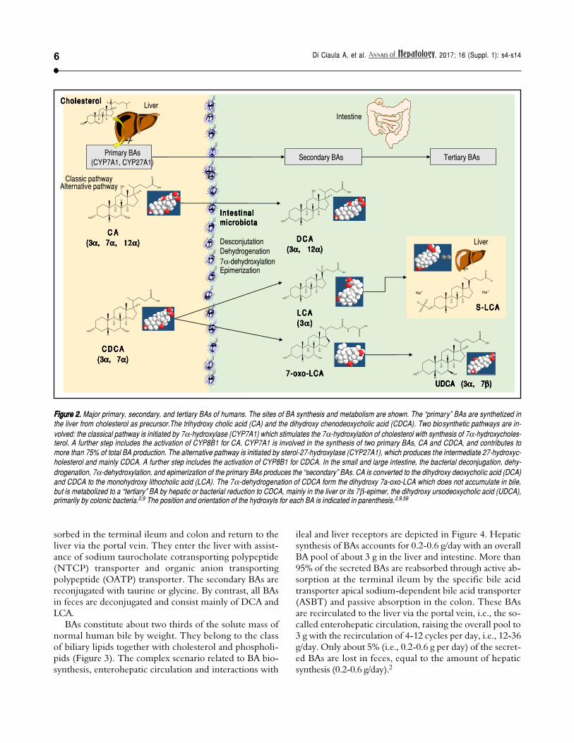
Figure 2.Figure 2.Figure 2.Figure 2.Figure 2. Major primary, secondary, and tertiary BAs of humans. The sites of BA synthesis and metabolism are shown. The “primary” BAs are synthetized inthe liver from cholesterol as precursor.The trihydroxy cholic acid (CA) and the dihydroxy chenodeoxycholic acid (CDCA). Two biosynthetic pathways are in-volved: the classical pathway is initiated by 7α-hydroxylase (CYP7A1) which stimulates the 7α-hydroxylation of cholesterol with synthesis of 7α-hydroxycholes-terol. A further step includes the activation of CYP8B1 for CA. CYP7A1 is involved in the synthesis of two primary BAs, CA and CDCA, and contributes tomore than 75% of total BA production. The alternative pathway is initiated by sterol-27-hydroxylase (CYP27A1), which produces the intermediate 27-hydroxyc-holesterol and mainly CDCA. A further step includes the activation of CYP8B1 for CDCA. In the small and large intestine, the bacterial deconjugation, dehy-drogenation, 7α-dehydroxylation, and epimerization of the primary BAs produces the “secondary” BAs. CA is converted to the dihydroxy deoxycholic acid (DCA)and CDCA to the monohydroxy lithocholic acid (LCA). The 7α-dehydrogenation of CDCA form the dihydroxy 7a-oxo-LCA which does not accumulate in bile,but is metabolized to a “tertiary” BA by hepatic or bacterial reduction to CDCA, mainly in the liver or its 7β-epimer, the dihydroxy ursodeoxycholic acid (UDCA),primarily by colonic bacteria.2,9 The position and orientation of the hydroxyls for each BA is indicated in parenthesis.2,9,59
Liver
Primary BAs(CYP7A1, CYP27A1)
C D C AC D C AC D C AC D C AC D C A(3(3(3(3(3ααααα, 7, 7, 7, 7, 7ααααα)))))
In tes t ina lIn tes t ina lIn tes t ina lIn tes t ina lIn tes t ina lmic rob io tamicrob io tamicrob io tamicrob io tamicrob io ta
DesconjutationDehydrogenation7α-dehydroxylationEpimerization
Intestine
Secondary BAs
D C AD C AD C AD C AD C A(3(3(3(3(3ααααα, 12, 12, 12, 12, 12ααααα)))))
L C AL C AL C AL C AL C A(3(3(3(3(3ααααα )))))
7-oxo-LCA7-oxo-LCA7-oxo-LCA7-oxo-LCA7-oxo-LCA
Tertiary BAs
Liver
S - L C AS - L C AS - L C AS - L C AS - L C A
UDCA (3UDCA (3UDCA (3UDCA (3UDCA (3ααααα, 7, 7, 7, 7, 7βββββ)))))
C AC AC AC AC A(3(3(3(3(3ααααα, 7, 7, 7, 7, 7α, 12αα, 12αα, 12αα, 12αα, 12α)))))
Cho les tero lCholes tero lCholes tero lCholes tero lCholes tero l
Classic pathwayAlternative pathway
sorbed in the terminal ileum and colon and return to theliver via the portal vein. They enter the liver with assist-ance of sodium taurocholate cotransporting polypeptide(NTCP) transporter and organic anion transportingpolypeptide (OATP) transporter. The secondary BAs arereconjugated with taurine or glycine. By contrast, all BAsin feces are deconjugated and consist mainly of DCA andLCA.
BAs constitute about two thirds of the solute mass ofnormal human bile by weight. They belong to the classof biliary lipids together with cholesterol and phospholi-pids (Figure 3). The complex scenario related to BA bio-synthesis, enterohepatic circulation and interactions with
ileal and liver receptors are depicted in Figure 4. Hepaticsynthesis of BAs accounts for 0.2-0.6 g/day with an overallBA pool of about 3 g in the liver and intestine. More than95% of the secreted BAs are reabsorbed through active ab-sorption at the terminal ileum by the specific bile acidtransporter apical sodium-dependent bile acid transporter(ASBT) and passive absorption in the colon. These BAsare recirculated to the liver via the portal vein, i.e., the so-called enterohepatic circulation, raising the overall pool to3 g with the recirculation of 4-12 cycles per day, i.e., 12-36g/day. Only about 5% (i.e., 0.2-0.6 g per day) of the secret-ed BAs are lost in feces, equal to the amount of hepaticsynthesis (0.2-0.6 g/day).2
7Bile Acid Physiology. , 2017; 16 (Suppl. 1): s4-s14
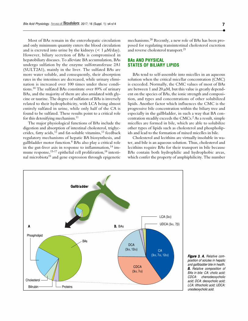
Figure 3.Figure 3.Figure 3.Figure 3.Figure 3. A. A. A. A. A. Relative com-position of solutes in hepaticand gallbladder bile in health.B.B.B.B.B. Relative composition ofBAs in bile: CA: cholic acid;CDCA: chenodeoxycholicacid; DCA: deoxycholic acid;LCA: lithocholic acid; UDCA:ursodeoxycholic acid.
Most of BAs remain in the enterohepatic circulationand only minimum quantity enters the blood circulationand is excreted into urine by the kidneys (< 1 μM/day).However, biliary secretion of BAs is compromised inhepatobiliary diseases. To alleviate BA accumulation, BAsundergo sulfation by the enzyme sulfotransferase 2A1(SULT2A1), mainly in the liver. The sulfated BAs aremore water soluble, and consequently, their absorptionrates in the intestines are decreased, while urinary elimi-nation is increased over 100 times under these condi-tions.10 The sulfated BAs constitute over 89% of urinaryBAs, and the majority of them are also amidated with gly-cine or taurine. The degree of sulfation of BAs is inverselyrelated to their hydrophobicity, with LCA being almostentirely sulfated in urine, while only half of the CA isfound to be sulfated. These results point to a critical rolefor this detoxifying mechanism.11
The major physiological functions of BAs include thedigestion and absorption of intestinal cholesterol, triglyc-erides, fatty acids,12 and fat-soluble vitamins,13 feedbackregulatory mechanisms of hepatic BA biosynthesis, andgallbladder motor function.8 BAs also play a critical rolein the gut-liver axis in response to inflammation,14 im-mune response,15-17 epithelial cell proliferation,18 intesti-nal microbiota19 and gene expression through epigenetic
mechanisms.20 Recently, a new role of BAs has been pro-posed for regulating transintestinal cholesterol excretionand reverse cholesterol transport.21
BAs AND PHYSICALSTATES OF BILIARY LIPIDS
BAs tend to self-assemble into micelles in an aqueoussolution when the critical micellar concentration (CMC)is exceeded. Normally, the CMC values of most of BAsare between 1 and 20 μM, but this value is greatly depend-ent on the species of BAs, the ionic strength and composi-tion, and types and concentrations of other solubilizedlipids. Another factor which influences the CMC is theprogressive bile concentration within the biliary tree andespecially in the gallbladder, in such a way that BA con-centration steadily exceeds the CMCs.2 As a result, simplemicelles are formed in bile, which are able to solubilizeother types of lipids such as cholesterol and phospholip-ids and lead to the formation of mixed micelles in bile.
Cholesterol and lecithins are virtually insoluble in wa-ter, and bile is an aqueous solution. Thus, cholesterol andlecithins require BAs for their transport in bile becauseBAs contain both hydrophilic and hydrophobic areas,which confer the property of amphiphilicity. The number
Gal lb ladderGal lb ladderGal lb ladderGal lb ladderGal lb ladder
Bilirubin Proteins
BAs
Phospholipid
Cholesterol
AAAAA B. B. B. B. B. BAs
LCA (3α)
UDCA (3α, 7β)
DCA(3α, 12α)
CDCA(3α, 7α)
CA(3α, 7α, 12α)

Metabolic e�ects
FGFR4/ β-Klotho
FGF 19/15
GLP-1/2
PYY
GPBAR-1
CYP7A1 CYP8B1
JNK/ERK1/2
Cholesterol
colon
ileum
microbiota
Enterohepatic circulation
4-12 cycles/day
CA CDCA (I)
DCA, LCA, UDCA (II, III)
FXR/RXR/FXRE
SHP
Portal blood
Active transport
Passive di�usion
L cells
FXR
RXR
duodenum
GPBAR-1
LRH-1
LXR HNF4α
≈80%
≈15%
≈5%
stomach
CCK motilin
GPBAR-1
GPBAR-1
Metabolic e�ects
Brown Adipose tissue
Muscle
≈100 mM
≈1 mM (DCA 0.8 mM)
BAAT/BACS
Oxysterols
GPBAR-1 ASBT
FXR
FGF19 GLP-1 GLP-2 PYY
ILEOCYTE NTCP
OATP
T/G
T/G
Fecal excretion T/G T/G
T/G
Proximal tubule
ASBT
MRP2 MRP3 MRP4
≈10-50% Spillover to peripheral
circulation
≈100umol
≈1-2 umol/day �ltration
Bile salt hydrolases (BHS)
OSTα/β
I-BABP
OSTα/β
MRP3 MRP4
BAs
bile
T/G
Portal blood
Hepatocyte
BSEP MRP2
Portal blood
VIP GPBAR-1
1
2
3 4
5
6
7
-
4a
FGFR4/ β-Klotho
Di Ciaula A, et al. , 2017; 16 (Suppl. 1): s4-s148
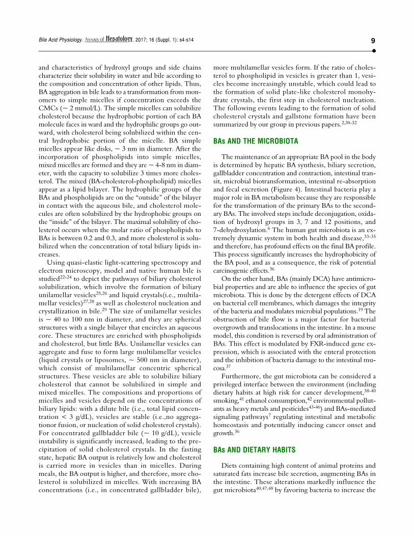
Figure 4.Figure 4.Figure 4.Figure 4.Figure 4. Bile acid (BA) biosynthesis, enterohepatic circulation and function through their receptors in the liver and intestine.Complex molecular mechanismsinvolve a set of nuclear receptors, i.e., farnesoid X receptor (FXR), retinoid X receptor (RXR), small heterodimer partner (SHP), liver receptor homologous-1(LRH-1), and liver X receptor (LXR).77 FXR plays a key role as main sensor of BAs and regulator of synthesis, secretion and metabolism of BAs in the liver, ile-um and colon.78,79 1.1.1.1.1. In the liver, the primary BAs (CA, CDCA) are mainly synthesized from cholesterol by the rate-limiting microsomal enzyme cholesterol 7α-hydroxylase (CYP7A1) and by CYP8B1 at a later step (the “classical pathway”) and by the CYP27A1 (the “alternative pathway”). BAs are conjugated to taurineor glycine mainly via two enzymes, BA CoA synthase (BACS) and BA-CoA-amino acid N-acetyltransferase (BAAT), secreted into bile by the bile salt exportpump (BSEP); the multidrug resistance-associated protein (MRP2) mediates secretion of organic substrates such as bilirubin, and glutathione. 2.2.2.2.2. Gallbladder:bile is stored and concentrated because water absorption occurs, as well as periodically released into the duodenum due to gallbladder contraction in the fast-ing state (about 20% emptying at the end of phase II of the migrating myoelectric complex80,81 under the control of the vagus and enterohormone motilin81
and especially after a meal due to the enterohormone cholecystokinin, CCK8). This rythmic activity is also modulated in concert with episodes of gallbladder re-laxation/refilling due to the effect of the vasointestinal peptide (VIP, released in the duodenum by gastric acid), BAs per se (acting on the gallbladder receptorGPBAR-1), and the intestinal FGF15/19 (following the BA/FXR interaction in the ileum) acting on the FGF4/β-Klotho receptor also expressed in the gallblad-der.8,82,83 Fasting serum BAs concentrations in healthy subjects are 0.2-0.7 µM to increase to 4-5 µM after each meal.2-5 3.3.3.3.3. BAs are efficiently (i.e., > 95%)reabsorbed in the terminal ileum. The remaining BAs enter the colon, undergo biotransformation to the secondary BAs by the resident gut microbiota, and un-dergo passive diffusion and reabsorption. Only 5% of BAs are lost in feces every day. The enterohepatic circulation of BAs includes their intestinal re-absorptionand continuous recirculation to the liver through the portal vein. About 10-50% of re-absorbed BAs undergo peripheral spillover into systemic circulation.84 4.4.4.4.4.Upon arrival in the terminal ileum, BAs activate FXR and increase the transcription of the enterokine fibroblast growth factor 19 (FGF19 in humans or FGF15in mice) which enters the portal circulation and regulates both gallbladder (see point 2) and liver effects (see point 5). BAs in the intestine also activate the Gprotein-coupled receptor (GPBAR-1) and stimulate the secretion of peptide YY (PYY), glucagon-like peptide 1 (GLP-1) and glucagon-like peptide 2 (GLP-2), allof which produce important metabolic effects on glucose metabolism,85 insulin metabolism and appetite acting on GPBAR-1 receptors located in the cells ofbrown adipose tissue and muscle.85 In the ileocyte, BA uptake, intracellular transport and secretion into the portal vein require the apical sodium dependent bileacid transporter (ASBT), the cellular intestinal BA binding protein (I-BABP), and the basolateral heterodimeric organic solute transporter (OSTα/β), respectively(see inset 4a for details). 5.5.5.5.5. The circulating FGF19 binds to hepatic FGF receptor 4 (FGFR4)/β-Klotho to activatec-Jun N-terminal kinase/extracellular signal-regulated kinase (JNK/ERK) signaling, which inhibits expression of CYP7A1 and CYP8B1 and hepatic BA synthesis, in synergy with the FXR-SHP inhibitorypathway.70,86 BAs enter the liver by sodium taurocholate cotransporting polypeptide (NTCP) and organic anion transporting polypeptide (OATP) transportersand act as physiological nuclear ligands for FXR, which regulates target gene transcription by binding toRXRs as a heterodimer.87 This results in increasedtranscription of the small heterodimer partner (SHP) expression. SHP, in turn, inhibits LRH-1, preventing the activation of target genes that participate in BAand fatty acid synthesis. In the absence of BAs, LRH-1 acts together with LXR to stimulate BA synthesis.55,88,89 FXR also regulates the enzymatic activity thatis involved in BA conjugation to glycine or taurine, and hepatic BA secretion by of BSEP and hepatic phospholipid secretion by ABCB4. BAs re-entering the liveralso interact with the liver GPBAR-1 expressed in Kupffer cells, in concert with the pathway activated by the FGFR4/β-Klotho. FXR activation also coordinatesBA detoxification enzymes (i.e., cytosolic sulfotransferase 2A1 [SULT2A1], aldol-keto reductase 1 B7 [AKR1B7], cytochrome P450 3A4/3a11 [CYP3A4/Cyp3a11], and UDP-glycosyltransferase 2B4 [UTG2B4]).90 6.6.6.6.6. The events leading to BA excretion from the hepatocyte into the portal vein are shown in the in-set. Specific transporters are the multidrug resistance protein 3 and 4 (MRP3, MRP4) and OSTα/β. 7.7.7.7.7. From the peripheral circulation, BAs also undergo renaluptake by the apical sodium/dependent bile acid transporter (ASBT) in the proximal tubule. Glomerular filtration of BAs are regulated by MRP2, 3, 4 transport-ers.91 Adapted from Ory, et al.92 and Inagaki, et al.,70 Garruti, et al.,77 Liu, et al.52
9Bile Acid Physiology. , 2017; 16 (Suppl. 1): s4-s14
more multilamellar vesicles form. If the ratio of choles-terol to phospholipid in vesicles is greater than 1, vesi-cles become increasingly unstable, which could lead tothe formation of solid plate-like cholesterol monohy-drate crystals, the first step in cholesterol nucleation.The following events leading to the formation of solidcholesterol crystals and gallstone formation have beensummarized by our group in previous papers.2,30-32
BAs AND THE MICROBIOTA
The maintenance of an appropriate BA pool in the bodyis determined by hepatic BA synthesis, biliary secretion,gallbladder concentration and contraction, intestinal tran-sit, microbial biotransformation, intestinal re-absorptionand fecal excretion (Figure 4). Intestinal bacteria play amajor role in BA metabolism because they are responsiblefor the transformation of the primary BAs to the second-ary BAs. The involved steps include deconjugation, oxida-tion of hydroxyl groups in 3, 7 and 12 positions, and7-dehydroxylation.6 The human gut microbiota is an ex-tremely dynamic system in both health and disease,33-35
and therefore, has profound effects on the final BA profile.This process significantly increases the hydrophobicity ofthe BA pool, and as a consequence, the risk of potentialcarcinogenic effects.36
On the other hand, BAs (mainly DCA) have antimicro-bial properties and are able to influence the species of gutmicrobiota. This is done by the detergent effects of DCAon bacterial cell membranes, which damages the integrityof the bacteria and modulates microbial populations.19 Theobstruction of bile flow is a major factor for bacterialovergrowth and translocations in the intestine. In a mousemodel, this condition is reversed by oral administration ofBAs. This effect is modulated by FXR-induced gene ex-pression, which is associated with the enteral protectionand the inhibition of bacteria damage to the intestinal mu-cosa.37
Furthermore, the gut microbiota can be considered aprivileged interface between the environment (includingdietary habits at high risk for cancer development,38-40
smoking,41 ethanol consumption,42 environmental pollut-ants as heavy metals and pesticides43-46) and BAs-mediatedsignaling pathways7 regulating intestinal and metabolichomeostasis and potentially inducing cancer onset andgrowth.36
BAs AND DIETARY HABITS
Diets containing high content of animal proteins andsaturated fats increase bile secretion, augmenting BAs inthe intestine. These alterations markedly influence thegut microbiota40,47,48 by favoring bacteria to increase the
and characteristics of hydroxyl groups and side chainscharacterize their solubility in water and bile according tothe composition and concentration of other lipids. Thus,BA aggregation in bile leads to a transformation from mon-omers to simple micelles if concentration exceeds theCMCs (~ 2 mmol/L). The simple micelles can solubilizecholesterol because the hydrophobic portion of each BAmolecule faces in ward and the hydrophilic groups go out-ward, with cholesterol being solubilized within the cen-tral hydrophobic portion of the micelle. BA simplemicelles appear like disks, ~ 3 nm in diameter. After theincorporation of phospholipids into simple micelles,mixed micelles are formed and they are ~ 4-8 nm in diam-eter, with the capacity to solubilize 3 times more choles-terol. The mixed (BA-cholesterol-phospholipid) micellesappear as a lipid bilayer. The hydrophilic groups of theBAs and phospholipids are on the “outside” of the bilayerin contact with the aqueous bile, and cholesterol mole-cules are often solubilized by the hydrophobic groups onthe “inside” of the bilayer. The maximal solubility of cho-lesterol occurs when the molar ratio of phospholipids toBAs is between 0.2 and 0.3, and more cholesterol is solu-bilized when the concentration of total biliary lipids in-creases.
Using quasi-elastic light-scattering spectroscopy andelectron microscopy, model and native human bile isstudied22-24 to depict the pathways of biliary cholesterolsolubilization, which involve the formation of biliaryunilamellar vesicles25,26 and liquid crystals(i.e., multila-mellar vesicles)27,28 as well as cholesterol nucleation andcrystallization in bile.29 The size of unilamellar vesiclesis ~ 40 to 100 nm in diameter, and they are sphericalstructures with a single bilayer that encircles an aqueouscore. These structures are enriched with phospholipidsand cholesterol, but little BAs. Unilamellar vesicles canaggregate and fuse to form large multilamellar vesicles(liquid crystals or liposomes, ~ 500 nm in diameter),which consist of multilamellar concentric sphericalstructures. These vesicles are able to solubilize biliarycholesterol that cannot be solubilized in simple andmixed micelles. The compositions and proportions ofmicelles and vesicles depend on the concentrations ofbiliary lipids: with a dilute bile (i.e., total lipid concen-tration < 3 g/dL), vesicles are stable (i.e.,no aggrega-tionor fusion, or nucleation of solid cholesterol crystals).For concentrated gallbladder bile (~ 10 g/dL), vesicleinstability is significantly increased, leading to the pre-cipitation of solid cholesterol crystals. In the fastingstate, hepatic BA output is relatively low and cholesterolis carried more in vesicles than in micelles. Duringmeals, the BA output is higher, and therefore, more cho-lesterol is solubilized in micelles. With increasing BAconcentrations (i.e., in concentrated gallbladder bile),
Di Ciaula A, et al. , 2017; 16 (Suppl. 1): s4-s1410
concentration of hydrophobic BAs (mainly DCA47,48) inthe total BA pool.39,49 In mice, a high-fat diet decreasesLactobacillales and increases the Clostridium subclusterXIVa, leading to an increase in serum levels of DCA. Themodified microbiota composition is suppressed by die-tary supplement of agaro-oligosaccharides (a naturalderivate from agarose).48 In Apcmin/+ mice, treatmentwith DCA alters the gut microbiota composition bycausing defective intestinal barrier function, intestinallow-grade inflammation, and cancer progression. Whenfecal microbiota is transplanted from DCA-treated miceto another group of Apcmin/+ animals, an increased tu-mor multiplicity is found likely due to activation of thetumor-associated Wnt/β-catenin signaling pathway. Ofnote, the cancer-promoting effects of BAs are blocked bygut microbiota depletion through antibiotic treatment.36
As demonstrated in animals, the pathways linking a high-fat diet to an alteration in intestinal microbiota are alsocorrelated with increased retention of hydrophobic BAsin the liver, leading to hepatocellular carcinoma in ani-mals with nonalcoholic steatohepatitis (NASH). Thispathogenic mechanism isalso supported by an inhibitionof key BA transporters secondary to high-fat diet-in-duced liver inflammation and to a down-regulation ofthe tumor suppressor gene CEBPα.50
BAs AS SIGNALING MOLECULES
As mentioned above, BAs display both hydrophilic andhydrophobic surfaces, which makes these molecules high-ly soluble, and detergent-like amphiphilic. The potency ofBAs as detergents depends on the distribution and orienta-tion of hydroxyl groups around the steroid nucleus of themolecule, a feature called hydrophobicity, which is quan-tified by high performance liquid chromatography(HPLC).2 The hydrophobicity of BAs, which is directlyrelated to cytotoxicity, is the following order: LCA>DCA > CDCA > CA > UDCA.
BAs are also being recognized as signaling molecules inthe human body because they are able to regulate metabol-ic and cellular functions by interaction with BA receptors.BAs interact with the nuclear receptor superfamily such asligand-activated FXR and GPBAR-1.1 FXR is a master BAsensor in the liver and ileum.51-53 The BA-FXR interactionis essential in BA homeostasis: the rank order of potencyis CDCA > LCA = DCA > CA in the conjugated and un-conjugated forms.54 FXR regulates a series of gene expres-sion that is involved in the synthesis, uptake, secretion andintestinal absorption of BAs, and all these processes are es-sential in the regulation of intracellular Bas.2,55,56 FXR acti-vation in the intestine increases expression of intestinalfibroblast growth factor 19 (i.e., FGF19 in humans orFGF15 in mice); in turn, the circulating FGF19 enters the
liver via the portal vein and reduces expression of hepaticcholesterol 7α-hydroxylase and BA synthesis.57
BAs also interact with GPBAR-1 that is mainly ex-pressed in Kupffer cells, but not hepatocytes.39,58 In thiscase, the rank order of potency is TLCA > TDCA >TCDCA > TCA.1 In the ileum, activation of GPBAR-1increases levels of peptide YY (PYY) with anorexigenic ef-fect (i.e., appetite reduction), as well as glucagon-like pep-tide-1 (GLP-1) and glucagon-like peptide-2 (GLP-2).60
GPBAR-1 is also expressed and metabolically active in thegallbladder, brown adipose tissue, skeletal muscle, macro-phages, and monocytes1,59 and in the enteroendocrine cellsof the intestine.61 In particular, GPBAR-1 signalling inskeletal muscle and brown adipose tissue results in localactivation of the type II iodothyronine deiodinase (DIO2)able to generate or transform the inactive thyroxine (T4)to active thyroid hormone (T3, a key regulator of metabo-lism and energy homeostasis). In Kupffer cells and macro-phages, GPBAR-1 activation inhibits LPS-inducedcytokine production.62 Such additional hormonal effectsof BAs are cAMP-mediated and might be particularly evi-dent after bariatric surgery with important and beneficialmetabolic effects, including increased energy expenditure,increased insulin secretion and/or sensitivity and decreaseinflammatory status.9,57,62 (see also chapter by Garruti, et al.in the present issue). The mechanisms governing BA bio-synthesis and the composition of the total BA pool are,therefore, of paramount importance for keeping the over-all digestive and metabolic functions of BAs in health.This aspect involves a number of nuclear receptors in theliver and intestine via FXR-dependent and -independentmechanisms.
FXR-dependent mechanisms
Overall, cholesterol 7α-hydroxylase (CYP7A1) is therate-limiting enzyme for regulating BA synthesis and is atarget gene of FXR. Several factors such as BAs, inflamma-tory cytokines, steroid hormones, and insulin may inhibitCYP7A1 transcription through the 5’-upstream region ofthe promoter.63-65 FXR is critical in this respect as a regu-latory factor of BA metabolism because it down-regulatesCYP7A1, sterol 12α-hydroxylase (CYP8B1), and sterol 27-hydroxylase (CYP27A1) transcription by a negative feed-back mechanism.
In the hepatocyte, one regulatory mechanism in-volves binding of BAs to FXR in the nucleus, the for-mation of the FXR/RXR heterodimer, and theactivationof small heterodimer partner (SHP), leadingto an inhibition of the activity of liver receptor homol-ogous-1 (LRH-1) and the CYP7A1 transcription.55,66
Furthermore, SHP displaces the promoter factorHNF4α from PGC-1α, thus contributing to CYP7A1
11Bile Acid Physiology. , 2017; 16 (Suppl. 1): s4-s14
and CYP8B1 transcription. FXR also plays a role in de-creasing BA cytotoxicity because it promotes expres-sion of the enzymes involved in conjugation of BAswith glycine or taurine, i.e., BA CoA synthase and BA-CoA-amino acid N-acetyltransferase.67,68 Excessive ac-cumulation of intrahepatic triglycerides during thesequence non-alcoholic fatty liver, steatohepatitis is as-sociated with abnormalities of gene expression of FXRand SHP and BA transporters.69
In the ileum, a second regulatory mechanism of BAsynthesis involves the secretion of FGF19 and activationof FGFR4 tyrosine kinase/β-klotho (a co-expressed mem-brane-bound glycosidase) signaling in the hepatocyte ba-solateral membrane.70,71 This pathway involves theJNK-mediated pathway and suppression of CYP7A1 tran-scription and points to the importance of the BAs/FXR/FGF19/FGFR4/CYP7A1 signaling cascade which nega-tively regulates BA biosynthesis in the liver in hu-mans.2,72,73
FXR-independent mechanisms
FXR-independent BA inhibition of CYP7A1 transcrip-tion might work by several parallel mechanisms to protectagainst BA toxicity during cholestasis and liver injury.
• Insulin receptor and activation of PI3K and AKT leadto phosphorylation of FoxO1 and inhibit CYP7A1transcription.
• Activation of the pregnane X receptor (PXR) and vita-min D receptor by LCA and binding to the BA re-sponse element (BARE)-I sequence in the CYP7A1promoter may inhibit CYP7A1 promoter activity.74
• Also, both PXR and vitamin D receptor inhibitCYP7A1 transcription by blocking HNF4α recruit-ment of PGC-1α to CYP7A1 chromatin.
• BAs also activate epidermal growth factor receptor(EGFR) and the Raf-1/MEK/ERK signaling pathway,thus inhibiting CYP7A1 transcription.
• The hepatocyte growth factor (HGF) is released fromhepatic stellate cells during liver regeneration and in-jury, and HGF stimulates HGF receptor cMet andMAPK pathways, leading to inhibition of CYP7A1transcription and BA synthesis.
• Kupffer cells secrete TGFβ-1 that activate its receptorTRβII and the SMAD signaling pathway in the hepato-cyte. SMAD3 enters the nucleus of hepatocyte andworks with HDACs and mSin3A to inhibit HNF4αactivation of CYP7A1 transcription. A tumour sup-pressor p53 interacts with HNF4α and inhibitsHNF4α activity. These alterations may inhibitCYP7A1 transcription.
• Under certain circumstances, i.e., endotoxin-induced
cholestasis, lipopolysaccharides released by bacteriastimulate the secretion of TNFα (alpha) and IL-1βfrom Kupffer cells, leading to activation of Toll-likereceptor 4. TNFα and IL-1β may inhibit CYP7A1 tran-scription by activating the TNF-α receptor and theMAPK/JNK pathway in the hepatocyte. JNK may in-hibit CYP7A1 and CYP8B1 transcription and BA syn-thesis by phosphorylating cJun no period75.76 andHNF4α.
CONCLUSIONS
BAs are synthesized mainly in the liver and are the majorlipid components of bile, as well as involved in hepatic cho-lesterol catabolism. BAs released by the gallbladder enter thegastrointestinal tract during the meal and are key regulators offat emulsion and solubilisation, two essential steps for thedigestion and absorption of cholesterol, triglycerides and fat-soluble vitamins. BAs also act as signaling molecules by acti-vating two main sensors in the body: the nuclear receptorFXR and the cell surface receptor GPBAR-1. In this way,BAs become key regulators of complex homeostatic path-ways at a systemic level ranging from their own homeostasistocholesterol, triglyceride, glucose and energy metabolisms.Additional regulations include cell proliferation, inflamma-tion, and tumor onset and progression.
Thus, maintaining the precise balance between BA spe-cies and amounts, as well as preventing the accumulationof excessive BAs in the body are of importance under-physiological or pathophysiological conditions that in-volve the liver, intestine, muscle and adipose tissues.
In addition, the pathways involving the intestinalmicrobiota and epigenetic factors regulate gene expres-sion and act as a common interface between environmen-tal factors (including diet, lifestyle, and exposure toenvironmental toxics) and the molecular events promot-ing the onset and the progress of disease. The high-fat diet,for example, increases the fecal concentration of the sec-ondary BAs that are a risk factor for the development ofcolorectal cancer. Of note, intestinal microbiota and theepigenome might be modulated by the primary preven-tion strategies (i.e., changes in dietary habits and lifestyle,and reduced exposure to environmental toxics) and thera-peutic tools. Future studies are needed to better clarifyhow these measures could influence pathogenic mecha-nisms, disease onset and the efficacy of the available thera-peutic tools.
ABBREVIATIONS
• BAs: bile acids• CA: cholic acid• CDCA: chenodeoxycholic acid
Di Ciaula A, et al. , 2017; 16 (Suppl. 1): s4-s1412
• CMC: critical micellar concentration• CYP7A1: cholesterol 7á-hydroxylase• DCA: deoxycholic acid• FGF: fibroblast growth factor• FXR: farnesoid X receptor• GPBAR-1: G-protein-coupled bile acid receptor-1
(also known as TGR5)• JNK: c-Jun N-terminal kinase• LCA: lithocholic acid• UDCA: ursodeoxycholic acid
CONFLICTS OF INTEREST
We declare that we have no conflicts of interest.
ACKNOWLEDGEMENTS
The present chapter is written in the context of theproject FOIE GRAS, which has received funding from theEuropean Union’s Horizon 2020 Research and Innovationprogramme under the Marie Sklodowska-Curie GrantAgreement No. 722619. Raquel Lunardi Baccetto andEmilio Molina-Molina are recipients of Foie Gras EarlyResearch Training Grant.
REFERENCES
1. Li T, Chiang JYL. Bile Acid Signaling in Metabolic Disease andDrug Therapy. Pharmacological Reviews 2014; 66: 948.
2. Wang DQH, Neuschwander-Tetri BA, Portincasa P. The BiliarySystem. 2nd Ed. Morgan & Claypool Life Sciences; 2017.
3. Ponz De Leon M, Murphy G, Dowling RH. Physiological fac-tors influencing serum bile acid levels. Gut 1978; 19: 32-9.
4. Schalm SW, LaRusso NF, Hofmann AF, Hoffman NE, vanBerge-Henegouwen GP, Korman MG. Diurnal serum levelsof primary conjugated bile acids. Assessment by specific ra-dioimmunoassays for conjugates of cholic and chenodeoxy-cholic acid. Gut 1978; 19: 1006-14.
5. LaRusso NF, Korman MG, Hoffman NE, Hofmann AF. Dy-namics of the enterohepatic circulation of bile acids. Post-prandial serum concentrations of conjugates of cholic acid inhealth, cholecystectomized patients, and patients with bileacid malabsorption. N Engl J Med 1974; 291: 689-92.
6. Ridlon JM, Kang DJ, Hylemon PB. Bile salt biotransformationsby human intestinal bacteria. J Lipid Res 2006; 47: 241-59.
7. Zhou H, Hylemon PB. Bile acids are nutrient signaling hor-mones. Steroids 2014; 86: 62-8.
8. Portincasa P, Di Ciaula A, Wang HH, Palasciano G, van Er-pecum KJ, Moschetta A, Wang DQ. Coordinate regulation ofgallbladder motor function in the gut-liver axis. Hepatology2008; 47: 2112-26.
9. Wahlström A, Sayin SI, Marschall H-U, Bäckhed F. Intestinalcrosstalk between bile acids and microbiota and its impacton host metabolism. Cell Metab 2016; 24: 41-50.
10. Alnouti Y. Bile Acid sulfation: a pathway of bile acid elimina-tion and detoxification. Toxicol Sci 2009; 108: 225-46.
11. Bathena SP, Mukherjee S, Olivera M, Alnouti Y. The profile ofbile acids and their sulfate metabolites in human urine andserum. J Chromatogr B Analyt Technol Biomed Life Sci2013; 942-943: 53-62.
12. Wang TY, Liu M, Portincasa P, Wang DQ. New insights intothe molecular mechanism of intestinal fatty acid absorption.Eur J Clin Invest 2013; 43: 1203-23.
13. Hofmann AF. The continuing importance of bile acids in liverand intestinal disease. Arch Intern Med 1999; 159: 2647-58.
14. Gong Z, Zhou J, Zhao S, Tian C, Wang P, Xu C, Chen Y, etal. Chenodeoxycholic acid activates NLRP3 inflammasomeand contributes to cholestatic liver fibrosis. Oncotarget2016; 7: 83951-63.
15. Tremblay S, Romain G, Roux M, Chen XL, Brown K, GibsonDL, Ramanathan S, et al. Bile Acid Administration Elicits anIntestinal Antimicrobial Program and Reduces the BacterialBurden in Two Mouse Models of Enteric Infection. Infect Im-mun 2017; 85.
16. Guo C, Xie S, Chi Z, Zhang J, Liu Y, Zhang L, Zheng M, et al.Bile Acids Control Inflammation and Metabolic Disorderthrough Inhibition of NLRP3 Inflammasome. Immunity 2016;45: 802-16.
17. Zhu C, Fuchs CD, Halilbasic E, Trauner M. Bile acids in regu-lation of inflammation and immunity: friend or foe? Clin ExpRheumatol 2016; 34: 25-31.
18. Dossa AY, Escobar O, Golden J, Frey MR, Ford HR, GayerCP. Bile acids regulate intestinal cell proliferation by modulat-ing EGFR and FXR signaling. Am J Physiol Gastrointest Liv-er Physiol 2016; 310: G81-92.
19. Yokota A, Fukiya S, Islam KB, Ooka T, Ogura Y, Hayashi T,Hagio M, et al. Is bile acid a determinant of the gut microbiotaon a high-fat diet? Gut Microbes 2012; 3: 455-9.
20. De Fabiani E, Mitro N, Gilardi F, Galmozzi A, Caruso D, Cre-stani M. When food meets man: the contribution of epigenet-ics to health. Nutrients 2010; 2: 551-71.
21. Wang DQ, Portincasa P, Tso P. Transintestinal cholester-ol excretion (TICE): A secondary, non-biliary pathwaycontributing to reverse cholesterol transport. Hepatology2017.
22. Mazer NA, Carey MC, Kwasnick RF, Benedek GB. Quasie-lastic light scattering studies of aqueous biliary lipid systems.Size, shape, and thermodynamics of bile salt micelles. Bio-chemistry 1979; 18: 3064-75.
23. Somjen GJ, Marikovsky Y, Lelkes P, Gilat T. Cholesterol-phospholipid vesicles in human bile: an ultrastructural study.Biochim.Biophys.Acta 1986; 879: 14-21.
24. Somjen GJ, Gilat T. Contribution of vesicular and micellarcarriers to cholesterol transport in human bile. J Lipid Res1985; 26: 699-704.
25. Carey MC, Small DM. The physical chemistry of cholesterolsolubility in bile. Relation to gallstone formation and dissolu-tion in man. J Clin Invest 1978; 61: 998-1026.
26. Holzbach RT. Metastability behavior of supersaturated bile.Hepatology 1984; 4: 155S-8S.
27. Olszewski MF, Holzbach RT, Saupe A, Brown GH. Liquidcrystals in human bile. Nature 1973; 242: 336-7.
28. Holzbach RT, Corbusier C. Liquid crystals and cholesterolnucleation during equilibration in supersaturated bile ana-logs. Biochim.Biophys.Acta 1978; 528: 436-44.
29. Wang X, Luo S, Liu Y. Effects of changes of plasma motilinlevel on the motility of gallbladder in patients with chronic re-nal failure. Chung.Hua.Nei.Ko.Tsa.Chih 1996; 35: 86-8.
30. Portincasa P, Moschetta A, Palasciano G. Cholesterol gall-stone disease. Lancet 2006; 368: 230-9.
31. Wang DQH, Portincasa P (eds.). Gallstones. Recent advanc-es in epidemiology, pathogenesis, diagnosis and manage-ment. 1st ed. New York, NY: Nova Science Publisher Inc.;2017, p. 1-676.
32. Portincasa P, Di Ciaula A, de Bari O, Garruti G, Palmieri VO,Wang DQ. Management of gallstones and its related complica-
13Bile Acid Physiology. , 2017; 16 (Suppl. 1): s4-s14
tions. Expert Rev Gastroenterol Hepatol 2016; 10: 93-112.33. Bonfrate L, Tack J, Grattagliano I, Cuomo R, Portincasa P.
Microbiota in health and irritable bowel syndrome: currentknowledge, perspectives and therapeutic options. Scand JGastroenterol 2013; 48: 995-1009.
34. Portincasa P, Bonfrate L, de Bari O, Lembo A, Ballou S. Irri-table bowel syndrome and diet. Gastroenterol Rep (Oxf)2017.
35. Long SL, Gahan CGM, Joyce SA. Interactions between gutbacteria and bile in health and disease. Mol Aspects Med2017; 56: 54-65.
36. Cao H, Xu M, Dong W, Deng B, Wang S, Zhang Y, Wang S, etal. Secondary bile acid-induced dysbiosis promotes intestinalcarcinogenesis. Int J Cancer 2017; 140: 2545-56.
37. Inagaki T, Moschetta A, Lee YK, Peng L, Zhao G, Downes M,Yu RT, et al. Regulation of antibacterial defense in the smallintestine by the nuclear bile acid receptor. Proc Natl AcadSci USA 2006; 103: 3920-5.
38. Ou J, DeLany JP, Zhang M, Sharma S, O’Keefe SJ. Associa-tion between low colonic short-chain fatty acids and highbile acids in high colon cancer risk populations. Nutr Cancer2012; 64: 34-40.
39. Ridlon JM, Wolf PG, Gaskins HR. Taurocholic acid metabo-lism by gut microbes and colon cancer. Gut Microbes 2016;7: 201-15.
40. O’Keefe SJ, Li JV, Lahti L, Ou J, Carbonero F, Mohammed K,Posma JM, et al. Fat, fibre and cancer risk in African Ameri-cans and rural Africans. Nat Commun 2015; 6: 6342.
41. Biedermann L, Zeitz J, Mwinyi J, Sutter-Minder E, Rehman A,Ott SJ, Steurer-Stey C, et al. Smoking cessation induces pro-found changes in the composition of the intestinal microbiotain humans. PLoS One 2013; 8: e59260.
42. Mutlu EA, Gillevet PM, Rangwala H, Sikaroodi M, Naqvi A, En-gen PA, Kwasny M, et al. Colonic microbiome is altered in al-coholism. Am J Physiol Gastrointest Liver Physiol 2012;302: G966-78.
43. Gao B, Chi L, Mahbub R, Bian X, Tu P, Ru H, Lu K. Multi-Om-ics Reveals that Lead Exposure Disturbs Gut MicrobiomeDevelopment, Key Metabolites, and Metabolic Pathways.Chem Res Toxicol 2017; 30: 996-1005.
44. Gao B, Bian X, Mahbub R, Lu K. Sex-Specific Effects ofOrganophosphate Diazinon on the Gut Microbiome and ItsMetabolic Functions. Environ Health Perspect 2017; 125:198-206.
45. Joly C, Gay-Queheillard J, Leke A, Chardon K, Delanaud S,Bach V, Khorsi-Cauet H. Impact of chronic exposure to lowdoses of chlorpyrifos on the intestinal microbiota in the Simu-lator of the Human Intestinal Microbial Ecosystem (SHIME)and in the rat. Environ Sci Pollut Res Int 2013; 20: 2726-34.
46. Joly Condette C, Bach V, Mayeur C, Gay-Queheillard J,Khorsi-Cauet H. Chlorpyrifos Exposure During Perinatal Peri-od Affects Intestinal Microbiota Associated With Delay ofMaturation of Digestive Tract in Rats. J Pediatr Gastroenter-ol Nutr 2015; 61: 30-40.
47. David LA, Maurice CF, Carmody RN, Gootenberg DB, But-ton JE, Wolfe BE, Ling AV, et al. Diet rapidly and reproduc-ibly alters the human gut microbiome. Nature 2014; 505:559-63.
48. Higashimura Y, Naito Y, Takagi T, Uchiyama K, Mizushima K,Ushiroda C, Ohnogi H, et al. Protective effect of agaro-oli-gosaccharides on gut dysbiosis and colon tumorigenesis inhigh-fat diet-fed mice. Am J Physiol Gastrointest Liver Phys-iol 2016; 310: G367-75.
49. Ridlon JM, Kang DJ, Hylemon PB, Bajaj JS. Bile acids and thegut microbiome. Curr Opin Gastroenterol 2014; 30: 332-8.
50. Xie G, Wang X, Huang F, Zhao A, Chen W, Yan J, Zhang Y,
et al. Dysregulated hepatic bile acids collaboratively promoteliver carcinogenesis. Int J Cancer 2016; 139: 1764-75.
51. Makishima M, Okamoto AY, Repa JJ, Tu H, Learned RM, LukA, Hull MV, et al. Identification of a nuclear receptor for bileacids. Science 1999; 284: 1362-5.
52. Liu H, Hu C, Zhang X, Jia W. Role of gut microbiota, bile ac-ids and their cross-talk in the effects of bariatric surgery onobesity and type 2 diabetes. J Diabetes Invest 2017.
53. Kuipers F, Bloks VW, Groen AK. Beyond intestinal soap—bile acids in metabolic control. Nat Rev Endocrinol 2014; 10:488-98.
54. Parks DJ, Blanchard SG, Bledsoe RK, Chandra G, ConslerTG, Kliewer SA, Stimmel JB, et al. Bile acids: natural ligandsfor an orphan nuclear receptor. Science 1999; 284: 1365-8.
55. Goodwin B, Jones SA, Price RR, Watson MA, McKee DD,Moore LB, Galardi C, et al. A regulatory cascade of the nu-clear receptors FXR, SHP-1, and LRH-1 represses bile acidbiosynthesis. Mol Cell 2000; 6: 517-26.
56. Wang L, Lee YK, Bundman D, Han Y, Thevananther S, KimCS, Chua SS, et al. Redundant pathways for negative feed-back regulation of bile acid production. Dev Cell 2002; 2:721-31.
57. Mazidi M, de Caravatto PP, Speakman JR, Cohen RV. Mecha-nisms of Action of Surgical Interventions on Weight-RelatedDiseases: the Potential Role of Bile Acids. Obes Surg 2017;27: 826-36.
58. Festa C, De Marino S, Carino A, Sepe V, Marchiano S, Cipri-ani S, Di Leva FS, et al. Targeting Bile Acid Receptors: Dis-covery of a Potent and Selective Farnesoid X ReceptorAgonist as a New Lead in the Pharmacological Approach toLiver Diseases. Front Pharmacol 2017; 8: 162.
59. de Aguiar Vallim TQ, Tarling EJ, Edwards PA. Pleiotropicroles of bile acids in metabolism. Cell Metab 2013; 17: 657-69.
60. Liu N, Zhao J, Wang J, Teng H, Fu Y, Yuan H. Farnesoid Xreceptor ligand CDCA suppresses human prostate cancercells growth by inhibiting lipid metabolism via targeting sterolresponse element binding protein 1. Am J Transl Res 2016;8: 5118-24.
61. Barrasa JI, Olmo N, Lizarbe MA, Turnay J. Bile acids in thecolon, from healthy to cytotoxic molecules. Toxicol In Vitro2013; 27: 964-77.
62. Schaap FG, Trauner M, Jansen PLM. Bile acid receptors astargets for drug development. Nature Reviews Gastroenter-ology & Hepatology 2013; 11: 55-67.
63. Chiang JY. Bile acid regulation of gene expression: roles ofnuclear hormone receptors. Endocr Rev 2002; 23: 443-63.
64. Chiang JY. Regulation of bile acid synthesis. Front Biosci1998; 3: d176-d93.
65. Chiang JY. Regulation of bile acid synthesis: pathways, nu-clear receptors, and mechanisms. J Hepatol 2004; 40: 539-51.
66. Miao J, Choi SE, Seok SM, Yang L, Zuercher WJ, Xu Y, Will-son TM, et al. Ligand-Dependent Regulation of the Activity ofthe Orphan Nuclear Receptor, Small Heterodimer Partner(SHP), in the Repression of Bile Acid Biosynthetic CYP7A1and CYP8B1 Genes. Mol Endocrinol 2011; 25: 1159-69.
67. Pircher PC, Kitto JL, Petrowski ML, Tangirala RK, BischoffED, Schulman IG, Westin SK. Farnesoid X receptor regulatesbile acid-amino acid conjugation. J Biol Chem 2003; 278:27703-11.
68. Solaas K, Ulvestad A, Söreide O, Kase BF. Subcellular or-ganization of bile acid amidation in human liver: a key issuein regulating the biosynthesis of bile salts. J Lipid Res 2000;41: 1154-62.
69. Aguilar-Olivos NE, Carrillo-Cordova D, Oria-Hernandez J,
Di Ciaula A, et al. , 2017; 16 (Suppl. 1): s4-s1414
Sanchez-Valle V, Ponciano-Rodriguez G, Ramirez-JaramilloM, Chable-Montero F, et al. The nuclear receptor FXR, but notLXR, up-regulates bile acid transporter expression in non-al-coholic fatty liver disease. Ann Hepatol 2015; 14: 487-93.
70. Inagaki T, Choi M, Moschetta A, Peng L, Cummins CL, Mc-Donald JG, Luo G, et al. Fibroblast growth factor 15 func-tions as an enterohepatic signal to regulate bile acidhomeostasis. Cell Metab 2005; 2: 217-25.
71. Jones S. Mini-review: endocrine actions of fibroblast growthfactor 19. Mol Pharm 2008; 5: 42-8.
72. Holt JA, Luo G, Billin AN, Bisi J, McNeill YY, Kozarsky KF,Donahee M, et al. Definition of a novel growth factor-de-pendent signal cascade for the suppression of bile acid bio-synthesis. Genes Dev 2003; 17: 1581-91.
73. Kim I, Ahn S-H, Inagaki T, Choi M, Ito S, Guo GL, Kliewer SA,et al. Differential regulation of bile acid homeostasis by thefarnesoid X receptor in liver and intestine. J Lipid Res 2007;48: 2664-72.
74. Chawla A, Repa JJ, Evans RM, Mangelsdorf DJ. Nuclear re-ceptors and lipid physiology: opening the X-files. Science2001; 294: 1866-70.
75. Gupta S, Stravitz RT, Dent P, Hylemon PB. Down-regulationof cholesterol 7alpha-hydroxylase (CYP7A1) gene expres-sion by bile acids in primary rat hepatocytes is mediated bythe c-Jun N-terminal kinase pathway. J Biol Chem 2001;276: 15816-22.
76. Dent P, Han SI, Mitchell C, Studer E, Yacoub A, Grandis J,Grant S, et al. Inhibition of insulin/IGF-1 receptor signalingenhances bile acid toxicity in primary hepatocytes. BiochemPharmacol 2005; 70: 1685-96.
77. Garruti G, Wang HH, Bonfrate L, de Bari O, Wang DQ, Portin-casa P. A pleiotropic role for the orphan nuclear receptorsmall heterodimer partner in lipid homeostasis and metabolicpathways. J Lipids 2012; 2012: 304292.
78. Modica S, Murzilli S, Salvatore L, Schmidt DR, Moschetta A.Nuclear bile acid receptor FXR protects against intestinal tu-morigenesis. Cancer Res 2008; 68: 9589-94.
79. Modica S, Gofflot F, Murzilli S, D’Orazio A, Salvatore L, Pel-legrini F, Nicolucci A, et al. The intestinal nuclear receptorsignature with epithelial localization patterns and expressionmodulation in tumors. Gastroenterology 2010; 138: 636-48.e12.
80. Luiking YC, Peeters TL, Stolk MFJ, Nieuwenhuijs VB, Portin-casa P, Depoortere I, vanBerge-Henegouwen GP, et al. Moti-lin induces gall bladder emptying and antral contractions inthe fasted state in humans. Gut 1998; 42: 830-5.
81. Portincasa P, Peeters TL, Berge-Henegouwen GP, Van Sol-inge WW, Palasciano G, van Erpecum KJ. Acute intraduode-nal bile salt depletion leads to strong gallbladder contraction,altered antroduodenal motility and high plasma motilin levelsin humans. Neurogastroenterol Motil 2000; 12: 421-30.
82. Housset C, Chrétien Y, Debray D, Chignard N. Functions ofthe Gallbladder. Compr Physiol 2016: 6(3): 1549-77.
83. Choi M, Moschetta A, Bookout AL, Peng L, Umetani M, Holm-strom SR, Suino-Powell K, et al. Identification of a hormonalbasis for gallbladder filling. Nat Med 2006; 12: 1253-5.
84. Dawson PA, Lan T, Rao A. Bile acid transporters. J LipidRes 2009; 50: 2340-57.
85. Brighton CA, Rievaj J, Kuhre RE, Glass LL, Schoonjans K,Holst JJ, Gribble FM, et al. Bile Acids Trigger GLP-1 ReleasePredominantly by Accessing Basolaterally Located G Pro-tein-Coupled Bile Acid Receptors. Endocrinology 2015; 156:3961-70.
86. Bjorkhem I, Blomstrand R, Lewenhaupt A, Svensson L. Ef-fect of lymphatic drainage on 7alpha-hydroxylation of cho-lesterol in rat liver. Biochem Biophys Res Commun 1978;85: 532-40.
87. Kemper JK. Regulation of FXR transcriptional activity inhealth and disease: Emerging roles of FXR cofactors andpost-translational modifications. Biochim Biophys Acta 2011;1812: 842-50.
88. Lu TT, Makishima M, Repa JJ, Schoonjans K, Kerr TA, Auw-erx J, Mangelsdorf DJ. Molecular basis for feedback regula-tion of bile acid synthesis by nuclear receptors. Mol Cell2000; 6: 507-15.
89. Nishimaki-Mogami T, Une M, Fujino T, Sato Y, Tamehiro N,Kawahara Y, Shudo K, et al. Identification of intermediates inthe bile acid synthetic pathway as ligands for the farnesoidX receptor. J Lipid Res 2004; 45: 1538-45.
90. Modica S, Bellafante E, Moschetta A. Master regulation ofbile acid and xenobiotic metabolism via the FXR, PXR andCAR trio. Frontiers in bioscience (Landmark edition) 2008;14: 4719-45.
91. Wagner M, Zollner G, Trauner M. New molecular insights intothe mechanisms of cholestasis. J Hepatol 2009; 51: 565-80.
92. Ory DS. Nuclear receptor signaling in the control of choles-terol homeostasis: have the orphans found a home? CircRes 2004; 95: 660-70.
Correspondence and reprints request:Prof. Piero Portincasa, M.D., Ph.D.
Clinica Medica “Augusto Murri”Department of Biomedical Sciences
and Human OncologyUniversity of Bari Medical School
Piazza Giulio Cesare 11, 70124 Bari-ItalyTel. +39-80-5478-227; Fax +39-80-5478-232.
E-mail: [email protected]