the brain and cranial nerves. major parts of the brain 1.brain stem – continuous with spinal cord ...
TRANSCRIPT

The Brain and The Brain and Cranial NervesCranial Nerves

Major Parts of the BrainMajor Parts of the Brain
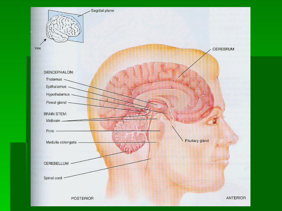
1.1. Brain stem – continuous with spinal Brain stem – continuous with spinal cordcord
MidbrainMidbrain PonsPons Medulla OblongotaMedulla Oblongota


Major Parts of the BrainMajor Parts of the Brain
2.2. Cerebellum – Posterior to brain stemCerebellum – Posterior to brain stem


Major Parts of the BrainMajor Parts of the Brain
3. Diencephalon – superior to the brain 3. Diencephalon – superior to the brain stemstem
ThalamusThalamus EpithalamusEpithalamus HypothalamusHypothalamus Subthalamus Subthalamus


Major Parts of the BrainMajor Parts of the Brain
4. Cerebrum – largest part of the brain 4. Cerebrum – largest part of the brain supported by the diencephalon and brain supported by the diencephalon and brain stemstem


Brain StemBrain Stem
1.1. Medulla OblongataMedulla Oblongata Continuous with spinal cordContinuous with spinal cord Contains both motor and sensory tractsContains both motor and sensory tracts

Brain Stem / MedullaBrain Stem / Medulla
Contains portrusions of white matter Contains portrusions of white matter called called pyramidscalled called pyramids
Brain Stem / MedullaBrain Stem / Medulla
The crossing of pyramids result in The crossing of pyramids result in neurons in the left cerebral cortex neurons in the left cerebral cortex controlling skeletal muscles on the right controlling skeletal muscles on the right side of the body and neurons in the right side of the body and neurons in the right cerebral cortex controlling skeletal cerebral cortex controlling skeletal muscles on the left sidemuscles on the left side

Brain Stem / MedullaBrain Stem / Medulla
There are reflex centers for regulation of There are reflex centers for regulation of heart rate, respiratory rate, heart rate, respiratory rate, vasoconstriction, swallowing, vomitingvasoconstriction, swallowing, vomiting

Brain Stem / PonsBrain Stem / Pons
Superior to the medullaSuperior to the medulla Helps control breathingHelps control breathing It relays nerve impulses related to It relays nerve impulses related to
voluntary skeletal movements from the voluntary skeletal movements from the cerebral cortex to the cerebellumcerebral cortex to the cerebellum


Brain Stem / MidbrainBrain Stem / Midbrain
Extends from the pons to the Extends from the pons to the diencephalondiencephalon
Cerebral Peduncles, superior colliculi, Cerebral Peduncles, superior colliculi, and inferior colliculi located hereand inferior colliculi located here


Brain Stem / MidbrainBrain Stem / Midbrain
Cerebral Peduncles – contain major Cerebral Peduncles – contain major motor tractsmotor tracts

Brain Stem / MidbrainBrain Stem / Midbrain
Superior colliculi – help with reflex head Superior colliculi – help with reflex head turning in response to visual stimuliturning in response to visual stimuli
Responsible for the pupillary reflex and Responsible for the pupillary reflex and accomodation reflex (adjusts shape of accomodation reflex (adjusts shape of lens for far versus close vision)lens for far versus close vision)

Brain Stem / MidbrainBrain Stem / Midbrain
Inferior coliculi – helps with reflex head Inferior coliculi – helps with reflex head turning in response to auditory stimuliturning in response to auditory stimuli
Reflex center for startle reflexReflex center for startle reflex

Cerebellum Cerebellum
Vermis - central constricted areaVermis - central constricted area Cerebellar hemispheres – lateral lobesCerebellar hemispheres – lateral lobes


CerebellumCerebellum
Cerebellar cortex – superficial layer of Cerebellar cortex – superficial layer of the cerebellum made up of gray matterthe cerebellum made up of gray matter

CerebellumCerebellum
Arbor Vitae – Deep to the gray mater Arbor Vitae – Deep to the gray mater made up of tracts (White Matter)made up of tracts (White Matter)


CerebellumCerebellum
Main function – Regulates posture and Main function – Regulates posture and balancebalance

Diencephalon / ThalamusDiencephalon / Thalamus
A pair of oval masses of grey matter, A pair of oval masses of grey matter, organized into nuclei, with interspersed organized into nuclei, with interspersed tracts of white mattertracts of white matter


Diencephalon / ThalamusDiencephalon / Thalamus
Intermediate mass – Joins the right and Intermediate mass – Joins the right and left halves of the thalamusleft halves of the thalamus

Diencephalon / ThalamusDiencephalon / Thalamus
Surrounds the third ventricleSurrounds the third ventricle
Diencephalon / ThalamusDiencephalon / Thalamus
It registers conscious recognition of pain, It registers conscious recognition of pain, temp., light touch, and pressure.temp., light touch, and pressure.
You need your cerebral cortex to You need your cerebral cortex to understand the nature of the pain.understand the nature of the pain.

Diencephalon / Diencephalon / Hypothalamus Hypothalamus
Inferior to the thalamusInferior to the thalamus


Diencephalon / Diencephalon / HypothalamusHypothalamus
Mammillary bodies – serve as reflexes Mammillary bodies – serve as reflexes related to the sense of smellrelated to the sense of smell

Diencephalon / Diencephalon / HypothalamusHypothalamus
Infundibulum – Connects the pituitary Infundibulum – Connects the pituitary gland to the hypothalamusgland to the hypothalamus

Diencephalon / Diencephalon / HypothalamusHypothalamus
Axons from the paraventricular and Axons from the paraventricular and supraoptic nuclei form the supraoptic nuclei form the hypothalamohypophyseal tract extends hypothalamohypophyseal tract extends through the infundibulum to the posterior through the infundibulum to the posterior pituitarypituitary
Diencephalon / Diencephalon / HypothalamusHypothalamus
FunctionsFunctions Control of the ANSControl of the ANS Production of hormonesProduction of hormones Regulation of emotional and behavioral Regulation of emotional and behavioral
patternspatterns Regulation of eating and drinkingRegulation of eating and drinking Control of body temperatureControl of body temperature

Diencephalon / Diencephalon / EpithalamusEpithalamus
Superior and posterior to thalamusSuperior and posterior to thalamus


Diencephalon / Diencephalon / EpithalamusEpithalamus
Pineal Gland – It secretes the hormone Pineal Gland – It secretes the hormone melatonin.melatonin.

Diencephalon / Diencephalon / SubthalamusSubthalamus
Below the thalamus Below the thalamus Helps control body movements Helps control body movements

Circumventricular OrgansCircumventricular Organs
Part of the diencephalon called CVOs Part of the diencephalon called CVOs that can monitor chemical changes in the that can monitor chemical changes in the blood because they lack a blood-brain blood because they lack a blood-brain barrierbarrier

Circumventricular OrgansCircumventricular Organs
Part of the hypothalamus, the pineal Part of the hypothalamus, the pineal gland, and the pituitary glandgland, and the pituitary gland


Circumventricular OrgansCircumventricular Organs
Thought to be the site of entry into the Thought to be the site of entry into the brain of HIV.brain of HIV.

CerebrumCerebrum
Largest part of the brainLargest part of the brain


CerebrumCerebrum
Cerebral cortex is composed of grey Cerebral cortex is composed of grey mattermatter
The deep grooves are called fissuresThe deep grooves are called fissures Shallower grooves are called sulciShallower grooves are called sulci


CerebrumCerebrum
Beneath the cortex lies cerebral white Beneath the cortex lies cerebral white mattermatter


CerebrumCerebrum
Longitudinal fissure separates it into right Longitudinal fissure separates it into right and left halvesand left halves

CerebrumCerebrum
The corpus callosum (a bundle of white The corpus callosum (a bundle of white fibers) connects it internally.fibers) connects it internally.


CerebrumCerebrum
Each cerebral hemisphere is separated Each cerebral hemisphere is separated into four lobes (frontal, parietal, temporal, into four lobes (frontal, parietal, temporal, and occipital)and occipital)


CerebrumCerebrum
White matter is under the cortex and White matter is under the cortex and consists of myelinated axons running in consists of myelinated axons running in three principle directionsthree principle directions

CerebrumCerebrum
Association fibersAssociation fibers connect and transmit connect and transmit nerve impulses between gyri in the same nerve impulses between gyri in the same hemispherehemisphere

CerebrumCerebrum
Commissural fibersCommissural fibers connect gyri in one connect gyri in one cerebral hemisphere to the cerebral hemisphere to the corresponding gyri in the opposite corresponding gyri in the opposite hemispherehemisphere

CerebrumCerebrum
Projection fibers transmit impulses from Projection fibers transmit impulses from the cerebrum to other parts of the brain the cerebrum to other parts of the brain and spinal cordand spinal cord

CerebrumCerebrum
Basal ganglia are paired masses of grey Basal ganglia are paired masses of grey matter internally located in each cerebral matter internally located in each cerebral hemispherehemisphere


CerebrumCerebrum
The Basal Ganglia function in controlling The Basal Ganglia function in controlling muscular movementsmuscular movements

CerebrumCerebrum
Huntington’s disease is a hereditary Huntington’s disease is a hereditary degenerative disorder of the basal nuclei.degenerative disorder of the basal nuclei.
Patients may suffer from abrubt, jerky, Patients may suffer from abrubt, jerky, almost continuous movements called almost continuous movements called choreachorea

CerebrumCerebrum
Limbic SystemLimbic System found in the cerebral found in the cerebral hemispheres and diencephalonhemispheres and diencephalon


CerebrumCerebrum
It functions in emotional aspects of It functions in emotional aspects of behavior and memory, and is associated behavior and memory, and is associated with pleasure and painwith pleasure and pain
We tend to remember things associated We tend to remember things associated with strong emotions. (Think back to the with strong emotions. (Think back to the events you remember from early events you remember from early childhood. What emotions pop up?)childhood. What emotions pop up?)

Lobes of the CerebrumLobes of the Cerebrum
Central Sulcus – Separates the frontal Central Sulcus – Separates the frontal lobe from the parietal lobelobe from the parietal lobe

Lobes of the CerebrumLobes of the Cerebrum
Precentral gyrus – immediately anterior Precentral gyrus – immediately anterior to the central sulcusto the central sulcus
Lobes of the CerebrumLobes of the Cerebrum
Postcentral gyrus – Located immediately Postcentral gyrus – Located immediately posterior to the central sulcusposterior to the central sulcus

Lobes of the CerebrumLobes of the Cerebrum
Lateral cerebral sulcus – Separates the Lateral cerebral sulcus – Separates the frontal lobe from the temporal lobefrontal lobe from the temporal lobe

Parieto-occipital sulcusParieto-occipital sulcus
Separates the parietal lobe from the Separates the parietal lobe from the occipital lobeoccipital lobe

Blood-Brain Barrier (BBB)Blood-Brain Barrier (BBB)
Prevents passage of many substances Prevents passage of many substances from blood into brain tissuefrom blood into brain tissue

Blood-Brain Barrier (BBB)Blood-Brain Barrier (BBB)
Endothelial cells of brain capillaries are Endothelial cells of brain capillaries are sealed together by tight junctionssealed together by tight junctions

Blood-Brain Barrier (BBB)Blood-Brain Barrier (BBB)
Processes of astrocytes press up against Processes of astrocytes press up against the brain capillaries and only allow the brain capillaries and only allow certain substances to pass from the certain substances to pass from the blood to neuronsblood to neurons

Blood-Brain Barrier (BBB)Blood-Brain Barrier (BBB)
Glucose (water soluble) crosses the BBB Glucose (water soluble) crosses the BBB via active transportvia active transport

Blood-Brain Barrier (BBB)Blood-Brain Barrier (BBB)
Other water soluble substances such as Other water soluble substances such as creatinine, urea, and most ions cross creatinine, urea, and most ions cross slowlyslowly

Blood-Brain Barrier (BBB)Blood-Brain Barrier (BBB)
Proteins and antibiotics cannot crossProteins and antibiotics cannot cross

Blood-Brain Barrier (BBB)Blood-Brain Barrier (BBB)
Lipid soluble substances such as oxygen, Lipid soluble substances such as oxygen, carbon dioxide, alcohol, and most carbon dioxide, alcohol, and most anesthetic agents cross easilyanesthetic agents cross easily
Protective coverings of Protective coverings of the Brainthe Brain
1.1. CraniumCranium
2.2. Cranial Meninges – continuous with the Cranial Meninges – continuous with the spinal meningesspinal meninges

Protective coverings of Protective coverings of the Brainthe Brain
Cranial Meninges made up of;Cranial Meninges made up of;Dura Mater – outerDura Mater – outerArachnoid Mater – middleArachnoid Mater – middlePia Mater – inner Pia Mater – inner

Protective coverings of Protective coverings of the Brainthe Brain
Subarachnoid space – between the Subarachnoid space – between the arachnoid mater and pia mater which arachnoid mater and pia mater which contains cerebrospinal fluidcontains cerebrospinal fluid

Protective coverings of Protective coverings of the Brainthe Brain
Three extensions of the dura mater Three extensions of the dura mater separate parts of the brainseparate parts of the brain

Protective coverings of Protective coverings of the Brainthe Brain
1.1. Falx Cerebri – Separates the two Falx Cerebri – Separates the two hemisphereshemispheres

Protective coverings of Protective coverings of the Brainthe Brain
2. Falx Cerebelli – Separates the two 2. Falx Cerebelli – Separates the two hemispheres of the cerebellumhemispheres of the cerebellum

Protective coverings of Protective coverings of the Brainthe Brain
3. Tentorium Cerebelli – Separates the 3. Tentorium Cerebelli – Separates the cerebrum from the cerebellum cerebrum from the cerebellum

Cerebrospinal FluidCerebrospinal Fluid
Clear colorless liquidClear colorless liquid

Cerebrospinal FluidCerebrospinal Fluid
Protects the brain and spinal cord against Protects the brain and spinal cord against physical and chemical injuriesphysical and chemical injuries
Cerebrospinal FluidCerebrospinal Fluid
Caries glucose, oxygen, and other Caries glucose, oxygen, and other chemicals from blood to neurons and chemicals from blood to neurons and neuroglianeuroglia

Cerebrospinal FluidCerebrospinal Fluid
Circulates through cavities in the brain Circulates through cavities in the brain and spinal cord and in the subarachnoid and spinal cord and in the subarachnoid space of the brain and spinal cordspace of the brain and spinal cord

CSF-filled cavities within CSF-filled cavities within BrainBrain
1. Lateral Ventricles – Within each 1. Lateral Ventricles – Within each hemisphere of the cerebrumhemisphere of the cerebrum


CSF-filled cavities within CSF-filled cavities within BrainBrain
2. Third Ventricle – A narrow cavity along 2. Third Ventricle – A narrow cavity along the midline superior to the hypothalamus the midline superior to the hypothalamus and between the R. and L. halves of the and between the R. and L. halves of the thalamusthalamus


CSF-filled cavities within CSF-filled cavities within BrainBrain
3. Fourth Ventricle – between the brain 3. Fourth Ventricle – between the brain stem and the cerebellumstem and the cerebellum


Circulation of CSFCirculation of CSF
CSF is formed by filtration from networks CSF is formed by filtration from networks of capillaries called choroid plexuses of capillaries called choroid plexuses (found in the ventricles) and in the brain (found in the ventricles) and in the brain tissuetissue

Circulation of CSFCirculation of CSF
It circulates through the;It circulates through the;1.1. Lateral ventriclesLateral ventricles2.2. Third ventricleThird ventricle3.3. Cerebral aqueductCerebral aqueduct4.4. Fourth ventricleFourth ventricle5.5. Then down through the central canal Then down through the central canal
and around the brain (subarachnoid and around the brain (subarachnoid space) space)

Circulation of CSFCirculation of CSF
Most of the fluid is absorbed by the Most of the fluid is absorbed by the arachnoid villiarachnoid villi
Sensory AreasSensory Areas
Sensory Areas of the cerebral cortex are Sensory Areas of the cerebral cortex are concerned with the reception and concerned with the reception and interpretation of sensory impulsesinterpretation of sensory impulses

Sensory AreasSensory Areas
Primary Somatosensory area – located in Primary Somatosensory area – located in the postcentral gyrusthe postcentral gyrus

Sensory AreasSensory Areas
Primary Somatosensory Area – Localizes Primary Somatosensory Area – Localizes exactly the points of the body where exactly the points of the body where sensations originatesensations originate

Sensory AreasSensory Areas
Primary Visual Area – located in the Primary Visual Area – located in the occipital lobe and receives impulses that occipital lobe and receives impulses that convey information for visionconvey information for vision
Sensory AreasSensory Areas
Primary Auditory Area – located in the Primary Auditory Area – located in the superior part of the temporal lobesuperior part of the temporal lobe

Sensory AreasSensory Areas
Primary Auditory Area – Helps you Primary Auditory Area – Helps you interpret pitch, rhythm, and loudness of interpret pitch, rhythm, and loudness of speechspeech

Sensory AreasSensory Areas
The primary gustatory area – it receives The primary gustatory area – it receives impulses for taste and is located in the impulses for taste and is located in the parietal lobeparietal lobe

Sensory AreasSensory Areas
Primary Olfactory Area – Located in the Primary Olfactory Area – Located in the medial aspect of the temporal lobe and medial aspect of the temporal lobe and receives impulses for smellreceives impulses for smell

Motor AreasMotor Areas
Govern muscular movementsGovern muscular movements

Motor AreasMotor Areas
Primary Motor Area – is in the precentral Primary Motor Area – is in the precentral gyrusgyrus

Motor AreasMotor Areas
Primary Motor Area - It has cell bodies of Primary Motor Area - It has cell bodies of neurons that descend to the cordneurons that descend to the cord

Motor AreasMotor Areas
Broca’s Area – Is located in the frontal Broca’s Area – Is located in the frontal lobe close to the lateral cerebral sulcus lobe close to the lateral cerebral sulcus and is the motor speech areaand is the motor speech area

Association AreasAssociation Areas
Association areas are concerned with Association areas are concerned with complex integrative functions such as complex integrative functions such as memory, emotions, reasoning, will, memory, emotions, reasoning, will, judgement, personality traits, and judgement, personality traits, and intelligenceintelligence

Association AreasAssociation Areas
Wernicke’s Area – Is a broad region in Wernicke’s Area – Is a broad region in the temporal and parietal lobesthe temporal and parietal lobes

Association AreasAssociation Areas
Wernicke’s Area – Responsible for Wernicke’s Area – Responsible for understanding language, but it may be understanding language, but it may be more involved in sounding out unfamiliar more involved in sounding out unfamiliar words.words.

AphasiaAphasia
Aphasia is the inability to use or Aphasia is the inability to use or comprehend words due to injury to the comprehend words due to injury to the association or motor speech areasassociation or motor speech areas

AphasiaAphasia
Receptive Aphasia – You have problems Receptive Aphasia – You have problems with comprehension but your own with comprehension but your own language is oklanguage is ok
AphasiaAphasia
Expressive Aphasia – You may Expressive Aphasia – You may comprehend language but your speech comprehend language but your speech does not make any sensedoes not make any sense

Cranial NervesCranial Nerves
Twelve pairs of cranial nerves originate Twelve pairs of cranial nerves originate from the brainfrom the brain

Cranial NervesCranial Nerves
Some cranial nerves (I, II, VIII) contain Some cranial nerves (I, II, VIII) contain only sensory fibers and are called only sensory fibers and are called sensory nervessensory nerves

Cranial NervesCranial Nerves
The rest are mixed nerves because they The rest are mixed nerves because they contain both sensory and motor fiberscontain both sensory and motor fibers