the botanical review - qucosa: startseite · to paleobotany and some of them are refinements of...
TRANSCRIPT

T H E B O T A N I C A L R E V I E W VOL. 4 0 J A N U A R Y - - M A R c H , 1 9 7 4 N o . 1
A P P R O A C H E S T O T H E I D E N T I F I C A T I O N
O F A N G I O S P E R M L E A F R E M A I N S
D a v i d L . D i l c h e r
Department of Plant Sciences Indiana University
Bloomington, Indiana
I. II.
III.
IV.
V, VI.
VII I .
A b s t r a c t . . . . . . . . . . . . . . . . . . . . . . . . . . . . . . . . . . . . . . . . . . . . . . . . . . . . . . . . . . . . . . . . . . . . . . . . . . . . . . . . . . . . . . . . . . . . . . . . . . 2
I n t r o d u c t i o n . . . . . . . . . . . . . . . . . . . . . . . . . . . . . . . . . . . . . . . . . . . . . . . . . . . . . . . . . . . . . . . . . . . . . . . . . . . . . . . . . . . . . . . . . . . . . 4
M e t h o d s o f I n v e s t i g a t i o n . . . . . . . . . . . . . . . . . . . . . . . . . . . . . . . . . . . . . . . . . . . . . . . . . . . . . . . . . . . . . . . . . . . . . . . . . 5
Ea r ly P a l e o f l o r i s t i c A p p r o a c h . . . . . . . . . . . . . . . . . . . . . . . . . . . . . . . . . . . . . . . . . . . . . . . . . . . . . . . . . . . . . . . . 5
C o m m u n i t y S t r u c t u r e . . . . . . . . . . . . . . . . . . . . . . . . . . . . . . . . . . . . . . . . . . . . . . . . . . . . . . . . . . . . . . . . . . . . . . . . . . . . 11
L e a f F o r m a n d V e n a t i o n . . . . . . . . . . . . . . . . . . . . . . . . . . . . . . . . . . . . . . . . . . . . . . . . . . . . . . . . . . . . . . . . . . . . . . . 12
T a x o n o m i c Use o f V e n a t i o n . . . . . . . . . . . . . . . . . . . . . . . . . . . . . . . . . . . . . . . . . . . . . . . . . . . . . . . . . . . . . . . . . . 14
Eco log ica l V a r i a t i o n o f V e n a t i o n C h a r a c t e r s . . . . . . . . . . . . . . . . . . . . . . . . . . . . . . . . . . . . . . . . 17
O u t l i n e o f L e a f A r c h i t e c t u r a l C l a s s i f i c a t i o n . . . . . . . . . . . . . . . . . . . . . . . . . . . . . . . . . . . . . . . . . . 18
T e c h n i q u e s fo r t h e S t u d y o f V e n a t i o n . . . . . . . . . . . . . . . . . . . . . . . . . . . . . . . . . . . . . . . . . . . . . . . . . . 54
P r e p a r a t i o n o f m o d e r n a n g i o s p e r m leaves . . . . . . . . . . . . . . . . . . . . . . . . . . . . . . . . . . . . . . . . . . . 54
P r e p a r a t i o n o f foss i l a n g i o s p e r m l eaves . . . . . . . . . . . . . . . . . . . . . . . . . . . . . . . . . . . . . . . . . . . . . . . 61
C u t i c u l a r A n a l y s i s . . . . . . . . . . . . . . . . . . . . . . . . . . . . . . . . . . . . . . . . . . . . . . . . . . . . . . . . . . . . . . . . . . . . . . . . . . . . . . . . . . . 68
T a x o n o m i c Use o f C u t i e u l a r C h a r a c t e r s . . . . . . . . . . . . . . . . . . . . . . . . . . . . . . . . . . . . . . . . . . . . . . . . . 72
T e r m i n o l o g y o f t h e S t o m a t a l C o m p l e x . . . . . . . . . . . . . . . . . . . . . . . . . . . . . . . . . . . . . . . . . . . . . . . . . . 74
Eco log ica l V a r i a t i o n o f C u t i c u l a r C h a r a c t e r s . . . . . . . . . . . . . . . . . . . . . . . . . . . . . . . . . . . . . . . . . . 76
S u r v e y o f A n g i o s p e r m C u t i c u l a r S t u d i e s in t h e Foss i l R e c o r d . . . . . . . . . . . . . . 77
T e r m i n o l o g y fo r C u t i c u l a r A n a t o m y . . . . . . . . . . . . . . . . . . . . . . . . . . . . . . . . . . . . . . . . . . . . . . . . . . . . . . 86
T e c h n i q u e s fo r t h e S t u d y o f C u t i c l e . . . . . . . . . . . . . . . . . . . . . . . . . . . . . . . . . . . . . . . . . . . . . . . . . . . . . 117
P r e p a r a t i o n o f cu t i c l e o f m o d e r n a n g i o s p e r m l eaves . . . . . . . . . . . . . . . . . . . . . . . . . . . . 117
P r e p a r a t i o n o f cu t i c l e o f foss i l a n g i o s p e r m leaves . . . . . . . . . . . . . . . . . . . . . . . . . . . . . . . 123
S t a t i s t i c a l A n a l y s i s . . . . . . . . . . . . . . . . . . . . . . . . . . . . . . . . . . . . . . . . . . . . . . . . . . . . . . . . . . . . . . . . . . . . . . . . . . . . . . . . . . 1 2 9
U s e o f t h e R e c o r d o f S ing le T a x o n . . . . . . . . . . . . . . . . . . . . . . . . . . . . . . . . . . . . . . . . . . . . . . . . . . . . . . . . 1 2 9
R e s u l t s o f a M o r p h o l o g i c a l A p p r o a c h . . . . . . . . . . . . . . . . . . . . . . . . . . . . . . . . . . . . . . . . . . . . . . . . . . . . . . . 1 3 0
I n f l u e n c e o n T a x o n o m y o f Foss i l L e a v e s . . . . . . . . . . . . . . . . . . . . . . . . . . . . . . . . . . . . . . . . . . . . . . . 1 3 0
I n f l u e n c e o n C o n c e p t s o f E v o l u t i o n . . . . . . . . . . . . . . . . . . . . . . . . . . . . . . . . . . . . . . . . . . . . . . . . . . . . . . 132
C o n c l u s i o n s . . . . . . . . . . . . . . . . . . . . . . . . . . . . . . . . . . . . . . . . . . . . . . . . . . . . . . . . . . . . . . . . . . . . . . . . . . . . . . . . . . . . . . . . . . . . . . . 139
A c k n o w l e d g m e n t s . . . . . . . . . . . . . . . . . . . . . . . . . . . . . . . . . . . . . . . . . . . . . . . . . . . . . . . . . . . . . . . . . . . . . . . . . . . . . . . . . . . . . 144
L i t e r a t u r e C i t e d . . . . . . . . . . . . . . . . . . . . . . . . . . . . . . . . . . . . . . . . . . . . . . . . . . . . . . . . . . . . . . . . . . . . . . . . . . . . . . . . . . . . . . . . . 145

THE BOTANICAL REVIEW
Merchants occasionally go through a wholesome, though troublesome and not always satisfactory, process which they term 'taking stock'. After all the excite- ment of speculation, the pleasure of gain, and the pain of loss, the trader makes up his mind to face facts and to learn the exact quantity and quality of his solid and reliable possessions.
The man of science does well sometimes to imitate this procedure, and, forgetting for the moment the importance of his own small winnings, to re-examine the common stock in trade, so that he may make sure how far the store of bullion in the cellar -- on the faith of whose existence so much paper has been circulating -- is really the solid gold of truth.
T. H. Huxley
A B S T R A C T
During the past 125 years the history of early angiosperms, interpreted through the fossil leaf record has been largely an exercise in paleofloristic studies, ignoring evolution. Imprecise identifications of ancient leaves "matched" to extant genera and families have been used as the basis for reconstructions of paleocommunities and paleoclimates. However, as the result of careful morphological studies of leaf form, venation and cuticular features new insights into the evolution of angiosperms are now available. In this paper considerations are given to the usefulness and shortcomings of leaf form, venation and cuticular analysis as diagnostic tools of plant identifica- tion. Many techniques for the study of the morphology of modern and fossil leaves are included in this paper as well as tables outlining features of leaf venation and the epidermis. Careful morphological studies of leaf form {such as the venation and epidermal characters emphasized in this paper) will provide better understanding of the relationships of living angiosperms and transform the fossil leaf record into useful data that can be used to study the evolution of the angiosperms.
K U R Z F A S S U N G
Die Geschichte der frtihen Angiospermen, wie sie sich in der CTberlieferung durch Blattabdrticke darstellt, wurde in den letzten 125 Jahren haupts~ichlich pal~iofloristisch betrachtet und die Evolution vernachl~issigt. Ungenaue Be- stimmungen yon fossilen Bl~ittern, die die Fossilien mit lebenden Gattungen und Familien in Verbindung brachten, wurden benutzt, um sowohl fossile Pflanzengesellschaften als auch klimatische Bedingungen zu rekonstruieren. Sorgf~iltige Untersuchungen jfingeren Datums yon Blattform, Aderungs- verlauf und Kutikula haben ein neues Verst~indnis der Angiospermen- Evolution m6glich gemacht. In dieser VerSffentlichung werden die MSglich- keiten und Grenzen der Analyse yon Blattform, Aderung und Kutikula als Hilfsmittel zur Bestimmung dargestellt. Es werden zahlreiche Methoden Zur Untersuchung der Morphologie rezenter und fossiler Bl~itter diskutiert und wesentliche Eigenschaften von Aderung und Epidermis in Form yon Tabellen zusammengestellt. Sorgf~iltige morphologische Untersuchungen yon Angio- spermenbl~ittern, wie sie in dieser Arbeit diskutiert sind, werden zu einem besseren Verstfindnis der Verwandtschaft rezenter Angiospermen unterein- ander ftihren und Daten yon fossilen Bl~ittern liefern, die direkt zur Ent- zifferung der Angiospermen-Evolution beitragen werden.

THE STUDY OF ANGIOSPERM LEAF REMAINS 3
RES UMEN
Durante los pasados 125 afios, la historia de los angiospermos primitivos, interpretada por las huellas de hojas fosilizadas, ha sido mayormente un ejercicio en los estudios paleofloristicos, ignorando la evoluci6n. Identifiea- ciones irnprecisas de hojas primitivas, eomparadas con g@neros y familias existentes, han sido usadas como la base para reconstrueciones de paleo- comunidades y paleoclimas. Sin embargo, como el resultado de diligentes estudios morfol6gicos de formas de hojas, disposieiones de las nervosidades, y caracteristicas eutieulares, han surgido nuevos informes sobre la evoluei6n de angiospermos. En este trabajo, se dan eonsideraciones alas utilidades y las insufieieneias de forma de hojas, disposieiones de las nervosidades, y anfilisis cutieulares eomo instrumentos diagn6sticos pfira investigaciones vegetales. Se incluyen en este trabajo muehas t@cnieas para el estudio de la morfologia hojas existentes y fosilizadas as/ eomo tablas bosquejando los rasgos de la disposiei6n de las nervosidades y epidermis de hojas. Los diligentes estudios morfol6gicos de formas de hojas (eomo la disposici6n de las nervosidades y caraeteristieas epid~rmicas acentuadas en este trabajo) nos darfin un mejor conocimiento de las relaeiones entre angiospermos vivos y transformarfin las huellas de hojas fosilizadas en datos fltiles que se pueden usar para estudiar la evoluei6n de los angiospermos.
PECEPAT
B Te'-IeHH8 ROC.qe,AHHX 125 .neT HCTOpHR paHHHX FiOI-{pb!TOCBI'fiBHHblX, HCTOYlHOB,-qHH~H Ha OCHOB8 OTFIBHc:QTHOB HCHOFI,::::QeI'qblX .qHCTbSB , COCT,::3BflFIIla B 3Ha'- . IHTef lBHO~ C T B n e H H npeHe6per -aPoLqee a B O - .,q�9 ynpaH-{HeHHe B O~.naCTH Fia.nBod~JqOpHCTHI4. H. HeTOHHO o n p e ~ e R e H H H e ~ p e B H H e ~ H C T b R , " R O ~ O - F H a H H b l e " n o ~ c o x p a H H B W H B C R pO~bl H C B M e ~ C T B a , C ~ H ~ H OCHOBOH ~RR BOCCTaHOB~BHHR n a R B o c o - o ~ e C T B H n a n e o H R H M a T O B . O~HaKO B p e 3 y n b T a T e T ~ a T e ~ b H b l X M o p ~ o R O F H H e C H H X H C C ~ e ~ O B a H H ~ ~OpMbl ~ H C T a , NH~HOBaHHf l H K y T H K y ~ R p H B I X n p H 3 H a H O B C 0 3 ~ a ~ H c b HOBL,t8 HOHRTHR O~ 3 B O ~ H H ROHpblTO- ceMeHHb fX . B HaCTOR~eM A o ~ a A e p a 3 6 H p a ~ T C a n O R B 3 H O C T b H HB~OCTaTHH ~OpMbl ~ H C T a , ~ H R H O B a - HHs H Oco6eHHOCTe~ KyTmKyRbl B KauecTBe AHaF-
HOCTHHBCKHX ~ p H 3 H a ~ O B npH p a c n o 3 H a B a H H H p e C T S H H H , B ~ o K n a ~ e yOOMHHaSTCR MHOmeCTBO pa3~HHHb!X T S X H H H e C ~ H X n p H e M O B , H C n O n b 3 y B M b l X opH HCC~B~OBaHHH MOB~O~OFHH COBBGP4s H HCHOO~eMhlx ~ H C T B B B , a T a H N 8 H cxeMk, l , ~a~0~He B O ~ H X HBOTaX O c o ~ e H H O C T H J]HCTOBOFO ~HDt {O-

4 THE BOTANICAL REVIEW
B6HHR H snH~epr~,r . Tu4aTeJqbHb~e r qop r HCCFIB,.~OBaHHFI (~Op.~lb! FIHCTa (THFla Bhl,.qeFIBHHb!X B
HaCmOmLqeN AoH~as H4HJTROBaHHR ~4 npmaHaROB
snHs o6ecneuam 6onee FRy6oHoe noHHNaHHe
COOTHOLUBHHI~I COBpBVIBHHblX noRpblmocBi 'qBHHblX H F IpeBpaTFIT CBB..O, BHHFI 06 HCI~OFIc3eI'Ib}X FIHCTbRX B
I-Io~qB3HLI8 .~@HHb~8, ROTOpb}E~ rqOH~HO 6y,.qeT HCtRO~Ob-
30Bc3Tb FIpH HCC..qB,~OBaHHH 3BOFI@U, HH r-IOl~p�88
INTRODUCTION
After a long history of the study of angiosperm leaves in the fossil record by general comparisons to the gross morphology of leaves of living plants, new ways of investigating fossil leaves and leaves of extant plants are being developed. Some of these techniques are new to paleobotany and some of them are refinements of techniques developed over a hundred years ago. When applied in tandem, these new and refined techniques provide a powerful wedge to open more widely the door to our understanding of the early history and subsequent evolution of angiosperm taxa, whether extinct or extant. Through a better understanding of the taxa in the fossil leaf record we can begin also to increase our understanding of time in relation to the evolution of angiosperms and the ecology of ancient vegetation.
Discontent with the long-standing paleofloristic lists which were published during the 19th and early 20th centuries has recently developed among paleobotanists working with angiosperm leaf re- mains. A group of paleobotanists who met during the first Latin American Congress of Botany in Mexico in December 1972 expressed their dissatisfaction with the taxonomic system presently used for fossil d icotyledonous leaves. They prepared and circulated a list of suggested recommendations which through international adop- tion might establish a better system for the classification of fossil leaf remains. This list calls for an illustrated dictionary of descriptive terminology, a method of coding leaves for computor input and organization of a data bank, preparation of a leaf atlas, and the development of a parataxonomic system for fossil leaves. In several paleobotanical laboratories in Europe where work on angiosperm leaf remains is being done, catalogues of reference material for the study of the cuticular features of fossil and extant angiosperms are being developed. Reference collections of cleared angiosperm leaves and cuticle are also being developed at a few laboratories in North America. An active concern for the development of a more precise and detailed method for studying fossil angiosperm leaf remains and their relationship to modern taxa is expressing itself in many parts of the world today and has led to a new method of investigation which uses a systematic approach to both gross form and fine features of venation and cuticutar anatomy.

THE STUDY OF ANGIOSPERM LEAF REMAINS
Such detailed research on fossil and extant leaves is, by its very nature, a tedious undertaking; to complete even a small survey requires a tremendous amount of time and patience. But as each survey is completed we expand our knowledge and understanding of fossil angiosperm leaf remains and their systematics. Although this new approach to the study of angiosperm leaf remains is exacting and the techniques are newly developed and still being perfected, the record of the early history and subsequent evolution of angiosperms which is resulting from this approach is more accurate and useful than the early paleofloristic lists which have long dominated the field of fossil angiosperm leaf studies.
METttOI)S OF INVESTIGATION
EARLY PALEOFLORISTIC APPROACH
For over 130 years paleobotanists have been publishing paleoflo- ristic accounts of angiosperms. A large part of this record has been based upon the remains of leaves, although fruits, seeds, and wood have played a role, and more recently studies of pollen have also become important. Nevertheless the record of leaves has been the source of information most widely used in studying the history of the group. The fossil leaf record has been important in discussions of the origin of the angiosperms (Sinnott and Bailey, 1915; Axelrod, 1952, 1960, 1964, 1970; Scott, Barghoorn and Leopold, 1960), their subsequent evolution and diversity (Wolfe and Barghoorn, 1960; Takhtajan, 1969; Delevoryas, 1971), and their distribution through time and space (Cain, 1944; Axelrod, 1959; Good, 1966) and in paleoclimatic interpretations {Wolfe and Hopkins, 1967; Axelrod and Bailey, 1969; MacGinitie, 1969; Doff, 1971; Wolfe, 1971; Dilcher, 1973).
An a t tempt to find modern counterparts for fossil leaf forms has been the philosophy which generally shaped the attitudes and re- search techn.iques of those who have published floristic accounts of the early angiosperms. Berry, one of the most practiced, able and prolific investigators of angiosperm leaf remains during the first half of the 20th Century, explained the philosophy behind his approach in his volume on the Lower Eocene Floras of Southeastern North America (1916, p. 73): "In a s tudy like this the chief emphasis should be based on comparisons with the existing relatives of the fossil forms . . . . "
The standard approach in the t axonomy of fossil angiosperm leaf remains for over 100 years was to search for similar modern forms and give the fossil leaf the name of that extant genus which, after a search of modem leaf types, was found to be the closest possible match. This prime objective of finding the modern relative of the fossil leaves studied not only has dominated past studies of fossil leaves but continues to permeate many areas of fossil angiosperm leaf

6 THE BOTANICAL REVIEW
studies today. With this objective in mind, paleobotanists have pre- sented to the world floral lists dominated by identifications of modern angiosperm genera and families from Cretaceous and early Tertiary sediments. Darwin called the sudden appearance of extant angiosperm genera in the Cretaceous an abominable mystery, appar- ently questioning neither the accuracy of identifications of early angiosperm remains as extant genera nor the philosophy which influenced paleobotanists to search for modern taxa in early and middle Cretaceous sediments.
The earlier researchers were quick to identify fossil leaves as angiosperm leaf forms they were familiar with (Arnold, 1959). For example, Lesquereux's early work, published in 1859 (Berry, 1916), on the angiosperm leaves found south of Somerville, Tennessee (Eocene age sediments}, lists such genera as Laurus, Prunus, Quereus, Fagus, Andromeda, and Elaeagnus. Berry (1916) corrected all of these genera to more exotic forms such as Neetrandra, Inga, Sophora, Banksia, Cassia and Chrysobalanus, although he still felt compelled to find a modern genus for each leaf form he described. One has the impression, from reading Berry's (1916, 1930) discussions of his taxonomic designations, that he sometimes found several modern leaf forms and a variety of illustrated fossil leaf forms in several families that might be suitable matches for some of the fossil leaves he described, but that he felt pressed to pick one taxon and defend it. Thus the taxonomic designations of early researchers were based on the best approximation that could be found by matching the gross form and venation of fossil and modern leaves. Von Ettings- hausen played a pioneering role in suggesting impor tant form and venation characters to be used in studying the t axonomy of fossil leaf forms. In 1861 he published a key of leaf types based upon venation patterns and presented a catalogue illustrating these forms. Little has been done to improve upon his a t tempt to categorize and illustrate leaf form and venation patterns until recently. However, von Ettingshausen's work represents a very incomplete cataloguing of gross form and venation. It is unfor tunate it was used so extensively by paleobotanists as an author i ty for leaf form and venation, encour- aging identifications to some extant genera which otherwise probably would not have been made.
The early success of finding modern taxa in ancient sediments set an example for those who followed. Most frequently the generic name of an extant genus was used for the fossil form although it was also common to slightly alter the extant generic name. This was done by adding a suffix such as -ires or -phyllum or a prefix such as pseudo-. Table I shows that in a selected list of floras from Creta- ceous and early Tertiary sediments 93.3% of the generic taxa as- signed to the fossil leaves had modern or slight modifications of modern generic names. Generally within the Cretaceous there are a few more strictly ext inct genera of leaf forms reported {7-17%} than in early Tertiary sediments (2-8%). These generic names as applied to

THE STUDY OF ANGIOSPERM LEAF REMAINS
fossil leaf material, whether altered or not, are treated by some as artificial form names with no thought of a relationship to a modern genus (e.g, Aralia, Dilcher and Dolph, 1970) and by others as indications of early species bearing a true generic relationship (e.g., Aralia, Lesquereux, 1894). This lack of precise meaning of a generic name has been compounded by the addition of other species to these genera by other authors.
Reid and Chandler {1933) reported on page 46 of their work on the London Clay fruits and seeds when discussing the question of extinct genera that "we have fairly definite knowledge that over two-thirds of the determined flora are ext inct ." However, when their floral list is examined many of these "ex t inc t " genera are allied to extant forms. When compared as the leaves were compared in Table I, about one third of the genera are given as modem genera, one third as "modif ied modern genera" and one third as truly extinct forms. It is significant to note that the percent of truly extinct genera of fruits and seeds (30%-35%) is considerably greater than any of the reports of leaf genera for Eocene times (Table I). It is also important to notice the relatively low percentage (30%-35%) of strictly modern generic names used by Reid and Chandler compared to the percent- ages (65%-100%) given for generic designations of Eocene age leaf genera (Table I). Certainly the angiosperm leaf record as published by earlier workers does not present the same evolutionary picture shown us by the fruits and seeds.
The recognition of modern families and genera from the leaf forms of early and middle Cretaceous, as well as Paleogene, fossil leaf material has been based upon general similarities of gross features of these fossil leaves to modern genera. Little use has been made of the more detailed morphology of the fine venation or features of the cuticle. When these are also considered the apparent modernness of the genera proposed by early workers often becomes less evident (Dilcher, 1971; Riepe and Dilcher, 1972; Wolfe, 1966, 1968, 1972a, 19725).
During the past few years I have been concerned with a reinvesti- gation of the taxa described by Berry (1916, 1924, 1930, 1941) from the Eocene beds of southeastern North America. The majority of the fossil plants described are leaf remains. Several paleobotanists have expressed to me doubts concerning the accuracy of Berry's original identifications, and even Berry indicates in his descriptions of some forms (e.g., Berry, 1930, Ficus mississippiensis cordata Berry n. var.) that the generic and family names given are probably incor- rect. For the fossil forms reinvestigated approximately 60% of the generic affinities assigned by Berry (1916, 1930) had to be revised, often changing the family affinities as well (Dilcher, 1971); some of these middle Eocene leaves are similar to those of extant genera, some only to extant families and some have not yet been placed in extant taxa at any level.

8 THE BOTANICAL REVIEW
T A B L E I
T A B U L A T I O N OF S E L E C T E D F L O R A S OF C R E T A C E O U S AND E A R L Y T E R T I A R Y AGE
Flora A u t h o r , Year N u m b e r of N u m b e r of N u m b e r of Ang iosperm Angiosperm Ang iospe rm Leaf
Famil ies t Jenera Genera Recogn ized as Exis t ing
Wilcox ( n o w recogn ized as a m i x t u r e of L o w e r & Middle E ocene )
Jackson ( U p p e r Eocene )
Cal iborne (Middle Eocene )
Dako ta ( U p p e r Cre taceous )
Laramie (Eocene )
Green River (Middle E ocene )
Green River (Middle E ocene )
Lower Cre taceous
Uppe r Cre taceous
U p p e r m o s t Cre taceous
P o t o m a c ( L o w e r Cre taceous )
Later E x t i n c t ( U p p e r Cre taceous , Pa ieocene)
Rocky Mrs. (Pa leocene)
Upper Cre taceous R o c k y Mts. for hills & Lower Medic ine Bow
Upper Cre taceous R o c k y Mrs. lance fo rm
Coppe r Basin ( U p p e r Eocene )
Tota l
Berry, 1930 73 + 1" 159 7
Berry, 1924 45 + 1 77 6
Berry, 1924 28 56 2
Lesq., 1891 48 + 1 98 10
Lesq., 1883 33 + 1 55 2
Lesq., 1883 39 + 1 83 2
MacGini t ie , 1969 36 + 1 61 0
Bell, 1956 15 + 1 19 3
Bell, 1957 30 + 1 49 5
Bell, 1949 22 35 6
Fon ta ine , 1889 20 29 2
Newber ry , 1898 31 + 1 60 6
Brown, 1962 39 § 1 79 8
Dorf, 1938 24 + 1 35 5
Dorf , 1942 25 + 1 40 4
Axel rod , 1966 14 20 0
955 69
* + 1 indica tes there are leaf r e ma ins assigned to incer ta sedis in add i t ion to those recognized to specific ang iospe rm families.

THE STUDY OF ANGIOSPERM LEAF REMAINS
T A B L E I (continued)
% Ext inc t Number of % Strict ly Number of Angiosperm Leaf Modern Angiosperm Leaf
Genera with Genera with Str ict ly Modern Modif ied Modern Generic Names Generic Names
% Modif ied Modern
4.4 104 65.4 48 30.2
7.8 49 63.6 22 28.6
3.6 38 67.9 16 28.6
10.2 59 60.2 29 29.6
3.6 42 76.4 11 20.0
2.4 69 83.1 12 14.5
1.6 51 83.6 10 16.4
15.8 5 26.3 11 57.9
10.2 24 49.0 20 40.8
17.1 15 42.9 14 40.0
6.9 6 20.7 21 72.4
10.0 49 81.7 5 8.3
10.1 54 68.4 17 21.5
14.3 18 51.4 12 34.3
10.0 20 50.0
0.0 20 100.0
16
0
261
40.0
0.0
7.2% 625 65.4% 27.3%

l0 THE BOTANICAL REVIEW
Sheffy (1972) in a detailed study of the modern and fossil leaves of Myrica reviewed the fossil record of this genus in North America. She writes, "A review of the megafossil record of Myrica in North America during Cretaceous and lower Tertiary times shows specific trends in both the general characteristics of the fossils recorded and in the scientific methods used by the paleobotanists." The record she discusses is broken into the following major headings, which indi- cates the nature of the study of early Tertiary leaf remains by earlier investigators. These headings are: "broadly defined form genus, in- sufficient and poorly documented fossil material, invalid association of dispersed organs, disregard for intraspecific variation, oversimplifi- cation of interspecific affinities, large number of provisional and questionable designations, confusion with the Proteaceae, compari- son to modern Myrica and fossil fruits and seeds" (Sheffy, 1972). She found that a large number of the fossil leaves previously assigned to Myrica were incorrectly identified and that many others could not confidently be placed in that genus because of their fragmentary nature or lack of well-preserved material for identification. Only when individual taxa are studied in detail and the morphology of the leaves of modern genera are known thoroughly can these more accurate identifications be made.
Every plant geographer, taxonomist and paleobotanist interested in the early history and distribution of any particular group of angiosperms must deal with published reports of earlier researchers. However researchers in systematics and paleobotany today often use the generic names provided by earlier workers as an indication of a supposed relationship to extant taxa with little real understanding of the precise nature of the fossil material originally described. Thus our view of the early evolution of angiosperms in the Cretaceous and early Tertiary reflects the results of the often inaccurate leaf- matching techniques used by earlier workers. Although several inves- tigators have used this early published record without questioning the basis for the identification of the fossil leaf remains or the confidence by which it was placed in an extant taxon, more and more investigators are coming to view it with skepticism. Stebbins (1950, p. 515-516) summarizes this att i tude when he writes, " I t is a well known fact that commonly preserved fossils, particularly the leaf impressions of the flowering plants, are the least diagnostic of all plant parts." Good (1966, p. 312) expresses similar doubts in writ- ing: " . . . leaves are the most plastic and variable of all plant organs, and that the number of types and designs of leaves is infinitely smaller than the total number of plant species, so that there are many plants with almost identical leaf forms and designs." He continues on page 313, " . . . it is generally admitted that identifica- tion and records based solely on detached fossil leaves . . . must be regarded with caution and treated to a certain extent as provisional, requiring confirmation or correction as and when means of doing this becomes available."
THE STUDY OF ANGIOSPERM LEAF REMAINS 11
More recently Cronquist (1968, p. 39-40) voiced similar reserva- tions: "The diversity of leaf form which can exist even within a closely related group of modern angiosperms is a constant source of difficulty to taxonomists, and many a 'new' species which has been confidently described and assigned to a particular genus on the basis of sterile material has turned out to belong to a quite different family. Attempts to match Lower Cretaceous leaves with those modern angiosperms may have led earlier paleobotanists to erroneous conclusions about the age of a number of families . . . . Paleobotany is replete with examples, continuing even until the present time, of drastic reinterpretation of the affinities of fossils consisting of im- prints of vegetative parts." Mason (1947, p. 210) in writing about the vegetational histories of floristic associations in western North America noted: "Unfor tunate ly the fossil record as revealed to us is so discontinuous and incomplete and fraught with incorrectly identi- fied angiosperm leaves, as to be very unreliable as a means of developing even the framework of the s tory."
A. C. Seward pointed the way to a more careful approach to the study of angiosperm leaf remains in 1931. In an at tempt to stimulate students to study the fossil record of angiosperms more critically, he wrote (p. 412), "My point is that we shall not be in a position adequately to deal with the subject . . . until more is known of the microscopical structure of the leaf-cuticle and a more critical study, including comparisons with existing plants, has been made of Euro- pean and other floras, several of which were described a good many years ago."
Kr~tusel (1953), Foster (1953) and M~idler (1953) presented discus- sions of their techniques and gave encouragement to develop mor- phological analysis of angiosperm leaf fossils at the Seventh Interna- tional Botanical Congress in Stockholm in 1950. It is interesting to note that these three, of a total of six papers given in the section on Paleobotany Technique, urged the study of epidermal characters and venation features of extant and fossil angiosperm leaves. Foster proposes the establishment of "slide herbaria" consisting of cleared leaves of modern plants while Mffdler proposed the establishment of a card system to contain references of an accurate account of the morphology and anatomy of extant and fossil leaves. Foster conti- nued his interest in the use of venation studies of modern leaves as a tool to understanding fossil leaves (Foster, 1952). It is to a great extent because of Foster's encouragement and persistent interest in venation that paleobotanists are now studying venation features with renewed interest.
COMMUNITY STRUCTURE
During the past 50 years paleobotanists have used the modern plant communi ty as a key to the fossil taxa which would be expected

12 THE BOTANICAL REVIEW
to be found associated together. Berry (1916) was cognizant of the communi ty relationships of the modern taxa to which he assigned the Eocene floras he described. In 1924, after a trip to Central and South America, he wrote, "At the time this report was w r i t t e n . . . I based most of my ecologic or climatic deductions on the literature, which later studies, made in the Tropics, proved to be somewhat misleading . . . Most of these fossil floras contain representatives of numerous genera that are now confined to the Equatorial Zone, but many of these genera are large and contain species that are adapted to a variety of habitats." Chaney and some of his students developed communi ty structure into a useful working taxonomic aid. They applied the hypothesis of harmonious continuity of the taxa in some communities through time to the younger Tertiary floras with some success. However it must be kept in mind that the stability of the taxa of communities through time is dependent upon the individual ecological tolerances of each organism in the communi ty (Dilcher, 1973). Wolfe (1969) has pointed out one conspicuous example of a taxonomic change of a fossil leaf form from Cinnamomum to Phila- delphus to Sassafras dependent upon the individual worker's under- standing of the paleocommunity. Graham (1972, p. 8) cites other such examples and adds that " I f to the problem of incorrect determi- nations there is added an increasing number of previously unreported genera now being recorded for various floras . . . it is apparent that knowledge of the species composition of Tertiary floras is in a state of f lux." Therefore it seems unwise to depend very heavily upon modern communi ty structure when at tempting to determine the component taxa of communities from late Cretaceous or early Ter- tiary times.
LEAF FORM AND VENATION
The gross form of angiosperm leaves, including such features as size, shape, nature of the margins, form of the apex, base and petiole, positioning of glands and nature of venation, has always been very important in the description of fossil leaves. The features of gross form are generally easily determined and some combinations of particular features seem to be unique to certain extant taxa. The use of particular features such as the presence of an inequilateral base and short petiole to suggest some fossil forms are leaflets and the presence of a swollen petiole to show affinities with particular families, and the use of " K e y " characters such as an emarginate apex, linear leaf form, and many others to identify leaf forms has been common in paleobotanical studies of angiosperm leaves. The variabi- lity of these characters has often resulted in the establishment of numerous species of certain fossil leaf genera. For instance, Berry (1916, 1930) described numerous species of the genus Sapindus based upon slight variations in the overall shape and the nature of the apex and base of the fossil leaf specimens. However a recent study of

THE STUDY OF ANGIOSPERM LEAF REMAINS 13
the cuticle and gross form of over 400 fossil Sapindus specimens, indicates that specimens exhibiting these variations in gross form probably belong to one leaf type (Dilcher, 1965b).
The gross form of extant angiosperm leaves is quite variable. However this has rarely been taken into account when describing fossil leaves. Berry discussed the variability of some modem leaf forms he was familiar with such as Sassafras {Berry, 1902), and Comptonia (Berry, 1906). However when he discusses more exotic taxa (e.g., genera of Proteaceae such as Knightia and Banksia, Berry, 1916) one has the impression that he had limited material at his disposal for comparison. The limited availability of extant angio- sperm leaf forms must have been a very common difficulty encoun- tered by early investigators of angiosperm leaf remains. Certainly this difficulty encouraged the use of illustrations in published studies of fossil and modem floras for extensive "picture matching" attempts to arrive at taxonomic determinations. The wide-spread use of "pic- ture matching" has been unfortunate for it does not allow a critical comparison of the fine features of leaf morphology, such as fine venation or cuticular analysis, because this information is rarely included in publications of fossil or modern floras. Wolfe {1972) recently wrote, " the use of an amateurish picture-matching tech- nique in identifying fossil dicotyledonous leaves has led to numerous unjustifiable floristic, vegetational, and evolutionary conclusions."
It is important when describing new taxa of angiosperm leaf remains, to have as large a number of fossil species {25-100) of each taxa to be described from each locality as possible; only when the gross form, fine venation and cuticular anatomy of a large number of specimens are studied can the variability of the taxa be properly understood. Dilcher and Dolph {1970) found the gross leaf form of nearly 75 specimens of Dendropanax to be quite variable and the fine venation and cuticular features of the same fossil specimens to be constant. Of course it is not always possible to have a large number of fossil specimens at your disposal for careful study; how- ever it is always possible to indicate the number of specimens examined in order to arrive at a particular description. I strongly recommend that paleobotanists describing new taxa of angiosperm leaf remains include the number of specimens upon which the description is based so that future workers will be able to relate the variability of leaf morphological characters to the number of speci- mens studied. Undue reliance has been placed on too many taxa in the literature that were identified on the basis of one or two specimens or even fragments of specimens. MacGinitie recognized this in 1953 when he wrote on page 79 of his Fossil Plants of the Florissant Beds, Colorado: "Paleobotanists rather easily fall into the error of overworking their material. Unless the characters are abso- lutely unique, it is never good practice to describe a new species from fragmentary material."

14 THE BOTANICAL REVIEW
The leaves of Quercus have always presented a problem to taxono- mists. Tucker (1974) illustrated several examples of parallelism of leaf form in Quercus. It is interesting to note that several of the different species which show similar gross leaf morphologies occur in similar habitats. Tucker concludes that this similarity in leaf mor- phology is adaptive and is the result of the ecological situation in which these species grow. Certainly through time a great number of parallelisms in leaf morphology of unrelated plants has occured. This may complicate the study of fossil angiosperm leaves, especially those of Cretaceous and early Tertiary age, but does not preclude their usefulness in unraveling the early history of the angiosperms.
TAXONOMIC USE OF VENATION
The classification of fossil specimens using gross leaf form, with- out considerations of fine venation and/or cuticular characters, pro- duces unreliable results. Considering the potential variability of gross leaf forms, including 1 ~ and 2 ~ venation patterns, for all plant species living today, and adding to this the probable variability of leaf forms of angiosperm taxa through over 100 million years of time, the reliability of basing identifications on gross form alone is reduced drastically. In addition it appears obvious, from looking at leaves of extant plants, that their leaf forms represent the products of a complex reticulum of evolution. However, gross leaf form and gross venation patterns, when studied in conjunction with fine venation and cuticular characters, can provide very reliable information about the fossil record of angiosperm leaves.
From the time of von Ettingshausen's interest in the venation of modern angiosperm leaves (1854a, 1854b, 1856, 1857, 1858a, 1858b, 1861, 1865, 1872) until Foster published a series of papers dealing with foliar venation (1950a, 1950b, 1953) little work was done in a systematic manner on the venation patterns of modern leaves. Foster's earlier work was concerned with leaf differentiation in angiosperms {1936, 1952) but he saw the potential of the diagnos- tic characters held within the venation of cleared leaves and strongly encouraged paleobotanists to use these characters in fossil material (1953). Some earlier practical applications had been made of vein- islet area (Levin, 1929). Gupta (1961) later published more observa- tions on the use of absolute vein-islet numbers and absolute veinlet termination numbers. Veinlet termination numbers were proposed by Hall and Melville {1951, 1954) as a technique for assessing the purity of fragments of a particular known leaf from a known locality in shipments of leaf fragments for pharmacological preparations.
Students working in Botany and Paleobotany at the University of California at Berkeley were certainly influenced by Foster. Pray completed a Ph.D. which dealt in part with the venation of some angiosperm leaves (1953) and published several papers on angiosperm venation (cited in Pray, 1963). Wolfe (1959) and Lucic (1970) both

THE STUDY OF ANGIOSPERM LEAF REMAINS 15
wrote masters theses dealing with details of the leaf venation of modern genera of angiosperms. Wolfe provides a brief t reatment of the Juglandaceae and Lucic wrote a rather detailed study of the fine venation of a large number of the species of Acer. Meyerhoff (1952) and Klucking (1962) both showed concern for venation features of angiosperm leaves in their Ph.D. dissertations. It is unfortunate that neither of these dissertations was published. Certainly this type of research shows the beginnings of new types of analyses being applied to paleobotanical problems. Also Mouton (1966, 1967) in Europe was asking similar questions and developing similar techniques. Pateo- botanists began to realize that the whole angiosperm leaf should be used in paleobotany, providing as many characters as possible, such as leaf form, venation and cuticular characters.
Wolfe (1959, 1966, 1968, 1969) has focused much of his taxo- nomic efforts on the study of fine venation of extant and fossil d icotyledonous leaves. In addition to other leaf characters he has used the nature of the ultimate venation of the marginal areas of leaves. He is developing a reference collection of several thousand cleared leaves (anonymous, 1972a). Read and Hickey (1972) present a revision of the classification of fossil palm and palm-like leaves based on venation studies. Riiffle (1968, 1969) related gross leaf form and primary venation of Upper Cretaceous leaf remains to the form and venation of leaves and seed leaves of certain extant angio- sperm families. It is important to conduct such surveys of gross and fine features of leaf morphology along recognized phylletic lines. Riepe and Dilcher {1972) compared the gross and fine venation as well as the general morphology of modern Sassafras leaves to Creta- ceous fossil remains previously assigned to Sassafras (Lesquereux, 1891; Berry, 1902). On the basis of fine venation they concluded that the Cretaceous leaf material designated as Sassafras by Les- quereux and Berry is not related to the modern genus (Fig. 9).
Stilrm (1971) surveyed the gross form, fine venation and cuticle of both modern and fossil leaves of the Lauraceae. In his very well illustrated work he devotes most of his efforts to the careful descrip- tion of the fossil material and has little discussion of the venation of extant lauraceous genera, although he does discuss recent cuticle in relation to the cuticle of the fossil leaves described.
Studies of the venation patterns of extant and fossil angiosperm leaves have also been of interest to Schorn and MacGinitie (personal communication). MacGinitie (1969) writes, "The paleobotanist , in the identification of fossil leaves, is now relying more on ultimate venation patterns, the arrangement and structure of the fourth- and fifth-order veinlets. If the third- and higher-order venation is not preserved, fossil leaves often cannot be identified."
Systems of descriptive terminology of gross leaf form and venation patterns of dicotyledonous leaves have been presented by paleo- botanists (von Ettingshausen, 1861; M~idler and Straus, 1971; Fergu- son, 1971; Walther, 1972) and taxonomists (KrUssman, 1960; Stace,

16 THE BOTANICAL REVIEW
1965; Mouton, 1966, 1967). The most comprehensive and usable system of such terminology has been published recently by Hickey (1973). He has combined previously published classifications of leaf architecture with his experience of studying leaf characters of extant and fossil leaves to produce a detailed classification which should be important in standardizing observations and terminology of leaf architecture for both the paleobotanist and the taxonomist. Because of its usefulness in the study of gross leaf form and venation patterns, a modification of the outline of leaf architectural classifica- tion published by Hickey is presented here in such a fashion as to enable rapid visual use of the classification and easy conversion to a computerized cataloging of leaf character data. A plea for the unifor- mity of the description of leaf characters as well as data bank storage of such information has been made by Weber (1972). Earlier M~dler and Straus (1971) outlined suggested characters of leaf form, vena- tion and cuticle for potential application to data bank storage. However their list of suggested characters was incomplete and, be- cause of the lack of definitions and illustrations of the characters, they do not lend themselves to precise description and standard application.
Patterns of leaf venation certainly diversified through time. Doyle and Hickey (personal communicat ion) have recently been investiga- ting the early diversity of Cretaceous angiosperm leaf form and venation. Doyle and Hickey (1972) and Hickey and Doyle (1972) report evolutionary trends in angiosperm leaves during Cretaceous time. The earliest angiosperm leaves appearing in the Aptian-Basal Albian? are rare, all simple with limited shape variation (elliptical, reniform, reniform-lobate, spatulate) and all have disorganized first rank (Hickey, 1971) venation. In the Middle Albian? the angiosperm leaves include forms which are peltate, ovate-cordate, palmately- lobed and pinnatifid to pinnately compound, some with second rank venation. Finally in the Upper Albian-Basal Cenomanian? there are numerous palmately-lobed leaves (platanoid-type) and other lobate and elliptical leaves which show third rank venation. This diversifica- tion of leaf form continued throughout the Cretaceous and into the lower Tertiary. At the same time numerous parallel lines of evolution of leaf form must have been established in many taxa, extant and extinct, in response to environmental pressures (Bailey and Sinnott, 1916; Wolfe and Hopkins, 1967; Axelrod and Bailey, 1969; Doyle and Hickey, 1972; Dilcher, 1973). As angiosperms adapted to diverse environments their leaves also were under pressure to modify their form. Inasmuch as venation is a function of leaf form, those diverse taxa which persisted in or invaded similar environments must have experienced pressures for parallel evolution of similar leaf structure. Therefore the angiosperm leaf record can be viewed as a reticulum of evolution requiring extremely careful and detailed study to unravel. The complications arising from such evolution must be faced in studying all aspects of leaf morphology, including cuticular features.

THE STUDY OF ANGIOSPERM LEAF REMAINS 17
ECOLOGICAL VARIATION OF VENATION CHARACTERS
In addition to the use of leaf venation in taxonomic research, it has also been proposed as a tool for interpreting paleoclimates. Manze (1968) measured the number of fine veins falling along a randomly-placed 1 cm. line for extant leaves of Fagus, Carpinus, Acer, Betula, Adansonia, Quercus, Persea, and several exotic genera grown at the Botanical Gardens in Cologne. He considered the abundance of fine veins along the 1 cm. line in relation to numerous environmental variables such as sun vs. shade, height, north vs. south facing, and moisture for various species. The sample size used was often small but for some species included over 200 leaves. He found a high correlation between the density of fine venation and relative humidity. He also found that leaves of the same genus and species may exhibit quite different densities of fine venation dependent upon certain factors of the environment in which the plants grew, presenting sufficient data to raise serious doubts concerning the taxonomic 'usefulness of the size of areolae and branching patterns of ultimate veinlets. Leaf size of a single species may also vary in relation to climate (Dilcher, work in progress).
The use of vein-islet areas (Levin, 1929; Gupta, 1961) or veinlet termination number (Hall and Melville, 1951, 1954; Gupta, 1961) still presents difficulties. The leaves of each species must be stan- dardized for each ecological setting before vein-islet areas or veinlet termination numbers can be used. The variance of these characters is not yet well understood.
Lucic (1970) investigated the relationship of ultimate venation characters of Acer to climate as part of his detailed studies of the venation of the genus. He found general correlation of venation types, which he established, with general rainfall patterns. In a detailed consideration of the influence of temperature and rainfall on fine venation, Lucic observed that areole size is less related to temperature and rainfall than are the freely ending veinlet degree of branching and freely ending veinlet frequency.
The leaf tissue organization of several species found in some Carolina shrub-bog areas and several species found in the Appala- chian Mountains were compared by Philpott (1956). When various species are compared from a mesic, swamp and shrub-bog a complete range of vein densities are found in each environmental setting.

18 THE BOTANICAL REVIEW
T A B L E I I
O UTLIN E OF L E A F A R C H I T E C T U R A L CLASSIFICATION*
Size A. Lamina length
1. to 2 cm 2. to 5 cm 3. to 10 cm 4. over 10 cm
B. Lamina area (one side) cm 2 1. Lep tophy l l 0-0.25 2. Nanophyl l 0.25-2.25 3. Microphyl l 2.25-20.25 4. Mesophyll 20.25-182.25 5. Macrophyl l 182.25-1640.25 6, Megaphyll 1640.25-X
Shape A. Lamina
1. Balance a. Whole lamina
1 ) symmetr ica l
2) asymmetr ica l
0 b. Base only
1 ) symmetr ica l
2) asymmetr ica l
*Slightly altered from Hickey 1973, reproduced with permission of the American Journal of Botany.

THE STUDY OF ANGIOSPERM LEAF REMAINS 19
2. Fo rm a. Oblong
1) l inear l /w ra t io 10:1 or more 2) lora te l /w ra t io 6:1 3 ) na r row ob long l /w ra t io 3 : 1 4) ob long l /w ra to 2:1 5) wide ob long l/'w ra t io 1.5:1 6) very wide ob long l /w ra t io 1.2:1 or less
"4/
b. Ell ipt ic 1 ) very na r row ell iptic 1/w ra t io 6 : 1 or more 2) na r row ell ipt ic l /w ra t io 3:1 3) ell iptic l /w ra t io 2:1 4) wide ell iptic l /w ra t io 1.5:1 5) subo rb i cu l a t e l /w ra t io 1.2:1 6) o rb icu la te l /w ra t io 1:1 7) ob la te 1 /wra t io 0.75 : l or less
c. Ovate 1 ) l anceo la te l /w ra t io 3 : 1 or more 2) na r row ova te l /w ra t io 2:1 3) ova te 1/w ra t io 1.5:1 4) wide ovate l /w ra t io 1.2:1 5) very wide ovate l /w ra t io 1:1 or less

20 THE BOTANICAL REVIEW
d. Obovate 1 ) nar row oblanceola te 1/w ratio 6 : 1 or more 2) ob lanceola te 1/w ratio 3 : 1 3) nar row obovate l /w ratio 2:1 4) wide obovate l /w ratio 1.2:1 5) very wide obovate l/w ratio 1:1 or less
e. Special shapes (including needle or awl shaped)
B. Apex -- tha t por t ion of the leaf bounded by approx imate ly the upper 25% of the leaf margin.
1. Acute
(9o~ / f \
2. Acumina te
/k 3. A t t enua te
4. Obtuse
5. R o u n d e d

THE STUDY OF ANGIOSPERM LEAF REMAINS 21
6. M u c r o n a t e
7. Re tu se
8. Em arg ina t e
9. T r u n c a t e
10. Other
C. Base - - t ha t p o r t i on o f the leaf b o u n d e d by a p p r o x i m a t e l y the lower 25% of the margin .
1. Acu t e
a. no rma l
190.~/' b. cuneate
e. d e e u r r e n t
2. Ob tuse
a. no rma l

22 THE BOTANICAL REVIEW
b. cuneate
c. deeurrent
3. Rounded
4. Truncate
5. Cordate
6. Auriculate
7. Saggitate
8. Hastate
9. Peltate
k2~./
54#
~ 4 5 '~

THE STUDY OF ANGIOSPERM LEAF REMAINS 23
III
10. Other
Margin A. Ent i re
B. Revolute or enrol led
J C. Lobed - - margin inden ted 1A or more of the d is tance to the midvein or
to the long axis o f the leaf.
D. Crenate s m o o t h l y rounded , w i t h o u t a po in t ed apex.
E. Erose irregular.

24 THE BOTANICAL REVIEW
F. Toothed -- margin having projections with pointed apices, indented less than 1A of the distance to the midvein or long axis of the leaf.
::': ('~"" :':" ~ A P C X
. i? '~" 'BA SAL SIDE
PARTS OF A TOOTH
1. Dentate axes approximately perpendicular to the tangent of the margin.
a. acute
~90~
b. acuminate
c. attenuate
d. obtuse
>90 ~ ~
e. mucronate

THE STUDY OF ANGIOSPERM LEAF REMAINS 25
2. Serrate axes inclined to the tangent of the margin.
a. Apical angle
1 ) acute
<90~
2) obtuse
" ~ >90~
b. Serration type
APICAL SIDE CONVEX
ili 2iiiiiii~ :.iiii::::iii::iiiiii::::::i::i::::iiiil y
8 ~!iiiiii',i!!iiiii~
;e , , , ,~
..1
m
m
STRAIGHT CONCAVE ACUMINATE
o0.
:::::::::::::::::::::::::::::::
~ i~iii~iiiiilililiiii~iiiii~ii~ ,
2
5
4
A B C D

26 THE BOTANICAL REVIEW
G. Sinuses - - be tween lobes, den ta t ions , serrat ions, or crenat ions .
1. R o u n d e d
1 2. Angular
H. Spacing
! 1. Regular interval varying by no more than 25%
2. Irregular interval varying by more than 25%
I. Series - - t ee th separated into size groups.
1. Simple - - t ee th all of one size

THE STUDY OF ANGIOSPERM LEAF REMAINS 27
2. C o m p o u n d -- t ee th in two or more sizes
J. E x t e n t
1. on comple t e margin
2. on upper 1A margin
IV Gland Posi t ion (includes neetaries , hyda thodes , tannin i ferous glands, etc.) .
APICAL
BASILAMINAR ~ r
PETIOLAR { '
U q[
GLAND POSITION

V
28 THE BOTANICAL REVIEW
A. P e t i o l a r - - o n t h e t i s s u e o f t h e p e t i o l e ; i n c l u d e s a c r o p e t i o l a r - - at t h e t o p o f t h e pe t i o l e .
B. B a s i l a m i n a r - - o n t h e fo l ia r t i s sue a t t h e ba se o f t h e b l ade . C. L a m i n a r - - g e n e r a l l y d i s t r i b u t e d o n t h e fo l ia r t i s sue . D. Apica l - - o n t h e l ea f a p e x . E. Marg ina l - - d i s t r i b u t e d o n t h e m a r g i n o r m a r g i n a l p r o c e s s e s .
1. A t t h e m a r g i n in e n t i r e m a r g i n a l leaves . 2. O n t h e t e e t h .
a. As a g l a n d u l a r t h i c k e n i n g . b. As a g l a n d u l a r s e t a o r br i s t le .
SETA
3. In t h e s i nus .
T e x t u r e A. M e m b r a n a c e o u s - - t h i n a n d s e m i - t r a n s p a r e n t , l ike a f ine m e m b r a n e . B. C h a ~ t a c e o u s - - o p a q u e a n d l ike w r i t i n g p a p e r . C. C o r i a c e o u s - - l e a t h e r y , t h i c k , s t i f f . D. O t h e r .
VI A t t a c h m e n t - - P e t i o l e ( u l e )
A. N o r m a l
B. I n f l a t e d
C. W i n g e d

THE STUDY OF ANGIOSPERM LEAF REMAINS 29
D. Absent
VII Vena t ion
A. Types of venat ion
1. P inna te - - with a single pr imary vein (midvein) serving as the origin for the higher order venat ion.
a. C ra spedod romous - - secondary veins te rmina t ing at the margin.
1) Simple - - all of the secondary veins and their branches te rminat ing at the margin.

30 THE BOTANICAL REVIEW
2) S e m i c r a s p e d o d r o m o u s -- secondary veins b r a n c h i n g jus t wi th in the margin, one of the b ranches t e rmina t i ng at the margin , the o t h e r jo in ing the supe rad j acen t sec- ondary .
3 ) M i x e d -- some of the secondary veins t e r m i n a t i n g at the margin and an approxi - ma te ly equal n u m b e r of (usual ly inter- vening) secondaries .
b. C a m p t o d r o m o u s -- s econda ry veins no t t e r m i n a t i n g at the margin .
1) B r o c h i d o d r o m o u s -- secondar ies jo ined toge the r in a series of p r o m i n e n t arches.

THE STUDY OF ANGIOSPERM LEAF REMAINS 31
2) E u c a m p t o d r o m o u s - - secondar ies u p t u r n e d and gradual ly d imin i sh ing apical ly inside the margin , c o n n e c t e d to the supe rad j acen t secondar ies by a series of cross veins with- ou t f o rming p r o m i n e n t margina l loops.
3) R e t i c u l o d r o m o u s - - secondar ies losing the i r i den t i t y t oward the leaf marg in by repea ted b r a n c h i n g in to a vein re t i cu lum.
4) C l a d o d r o m o u s - - secondar ies freely ramif ied t o w a r d the margin .

32 THE BOTANICAL REVIEW
c. H y p h o d r o m o u s - - all b u t the p r ima ry vein absen t , r u d i m e n t a r y , or concea led wi th in a cor iaceous or f leshy mesophy l l .
/ 2. Pa ra l l e lod romous - - t w o or more p r ima ry veins or ig ina t ing
beside each o t h e r at the leaf base and r u n n i n g parallel to the apex where t hey converge.
3. C a m p y l o d r o m o u s - - several p r imary veins or the i r b ranches , o r ig ina t ing at , or close to, a single p o i n t and r u n n i n g in s t rongly developed, recurved arches be fo re converg ing t o w a r d the leaf apex. Vein p a t t e r n conve rgen t above and below.

THE STUDY OF ANGIOSPERM LEAF REMAINS 33
4. A c r o d r o m o u s - - t w o or more pr imary or s t rongly developed secondary veins running in convergent arches toward the leaf apex. Arches n o t recurved at base.
a. Basal
( 1) Perfec t - - deve loped > 2/3 d is tance to apex
/ 2) Impe r f ec t - - d e v e l o p e d < 2/3 d is tance to apex
b. Suprabasal
1) Per fec t - - developed >2 /3 dis tance to apex
2) Imper fec t - - developed < 2 / 3 dis tance to apex

34 THE BOTANICAL REVIEW
5. A e t i n o d r o m o u s - - three or more pr imary veins diverging radially f rom a single point .
6. Pa l inac t inodromous - - primaries having one or more subsidiary po in t s o f radiat ion above the lowest point , e.g., Platanus.
(The fol lowing categories apply to 5 and 6 above)
a. Posi t ion of 1 ~
1 ) Basal
a) Perfect - - Lat. 1 ~ veins cover 2/3 lamina
(1) marginal

THE STUDY OF ANGIOSPERM LEAF REMAINS 35
(2) re t icula te
2) Suprabasal
a) Per fec t - - Lat. 1 ~ veins cover 2/3 lamina
(1) marginal
(2) re t icula te
b) Impe r f ec t - - Lat. 1 ~ veins cover >2 /3 lamina
(1) marginal
+ (2) re t iculate

36 THE BOTANICAL REVIEW
(e) f label la te
B. V e n a t i o n
1. P r imary Veins (1 ~ ) - - t he t h i ckes t vein(s) of the leaf, occur r ing e i the r singly as the midve in , or as a series o f veins of re lat ively equal th ickness .
a. Size - - d e t e r m i n e d m i d w a y b e t w e e n the leaf apex and base as to the ra t io o f vein w i d t h (vw) to leaf w i d t h (LW); vw/LW x 100% = Size
1 ) Massive > 4 %
/t 2) S t o u t 2-4%

THE STUDY OF ANGIOSPERM LEAF REMAINS
3 ) Modera te 1.25-2%
\ \
37
4) Weak <1 .25%
b. Course
1 ) Straight
2) Markedly curved

38 THE BOTANICAL REVIEW
3) Sinuous
4 ) Zig-zag
2. Secondary Veins (2 ~ ) - - the size class of veins (which arise f rom the primaries).
a. Angle o f divergence - - measured above the po in t o f branching
1) acute - - a n g l e less than 80 ~ a) nar row <45 ~ b) mode ra t e 45-65 ~ c) wide 65-80 ~
2) r ight angle 80-100 ~ 3) ob tuse > 100 ~
b. Variat ions in 'angle of divergence 1) un i fo rm

THE STUDY OF ANGIOSPERM LEAF REMAINS
2) u p p e r m o r e o b t u s e t h a n lower
3) u p p e r m o r e acute t h a n lower
4} lowes t pa i r m o r e acu te t h a n pairs above
39
5) lower and u p p e r s econda ry veins more o b t u s e t h a n midd le sets
6) m o r e acute o n one side of the leaf t h a n on the o t h e r

40 THE BOTANICAL REVIEW
7 ) varies irregularly
c. Relative thickness of secondary veins -- a measure of the width of the middle secondary veins compared to those of the primary and tertiary orders. Such relative estimates of thickness for this and succeeding vein orders are essentially a measure of the proportional reduction in width from one vein order to the next. This is a useful character only in cases of marked departure from the width expected in the proportional reduction series.
1) thick -- proportionally wide, in relation to tile primary and tertiary orders or to the second- aries in other leaves of similar size.
2) moderate -- the general case. 3) fine to hairlike -- proportionally narrow in re-
lation to the primary and tertiary vein orders or to the secondaries in other leaves of similar size.
d. Course 2 ~
2) brah~.r
l 1 ) straight
t 3) recurved

THE STUDY OF ANGIOSPERM LEAF REMAINS
4) curved
a) uniformly
b) abruptly
5) sinuous or zig-zag
6) unbranched
41

42 THE BOTANICAL REVIEW
e. Behavior of loop-forming branches (if any). 1 ) joining superadjacent secondary at acute angle (a). 2) joining superadjacent secondary at right angle (b). 3) joining superadjacent secondary at obtuse angle (c). 4) enclosed by secondary arches, 3" or 4 r arches (d).
IES
rAL
5) forming an intramarginal vein

THE STUDY OF ANGIOSPERM LEAF REMAINS 43
f. In tersecondary Veins - - thickness in te rmedia te be tween that o f the second and third order veins; generally originating f rom primary vein, interspersed among the secondary veins.
1 ) simple - - consist ing of a single vein segment (e, of i l lustrat ion for e. 1-4 ).
2) compos i te - - made up of coalesced tert iary vein segments for over 50% of its length.
g. Intramarginal vein.
3. Tert iary Veins (3 ~ ) - - the nex t finest branches o f the secondary veins and those branches of equal thickness f rom the primaries.
a . Angle o f origin - - (def ined above). When the predomi- nant angle of ter t iary origin on the exmedia l ( lower) side of the secondary veins is compared with that on the admedial (upper) side of the secondary veins, the com- binat ions shown on Table 2 are possible. This trait is of diagnostic value. As a rule, in those ter t iary veins which originate on the admedial side of the secondary veins and curve to join the pr imary forming the midvein, the angle of ter t iary vein origin on the midvein equals the angle of ter t iary vein origin on the exmedia l side of the secondary veins of the leaf. Depar ture f rom this rule is a t axonomica l ly significant feature.

44 THE BOTANICAL REVIEW
Angle of 3 ~ origin on the Admed ia l (Upper ) Side of the 2 ~ 's
Angle of 3 ~ origin on the Exmedial (Lower) Side of the 2 ~' 's
Acute Right Obtuse
Acu te AA (f) RA (i) OA (1)
Righ t A R (g) R R (j) OR (m)
Ob tuse AO (h) RO (k) OO (n)
4ES
TAL
b. Pa t t e rn 1) Rami f i ed - - te r t ia ry veins b r a n c h i n g in to h igher
orders w i t h o u t re jo in ing the secondary veins a) exmedia l - - b r a n c h i n g o r i en t ed toward
the margin

THE STUDY OF ANGIOSPERM LEAF REMAINS 45
b) admedial - - branching or ien ted toward the leaf axis
c) transverse - - branching or ien ted across intercostal area
2) Ret icula te - - ter t iary veins anas tomos ing with o the r ter t iary veins or with the secondary veins
a) r andom ret iculate - - angles of anasto- moses vary
b) or thogonal re t iculate - - angles of anasto- moses p r edominan t ly right angles
f
3) Percur ren t - - tert iaries f rom the oppos i t e second- arms ]ommg
a) course
(1) simple - - u n b r a n c h e d (k, l, m)

46 THE BOTANICAL REVIEW
(2) f o r k e d - giving rise to th i rd o rder r ami f i ca t ions
k J Longle of
< / J . , / / / ~ l l~.d~vergence
I ~ ' ' f
\
(3) s t ra ight - - p a s s i n g across the in te rcos ta l area w i t h o u t a no t i ceab le change in course (h, 1).
(4) convex - - midd le p o r t i o n of the vein curving away f rom the cen te r of the leaf (f).
(5) concave - - m i d d l e p o r t i o n of the vein curving toward the cen te r of the leaf (n).
(6) r e t ro f l exed - - fo rming a single S-shaped curve concave apical ly and convex basal ly (o).
(7) r e c u r r e d - - curv ing inward f rom p o i n t of origin on the adaxial side of a s econda ry vein to t e r m i n a t e on the midve in of the leaf (m, n).
(8) s inuous - - r epea ted ly changing d i rec t ion of curvature .
b) re la t ionsh ip to midve in
MID-VEIN- 5* ANGLE

THE STUDY OF ANGIOSPERM LEAF REMAINS 47
(1) a p p r o x i m a t e l y a t r igh t angles
(2) l o n g i t u d i n a l - - a p p r o x i m a t e l y para l le l
( 3 ) o b l i q u e
(a) 3 ~ ~ c o n s t a n t
(b ) 3 ~ ~, d e c r e a s e s - o u t w a r d
(c) 3 ~ ~ dec rea se s - - u p w a r d

48 THE BOTANICAL REVIEW
/ ~ t c) a r r a n g e m e n t (1) p r e d o m i n a n t l y a l t e rna te - -
jo in ing each o t h e r w i th an offse t
(2) p r e d o m i n a n t l y oppos i t e - - jo in ing each o t h e r s m o o t h l y
h r 2 o (3) a l t e rna t e and oppos i t e in a b o u t equal p r o p o r t i o n s
(4) d i s t an t - - in terval b e t w e e n veins 0.5 cm or greater
(5) close - - interval b e t w e e n veins less t h a n 0.5 cm
4. Higher o rder v e n a t i o n
a. Highes t vein o rde r of leaf: 3 ~ , 4 ~ , 5 ~ , 6 ~ , 7 ~ .
b. Highest ve in order showing e x c u r r e n t b ranch ing : 2 ~ , 3 ~ , 4 ~ , 5 ~ ' 6 ~
c. Q u a t e r n a r y veins
1) Size a) th ick - - wide r t h a n e x p e c t e d b) t h in - - n a r r o w e r t h a n e x p e c t e d

THE STUDY OF ANGIOSPERM LEAF REMAINS
2) Course a) relatively randomly or iented
.> 4 ~ 4 * I o
49
b) or thogonal - - arising at right angles
3 ~ 2 ~
d. Quinternary veins (analyzed as in c 1 above). 1) Size (as above)
a) thick b) thin
2) Course (analyzed as in c 2 above). a) random (as above) b) or thogonal (as above)
e. Marginal u l t imate venation.
1 ) Incomple te - - freely ending veinlets adjacent to the margin

50 THE BOTANICAL REVIEW
li 2) Looped - - marginal u l t imate venat ion recurved
to form loops
3) Fimbria te - - higher vein orders fused into a vein running just inside of the margin (fimbrial vein)
:IMBRIAL VEIN
5. Areoles - - the smallest areas of the leaf tissue su r rounded by veins which taken toge ther form a cont iguous field over mos t o f the area of the leaf.
a. Deve lopment
1 ) Well developed -- meshes of relatively consis- t en t size and shape

THE STUDY OF ANGIOSPERM LEAF REMAINS 51
2) Imper fec t - - meshes of irregular shape, more or less variable in size
3) Incomple te ly closed meshes - - one or more sides o f the mesh no t bounded by a vein, giving rise to anomalous ly large meshes of highly irregular shape
4) Areola t ion lacking
b. Arrangement
1 ) R a n d o m

52 THE BOTANICAL REVIEW
c. Shape
2) Oriented
N
d. Size
1 ) Triangular
2) Quadrangular
\
3) Pentagonal
4) P o l y g o n a l - with more than 5 sides
5) Rounded
6) Irregular
1) Very large > 2 mm 2) Large 2-1 mm 3) Medium 1-0.3 mm 4) Small < 0.3 mm

THE STUDY OF ANGIOSPERM LEAF REMAINS 53
e. Vein le t s
1 ) Vein le t s n o n e
2) S i m p l e - w i t h o u t b r anches a) Linear
b) Curved
3 ) Branched a) Once
b) Twice
@ c) Three t imes

54 THE BOTANICAL REVIEW
TECHNIQUES FOR THE STUDY OF VENATION
During the past 130 years various techniques have been used to study the comparative morphology of the venation of modern and fossil leaves. Frequently the venation of the leaves of modern and fossil plants has been observed by the naked eye or by the use of a dissecting ~ without any special preparation of the leaf material. Although general observation is important, it yields little information about the details of venation and a knowledge of the features of both gross and fine venation is essential to understand the relationships between modern and fossil plants.
By necessity, the techniques used vary with the nature of the fossil material studied. The following methods of preparing the venation of modem and fossil leaves have been used by paleobotanists to com- pare modern and fossil angiosperm leaves and morphologists and taxonomists to study the detailed venation of modern leaves.
Any technique for the study of leaf morphology which has been worked out by a particular investigator is a procedure which he has found successful for a particular type of extant or fossil leaf material. Therefore, the techniques outlined in this review for the investigation of venation and cuticle may often need to be modified according to the specific results desired and the material being studied. It is important for the paleobotanist studying angiosperm leaf remains to continue searching for new techniques and adapting old ones.
I. Preparation of Modern Angiosperm Leaves
A. Leaf Skeletons. The venation of leaves consists of cells which are more resistant to decay and maceration than many of the other tissues of the leaf. Thus leaf skeletons can be prepared by the differential destruction of the leaf tissues. The preparation of such leaf skeletons is not new but has been used by various persons for hundreds or perhaps thousands of years to prepare leaf skeletons for study or as works of art. Leaf skeletons may be prepared naturally or artificially; both procedures are given here.
Natural Skeletonization of Leaves. Nearly every naturalist has, at some time, observed skeletons of leaf venation on the forest floor or in streams or ponds. This natural breakdown of the less resistant tissues around the venation of leaves is not well understood. It appears to be directly related to factors affecting cycling of organic matter and nutrients through any particular ecosystem. Agents of litter break- down are discussed by Edwards and Heath (1963), Steubing (1970) and Edwards et al. (1970) in reference to temperate forests. However, the agents mentioned are ubiquitous, and probably similar genera or species play an important part in litter breakdown around the world. The tissues in leaves are attacked by a multi tude of organisms before and especially after they are shed. Microorganisms, such as bacteria and

THE STUDY OF ANGIOSPERM LEAF REMAINS 55
fungi, as well as protozoa, nematodes, earth worms, insects, mites, millipedes and even vertebrates are agents of leaf decomposit ion in forest litter. This decomposit ion ranges from a slow chemical break- down of the soft tissues by microorganisms to a mechanical breakdown of the tissues through chewing or tearing by animals. DeVries, Bredemeijer and Heinen (1967) indicate the cellulosic component of the cuticular layer is broken down prior to the cutin. Reporting on a detailed investigation of the breakdown of the cuticular layer, they show the progressive decomposit ion of this layer and demonstrate the decay of leaf cutin by exo-enzyme action of soil microorganisms.
According to Edwards et al. (1970), breakdown of leaf litter must follow a particular sequence of decomposit ion in order for the soft tissues to be selectively decomposed while the venation is left intact; they point out that " : . . for several weeks after fresh deciduous litter reaches the forest floor, it is not eaten by the litter fauna, although it is invaded by some microorganisms, especially fungi". In the natural skeletonization of leaves it seems to be important that the micro- organisms act on the leaf first, perhaps accompanied or followed by small fauna such as protozoa or mites which further decompose the soft tissues but do not chew or tear away whole fragments of the leaves.
If this process were bet ter understood and could be controlled, it might be a practical method to use in laboratory preparation of leaf skeletons for scientific investigation; just as the dermestid beetle is used to aid in the preparation of animal skeletons, a carefully chosen culture of microorganisms and fauna might prove to be useful in the preparation of leaf skeletons.
Stehli and Briinner (1968) outline the following procedure for the natural preparation of leaf skeletons. Allow the leaves to sit in rain water for some time to rot out the softer tissues of the leaf blade, then clean off the remaining loose epidermis or other tissues, bleach, wash and dry.
Artificial or Chemical Preparation of Leaf Skeletons. Heat leaves in a 5-20% sodium hydroxide (Na OH) or potassium hydroxide (K OH) solution to just under boiling until they turn brown and the epidermis appears to bubble up or loosen from the venation. Heating usually takes 5-10 minutes. Then place the leaf in a pan of cold water and rub between your fingers until the loose epidermis slips from the leaf. Press the leaf firmly between your fingers to aid in removing the mesophyll. If the mesophyll is difficult to remove, return the leaf to the hot Na OH or K OH solution for a short time and again remove and rub between your fingers until the mesophyll is cleaned from the venation (Stiirm, 1971, and personal communication; Stehli and Fischer, 1964). The careful use of an ultrasonic vibrator aids in cleaning the mesophyll from the venation. Place the cleaned leaf skeleton in dilute (2-5%) solution of potassium hypochlori te (K CLO2), calcium hypochlori te [Ca (OC1)2 ], sodium hypochlori te (Na HCLO 2 ) or hydrogen peroxide (H 2 02 ) to bleach the preparation. Wash the leaf skeleton and then dry by

-~ .o
0
o
. ~ 0
0 ~ ~'~
0 ~
THE STUDY OF ANGIOSPERM LEAF REMAINS 57
pressing between sheets of blotting paper. For storage the rather delicate leaf skeletons should be lightly fastened down on dark colored cards or stiff sheets in notebooks to avoid damage. I have used this technique successfully with dried leaves as shown in Figure 2, but I am unsure of its success with fresh material.
The following skeletonization technique appeared in Better Homes and Gardens (Nov. 1972) and may be used by those with only household chemicals available: Place leaves in a solution of three tablespoons of lye, a quarter bar of yellow kitchen soap, and one quart water and boil for about two hours in a glass container. Rinse carefully, then pound (with the rounded end of a wooden handle) and brush away pulpy material between veins. Soak the leaves in household bleach (5% sodium hypochlorite solution), then rinse in cold water and dry the leaves fiat.
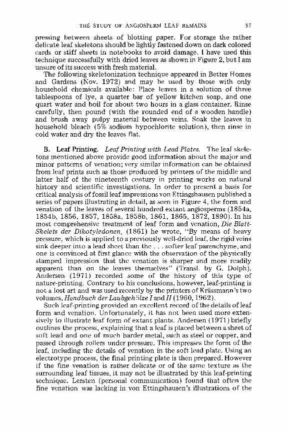
B. Leaf Printing. Leaf Printing with Lead Plates. The leaf skele- tons mentioned above provide good information about the major and minor patterns of venation; very similar information can be obtained from leaf prints such as those produced by printers of the middle and latter half of the nineteenth century in printing works on natural history and scientific investigations. In order to present a basis for critical analysis of fossil leaf impressions von Ettingshausen published a series of papers illustrating in detail, as seen in Figure 4, the form and venation of the leaves of several hundred extant angiosperms (1854a, 1854b, 1856, 1857, 1858a, 1858b, 1861, 1865, 1872, 1890). In his most comprehensive treatment of leaf form and venation, Die Blatt- Skelets der Dikotyledonen, (1861) he wrote, "By means of heavy pressure, which is applied to a previously well-dried leaf, the rigid veins sink deeper into a lead sheet than t h e . . , softer leaf parenchyme, and one is convinced at first glance with the observation of the physically stamped impression that the venation is sharper and more readily apparent than on the leaves themselves" (Transl. by G. Dolph). Andersen (1971) recorded some of the history of this type of nature-printing. Contrary to his conclusions, however, leaf-printing is not a lost art and was used recently by the printers of KriJssmann's two volumes, Handbuch der LaubgehSlze I and H (1960, 1962).
Such leaf-printing provided an excellent record of the details of leaf form and venation. Unfortunately, it has not been used more exten- sively to illustrate leaf form of extant plants. Andersen (1971) briefly outlines the process, explaining that a leaf is placed between a sheet of soft lead and one of much harder metal, such as steel or copper, and passed through rollers under pressure. This impresses the form of the leaf, including the details of venation in the soft lead plate. Using an electrotype process, the final printing plate is then prepared. However if the fine venation is rather delicate or of the same texture as the surrounding leaf tissues, it may not be illustrated by this leaf-printing technique. Lersten (personal communication) found that often the fine venation was lacking in yon Ettingshausen's illustrations of the

58 THE BOTANICAL REVIEW
THE STUDY OF ANGIOSPERM LEAF REMAINS 59
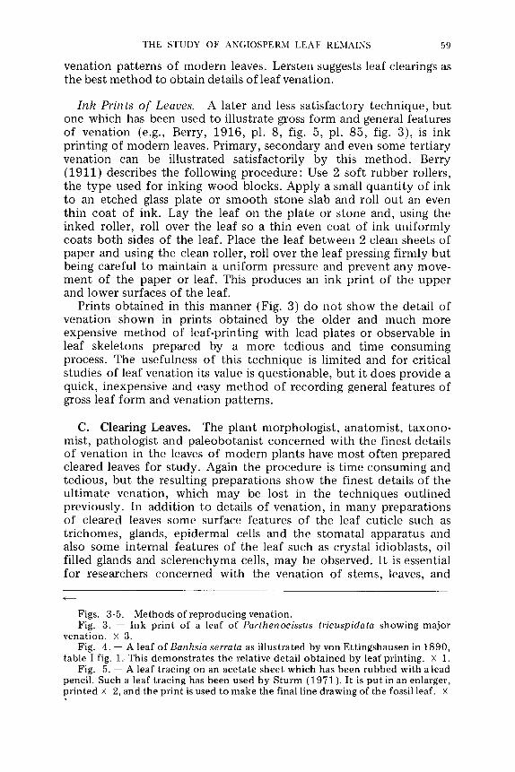
vena t ion pa t t e rns o f m o d e r n leaves. Lers ten suggests leaf clearings as the best m e t h o d to ob ta in details o f leaf vena t ion .
Ink Prints o f Leaves. A later and less sa t i s fac to ry t echn ique , bu t one which has been used to i l lustrate gross f o r m and general fea tures of vena t ion (e.g., Berry, 1916, pl. 8, fig. 5, pl. 85, fig. 3), is ink pr in t ing of m o d e r n leaves. P r imary , s econda ry and even some te r t i a ry vena t ion can be i l lus t ra ted sa t i s fac tor i ly by this m e t h o d . Berry (1911) describes the fol lowing p r o c e d u r e : Use 2 sof t r u b b e r rollers, the t y p e used for inking w o o d blocks. App ly a small quan t i t y of ink to an e t ched glass pla te or s m o o t h s tone slab and roll ou t an even thin coa t of ink. Lay the leaf on the p la te or s tone and, using the inked roller, roll over the leaf so a thin even coa t o f ink u n i f o r m l y coats b o t h sides of the leaf. Place the leaf be tween 2 clean sheets of pape r and using the clean roller, roll over the leaf pressing f i rmly bu t being careful to ma in ta in a u n i f o r m pressure m e n t of the pape r or leaf. This p roduces an and lower surfaces of the leaf.
Prints o b t a i n e d in this m a n n e r (Fig. 3) do vena t ion shown in pr in ts ob t a i ned by the
and p reven t any move- ink pr in t o f the u p p e r
n o t show the detai l o f o lder and m u c h m o r e
expens ive m e t h o d of leaf-pr in t ing with lead plates or observable in leaf ske le tons p repa red by a m o r e ted ious and t ime c o n s u m i n g process. The usefulness of this t echn ique is l imi ted and for crit ical studies of leaf vena t ion its value is ques t ionab le , bu t it does prov ide a quick, inexpens ive and easy m e t h o d of record ing general fea tures of gross leaf f o r m and vena t ion pa t te rns .
C. Clear ing Leaves. The p lan t morpho log i s t , ana tomis t , t a x o n o - mist , pa tho log i s t and p a l e o b o t a n i s t conce rned with the f inest detai ls o f vena t ion in the leaves of m o d e r n p lants have m o s t o f t en p r epa red c leared leaves for s tudy . Again the p r o c e d u r e is t ime consuming and tedious , bu t the resul t ing p repa ra t ions show the f inest detai ls o f the u l t ima te vena t ion , which m a y be lost in the t echn iques ou t l ined previously . In add i t ion to detai ls o f vena t ion , in m a n y p repa ra t ions of c leared leaves some surface fea tures of the leaf cut ic le such as t r i chomes , glands, ep ide rma l cells and the s toma ta l appa ra tu s and also some in ternal fea tures of the leaf such as crys ta l idioblasts , oil filled glands and s c l e r e n c h y m a cells, m a y be observed. I t is essential for researchers c o n c e r n e d with the vena t ion of s tems, leaves, and
Figs. 3-5. Methods of reproducing venation. Fig. 3. -- Ink print of a leaf of Parthenocissus tricuspidata showing major
venation, x 3. Fig. 4. -- A leaf of Banksia serrata as illustrated by yon Ettingshausen in 1890,
table I fig. 1. This demonstrates the relative detail obtained by leaf printing, x 1. Fig. 5. -- A leaf tracing on an acetate sheet which has been rubbed with a lead
pencil. Such a leaf tracing has been used by Sturm (1971). It is put in an enlarger, printed • 2, and the print is used to make the final line drawing of the fossil leaf. •

60 THE BOTANICAL REVIEW
floral organs that these plant organs also be cleared. Lersten (1967) published a very useful list of 31 selected references of botanical clearing methods; I will not repeat a listing of those references here but refer the interested reader to his bibliography of techniques.
The following schedule for clearing modern angiosperm leaf mate- rial has been modified by Sheffy and Potter at Indiana University from a procedure published by Payne (1969).
Place the dried leaf in a petri dish or beaker. Add 10% sodium hydroxide (Na OH) at room temperature. If the leaf material is very delicate 5% Na OH may be used. Heat slowly to just below the boiling point. Maintain the heat for 5-10 minutes (thin leaves may require less time). Check leaf material frequently in order to judge the progress of the clearing and remove when the leaf is translucent. Wash in water 2 times. If necessary, the previous steps may be repeated until the leaf is translucent. Bleach the leaf in 50% commer- cial bleach [5% sodium hypochlorite (Na HC1 02 )] solution for 10-15 minutes. Watch carefully and remove when the leaf becomes cream colored or white. Wash in water 2 times. Dehydrate in an alcohol series of 50% ETOH (ethyl alcohol), 75% ETOH, leaving the leaf about 10-15 minutes in each solution. Place in stain solution of 1% safranin 0 in 95% ETOH for 20 minutes or longer. Some leaf material requires several hours to become well stained. Wolfe (per- sonal communication) suggests that a few drops of acetic acid com- bined with heat may be used to speed up the staining of cleared leaves. Wash the stain out in 100% ETOH until desired level of staining is obtained. Transfer to solution of equal parts of absolute ETOH and xylene. Transfer to 100% xylene before mounting in HSR (Harleco synthetic resin) or Canada balsam. Mount the leaf be- tween two glass slides. Weight the slides, place on a drying table and check each day, adding more mounting medium as needed. HSR dissolved in toluene may be used to decrease the drying time of the slides.
There are many variations of this method of clearing leaves. The above schedule clears leaves rapidly because of the use of heat but the slides dry slowly and care must be taken to avoid bubbles forming from the margins of the preparations as they dry.
A modification of this schedule for clearing leaves was published by Foster (1952) and modified by Hickey (1973) follows; it is sufficiently different that it warrants being listed as an alternative procedure.
Place leaf in a petri dish and weight it down with a plastic screen. If the leaf is thin textured add a 5% sodium hypochlorite (Na H C102) solution (commercial bleach), and allow to clear 24-28 hours. If the leaf is medium or thick textured add 5% sodium hydroxide (Na OH) solution at room temperature; change this solution daily, then weekly until no discoloration of the leaf is apparent. Soaking in 5% Na OH at 45~ may be used, with care, to speed up the clearing. Rinse leaves in water. Place in 250% solution chloral hydrate [C C13 CH (OH)2 ] for 12-16 hours to clear the leaf. Fill a bottle of chloral

THE STUDY OF ANGIOSPERM LEAF REMAINS 61
hydrate crystals with water and let stand until dissolved in order to produce a saturated solution of chloral hydrate. Rinse in water to remove chloral hydrate. Dehydrate in ethanol (ETOH) and toluene series. Stain with safranin 0 in a 50% absolute ETOH and 50% toluene solution until a uniform density of color is obtained. Mount in HSR dissolved in toluene between 2 glass slides, weight, and check frequently adding more mounting medium when necessary.
The technique used by Wolfe is basically similar to that followed by Hickey (Wolfe, personal communication) with the following changes. Wolfe uniformly soaks all leaves in 5% Na OH for 48 hours changing the solution once. Then rinses in H2 O, followed by a rinse in acetic acid (to acidify the solution so the bleach will be effective), and then transfers to commercial bleach. The leaves bleach typically in a few minutes or 1/2 to 2 hours (some may take longer). This is followed by a rinse in H2 O, then the leaves are placed in the chloral hydrate (250% solution) for 48 hours, rinsed, dehydrated, stained as outlined in the above schedules and mounted in Permount (product of Fisher Scientific). Most of the cleared leaves are mounted between 31A '' X 4" glass slides produced by Arthur Thomas Inc. These are then dried in ovens and stored in wooden cabinets (Nega Files) commercially available for the storage of 31A '' X 4" lantern slides. Some slides 41/~ '' X 5" and 5" X 10" are made to accommodate larger leaves. Also some of the longer leaves are cut 2 or 3 times so they will fit on a standard size slide. Figure 1 demonstrates the venation of a cleared leaf as projected directly onto photographic paper.
II. Preparation of Fossil Angiosperm Leaves
A. Untreated Fossil Leaves. The majority of angiosperm leaf remains which have been studied were observed in the laboratory in the same condition as they were found in the field. Depending upon the type of preservation and the nature of the fossil leaves there is often little which is required or possible to make the leaves more acceptable for critical investigation. Generally in every paleobotany laboratory fossil material which is unpacked from field collecting is carefully examined and may be tr immed before being stored for future investigations. When a particular fossil leaf is selected for investigation it is examined carefully with a dissecting microscope, then cleaned, and further exposed if necessary, to yield opt imum information of the gross leaf form and fine venation. The t reatment given the fossil after the above general procedures are completed, depends upon what information the investigator is interested in obtaining, tempered by what information the investigator feels can be obtained from the particular fossil leaf material.
Early investigators generally studied angiosperm leaf remains in an unaltered state and the majority of paleobotanists studying angio- sperm leaf remains continue to do so. The only significant change in the use of fossil material has been in the level of observations of gross form and venation patterns made. In several cases even the level of

62 THE BOTANICAL REVIEW
observations made has not changed much in the past 100 years. To increase our present understanding and to avoid perpetuating the numerous incorrect identifications of angiosperm leaf remains re- corded in the past, it is important that we extract the maximum amount of information possible from the fossil remains.
In order to obtain maximum information of gross form and venation patterns from untreated leaf impressions and oxidized mate- rial several techniques have been used. The most obvious is the use of oblique lighting (Fig. 9), reflected lighting, direct lighting (Fig. 7), or any combination of these, in order to obtain a good photographic record of the fossil material. Often several photographs taken with varying lighting techniques and magnifications may be necessary to illustrate features of the fine venation. It is often possible to produce a photograph of a fossil leaf which illustrates the leaf form and fine venation more clearly or in sharper contrast than it appears with direct lighting in the original specimen. However photographs of leaf remains retouched to enhance leaf outline and venation patterns (e.g., Berry, 1930, plate 16, fig. 5; MacGinitie, 1962, plate 3, fig. 7; Axelrod, 1966, plate 12, figs. 2-4) should be used with great caution. Whenever a reader can identify such a bastard photograph he should regard it as such, realizing that it is satisfactory neither as a photo- graph nor as a line drawing. In place of an altered hybrid photograph both a line drawing and a photograph of the fossil leaf should be illustrated.
The use of infrared, photography for illustrating fossil leaf remains has not been satisfactorily explored. Some attempts at its use have been made by Tralau and K. Kilpper (personal communication of M. Stfirm and W. Friedrich); however I know of no published discussion of its application to the study of fossil leaf remains. Wolfe had the fossils he collected from the Tertiary of Alaska photographed using infrared film. In spite of the poor quality of reproduction of plates II-V in Wolfe's paper on the Alaskian Tertiary (Wolfe, 1972) by Elsevier Publishing Company, the details o f the leaves are easier to see in the plates than in the original specimens (Wolfe personal communication). Sturm showed me an Engelhardia fruit which was preserved as a thin carbon film in a lignitic matrix. It was difficult to see the outline of the fossil and the venation of the specimen. However Dr. Tralau photographed the specimen with infrared film and the fruit outline and venation were much enhanced. Although at present photographing with infrared sensitive film, recording infrared waves which focus in a different plane than white light, is difficult because lenses are not corrected for invisible infrared light. The use of infrared photography may allow observations of fine venation which were previously difficult or impossible to observe and illus- trate.
Line drawings of angiosperm leaf remains have been and still are a common and useful means of illustrating leaf form and fine venation. Often it is easier to observe slight differences in form and venation patterns from line drawings of the material than from observing the

Fig. 6. - - P e n n s y l v a n i a n age i r o n s t o n e n o d u l e f r o m I n d i a n a w i t h a f e rn leaf , Pecopteris sp. , p r e s e r v e d . A p h o t o g r a p h i c p r i n t o f t h e n o d u l e was u s e d as t h e ba se ove r w h i c h t h e l ine d r a w i n g was m a d e . T h e p r o c e s s is e x p l a i n e d in t h e t e x t . Pre- p a r e d by Mr. T h o m a s Held . x 7.

64 THE BOTANICAL REVIEW
material itself. However line drawings which present only the leaf outline and features of the primary and secondary venation are of limited value. Line drawings should never be done as free-hand sketches of fossils.
There are several techniques for preparing accurate detailed draw- ings. The most obvious technique is the use of a camera lucida at tachment to a dissecting microscope. Good quality line drawings can also be made by projecting a negative of a leaf in an enlarger and tracing its form and venation pattern or by printing an enlargement of a negative on a non-glossy finish paper and inking on the photo- graph or print with India ink. After the ink has completely dried, the print is submerged in a weak iodine and water solution until the photograph has disappeared. The drawing is then rinsed in water and placed in photographic fixer until all traces of the iodine are gone and then washed and dried as a normal photographic print. This technique is very easily done and can be useful in illustrating leaf form and venation of extant and fossil material (e.g., Dilcher and Mehrotra, 1969, figs. 14, 15). Any portion of the original photo- graph may be retained by coating both the top and bot tom surfaces of the area you wish to retain with rubber cement, which can be easily removed after bleaching and washing the print. In this manner you can produce a line drawing for one half of the leaf and a photograph for the other half (Fig. 6).
Stiirm (personal communication) uses an acetate overlay to pro- duce line drawings of fossil leaves. He cuts a thick clear acetate sheet to the size of the piece of matrix in which the fossil leaf occurs and tapes it securely in place. With the use of a sharp needle the leaf outline and venation pattern, including extremely fine venation, are scratched into the acetate. The acetate is then removed and rubbed with a soft lead pencil so as to fill the scratch marks with graphite (Fig. 5). This is then put in an enlarger and used as a negative to produce an enlarged print of the fossil leaf. The print is then placed on a light table and traced on to tracing paper in ink. When this line drawing is reduced to natural size for publication, it results in a clear, sharp and detailed line drawing of the original fossil leaf (e.g., Stiirm 1971). Walther (personal communication) uses a frosted acetate sheet placed over the fossil leaf material and traces the leaf form and venation on the sheeting much as one would use tracing paper to produce line drawings. Certainly there are numerous techniques which can be used to produce accurate and detailed line drawings. The samplings given here are presented for the benefit of those who might not know about them and might find them useful, and to emphasize that line drawings can be an important and sometimes necessary tool in the investigation and illustration of angiosperm leaves.

THE STUDY OF ANGIOSPERM LEAF REMAINS 65
B. Special Treatments for Fossil Leaves. Clearing Unaltered Remains. Relatively unaltered leaf remains are occasionally found in clay sediments or lignites which have not been extensively com- pressed or deformed. Such mummified leaves may flake off clay or lignitic sediments in which they were preserved (Bandulska, 1923; Dilcher, 1965a, 1965b) or may have been deposited in such quanti- ties that they formed papery layers of compressed leaves (Barthel, Kva~ek, Rilffle, 1966; Jung, Knobloch, Kva~ek, 1971).
The technique I use to clear opaque unaltered leaf material is the following. Place the leaves in 5% potassium hydroxide (K OH), holding the material between 2 layers of glass to keep it from curling or breaking apart, if this is a problem. Change the KOH after one day and observe the progress of the clearing; clearing is usually complete after the second day. After the leaf is cleared (leaf lamina fades to a light brown or rust color that contrasts with the venation which remains dark brown), the leaf material is carefully rinsed in water, dehydrated in an alcohol series (50%, 95%, 100% ETOH to equal parts of 100% ETOH and xylene), placed in xylene and mounted in Canada balsam or HSR (Harleco Synthetic Resin). The newly cleared leaves may be mounted directly in glycerine jelly from water or dried, stored in envelopes and later photographe d with transmitted light while submerged in xylene under glass. I have photographed over 400 cleared leaves under xylene and the details of the fine venation are extremely clear. This photographic method permits quicker processing of the fossils and easier storage because no per- manent slides of the whole leaves are prepared and the leaf material remains available for repeated analysis of the cuticle with light microscopy and SEM and for sectioning.
Transfers of Compressed Leaf Remains. Angiosperm leaf remains are most frequently found as impressions or compressions in a variety of matrixes. Compression leaf fossils are found in varying states of preservation and the amount of organic material preserved depends upon the amount of oxidation which the fossil has under- gone. Often in leaf compressions, especially those leaves preserved in softer sediments, a fragmented cuticular film may be preserved and due to differential oxidation, the venation may contrast well with the leaf lamina and the matrix. In other less oxidized sediments the leaves may appear as opaque black outlines on the matrix (Fig. 7).
Some European paleobotanists have developed techniques to make transfers of compressed angiosperm leaf remains (Voight, 1936, 1949; Kr~iusel, 1950). The transfer technique was first extensively used for fossils found in the Geisel Valley near Halle am Salle, Germany, during the early 1930's (Voight, 1936). It is widely used by paleobotanists such as RUffle, J~ihnichen and Litke at the Institute for Palaeontology in East Berlin. Leaf transfers have been used mainly for leaf compression material preserved in clay and occasion- ally for leaves preserved in brown coal or lignite. The technique is as follows (personal communicat ion, J~ihnichen and Rilffle). Use Geisel-

66 THE BOTANICAL REVIEW

THE STUDY OF ANGIOSPERM LEAF REMAINS 67
tallack in acetone solution (available from Wilhelm Schmidt, Jr. K. G., 7022 Leipzig, Lumunbastrasse 27, D D R ) a n d thin further with acetone; apply 2-3 coats to leaf remains and allow to dry. Peel the transfer from the matrix and wash in water to remove loose matrix. If matrix is not washed off in water submerge in 3% HF, macerate in Schultze's solution, 1 part saturated solution of potassium chlorate (K C103 ) to 2 parts concentrated nitric acid (NHO3), rinse in water, bleach in hydrogen peroxide (H2 O2), again rinse in water, and mount in glycerine jelly or dry and store in envelopes.
E. Fjeldso Christensen has modified the above technique. He first removes cuticle from the compression and prepares it for cuticular examination. After satisfactory cuticular preparations have been pre- pared, he paints several {4-5) layers of collodion dissolved in ether over the leaf impression. Before this collodion film dries hard (at which time it may crack badly), he puts the whole block of clay in warm water and loosens the edges of the collodion from the clay. Then, using a soft stream of water, he gradually works the collodion film free from the clay block. The film is then cut to the approxi- mate size of the leaf and immersed in 30% HF for 10-30 minutes. It is then washed in water, transferred to absolute alcohol and brushed to remove any clay still adhering to the film. The film is then flattened on a glass plate, blotted with paper towels to remove as much alcohol as possible arid moved directly into xylene before it drys out. It is finally mounted between glass in a mounting medium compatible with xylene such as Canada balsam.
Mr. Christensen's fossil leaf material consists of carbonaceous leaf compressions in a clay matrix from Miocene deposits in Denmark. Only the gross venation could be seen in unprepared material while the transfers prepared by this collodion film method show very clearly the major and minor veins in the leaves (Koch, Friedrich, Christensen, and Friis, 1973, plate 4, figs. 3 and 4).
I experimented with this technique using flexible collodion and found that the collodion must be thinned with 3 parts absolute ETOH and 1 part ether until, when it is painted on a test protion of the matrix in which the fossil is preserved, it does not produce bubbles upon drying. Applying alcohol and ether directly to the fossil being coated also helps reduce bubbles. The transfer is very acid resistant but swells and distorts badly in 100% ETOH and swells slightly when mounted in a xylene base medium. Such problems do not occur with air-dried transfers or those mounted in glycerine jelly or a toluene base medium (e.g., Harleco Synthetic Resin dissolved in toluene); I would not advise the use of alcohol to dehydrate the
4-- - -
Figs. 7-9. V e n a t i o n of fossi l leaves. Figs. 7 & 8. - - A leaf of Ocotea obtusifolia f rom middle Eocene age clay
deposi ts in wes te rn Tennessee . Fig. 6 shows the leaf compre s s ion as it was f o u n d on the clay mat r ix . Fig. 7 shows the same leaf compres s ion af ter i t was coa t ed wi th col loidin , r e m o v e d f rom the clay ma t r ix , cleared, and m o u n t e d , x 1.5.
Fig. 9. - - Leaf impress ion of the so-called Sassafras cretaceum Newber ry f rom the Dako ta F o r m a t i o n of Kansas. It was p h o t o g r a p h e d by Mr. Charles Beeker wi th obl ique l ight ing to enhance the vena t ion . • 6.

68 THE BOTANICAL REVIEW
transfers. I had no problem with the collodion cracking when it dried but found that both water and HF may lightly cloud the collodion if it is not well airdried. The collodion transfers can be easily removed from a clay matrix by pulling, allowing small sections or parts of leaves to be prepared for detailed analysis.
The leaf transfer technique can be very useful and should find wider use in research on angiosperm leaf compression material. Frequently fine venation is preserved but cannot be clearly seen in leaf compressions (Fig. 7). However when transfers of somewhat oxidized leaves are made and mounted, the result is often a very fine cleared leaf preparation that shows the ultimate venation of the fossil. The transfer technique has the additional advantage of re- moving the entire fossil leaf from the sediment intact so that then this leaf can be macerated and/or bleached and cleared by a mount- ing medium in order to enable the observation of the fine venation (Fig. 8). However the value of making transfers of fossil leaves or otherwise treating or tampering with fossil leaf material, if it is not necessary to obtain data for a particular study, seems doubtful. Often some fine venation features can be observed in the clean impression remaining in the matrix after the transfer is pulled away. The dried transfers are easily stored for reference. Cuticular features can frequently be observed in the leaf transfer. However for best results, separate slides should be made for the study of fine venation and cuticle even when transfer methods are used for both. The best preparation for the study of fine venation often is not the best preparation for studying cuticle and vice versa.
Autoradiographic Technique. BjSrlykka (1965) described in a short report a technique for examinging details of invertebrate fossils using radium which may be l~ss readily absorbed by a fossil than by the surrounding matrix. Thus, if the fossil is painted with a dilute solution of radium and covered with autoradiographic stripping film the resulting negative may show features of the fossil not otherwise easily observed. Walther Friedrich (personal communication) sug- gested the use of this technique to me for angiosperm leaf remains, but as far as I know it has never been tried.
CUTICULAR ANALYSIS
Much of the morphology and anatomy preserved in angiosperm leaf remains has often been neglected by many investigators. Fossil cuticle has received little attention although its study yields as much, if not more, information than a study of pollen or venation, and has great potential application to taxonomic, ecological and stratigraphic questions.
The cuticular membrane of plants is a heterogeneous collection of superficial waxes, cutin, cuticular waxes, cellulose and pectin, cover- ing and intimately associated with the external surfaces of the cell wall (Fig. 10) (Halloway, 1971). Cutin, a chief component of the

THE STUDY OF ANGIOSPERM LEAF REMAINS
SuDerflcpal w a x
69
Cutbcular membran
Cutm
Cubcu la r w a x
Cel lulose
Pect in
Cell wa l l Cel lu lose ( + Pect in)
Fig. 10. -- Fine structure of the cuticular membrane. (Reproduced with per- mission of the publisher from Halloway, 1971.)
membrane, is a complex polymer of hydroxy- fa t ty acids with ester linkages (Martin and Juniper, 1970). The cuticular membrane not only is integrated with the outer walls of the epidermal cells but also frequently is a componen t of the outer portions of their anticlinal walls. When the cellulosic cell walls of the epidermal cells are de- s troyed, cuticular pegs frequent ly remain, marking the position of anticlinal walls (Figs. 19-22). Thus because of its chemistry the cuticular membrane is resistant and because of its intimate associa- tion with the epidermal cell walls it preserves much of the character of the epidermis. In addition, such features as trichomes, guard cells, and sculpturing or ornamentat ion of the outer leaf surface may be retained in the cuticular membrane. The stomatal complex has a number of characters itself which may be diagnostic {Figs. 11 & 12).
Fossil cuticle, as well as cuticle of living leaves, contains a host of characters which may be impor tant in determining relationships at specific, generic and family levels. The study of cuticular remains of fossil leaves may sometimes be essential to understanding the history of an extant taxon. For example, Schneider (1965), Ferguson (1971) and Sheffy (1972) all indicate that it is impossible to identify the leaf remains of the genus Myrica unless particular cuticular features of the t r ichome stalk or base are observed. The fact that trichomes, as well as the area of t r ichome at tachment , can be an important taxonomic tool has also been shown by Stace {1965, 1969a, 1969b). Uphof wrote the following in the introduct ion to his book on plant hairs (1962): "Tr ichomes and emergences show a remarkably wide range of variation in form and function as well as in physical and chemical properties. The form in which they appear often differs

70 THE BOTANICAL REVIEW
stomatal aperture ~ T-piece at ~ stornatal pole
outerstornatall,dge ~ / ~ rt:~n~ n i:i a, t $ :~!i,::~y
Fig. 11. - - The s toma ta l c o m p l e x in surface view. ( R e p r o d u c e d wi th the pe rmiss ion of the pub l i sher f rom Stace, 1965 . )
sub-stornatal back cavity
subsidiary ce l l / ~
front cavity
tuner stornatal ledge
~ ~ outer wall of guard cell
outer stomatal ledge
stomatal aperture Fig. 12. - - The s toma ta l com p l ex in vert ical sect ion. ( R e p r o d u c e d wi th per-
miss ion of the publ i shers f rom Stace, 1965 . )

THE STUDY OF ANGIOSPERM LEAF REMAINS 71
from species to species or from genus to genus, but it may also be characteristic for a group of higher rank, e.g., for a family. This aspect of their diversity was studied especially by Solereder (1899, 1908), by Solereder & Meyer (1929), and by Metcalfe & Chalk (1950)." Hummel and Staeche (1962) discuss the distribution of five basic types of trichomes which they review; 1) single-celled hairs, 2) star-shaped or shield-shaped hairs, 3) hairs consisting of a file of two or more cells, 4) hairs with a single cell or file of cells terminating with an enlarged unicellular or muticellular head, 5) hairs consisting of two or more files of cells. This is an a t tempt to organize the large amount of knowledge known of tr ichome types of the angiosperms.
Stace (1965) published the most useful, broad and comprehensive monograph on the application of cuticular analysis which has been written. He discusses a variety of topics, most of which should be of interest to the systematic botanist and paleobotanist. In his preface he acknowledges the superficial level of our present understanding of angiosperm cuticle but notes the increased interest in this area by both botanists and paleobotanists and encourages the wider use of cuticular analysis in all areas of plant systematics. The review by Stace is reasonably thorough in its t reatment of the history of cuticular studies and its use by both botanists and paleobotanists; I will not a t tempt in this review to rewrite what is already available in such a readable form. A short survey of cuticular studies of leaves was written by Sinclair and Sharma (1971). Priestley's (1943) brief review paper on the cuticle of angiosperms is of little use to system- atic botanists.
Stebbins and Khush (1961) present a short but very interesting paper on the stomata of the monocotyledons . They consider the stomatal complex systematically, in relation to the growth habit, and also the geographic distribution of stomatal types. They suggest that the specialized stomatal complex, once adapted, persisted even though the habitat in which the early evolution of the stomatal complex took place may have changed. Three types of s tomata are recognized, the most primitive one having many subsidary cells. Tomlinson (1974) in a recent review of the usefulness of the stoma- tal complex development as a taxonomic tool in the monocoty ledons disagrees with much of what Stebbins and Khush (1961) wrote. Tomlinson studied the development of about 100 species of mono- cotyledons representing a large number of families. He suggests that it is premature to speculate about the phylogenetic significance of stomatal patterns in the monocotyledons . He further suggests that the stomatal complex has systematic value only when used in com- bination with several other characters of known taxonomic useful- ness.
The leaf epidermis of the Magnoliaceae and related families has been studied by Baronova (1972). The stomatal complex, as well as other epidermal features, further suggest the presently accepted relationships of the families in the Magnoliales. She discusses many

72 THE BOTANICAL REVIEW
of the genera used in each family, relating the epidermal features to certain generic and family relationships. She found that most of the Magnoliales have paracytic stomatal complex and considers this a primitive type within the angiosperms.
It is important for systematic botanists to be informed of the most current work on the fine structure and function of the epidermis and cuticle. Some useful information is available on the fine structure of the cuticle in Ecology of Leaf Surface Microorganisms fed. by Preece and Dickinson, 1971), but probably the most complete, recent survey of plant cuticles is by Martin and Juniper (1970). The latter book is not direeted towards systematic uses of cuticle but is useful nevertheless.
The following section of this review paper is primarily concerned with systematic uses of angiosperm cuticle of fossil leaf remains.
TAXONOMIC USE OF FOSSIL CUTICULAR CHARACTERS
In order to use cuticular analysis for systematic s tudy we must first assess the characters available in the cuticle of the taxon under investigation and then a t tempt to understand the variability of these characters in various ecological settings and throughout the range of the taxa and related taxa being studied. This s tudy of extant taxa must be done before one can use cuticular analysis to good advantage with fossil angiosperm leaves. A thorough s tudy of the fine venation and cuticular characters of both extant species and the fossil material is essential to unraveling the evolution of extant genera. Unfortu- nately, little has been published on the detailed morphology of the extant forms of angiosperm leaves and thus the paleobotanist must do much of this work on modern leaves himself.
However, published information is available to guide the selection and use of characters for cuticular study. Stace (1965) reviews the use of particular characters of the cuticle point by point and presents very useful discussions of each character suggested; I will not a t tempt to review these characters here but refer the reader to Stace.
One of the characters commonly used in cuticular analysis requires some discussion at this point. Various names have been proposed to describe the arrangement of the epidermal cells surrounding the guard cells. The term stomatal complex (Stace, 1965; van Cotthem, 1970; Fryns-Claessens and van Cotthem, 1973) is used here to refer to these cells (Figs. 11 & 12). The most current and widely used terms for the mature stomatal complex come from Metcalfe and Chalk (1950) and van Cotthem (1970). A few subsequent additions appear in this review. These terms are used to refer to the arrange- ment of cells in the stomatal complex as observed in mature leaves only. Pant (1965), Payne (1970) and numerous others cited by Fryns-Claessens and van Cotthem (1973), have been concerned with the development of the cells in the stomatal complex. They have proposed terms which are dependent upon observing the origin of

THE STUDY OF ANGIOSPERM LEAF REMAINS 73
the subsidiary cells of the stomatal complex. Certainly the systematic usefulness of cuticular analysis is greatly enhanced by an under- standing of development; however, in paleobotany or systematics based on herbarium material where development cannot be observed, little use can be made of developmental terminology. Whenever developmental sequences are suggested for mature fossil leaves or mature extant leaves, it should be understood that these do not represent developmental sequences bu t only an educated guess of possible patterns of development.
A number of terms have been proposed to refer to the various types of stomatal complex. Some of these terms refer to develop- merit and others to the mature condition, thus some confusion may result. In their excellent review, Fryns-Claessens and van Cot them (1973) present a classification of the ontogenetic types of s tomata that at tempts to integrate the terminology of the stomatal complex of mature leaves with that used for the ontogeny. Table III is an a t tempt to present a clear cross listing of the ontogenetic types of stomatal classification with that proposed for mature leaves. I suggest these terms could be standardized by adding suffixes used to describe development to the terms used to describe the stomatal complex observed in mature leaves (Stace, 1965; Fryns-Claessens and van Cotthem, 1973); the suffixes would be -mesogenous (subsidiary cells develop from the same initial as the guard cells), -mesoperigenous (some of the subsidiary cells develop from the same initial as the guard cells) and -perigenous (none of the subsidiary cells develop from the same initial as the guard cells). In mature leaves, it is frequently, but not always, possible to distinguish any conspicuous subsidiary cells. The subsidiary cells may stain differently, have different surface textures and/or may be distinguished by their arrangement and/or size. However as this is not always the case Table III is arranged so that one ontogenetic type may correspond to more than one mature stomatal complex type and, as suggested by Fryns-Claessens and van Cot them (1973), more than one ontogenetic type may correspond to a single mature stomatal type. The 31 mature types of stomatal complex proposed in Table III and illus- trated in Table IV are based upon the work of van Cot them (1970) and Fryns-Claessens and van Cot them (1973) and are arranged to relate to the 26 ontogenetic types of stomatal complex proposed by Fryns-Claessens and van Cotthem. Table III is a temporary working summation of stomatal types that should be expanded and reworked as more information becomes available. I suggest that new terms for ontogenetic types proposed in the future be based upon Greek root words that describe the arrangement of cells in the stomatal complex rather than being derived from the plant in which the condition was first observed. For such new ontogenetic types, the terminology for their mature stomatal complex should also be given.

74 T H E B O T A N I C A L R E V I E W
�9 0
0
0
0 z
"o
0 . . . ~ oO
N ~
0
o~
o ' ~
~ 0
o~.~
o
o
o
o
0 0
o .~, o .~ �9 �9
�9 ~ 2 '~ 2
0
~
~ .~ ~ . ~
~ o ~
0
0
0
o �9 0 0
l~ 0 ~ ,~ ~ 0
0
o
g ~ " n o ~ �9 ~ ~ ~ . ~ o
~ . e ~ ~
0 0 0
o~ o~
�9 r~ ~
"D. 0
0 �9
0
o
0 0 " ~
-~ o ,.~

T H E S T U D Y O F A N G I O S P E R M L E A F R E M A I N S 75
0 C
0
0 <3
0 0 �9 0 �9
o O ~ Z
~'~ ~
. ~ ~ o
0 0 C
a a
0 0
CuC~ ~ 0
0
0 0 0 0
| o o o ~ 0 0 ~ ~
--"C "C 0
0 := 0 :=
o
~ o =~
O. w~ 0
~ 0: ~ ~ U 0 0 ~ 0 U O) ~ 0 ~

76 THE BOTANICAL REVIEW
In order to facilitate cataloging cuticular characters and compari- sons of one form of cuticle to another 1 have prepared Table IV, which has been modified from Stace (1965). When cuticle is being used to establish taxonomic affinities of an unknown leaf, all charac- ters given in the table should not be given equal consideration by the investigator. In different ecological situations cell size, stomatal fre- quency, trichome frequency, and anticlinal wall configuration may vary while the positioning of the stomata, the orientation of the stomata, the nature of the trichome and the type of stomatal complex is less likely to vary. Also leaves of one species may have more than one type mature stomatal complex. However when the many features of cuticle are considered together, with an under- standing of their variability, they are extremely useful, sometimes essential, in sorting out the taxonomy of angiosperm leaves.
The cross listing of mature and ontogenetic classifications given in Table III was difficult to arrange without making original observa- tions of the stomatal complex. Table III is based mainly upon the review paper by Fryns-Claessens and van Cotthem (1973). Some ontogenetic types which they propose for mature types are split and thus these are repeated on the table. This splitting was necessary because of the addition of several mature types of stomata; in some cases the ontogenetic types fit more than one mature type depending upon the ability to distinguish one or more subsidary cells from surrounding epidermal cells. In fossil cuticle they may be distin- guished by their size, shape, surface texture or stainability. For some mature types no development is given. These were observed as fossils or found in the literature without reference to their ontogeny. Polocytic and pericytic types of the stomatal complex which have been reported only on ferns are given to complete the list of types even though they do not apply to angiosperms. The terms hypocytic and epicytic for mature stomatal types by van Cotthem (1971) are not given here because they deal with placement of the guard cells rather than arrangement of the subsidary cells.
ECOLOGICAL VARIATION OF CUTICULAR CHARACTERS
The ecological variability of cuticle is well established; considera- tions of this topic have been common in the literature on angiosperm cuticles. Stace (1965) and Sinclair and Sharma (1971) survey much of the earlier literature on ecological variations of cuticle. Only a few current papers which are not included in their reviews are given here.
The effects of light intensity on the variation of cuticular charac- ters has been studied (Dilcher and Zeck, 1968; Pazourek, 1970) as has the effects of moisture (Gindel, 1969). Sharma and Dunn (1968, 1969) and Sharma (1972) also considered light intensity and mois- ture, and in addition other environmental factors such as position of the leaf on the plant and position of the area sampled on the leaf. Sharma and Dunn (1969) found that in Datura the pattern of

THE STUDY OF ANGIOSPERM LEAF REMAINS 77
stomatal development, stomatal index (except for extreme environ- ments), and type of trichomes remained constant while stomatal frequency {also see Gupta, 1961), tr ichome frequency, epidermal cell frequency, anticlinal wall pattern and the surface texture varied under different environmental conditions. It is important not to put too much taxonomic emphasis upon the characters that vary with environmental changes. However it is difficult to evaluate the signifi- cance of slight variations in cuticular characters of fossil material. If two fossil leaves are from the same horizon and locality and are identical in gross form and fine venation but have varying cell sizes and shapes and stomatal frequencies with other cuticular characters remaining constant, the variation is probably environmental. Because of the difficulty in assessing the significance of minor variations in cuticular characters of fossil leaves, extreme care should be taken not to over-use subtle variations to establish new species. However in recent work we have found that cuticular variations can be observed through time while gross leaf features remain constant (Dilcher, 1971).
R(iffle (1964) discusses cuticular characters of Tertiary leaves as indicators of the ecology in which they grow. Some cuticular fea- tures of leaves from extreme environments are obvious, as are the associated leaf forms. A few monographs of tropical angiosperms have included cuticular characters, and the nature of leaf surfaces in temperate environments is well known. However I know of no carefully done field study that relates cuticular form to various non-temperate environments. It is potentially possible that modern and fossil angiosperm leaf cuticles might be very useful environ- mental indicators when the leaf epidermis and its response to the environment is bet ter understood.
SURVEY OF ANGIOSPERM CUTICULAR STUDIES IN THE FOSSIL RECORD
It is quite natural that the first reference to angiosperm leaf cuticle comes from fossil cuticle which was naturally oxidized and ready for observation directly from the sediment (Brodie, 1842, according to Stace, 1965). Bornemann, called the " founder of cuticular analysis" by Barthel (1966), was probably the first person to make use of cuticular characters in living plants. He was concerned also with fossil remains; in 1856 according to Barthel he described the cuticle of Cycadophyllum in a pioneering work in paleobotanical studies of the cuticles of gymnosperms. During the next 100 years numerous papers by various paleobotanists making several important contribu- tions to our understanding of gymnosperms (e.g., Florin's work on gymnosperm cuticle, 1931, 1933) followed. Much less attention was given to fossil angiosperm leaf cuticles, however, until Bandulska (1923) began to publish on the cuticles of some dicotyledonous leaf remains from the Eocene clay deposits exposed by the sea in cliffs by Bournemouth, England. She published a series of papers (Bandulska,

78 THE BOTANICAL REVIEW
1923, 1924, 1926, 1928, 1931) all concerned with the cuticular features of fossil and extant angiosperms. She searched the system- atic literature, some of which included systematic anatomical details of leaf cuticles (e.g., Solereder, 1908), and prepared the first set of modern reference slides of the cuticle of extant angiosperm leaves for use in her research. The Bournemouth flora is a large flora but b e c a u s e of the detailed, time-consuming nature of her work, it progressed slowly, and only a few angiosperm leaves were ever described.
Berry undoubtedly found much preserved cuticular material when working on the Eocene floras of southeastern North America (Berry, 1916, 1924, 1930, 1941). He described the cuticular character in one leaf-type, which he identified by gross form, noting that the details and variations of angiosperm cuticle are so diverse and little known that it could not, at that time, be used as a satisfactory taxonomic tool (Be~ry, 1933). He did no other work on plant cuticles.
Strauss (1930) mentions and figures a few fragments of cuticular remains in his work on the Pliocene age Willershausen flora. However he did not use these cuticular remains for detailed taxonomic com- parisons with the cuticle of the extant genera to which he assigned the fossil specimens. The cuticles of Deuralquea and Listea leaves of Paleocene age were described by Stockmans (1932).
Elise Hofmann studied leaf compression material from the Geisel Valley brown coal mines in Germany and published a report of her work in 1932. She worked with well-preserved leaves found in leaf-coals, preparing slides of the cuticles and attempting to identify the leaves by gross morphology. Many of the leaves she was not able to identify and for these she established the genus Folium [Stace (1965) suggests that the generic name Dicotylophyllum, used by Bandulska (1923) for fossil d icotyledonous leaves of doubtfu l affini- ty, has priority over Folium]. Both Folium Hofmann and Dicotylo- phyllum Bandulska should be put in synonymy with Dicotylo- phyllum Saporta (Andrews, 1970). Hofmann (1932) gave species names based upon the nature of the cuticle to several leaf forms without describing details of gross form, and often having only the upper or lower epidermis preserved. This is more an abuse of cuticu- lar anatomy than a satisfactory use of the tool. However she does provide some information concerning the cuticular features of a few forms she identified as existing fossil leaf types, but apparently without surveying the cuticular anatomy of extant forms as Bandulska had done (Bandulska, 1923, 1924, 1926, 1928, 1931). Beyn (1940) published a much more carefully done study of the leaf compressions from the Geisel Valley area by Halle (Saale), Germany.
One of the earliest and most complete reviews of techniques, considerations of the value and demonstrat ion of the uses of cuti- cular characters of angiosperm leaves as applied to paleobotany, was published by Jurasky in three parts (1934, 1935a, 1935b). He discusses the problem of ecological variability of cuticular form,

THE STUDY OF ANGIOSPERM LEAF REMAINS 79
stressing the usefulness of cuticular characters on a species level but indicating the difficulty of their use at higher taxonomic levels. He recognizes the usefulness of cuticle in heterophyllic forms, but is generally cautious not to over-state the usefulness of cuticular characters for broadly based schemes of phylogeny or the evolution of cuticular characters through time.
Hunger (1939) published a flora from Eocene brown coal deposits of the Zeitz-Weissenfelser district of Germany in which he described several angiosperm leaves for which he illustrated and described the zuticle. He had little extant material available and mainly based his identifications upon previously published descriptions of the gross form of fossil leaves. The cuticular material from these leaves is described but is not used seriously in his systematic treatment of the fossils. In 1938 Hunger published a short note on the peltate tri- chomes he found in cuticular material from this Eocene brown coal, mentioning several modern angiosperm families which have peltate trichomes. One has the feeling most of his experience with compara- tive modern material came from Solereder's, Systematische Ana- tomic der Dikotyledonen (1899).
Interest in the use of cuticular characters in fossil angiosperm leaf systematics grew during the 1920's and 1930's. It had been stimu- lated by information concerning the cuticle of dicotyledons made available in Solereder (1899) and the English translation of Solereder by Boodle and Fritsch (1908). Then Bandulska (1923, 1924, 1928, 1931) demonstrated some success in the application of cuticular characters to fossil leaves; Florin (1931) published his work on extant gymnosperm leaf epidermal anatomy; Harris (1932, 1935, 1937) published his work on The Fossil Flora o f Scoresby Sound East Greenland making extensive use of cuticles; several researchers began surveying cuticular characters associated with angiosperm groups (see Stace, 1965, for references) and the ecological variability of some cuticular characters was studied. In 1932 Odell seriously questioned the usefulness of cuticular characters of angiosperms in paleobotany. She critically examined, point by point, the value of each character and concluded that "the modern method of naming fossil Angiosperms from a combination of the form, venation, and epidermal structure of their vegetative organs is quite inadequate for specific or even generic diagnosis." Edwards (1935) and Stace (1965) took strong opposition to this view, pointing out that Odell exam- ined each character, one by one, and applied separate analysis to each of them, but that when taken together they can provide useful systematic information. Odell's objections are not generally thought to be damaging criticisms of cuticular analysis today.
Little use was made in paleobotany of cuticular analysis of angio- sperm remains from the late 1930's until 1950. M//dler published a note in 1950 discussing the problems of dealing with Tertiary leaf remains in which he emphasized the importance of cuticular analysis in the development of the whole area of Tertiary leaf studies.

80 THE BOTANICAL REVIEW
Following Madler's "Thoughts on Tertiary Leaves" (1950) a series of 7 papers of which Weyland was the continuing author in collabo- ration with Kr~usel, Kilpper and Berendt (Kr~usel and Weyland, 1950, 1954, 1959; Weyland, 1957, 1959; Weyland and Kilpper, 1963; and Weyland, Kilpper and Berendt, 1967) was published. This series of papers represents one of the most extensive surveys of angiosperm cuticular remains. Weyland and Kr//usel developed a reference collection of several thousand cuticular preparations of extant angiosperms which are frequently referred to in the series. The majority of the fossil leaf cuticles described in these papers came form the Rhine Valley brown coal sediments of Miocene Age. The fossils described are never treated in a floristic manner but each leaf form is carefully analyzed with details of the cuticle forming an integral part of the determination of each genus and species. It is unfor tunate that fine venation was neither analyzed nor described, nor was even a complete description of major venation generally included in these publications. The nomenclature used assigns nearly every leaf form to an extant family, and to an extant genus or a slightly modified name of an e~tant genus.
The modern reference collection used in determining these assign- ments, however large, was often not extensive for each generic form studied nor were families studied in detail. It appears from reading this series that cuticular preparations of the fossil leaves were made, some notes taken of their gross form and major venation, and theh these were matched with modern leaves and associated cuticle of one or two species of a particular genus or genera of a particular family. Thus, the fossil material formed the basis for comparison, and sampling of extant leaf material though general, was not exhaustive. Certainly this series of papers set a definite pattern for the current research with angiosperm cuticular remains. Some taxonomic revi- sions of this work have been suggested by more research. However this series marks the first a t tempt to apply the careful application of cuticular analysis in conjunction with gross morphology to the de- scription of large numbers of Tertiary angiosperm leaf remains.
By 1955 cuticular analysis was becoming recognized as important in the description and systematics of Tertiary angiosperm leaf floras and was accepted as an essential tool by many. Since that time, a large number of studies have been published using cuticular analysis along with leaf form and venation when discussing specific floras or specific taxonomic groups. Most of this work has been done by European paleobotanists.
The majority of published research dealing with Tertiary angio- sperm leaves coming from East Germany during the last 15 years has made extensive use of cuticular material. J~ihnichen has published a number of papers dealing with specific taxonomic units such as the Lauraceae (J~ihnichen, 1955, 1958), the subgenus Euquercus (J~ihni- chen, 1966), and Castanopsis (J~ihnichen, 1956) and a few short floristic studies (J~ihnichen, 1959, 1965, 1969) in which cuticular

THE STUDY OF ANGIOSPERM LEAF REMAINS 81
analysis has played a major role. In 1963 Mai published a floristic account of leaves and fruits from the Miocene diatomite near Seif- hennersdorf, East Germany, using marginal fine venation and brief comparisons of cuticle, when available. In a large comprehensive study of the upper Miocene floras of the Randecker Maar area, East Germany, Riiffle (1963) gives a detailed account of a rich flora making extensive use .of cuticular analysis. He also studied the ecological value of cuticular characters in Tertiary leaves (R[iffle, 1964), worked with upper Cretaceous leaves from the Hartz moun- tain area which had previously been assigned to Proteaceae and Myricaceae, reassigning them to the Monimiaceae on the basis of their fine venation and cuticular characters (R[iffte, 1965), published accounts of the venation and cuticular characters of upper Creta- ceous leaves with special reference to Credneria (R[iffle, 1968), and published a few short notes on particular forms of leaves in the Fagaceae and Symplocaceae (Rilffle and Palamarev, 1965; Barthel, KvaSek and Riiffle, 1966). Peters (1963) described an Upper Miocene flora from brown coal deposits near Wackersdorf, West Germany, in which cuticular analysis forms a major part of the work. The analysis of the upper Miocene flora is continuing using material from other pits in the Wackersdorf area (Jung, Knobloch, KvaSek, 1971). As a part of this continued study a careful analysis of the leaves is in progress, using cuticular analysis (Knoblock, 1971; Jung, personal communication).
Walther (1964, 1967) became interested in cuticular analysis while working on the Seifhennersdorf flora which Mai (1963) also studied. He outlines specific techniques for preparation of cuticles from the Miocene diatomite. He presents a careful s tudy of leaf form, venation and cuticle when it is preserved. Walther has also made extensive use of cuticular characters in addition to venation patterns and gross form in his studies of Hedera (1970) and Acer (1968, 1972). Walther's work on Acer, published in 1972, is the most extensive monograph available of the European fossil members of the genus.
The flora of the lower seam of the Niederlaustiz, East Germany, was the subject of an extensive s tudy by Litke (1966) which though well done may not please some researchers because he could not " f i t " all of his material into published taxa. The use of cuticular characters predominates over the use of other leaf characters in this study, which is unfortunate; perhaps there was little material and poor preservation of venation. However, he describes the cuticle in detail, so it was unquest ionably well preserved. Litke continued his use of cuticular analysis in studies on fossil grasses (1968) and a small flora from the lower Miocene of northwest Sachen, East Germany (1968), and in interpreting a transition in climate during the younger Tertiary (1967). It is unfor tunate Litke is no longer working in paleobotany (personal communicat ion, J~ihnichen).

82 THE BOTANICAL REVIEW
Schneider (1965) presented a short report of a few leaf remains from the upper Oligocene brown coals of Bitterfeld, East Germany, based mainly upon careful cuticular studies. He then combined a consideration of plant remains, consisting mainly of angiosperm cuticles, with a stratigraphic and petrographic study of the Miocene, upper Lausitz brown coals (Roselt and Schneider, 1969; Schneider, 1969; Schneider, 1972). In the paper by Roselt and Schneider (1969) an artificial nomenclature for dispersed cuticles and their associated characters is proposed, along with several examples of the use of this nomenclature. A demonstrat ion of the application of this system to Miocene brown coal sediments is given in the paper by Schneider (1969). Certainly many researchers have observed dis- persed cuticular fragments when examining slides of pollen. However until these papers were published no systematic method had been proposed and demonstrated for the application of dispersed cuticles to stratigraphy. Few others have tried to apply these techniques to sediments, probably because when organic remains of plant cuticles are well preserved, pollen is usually also well preserved, and at present pollen is bet ter unders tood as a stratigraphic tool. Also most researchers currently working with cuticular analysis seem to be more interested in its application to the systematics and paleogeo- graphy of fossil leaves than its application in stratigraphy. Some of these envision being able to identify previously described leaf types from small fragments of dispersed cuticle. This would then enable them to determine what elements a particular flora consisted of when only cuticular fragments of leaves were preserved. I find this application of dispersed cuticle difficult to accept, save on a very limited basis, for reasons I will explain later. However vertical sampling in sediments or close stratigraphic units may be bet ter mapped by dispersed cuticular analysis than pollen analysis, because cuticle may be a more sensitive index of change through time. The general usefulness and applicability of dispersed cuticles in strati- graphy remain an interesting and open area.
Two other significant works using cuticular analysis have dealt with Tertiary sediments in Germany. Ferguson (1971) described an extensive Miocene age flora found near Kreuzau, West Germany. This publication is particularly interesting because of the manner in which the flora is treated. Ferguson considered gross leaf form, major and fine venation and cuticular characters. He tried to examine several fossil leaves to establish the morphological characters of each fossil type whenever possible; unfortunately the number of specimens was sometimes limited. Included in his leaf studies are detailed data on venation characters and a large list of extant angiosperm leaf material from herbaria, so the reader is aware of the basis for his comparisons with extant angiosperms. Cuticular analysis also forms part of Fergu- son's comparison with extant material. He combined an analysis of the fossil cuticle with careful observations of the cuticle of modem angiosperms. He took a bold taxonomic step in the systematics of

THE STUDY O F ANGIOSPERM LEAF REMAINS 83
the dicotyledonous leaf remains he studied. Of the 65 taxa of dicotyledonous leaves he identified, only 29 were referred to extant families and 17 to extant genera. This is certainly not in keeping with the usual taxonomic t reatment of other Miocene floras. Perhaps because the detailed nature of Ferguson's investigation provided a greater number of taxonomic characters with which to work, he was able to evaluate the degree of relationship of the Miocene and extant leaves more critically than had been done previously. Certainly this work presents a new facet to the question of the " m o d e m " nature of the angiosperms during Miocene times and is contradictory to most published records of Neogene leaf, pollen, fruit and seed, and wood remains.
Because of the detailed work involved when minute details of fine venation and cuticular characters are analyzed and compared with extant vegetation and previously described fossil forms, it becomes nearly impossible to publish an entire flora. Were the length of time necessary to research such a flora available, few journals or publishers could accommodate the large manuscript that would result. There- fore the trend in publication of morphological research on angio- sperm leaf remains may well be to publish particular taxonomic sections of a fossil flora or from several floras.
A very detailed study of the Lauraceae from an Eocene locality near Darmstadt, West Germany, was published by St[irm (1971). He studied in detail the leaf types, form, venation and cuticular charac- ters of extant Lauraceae and 7 genera of fossil leaves. Leaves of the various living genera of the Lauraceae are extremely difficult to identify by gross form and venation wi thout the aid of flowering material (personal communication, W. M. Hathaway). Stiirm, how- ever, used cuticular analysis almost exclusively in his identification of the fossil leaves. The Lauraceae is estimated to have some 2,000 to 2,500 species living today. It was also an abundant family in the Paleogene and has attracted some interest (e.g., J~hnichen, 1955, 1958; Schakryl, 1965; Kva6ek, 1971). However, because of its size it is difficult to carry out any type of detailed survey. Sttirm's survey of the fossil members of this family in the Eocene is the most complete work available.
Some significant contr ibutions to the use of cuticular analysis have also been made by workers from other areas of Europe. Givulescu (1968) published a flora of Oligocene-Miocene age from Romania using cuticular analysis for the first time in that area of Europe. In Czechoslovakia, Kva6ek, Knobloch, Bt]tek and Holy have used cuti- cular analysis in Tertiary angiosperm leaf studies. Kva6ek (1971) studied fossil leaves of the Lauraceae using cuticular analysis to distinguish 17 species which could then be used as stratigraphic indicators in the Bohemian Tertiary. KvaSek (1972) published a detailed paper on Engelhardia leaves and has been involved in a number of studies of particular taxa using cuticular characters (Kva6ek and Knobloch, 1967; Knobloch and KvaSek, 1965a, 1965b;

84 THE BOTANICAL REVIEW
Kva6ek and Bfi~ek, 1966; Bfi~ek, Holy and Kva~ek, 1967). These studies show the potential for the use of detailed approaches to particular taxa. In them cuticular characters seem to be utilized more extensively than venation patterns or gross form and there is no indication that an extensive survey was made of extant leaf material. Nevertheless these studies show the useful and sometimes necessary application of cuticular analysis to Tertiary angiosperm leaf studies. Shakryl (1972a, 1972b) published reports of Oeotea in Upper Mio- cene sediments and Aniba in Pliocene sediments of Abkhazia in south Russia. She deals with the leaf forms as well as cuticular analysis, drawing upon her exprience with the cuticular anatomy of the Lauraceae (Shakryl, 1965).
Berry's short paper on the cuticle of Combretum in 1933 was the only interest shown in the cuticle of fossil angiosperms of North America until Pierce (1961) studied the cuticular characters of Creta- ceous leaves. Pierce eventually laid this interest aside to work on pollen analysis. More recently I began work on the Middle Eocene flora of southeastern North America using cuticular analysis (Dilcher, 1963). Cuticular remains are well preserved in the clay sediments of early Tertiary age in southeastern North America. A few short reports of specific taxa from this material using cuticular analysis and fine venation studies have been published (Dilcher, 1963, 1969; Dilcher and Mehrotra, 1969; Dilcher and Dolph, 1970). Several papers have been given on investigations based upon cuticular and fine venation studies, only a few of which have been published in detail at this time (abstracts of papers presented by: Dilcher, 1965, 1968; Anderson and Dilcher, 1968; Dilcher and Zeck, 1968; Dilcher and Mehrotra, 1969; Daghlian and Dilcher, 1971; Daghlian and Dilcher, 1972; Dolph, 1972, 1973). A brief survey of the revisions proposed for this Middle Eocene flora, based upon studies of cuti- cular characters and fine venation features, indicates the importance of cuticular characters in the systematics of fossil angiosperm leaves (Dilcher, 1971).
One of the most detailed studies ever worked out for particular fossil angiosperm leaf forms has recently been completed by Sheffy (1972). She studied the leaf anatomy of the genus Myrica giving particular attention to the gross form, venation and cuticular charac- ters of nearly all the extant species of the genus. Then she applied this information to the fossil record of the genus and found that nearly all of the Cretaceous and early Tertiary reports of the genus in North America could not be substantiated. The evolution of the present leaf form of the genus, using cuticular characters as an integral part of the comparison, can not be established earlier than Oligocene time (Sheffy, 1972; Schneider, 1965).
Since the 1950's when Pierce expressed a passing interest in cuticular analysis of angiosperm remains of Cretaceous age, no fur- ther work was done on material of this age in North America. However some work is now being done with the cuticles of upper

THE STUDY OF ANGIOSPERM LEAF REMAINS 85
Lower Cretaceous angiosperms (Mersky, 1 9 7 3 ) a n d lower Upper Cretaceous angiosperms (Dilcher and Reynolds, work in progress) in North America.
Krassilov has been working with Upper Cretaceous sediments in far eastern Russia. He recognizes the need to revise the t axonomy of Cretaceous angiosperms and feels cuticular structure can give some evidence of natural affinities. Krassilov has published several papers using morphological approaches which are referenced in a recent contr ibut ion on Cretaceous angiosperm cuticles (Krassilov, 1973).

86 THE BOTANICAL REVIEW
T A B L E I V
T E R M I N O L O G Y FOR CUTICULAR ANATOMY
II
Cuticular thickness - - a general izat ion based upon sect ions and /o r ease of p repara t ion and handling.
A. th in B. m ed ium C. thick
Epidermal cell fo rm - - same analysis to be repea ted for A, B and C. A. upper epidermis - - non venous areas B. lower epidermis - - non venous areas C. leaf margin cells
1. shape a. i sodiametr ic - - length and wid th more or less equal
b. rectangular - - length 1.5-2.5 t imes wid th
2. size a. length - - give measuremen t s of range of length and average
length b. wid th - - give measu remen t s of range of wid th and average
wid th 3. a r rangement
a. r andom - - mix tu re of fol lowing types b. non r andom - - generally
1 )' te t ragonal , 4-sided cells
- [ J l

THE STUDY OF ANGIOSPERM LEAF REMAINS
2) pentagonal, 5-sided cells
87
3) hexagonal, 6-sided ceils
4) polyagonal, > 6-sided cells 5) linear, elongate cells
a) long and short cells alternating
b) oblique end walls
_ J ~ ,,
c) square (_+90 ~ ) end walls [as shown in a) above ]
d) end walls enclose (wrap around) adjacent cells

88 THE BOTANICAL REVIEW
4. ant ic l ina l cell wall p a t t e r n (cell ou t l i ne ) a. s t ra igh t
b. r o u n d e d
c. u n d u l a t e
1 ) t y p e of u n d u l a t i o n , giving wave leng th and wave a m p l i t u d e
a) x ( )
c)
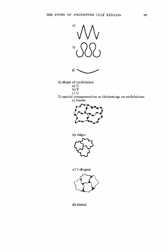
THE STUDY OF ANGIOSPERM LEAF REMAINS 89
2) shape of undulat ion a) U b )V c) z~
3) special ornamentation or thickenings on undulations a) knobs
b) ridges
c) T-shaped
@ d) absent

90 THE BOTANICAL REVIEW
5. surface o r n a m e n t a t i o n a. absen t
b. p resen t 1) s t r i a t ions
'/~//(:di("
2 ) papi l lae
3) t h i c k e n e d areas
4) t h i n areas
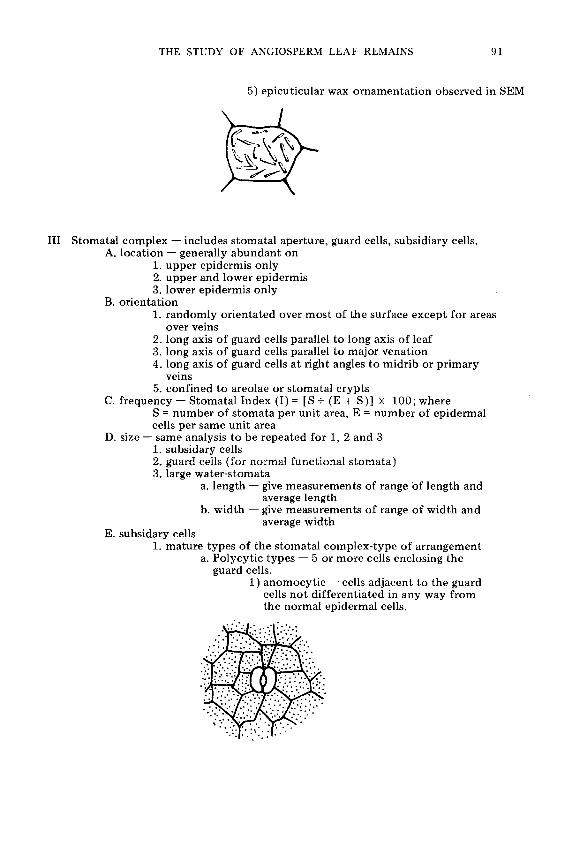
THE STUDY OF ANGIOSPERM LEAF REMAINS 91
5) epicut icular wax o r n a m e n t a t i o n observed in SEM
III Stomatal comp lex - - includes s tomata l aper ture , guard cells, subsidiary cells. A. loca t ion - - generally abundan t on
1. upper epidermis only 2. uppe r and lower ep idermis 3. lower ep idermis only
B. o r ien ta t ion 1. r andomly o r i en ta ted over m o s t of the surface excep t for areas
over veins 2. long axis o f guard cells parallel to long axis o f leaf 3. long axis o f guard cells parallel to major venat ion 4. long axis o f guard cells at r ight angles to midr ib or p r imary
veins 5. conf ined to areolae or s tomata l c ryp ts
C. f requency - - S tomata l Index (I) = [S + (E + S)] • 1 0 0 ; w h e r e S = number of s toma ta per uni t area, E = n u mh er of epidermal cells per same unit area
D. size - - same analysis to be repea ted for 1, 2 and 3 1. subsidary cells 2. guard cells ( for normal funct ional s t o ma t a ) 3. large wate r - s tomata
a. length - - give measu remen t s of range o f length and average length
b. wid th - - give measu remen t s of range of wid th and average wid th
E. subsidary cells 1. mature types of the s tomata l c o m p l e x - t y p e of a r rangement
a. Polycyt ic types - - 5 or more cells enclosing the guard cells.
1) anomocy t i c - - cells ad jacent to the guard cells no t d i f fe ren t ia ted in any way f rom the normal epidermal cells.

92 THE BOTANICAL REVIEW
2) cyc locy t ic - - single ring of small cells en- closing the guard cells.
....:::.
3) a m p h i c y c l o c y t i c - - d o u b l e r ing of small cells enc los ing the guard cells.
...;.
4) a t i nocy t i c - - single r ing of s o m e w h a t en- larged or e longate cells enc los ing the guard cells.

THE STUDY OF ANGIOSPERM LEAF REMAINS 93
b. Anisocytic types -- 3 cells (may be unequal in size) enclosing the guard cells.
5) anisocytic -- single ring of 3 cells (2 larger, 1 smaller) enclosing the guard cells.
..::-
~ 1 7 6
~ ~:'i!"::"
6) amphianisocytic -- double ring of cells enclosing the guard cells with the inner ring consisting of 3 cells (2 larger, 1 smaller); outer ring may be incomplete consisting of 2-3 or 4 cells.
ii i :

94 THE BOTANICAL REVIEW
c. Diacytic types -- 2 cells enclosing the guard cells at right angles to the long axis of the guard cells.
7) diacytic -- single ring of 2 cells enclosing the guard cells at right angles to the long axis of the guard cells.
" "
�9
o'*. " .~ ~ i ':..'..'.;:':
8) amphidiacytic -- double ring of 4 cells enclosing the guard cells at right angles to the long axis of the guard cells.
i
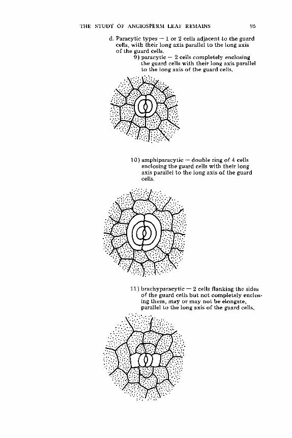
95
0
d. Paracytic types -- 1 or 2 cells adjacent to the guard cells, with their long axis parallel to the long axis of the guard cells.
9) paracytic -- 2 cells completely enclosing the guard cells with their long axis parallel to the long axis of the guard cells.
�9 .
THE STUDY OF ANGIOSPERM LEAF REMAINS
10) amphiparacytic -- double ring of 4 cells enclosing the guard cells with their long axis parallel to the long axis of the guard cells.
* . . . * . * . ~ �9 , ~
11) brachyparacytic -- 2 cells flanking the sides of the guard cells but not completely enclos- ing them, may or may not be elongate, parallel to the long axis of the guard cells.
�9 , . 0 . ,

96 THE BOTANICAL REVIEW
12) amphibrachyparacyt ic - - 4 cells f lanking the sides of the guard cells but no t com- pletely enclosing them, may or may not be elongate, parallel to the long axis of the guard cells.
13) hemiparacyt ic - - 1 of the cells adjacent to one guard cell enclosing it and parallel to its long axis, the o ther guard cell having 3 or more normal epidermal cells surround- ing it.
~ ," ~ �9
e. Tet racyt ic types - - 4 cells adjacent to and enclosing the guard cells.
14) para te t racyt ic - - 2 elongate cells lateral and parallel to tlae guard cells, 2 narrow polar cells.

THE STUDY OF ANGIOSPERM LEAF REMAINS 97
,o
15) a m p h i p a r a t e t r a c y t i c - - 2 e longa te cells la teral and parallel to the guard cells, 2 n a r r o w polar cells, all of wh ich is sur- r o u n d e d by a r ing of small cells.
!i I, ii i
16) b r a c h y p a r a t e t r a c y t i c - - 2 s h o r t cells lat- eral and parral lel to t he guard cells, 2 wide polar cells.
, , - , % : *
17) a m p h i b r a c h y p a r a t e t r a c y t i c - - 2 sho r t cells la teral and parallel to the guard cells, 2 wide po la r cells, all o f wh ich is s u r r o u n d e d by a r ing 'of small cells.
.'.: �9 .:........:.~:..: .:.
�9 ii:!l..'..'..'..' , .: . . . .:
. a " :.'::"." : :::

98 THE BOTANICAL REVIEW
18) s t au rocy t i c - - 4 cells, more or less equal in size, w i th the ant ic l ina l walls of the subsi- dary cells e x t e n d i n g a t r ight angles f rom the ~oles and midd le of the guard cells.
:. ""~.~ ...
19) a n o m o t e t r a c y t i c - - 4 cells enclos ing guard cells in an i rregular and var iable pa t t e rn .
.:!.t ': : :~ :~ ""
�9 . . . , * . , . * , ,
f. Hexacy t ic types - - 4 cells ad j acen t to the guard cells wi th 2 add i t iona l ( lateral or polar ) cells wh ich can be d i s t inguished f rom the ep idermal ceils.
20) p a r a h e x a c y t i c - - m o n o p o l a r - - 4 e longa te cells lateral and parallel to the guard cells, 2 n a r r o w polar cells.
: .:.." ~'::::." . ..[.:- ; . . .
0 * ,

THE STUDY OF ANGIOSPERM LEAF REMAINS 99
21) p a r a h e x a c y t i c - - d i p o l a r - - 2 e l o n g a t e cel ls la te ra l a n d para l le l t o t h e guard cells , 4 n a r r o w p o l a r cells.
!if! I::::':'':'~:'~:" �9 : I'. �9 . . . :~: . : . , . . , , . , . . . ~:.. �9 .- !:: I . ; -~ .~ : .~ . . . . !~.. .- ~
: ' i ' ~ : ' .
i l I ' ' ; ~i ~ . , '::'1,: .' l:.:
.': !! .'::'1:';I... ':':'
22) b r a c h y p a r a h e x a c y t i c - - m o n o p o l a r - - 4 s h o r t cells la tera l to t h e guard cells , 2 w i d e p o l a r cells .
o , , � 9 1 7 6

100 THE BOTANICAL REVIEW
23) brachyparahexacytic - - d i p o l a r - 2 short cells lateral to the guard cells, 4 wide polar cells.
~176176 ~ o o _ �9 �9 o
:.:,'.:~: " ' o ~ ,
: ~ o 0 , ~ 1 7 6 ":~
g. Polocytic types -- 1 cell nearly, but not completely, enclosing the 2 guard cells.
24) polocytic - - 1 cell nearly enclosing both guard cells except for one pole which is covered by a single epidermal cell.
�9
' ' " ~
.::-~
25) copolocytic - - 1 cell (subtended by a cres- cent-shaped cell) nearly enclosing both guard cells except for one pole which is covered by a single epidermal cell.
�9 : . . . . " ~!:." ?.:
0~149

THE STUDY OF ANGIOSPERM LEAF REMAINS 101
26 ) ax i l locyt ic - - 1 cell near ly enclos ing b o t h guard cells excep t for one free pole wh ich is covered by 2 cells wi th a c o m m o n ant i- cl inal wall e x t e n d i n g f rom the pole paral- lel to the long axis o f the guard cells�9
�9
�9 i .i:i~.
.. , . . . . . .
27) coax i l locy t i e - - 1 cell ( s u b t e n d e d by a c rescen t - shaped cell) near ly enclos ing b o t h guard cells e x c e p t for one free pole which is covered b y 2 cells w i th a c o m m o n ant ic l ina l wall e x t e n d i n g f rom the pole parallel to the long axis of the guard cells�9
. . . . . �9 : . - : : : : : " . ~ ' ~ ' : : . : ' . ' : . . .
�9 ": ! : ' . : ' : . . : r ".'.::"
.:" '2:"

102 THE BOTANICAL REVIEW
h. Per icyt ic types - - 1 cell encloses b o t h guard cells�9 28) desmocy t i c - - 1 cell enc los ing b o t h guard
cells wi th one ant ic l inal wall e x t e n d i n g f rom one of the poles c u t t i n g the cell once.
�9 ......:.:i::E::.i! '.:!.:."~: : ~::.. :.._~ E:!"...i -!
:!:ii:.... :..j_:~.:.: :-...
29) per icy t ic - - 1 celt enc los ing b o t h guard cells.
30) coper icy t i c - - 1 cell ( s u b t e n d e d by a cres- cen t - shaped cell) enclos ing b o t h guard cells.
... ~ ,.:...... :i:.!-:.i::!i~ii~!.

THE STUDY OF ANGIOSPERM LEAF REMAINS 103
31 ) a m p h i p e r i c y t i c - - 1 cell enclos ing b o t h guard cells enc losed by a s econd single cell.
' : : . . : . . . . ~ i!.'.: :
2. d e v e l o p m e n t f r o m p r o t o d e r m ini t ial (see t e x t for de f in i t ions ) a. m e s o g e n o u s

b. mesoperigenous
104 THE BOTANICAL REVIEW
c. perigenous
ili:i:~ilill
/
\

THE STUDY OF ANGIOSPERM LEAF REMAINS 105
3. o r n a m e n t a t i o n a. a b s e n t
b. s t r ia te
c. papi l la te 1) a rching over s toma ta l ape r tu re a n d / o r guard
cells
2) raised d i rec t ly over subs id iary cells

106 THE BOTANICAL REVIEW
3) r andomly directed
d. th ickened areas on perielinal wal
F. guard cells 1. pos i t ion - - relative to epidermis
a. raised
b. + level

THE STUDY OF ANGIOSPERM LEAF REMAINS 107
c. s u n k e n 1 ~ s toma ta l ape r tu re o p e n
I I
2) s t oma ta l ape r tu re occ luded
2. o r n a m e n t a t i o n a. ou te r s t o m a t a l ledge consp i cuous
1 ) p la in
2) o rna t e
1) 2)
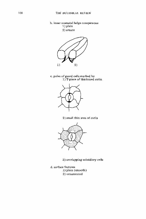
108 THE BOTANICAL REVIEW
b. inner stomatal ledge conspicuous 1 ) plain 2) ornate
1) 2)
c. poles of guard cells marked by 1 ) T-piece of thickened cutin
2) small thin area of cutin
3) overlapping subsidiary cells
d. surface features 1) plain (smooth) 2) ornamented

THE STUDY OF ANGIOSPERM LEAF REMAINS 109
IV Tr ichomes (hairs and glandular hairs) A. absent B. p resen t
1. occur rence on the leaf a. uppe r epidermis b. upper and lower ep idermis c. lower epidermis
2. n u m b e r of types p resen t 3. f r equency - - Tr i chome Index (I) = [T + (E + T)] • 100; where
T = n u m b e r of t r i chomes per un i t area, E = n u m b e r of epidermal cells per same uni t area
4. occurrence in relat ion to o the r t r i chomes a. singly
b. in tuffs
5. nature of t r i chome base a. t r i chome foo t ( a t t a c h m e n t )
1) scarcely mod i f i ed

11 0 THE BOTANICAL REVIEW
2) peg-like
3 ) 2-celled
4) mul tieellular
b. t r i chome basal cells 1) unmodi f i ed epidermal cells

THE STUDY OF ANGIOSPERM LEAF REMAINS
2) modified a) radial
b) pocket-like
111
c. trichome base 1) unmodified
2) thickened

112 THE BOTANICAL REVIEW
6. nature of t r i chome a. single cell
b. two-cel led
c. mult icel lular
7. nature of basal cells o f the t r i chome stalk a. single cell

THE STUDY OF ANGIOSPERM LEAF REMAINS 113
b. m u l t i c e l l u l a r file, 1-cell t h i c k
c. m u l t i c e l l u l a r fi le, 2-cel ls t h i c k
d. m u l t i c e l l u l a r
8. n a t u r e o f t r i c h o m e h e a d o r t ip a. n o c o n s p i c u o u s h e a d o n t r i c h o m e

114 THE BOTANICAL REVIEW
b. single-celled head
c. head 2-5 cells
d. rounded cluster of < 5 cells
e. f la t tened single-celled head

THE STUDY OF ANGIOSPERM LEAF REMAINS 115
f. f la t tened mult icel lular head 1 ) one cell thick
a) cells arranged radially
b) files of cells arranged radially
2) more than one cell thick a) cells arranged radically
b) files of cells arranged radially

116 THE BOTANICAL REVIEW
g. b ranched hairs 1) b ranched once
f
2) b ranched more than once
9. t r i chome size - - the range of the length and wid th or d iamete r and average of the l eng th and wid th or d iameter
a. t r i chome a t t a c h m e n t area (pore) b. t r ichome-base cells c. t r i chome d. individual or specialized parts o f the t r i chome
10. o rnamen ta t i on of t r i chomes a. absent b. p resen t
V Presence of o the r features on leaf epidermis (see Stace 1965, for a discussion of these features)
A. domat ia B. cork-warts C. h y d r a t h o d e s D. glandular epidermal cells

THE STUDY OF ANGIOSPERM LEAF REMAINS 117
TECHNIQUES FOR THE STUDY OF CUTICLE
As a result of the interest shown of plant cuticles in several rather diverse areas of science for a variety of applications, many techniques have been developed for their preparation. Cuticular analysis has been found to be a useful systematic tool whenever leaf material, whole or fragmentary, must be identified to a specific known extant species. Taxonomists, the drug industry, animal nutritionists, animal toxicologists, and police departments have all found cuticle useful in plant identification. Cuticular analysis has also been used for pur- poses other than systematics. Plant physiologists and ecologists con- cerned with the leaf epidermis have developed specific techniques for rapid evaluation of epidermal features, while the biochemist has developed quite different methods for isolating the cuticular mem- brane or component parts of the cuticle for fine structure and chemical analysis.
The techniques for preparing fossil cuticle vary also. The variation is not dependent upon the age or taxonomic affinities of the cuticle but upon the nature of preservation and the information the individ- ual investigator hopes to obtain from the fossil material. Each fossil locality, in fact each species at each locality, may require individual modifications of any given preparation technique for opt imum infor- mation to be obtained from the specimen. Each fossil specimen should be treated as a unique specimen and eventually the investiga- tor will be able to judge from experience the best preparation techniques to use.
The techniques discussed in this paper are only those which have direct application to studies of the morphology and systematics of angiosperm leaf cuticle of modern and fossil plants.
I. Preparation of Cuticle of Modern Angiosperm Leaves
A. Whole Mounts of Cuticle. The details of cuticular characters can be studied best when whole mounts of the cuticle are prepared (Figs. 13-15). For such preparations I cut a 1 cm. square (modified according to leaf size) from the median area of the leaf along the margin. This is then cut in half perpendicular to the leaf margin. These rectangles are placed in bleach (5% sodium hypochlori te Na H C1 02 ) until bleached white. Usually the mesophyll disintegrates within 4-12 hours in the bleach and the cuticles remain intact. The preparations are then placed in H2 O and very carefully, often under a dissecting microscope with the use of flattened dissecting needles, the upper and lower epidermis are worked apart and the mesophyll brushed away. The upper and lower epidermis remain attached at the margin of the leaf which acts as a sort of hinge. The preparations are then dehydrated in alcohol (50% ETOH, 95% ETOH, 100% ETOH), stained with 1% safranin 0 in 100% ETOH for 15 minutes to 6 hours or longer, allowed to destain in 100% ETOH if necessary, transferred

118 THE BOTANICAL REVIEW

THE STUDY OF ANGIOSPERM LEAF REMAINS l l 9
to xylene and mounted in H.S.R. (Harleco Synthetic Resin). Mount- ing is most easily accomplished by partially submerging a slide in the xylene solution and carefully sliding or floating the specimen onto the slide. The cuticle should be positioned with the outer surface of the upper and lower epidermis uppermost, remaining hinged by the margin.
The leaves or leaflets used are usually obtained from herbarium material and careful records should be kept of the data given on each herbarium sheet, along with additional leaves procured for clearing and not cut for cuticular analysis. It is important to keep careful records of the extant material used for comparative purposes because mistakes can easily be made in the identification of herbarium material. Pant and Mehra (1964) killed and fixed living leaves in Farmer's solution and then made epidermal peels which they stained with acetocarmine in order to make observations of the development of stomata.
A variety of solutions have been used to macerate leaf tissue in order to free the cuticular membrane. In addition to bleach (eau de Javelle), two solutions commonly used are Schulze's solution (1 part saturated solution of potassium chlorate, K C1 03, and 2 parts concentrated Nitric acid, HNO3), often used in macerating fossil tissue and a solution of 1 part hydrochloric acid (H C1) to 3 or 5 parts ethanol and ammonia. Stiirm (personal communication) found the leaf epidermis was often freed by hot 5-20% sodium hydroxide (Na OH) as well as hot 5-20% potassium hydroxide (KOH). Alvin and Boulter (personal communication) have experimented with 5, 10 and 20% chromium trioxide (Cr03) for varying lengths of time and found it to be very satisfactory in the maceration of leaf tissue from cuticle of gymnosperm leaves prepared for scanning electron microscope studies (Figs. 19-20). Stace (1965) suggests the use of 10% Cr03 and concentrated HNO3 for cuticular preparations. Kva(~ek (1966) used 30% hydrogen peroxide (H20) made basic with 10% potassium hydroxide (KOH). He left the leaf specimen in this solution for 2-3 days at room temperature or 30-60 minutes at 70~ This was followed by submersion on Schulze's solution for 3-15 minutes and then 10% potassium hydroxide (KOH) to disintegrate the mesophyll and separate the cuticle. A more drastic, but apparently successful method is the use of acetolysis for releasing cuticular membranes (Huynh, 1971). The acetolysis technique, which had not previously
4-----
Figs. 1 3 - 1 5 . Light microscopy of the lower epidermis of modern angiosperm leaves.
Fig. 13. - - The lower epidermis of Nectrandra latifolia. Typical of the Laura- ceae, there are 2 subsidiary cells (paracyt ic) enclosing the slightly sunken guard cells, x 400.
Fig. 14. - - Prostrate t r ichome lying in a modif ied epidermal pocket . This is c o m m o n in the lower epidermis of all species of Sapindus. Lower epidermis of Sapindus saponaria (f. 3 microcarpa), x 600.
Fig. 15. - - The lower epidermis of Talisia angustifolia showing the numerous (usually 5 or more) epidermal cells ( anomocyt ic ) surrounding and over-lying the guard cells. This is typical in the Sapindaceae (Note also Fig. 14). x 630.

120 THE BOTANICAL REVIEW
been applied to the preparation of cuticles is widely used in the preparation of pollen and spores. It consists of the use of a mixture of 9 parts acetic anhydried [(CH3 CO)2 O] and 1 part concentrated sulfuric acid (H2 SO4 ) heated to 100~ Huynh found this produced rapid destruction of the cellulose resulting in clean cuticular mem- branes in 5-10 minutes. Brief discussions and literature references for many macerating techniques are available in Sinclair and Dunn (1961), Stace (1965), Sinclair and Sharma (1971) and Huynh (1971).
Probably the most important new tool available for studying surface detail is the scanning electron microscope (Figs. 16-22). Its application to the features of cuticle has just begun to be studied. There is little information presently available concerning the tech- niques of sample preparation and the effects of varying KV (kilovolt- age) and beam penetration of the material. I have learned (from work done with Alvin) that using a low or medium KV (10-maximum 20) for cuticular samples seems to produce the best results. Both the external (Figs. 16-18) and internal (Figs. 19-22) surfaces of the epidermis or cuticular membrane should be studied with the SEM. Boulter (1970) and Alvin are using the SEM to study taxodiaceous cuticles, Alvin and Ferguson are using the SEM with the cuticles of Amentotaxis, and Cutler is studying monocotyledonous cuticles using the SEM. Alvin (1970) and Baker and Parsons (1971) have written useful papers discussing the preparation of leaf cuticle for SEM observation.
B. Replicas of Cuticles. A quick and easy way to obtain infor- mation concerning the surface features of leaves is to make a replica of the surface detail and examine it under a microscope. For many years such replicas have been observed embossed in herbarium glue that peeled away from leaf surfaces. This technique simply requires a material which will conform to-the patterned surface of a leaf and then be easily peeled away leaving the surface pattern impressed in the material, which can then be studied by transmitted light micro- scopy. Sinclair and Dunn (1961) survey various methods used for making surface impressions since 1902 and recommend the use of Archer's herbarium mounting plastic. A few references to methods of preparing replicas of leaf surfaces which Sinclair and Dunn do not mention, follow. Turtox Service Leaflet No. 31 proposes placing a plastic cover slip on the surface of a leaf wet with a solvent for the plastic. Long and Clements (1934) used collodion films for making stomatal counts. Bennett and Furmidge (1956) recommended the
Figs. 16-18. SEM microscopy of the external surfaces of modern angiosperm leaves.
Figs. 16 & 17. - - The upper (Fig. 16) and lower (Fig. 13) epidermis of Oremunnea pterocarpa showing the di f ference in surface texture , abundance of pel tate t r ichomes and presence of s tomata. • 250.
Fig. 18. - - Lower epidermis ofAlfaroa guatemalensis which is closely related to Oremunnea pterocarpa. A few guard cells and many conspicuous papilate sub- sidiary cells can be seen. • 500.

THE STUDY OF ANGIOSPERM LEAF REMAINS 121

122 THE BOTANICAL REVIEW

THE STUDY OF ANGIOSPERM LEAF REMAINS 123
use of cellulose acetate dissolved in acetone painted on the leaf surface and North (1956) recommends the use of dye (night blue) in the cellulose acetate solution for bet ter viewing of the leaf peels. Sampson (1961) and later Miller and Ashby (1968) suggest the use of a double replica technique, in which a primary replica of a silicone rubber monomer is made of the leaf surface and from this a second- ary rephca xs made of m~rocellulose (clear nail varnish). This tech- nique has the advantage of being able to produce replicas from wet leaf surfaces.
Horanic and Gardner (1967) proposed the use of Rhoplex AC-33, a viscous emulsion of acrylic polymers, as an improvement over earlier techniques of making replicas. Tur tox Service Leaflet (1960) and Payne (1968) suggest the use of cellulose acetate film (it can be purchased in sheets of various thickness), which is placed on a leaf surface wet with acetone, to make good replicas in a short time with very few materials or mess involved. Techniques for making replicas of the surface features of leaves have been discovered, rediscovered, modified and remodified independent ly by many investigators as the need for their use arose. There may be other techniques not listed here but those given provide a starting point for anyone interested in investigating these techniques. I have found the technique outlined by Payne (1968) the easiest and most satisfactory for general use. It can also be used to remove dense coatings of tr ichomes on leaves, if they are bothersome in observing cells or the stomatal complex.
The use of the carbon replica technique has been impor tant in the investigation of fine detail of epicuticular waxes with the electron microscope. Martin and Juniper (1970) discuss this technique in some detail as well as mentioning other techniques useful for the examination of surface features of leaves by electron microscopy.
II. Preparation of Cuticle of Fossil Angiosperm Leaves
The preparation of fossil cuticle must be done with more care and control than the preparation of cuticles of extant angiosperms be- cause the material is often much more fragile or fragmentary and is not often as readily duplicated as extant material. Each preparation must be watched carefully and judged independent ly of other sam- ples to assure the best results.
+ _ _
Figs. 19 & 20. -- SEM microscopy of the interior surface of a modern gymno- sperm leaf. The lower epidermis of Sequoia sempervirens prepared with chromium trioxide and photographed by Mr. Carl Medsker. Note the nature of the anticlinal extensions of cutin and the granular nature of the cutin. Boulter (personal communication) has suggested that this granular character is typical of the Taxo- diaceae. Fig. 19 • 250 and Fig. 20 • 500.

124 THE BOTANICAL REVIEW
A. Whole Mounts of the Cuticle. Whole mounts or mounts of as large areas of the cuticular membrane as possible have most common- ly been used in studies of fossil cuticle. When a compression of an angiosperm leaf is exposed the entire leaf, upper and lower epidermis and mesophyll cells if preserved, may adhere to one surface of the matrix, or the leaf may break along the mesophyll leaving the upper epidermis on one piece of the matrix and the lower epidermis on the other piece of matrix. When the entire leaf adheres to one side of the matrix the leaf will often crack and curl up into small pieces as it dries. It has been suggested that if the matrix is allowed to dry out very slowly the cuticle will not curl or crack, but it has been m y experience that even when fossils are allowed to dry out within the sediment, cracking and curling of well preserved leaves still occurs. To avoid this problem, spray the material when it is first exposed with a thin film of transparent plastic (e.g., Krylon, no. 1303, crystal clear acrylic spray coating which dissolves in acetone, product of Borden, Inc., New York) for which you have a suitable solvent so cuticular preparations can be made at a later time. Walther (1964) discussed methods of collecting fossil leaf material in the field. He secured some of the loose cuticular material with glycerine jelly and coated some badly fragmented material with an acetone soluble lacquer. Glass or plastic vials and envelopes are convenient for collecting loose cuticle in the field or retaining it for study when working in the laboratory.
Two basic types of techniques have been successfully employed in studying fossil cuticle as whole mounts. Samples of the cuticles have simply been removed from the matrix and macerated or the whole leaf or parts of it have been removed from the matrix as a transfer and processed. These are described below.
Preparation of Cuticular Fragments. Remove the cuticular mate- rial from the compression. This may be done by brushing the loose cuticular material into a test tube, picking the larger cuticular frag- ments off with forceps, or scraping the compression with a flattened probe if the cuticle adheres tightly to the matrix. It is wise to use a small paper funnel when placing the cuticular fragments into a test tube in order to catch all the fragments being brushed or scraped from the fossil leaf. If the fossil specimen has been treated with a plastic spray (e.g., Krylon) it will be necessary to wash the cuticular material with an appropriate solvent (e.g., acetone for Krylon) before maceration. Bleach the cuticular material in 5% sodium hypochlorite (Na H C1 02 ) solution until the fragments are bleached white (5 to 10 minutes). A commercial bleach such as Chlorox or Hilex is satisfactory. They usually sink to the bot tom of the sodium hypo- chlorite solution when bleaching nears completion. Add water to dilute the bleach, centrifuge if necessary, and decant. Schultze's solution may be used if bleach is found to be too harsh. Wash cuticular material with water, centrifuge if necessary, decant. Next

THE STUDY OF ANGIOSPERM LEAF REMAINS 125
proceed with either a water stain process (A) or an alcohol stain process (B). I prefer the latter. A. Stain cuticle in a wa te r stain. Many water stains are satisfactory. I have found 2% safranin 0 in water stains the cuticle well. Cuticle should be stained for 10 min- utes-24 hours. Add water, centrifuge if necessary, and decant. Wash cuticular material with water, centrifuge if necessary, and decant. The cuticle may be mounted in glycerine jelly at this point or dehydrated in an alcohol series to xylene and mounted in H.S.R. B. Dehydrate in 95% alcohol. Stir well, allow the cuticle to remain in the alcohol about 10 minutes (no longer). The cuticle may be mounted in diaphane at this point and should be stained in water or 95% ETOH if desired. Place 1% safranin 0 in 100% EOTH and allow to stain 10 minutes to 24 hours. Centrifuge if necessary, decant, transfer to 100% ETOH and allow to destain for 10 minutes or longer. Centrifuge if necessary, decant. Add xylene to the cuticular material. The cuticles may be kept in xylene for some time before mounting (if any alcohol is present the safranin will slowly wash out). Mount in H.S.R. or suitable mounting medium. To mount , pipette a concentrated number of small fragments or hand manipu- late larger fragments on to a slide. Absorb the excess xylene, add the mounting medium, and cover with a cover slip. Place a weight on the cover slip and dry for several days.
Cuticular fragments, when well preserved, can be manipulated by hand and moved from solution to solution. However when they are extremely fragmentary I treat them much as a pollen sample, pro- cessing them in a test tube with the aid of a centrifuge. Walther (personal communicat ion) processes cuticular fragments in a depres- sion slide changing solutions with a small pipette.
Several maceration techniques have been used with fossil cuticle. The most commonly used solutions for maceration of fossil cuticle are those mentioned by Ferguson (1971) and StUrm (1971). Both used Schulze's solution (1 part K C103 and 2 parts concentrated HN 03 ) for the initial maceration. Ferguson followed this with 10-25% ammonia (HN3) while St~irm followed it with hydrogen peroxide (H2 02 ). Stiirm also sometimes carried out his maceration with con- centrated nitric acid (HN O3) for 1/2-1 hour followed by a dilute ammonia (HN4 OH) solution. He mounted his material in glycerine jelly with stain in it. Glycerine jelly seems to be a particularly popular mounting medium for cuticular material in Europe.
One difficulty in observing preparations of loose fragments of fossil cuticle prepared by maceration is the problem of interpreting which side of the cuticle is facing up in the final preparation. This can be done by very carefully studying the cuticle under high magnification and slowly focusing through several optical focal planes of the cuticle. By interpreting the sequence of structures observed, the disposition of the cuticle can usually be understood. Occasionally the surface details of the cuticle are difficult or impos- sible to determine accurately when the material is mounted inside

126 THE BOTANICAL REVIEW

THE STUDY OF ANGIOSPERM LEAF REMAINS 127
up. It might be useful, if this is a problem, to use a reversible duralumin microscope slide developed by Sims and Lyon (1963) in which the material is mounted between 2 cover slips which fit into an aluminum slide-like frame, allowing both sides of the microscope preparation to be observed at high magnifications.
Whole mounts of fossil cuticular material should also be prepared for and observed with an SEM on a regular basis. Little work has been published on the techniques which are best used when pre- paring fossil cuticle for SEM observation; however, techniques are being developed (Alvin and Dilcher, work in progress). Just as with extant cuticular material, both the external and internal features of fossil cuticle should be studied. Internal features of fossil cuticle are shown in Figures 21 and 22.
Transfer of Leaf Material for Cuticular Preparation. Making trans- fers of Tertiary angiosperm leaves has been a popular method of preparing leaves for general study. The technique for preparing whole leaf transfers of compressed remains is discussed earlier in this review in detail. This same technique is useful for cuticular preparations as noted by RUffle (1963) and Walther (1964) and may be applied to whole leaves or only to small sections of leaves. The most common method of making transfers is by coating the fossil with a liquid such as collodion or a clear lacquer.
The transfer method is very useful when the fossil cuticle is so fragmentary that it tends to disintegrate completely when being macerated, and sometimes, even when small fragments of cuticle could be successfully prepared by maceration, it may be important to relate the orientation of the stomatal complex to the whole leaf or to other stomatal or epidermal features. Transfers maintain the epidermal cells in place and intact.
When the transfer method is used to study cuticle it is often necessary to wash or dissolve away any matrix adhering to the fossil and to macerate the leaf in order to clear the cuticle. These tech- niques are discussed earlier. If both the upper and lower epidermis are preserved and remain together on the same piece of matrix, it is usually difficult or impossible to observe cuticular characters of both on a single preparation because you must focus through one to see the other. However if the transfer is cut, and some preparations are mounted with the upper surface uppermost while other preparations are mounted with the lower surface uppermost, better observations can be made of both surfaces. Another technique is the use of Sims and Lyon's (1963) double cover slip preparation discussed earlier.
< _ _ . I
Figs. 21 & 22. - - SEM mic roscopy of the in te r io r of fossil ang iosperm leaves. The uppe r ep idermis (Fig. 21 ) and lower ep idermis (Fig. 22) of two d i f fe ren t leaves f rom Middle Eocene clay deposi ts of wes te rn Tennessee . No te the d i f fe rence in the na tu re of the ant ic l inal ex tens ions of the cut in . X 500.

128 THE BOTANICAL REVIEW
Fossil leaves may have undergone a certain amount of natural maceration or oxidation in the sediments in which they are found. When this has happened they often split open along the mesophyll and are exposed as part and counter part. Working with such material McConnaha (student research project at Indiana University) sug- gested the use of clear celluloid tape for making transfers of the cuticle. A large, naturally-macerated square of cuticle can be lifted intact from the surface of a compression and mounted so the position and orientation of the cuticle (with reference to inside or outside surfaces) can be controlled. Probably the best tape for this preparation method is Scotch Brand #853 manufactured by 3M. The tape is cut in a small square, pressed on the fossil so the adhesive is in contact with the fossil over the entire surface of the tape, peeled away, brushed lightly to remove loose clay particles, dipped in xylene and mounted in H.S.R. (Harleco Synthetic Resin). The cellu- loid tape method is useful for some purposes, but the preparations are not as satisfactory as a carefully done collodion transfer. The adhesives on the pressure sensitive celluloid tape swell in organic solvents distorting the original preparation after 2-3 days. Also, tape is not as resistant to chemicals or water and does not hold the fossil cuticle as firmly as collodion to allow cleaning of the clay or other matrix from the cuticle. We have experimented with both transfer techniques and use the collodion for preparations to be mounted in a mounting medium soluble in organic solvents.
Collodion transfers can also be used for observations of the leaf surfaces in the SEM. I have found that the cuticle should be cleaned chemically before SEM observations are made and use warm HF for 1 hour to accomplish this. C. R. Hill (personal communication) for Mesozoic cuticles uses a layer of acetone-based clear nail varnish followed by a celloidin-based glue to build up a layer about 1 mm thick. The matrix is then dissolved away with HF and the specimen washed, coated, and observed in the SEM.
Alvin (1970) studied the leaf surfaces of Cretaceous gymnosperms using the SEM. He cleaned the leaf cuticles with HF acid and macerated them in Schultze's solution before observing them under the SEM.
There are few published accounts of the use of replicas in studying fossil cuticle. Fossil cuticle is generally not well enough preserved to permit the use of acetate film or other such materials used for making replicas of modern cuticle. However when the sediment is very fine, filling in along the surface contours of the eipdermis, a natural replica may result. Such natural replicas may be cast and observed in oblique lighting or by the scanning electron microscope. Alvin has made successful casts of the cuticle of fossil leaves of Amenota tox i s and some angiosperm leaf impressions from Alum Bay. He used silicone rubber for casting the fossil leaf and then studied the surface detail with the SEM (Alvin personal communica- tion). Chaloner and Gay (1973) have developed the use of rubber

THE STUDY OF ANGIOSPERM LEAF REMAINS 129
latex casts for SEM observation of anatomical detail of Carboni- ferous impressions and this method appears to have considerable potential for revealing epidermal features. Chaloner and Gay illus- trate the results of this technique as applied to several lycopod stem impressions. Details of lycopod epidermal cells and stomata were well preserved and could be observed with the SEM.
STATISTICAL ANALYSIS
There is no published record of detailed statistical studies of the form and/or anatomy of angiosperm leaf remains although some authors have presented data on the variability of gross leaf form (Tanai, 1960, 1972; Hantke, 1965) or venation (Ferguson, 1971). These studies are important contributions to paleobotany because they indicate the usefulness of careful measurements of fossil material. Tanai (1972) has presented leaf indices of several species of living beech. Hantke (1965) presents detailed leaf proportion dia- grams for Acer trilobatum based on a large number of leaves of various sizes which show that the proportions of the leaves remained quite constant throughout a range of sizes. Ferguson (1971) gives numerous graphs illustrating the relationship of the distance between secondary veins and the positioning of the veins (vein interval) on the leaf. He did this for several specimens of individual species as well as graphing breadth of lamina vs. length of lamina. The results show a distinct correlation of features among different fossil specimens.
Except for a few tables giving the cuticular features of various species of extant angiosperm genera, such as Rhus (Kr~iusel and Weyland, 1954) and Myrica (Sheffy, 1972), no detailed account of a comparative nature has been attempted using cuticle. These studies are not in fact statistical treatments of the data.
In order to appreciate the statistical significance of the features now being used to describe fossil leaves, it is important to develop new statistical tests or to use existing ones and apply them to the data available. Dolph, in studying the fossil leaves of Apocyno- phyllum mississippiensis, has developed an array of statistical tests which he has applied to his data (Dolph, 1973, 1974). This type of approach is in fact a form of numerical taxonomy modified to the special problems of leaf t axonomy of fossil angiosperms. Hopefully the use of a sound statistical approach will allow us to make better use of the data available.
USE OF THE RECORD OF A SINGLE TAXON
As the taxonomic relationships of the various organs or parts of fossil plants in various floras are better understood, it may be possible to study a taxon in detai l at a particular time in the past. In some fossil floras specimens of leaves, fruits and/or seeds, pollen, wood and flowers may be found and identified to an extant genus.

130 THE BOTANICAL REVIEW
For example, in the middle Eocene clay deposits of Kentucky and Tennessee leaves, fruits and pollen belonging to Engelhardia have been identified (Berry, 1930; Elsik and Dilcher, in press; Potter and Dilcher, 1971) and such an assemblage has also been identified for the extant genus Nyssa (Berry, 1930; Gray, 1961; Dilcher and McQuade, 1967). I do not suggest that the same genus and species name be given to various parts of a plant which appear to be related to the same genus and are found at the same locality as MacGinitie (1969) did with fruits and leaves of Lomatia in the Green River flora. Neither the fruits nor leaves now appear to belong to Lomatia or the family Proteaceae (MacGinitie personal communication). However there are numerous possibilities for paleobotanists to exam- ine in detail the various plant parts of a particular taxon present in the fossil record at a particular time which may be somewhat similar to an extant genus. Using such an approach we would be in a position to discuss, with some understanding, the evolutionary status of an extant taxon at a particular time in its history. As far as I know a detailed morphological s tudy of the various parts of an angiosperm genus or species collected from one time and place in the fossil record has not been done.
RESULTS OF A MORPHOLOGICAL APPROACH
INFLUENCE ON. TAXONOMY OF FOSSIL LEAVES
A carefully done morphological study of angiosperm leaf remains, including critical analysis of gross form, venation and cuticle, should result in better understanding of the fossil material. When the result- ing information is related to similar information about extant taxa a more meaningful comparison can be made between them. However it has been far too common in past studies of angiosperm leaves to make rather positive statements of the relationship of fossil leaves to extant genera with only fragmentary evidence to support the rela- tionship. As noted previously in this review, the early published record is replete with comparisons of fossil leaves to extant taxa based upon gross leaf form and major venation patterns without a careful survey of these characters in angiosperms living today.
In descriptions of fossil leaf remains, a concise statement of the type of analysis used to study the fossil leaves, the number of fossils of each leaf type available for study, and the number and names of similar fossil and extant leaves, including the types of analysis used in studying them should be presented. Then the reader will be able to understand the basis on which the taxa described was identified and be able to evaluate the conclusions which may be based upon the leaf identifications. When such questions are asked of much of the past work on fossil leaves, it appears that often only a few fossil speci- mens were examined, no critical analysis of any sort was applied to

THE STUDY OF ANGIOSPERM LEAF REMAINS 131
the fossil mater ia l and on ly general re fe rences are given to one or two similar fossil and e x t a n t leaf types . F o r e x a m p l e Ber ry (1916) wri tes of Myrica elaeanoides, "Myrica elaeanoides, if it is a Myrica, m u s t have been rare in the Wilcox f lora or else an i n h a b i t a n t o f areas r e m o t e f r o m fossi l izat ion, fo r it has been de t ec t ed on ly twice in the large co l lec t ions subsequen t ly made . There is some r e semblance to t h e larger leaves o f the Wilcox species Nectrandra pseudocoriaceae Berry, bu t the t w o f o r m s are bel ieved to be pe r f ec t l y d i s t i nc t . " And of Myrica wilcoxensis, " I t is a l m o s t ident ica l wi th a n u m b e r of exis t ing species, as for e x a m p l e Myrica cerifera... I t is also close to the exis t ing Eurasia t ic Myrica gale Li nne . " S h e l l y (1972) p roves these s t a t e m e n t s are incor rec t . Because there is so l i t t le subs tan t ive da ta assoc ia ted wi th t h e m , the accuracy o f such iden t i f i ca t ions is d i f f icu l t to d e t e r m i n e w i t h o u t r e -examin ing the mater ia l . T h e r e f o r e I p r o p o s e t h a t fu tu re pub l i ca t ion of descr ip t ions and discussions of fossil ang iospe rm leaves inc lude i n f o r m a t i o n which al lows the reader to d e t e r m i n e easily the basis fo r the t a x o n o m i c ass ignment . I t is suggested t ha t the fo l lowing i n f o r m a t i o n be included:
Fossil Name, author Diagnosis:
gross form: venation pattern (including fine venation): cuticular analysis:
Number of leaves (whole or fragmentary) examined from each horizon or precise locality:
Types of analysis used: gross f o r m - venation pattern, major venation, fine venation -- euticular analysis, upper epidermis, lower epidermis --
Fossils examined for comparison, types of analysis used -- Extant leaves examined for comparison, types of analysis used -- Discussion:
level of confidence of identification -- other related organs or associated species important in the
identification -- time range of the species identified and of the genus if applic-
able -- geographic range of the species and of the genus if applicable
any additional information --
The a d o p t i o n o f this p lan would give pa l eobo tan i s t s m o r e precise d i rec t ion in the i r a p p r o a c h to the inves t iga t ion and analysis o f a n g i o s p e r m leaf mate r ia l and readers m o r e i n f o r m a t i o n for in terpre- t a t i on of the t a x o n o m y . The readers o f pa leof lor i s t ic papers o f t en c o n c e n t r a t e on the i n t e rp r e t a t i ons made and ignore the sys t ema t i c s of the fossils u p o n which the i n t e rp r e t a t i ons are based. Par t of the r eason for this is t ha t the sy s t ema t i c sec t ions of pa leof lor i s t ic s tudies o f t en p rov ide li t t le conc re t e da ta t ha t wou ld al low the reader to

132 THE BOTANICAL REVIEW
make a fair evaluation of the evidence used to substantiate identifi- cations of taxa.
When this sort of morphological approach is applied to angiosperm fossil floras previously identified and described by less precise and detailed techniques, there are certain to be extensive taxonomic revisions in the fossil record. As mentioned earlier, work completed so far on the reinvestigation of the Eocene floras of southeastern North America (Dilcher, 1973) has resulted in a revision of 60%-70% of Berry's (1930) family and generic identifications. More drastic taxonomic revisions of angiosperm leaves are resulting from work on the Lower Cretaceous deposits in eastern North America (Doyle and Hickey, personal communication).
Both venation and cuticle of a leaf consist of a complex collection of characters (Table II and Table IV). Because some of these features may be conservative while others vary more easily throughout time or in different environments, the relative conservancy of venation and cuticle as a whole is difficult to compare. Even the gross form of leaves varies with changes in environment (Dilcher, 1973).
A number of angiosperms are known to be heterophyllous. In such case the cuticle is more conservative than leaf form or venation. Gross leaf shape may vary from mature to immature foliage, and be accompanied by significant venation differences as in Dendropanax- Gillbertia (Dilcher and Dolph, 1970). Also variation in leaf margins accompanied by differences in marginal venation patterns may occur as in the leaf dimorphism of Populus trichocarpa (Critchfield, 1960).
Gross leaf form and major venation patterns appear to be relative- ly stable characters, except in the case of leaf dimorphism or ecolo- gical variations, where leaf size, fine venation, areole shape and size, and marginal venation are responsive to environmental variations. This variation is discussed earlier in this review. The stomatal com- plex, stomatal orientation and positioning and, generally, trichome type and positioning are stable characters while other cuticle charac- ters vary more with environmental changes which were discussed earlier in this paper. Which characters are more stable in any one taxon at any one period of geologic time is not known. Certainly all these characters have changed through time.
INFLUENCE ON CONCEPTS OF EVOLUTION
An effective morphological approach to leaf remains is having, and will continue to have, a profound influence on our understanding of the evolution of angiosperms. I am referring here not only to evolu- tion leading to the origin of angiospermy, but evolution subsequent to the origin o f angiospermy as well. The detailed angiosperm pollen record of the Cretaceous (Doyle, 1969; Muller, 1970) illustrates the discrepancy between identifications to extant angiosperm taxa of leaves and pollen. This difference is clearly seen by a comparison of Table I with figures 23 and 24. Muller (1970) writes, "In general it

THE STUDY OF ANGIOSPERM LEAF REMAINS 133
90 mill. yr.
top Cenomanian
phase
II 100 mill. yr.
top Albian
top Aptian
phase
I
140 mill. yr.
top Jurassic
O,.
~" ~ ~ 8 ~ ~" = , , o ~ "r_ ~
= o o - ~ _ _
0 0 0 > 0 v O .
ca .1~ ea 6m
0 a .
G,I O .
, @@�9 !
I I I I I I I I I I
...... | -
I I I I I I I I I I I I
J
! I ! s
114' I
l
l
I
Fig. 23. - - Ini t ial r ad ia t ion of ang iosperm pol len types in the fossil record. ( R e p r o d u c e d wi th permiss ion of the pub l i she r f rom Muller, 1970) .

134 THE BOTANICAL REVIEW
O 0
Quaternary o, io-ene.
10
Miocen~
30 Oligocene
4O
Eocene
5O
Palaeocene 60
Senonian 70
8O Turonian
9O
Cenomanian 100
Albian
5 0 5 10 i
n e w
super new orders orders
0 5 10 15
new families
Fig. 24. -- The appearance of new angiosperm taxa as summarized from palynological data by Muller, 1970. (Reproduced with permission of the pub- lisher from Muller, 1970.)
would appear that paleobotanists have been more inclined to place leaf fossils from the mid-Cretaceous into extant families or even genera, while for pollen this is only possible from the Senonian upwards. Even allowing for the possibility that some of the identifi- cations of early leaves are erroneous, this discrepancy could indicate that pollen and leaf morphology evolved independent ly ."
The appearance of new angiosperm families in the fossil record (Fig. 24) is very different from the perspective obtained from observ- ing the floristic studies in Table I. Muller (1970) is too kind to the paleobotanists when he suggests " the possibility that some of these identifications of early leaves are er roneous ," when the probabil i ty that most of them are incorrect at the generic level and many at the

THE STUDY OF ANGIOSPERM LEAF REMAINS 135
family level would actually be more accurate. However this still leaves open the point raised by Muller that "this discrepancy could indicate that pollen and leaf morphology evolved independent ly ." There is no reason to assume that the two organs evolved in step. Axelrod (1970) extends such an argument into the Early and mid- Cretaceous suggesting that the pollen record is a poor indication of the origin and evolution of angiosperms. It is important to separate the analysis of two plant organs (leaves and pollen) when assessing their evolutionary significance at a particular time. Even today leaf analysis and pollen analysis of the same group may not provide the same degree of information concerning the evolution of a group. In this regard it behooves the paleobotanist to be careful not to use pollen evidence to establish leaf identities. For example, a study of the fossil leaf record of the family Proteaceae now indicates that no fossil leaves from North America assigned to this family validly represent it in North America (Dilcher, MacGinitie, unpublished data), although the pollen record of Proteacidites has been used to indicate the presence of the family (Muller, 1970) there. There are also numerous records of the pollen of Myrica from Late Cretaceous time while a re-examination of the leaf record, using cuticular analy- sis, provides no evidence of the presence of Myrica at that time (Shelly, 1972). Perhaps studies of pollen and leaf remains can act as a double check on each other in determining the uncertain, or difficult to establish, identifications to modern affinities of fossil pollen and leaves. Perhaps the degree of modernness of different organs makes the identification of extant taxa possible at different times for different organs as suggested by Muller (1970) and Axelrod (1970).
A discrepancy also exists in studies of the leaf record of angio- sperms. For instance, studies of Tertiary angiosperm leaf remains done by Leopold and MacGinitie (1972), Wolfe (1972c) and Tanai (1972) in western North America, Alaska and Japan record the presence of many extant genera from both Paleocene and Neogene times, while Ferguson (1971), in his study of a Neogene age angio- sperm flora in Germany, places less than 1/3 of his recognized leaf taxa in extant genera. The details of leaf anatomy examined in these four investigations varied. The question arises; is the percentage of Tertiary leaf remains assigned to modern taxa at the generic level a function of the completeness with which the leaf remains and related modern taxa have been studied?
Wolfe (1972a, 1972b) recognized evolution of Cretaceous and Early Tertiary leaf forms. He indicates the difficulty of placing some Paleocene leaf material in extant taxa even at the ordinal level, referring to them as "Cretaceous relics." He assigns a few leaves of Middle and Late Eocene times to extinct genera. Studies by Leopold and MacGinitie (1972) and Tanai (1972) give only slight indications of extinct or unrecognizable angiosperm taxa during the Tertiary. Their work seems to indicate the presence and continued existence of extant angiosperm genera from Late Eocene times to the present.

136 THE BOTANICAL REVIEW
Ferguson's data, on the other hand, leads to the conclusion that many of the angiosperm genera in the Miocene leaf record can not be related to extant genera at the generic level. Perhaps this discrepancy is the result of a combination of two factors: 1) This was Ferguson's (1971) first a t tempt at surveying a fossil flora and he had to cope with learning all of the background information necessary to judge the taxonomic affinities of his fossil leaves. Ferguson not only worked without the aid of an experienced angiosperm leaf morpho- logist, he enlarged the scope of his undertaking by adding numerous characters little studied by leaf morphologists. It was humanly im- possible to research the limits of variability and similarity of leaf characters for all the genera and families involved. Thus Ferguson's results represent an a t tempt to relate fossil leaves to extant genera and families incompletely sampled for the detailed morphological analysis applied to their leaves. Therefore he may not have been able to find extant affinities because of his sampling of extant forms. 2) Ferguson analyzed his material in detail wi thout always having ana- lyzed the characters in a taxonomic situation. When a close look is made at any plant organ morphologically, differences may be found. It is often difficult to interpret these differences, especially when the time span of several million years is interposed.
In any case it appears that the more information available the more difficult it is to draw close relationships between living and fossil leaf forms. This question remains open, but perhaps more evidence of the evolution of various taxa during the Tertiary will be forthcoming as detailed studies are completed.
Boulter (1971) suggests the possible complete extinction of many or even most members of the northwestern European Neogene flora. Repeatedly in the systematic section of the paper he indicates that Neogene pollen types represent extinct species "dissimilar from any living today" (Boulter, 1971, p. 402). The assignment of fossil forms to living taxa has also been discussed by Leopold (1969). She presents fruit and seed data mainly from Reid, Chandler and Elias to support a progressive evolution of modem genera and species during the Cenozoic. The obvious fact is that more living genera and species can be found ion the fossil record of the Neogene than the Paleogene. It is interesting to note that the latest records of extinct genera of plants involve herbaceous forms (Leopold, 1969) that probably are diversifying rapidly during the Neogene.
There is often very little data, other than the opinion of the investigator, to substantiate many angiosperm fossil leaf identifi- cations, and virtually no insight into the evolution of taxa has been reported in studies of fossil leaf remains. Angiosperm fossil leaf taxa have been treated as static entities. Listing the "similar living species" for Paleogene leaves identified to extant genera (Axelrod, 1966; MacGinitie, 1969) discourages consideration of the part these fossils played in the evolution of extat~t taxa.
It is time that paleobotanical investigations of angiosperm leaf remains begin to reflect a post-Darwinian concept of the evolution of

THE STUDY OF ANGIOSPERM LEAF REMAINS 137
angiosperms. Some studies are in progress on the morphological characters of Cretaceous and Tertiary leaves, as mentioned earlier in this review, which should point the way to the use of fossil leaves in the interpretation of the evolution of the angiosperms.
When leaf remains are examined in detail, consistent and signifi- cant differences may sometimes be noticed in similar leaves from different ages and/or localities. Such differences in leaf remains might be used as important sensitive stratigraphic tools. Perhaps genetic (Miki and Hikita, 1951) or environmental changes which plants undergo may be most easily observed in fossil foliage than in other plant remains. It is well known that changes in the chro- mosome number of a plant often result in a size change of the guard cells and/or epidermal cells (Sax, 1937). Also, as mentioned earlier, environmental changes are reflected in the fine venation and cuti- cular characters of leaves.
Investigations at Indiana University of the gross leaf form, vena- tion patterns and cuticular characters of angiosperm leaves has shown that some fossil leaves having similar gross morphology and venation patterns may have different cuticular characters (Dilcher, 1971). The extinct genus Dryophyllum is one of the most common leaves found in the middle Eocene sediments of Kentucky and Tennessee. The age of the clay pits from which leaf floras have been collected appear to vary in age (based upon pollen data, Tschudy, personal communi- cation) from lower Middle Eocene to upper Middle Eocene, a time interval of 5-7 million years. Throughout this interval the gross morphology, venation patterns and stomatai complex of D. tennes- seensis are reasonably uniform but the nature of the trichomes varies markedly. Cuticular variations that are location or perhaps time specific, have also been observed in Knightiophyllum (Dilcher and Mehrotra, 1969), palms (work in progress), Apocynophyllum (Dolph, 1973) and Sapindus (work in progress). In fact many leaf forms common to several pits that have been examined in detail show some variations of cuticular characters which appear to be pit specific. Stiirm (1971) described Ocotea multipora from Middle Eocene deposits near Darmstadt, West Germany, which is identical in gross form and venation to Ocotea obtusifolia Berry, described in detail by me (Dilcher, 1963). The only difference is in the number and positioning of the "pores" of trichome bases on the cuticle. These details in leaf morphology may prove useful in understanding the changes in leaves through time.
I have found in the leaf forms studied that some of the cuticular features, such as trichome type, positioning, frequency, surface pat- terns, cell size and anticlinal wall configuration and cell arrange- ments, may show changes through small time intervals. The question, which characters in leaves are the most conservative through time, can not be answered now. It appears that some cuticular characters are more sensitive to change through time than major venation patterns or gross form. However, it is entirely possible that in some angiosperm taxa gross form or venation patterns may change more

138 THE BOTANICAL REVIEW
rapidly through time than some cuticular features. Certainly in heterophyllous leaf forms of one species major venation patterns may vary considerably, as in the dimorphic leaves of Populus euphra- tica and Hedera helix (Jurasky, 1935). In such cases cuticle may be more useful taxonomically than either gross forms or venation. Probably the evolution of venation patterns is closely tied to gross form of leaves while the evolution of most cuticular characters is independent of either gross form or venation. However both are influenced by, and an expression of, the genetics and environment of a particular leaf.
Once the evolution of particular characters in angiosperm leaf remains is recognized they can be used as stratigraphic tools. The present practice of assigning extant generic names to fossil leaves of all ages has discouraged this use and encouraged the use of the varying composition of assemblages of leaves as a stratigraphic tool as discussed by Wolfe (1969, 1972). On the stratigraphic use of fossil plants, Hughes (1963) writes, " I t is no longer of much value that Nipa existed for 60 million years and thus has value as a stratigraphic indicator of that magnitude; what is required is to know where the plant came from, how the known organs of Nipadites of the Eocene differed from those of the recent plant and . . . where best to draw generic limits in t ime." Hughes and Moody-Stuary (1969) and Hughes (1970) have proposed the Biorecord to replace the genus because they feel the present use of genus in the fossil record is tied to pre-evolutionary concepts that often create an incompletely de- fined enti ty for which the evolutionary significance (thus strati- graphic use) is lost. This is certainly true of many of the generic taxa to which angiosperm leaf remains are presently assigned.
It is time to determine the limits of extant angiosperm genera and families in the fossil record. Only by a new approach, such as outlined in this review, can we begin to understand the early fossil record of angiosperm leaves. This will not be a simple task nor can it be approached with a closed mind. Certainly much of the work done in the past has value, but it should be re-investigated, using new methods of analysis and more complete reference collections than were available in the past. The paleobotanist must be complete, indicating what his data consist of and the basis for his indentifi- cations. If fossils are related to extant groups or taxa then a reason- able survey should have been made of the extant forms. Depending upon the data available and upon the degree and thoroughness of comparisons made with extant and related fossil leaves, the paleo- botanist should give some indication of the level of confidence with which he places a fossil leaf in a particular taxa. The taxonomy of this fossil leaf can then be evaluated accordingly by the scientific community .

THE STUDY OF ANGIOSPERM LEAF REMAINS 139
CONCLUSIONS
Several approaches have been used in the study of angiosperm leaf remains. Many of them are reviewed in this paper. The time for leaf matching, using general characters to establish taxonomic affinities of fossils, is past and the use of ecological associations to aid in identifications of fossil leaves of Cretaceous and Paleogene age should be discouraged. The more information available from a de- tailed and conclusive study of a fossil leaf form, the better it can be compared to living taxa and/or other fossil leaf forms. It is necessary to know the limits of the variability of characters and their distribu- tion through taxonomic groups before they can be used as an effective taxonomic tool. Taxonomic affinities given for fossil leaves should be evaluated on the basis of: 1) the amount of information used to arrive at a systematic determination, realizing some charac- ters may be more important than others, and 2) the extent to which the distribution of these characters was surveyed in extant and other fossil taxa. There is still the need for a great deal of living reference material to be sampled and for experimental work to be done in order to learn the importance and stability of characters used in assigning fossil leaves to extinct or extant genera. Because of the amount of detailed analysis required for each taxonomic determina- tion, it is questionable if comprehensive floristic studies of large fossil floras should be at tempted. Rather a detailed analysis of a genus, family or order, in which some careful work has been done on living and fossil leaf forms, might be more appropriate.
The extensive use of extant generic names for fossil leaf remains from Cretaceous and Paleogene times has not provided a satisfactory understanding of angiosperm evolution. A pre-anatomical approach to the study of angiosperm leaf remains has often resulted in a pre-evolutionary interpretation of the data.
Many investigators are still reporting extant generic names for many early angiosperm leaf forms. These come mainly from studies in which there is little evidence of a detailed analysis of leaf ana- tomy. A few careful studies have been done demonstrating what characters are necessary to assign a fossil leaf to an extant genus and surveying leaves through time in order to interpret the leaf record of that extant genus.
It is necessary for those who use the fossil record of angiosperm leaves to evaluate it critically and apply it carefully. Paleoecological, paleophytogeographical and paleosystematic interpretations are only as valid as the data upon which they are based. If they are dependent upon identifications of fossil angiosperm leaves, then those identifi- cations must also be evaluated before any general conclusions can be accepted. The paleobotanist should recognize this and provide infor- mation of the basis for his identifications of fossil leaves. If this is not done, then little confidence can be placed in any conclusions drawn from the leaf identifications.
140 THE BOTANICAL REVIEW
The a n a t o m y of angiosperm leaves can provide a powerfu l tool in unders tand ing the systematics and evolut ion of fossil angiosperm leaves. I t should be used m u c h more than it has been. I hope this review will s t imulate such research.
SCHL USS F OL GE R UN GE N
Zahlreiche unterschiedliche Methoden sind zur Untersuchung yon fossilen Angiospermenbl~ittern benutzt worden und viele yon Ihenen werden in dieser Arbeit diskutiert. Es reicht heute nicht mehr aus taxonomische Zusammen- gehSrigkeit nur durch den Vergleich der Form und einiger weniger ~iuf~er- licher Charakteristika festzulegen. Das Auftreten einer Pflanze in einer best- immten 5kologisch interpretierbaren Pflanzengemeinschaft sollte bei Formen der Kreide und des Alt-Terti~irs nicht als Bestimmungshilfe benutzt werden. Je mehr Informationen fiber eine fossile Blattform aus detailierten und exakten Untersuchungen bekannt sind, desto besser l~if~t sie sich mit le- benden und/oder anderen fossilen Formen vergleichen. Es ist notwendig, die Grenzen der Variabilit~it yon Eigenschaften und ihre Verbreitung in verschie- denen taxonomischen Gruppen zu kennen, bevor sie erfolgreich bei taxono- mischen Problemen angewandt werden kSnnen. Angaben fiber die Verwandt- schaftsbeziehungen fossiler Bl~itter sollten auf Grund folgender Gesicht- spunkte beurteilt werden: 1) Wie umfangreich die Daten sind, die zu der systematischen Einstufung fiihrten, wobei zu beachten ist, daf$ einige Eigen- schaften wesentlicher sein kSnnen als andere; 2) Wie ausfUhrlich die Verbrei- tung dieser Eigenschaften in rezenten und fossilen Pflanzen untersucht wurde. Umfangreiches Vergleichsmaterial von lebenden Pflanzen muf~ noch gesammelt und untersucht werden und zahlreiche Experimente sind durch- zufiihren, bevor die Bedeutung und Stabilit~it aller Eigenschaften richtig interpretiert werden kSnnen, die heute benutzt werden, um fossile Bl~itter rezenten oder fossilen Gattungen zuzuordnen. Da so umfangreiche und detailierte Untersuchungen fiir jede einzelne Bestimmung notwendig sind, ist es fraglich, ob floristische Untersuchungen grSfSerer fossiler Floren ~iberhaupt versucht werden sollten. Sorgfiiltige Revisionen von Gattungen, Familien oder Ordnungen mit genauer Untersuchung rezenter und fossiler Formen scheinen gegenw~irtig angebrachter zu sein.
Der weitverbreitete Gebrauch von Gattungsnamen rezenter Angiospermen fiir fossile Bl~itter aus der Kreide und dem Alt-Terti~ir hat nicht zu einer zufriedenstellenden Interpretation der Angiospermen-Evolution beigetragen. Eine pr~i-anatomische Methodik bei der Untersuchung von Blattresten hat oft zu einer pdi-evolution~iren Interpretation der Daten gefiJhrt. Trotzdem benutzen noch zahlreiche Forscher Namen lebender Gattungen fur Blattreste friJher Angiospermen. Dies kann besonders in solchen Arbeiten gefunden werden, in denen offensichtlich wenig Weft auf eine genaue Analyse der Blattanatomie gelegt wurde. Einige sorgfiiltige Untersuchungen haben aber bereits gezeigt, welche Eigenschaften beachtet werden miJssen, um ein Fossil mit Sicherheit einer lebenden Gattung zuordnen zu kSnnen und um Blatt- reste iJber geologische Zeitr~iume zu verfolgen und das Vorkommen einer lebenden Gattung in verschiedenen Stufen nachzuweisen.
Daten fiber fossile Angiospermenbl~itter sollten kritisch gesehen und ent- sprechend vorsichtig angewandt werden. PalSkologische, pal~iophytogeo- graphische und pal~iosystematische Interpretationen kSnnen nicht besser sein als die Daten, auf denen sie beruhen. Wenn sich die Interpretationen auf die
THE STUDY OF ANGIOSPERM LEAF REMAINS 141
Best immung fossiler Angiospermenbl~it ter beziehen, miissen diese Bestim- mungen beur te i l t werden, bevor Schluftfolgerungen al lgemeiner Ar t akzep- t ier t werden kSnnen. Es sollte daher als selbstverst/indlich angesehen werden, daft in jeder Arbei t mi tge te i l t wird, auf Welchen Eigenschaften, Methoden und Daten die Best immungen beruhen. Wenn dies n icht geschieht, sind die Schlu3folgerungen aus den Bla t tbes t immungen kaum glaubwiirdig.
Die Ana tomie der Angiospermenbl~i t ter kann wesent l iche Daten zum Verst/indnis der Sys temat ik und Evolut ion der fossilen Angiospermenbl~i t ter l iefern. Sie sollte bedeu tend h/iufiger benu tz t werden als es bisher der Fall war. Es b le ib t zu hoffen, dal~ dieser fJberbl ick zu wei teren Forschungen anregen wird.
CONCL USIONES
Se han ut i l izado varios m~todos en el es tudio de los restos vegetales de angiospermos. Se repasan muchos de ellos en este t rabajo. Ha pasado el m omen to de comparac iones de hojas, u t i l izando caracterfst icas generales para es tablecer af inidades taxonbmicas de fSsiles, y se debe desalentar el uso de asociaciones ecolbgicas para ayudar en la ident i f icacibn de hojas fosili- zadas de edad gredosa y paleog~nica. Cuanta m~s informaciSn se disponga de un es tudio deta l lado y conclusivo sobre una forma de una hoja fosilizada, lo mejor se puede comparar con clasificaciones vivientes y /o con otras formas de hojas fosilizadas. Es loreciso saber los lfmites de las variaciones de caracterfst icas y su dis t r ibucibn po t grupos t axon6micos antes de que se puedan usar ef icazmente como ins t rumentos taxonSmicos . Afinidades taxo- nbmicas dadas para hojas fosilizadas se deben considerar a base de: 1) la cant idad de informacidn ut i l izada para llegar a una de te rminac idn sistemfi- tica, t omando en cuenta que algunas caracterfst icas pueden ser m~is impor- tantes que otras y 2) g qu~ grado se han examinado estas caracterfst icas segfln clasificaciones existentes y de ot ros fSsiles. Es necesario todavfa estudiar muchos ejemplares vivientes y llevar a cabo labores exper imenta les para en tender la impor tanc ia y es tabi l idad de las caracterfst icas, usadas en asignar hojas fosilizadas a g~neros existentes y ext intos . Debido a la cant idad de an~lisis de ta l lado requer ido para cada de terminaci0n taxonbmica , es dudoso que puedallevarse a cabo un es tudio comprensivo de grandes floras fosilizadas. En su lugar, ser~ mils ap rop iado un anfilisis de ta l lado de un g~nero, una familia, o un orden en los cuales se hallen l levado a cabo es tudios cuidadosos sobre formas de hojas vivientes y fosilizadas.
E1 uso extensivo de nombres gen6ricos exis tentes para los restos de hojas fosilizadas de las eras gredosa y paleog~nica no ha p roporc ionado un cono- c imiento sat isfactor io de la evoluci6n de los angiospermos. Un m~todo no anatSmico de estudiar los restos vegetales de angiospermos ha resul tado a menudo en una in terpre tac ibn no evolutiva de los datos, aunque muchos investigadores todavfa repor tan hombres gen~ricos existentes para muchas formas iniciales de angiospermos vegetales. Estos se derivan pr inc ipa lmente de estudios en que hay poca evidencia de anfilisis de ta l lado de la ana tomfa de las hojas. Unos pocos es tudios cuidadosos se han hecho demos t r ando qu~ caracterfst icas son necesarias para asignar una hoja fosil izada a un g~nero exis tente y examinando hojas a trav~s de los t iempos para in te rpre ta r las huellas vegetales de ese g~nero existente . Es necesario que los que empleen las huellas fosilizadas de las hojas de angiospermos las evaluen cr f t icamente y las apl iquen di l igentemente. In te rpre tac iones paleoecol6gicas, paleofi togeo-

142 THE BOTANICAL REVIEW
gr~ficas y paleosistem~ticas son solamente tan v~lidas como los datos en los cuales se basen. Si dependen en la identificaci6n de hojas fosilizadas de angiospermos, entonces esas identificaciones se deben tambi~n evaluar antes de poder aceptar cualquier conclucibn general. E1 paleobotanista debe re- conocer esto y proveer informes sobre la base para su identificaci6n de las hojas fosilizadas. Si esto no se hace, entonces no se le puede dar mucha creencia a minguna conclusibn basada en la identif icacibn de las hojas.
La anatom~a de las hojas de angiospermos puede proveer un ins t rumento poderoso para entender la metodologfa y la evoluci6n de las hojas fosilizadas de angiospermos. Se debe utilizar mucho m~s que se ha utilizado. Ojal~ que este estudio estimule tal investigaci6n.
BB]BOZ~
0pH HccRe~OBaHHH OCTaTKOB nHCmbeB noKpblTO-
ceHeHHblX npHMeHR~HOb paanHHHSle no~xo~s~, mHorHe Ha
HOTOpSWx paaSHpa~Tc~ [] HaCToR~eM ~OKna~e. Opo~na
yme nopa conocTaS~eHH~ RHCTbeB C HcnoabaosaHHem
o6~mx npmaHa~os npH yCTaHOS~eHHH TaKCOHOaHHecKoFo pO~CTBa
HcKonaemslx, H He cne~yem nOOLL~OHTb onpeAenemme HcKonaemu•
~HCTbeB meJ]OBOFO nepmo~a H na~eomeHa c noi~iou~s�9 aKO~OFHHe-
CKHX caa3eA, qeH 6o~bme HmeeTcs CBB~eHHA OT no~po6HOmO H y~BAHTeRbHOFO HCCRB~OBaHHR ~opHbl HCKOnaB~OFO AHCTa, TeM RBFHe MO~HO CpaBHHTb HX C COBpeHeHHblMN TaKCOHaHH H/H~H ApyFHrqH ~opmamu HoKonaei4blX ;~HCTbeB. Heo6xo~mi'qo yaHaTb npe~enb~ HaMeHHHBOCTH npHaHaKoB H HX pacnpe~eneHHe e TaKCOHO;4HHeCKHX Fpynnax , npem~e Hei'q HX ynoTpe@neHHe B KaHeCTBe a~eKTHBHOFO TaKCOHO,~HHeCKOFO cpe~oTBa OKameTc~ BO3IflO~HblM, TaKCOHO~HHBCKHe CBR3H, ~R HCKO~ae,%IX ~HCTbBB, caeAyeT Oq~HHBaTb Ha OOHOBe: 1) KO~HH~OTB~ CB8~HHH, Hcno~b3OBaHHblX ~ R HX CHCTe~aTHHBCKOFO o n p G ~ e H H R , npHsHt~aa BO 8SH~aHHe, ~TO He~omopble npHaHa~H Momym ~blTb samsee Apymmx, H 2) cmenesH, AO SoTopoA p a c - HpeAe~eHHe 9THX npH3HaKOB ~blnO HCC~s B coxpaHHB- �9 HXCs H ApyFHX HCKOnaBHblX TaKCOHaX, OpO~onma~T OCTaBaT~Cs HeO~XO~H~bIHH npHoSpeTeHHe 06pa3qOB ~O~b~OFO KO~HHeCTBa COBpeHeHHOFO cnpaBOHHOFO ?~aTepmana, a TaKme npoBe~eHHe OnblTOB C qB~b~ oops Ba~HOCTH H yCTOHHHBOCTH HpH3HaKOB, HCnO~b3ye[~blX OpH OTHeCeHHH HcKon~8~blX ~HCTBeB H BBI~ep~HI4 H~H coxp~HHBWHMCR po~aM, Ha-aa o ~ 8 H ~ no~po~Horo aHanHaa, HBO~XO~HMOFO ~nR Kem~OFO TaKCOHOMHHeCKOFO oRpeAeneHHR, COMHHTe~bHO, c~eAyeT ~H npeAnpHHHMaTb BOeOTOpOHHHe ~OpHOTHHeCKHe HCCnB~OBaHHR 5Onb~HX HCKOnae,4blx Chop. C~opee, noApo0-

THE STUDY OF ANGIOSPERM LEAF REMAINS 143
Hbl~ aHanHa po,sa, ceMe~CTBa H~H no,a, Ptnacca, [] HOTqOBIX Koe-HaHam TLI4aTB~bHaB padoTa Hc:~.i], COBpeHBHHblHH H HcKonaeMbll'qH r .FIHCTbeB dblYla npoBe,~,eHa paHee, BO3- IVlO}-I{HO OK83aJlCl:l dbl do.Flee ~rlBCTHbliVl,
IIIHpoHoe ynoTped~eHHe coxpaHHBLUHXCa pO,qOBbiX HrleH ~lIR HCKORaBMUIX OCTaTKOB .,qHCTIoBB C MB.FIOBOFO FISIDHO,..~a H naneoFeHa He ~ano y,I],OBFIBTBOpHTBrlBHOI- 0 nOHRTHR Od
3BOO~OO, HH nOHpbITOOBIqeHHUX. ~O-aHaTOMHHeCKHA no,qxo~ K
HCOJ]e,qOBaHH~O OCTc:OTKOB ,qHCTbBB HBCTO HOH'-IaFICR He 3BO-
.O~OU, HOHHSlrl HCTOnSOBaHHei'I ,AaHHblX, O,AHaKO HOO,'le,qoBame.nH
ace eqe npo~onm@om coodLu, amb coxpaHHBLUHeCa pOAOBUe
ma3BaHmn 3,~n VIHOFHX r JqHCTSeB noKpumocemeHHb~X.
TaKme pe3ynbmaTs~ nponanm~OTCn [] Hccne~qoBaHHnX, B H OTOpblX
,'taro w1eeTcn no~ta3aHmA o BHHMaTeRbHOM aHa nH3e aHamOmmm
OHCTa. CyLqecmsyeT HecHOnbHO TLU,aTenbHblX HCCRB,Z],OBaHHA,
yKaasIBa~u4Hx Ha npm3HaKm, HeOdXO3,Hmble ~nn TOt 0, HTOdS~
OTHBCTH HcKonaemblA nHOT K coxpaHHBLUeM,vcm po,z~y, m
npocaemmBaHzu, mx OHOTbR Ha npoTameHmm BpemeHH c qenb~o
HCTOnKORaHHm aHCTOBOr-O oTneqamHa ~qaHHOFO coxpaHHBLUeFocn
po,qa.
Om padOTHHHOB, nOnb3yH:zU, HXCm oTneHamKamH mc~onaeab~x
~mcmbeB noHpJmoceMeHHblX, mpedyeTCm s.pmmmHecKoe Ft HHV1 OTHOUJeHHB H OCTOpOH-IHOB HCCFIS,D, OBaHHB MaTepHa.qa. F]a: ]eoaHo~o- FHHeCK.He, na.neOCHTOFeOFpaCHHecKHe H na.neocHcTeMaTHHecKHe HCTOYlKOB~HHR RBJ]FItOTCR YlHLLIb H~CTO.FIbKO H4B ,.D, el4CTBHTBIIbHblMH, HcqCH.OFIBHO ~B!~ICTBHTIBFIBHBI ~aHHblB, Ha KOTOpblx OHH OCHOBc~Hbl, ECJqH OHH 3c~BHCRT OT onpB~BJqBHHFI HcKonaeMblX JqHCTbBB FIOKpblTOCBMSHHblX~, TO 3TH onpe,qe~]eHHR CJ-IB,I~_.VlOT TSHHM ~E~ odpaaor~ oIAeHHTB, npem.,Ae He~,~ r{aEHe-~Hdo 06LU, He BblBO,.qbl MOFyT dbfTb npHHFITbl. FlaFIBOdOTaHHI4 ,qO.qH.leH COaHaBaTIo 3TO H ,.qaBaTb CBe,AeHHR Od OCHOBaHHH, Ha HOTOpOr'l OH OFIpB,I],B.oRFI HcKonaei'lble .nHCTba. EC.nH 3TOFO c,~e.naHo He dy~eT , TO Bp~,q ~qH MO}+{HO d_v,.qeT ,.qoBepmTb KaKH,'I-.,qHdO BblBO,Z],a('l, C,.8,BFlaHHblIfl H80CHOBB paono3HaBc:qHHH FIHCTBeB.
AHaTO?qHs OHOTbeB noHpbGoceMeHHblX r'1omeT OH a3ambOm
lqOmyHHi-1 opy,.qHeM npH OCBOeHHH CHCTBMaTHI4.H m 3BOFIIOU, HH HCHO-
nae~b~X .aHCTbeB nOKpSGoceMeHHb~X. E~o o.ne~,yeT n o n b 3 0 - BaTheR Fopa3,.a,o 60~lbLUe, HeM B npOLU.nOM. ~OV,..Oa,..qHHH,. Ha~BBTCR BblIJJBH3YlOH-(BHHblIVl odaopom BO3~.V,qHTB HHTBpBc t4
TaPtorfly HCCFIB,.D, OBc::qHHI-O,

144 THE BOTANICAL REVIEW
ACKNOWLEDGMENTS
This paper is the product of those who have worked on the morphology of angiosperm leaves. Researchers concerned with the morphology of modern and fossil angiosperm leaves developed the techniques given, and shaped the attitudes expressed, in this review paper. The people who have contr ibuted to this review are men- t ioned in the text and are too numerous to recount here. I wish to acknowledge the help and suggestions given to me in developing or modifying special techniques by those who have worked as techni- cians, graduate assistants or s tudent assistants in the paleobotany laboratory at Indiana University. The careful reading of this manus- cript by Professor T. Delevoryas, Dr. J. Wolfe and Dr. L. Hickey is very much appreciated. Help given with the German and Russian translations by Dr. H. Pfefferkorn and Miss M. Baronova is gratefully acknowledged.
Support for research which resulted in the writing of this manu- script was provided to the author by the National Science Founda- tion (N.S.F. Grant No. GB 12803). Much of the writing of the manuscript was completed while the author was a Guggenheim Fellow in 1972-73. A special word of thanks is extended to the Guggenheim Foundation, which made possible visits to numerous paleobotanical laboratories to gather the information, and time to assemble the data, presented in this review.
When we no longer look at an organic being as a savage looks at a ship, as at something wholly beyond his comprehension; when we regard every production o f nature as one which has had a history; when we contemplate every complex structure and instinct as the summing up o f many contrivances, each useful to the possessor, nearly in the same way as when we look at any great mechanical invention as the summing up o f the labour, the experience, the reason, and even the blunders o f numerous workmen; when we thus view each organic being, how far more interesting, I speak from experience, will the study o f natural history become!
C H A R L E S D A R WIN On the Origin o f Species, (1859)

THE STUDY OF ANGIOSPERM LEAF REMAINS 145
L I T E R A T U R E CITED
Alvin, K. L. 1970. The s tudy of fossil leaves by SEM. Scanning Elect ron Microscopy 3: 121-218.
Andersen, F. J. 1971. A lost art remembered�9 Gard. Jour . 21: 41-47. Anderson, G. J. & D. L. Dilcher. 1968. Cut icular analysis of the ext inc t
genus Dryophyllum of the Fagaceae. Ind. Acad. Sci. 7 7 : 1 3 0 - 1 3 1 (Abstract) �9
Andrews, H. N. 1970. Index of Generic Names of Fossil Plants, 1820-1965. Geol Survey Bull. 1300, U.S. Gov. Print ing Office, Wash. 354 pp.
Anonymous . 1972a. Leaf skeletons hold clues to prehis tor ic t imes. Missouri Bot. Gard. Bull. 61, no. 4.
Anonymous �9 1972b. Leaf art. Bet ter Homes & Gard. , Nov. 226 pp. Arnold , C. A. 1969. Pa leobotany . Chpt. (pp. 103-108) In: A Shor t His tory
of Botany in the Uni ted States, Edi tor J. Ewan, XI In ternat . Bot. Congress, Hafner, N.Y. 174 pp.
Axel rod , D. I. 1952. A theory of angiosperm evolution�9 Evolut ion 6: 29-60. �9 1958. Evolut ion of the Madro-Ter t iary geoflora. Botan. Rev.
24: 433-509. 1960. The evolut ion of f lowering plants. Evolut ion af ter Dar-
win, Vol I, The Evolut ion of Life, Univ. of Chicago Press, Chicago, 227-305. S. Tax, edi tor .
�9 1966. The Eocene Copper Basin flora of Nor theas tern Nevada�9 Univ. of Calif. Pub. Geol. Sci. 59: 1-83.
�9 1970. Mesozoic pa leogeography and early angiosperm history�9 Bot. Rev. 36: 277-319.
& H. P. Bailey�9 1969. Pa leotempera ture analysis of Ter t iary floras. Palaeogeography, Palaeocl imatol . , Palaeoecol. 6: 163-195.
Bailey, I. W. & E. W. Sinnot t . 1916. The cl imatic d i s t r ibu t ion of certain types of angiosperm leaves. Am. Jour. of Bot. 3: 24-39.
Baker, E. A. & E. Parsons�9 1971. Scanning e lec t ron microscopy of p lant cuticles�9 Jour. Microscopy 94: 39-49�9
Bandulska, H. 1923. A pre l iminary paper on the cut icular s t ructure of cer tain d i co ty l edonous and coniferous leaves from the Middle Eocene flora of Bournemouth . Jour. Linn. Soc. Bot. 46: 241-266.
�9 1924. On the cuticles of some recent and fossil Fagaceae. Jour. Linn. Soc. Bot. 46: 427-441.
�9 1926. On the cuticles of some fossil and recent Lauraceae. Jour. Linn. Soc. Bot. 47: 383-424.
�9 1928. A c innamon from the Bournemouth Eocene. Jour. Linn. Soc. Bot. 48: 139-147.
�9 1931. On the cuticles of some recent and fossil Myrtaceae. Jour. Linn. Soc. Bot. 48: 657-671.
Baranova, M. 1972. Sys temat ic a n a t o m y of the leaf epidermis in the Magnolia- ceae and some related families�9 Taxon 21 : 447-484.
Barthel, M. 1966. Johann Georg Bornemann - - begriinder der kut ikularanalyse . Hall. Jb. f. Mit teldt . Erdg. 7: 7-10.
�9 Z. KvaSek, & L. Riiffle. 1966. Symplocaceen-Bl~it ter im Eoz~in des Geiseltales. Monatsber iehte Deutseh. Akad. Wiss. Berlin 8: 354-359.
Bell, W. A. 1949. Uppermos t Cretaceous and Paleocene floras of western Alberta�9 Geol. Survey of Canada, Bull. 13: 1-231.

146 THE BOTANICAL REVIEW
�9 1956. Lower Cretaceous floras of western Canada�9 Geological Survey of Canada, Memoir 285: 1-331.
�9 1957. Flora of the Upper Cretaceous Nanaimo group of Vancouver Island, British Columbia. Geological Survey of Canada, Memoir 293: 1-84.
Bennett, S. H. & S. G. Furmidge. 1956. Impression methods for the study of the distribution of deposits and organisms on plant surfaces�9 Nature 178: 152-153.
Berry, E. W. 1902. Notes of sassafras�9 Bot. Gaz. 34: 426-450. �9 1906. Living and fossil species of Comptonia. Am. Naturalist
40: 485-524�9 �9 1911. A method of making leaf prints. Torreya 11: 62-64. �9 1916. The lower Eocene floras of southeastem North America.
U.S.G.S. Prof. Paper 91. 481 pp. �9 1924. The Middle and Upper Eocene floras of southeastern
North America. U.S.G.S. Prof. Paper 92. 206 pp. �9 1930. Revision of the lower Eocene Wilcox flora of the south-
eastern States�9 U.S.G.S. Prof. Paper 156. 196 pp. �9 1933. The cuticle of an Eocene Combretum. Jour. Wash. Acad.
Sci. 23: 505-508�9 �9 1941. Additions to the Wilcox flora from Kentucky and Texas.
U.S.G.S. Prof. Paper 193E: 83-99. Beyn, W. 1940. Die Einschaltungelformter Pflanzenreste in das braunkohlen-
profil des mittleren Geiseltales. Nova Acta Leopol. N.F. 8: 377-438�9 BjSrlykke, K. 1965. An autoradiographic technique for illustration of struc-
tures in sedimentary rocks. Norsk Geologisk Tidsskrift 45: 357-358�9 Bornemann, J. G. 1856. t3ber organische Reste der Lettenkoklengruppe
Thfiringens. Ein Beitrag zur Favena und Flora dieser Formation der jetztweltlichen cycadeegattungen. IX, 85 p., 12 tab., Leipzig (Verl. Wilch, Engelmann).
Boulter, M. C. 1970. Lignified guard cell thickenings in the leaves of some modern and fossil species of Taxodiaceae (Gymnospermae). Biol J. Linn. Soc. 2: 41-46�9
�9 1971. A palynological study of two of the Neogene plant beds in Derbyshire�9 Bull. Brit. Museum, Geol. 19(7): 361-410.
Brodie, P. B. 1842. Notice on the occurrence of plants in the plastic clay of the Hampshire coast. Proc. Geol. Soc. Lond. 3: 592.
Brown, R. W. 1944. Temperate species in the Eocene flora of the southeast- ern United States�9 Wash. Acad. Sci. Jour. 34: 349-351.
1960. Corkwood in the Eocene flora of the southeastern United States�9 Jour. Paleontology 34: 423-432.
Bfi~.ek, (~., F. Holy, & Z. Kva6ek. 1967. Eine bemerkenswerte Art der Familie Platanaceae Lindl. (1836) in nordbShmischen Terti/ir. Monats- berichte Deut. Akad. Wiss. Berlin 9: 203-315.
Cain, S. A. 1944. Foundation of plant geography. Harper & Bros., N. Y. 556 PP.
Chaloner, W. G. & M. M. Gay. 1973. Scanning electron microscopy of latex casts of fossil plant impressions�9 Palaeontology 16: 645-649.
Cotthem, W. van. 1970. A classification of stomatal types�9 Bot. Jour. Linn. Soc. 63: 235-246�9
Critchfield, W. N. 1960. Leaf dim.orphism in Populus Trichoca~pa. Am. Jour. Bot. 47: 699-711.

THE STUDY OF ANGIOSPERM LEAF REMAINS 147
Cronquist, A. 1968. The Evolution and Classification of Flowering Plants. Houghton Mifflin Company, Boston. 396 pp.
Daghlian, C. P. & D. L. Dilcher. 1971. Philodendron leaves from Eocene sediments in Tennessee�9 Ind. Acad. Sci. 80:95-96 (abstract).
�9 1972. Middle Eocene Sabaloid Palms. Indiana Acad. of Sci. 81: 94-95 (abstract).
Darwin, C. 1859. Origin of the species by means of natural selection. Reprint, of 1st Ed. Wats & Co., London, 426 pp.
Delevoryas, T. 1971. Biotic provinces and the Jurassic-Cretaceous floral transition�9 Proc. North Amer. Paleontol. Conven., part L: 1660-1674.
De Vries, H., G. Bredem@er & W. Heinen. 1967. The decay of cutin and cuticular components by soil microorganisms in their natural environ- ment. Acta Bot. Neerl., 16: 102-110.
Dilcher, D. L. 1963. Cuticular analysis of Eocene leaves of Ocotea obtusi- folia. Amer. Jour. Bot. 50: 1-8.
1965a. Epiphyllous fungi from Eocene deposits in western Tennessee, U.S.A. Palaeontographica, Abt. B. 116: 1-54.
�9 1965b. Sapindaceous leaflets from Eocene deposits of Tennes- see. Amer. Jour. Bot. 52 :639 (abstract)�9
�9 1968. Revision of Eocene palms from southeastern North America based upon cuticular analysis�9 Am. Jour. Bot. 5 5 : 7 2 5 (ab- stract)�9
�9 1969. Podocarpus from Eocene of North America. Science 164: 299-301.
1971. A revision of the Eocene flora of southeastern North America�9 The Paleobotanist 20: 7-18.
' . 1973. A Paleoclimatic Interpretation of the Eocene Floras of Southeastern North America. Chpt. 2. In: Vegetation and Vegetational History of Northern Latin America, ed. by A. Graham, Elsevier Pub. Co., Amsterdam�9 39-59�9
& G. E. Dolph. 1970. Fossil leaves of Dendropanax from Eocene sediments of southeastern North America. Amer. J. Bot., 57: 153-160�9
& J. F. McQuade. 1967. A morphological study of Nyssa endocarps from Eocene deposits in western Tennessee. Bull. Torrey Bot. Club 94: 35-40�9
& B. Mehrotra. 1969a. A re-evaluation of early Tertiary North American Proteaceae. XI Internat. Bot. Cong., Abstracts p. 47.
�9 1969b. A study of leaf compressions of Knightiophyllum from Eocene deposits of southeastern North America�9 Amer. J. Bot. 56: 936-943.
& C. A. Zeck. 1968. A study of the factors controlling variation of cuticular characters�9 Ind. Acad. Sci. 78 :115 (abstract).
Dolph, G. E. 1972. A review of the fossil Apocynaceae from the Eocene of western Tennessee and Kentucky�9 Indiana Acad. Sci. Proc. 8 1 : 9 3 (abstract).
�9 1973. An analysis of population variability in Apocynophyllum mississippiensis. Amer. Jour. Bot. 60(4, sup.): 16 (abstract).
�9 1974. Studies in the Apocynaceae, part I: A statistical analysis of Apocynophyllum mississippiensis. Ph.D. dissertation, Department of Plant Sciences, Indiana University, Bloomington, Indiana, 141 pp.

148 THE BOTANICAL REVIEW
Dorf, E. 1938. Upper Cretaceous floras of the Rocky Mountain region�9 I: Stratigraphy and palaeontology of the Fox Hills and lower Medicine Bow formations of southern Wyoming and northeastern Colorado. Carnegie Institution of Washington Pub. 508: 1-78.
- - . 1942. Upper Cretaceous floras of the Rocky Mountain region. II: Flora of the Lance Formation at its type locality, Niobrara County, Wyoming. Carnegie Institution of Washington Pub. 508: 78-159.
- - . 1964. The use of fossil plants in a paleoclimatic interpretation. In: Nairn, A. E. M. (ed.), Problems in Palaeoclimatology. Interscience, London, 13-30�9
�9 1969. Paleobotanical evidence of Mesozoic and Cenozoic cli- matic changes. Proc. North Amer. Paleontol. Conven., part D: 323-346.
Doyle, J. A. 1969. Cretaceous angiosperm pollen of the Atlantic coastal plain and its evolutionary significance�9 Jour. Arnold Arboret. 50: 2-35.
& L. J. Hickey. 1972. Coordinated evolution in Potomac Group angiosperm pollen and leaves. Amer. Jour. Bot., 59 :660 {abstract}.
Dunn, D. B., G. K. Sharma & C. C. Campbell. Stomatal patterns of dicotyle- dons and monocotyledons. Am. Midl. Nat. 74: 185-195�9
Edwards, C. A. & G. H. Heath. 1963. The role of soil animals in breakdown of leaf material. Pub. In: Soil Organisms, p. 76-84 ed. by J. Doeksen and J. van der Drift, North Holland Pub. Co., Amsterdam. 453 pp.
& D. E. Reichle, D. A. Crossley. 1970. The role of the soil invertebrates in turn over of organic matter and nutrients chpt. 12 (pp. 147-172). In: Ecological Studies, analysis and synthesis, Vol. 1. Analy- sis of Temperate Ecosystems, ed. by D. E. Reichle, Springer-Verlag, N.Y. 304 pp.
Edwards, W. N. 1935. The systematic value of cuticular characters in recent and fossil angiosperms. Biol. Rev. 10: 442-459.
Elsik, W. & D. L. Dilcher. In press�9 Palynology and age of clays exposed in Lawrence clay pit, Henry County, Tennessee�9 Palaeontographica, Abt. b.
Ferguson, D. K. 1971. The Miocene flora of Kreuzau, Western Germany�9 1. The Leaf-remains. North-Holland Pub. Co. Amsterdam�9 297 pp.
Florin, R. 1931. Untersuchungen zur stammesgeschichte der Coniferales und Cordaitales. I. Morphologie und epidermisstruktur der assimilation- sporgane bei den rezenten Koniferen. K. Svenska Vetensk, Akad, Handl., 10 :588 pp.
�9 1933. Studien fiber die Cycadales des Mesozoikums. K. Svenska Vetensk. Akad. Handl. 12 :134 pp.
Fontaine, W. M. 1889. The Potomac or younger Mesozoic flora�9 U.S. Geol. Survey, Monographs 15 :377 pp.
Foster, A. S. 1936. Leaf differentiation in angiosperms. Bot. Rev. 2: 349-372.
�9 1950a. Morphology and venation of the leaf in Quiina acutan- gula Ducke. Amer. Jour. Bot. 37: 159-171.
�9 1950b. Venation and histology of leaflets in Touroulia guianen- sis Aubl. and Foresia tricarpa Pires. Amer. Jour. Bot. 37: 848-862�9
�9 1952. Foliar venation in angiosperms from an ontogenetic standpoint. Am. J. Bot. 39: 752-766�9
�9 1953. Techniques for the study of venation patterns in the leaves of angiosperms. Proc. 7th Int. Bot. Cong�9 Stockholm 1950: 586-587.

THE STUDY OF ANGIOSPERM LEAF REMAINS 149
Fryns-Claessens, E., & W. van Cot them. 1973. A new classif ication of the ontogenet ic types of stomata�9 Bot. Rev. 39: 71-138.
Gindel, I. 1969. S tomata l number and size as related to soil mois ture in tree xe rophy te s in Israel�9 Ecol. 50: 263-267.
Givulescu, R. 1968. Ein neuer Beitrag zur Kenntnis der fossilen Flora yon Corus bie Cluj (Rum/inien). Geologie 17: 572-605�9
Good, R. 1964. The geography of the f lowering plants. John Wiley & Sons Inc., New York. 518 pp.
Graham, A. 1972. Outl ine of the origin and historical recogni t ion of floristic affinit ies between Asia and Eastern Nor th America�9 Chpt. 1, In: Floris t ics & Paleoflorist ics of Asia & Eastern Nor th America, ed. by A. Graham, Elsevier Pub. Co., Amsterdam, 1-16.
Gray, J. 1961. Tempera te pol len genera in the Eocene (Claiborne) flora, Alabama. Science 132: 808-810�9
Gupta, B. 1961. Corre la t ion of tissue in leaves�9 I. Absolu te vein-islet numbers and absolute veinlet t e rmina t ion numbers�9 II. Absolu te s tomatal num- bers. Ann. of Bot. New ser. 25: 65-77.
Hall, J. P. & Melvill, C. 1951. Veinlet t e rmina t ion number�9 A new character for the d i f ferent ia t ion of leaves�9 Jour. Pharm. & Pharmacol . 3: 934-941�9
�9 1954. Veinlet t e rmina t ion number -- some fur ther observa- tions. Jour. Pharm. & Pharmacol . 6: 129-133�9
Hantke, R. 1965. Die fossilen Eichen und Ahorne aus der Molasse der Schweiz und von Oehingen (Siid-Baden). Neujahrsbla t t Naturforsch. Ges. Ziirich. 167. StOck, 140 pp.
Harris, T. M. 1932. The fossil f lora of Scoresby Sound east Greenland, pt. 3: Caytoniales and Bennett i tales . Medd. om Gronland Bd. 85 (5) 130 pp.
�9 1935. The Fossil Flora of Scoresby Sound east Greenland, pt. 4: Ginkoales, Coniferales, Lysopodia les and isolated fruct if icat ions. Medd. om Gronland Bd. 112 (1) 176 pp.
�9 1937. The Fossil Flora of Scoresby Sound east Greenland, pt. 5: Strat igraphic relat ions of the p lant beds. Medd. om Gronland, Bd. 112 (2) 112 pp.
Hickey, L. J. 1971. Evolu t ionary significance of leaf archi tectural features in the woody dicots. Amer. J. Bot. 5 8 : 4 6 9 (abstract)�9
1973. Classification of the archi tecture of d i co ty l edonous leaves�9 Amer. J. Bot. 60: 17-33.
& J. A. Doyle�9 1972. Fossil evidence on evolut ion of angiosperm leaf venation. Amer. Jour. Bot., 5 9 : 6 6 1 (abstract}�9
Hofmann, E. 1932. Epidermisres te und Blat tabdri icke aus den Braunkohlen- lagern ded Geiseltales. Nov. Act. Leopold , Neue Folge 1: 59-74�9
�9 1932. Die Bedeutung der kut ikularanalyse fiir die pal~iobotanik. Verhandl. Zool. Botan. Ges. Wien 82: 19-21�9
�9 1934. Pal~iohistologie der Pflanze Grundziige einer Gewebelehre fiber Fossile Pflanzen, Julius Springer Verlag, Wien, 308 pp.
Halloway, P. J. 1971. The chemical and physical characteris t ics of leaf surfaces�9 pp. 39-65, Ecology of leaf surface Micro-organisms,ed. by T. F. Preece and C. H. Dickinson, Academic Press, London. 640 pp.
Horanic, G. F. & F. E. Gardner. 1967. An improved me thod of making epidermal imprints�9 Bot. Gaz. 128: 144-150.
Hughes, N. F. 1963. The assignment of species of fossils to genera�9 Taxon 12: 336-337.

150 THE BOTANICAL REVIEW
�9 1970�9 Remedy for the general da ta handling failure of palaeon- tology. System�9 Assoc. Spec. vol. no�9 3, "Data Processing in Biology and Geology ," ed. J. L. Cutbil l , 321-330�9
�9 & J. C. Moody-Stwar t . 1969. A me thod of strat igraphic correla- t ion using early Cretaceous spores�9 Palaeonto logy 12 :84 -111 .
Hummel, K. & K. Staesche. Die verbrei tung der haa r typen in den nati lr l ichen verwandtschaf tsgruppen. Handbuch der p f lanzenana tomie , bd. IV, t iel 5, Gebruder Borntraeger, Berlin, 209-271.
Hunger, R. 1938. Schildhaare aus der eoz~ine braunkohle des Zeitz-Weissen- felser reviers. Zeit. Natwiss. 92: 96-99�9
1939. Bios t ra tonomie und Pa leobotan ik der bl~itterkohlenvor- k o m m e n des eoz~inen humodi l s de Zeitz-Weissenfelser reviers. Inaugu- ral-Dissertation, Wilhelm Knapp, Halle (Saale), Verlag. 37 p.
Huynh, K. 1971. The appl ica t ion of acetolysis for releasing leaf cut icular membranes of Pandanus in t axonomic studies�9 Stain Tech. 46: 227-232�9
J~ihnichen, H. 1955. Beitr~ige zur F lora der terti~iren plast ischen Tone yon Preschen bei Bilin CSR. Lauraceae I. Min. Geol Jhb. Dresden 1: 59-79�9
�9 1956. Ober Castanopsis furcinervis (Rossm.) Kr. & Wld. (Faga- cee) aus der altt~irti~iren braunkohle der Ukraine�9 Jb. Staat l , Mus. Min. Geolog. 2: 142-146.
�9 1958. Beitrage zur F lora der terti~iren plast ischen Tone yon Preschen bei Bilin/CSR. Lauraceae II. Jb. Staatl . Mus. Mineral. Geol. 1958: 60-95�9
�9 1959. Die Kut ikularanalyse fossiler Bl~itter im Rahmen der mikropal~iobatnischen Komplexme thode . Z. Geol. Jhrg. 8: 697-804�9
�9 1965. Beitr~ige zur terti~ir f lora der Laust iz- inkohl te Blat ter und epidermiss t rukturen . Monatsber ich. Deut. Ak. Wiss. Berlin 7: 664-670.
�9 1966. Morphologisch-anatomische Studien fiber s t rukturbie- tende, ganzrandige Eichenbl~itter des Subgenus Euquercus-Quercus lusatica n. sp. im Terti~ir Mit te leuropas. Monatsber ichte Deut. Akad. Wiss. Berlin 8: 477-512.
�9 1969. Revision zu original en s t ruk tu rb ie tender Bl~itter aus der Lausi tzer und Niederrheinschen Braunkohle. Geologie 18: 77-111�9
Jung, W., E. Knobloch & Z. KvaSek. 1971. Makroflor is t ische Untersuchung- en im Braunkohlenterti~ir der Oberpfalz. Mitt. Bayer Staatssamml. Pal~iont. hist. Geol. 11: 223-249�9
Jurasky, K. A. 1934-35. Kut ikularanalyse I-III. Biologia Generalis 10: 383-402; 11: (1) 227-244; 11: (2) 1-26.
Kirchheimer, F. 1942. Laubbl~itter aus dem ~iltesten Terti~ir der Lausitz. Planta 33: 1.
Klucking, E. P. 1962. An Oligocene flora from the western Cascades, with an analysis of leaf s tructure. Ph.D. Thesis, Univ. of Calif., Berkeley, Dept. of Geology.
Knobloch, E. 1971. Die braunkohle yon Wackersdorf , ein d o k u m e n t aus der g e s c h i c h t e der pf lanzenwelt . Beyerischer Braunkohlen Bergbau, Zeitsch. Bayer. Braunk.-Ind. A.G.H. 80: 1-11.
& Z. KvaSek. 1965a. Byttneriophylum tiliaefolium (A1. Braun) Knobloch et KvaSek in den terti~iren Floren der Nordhalbkugel . Sborn- ik Geol. Ved. 5: 123-166�9
�9 1965b. Einige neuse Erkenntnisse fiber "Ficus" titliaefolia (A1. Braun) Heer. H. Jb. Geol. Palaont. Abh. 121: 13-208.

THE STUDY OF ANGIOSPERM LEAF REMAINS 151
Koch, B. E., W. L. Friedrich, E. F. Christensen, & E. M. Friis. 1973. Den Miocaene brunkulsflora og dens geologiske miljo ~ i Soby-fasterholt omr~det sydost for herning. Dansk. geol. Foren., Arsskrift for 1972: 1-57.
Krassilov, V. 1973. Cuticular structure of Cretaceous angiosperms from the far east of the USSR. Palaeon. Abt. B, 142: 105-116�9
Kr~iusel, R. 1950. Die pal~iobotanischen Untersuchungsmethoden. Gustav Fischer Verlag, Jena 98 pp.
�9 1953. Neue Praparationsmethoden. Proceed�9 7th Inter�9 Bot. Cong., Stokholm 1950: 585-586�9
& H. Weyland. 1950. Kritsche Untersuchungen zur kutikular- analyse terti~irer Bl~itter I. Palaeontographica, Abt. B, 91: 7-92.
�9 1954. Kritsche Untersuchungen zue kutikularanalyse terti~irer Bl~itter II. Palaeontographica, Abt. B, 96: 106-163�9
�9 1959. Kritsche Untersuchungen zur kutikularanalyse terti~irer Bl~itter IV. Dikotyledonen aus der Braunkohle. Palaeontographica, Abt. B, 105: 101-124.
Kriissmann, G. 1960-61. Handbuch der Laubgeholze I and II. Berlin�9 KvaSek, Z. 1966. Ein kombiniertes Verfahren fur die Gewinnung von Blatte-
pidermen. Preslia 38:205-207 (in Czech�9 with Ger. summary). �9 1971. Fossil Lauraceae in the stratigraphy of the North-
Bohemian Tertiary. Sbornik Geol. Ved, Paleo. 13: 47-86�9 �9 1972. Engelhardia-leaves in the European Tertiary. Gasopis
min. geol. 17: 25-31�9 & C. BS~ek. 1966. Einige interessante Lauraceen und Symplo-
caceen des nordb6mischen Terti~irs. Vestnik Ustred. Ustavu Geol. 41: 291-294�9
& E. Knobloch. 1967. Zur nomenklatur der gattung Daphno- gene Ung. und die neue art Daphnogene pannonica sp. n. Vestink Ustred. Ustavu Geol. 42: 201-210.
Leopold, E. B. 1969. Late Cenozoic palynology. Chp. 17 (pp. 377-438)In: Aspects of Palynology ed. by R. H. Tschudy & R. A. Scott, Wiley- Interscience, J. Wiley & Sons, N. Y. 510 pp.
& H. D. MacGinitie. 1972. Development and affinities of Terti- ary floras in the Rocky Mountains. Chpt. 12. In: Floristics and Paleofloristics of Asia and Eastern North America. ed. by A. Graham. Elsevier Pub. Co. Amsterdam. 147-200.
Lesquereux, L. 1883. The fossil Flora of the Western Territories. Part III. The Cretaceous and Tertiary Floras. U.S. Geol. Survey, Report, 8 :283 PP.
�9 1891. The flora of the Dakota group. U�9 Geol. Survey, Monographs, 17: 1-400.
Levin, F. A. 1929. The taxonomic value of vein-islet areas. Quart. Jour. Pharm. & Pharmacol. 2: 17-43.
Litke, R. 1966. Kutikularanalytische Untersuchungen im Niederlausitzer UnterflSz. Pal~iont. Abh. B. 2(2): 193-426.
1967. Kutikularanalytischer Nachweis t'fir einen Wechsel von warmgem~issigtem zu warmem Klima im Jungterti~ir. Abh. zentr, geol. Inst. H. 10: 123-127.
�9 1968. Pflanzenreste aus dem untermioz~in in Nordwestsachsen. Palaeontographica Abt. B 123: 173-183.
�9 1968. Ober den Nachweis terti~irer Gramineen. Monatsberichte Deutsch. Adad. Wiss. Berlin 10: 462-471.

152 THE BOTANICAL REVIEW
Long, F. and F. E. Clements. 1934. The method of collodion films for stomata. Amer. Jour. Bot. 21: 7-16.
Lucic, P. C. 1970. Detailed leaf venation studies of selected species of Acer. M.A. Thesis, Univ. of Calif., Berkeley, 174 pp.
MacGinitie, H. D. 1953. Fossil plants of the Florissant beds, Colorado�9 Carnegie Instit. of Washington Pub. 599, Wash. D.C. 198 pp.
�9 1962. The Kilgore flora, a late Miocene flora from northern Nebraska�9 Univ. Calif. Pub. Geol Sci. 35: 67-158.
�9 1969. The Eocene Green River flora of northwestern Colorado and northeastern Utah. Univ. Calif. Publ. Geol Sci. 83 :140 pp.
M~idler, K. 1939. Die plioz~ne Flora von Frankfurt am Main. Abh. Senskenb. naturf. Ges. 446: 1-202�9
�9 1950. Gedanken fiber die Probleme der terti~irin Bl~itter. Palae- ontographica, Abt. B, 91: 1-6.
�9 1953. i)ber die MSglichkeiten einer planm~issigen morpholo- gischen Analyse der dikotylen Bl~itter. Proc. 7th Internat. Bot. Cong�9 Stockholm, 1950: 587-588�9
& A. Straus. 1971. Ein System der blattformen mit spezieller anwendung fur die bestimmung Neogener blattreste (Mioz~in und Plioz~in). Bot. Jb., 90: 562-574�9
Mai, D. H. 1963. Beitdige zur kenntnis der Terti~irflora von Seifhennersdorf (Sachsen). Jb. Staatl. Mus. Mineral. Geol. 1963: 39-114.
Manze, U. 1968. Die Nervaturdichte der Bl~itter als Hilfsmittel der Pal~iokli- matologie Sonderveroffentl. Geol. Inst. Univ. KSln, 14: 1-103.
Martin, J. T. & B. E. Juniper�9 1970. The Cuticles of Plants. St. Martin's Press, New York. 347 pp.
Mason, H. L. 1947. Evolution of Certain Floristic Associations in western North America. Ecol. Monographs, 17: 201-210�9
Mersky, M. L. 1973. Lower Cretaceous (Potomac Group) angiosperm cuti- cles. Amer. Jour. Bot. 60:17-18 (abstract)�9
Metcalfe, C. R. & L. Chalk. 1950. Anatomy of the dicotyledons�9 Volumes I and II, Oxford University Press, London, 1500 pp.
Meyerhoff, A. A. 1952. A study of the leaf venation of Betulaceae, with its application to paleobotany. Ph.D. Thesis, Stanford Univ�9 247 pp.
Miki, S. and S. Hikita. 1951. Probable chromosome number of fossil Sequoia and Metasequoia found in Japan. Science 113: 3-4.
Miller, N. A. & W. C. Ashby. 1968. Studying stomates with polish�9 Turtox News 46: 322-324�9
Moore, R. & B. Ratcliffe. 1971. The record in the rocks. Audubon 73: 13-29�9
Mouton, J. A. 1966. Sur la systematique foliaire en paleobotanique. Bull. de la Ser. bot. Fr. 113: 492-502.
�9 1967. Architecture de la nervation foliare. 92 e Congres national des societetes savantes, Strasbourg et Colmar. III: 165-176.
Muller, J. 1970. Palynological evidence on early differentiation of angio- sperms. Biol. Rev., 45: 417-450�9
Newberry, J. S. 1898. The later extinct floras of North America. U. S. Geol. Survey, monographs, 35: 1-295�9
North, C. 1956. A technique f o r measuring structural features of plant epidermis using cellulose acetate films. Nature 176: 1186-1187.
Odell, M. E. 1932. The determination of fossil angiosperms by the character- istics of their vegetative organs. Ann. Bot. 46: 941-963.

THE STUDY OF ANGIOSPERM LEAF REMAINS 153
Pant, D. D. 1965. On the ontogeny of stomata and other homologous structures. Plt. Sci. Ser. 1: 1-24.
& B. Mehra. 1964. Ontogeny of stomata in some Ranuncula- ceae. Flora 155: 179-188.
Payhe, W. W. 1968. The use of cellulose acetate film for the production of epidermal casts. Ward's Bull. 7(52): 6, 7.
1969. A quick method for clearing leaves�9 Ward's Bulletin 8(61): 4, 5.
1970. Helicocytic and allelocytic stomata: unrecognized patterns in the dicotyledonae. Amer. J. Bot. 57: 140-147.
Pazourek, J. 1970. The effect of light intensity on the stomatal frequency in leaves of Iris hollandica hort., var. Wedgwood. Biol. Plant�9 12: 208-215.
Peters, I. 1963. Die Flora der oberpf~ilzer Braunkohlen und ihre iSkologische und Stratigraphische Bedeutung. Palaeontographica Abt. B. 112: 1-50.
Philpott, J. 1956. Blade tissue organization of foliage leaves of some Carolina shrub-bog species as compared with their Appalachian mountain affini- ties. Bot. Gaz. 118: 88-105.
Pierce, R. L. 1961. Lower Upper Cretaceous plant microfossils from Minne- sota. Minn. Geol. Sur. Bull. 42, 86 pp.
Potter, F. W. Jr. and D. L. Dilcher. 1971. Reevaluation of Engelhardia of the southeastern United States�9 Indiana Acad. Sci., Proc., 8 1 : 9 4 (ab- stract).
Pray, T. R. 1953. Morphological and histogenetic studies on the foliar venation of certain angiosperms. Ph.D. dissertation, University of Calif., Berkeley, 264 pp.
�9 1963. Origin of vein endings in angiosperm leaves. Phytomorph. 13: 60-81.
Preece, T. F. & C. H. Dickinson�9 1971. Ecology of leaf surface micro- organisms. Academic Press, London, 640 pp.
Priestley, J. H. 1943. The cuticle in Angiosperms�9 Bot. Rev. 9: 593-616. Read, R. W. & L. J. Hickey. 1972. A revised classification of fossil palm and
palm-like leaves. Taxon 21: 129-137�9 Reid, E. M. & M. E. J. Chandler�9 1933. The London Clay Flora. British
Museum (Natural History), 560 pp. Riepe, R. A. & D. L. Dilcher. 1971. A comparison of modem and Cretaceous
Sassafras leaves. Indiana Acad. Sci., Proc., 81:91-92 (abstract). Roselt, G. & W. Schneider. 1969. Cuticulae dispersae, ihre Merkmale,
Nomenklatur und Klassifikation. Pal~iont. Abh. B. 3(1): 1-128. Rilffle, L. 1963. Die obermiozane (sarmatische) Flora vom Randecker Maar.
Paltiont. Abh�9 1: 139-296. �9 1964. Okologische bewertung yon kutikularmerkmalen Terti~ir-
zeitlicher Bl~itter. Wiss Z. Humboldt-Univ. Berlin, Math�9 R. 12: 25-46.
�9 1965. Monimiaceen-Bl~itter im ~ilteren Senon yon Mitteleuropa. Geologie 14: 78-105.
�9 1968�9 Merkmalskomplexe bie ~ilteren Angiospermen-Bl~ittern und die kutikula yon Credneria zenker (Menispermaceae). Palaeonto- graphica Abt. B, 123: 132-143�9
�9 1969. Die telomtheorie bei der deutung yon Angiospermen- organen und ihrer herkunft. P~idagogische Hochsch. Potsdam, Wissen- schaf. Zeitsch. Math.-Naturwiss. Reihe, 13: 253-272.

154 THE BOTANICAL REVIEW
& E. Palamarev. 1965. Quercophy.llum brezanii -- eiene neue fossile art aus dem Pal~iogen Bulgariens. Bulgarian Acad. Sci. 16: 149-154.
Sampson, J. 1961. A method of replicating dry or moist surfaces for examination by light microscopy. Nature 191: 932-933.
Sarvelle, P., J. R. Meyer, & A. Owings. 1961. A scotch tape method for counting or measuring stomata�9 Crop. Sci. 1(1): 81-82.
Sax, K. & H. G. Sax. 1937. Stomata size and distribution in diploid and polyploid plants. Jour. Arnold�9 Arb. 18: 164-172.
Schneider, W. 1965. Blattreste aus der oberologoz~nen Braunkohle yon Bitterfeld. Geologie 14: 1236-1251.
�9 1966. Beziehungen zwischen Pflanzeninhalt und petrograph- ischer Beschaffenheit yon Weichbraunkohlen am Beispiel der mioz~inen Braunkohlen der Oberlausitz. Ber. detusch. Ges. geol. Wiss. A. Geol. Pal~iont. 11: 615-633�9
�9 1969. Cuticuae dispersae aus dem 2. Lausitzwer F15z (Mioz~in) und ihre fazielle Agussage. Freiberger Forschungshefte C 222 Pal~iont., 74 pp.
1972. Laurophyllum rugaturn, Kva~ek & B ~ e k aus dem Mioz~in der Lausitz. Pal~ion. Abh. Abt. B, 3: 855-859�9
Scott, R., E. S. Barghoorn, & E. B. Leopold. 1960. How old are the angiosperms? Am. Jour. Sci., Bradley volumes, 258--A: 284-299�9
Seward, A. C. 1931. Plant life through the ages. The Macmillan Company, New York, 601 pp.
Shakryl, A. K. 1965. Epidermis of the Lauraceae .for the diagnosis of some modern and fossil species (in Russian). Akad. Nauk, Grus. SSR, Suchum, Bot. Sad, Tiflis, 78 pp.
�9 1972. Representation of the genus Ocotea in the sarmatian deposits of Abkhazia (in Russian). Bull. Acad. Sci. Georgian SSR. 68: 501-504.
�9 1972. Representatives of the neotropical genus Aniba in the Pliocene deposits of Duab (in Abkhazia) (in Russian). Bull. Acad. Sci. Georgian SSR 68: 497-500.
Sharma, G. K. 1972. Environmental modifications of leaf epidermis and morphological features in Verbena canadenis. The Southwest�9 Nat. 17: 221-228.
& B. D. Dunn. 1968. Effect of environment on the cuticular features in Kalanchoe fedschenleoi. Bull. Torrey Bot. Club. 95: 464-473.
1969. Environmental modification of leaf surface traits in Datura stramonium. Canadian Jour. Bot. 47: 1211-1216�9
Sheffy, M. V. 1972. A study of the Myriaceae from Eocene sediments of southeastern North America. Ph.D. dissertation, Department of Plant Sciences, Indiana University, Bloomington, Indiana, 152 pp.
Sims, A. L. & A. G. Lyon. 1963. A reversible duralumin microscope slide�9 Jour. Sci. Instr. 40: 370.
Sinclair, C. B. & D. B. Dunn. 1961. Surface printing of plant leaves for phylogenetic studies. Stain Tech. 36: 299-304�9
& G. K. Sharma. 1971. Epidermal and cuticular studies of leaves�9 Jour. Tenn. Acad. Sci. 46: 2-11.
Sinnott, E. W. & I. W. Bailey. 1915: Investigations of the phylogeny of the angiosperms. 5. Foliar evidence as to the ancestry and early climatic environment of the angiosperms. Am. Jour. Bot. 2: 1-22.

THE STUDY OF ANGIOSPERM LEAF REMAINS 155
Solereder, H. 1899. Systematische anatoime der dicotyledonen. F. Enke, Stuttgart, 979 pp.
�9 1908. Systematic anatomy of the dicotyledons. Trans. L. A. Boodle & F. E. Fritsch; revis. D. H. Scott, Oxford Press, 1182 pp.
& F. J. Meyer�9 1929. Systematishe anatomie der monokotyle- donen. Berlin�9
Stace, C. A. 1965. Cuticular studies as an aid to plant taxonomy. Bull. Brit. Mus. Bot. 4(1): 1-78.
�9 1965. The significance of the leaf epidermis in the taxonomy of the Combretaceae. I. A general review of tribal, generic and specific characters�9 Jour. Linn. Soc., Bot. 59: 229-252�9
�9 1969. The significance of the leaf epidermis in the taxonomy of the Combretaceae. II. The genus Combretum subgenus Combretum in Africa. Jour. Linn. Soc. Bot. 62: 131-168�9
�9 1969. The significance of the leaf epidermis in the taxonomy of the Combretaceae. III. The genus Combretum in America. Brittonia 21: 130-143�9
Stebbins, G. L. & G. S. Khush. 1961. Variation in the organization of the stomatal complex in the leaf epidermis of monocotyledons and it's bearing on their phylogeny. Am. Jour. Bot. 48: 51-59�9
Stebbins, Jr., G. L. 1950. Variation and Evolution in Plants�9 Columbia University Press, New York, 643 pp.
Stebbins, G. L. & G. S. Khush. 1961. Variation in the organization of the stomatal complex in the leaf epidermis of monocotyledons and its bearings on their phylogeny. Am. J. Bot. 48: 51-59�9
Stehli, G. & G. Briinner. 1968. Pflanzensammeln-aber richtig. Kosmos, Franckh'sche Verlagshand., W. Keller & Co., Stuttgart, pp. 1-130.
& W. G�9 Fischer�9 1964. Pflanzensammeln-aber richtig. Kosmos, Franckh'sche Verlagshandlung, W. Keller & Co., Stuttgart, 96 pp.
Steubing, L. 1970. Soil Flora: Studies of the number and activity of microorganisms in Woodland soils. Chpt. 1 (p. 131-147) in Ecological Studies analysis and synthesis, vol. 1. Analysis of Temperate Eco- systems, ed. by D. E. Reichle, Springer-Verlag, N. Y. 304 pp.
Stockmans, F. 1932. Sur des epidermes de dicotyledomees (Dewalquea gelindenensis Saporta et Marion et Litsea elatinervis Saporta et Marion) des Marnes de Gelinden (Paleocene), Bull. Mus. R. Hist. Nat. Belg. 8: 1-10.
Straus, A. 1930. Dikotyle Pflanzenreste aus dem Oberpliozan von Willer- shausen (Krein Osterode, Harz) I. Jahbr, Preuss. Geol. Landes. Berh 51{1): 302-336.
Stfirm, M. 1971. Die eoz~ine flora von Messel bei Darmstadt. I. Lauraceae. Paleontographica Abt. B. 134: 1-60.
Takhtajan, A. 1969. Flowering plants origin and dispersal. Translation by C. Jeffrey, Smithsonian Institution Press, City of Washington, 310 pp.
Tanai, T. 1960. On the fossil beech leaves from the Ningyo-toge area, in Chugoku district, Japan. Trans. Proc. Palaeontol. Soc. Japan 37: 193-200�9
�9 1972. Tertiary history of vegetation in Japan. Chpt. 14 in Floristics & Paleofloristics of Asia and Eastern North America. ed. by A. Graham. Elsevier Publ. Co. Amsterdam. 235-255.
Tomlinson, P. B. 1974. Development of the stomatal complex as a taxo- nomic character in the monocotyledons. Taxon 23: 109-128.
Tucker, J. M. 1974. Patterns of parallel evolution of leaf form in new world oaks. Taxon 23: 129-154�9

156 THE BOTANICAL REVIEW
Tur tox Service Leaflet�9 1960�9 Micro-replicas�9 Tur tox Ser. Leaflet no. 31, p. 1-4, Tur tox ser. dept . , Gen�9 Biol. Sup�9 House, Chicago, Illinois�9
Unger, F�9 1850. Genera et species p lan tarum fossilium. Vindobonae , Apud Wilhelmum Braumfiller, Wein, 627 pp.
Uphof , J�9 C�9 Th�9 1962. Plant hairs�9 Handbuch der pf lanzenanatomie , bd�9 IV, teil 5, Gerbri lder Borntraeger, Berlin, 206 pp.
Voight, E�9 1936�9 Die Lackf i lmmethode , ihre bedeutung und Anwendung in der Pal~iontologie, Sedimentpe t rographie , und Bodenkunde�9 Z�9 dtsch, geol. Ges�9 88: 272-292�9
1949�9 Der Block-Lackfi lm Z. dtsch�9 geol. Ges. 99(1947): 124-130�9
Von Ett inghausen, C. 1854a�9 ~2ber die Nervat ion der Bl~itter und bla t tar t igen Organe bei den Euphorbiaceen , mi t besonderer Rfickischt auf die vorwel t l ichen Formen�9 Sitz�9 Bet. Kaiserl. Akad. Wiss�9 Math. Naturw�9 CI., 12: 138-160.
�9 1854b�9 ~2ber die Nervat ion der Bl~itter der Papil ionaceen. Sitz. Ber. Kaiserl. Akad�9 Wiss�9 Math�9 Naturw, CI�9 12: 600-663�9
�9 1856�9 ~2ber die Nervat ion der Bl~itter bei den Celastrineen. Sitz�9 Ber. Kaiserl. Akad. Wiss., Math. Naturw, CI., 22: 269-271.
�9 1857. t )ber die Nervat ion der Bl~itter bei den Celastrineen. Denkschr. Kaiserl. Akad. Wiss., Math. Naturw. Cl., 13: 43-83�9
�9 1858a. ~)ber die Nervat ion der Bombaceen. Denkschr. Kaiserl. Akad. Wiss., Math. Naturw. Cl., 14: 49-62�9
�9 1858b. Die Blat tskelete der Apetalen, line Vorarbei t zur Inter- p re ta t ion der fossilen Pflanzenreste. Denkschr. Kaiserl. Akad. Wiss., Math. Naturw. Cl., 15: 181-272�9
1861. Die Blat tskelete der Diko ty ledonen mi t besondere Rficksicht auf die Untersuchung und Best immung der fossilen Pflanz- enreste. KSn. Hof. und Staatsdruckerei , Wein, 308 pp.
�9 1865. Beitrag zur Kenntnis der Nervat ion der Gramineen. Sitz. Ber. Kaiserl. Akad. Wiss., Math. Naturw. Cl., 52: 405-432�9
1872. f2ber die Blat tskelete der Loranthaceen. Denkschr. Kaiserl. Akad. Wiss., Math. Naturw. CI., 32: 51-84.
�9 1890. ~2ber fossile Banksia-Arten und ihre Beziehung zu den lebenden. Sitzungsberich. Kais. Akad. Wiss. Wien. Math.-Natur . 99(1): 1-16.
Walker, N. E. & D. B. Dunn. 1967. Envi ronmenta l modi f ica t ion of cut icular characteris t ics of Alaska pea plants. Trans. Missouri Acad. Sci., 1: 17-24�9
Walther, H. 1964. Pal i ionbotanische untersuchunge im terti~ir von Seifhen- nersdorf . Jb. Staatl . Mus. Mineral. Geol. 1964: 1-131�9
�9 1967. Erg~inzungen zur f lora yon Sei fhennersdorf (Sachsen). Abh. Staatl . Mus. Mineral�9 Geol. 12: 259-277�9
�9 1968. Zur Nomenk la tu r von Acer " t r i l oba tun . " Monatsber ichte Deutsh. Akad. Wiss. Berlin 10: 630-638.
�9 1970. Die Gat tung Hedera L. im Terti~ir yon Salzhausen. Abh. Staatl . Mus. Mineral. Geol. 16: 211-234.
�9 1972. Studien fiber terti~ire Acer Milteleuropas. Abh. Staatl . Mus. Mineral. Geol. Dresdan 19: 1-309.
Weber, R. 1972. Consideraciones metodologicas sobre la t axonomia de las hojas fossiles de las d icot i ledoneas . Sobre t i ro de las Mem. Symp. I. Congresso Lat inoan. Mex. Bot., Soc. Bot. Mex. p. 135-151.

THE STUDY OF ANGIOSPERM LEAF REMAINS 157
Weyland, H. 1957�9 Kritische Untersuchungen zur kutikularanalyse terti~irer Bliitter III. Monocotylen der rheinischen Braunkohle. Palaeonto- graphica. Abt. B 103: 34-74�9
1959. Kritische Untersuchungen zur kutikularanalyse terti~irer Bl~itter V. Weiteres tiber monocotylen der rheinischen Braunkohle. Palaeontographica, Abt. B 106: 1-10.
& K. Kilpper. 1963. Kritische Untersuchungen zur kutikular- analyse terti~irer Bl~itter VI. Weistere Dicotyledonen aus der rhein- ischen Braunkohle. Palaeontographica, Abt. B 93-116.
& Berendt, W. 1966. Die Stacheln tragende Palme der Nieder- rheinischen Braunkohle. Palaeontographica, Abt. B 118: 74-92�9
�9 1967. Kritische Untersuchungen zur Kutikutaranalyse terti~irer BUitter VII. Nachtr~ige zu frtiheren Arbeiten fiber Epidermen aus der niederrheinischen Branukohle und neue Arten. Palaeontographica, Abt. B 120: 151-168.
Wolfe, J. A. 1959. Tertiary Juglandaceae of western North America�9 Masters thesis in paleontology in the Graduate Division of the University of California, Berkeley, 110 pp.
�9 1964. Miocene floras from Fingerrock wash southeastern Nevada�9 U.S. Geol. Surv., Profess. Papers, 454-N: N1N36.
�9 1966. Tertiary plants from the Cook Inlet region, Alaska�9 U.S. Geol. Surv., Profess. Papers, 398-b:- B1-B32.
�9 1968. Paleogene biostratigraphy of nonmarine rocks in King County, Washington�9 U.S. Geol. Surv., Profess�9 Papers, 571: 1-33.
�9 1969. Paleogene floras from the Gulf of Alaska region. U.S. Geol. Survey, open file report, 111 pp.
�9 1971. Tertiary climatic fluctuations and methods of analysis of T e r t i a r y floras. Palaeongeography, Plaeoclimatol, Palaeoecol., 9: 27-57.
�9 1972a. Significance of comparative foliar morphology to paleo- botany and neobotany. Amer. Jour. Bot. 59: 664, (abstract)�9
1972b. Phyletic significance of Lower Cretaceous dicotyle- donous leaves from the Patuxent Formation, Virginia. Amer. Jour. Bot. 59: 664, (abstract).
�9 1972c. An interpretation of Alaskan Tertiary floras�9 Chpt. 13. In: Floristics & Paleofloristics of Asia & Eastern North America. ed. by A. Graham. Elsevier Pub. Co., Amsterdam 210-233.
& E. S. Barghoorn. 1960. Generic change in Tertiary floras in relation to age. Amer. Jour. Sci., Bradley Volume, 258-A: 388-399�9
& D. M. Hopkins. 1967. Climatic changes recorded by Tertiary land floras in northwestern North America�9 In: K. Hatai (ed.), Tertiary Correlations and Climatic Changes in the Pacific -- Symp. Pacific Sci. Congr., l l t h , Tokyo, Aug.-Sept. 1966, 25: 67-76�9
REFERENCES ADDED IN PROOF
Napp-Zinn, K. 1973. Anatomie des Blattes, II Blattanatomie der Angiospermen, A. topographische Anatomie des Angiospermenblattes, 1. Lieferung, Hand- buch der Pflangenatomie, Bd. VIII, teil 2A, Gebriider Borntraeger, Stutt- gart, 764 pp.
Wolfe, J. 1973. Fossil forms of Amentiferae, Brittonia 25: 334-355.