the biotechnology education company ® - los angeles · pdf file ·...
TRANSCRIPT

Updated
Revised
and
The Biotechnology Education Company ®
EDVOTEK, Inc. • 1-800-EDVOTEK • www.edvotek.com
EVT 001227K
EDVO-Kit #
315In Search ofthe Sickle Cell Gene
Storage: See Page 3 for specifi c storage instructions
EXPERIMENT OBJECTIVE:
In this experiment, you will learn about an important application of biotechnology to biomedical diagnosis, as it related to sickle cell anemia.
131
In Search of the Sickle Cell Gene
EVT 001227K
2
xxx315EDVO-Kit #
EDVOTEK - The Biotechnology Education Company® • 1-800-EDVOTEK • www.edvotek.com
EDVOTEK, The Biotechnology Education Company, and InstaStain are registered trademarks of EDVOTEK, Inc.. Ready-to-Load and UltraSpec-Agarose are trademarks of EDVOTEK, Inc.
Page
Experiment Components 3Experiment Requirements 4Background Information 5
Experiment Procedures Experiment Overview and General Instructions 11 Module One: Agarose Gel Electrophoresis 12 Module Two: Southern Blot Transfer 14 Study Questions 18 Instructor's Guidelines Notes to the Instructor 20 Pre-Lab Preparations 22 Experiment Results and Analysis 23 Study Questions and Answers 24
Appendices
A Agarose Gel Preparation For Southern Blot Analysis 26
B Quantity Preparations for Agarose Gel Electrophoresis 27 Material Safety Data Sheets 28
Table of Contents
132

In Search of the Sickle Cell Gene
EVT 001227K
3
EDVOTEK - The Biotechnology Education Company® 1-800-EDVOTEK • www.edvotek.com
FAX: (301) 340-0582 • email: [email protected]
315EDVO-Kit #
Experiment Components
DNA Samples for Electrophoresis
A Sickle cell gene sampleB Sickle cell trait (carrier) sampleC Normal gene sampleD Mother's DNA sampleE Child's DNA sampleF Father's DNA sample
Components for Membrane Transfer
• 5 Pre-cut Southern Blot Nylon Membrane (7 x 7 cm)• 5 Pre-cut Blotting Filter Paper (7 x 7 cm)• Blue-Blot DNA Stain™ Solution (10x)• NaCl• NaOH
Other Reagents & Supplies
• UltraSpec-Agarose™ powder • Concentrated electrophoresis buffer
All components are intended for educational research only. They are not to be used for diagnostic or drug purposes, nor admin-istered to or consumed by humans or animals.
This experiment is a simu-lation. THIS EXPERIMENT DOES NOT CONTAIN HUMAN DNA. None of the experiment components are derived from human sources.
This experiment contains enough reagents for 5 groups.
Components & Requirements
Storage: Store entire
experiment in the refrigerator
Before use, check all volumes of Standard DNA fragments and DNA samples for electrophoresis. Evaporation may have caused samples to become more concentrated.
If needed, tap tubes or centrifuge, then add distilled water to slightly above the 1.0 cm level and mix.
4.1
cm
ApproximateVolume
Measurements
0.5 cm tube120 µl
1.0
cm
133

In Search of the Sickle Cell Gene
EVT 001227K
4
xxx315EDVO-Kit #
EDVOTEK - The Biotechnology Education Company® • 1-800-EDVOTEK • www.edvotek.com
Mon - Fri 9 am - 6 pm ET
(1-800-338-6835)
EDVO-TECH SERVICE
1-800-EDVOTEK
Mon - Fri9:00 am to 6:00 pm ET
FAX: (301) 340-0582Web: www.edvotek.comemail: [email protected]
Please have the following information ready:
• Experiment number and title• Kit lot number on box or tube• Literature version number (in lower right corner)• Approximate purchase date
Technical ServiceDepartment
Online Orderingnow available
Visit our web site for information about EDVOTEK’s complete line of “hands-on” experiments forbiotechnology and biology education.
Requirements
• Horizontal gel electrophoresis apparatus • D.C. power supply • Automatic micropipets with tips • Balance • UV Transilluminator • Waterbath (65°C) • 80°C icubation oven • Microcentrifuge (optional) • Microwave, hot plate or burner • Assorted glassware (beakers, fl asks and graduated cylinders) • 250 ml fl asks or beakers • Hot gloves or beaker tongs • Safety goggles and disposable laboratory gloves • Plastic wrap • Paper towels • Forceps • Small plastic trays for soaking gels (clean, recycled lids from micropipet tips work well) • Distilled or deionized water • Concentrated HCl
134

5In Search of the Sickle Cell Gene
315EDVO-Kit #
The Biotechnology Education Company® • 1-800-EDVOTEK • www.edvotek.com
Duplication of this document, in conjunction with use of accompanying reagents, is permitted for classroom/labora-tory use only. This document, or any part, may not be reproduced or distributed for any other purpose without the written consent of EDVOTEK, Inc. Copyright © 1997, 1998, 1999, 2001, 2006 EDVOTEK, Inc., all rights reserved EVT 001227K
Backg
rou
nd
Info
rmatio
n
The Sickle Cell Gene
A single nucleotide change in the DNA sequence of an important gene can affect health and disease. A large number of genetic diseases are identifi ed where such changes have been correlated to the changes in a single nucleo-tide. More recently, mutations in oncogenes and tumor suppressor genes such as p53, have been associated with lung, colon and breast cancer. Other mutations in genes such as the BRCA 1 and II genes have been identifi ed as markers with potential as diagnostic tools for breast cancer.
Human genetics follows the basic fi ndings of the Augus-tine monk, Gregor Mendel, who studied plant genetics in the mid-1800’s. Mendelian genetics, which predicts traits inherited by offspring, is based on the inheritance of two alleles, or forms of the gene. These two alleles are inherited one from each parent. Alleles, and corresponding traits, can be either dominant or recessive. When a dominant allele is inherited, the trait coded by that allele will be apparent in the offspring. The presence of a dominant allele will, in ef-fect, mask the trait coded by the recessive allele. To observe a recessive trait, it is required that both parental alleles be the recessive type. If both alleles are the same type, either both recessive or both dominant, the individual is homozy-
gous with respect to that trait. If an individual has one dominant and one recessive, the individual is heterozygous for the particular trait.
Mendelian inheritance can be demonstrated with a 2 x 2 matrix, as shown in Figure 1. Parental alleles are placed on the sides of the matrix, and the genotype (what is genetically inherited) and phenotype (the way we look) of the offspring can be predicted. By convention, the dominant allele is denoted by an uppercase letter and the recessive allele by a lowercase letter. For example, assuming both parents each carry one dominant allele and one recessive allele, we can predict that 3/4 of their children will have the domi-nant phenotype and 1/4 of their children will have the recessive phenotype. Genotypically, 1/4 of the children will carry two dominant alleles; 1/2 of the children will carry one dominant and one recessive allele, and 1/4 will carry two recessive alleles. These estimates would be observed if there are a large number of offspring from two parents, as in the case of insects or plants.
Hemoglobin, which is present in red blood cells, is the carrier of oxygen to cells in the body. In capillaries carbon dioxide, which is a by product of me-tabolism, enters red cells and is converted to carbonic acid. The acidic pH re-duces the affi nity of oxygen binding to hemoglobin resulting in the release of oxygen in cells. Likewise when the bound carbon dioxide is released from red cells in the lungs there is an increase in pH which favors the binding of oxygen to hemoglobin. In individuals who suffer from certain blood diseases such as sickle cell anemia, the binding and subsequent transport of oxygen is compromised due to the mutation of a single nucleotide. This results in a
T
T
t
t
TT Tt
Tt tt
Genotype
Phenotype
1/41/21/4
3/41/4
TTTttt
dominantrecessive
Figure 1
135

Duplication of this document, in conjunction with use of accompanying reagents, is permitted for classroom/labora-tory use only. This document, or any part, may not be reproduced or distributed for any other purpose without the written consent of EDVOTEK, Inc. Copyright © 1997, 1998, 1999, 2001, 2006 EDVOTEK, Inc., all rights reserved EVT 001227K
6
315In Search of the Sickle Cell Gene
The Biotechnology Education Company® • 1-800-EDVOTEK • www.edvotek.com
EDVO-Kit #B
ackg
rou
nd
Info
rmat
ion
The Sickle Cell Gene
defi ciency of oxygen and carbon dioxide exchange in the patient. In sickle cell anemia patients, the substitution of the polar side chain (Glu) with a nonpolar hydrophobic side chain (Val) results in the polymerization of the unoxygenated form of hemoglobin and subsequent precipitation of such polymers in red blood cells. The precipitation gives red blood cells a sickle shape due to the lack of diffusion through capillaries.
In the United States, sickle cell anemia is of special interest since it is esti-mated that 8% of African Americans are carriers of the sickle trait. There-fore, pregnancies at risk of an offspring suffering from sickle cell anemia is 8% x 8%, which equals 0.64%. It is of interest to note that heterozygous individuals for Hb S have a high resistance to the malaria parasite, part of
whose life cycle is spent in red blood cells. His-torically, sickle cell anemia provided a selective advantage in some regions of the world such as parts of Africa. This can also explain the reason for the high frequency of this homozygous gene amongst African Americans.
Hemoglobin is made up of two α chains and two β chains. The gene where the α is located is on the short arm of chromosome 16, while the β-globin gene cluster is on the short arm of chromosome 11. In addition to the adult form of Hb encoded within the β Hb cluster are the Hb forms that substitute for the adult β Hb during the various stages of development. Hemoglobin S (Hb S) is the variant form of adult hemoglobin A (Hb A) in which an amino acid substitution occurs in the β globin chain. The amino acid substitution is that of Valine (Val) in Hb S for the glutamic acid (Glu) normal Hb A hemoglobin (Figure 2). This signifi cant fi nding was reported in 1957 by Vernon Ingram who was able to determine this structural change using peptide mapping analysis which ushered molecular medi-cine. These procedures are tedious and diffi cult. It should be noted that this predates Polymerase Chain Reaction (PCR) and DNA Sequencing.
The single base mutation is an A to T in the triplet codon of the amino acid residue number 6 from the amino acid end in the beta chain of hemoglobin. This change introduces an amino acid with a polar (neutral) side chain valine instead of the acidic glutamic acid (negative) residue and changes the property of the hemo-globin molecule. This substitution also changes
Figure 2: Effect of the specifi c point mutation (A –> T) results in the altered amino acid code.
Normal Hemoglobin
Sickle Cell Hemoglobin
The only structural
difference is one change in the sequence of each beta globin chain.
Glutamic acid
Proline
Valine
Histidine
Leucine
Threonine
Glutamic acid
Lysine
Valine
Proline
Valine
Histidine
Leucine
Threonine
Glutamic acid
GAG
CCT
GAG
CCT
GTG
GAG
Lysine
136

7In Search of the Sickle Cell Gene
315EDVO-Kit #
The Biotechnology Education Company® • 1-800-EDVOTEK • www.edvotek.com
Duplication of this document, in conjunction with use of accompanying reagents, is permitted for classroom/labora-tory use only. This document, or any part, may not be reproduced or distributed for any other purpose without the written consent of EDVOTEK, Inc. Copyright © 1997, 1998, 1999, 2001, 2006 EDVOTEK, Inc., all rights reserved EVT 001227K
Backg
rou
nd
Info
rmatio
n
The Sickle Cell Gene
the electrophoretic mobility of Hb S compared to Hb A. At slightly basic pH, such as 8.4, Hb S will be relatively more positive than Hb A and therefore will travel slower towards the positive (anode) electrode. This change in mobility is used as a diagnostic test of the presence of Hb S.
With the advent of biotechnology, fetal DNA from cells can be obtained by amniocentesis and analyzed with a high degree of accuracy. DNA from a few cells can provide suffi cient DNA to amplify using Polymerase Chain Reaction (PCR). Alternative methods can include growing cells in culture to yield suffi cient DNA for analysis. The basis of the test is the recognition by restriction enzymes of specifi c palindromic sequences in DNA. In the nor-mal β globin gene, the sequence of nucleotides that specifi es amino acids 5, 6 and 7 (Pro-Glu-Glu) are CCT- GAG-GAG. The point mutation in codon 6 converts the A to T changing the sequence CCT-GTG-GAG. The palindrome recognition site of the restriction enzyme Mst II is CCTNAGG, where N can be any of the four nucleotides. Close examination of the sequence shows that Mst II will recognize the normal β globin CCT-GAG-G where N is a G, but not the mutated form. The restriction enzyme digests can then be analyzed by electrophoresis and Southern blotting using the appropriate probe for the β globin gene. Alternatively, DNA Sequencing can be used to determine the conversion of A –> T that is the basis of the sickle cell trait and sickle cell anemia.
Other blood disease such as β-thalassemias are attributed to various point mutations or other translational product aberrations. Almost 400 different hemoglobin (Hb) variants of known structure have been identifi ed. The early recognized variants were historically assigned alphabetical initials based sequence of discovery or hematologic features.
ABOUT POLYMERASE CHAIN REACTION
PCR has two important advantages. The fi rst is sensitivity, which allows for DNA fi ngerprinting identifi cation using much smaller amounts of DNA since PCR amplifi es DNA. The second advantage is the speed of PCR analysis, which allows critical questions to be answered more quickly as compared to Southern Blot analysis.
PCR amplifi cation requires the use of a thermostable DNA polymerase, such as Taq polymerase. Purifi ed from a bacterium known as Thermus Aquaticus that inhabits hot springs, Taq polymerase is commonly used in PCR because it remains stable at near-boiling temperatures. Also included in the PCR reaction are the four deoxynucleotides (dATP, dCTP, dGTP, and dTTP) and two synthetic oligonucleotides, typically 15-30 base pairs in length, known as "primers". These components, together with the DNA to be amplifi ed, are incubated in an appropriate buffer that contains Mg2+. The primers are designed to correspond to the start and end of the DNA to be amplifi ed, known as the "target".
137

Duplication of this document, in conjunction with use of accompanying reagents, is permitted for classroom/labora-tory use only. This document, or any part, may not be reproduced or distributed for any other purpose without the written consent of EDVOTEK, Inc. Copyright © 1997, 1998, 1999, 2001, 2006 EDVOTEK, Inc., all rights reserved EVT 001227K
8
315In Search of the Sickle Cell Gene
The Biotechnology Education Company® • 1-800-EDVOTEK • www.edvotek.com
EDVO-Kit #Th
e Ex
per
imen
t
3' 5'
3' 5'
5' 3'
5' 3'
5'
5' 3' 3' 5'
5' 3'
5' 5'
Denature 94°C
5'
Extension72°C
3' 5'
Separation of 2 DNA strands
=
Primer 1 =
Primer 2 =
5' 3' 5'
Anneal 2 primers
45°C
3' 5' 5'
5' 5'
3' 5' 5'
5'
5' 3'
5'
5' 5'
5' 3'
5' 3'
5' 3'
5' 3'
5' 3'
5' 3'
5'
5' 3'
Cyc
le 1
C
ycle
2
Cyc
le 3
Target Sequence
5' 3'
5' 3'
5' 3'
The Sickle Cell Gene
Figure 3: Polymerase Chain Reaction
138
9In Search of the Sickle Cell Gene
315EDVO-Kit #
The Biotechnology Education Company® • 1-800-EDVOTEK • www.edvotek.com
Duplication of this document, in conjunction with use of accompanying reagents, is permitted for classroom/labora-tory use only. This document, or any part, may not be reproduced or distributed for any other purpose without the written consent of EDVOTEK, Inc. Copyright © 1997, 1998, 1999, 2001, 2006 EDVOTEK, Inc., all rights reserved EVT 001227K
The Exp
erimen
t
The Sickle Cell Gene
The PCR reaction mixture (which contains the DNA polymerase, buffer, deoxynucleotides, primers, and template) is subjected to sequential heating/cooling cycles at three different temperatures (Figure 3).
• In the fi rst step, the enzyme reaction is heated to near boiling (92° - 96°C.) to denature or "melt" the DNA. This step, known as "denatur-ation" disrupts the hydrogen bonds between the two complimentary DNA strands and causes their separation.
• In the second PCR step, the mixture is cooled to a temperature that is typically in the range of 45° - 65°. In this step, known as "annealing", the primers, present in great excess to the template, bind to the sepa-rated DNA strands.
• In the third PCR step, known as "extension", the temperature is raised to an intermediate value, usually 72°C. At this temperature the Taq polymerase is maximally active and adds nucleotides to the primers to synthesize the new complimentary strands.
ABOUT SOUTHERN BLOTS
Analysis of complex DNA is facilitated by Southern blot analysis. After electrophoresis, the gel is sequentially treated in HCl and NaOH. The HCl treatment introduces apurinic sites in DNA which makes phophodiester bonds at these sites labile and introduces nicks in double-stranded DNA. These Apurinic sites result when the purine base is removed, as in adenine residue from the A=T base pair. The NaOH treatment disrupts the the in-terstrand hydrogen bonds between the base pairs. The sequential acid and base treatments therefore result in the formation of small fragments from large DNA fragments. This facilitates the transfer of DNA fragments onto the nylon membrane. This procedure causes the double stranded restric-tion fragments to be converted into single stranded form. A replica of the electrophoretic pattern of DNA fragments in the gel is made by transferring (blotting) to a membrane of treated nylon. This is done by placing the nylon membrane on the gel after electrophoresis and transferring the fragments to the membrane by capillary action or by electrotransfer. The DNA becomes permanently adsorbed to the membrane, which can be manipulated much more easily than the gel. At this point the DNA is not visible on the nylon membrane.
Analysis of the transferred DNA is often done by hybridization with a DNA probe. Currently, non-isotopic detection systems are employed to detect DNA bound to the membrane. These probes are chemically synthesized and can be easily labelled by fl uorescent tags or by radioisotopes.
139

Duplication of this document, in conjunction with use of accompanying reagents, is permitted for classroom/labora-tory use only. This document, or any part, may not be reproduced or distributed for any other purpose without the written consent of EDVOTEK, Inc. Copyright © 1997, 1998, 1999, 2001, 2006 EDVOTEK, Inc., all rights reserved EVT 001227K
10
315In Search of the Sickle Cell Gene
The Biotechnology Education Company® • 1-800-EDVOTEK • www.edvotek.com
EDVO-Kit #Th
e Ex
per
imen
t
A solution containing the single-stranded probe is incubated with the mem-brane containing the transferred, single-stranded DNA fragments. Under the proper conditions, the probe will only base pair (hybridize) to those frag-ments containing the complementary repeated sequences. The membrane is then washed to remove excess probe and is exposed to a sheet of x-ray fi lm. Only those DNA fragments that have hybridized to the probe will reveal their positions on the fi lm because the localized areas of radioactivity cause exposure of the x-ray fi lm. This process is known as autoradiography. The hybridized fragments appear as discrete bands (fi ngerprint) on the fi lm and are in the same relative positions as they were in the agarose gel after elec-trophoresis. The reason that well-defi ned bands can be visualized is because only a small fraction of the hundreds of thousands of fragments present contain sequences complementary to the probe.
In this experiment, two parents will be tested to determine if they are carri-ers of the sickle cell trait. In this hypothetical case, the parents have con-cerns about the possibility that their child is a carrier of the sickle cell gene. They have decided to determine the hemoglobin status of their child. In this experiment, simulated DNA digests will be separated by gel electrophoresis, followed by a Southern Blot, and then analyzed.
The Sickle Cell Gene
140

11In Search of the Sickle Cell Gene
315EDVO-Kit #
The Biotechnology Education Company® • 1-800-EDVOTEK • www.edvotek.com
Duplication of this document, in conjunction with use of accompanying reagents, is permitted for classroom/labora-tory use only. This document, or any part, may not be reproduced or distributed for any other purpose without the written consent of EDVOTEK, Inc. Copyright © 1997, 1998, 1999, 2001, 2006 EDVOTEK, Inc., all rights reserved EVT 001227K
The Exp
erimen
t
EXPERIMENT OBJECTIVE:
In this experiment, you will learn about an important application of biotech-nology to biomedical diagnosis, as it related to sickle cell anemia.
BRIEF DESCRIPTION OF THIS EXPERIMENT:
Mutations in DNA can be inherited from one or both parents. Many germline genetic diseases are passed on from one generation to the next in Mendelian genetics. Thus if one parent is a carrier of a gene mutation that causes a genetic disease while the other does not, the offspring could be a carrier of the gene in one chromosome while the other chromosome will
carry the normal gene. Such individuals could be carriers of the trait for the disease but usually do not manifest clinical traits.
In this experiment, you will separate DNA samples by electrophoresis, after which you will perform a Southern blot. You will then analyze the results of simulated DNA from hypothetical parents and offspring.
LABORATORY SAFETY
1. Wear gloves and goggles routinely as good laboratory practice.
2. Exercise extreme caution when working with equipment that is used in conjunction with the heating and/or melting of reagents.
3. DO NOT MOUTH PIPET REAGENTS - USE PIPET PUMPS.
4. Exercise caution when using any electrical equipment in the laboratory.
5. Always wash hands thoroughly with soap and water after handling reagents or biological materials in the laboratory.
LABORATORY NOTEBOOK RECORDINGS:
Record experimental results in your laboratory notebook or on a separate worksheet. Before starting the Experiment, write a hypothesis that refl ects the experiment and predict experimental outcomes. During the Experiment, record (draw) your observations, or photograph the results. Following the Experiment, formulate an explanation from the results and determine what could be changed in the experiment if the experiment were repeated. Write a hypothesis that would refl ect this change.
Experiment Overview and General Instructions
141

Duplication of this document, in conjunction with use of accompanying reagents, is permitted for classroom/labora-tory use only. This document, or any part, may not be reproduced or distributed for any other purpose without the written consent of EDVOTEK, Inc. Copyright © 1997, 1998, 1999, 2001, 2006 EDVOTEK, Inc., all rights reserved EVT 001227K
12
315In Search of the Sickle Cell Gene
The Biotechnology Education Company® • 1-800-EDVOTEK • www.edvotek.com
EDVO-Kit #Th
e Ex
per
imen
t
AGAROSE GEL REQUIREMENTS FOR THIS EXPERIMENT
• Recommended gel size: 7 x 7 cm
• Number of sample wells required: 6
• Agarose gel concentration: 0.8%
PREPARING THE AGAROSE GEL
1. Close off the open ends of a clean and dry gel bed (casting tray) by us-ing rubber dams or tape.
2. Place a well-former template (comb) in the fi rst set of notches at the end of the bed. Make sure the comb sits fi rmly and evenly across the bed.
3. To a 250 ml fl ask or beaker, add agarose powder and buffer as indicated in the Reference Tables (Appendix A) provided by your instructor. Swirl the mixture to disperse clumps of agarose powder.
4. With a marking pen, indicate the level of the solution volume on the outside of the fl ask.
5. Heat the mixture using a microwave oven or burner to dissolve the aga-rose powder.
6. Cool the agarose solution to 60°C with careful swirling to promote even dissipation of heat. If detectable evaporation has occurred, add distilled water to bring the solution up to the original volume marked in step 4.
After the gel is cooled to 60°C:
7. Place the bed on a level surface and pour the cooled agarose solution into the bed.
8. Allow the gel to completely solidify. It will become fi rm and cool to the touch after approximately 20 minutes.
9. After the gel is solidifi ed, be careful not to damage or tear the wells while removing the rubber dams or tape and comb(s) from the gel bed.
10. Place the gel (on its bed) into the electrophoresis chamber, properly oriented, centered and level on the platform.
11. Fill the electrophoresis apparatus chamber with the appropriate amount of diluted (1x) electrophoresis buffer (refer to Table B on the instruction sheet from the Appendix provided by your instructor).
Module One: Agarose Gel Electrophoresis
If you are unfamiliar with agarose gel preparation and electrophoresis, detailed instructions and helpful resources are available at www.edvotek.com
Important Note
Continue heating until the fi nal solution appears clear (like water) without any un-dissolved particles. Check the solution carefully. If you see "crystal" particles, the agarose is not completely dissolved.
142

13In Search of the Sickle Cell Gene
315EDVO-Kit #
The Biotechnology Education Company® • 1-800-EDVOTEK • www.edvotek.com
Duplication of this document, in conjunction with use of accompanying reagents, is permitted for classroom/labora-tory use only. This document, or any part, may not be reproduced or distributed for any other purpose without the written consent of EDVOTEK, Inc. Copyright © 1997, 1998, 1999, 2001, 2006 EDVOTEK, Inc., all rights reserved EVT 001227K
The Exp
erimen
t
LOADING THE SAMPLES
12. Make sure the gel is completely submerged under buffer before loading the samples and conducting electrophoresis. Load 18-20 µl of each DNA sample.
Lane Tube 1 A Sickle cell gene sample 2 B Sickle cell trait (carrier) sample 3 C Normal gene sample 4 D Mother's DNA sample 5 E Child's DNA sample 6 F Father's DNA sample
Reminder:
Before loading the samples, make sure the gel is properly oriented in the apparatus chamber.
Electrophoresis can be completed in 15-20 minutes under optimal conditions. For Time and Voltage recommendations, refer to Table C (Appendix A).
Module One: Agarose Gel Electrophoresis
_Black
+RedSample
wells
RUNNING THE GEL
13. After the DNA samples are loaded, properly orient the cover and care-fully snap it onto the electrode terminals.
14. Insert the plugs of the black and red wires into the corresponding inputs of the power source.
15. Set the power source at the required voltage and conduct electrophore-sis for the length of time determined by your instructor.
16. Check to see that current is fl owing properly - you should see bubbles forming on the two platinum electrodes.
17. After the electrophoresis is completed, disconnect the power and re-move the gel from the bed for Southern blot analysis.
143

Duplication of this document, in conjunction with use of accompanying reagents, is permitted for classroom/labora-tory use only. This document, or any part, may not be reproduced or distributed for any other purpose without the written consent of EDVOTEK, Inc. Copyright © 1997, 1998, 1999, 2001, 2006 EDVOTEK, Inc., all rights reserved EVT 001227K
14
315In Search of the Sickle Cell Gene
The Biotechnology Education Company® • 1-800-EDVOTEK • www.edvotek.com
EDVO-Kit #Th
e Ex
per
imen
t
Quick Reference:
The depurination procedure must be brief (no longer than 8 minutes). Prolonged exposure to HCl com-pletely depurinates DNA strands. Subsequent treatment with a dena-turation solution would fragment the depurinated DNA molecules into very short oligonucleotides, which are poor targets for probe-based detection.
During this procedure, the bromo-phenol blue tracking dye in the gel will change color. After 8 minutes, the dye will be greenish to slightly yellow in color.
OVERVIEW
In this module, you will transfer the DNA fragments from the agarose gel, to a nylon membrane. After the transfer, the membrane will be baked for a short time to fi x the DNA to the membrane.
DEPURINATION AND DENATURATION (Approximately 1 hour)
The HCl treatment introduces apurinic sites in DNA which makes phophodiester bonds at these sites (obtained upon hydrolysis of purine bases) labile and introduces nicks in double-stranded DNA. Apurinic sites result when the purine base is removed, such as an adenine residue from the A/T base pair. The NaOH treatment disrupts the the interstrand hydro-gen bonds between the base pairs. The sequential acid and base treatments result in the formation of small fragments from larger DNA fragments. This procedure causes the double stranded restriction fragments to be converted (melt) into single stranded form and that facilitates the transfer of DNA fragments onto the nylon membrane.
Module Two: Southern Blot Analysis
1. After electrophoresis, depurinate the agarose gel by placing it in a tray containing 200 ml of 0.25 M HCl. Leave at room temperature for 8 min-utes. Make sure gel is immersed in the liquid.
Stop depurination if the dye becomes completely yellow before 8 minutes.
2. Carefully discard the HCl solution; do not reuse the solution.
3. Rinse the agarose gel with several changes of 200 ml distilled water.
4. Soak the agarose gel for 15 minutes in 200 ml of DNA Denaturation Solution (0.5 M NaOH/0.6 M NaCl). Make sure gel is immersed in the liquid.
5. Periodically shake the tray to immerse the gel, which will fl oat because of the density of the solution.
6. Discard the solution.
7. In a second 200 ml of DNA Denaturation Solution (0.5 M NaOH/0.6 M NaCl), continue soaking the agarose gel for 15 minutes. Do not discard the DNA Denaturation Solution. Save the solution to wet the nylon membrane in Step 12.
144

15In Search of the Sickle Cell Gene
315EDVO-Kit #
The Biotechnology Education Company® • 1-800-EDVOTEK • www.edvotek.com
Duplication of this document, in conjunction with use of accompanying reagents, is permitted for classroom/labora-tory use only. This document, or any part, may not be reproduced or distributed for any other purpose without the written consent of EDVOTEK, Inc. Copyright © 1997, 1998, 1999, 2001, 2006 EDVOTEK, Inc., all rights reserved EVT 001227K
The Exp
erimen
t
SETTING UP THE SOUTHERN BLOT TRANSFER(Approximately 30 minutes)
After the second denaturation of the gel, set up the Southern Transfer:
8. Place a sheet of plastic wrap (such as Saran Wrap) on a fl at level lab bench.
9. Remove the gel from the tray and place it (well side down) directly onto the plastic wrap. Inverting the gel places the smooth surface on top for contact with the membrane.
10. Wearing gloves and using forceps and scissors, trim the nylon membrane to the size of the gel.
11. Carefully pick up a membrane at the edges with clean forceps.
12. Slightly bend the membrane in the middle and slowly wet the mem-brane (from the middle out) in the DNA Denaturation Solution con-tained in the tray from step 9.
13. Release the membrane and gently submerge it for 5 minutes until it is thoroughly saturated with DNA Denaturation Solution.
14. Use forceps to remove the saturated membrane from the DNA Denatur-ation Solution and place it on top of the inverted agarose gel.
15. Trim the white blotting fi lter paper to the same size as the gel and the membrane.
WEAR GLOVES - DO NOT TOUCH THE NYLON MEMBRANE WITH BARE HANDS.
Areas of the membrane touched by ungloved hands will leave oil resi-dues and will not bind DNA during transfer. Many gloves contain powder, which will increase the background on the membrane. Put on gloves and wash them under tap water to remove any residual powder. Handle the membrane with clean forceps.
16. Place the white fi lter paper on top of the membrane.
17. Roll a 5 or 10 ml pipet across the fi lter paper to remove air bubbles.
18. Carefully place a stack of paper towels approximately 4 - 5 cm thick on top of the fi lter paper.
19. Place an empty tray on top of the paper towels. Put a small object, such as an empty 400 ml beaker, inside the tray for weight.
20. Allow the blot transfer to progress 3-4 hours or overnight.
Gel
Nylon membrane
Filter paper
Paper towels
OPTIONAL STOPPING POINT
Module Two: Southern Blot Analysis
145
Duplication of this document, in conjunction with use of accompanying reagents, is permitted for classroom/labora-tory use only. This document, or any part, may not be reproduced or distributed for any other purpose without the written consent of EDVOTEK, Inc. Copyright © 1997, 1998, 1999, 2001, 2006 EDVOTEK, Inc., all rights reserved EVT 001227K
16
315In Search of the Sickle Cell Gene
The Biotechnology Education Company® • 1-800-EDVOTEK • www.edvotek.com
EDVO-Kit #Th
e Ex
per
imen
t
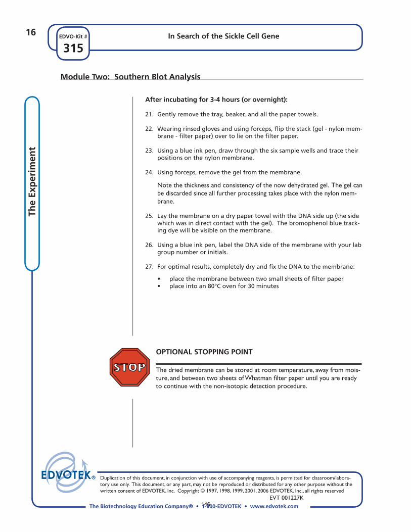
After incubating for 3-4 hours (or overnight):
21. Gently remove the tray, beaker, and all the paper towels.
22. Wearing rinsed gloves and using forceps, fl ip the stack (gel - nylon mem-brane - fi lter paper) over to lie on the fi lter paper.
23. Using a blue ink pen, draw through the six sample wells and trace their positions on the nylon membrane.
24. Using forceps, remove the gel from the membrane.
Note the thickness and consistency of the now dehydrated gel. The gel can be discarded since all further processing takes place with the nylon mem-brane.
25. Lay the membrane on a dry paper towel with the DNA side up (the side which was in direct contact with the gel). The bromophenol blue track-ing dye will be visible on the membrane.
26. Using a blue ink pen, label the DNA side of the membrane with your lab group number or initials.
27. For optimal results, completely dry and fi x the DNA to the membrane:
• place the membrane between two small sheets of fi lter paper • place into an 80°C oven for 30 minutes
OPTIONAL STOPPING POINT The dried membrane can be stored at room temperature, away from mois-ture, and between two sheets of Whatman fi lter paper until you are ready to continue with the non-isotopic detection procedure.
Module Two: Southern Blot Analysis
146

17In Search of the Sickle Cell Gene
315EDVO-Kit #
The Biotechnology Education Company® • 1-800-EDVOTEK • www.edvotek.com
Duplication of this document, in conjunction with use of accompanying reagents, is permitted for classroom/labora-tory use only. This document, or any part, may not be reproduced or distributed for any other purpose without the written consent of EDVOTEK, Inc. Copyright © 1997, 1998, 1999, 2001, 2006 EDVOTEK, Inc., all rights reserved EVT 001227K
The Exp
erimen
t
Module Two: Southern Blot Analysis
NON-ISOTOPIC DETECTION OF DNA
During this procedure you will visualize the DNA on the membrane. Blue Blot DNA Stain™ is a non-isotopic reagent, developed by EDVOTEK for class-room use, that eliminates all the associated hazards of working with radioac-tive isotopes or chemicals used in non-isotopic labeling.
28. Place the membrane with the DNA side up in 100 ml of dilute Blue-Blot™ solution.
29. Soak the membrane at room temperature for 10 to 15 minutes.
30. Remove the membrane with forceps and rinse in 200 ml of distilled wa-ter.
31. Replenish the distilled water once, or until the membrane is destained and DNA bands are clearly visible.
147

148

Edvotek315“SouthernBlot”–StudyQuestions Name:1.Sicklecelltraitandsicklecellanemiaareduetoamutationinwhatgene?Howdothegenotypesdifferbetweenthetwoconditions?2.Describehowthesicklecellgeneandproteindifferfromthewild-typeversions.3.WhatisrunonagelwhenperformingaSouthernBlot?WhataboutaNorthernBlot?4.Whatisrunonagelwhenperformingawesternblot?Whatistheprobeforawesternblot?5.WhataretherolesofHClandNaOHwhentreatingthegelafterelectrophoresis?6.Describewhatitmeanstocarryout“hybridizationwithaDNAprobe”.
149

150