the bark-beetle-associated fungus, endoconidiophora polonica, utilizes … · the...
TRANSCRIPT
The Bark-Beetle-Associated Fungus, Endoconidiophorapolonica, Utilizes the Phenolic Defense Compounds of ItsHost as a Carbon Source1[OPEN]
Namita Wadke, Dineshkumar Kandasamy, Heiko Vogel, Ljerka Lah, Brenda D. Wingfield,Christian Paetz, Louwrance P. Wright, Jonathan Gershenzon, and Almuth Hammerbacher2*
Max Planck Institute for Chemical Ecology, 07745 Jena, Germany (N.W., D.K., H.V., C.P., L.P.W., J.G., A.H.);University of Potsdam, 14476 Golm, Germany (L.L.); and Department of Genetics, Forestry and AgriculturalBiotechnology Institute, University of Pretoria, Pretoria 0028, South Africa (B.D.W.)
ORCID IDs: 0000-0003-4988-6263 (L.L.); 0000-0001-5998-6079 (L.P.W.); 0000-0002-1812-1551 (J.G.); 0000-0002-0262-2634 (A.H.).
Norway spruce (Picea abies) is periodically attacked by the bark beetle Ips typographus and its fungal associate,Endoconidiophora polonica, whose infection is thought to be required for successful beetle attack. Norway spruceproduces terpenoid resins and phenolics in response to fungal and bark beetle invasion. However, how the fungalassociate copes with these chemical defenses is still unclear. In this study, we investigated changes in the phenoliccontent of Norway spruce bark upon E. polonica infection and the biochemical factors mediating these changes.Although genes encoding the rate-limiting enzymes in Norway spruce stilbene and flavonoid biosynthesis wereactively transcribed during fungal infection, there was a significant time-dependent decline of the correspondingmetabolites in fungal lesions. In vitro feeding experiments with pure phenolics revealed that E. polonica transformsboth stilbenes and flavonoids to muconoid-type ring-cleavage products, which are likely the first steps in thedegradation of spruce defenses to substrates that can enter the tricarboxylic acid cycle. Four genes were identified inE. polonica that encode catechol dioxygenases carrying out these reactions. These enzymes catalyze the cleavage of phenolicrings with a vicinal dihydroxyl group to muconoid products accepting a wide range of Norway spruce-produced phenolicsas substrates. The expression of these genes and E. polonica utilization of the most abundant spruce phenolics as carbonsources both correlated positively with fungal virulence in several strains. Thus, the pathways for the degradation ofphenolic compounds in E. polonica, initiated by catechol dioxygenase action, are important to the infection, growth, andsurvival of this bark beetle-vectored fungus and may play a major role in the ability of I. typographus to colonize sprucetrees.
Norway spruce (Picea abies), an ecologically andeconomically important conifer in European borealand mountainous regions, is susceptible to attack bythe bark beetle Ips typographus after abiotic stress.This insect species kills mature Norway spruce treesby mass attack using aggregation pheromones toattract conspecifics (Wermelinger, 2004; Kausrud
et al., 2012; Schiebe et al., 2012). Bark beetles areassociated with various species of blue-stainingascomycete fungi that are introduced into the hostduring an attack (Kirisits, 2010). These blue-stainfungi ultimately grow into the cambium and sapwoodof trees, where they occlude the vascular bundles andnegatively affect wood quality (Kuroda, 2005). Un-der severe infestation, this can lead to tree mortal-ity. Fungal infestation is hypothesized to enhancebark beetle brood development by exhausting thetree’s defenses at an early stage during host coloni-zation (Paine et al., 1997; Krokene and Solheim, 1998,2001; Nagy et al., 2004; Franceschi et al., 2005; Zhaoet al., 2011).
Norway spruce defenses include many differentchemical compounds that are thought to protect frominsect damage andmicrobial infection. The best studiedchemical defense in this tree species are oleoresins, ablend of monoterpenenoids, sesquiterpenenoids, andditerpenenoids, stored in specialized resin ducts (Keelingand Bohlmann, 2006). Another well-studied class of de-fenses are phenolic substances, including stilbenes, flavo-noids, and proanthocyanidins, that are stored in concentric
1 This work was supported by the Jena School of Microbial Com-munication and the Max Planck Society (to J.G., N.W., and A.H.).
2 Present address: Department of Microbiology, Forestry and Ag-ricultural Biotechnology Institute, University of Pretoria, Private BagX20, Pretoria 0028, South Africa.
* Address correspondence to [email protected] author responsible for distribution of materials integral to the
findings presented in this article in accordance with the policy de-scribed in the Instructions for Authors (www.plantphysiol.org) is:Almuth Hammerbacher ([email protected]).
A.H., J.G., and L.P.W. conceived the study and procured funding;N.W., D.K., C.P., H.V., and A.H. did the experiments; B.D.W. andL.L. contributed sequencing data; L.L. constructed the phylogeny;A.H., L.P.W., and J.G. wrote the article.
[OPEN] Articles can be viewed without a subscription.www.plantphysiol.org/cgi/doi/10.1104/pp.15.01916
914 Plant Physiology�, June 2016, Vol. 171, pp. 914–931, www.plantphysiol.org � 2016 American Society of Plant Biologists. All Rights Reserved. www.plantphysiol.orgon July 11, 2018 - Published by Downloaded from
Copyright © 2016 American Society of Plant Biologists. All rights reserved.
rows of phloem parenchyma cells (also known as poly-phenolic parenchyma cells) in spruce bark (Li et al., 2012).It has been shown that concentrations of both terpenoids
and phenolics increase in spruce bark upon wounding orfungal infection (Martin et al., 2002; Hammerbacher et al.,2011, 2014).
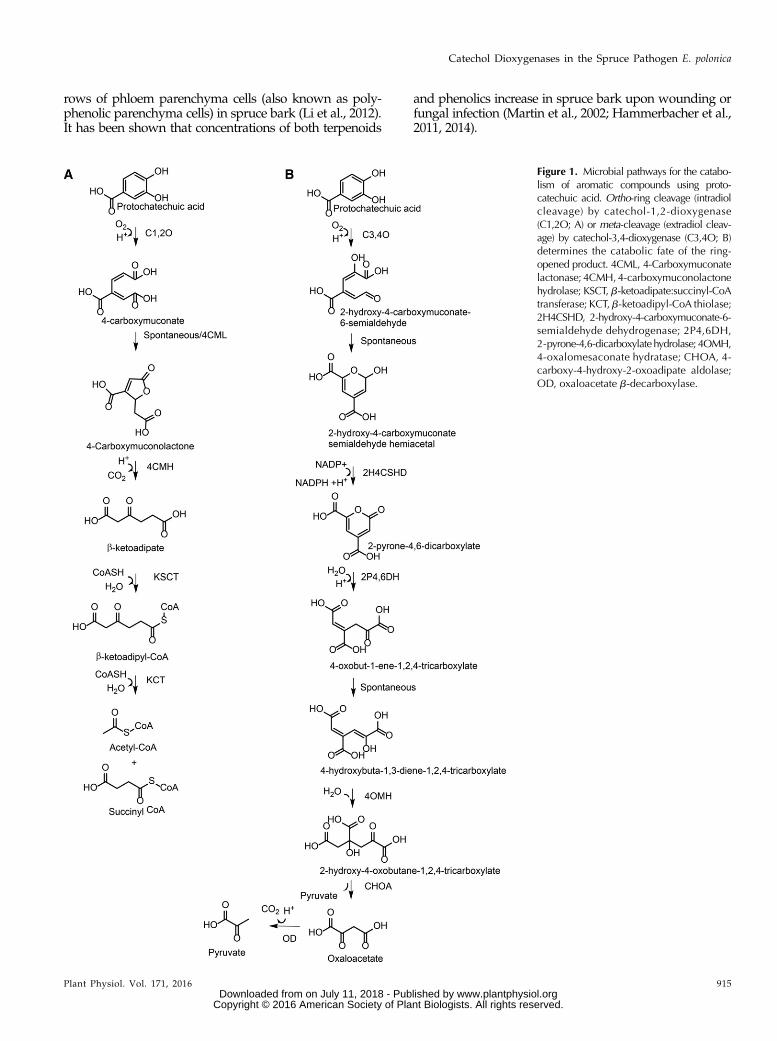
Figure 1. Microbial pathways for the catabo-lism of aromatic compounds using proto-catechuic acid. Ortho-ring cleavage (intradiolcleavage) by catechol-1,2-dioxygenase(C1,2O; A) or meta-cleavage (extradiol cleav-age) by catechol-3,4-dioxygenase (C3,4O; B)determines the catabolic fate of the ring-opened product. 4CML, 4-Carboxymuconatelactonase; 4CMH,4-carboxymuconolactonehydrolase; KSCT, b-ketoadipate:succinyl-CoAtransferase; KCT,b-ketoadipyl-CoA thiolase;2H4CSHD, 2-hydroxy-4-carboxymuconate-6-semialdehyde dehydrogenase; 2P4,6DH,2-pyrone-4,6-dicarboxylatehydrolase; 4OMH,4-oxalomesaconate hydratase; CHOA, 4-carboxy-4-hydroxy-2-oxoadipate aldolase;OD, oxaloacetate b-decarboxylase.
Plant Physiol. Vol. 171, 2016 915
Catechol Dioxygenases in the Spruce Pathogen E. polonica
www.plantphysiol.orgon July 11, 2018 - Published by Downloaded from Copyright © 2016 American Society of Plant Biologists. All rights reserved.
The mechanisms by which blue-stain fungi exhaustspruce chemical defenses are just beginning to be studied.Both terpenoids and phenolics are present at high con-centrations in healthy spruce bark and also are synthe-sized de novo upon mechanical damage and fungalinfection (Martin et al., 2002; Hammerbacher et al., 2011,2014). Recently, astringin, a stilbene glycoside present inthe concentric rows of phloem parenchyma cells, wasshown to be catabolized by Endoconidiophora polonica(Hammerbacher et al., 2013). E. polonica (formerly knownas Ceratocystis polonica; de Beer et al., 2014) is the mostvirulent blue-stain fungus transmitted by I. typographus(Krokene and Solheim, 1998). Although spruce trees stillactively synthesize astringin in fungus-infected bark, a netloss of this compound occurs within the fungal lesions(Hammerbacher et al., 2013). This reduction in astringinwas attributed to fungal biotransformation processes, in-cluding the formation of astringin dimers, aglycones, andring-cleavage products with muconoid carbon skeletons,but specific genes or enzymes involved in these processeswere not identified. It also has been reported thatGrosmannia clavigera, a blue-stain fungus associated withthe mountain pine beetle, couldmetabolize the oleoresinmonoterpene defenses of pine trees (Pinus spp.) by oxi-dative conversion (Wang et al., 2014).
Astringin-derived muconoids may represent the firststep of a catabolic pathway for the utilization of aro-matic compounds as a carbon source by blue-stainfungi in spruce, similar to the catabolic pathways de-scribed for the degradation of small aromatic compoundsby soil-dwelling bacteria and fungi (Fig. 1; Harwood andParales, 1996; Vaillancourt et al., 2006). The first step inthe catabolism of simple 3,4-dihydroxylated (catecholic)phenolic compounds by soil microbes is the productionof ring-cleaved muconoid derivatives, which is cata-lyzed by dioxygenase enzymes. Intradiol dioxygenasescleave 3,4-dihydroxylated aromatic rings between thetwo hydroxyl groups to produce a muconolactone,which is further transformed to b-ketoadipate. Afteractivation by twoCoAmolecules,b-ketoadipate isfinallyprocessed to form acetyl-CoA and succinyl-CoA. Extra-diol dioxygenase enzymes cleave the 3,4-dihydroxylatedaromatic ring adjacent to the two hydroxyl groups of thesubstrate to produce a muconate semialdehyde, which isfurther transformed to a range of organic acids includingpyruvate and oxaloacetate. Products of both pathwaysare then utilized by the tricarboxylic acid cycle for theproduction of energy-rich NADH and CO2. These path-ways have special significance in nature and human so-ciety due to their involvement in the degradation of thephenolic biopolymer lignin as well as soluble phenolics inlitter and other organic waste (Dashtban et al., 2009; Gallet al., 2014). It would be interesting to determine if plantpathogens growing on hosts containing high concentra-tions of phenolics also employ such pathways for phe-nolic degradation and exploitation as energy sources.Blue-stain fungi harboring these pathways could poten-tially be of use as bioremediators of waste affluent frompulp mills or could be exploited to degrade phenolicwaste from other industrial processes.
The degradation of phenolic compounds plays a vitalrole in the survival, growth, and virulence of plantpathogens on various herbaceous (El Hadrami et al.,2015; Lowe et al., 2015) and woody (Hammerbacheret al., 2013) host plants. Here, we continue our investi-gation of phenolic degradation in E. polonica, a blue-stain fungus transmitted by bark beetle attack. Not onlystilbenes but also flavonoids were found to be degradedin Norway spruce bark. We isolated transcripts encodingcatechol dioxygenases, enzymes with a broad substraterange that catalyze the first step in phenolic catabolism byE. polonica, converting stilbenes and flavan-3-ols to ring-cleaved muconoid products.
RESULTS
Changes in Phenolic Biosynthesis and Accumulation inNorway Spruce Bark after E. polonica Infection
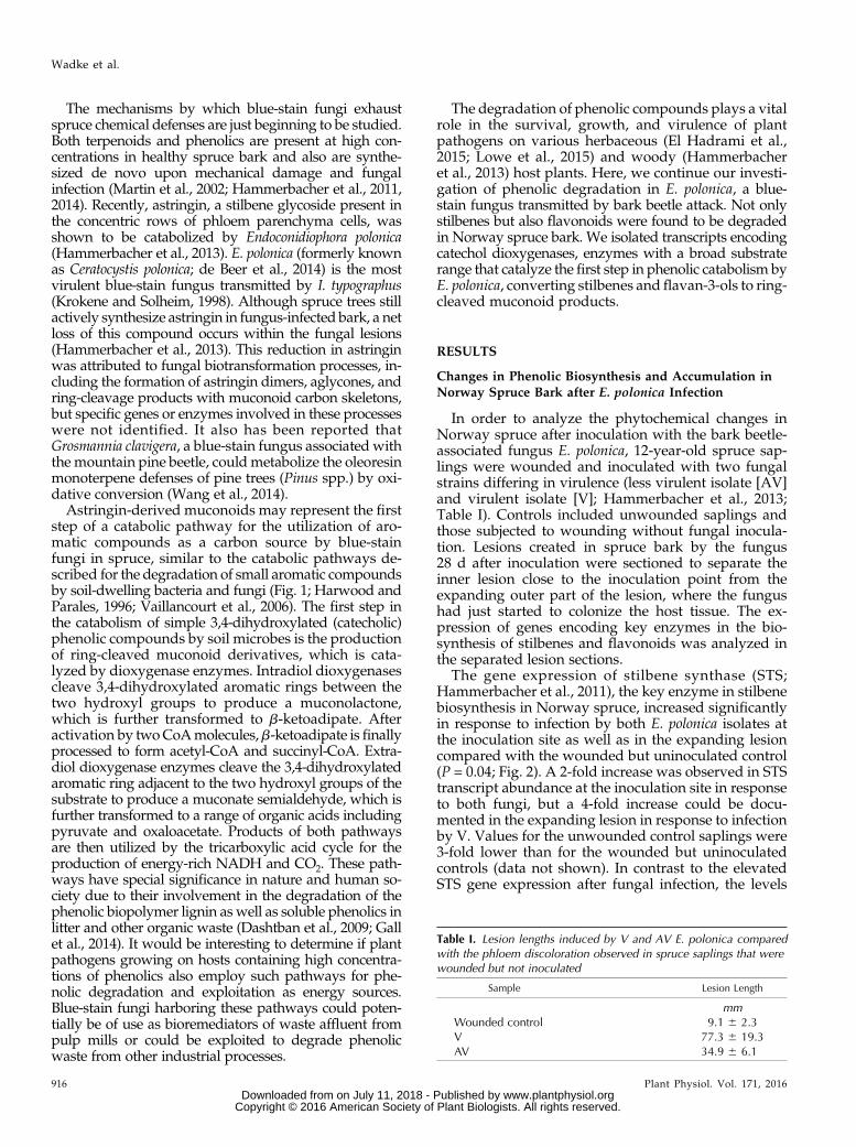
In order to analyze the phytochemical changes inNorway spruce after inoculation with the bark beetle-associated fungus E. polonica, 12-year-old spruce sap-lings were wounded and inoculated with two fungalstrains differing in virulence (less virulent isolate [AV]and virulent isolate [V]; Hammerbacher et al., 2013;Table I). Controls included unwounded saplings andthose subjected to wounding without fungal inocula-tion. Lesions created in spruce bark by the fungus28 d after inoculation were sectioned to separate theinner lesion close to the inoculation point from theexpanding outer part of the lesion, where the fungushad just started to colonize the host tissue. The ex-pression of genes encoding key enzymes in the bio-synthesis of stilbenes and flavonoids was analyzed inthe separated lesion sections.
The gene expression of stilbene synthase (STS;Hammerbacher et al., 2011), the key enzyme in stilbenebiosynthesis in Norway spruce, increased significantlyin response to infection by both E. polonica isolates atthe inoculation site as well as in the expanding lesioncompared with the wounded but uninoculated control(P = 0.04; Fig. 2). A 2-fold increase was observed in STStranscript abundance at the inoculation site in responseto both fungi, but a 4-fold increase could be docu-mented in the expanding lesion in response to infectionby V. Values for the unwounded control saplings were3-fold lower than for the wounded but uninoculatedcontrols (data not shown). In contrast to the elevatedSTS gene expression after fungal infection, the levels
Table I. Lesion lengths induced by V and AV E. polonica comparedwith the phloem discoloration observed in spruce saplings that werewounded but not inoculated
Sample Lesion Length
mmWounded control 9.1 6 2.3V 77.3 6 19.3AV 34.9 6 6.1
916 Plant Physiol. Vol. 171, 2016
Wadke et al.
www.plantphysiol.orgon July 11, 2018 - Published by Downloaded from Copyright © 2016 American Society of Plant Biologists. All rights reserved.
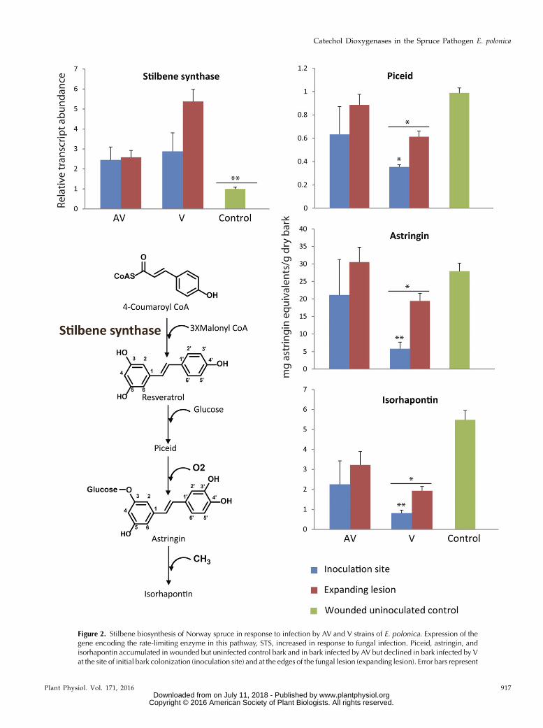
Figure 2. Stilbene biosynthesis of Norway spruce in response to infection by AV and V strains of E. polonica. Expression of thegene encoding the rate-limiting enzyme in this pathway, STS, increased in response to fungal infection. Piceid, astringin, andisorhapontin accumulated in wounded but uninfected control bark and in bark infected by AV but declined in bark infected by Vat the site of initial bark colonization (inoculation site) and at the edges of the fungal lesion (expanding lesion). Error bars represent
Plant Physiol. Vol. 171, 2016 917
Catechol Dioxygenases in the Spruce Pathogen E. polonica
www.plantphysiol.orgon July 11, 2018 - Published by Downloaded from Copyright © 2016 American Society of Plant Biologists. All rights reserved.

of the stilbene glucosides piceid, astringin, and iso-rhapontin observed in the fungal lesions were generallylower than those in the wounded but uninoculatedcontrols. These reduced metabolite levels were mostevident at the inoculation site (P = 0.04 for piceid, P =0.004 for astringin, and P = 0.003 for isorhapontin) andto a lesser degree in the expanding lesion of V. Metab-olite levels differed significantly in lesions induced by Vcompared with AV and the wounded control (P = 0.05for piceid, P = 0.01 for astringin, and P = 0.02 for iso-rhapontin). Stilbene concentrations also were lower inthe inner lesions induced by AV compared with thebark of control saplings, but these differences were notstatistically significant (Fig. 2).
Chalcone synthase (CHS) encodes an enzyme familycatalyzing the rate-limiting step in flavonoid biosyn-thesis (Hammerbacher et al., 2014). Similar to STS geneexpression, transcript accumulation of CHS increasedby more than 3-fold in the lesion sections of V (P = 0.05)and to 1.5-fold in the expanding lesion of AV (P. 0.05;Fig. 3) compared with the levels of CHS transcriptin wounded but uninoculated control saplings. Nar-ingenin, the direct product of CHS, is oxidized in twoenzymatic steps to form the dihydroflavonol, taxifolin,that occurs in high concentrations in nonwoundedspruce bark. In our experiment, lower levels of thiscompound were detected in fungus-infected bark sec-tions than in the wounded control sections (P = 0.01).Especially in the inner lesion of V close to the inocu-lation point, a very strong decrease in taxifolin wasobserved (P = 0.005). Taxifolin also serves as a sub-strate for the biosynthesis of other flavonoids inNorway spruce and is reduced in twoNADPH-dependentreduction steps to the flavan-3-ol catechin, which issubsequently polymerized to the flavan-3-ol dimer,PA B1 (Hammerbacher et al., 2014). Both catechin andPA B1 content declined in the inner lesions induced byAV and V (P, 0.001 for catechin and P = 0.1 for PA B1)compared with the expanding lesions and the woundedcontrols (P , 0.05). However, catechin levels in-creased in the expanding lesion of AV and PA B1 con-tent increased in the expanding lesions of both fungalisolates when compared with the wounded control, al-though these increases were only significant for thenonvirulent isolate (P , 0.01 for catechin and P = 0.04for PA B1; Fig. 3).
Taken together, these results show that, although thegenes encoding the rate-limiting enzymes in stilbeneand flavonoid biosynthesis are actively transcribedduring fungal infection, there are significant declines instilbene and flavonoidmetabolites in the lesions createdduring infection by E. polonica. This decline was highestin the inner lesions close to the inoculation site. Acomparison of the lesions of the two E. polonica isolates
clearly showed that, while the transcription of genesinvolved in polyphenol biosynthesis was more activein response to V, metabolite levels were lower in thelesions created by this fungus compared with thosefrom the lesions created by AV.
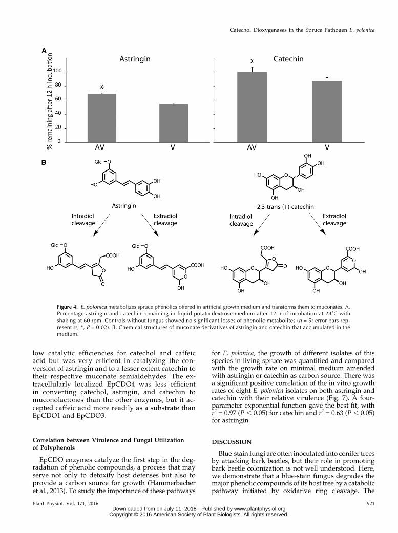
Biotransformation of Astringin and Catechin byE. polonica in Vitro
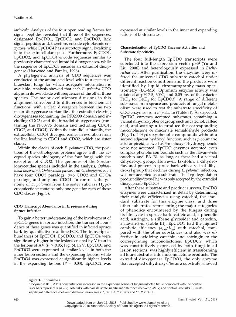
To explore the hypothesis that E. polonica is directlyresponsible for the decline of polyphenolic defensemetabolites in its spruce host, AV and V were grownin artificial medium supplemented with the stilbeneastringin or the flavan-3-ol catechin. The concentrationsof both metabolites declined in the medium 12 h afterincubation (Fig. 4A), with lower metabolite levels re-covered frommedium colonized by V (P = 0.02 for bothmetabolites) compared with AV. Biotransformation ofcatechin by both fungal isolates was lower than thatobserved for astringin (P, 0.001). For bothmetabolites,muconoid biotransformation products were recoveredfrom the medium (Fig. 4B).
The decline in metabolites in the fungus-colonizedartificial medium in this experiment effectively mirrorsthe metabolite decline observed in infected spruce bark(Figs. 2 and 3). Taken together, these results clearlyshow that E. polonica can metabolize the polyphenolicdefense compounds and that the muconoids recoveredfrom E. polonica-colonized medium represent catabo-lites of this process.
Identification and Phylogenetic Analysis of CatecholDioxygenase Genes in E. polonica and OtherBlue-Stain Fungi
To identify enzymes in E. polonica involved in thecatabolism of Norway spruce polyphenols, an EST li-brary of this fungus was sequenced using an IlluminaHiSeq 2000 sequencer (MWG Operon). A total of 42million reads were assembled into approximately 32,000contigs, from which 12 contigs were highly similar tocoding regions of catechol dioxygenase (CDO) genesfrom GenBank. CDOs are a family of enzymes catalyz-ing the cleavage of phenolic rings with a vicinal dihy-droxyl function. Further analysis showed that these 12CDO contigs could be reassembled into three partialtranscripts and one full-length transcript. The partialopen reading frames were then completed using theRACE method. To confirm the identity of these CDOsequences and verify that all CDO transcripts wereidentified in E. polonica, BLAST searches were con-ducted using the unpublished partial genomes of bothE. polonica and its closest relative, Endoconidiophora
Figure 2. (Continued.)SE (n = 5). Asterisks with bars indicate significant differences between AV, V, and control; asterisks illustrate significant differencesbetween different lesion areas: *, 0.01 , P , 0.05 and **, P , 0.01.
918 Plant Physiol. Vol. 171, 2016
Wadke et al.
www.plantphysiol.orgon July 11, 2018 - Published by Downloaded from Copyright © 2016 American Society of Plant Biologists. All rights reserved.
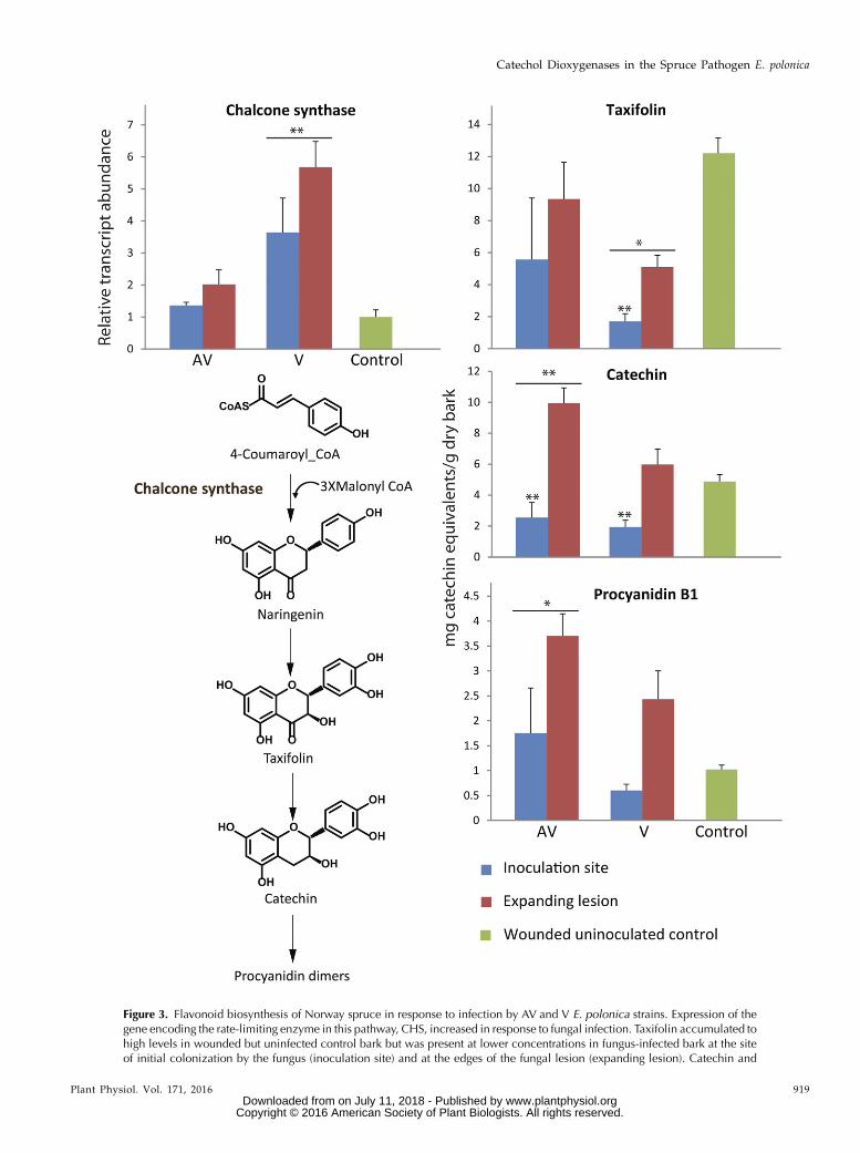
Figure 3. Flavonoid biosynthesis of Norway spruce in response to infection by AV and V E. polonica strains. Expression of thegene encoding the rate-limiting enzyme in this pathway, CHS, increased in response to fungal infection. Taxifolin accumulated tohigh levels in wounded but uninfected control bark but was present at lower concentrations in fungus-infected bark at the siteof initial colonization by the fungus (inoculation site) and at the edges of the fungal lesion (expanding lesion). Catechin and
Plant Physiol. Vol. 171, 2016 919
Catechol Dioxygenases in the Spruce Pathogen E. polonica
www.plantphysiol.orgon July 11, 2018 - Published by Downloaded from Copyright © 2016 American Society of Plant Biologists. All rights reserved.
laricicola. Analysis of the four open reading frames forsignal peptides revealed that three of the sequences,designated EpCDO1, EpCDO2, and EpCDO3, lacksignal peptides and, therefore, encode cytoplasmic en-zymes, while EpCDO4 has a secretory signal localizingit to the extracellular space. Furthermore, EpCDO1,EpCDO2, and EpCDO4 encode sequences similar topreviously characterized intradiol dioxygenases, whilethe sequence of EpCDO3 encodes an extradiol dioxy-genase (Harwood and Parales, 1996).
A phylogenetic analysis of CDO sequences wasconducted at the amino acid level with four species ofblue-stain fungi for which adequate information isavailable. Analysis showed that each E. polonica CDOaligns in its own clade with sequences of the other threespecies. The major evolutionary divisions in thisalignment correspond to differences in biochemicalfunctions, with a clear divergence between the twomajor dioxygenase subfamilies: the extradiol-cleavingdioxygenases (containing the PF02900 domain and in-cluding CDO3) and the intradiol dioxygenases (con-taining the PF00775 domain and including CDO1,CDO2, and CDO4). Within the intradiol subfamily, theextracellular CDO4 diverged earlier in evolution fromthe line leading to CDO1 and CDO2, which are sisterclades.
Within the clades of each E. polonica CDO, the posi-tions of the orthologous proteins agree with the ac-cepted species phylogeny of the four fungi, with theexception of CDO2. The genomes of the Sordar-iomycetidae species included in the analysis, Ophios-toma novo-ulmi, Ophiostoma piceae, and G. clavigera, eachhave four CDO3 paralogs, two CDO2 and CDO4paralogs, and only one CDO1. In contrast, the ge-nome of E. polonica from the sister subclass Hypo-creomicetidae contains only one gene for each of theseCDO clades (Fig. 5).
CDO Transcript Abundance in E. polonica duringSpruce Infection
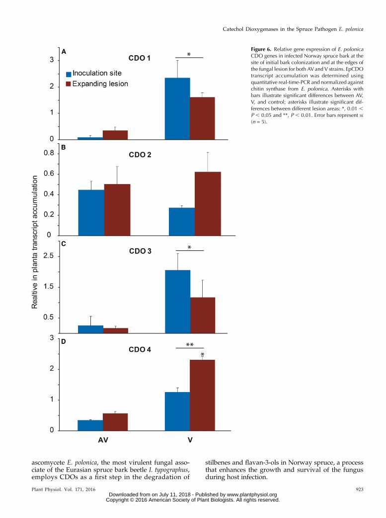
To gain a better understanding of the involvement ofEpCDO genes in spruce infection, the transcript abun-dance of these genes was quantified in infected sprucebark by quantitative real-time-PCR. The transcript a-bundances of EpCDO1, EpCDO3, and EpCDO4 weresignificantly higher in the lesions created by V than inthe lesions of AV (P . 0.05; Fig. 6). In V, EpCDO1 andEpCDO3 were expressed at similar levels in both theinner lesion sections and the expanding lesions, whileEpCDO4 was expressed at significantly higher levelsin the expanding lesions (P = 0.03). EpCDO2 was
expressed at similar levels in the inner and expandinglesions of both isolates.
Characterization of EpCDO Enzyme Activities andSubstrate Specificity
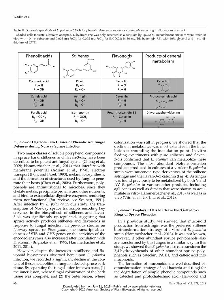
The four full-length EpCDO transcripts weresubcloned into the expression vector pH9 (Yu andLiu, 2006) and heterologously expressed in Esche-richia coli. After purification, the enzymes were of-fered the universal CDO substrate catechol underdifferent reaction conditions and the products wereidentified by liquid chromatography-mass spec-trometry (LC-MS). Optimum enzyme activity wasattained at pH 7.5, 30°C, and 0.05 mM of the cofactorFeCl3 (or FeCl2 for EpCDO3). A range of differentsubstrates from spruce and products of fungal metab-olism were used to test the substrate specificity ofCDO enzymes from E. polonica (Table II). As expected,EpCDO enzymes accepted substrates containing avicinal dihydroxyphenol group such as catechol, caffeicacid, and astringin to produce the correspondingmuconolactone or muconate semialdehyde products(Fig. 1). 4-Hydroxyphenolic compounds without asecond adjacent hydroxyl function, such as p-coumaricacid or piceid, as well as 3-methoxy-4-hydroxyphenolswere not accepted. EpCDO enzymes accepted evencomplex phenolic compounds such as the flavan-3-olscatechin and PA B1 as long as these had a vicinaldihydroxyl group. However, taxifolin, a dihydro-flavonol present in spruce bark with a vicinal dihy-droxyl group that declines during E. polonica infection,was not accepted as a substrate. The Trp degradationproduct dihydroxy-Phewas only accepted by the extradioldioxygenase EpCDO3.
After these substrate and product surveys, EpCDOenzymes were characterized in detail by determiningtheir catalytic efficiencies using catechol, the stan-dard substrate for this enzyme class, and threeother substrates representing the major categoriesof phenolics encountered by the fungus duringits life cycle in spruce bark: caffeic acid, a phenolicacid; astringin, a stilbene glycoside; and catechin,a flavan-3-ol (Table III). EpCDO1 had the highestcatalytic efficiency (kcat/Km) with catechol, com-pared with the other substances, and also was ef-fective in oxidizing catechin and astringin to thecorresponding muconolactones. EpCDO2, whichwas constitutively expressed by both fungi in alllesion sections, was highly efficient in transformingall four substrates into muconolactone products. Theextradiol dioxygenase EpCDO3, the only enzymethat accepted dihydroxy-Phe as a substrate, showed
Figure 3. (Continued.)procyanidin B1 (PA B1) concentrations increased in the expanding lesion of fungus-infected tissue compared with the control.Error bars represent SE (n = 5). Asterisks with bars illustrate significant differences between AV, V, and control; asterisks illustratesignificant differences between different lesion areas: *, 0.01 , P , 0.05 and **, P , 0.01.
920 Plant Physiol. Vol. 171, 2016
Wadke et al.
www.plantphysiol.orgon July 11, 2018 - Published by Downloaded from Copyright © 2016 American Society of Plant Biologists. All rights reserved.
low catalytic efficiencies for catechol and caffeicacid but was very efficient in catalyzing the con-version of astringin and to a lesser extent catechin totheir respective muconate semialdehydes. The ex-tracellularly localized EpCDO4 was less efficientin converting catechol, astingin, and catechin tomuconolactones than the other enzymes, but it ac-cepted caffeic acid more readily as a substrate thanEpCDO1 and EpCDO3.
Correlation between Virulence and Fungal Utilizationof Polyphenols
EpCDO enzymes catalyze the first step in the deg-radation of phenolic compounds, a process that mayserve not only to detoxify host defenses but also toprovide a carbon source for growth (Hammerbacheret al., 2013). To study the importance of these pathways
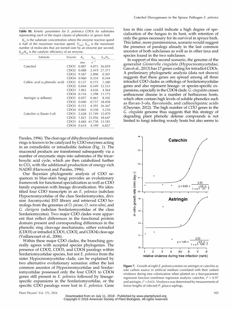
for E. polonica, the growth of different isolates of thisspecies in living spruce was quantified and comparedwith the growth rate on minimal medium amendedwith astringin or catechin as carbon source. There wasa significant positive correlation of the in vitro growthrates of eight E. polonica isolates on both astringin andcatechin with their relative virulence (Fig. 7). A four-parameter exponential function gave the best fit, withr2 = 0.97 (P , 0.05) for catechin and r2 = 0.63 (P , 0.05)for astringin.
DISCUSSION
Blue-stain fungi are often inoculated into conifer treesby attacking bark beetles, but their role in promotingbark beetle colonization is not well understood. Here,we demonstrate that a blue-stain fungus degrades themajor phenolic compounds of its host tree by a catabolicpathway initiated by oxidative ring cleavage. The
Figure 4. E. polonica metabolizes spruce phenolics offered in artificial growth medium and transforms them to muconates. A,Percentage astringin and catechin remaining in liquid potato dextrose medium after 12 h of incubation at 24˚C withshaking at 60 rpm. Controls without fungus showed no significant losses of phenolic metabolites (n = 5; error bars rep-resent SE; *, P = 0.02). B, Chemical structures of muconate derivatives of astringin and catechin that accumulated in themedium.
Plant Physiol. Vol. 171, 2016 921
Catechol Dioxygenases in the Spruce Pathogen E. polonica
www.plantphysiol.orgon July 11, 2018 - Published by Downloaded from Copyright © 2016 American Society of Plant Biologists. All rights reserved.
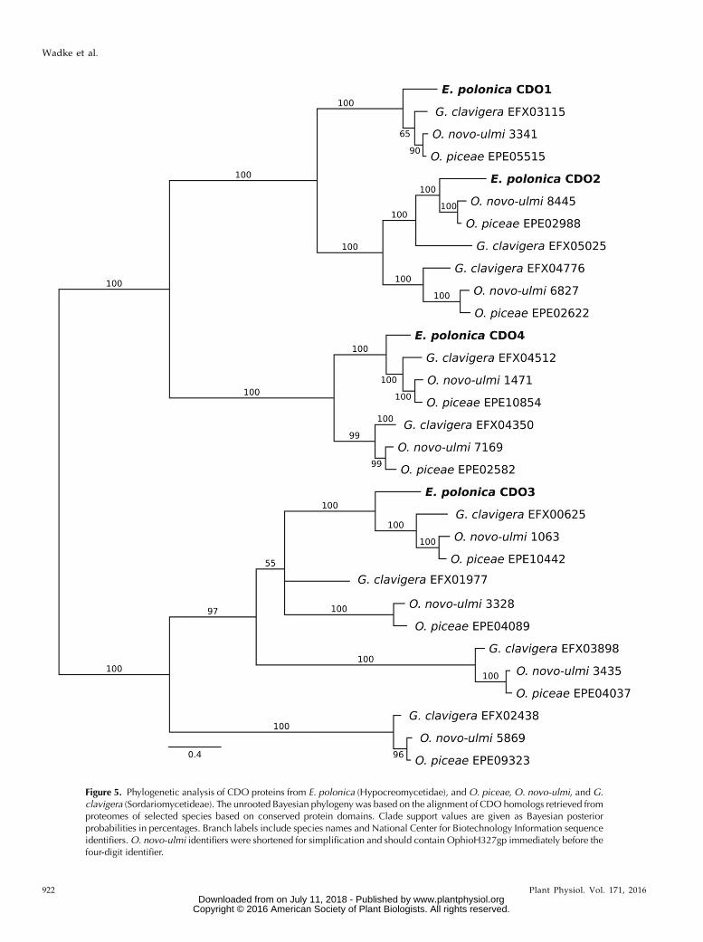
Figure 5. Phylogenetic analysis of CDO proteins from E. polonica (Hypocreomycetidae), and O. piceae, O. novo-ulmi, and G.clavigera (Sordariomycetideae). The unrooted Bayesian phylogeny was based on the alignment of CDO homologs retrieved fromproteomes of selected species based on conserved protein domains. Clade support values are given as Bayesian posteriorprobabilities in percentages. Branch labels include species names and National Center for Biotechnology Information sequenceidentifiers.O. novo-ulmi identifiers were shortened for simplification and should contain OphioH327gp immediately before thefour-digit identifier.
922 Plant Physiol. Vol. 171, 2016
Wadke et al.
www.plantphysiol.orgon July 11, 2018 - Published by Downloaded from Copyright © 2016 American Society of Plant Biologists. All rights reserved.
ascomycete E. polonica, the most virulent fungal asso-ciate of the Eurasian spruce bark beetle I. typographus,employs CDOs as a first step in the degradation of
stilbenes and flavan-3-ols in Norway spruce, a processthat enhances the growth and survival of the fungusduring host infection.
Figure 6. Relative gene expression of E. polonicaCDO genes in infected Norway spruce bark at thesite of initial bark colonization and at the edges ofthe fungal lesion for both AVand V strains. EpCDOtranscript accumulation was determined usingquantitative real-time-PCR and normalized againstchitin synthase from E. polonica. Asterisks withbars illustrate significant differences between AV,V, and control; asterisks illustrate significant dif-ferences between different lesion areas: *, 0.01 ,P , 0.05 and **, P , 0.01. Error bars represent SE
(n = 5).
Plant Physiol. Vol. 171, 2016 923
Catechol Dioxygenases in the Spruce Pathogen E. polonica
www.plantphysiol.orgon July 11, 2018 - Published by Downloaded from Copyright © 2016 American Society of Plant Biologists. All rights reserved.
E. polonica Degrades Two Classes of Phenolic AntifungalDefenses during Norway Spruce Infection
Twomajor classes of soluble polyphenol compoundsin spruce bark, stilbenes and flavan-3-ols, have beendescribed to be potent antifungal agents (Chong et al.,2009; Hammerbacher et al., 2014) that interfere withmembrane potential (Adrian et al., 1998), electrontransport (Pont and Pezet, 1990), melanin biosynthesis,and the formation of structures used by fungi to pene-trate their hosts (Chen et al., 2006). Furthermore, poly-phenols are antinutritional to microbes, since theychelate metals, precipitate proteins and other nutrients,and bind to extracellular digestive enzymes, renderingthem nonfunctional (for review, see Scalbert, 1991).After infection by E. polonica in our study, the tran-scription of Norway spruce transcripts encoding keyenzymes in the biosynthesis of stilbenes and flavan-3-ols was significantly up-regulated, suggesting thatspruce actively produces stilbenes and flavonoids inresponse to fungal infection. In previous studies onNorway spruce or Picea glauca, the transcript abun-dances of STS and CHS genes or the activities of theencoded enzymes also increased after inoculation withE. polonica (Brignolas et al., 1995; Hammerbacher et al.,2011, 2014).
However, despite the increases in stilbene and fla-vonoid biosynthesis observed here upon E. polonicainfection, we recorded a significant decline in the con-tent of these metabolites in fungus-infected spruce barktissue. By separating the fungal lesion into two parts, (1)the inner lesion, where fungal colonization of the barktissue was complete, and (2) the outer lesion, where
colonization was still in progress, we showed that thedecline in metabolites was most extensive in the innerlesion surrounding the inoculation point. In vitrofeeding experiments with pure stilbenes and flavan-3-ols confirmed that E. polonica can metabolize thesecompounds. The most abundant biotransformationproducts produced in cultures of a virulent E. polonicastrain were muconoid-type derivatives of the stilbeneastringin and the flavan-3-ol catechin (Fig. 4). Astringinwas found previously to be metabolized by both V andAV E. polonica to various other products, includingaglucones as well as dimers that were shown to accu-mulate in vitro (Hammerbacher et al., 2013) aswell as invivo (Viiri et al., 2001; Li et al., 2012).
E. polonica Employs CDOs to Cleave the 3,4-HydroxyRings of Spruce Phenolics
In a previous study, we showed that muconoidproduction from astringin was the preferred stilbenebiotransformation strategy of a virulent E. polonicastrain (Hammerbacher et al., 2013). It was not known,however, if other abundant spruce polyphenols alsoare transformed by this fungus in a similar way. In thisstudy, we showed that E. polonica also can transform the3,4-hydroxyphenols of other abundant spruce poly-phenols such as catechin, PA B1, and caffeic acid intomuconoids.
The formation of muconoids is a well-described bi-otransformation strategy of soil bacteria and fungi forthe degradation of simple phenolic compounds suchas catechol and protochatechuic acid (Harwood and
Table II. Substrate specificity of E. polonica CDOs for phenolic defense compounds commonly occurring in Norway spruce bark
Shaded cells indicate substrates accepted. Dihydroxy-Phe was only accepted as a substrate by EpCDO3. Recombinant enzymes were tested invitro with 10 mM substrate and 0.005 mM FeCl3 (or 0.005 mM FeCl2 for EpCDO3) in 50 mM Tris buffer, pH 7.5, with 10% glycerol and 1 mM di-thiothreitol (DTT).
924 Plant Physiol. Vol. 171, 2016
Wadke et al.
www.plantphysiol.orgon July 11, 2018 - Published by Downloaded from Copyright © 2016 American Society of Plant Biologists. All rights reserved.
Parales, 1996). The cleavage of dihydroxylated aromaticrings is known to be catalyzed by CDO enzymes actingin an extradiolitic or intradiolitic fashion (Fig. 1). Themuconoid products are transformed subsequently via anumber of enzymatic steps into substrates of the tricar-boxylic acid cycle, which are then catabolized furtherto CO2 with the additional production of energy-richNADH (Harwood and Parales, 1996).Our Bayesian phylogenetic analysis of CDO se-
quences in blue-stain fungi provides an evolutionaryframework for functional specialization as well as CDOfamily expansion with lineage diversification. We iden-tified four CDO transcripts in an E. polonica (subclassHypocreomycetidae of the class Sordariomycetes, divi-sion Ascomycota) EST library and retrieved CDO ho-mologs from the genomes of O. piceae, O. novo-ulmi, andG. clavigera (subclass Sordariomycetidae of the classSordariomycetes). Two major CDO clades were appar-ent that reflect differences in the functional proteindomain present and corresponding differences in thephenolic ring cleavage mechanisms, either extradiol(CDO3) or intradiol (CDO1, CDO2, and CDO4) cleavage(Vaillancourt et al., 2006).Within these major CDO clades, the branching gen-
erally agrees with accepted species phylogenies. Thepresence of CDO2, CDO3, and CDO4 paralogs withinSordariomycetidae species, but not E. polonica from thesister Hypocreomycetidae clade, can be explained bytwo alternative evolutionary scenarios: either the lastcommon ancestor of Hypocreomycetidae and Sordar-iomycetidae possessed only the four CDO1 to CDO4genes still present in E. polonica followed by lineage-specific expansions in the Sordariomycetidae, or thespecific CDO paralogs were lost in E. polonica. Gene
loss in this case could indicate a high degree of spe-cialization of the fungus to its host, with retention ofonly the genes necessary for its survival in spruce bark.This latter, more parsimonious, scenario would suggestthe presence of paralogs already in the last commonancestor of both subclasses as well as in other taxa andspecies found in the two subclasses.
In support of this second scenario, the genome of thegeneralist Glomerella cingulata (Hypocreomycetidae;Gan et al., 2013) has 17 genes coding for intradiol CDOs.A preliminary phylogenetic analysis (data not shown)suggests that these genes are spread among all threeintradiol CDO clades as orthologs of Sordariomycetidaegenes and also represent lineage- or species-specific ex-pansions, especially in theCDO4 clade.G. cingulata causesanthracnose disease in a number of herbaceous hosts,which often contain high levels of soluble phenolics suchas flavan-3-ols, flavonoids, and caffeoylquinic acids(Cheynier, 2012). The high number of CDO genes in theG. cingulata genome thus suggests that this strategy ofdegrading plant phenolic defense compounds is notlimited to fungi infecting woody hosts but also seems to
Figure 7. Growth of eight E. polonica strains on astringin or catechin assole carbon source in artificial medium correlated with their rankedvirulence during tree colonization when plotted on a four-parameterregression function (nonlinear regression analysis: catechin, r2 = 0.97and astringin, r2 = 0.63). Virulence was determined bymeasurements oflesion lengths of infected P. glauca saplings.
Table III. Kinetic parameters for E. polonica CDOs for substratesrepresenting each of the major classes of phenolics in spruce bark
Km is the substrate concentration where the enzyme reaction speedis half of the maximum reaction speed, Vmax; kcat is the maximumnumber of molecules that are turned over by an enzyme per second;kcat/Km is the catalytic efficiency of an enzyme.
Substrate Enzyme Km kcat kcat/Km
mM s21
Catechol CDO1 0.087 4.873 56.029CDO2 0.088 2.419 27.371CDO3 9.587 2.890 0.301CDO4 0.960 0.234 0.244
Caffeic acid (a phenolic acid) CDO1 0.137 0.175 1.280CDO2 0.044 0.549 12.533CDO3 1.063 4.636 4.364CDO4 0.116 1.298 11.175
Astringin (a stilbene) CDO1 0.057 0.461 8.102CDO2 0.040 0.737 18.458CDO3 0.121 4.391 36.367CDO4 0.065 0.338 5.210
Catechin (a flavan-3-ol) CDO1 2.628 31.749 12.079CDO2 1.821 33.956 18.647CDO3 4.684 61.738 13.181CDO4 0.614 4.199 6.837
Plant Physiol. Vol. 171, 2016 925
Catechol Dioxygenases in the Spruce Pathogen E. polonica
www.plantphysiol.orgon July 11, 2018 - Published by Downloaded from Copyright © 2016 American Society of Plant Biologists. All rights reserved.
be a common trait of plant pathogenic fungi infectingherbaceous species.
The heterologously expressed EpCDO genes all ac-cepted catechol and protocatechuic acid as substratesand also converted more complex phenolic compoundssuch as caffeic acid, astringin, catechin, and procyanidindimers to muconoid products. Interestingly, althoughthese enzymes readily accepted complex flavan-3-ols,such as PA B1, as substrates, they could not convert fla-vonols and dihydroflavonols to the respective muco-noids. Thus, the depletion of the dihydroflavonol taxifolinin spruce bark colonized by E. polonica is not due to thedirect action of CDO enzymes. E. polonicamight thus usean alternative biodegradation strategy for taxifolin, forexample startingwith an initial cleavage of the C-ring andfurther degradation to protocatechuic acid, as has beendescribed for Pseudomonas putida (Pillai and Swarup,2002).
As expected, EpCDO enzymes also did not cleaveany compounds lacking 3,4-dihydroxylated rings, suchas the stilbenes, piceid, and isorhapontin. Yet, theconcentrations of these compounds also were signifi-cantly lowered in fungus-infected bark, indicating thatE. polonica must have other biotransformation strategiesto catabolize these phenolic rings. For example, it hasbeen shown that E. polonica can polymerize stilbenes toform compounds that are less soluble in aqueous so-lution (Hammerbacher et al., 2013). Alternatively, a 3,4-dihydroxyl function in a phenolic ring system can beformed in situ from other substitution patterns, yield-ing compounds that are then subject to extradiol orintradiol cleaving CDOs. For example, bacteria havebeen shown to demethylate the methoxyl functions incompounds such as vanillin to yield dihydroxylatedproducts that can serve as substrates for ring-openingdioxygenases (Chen et al., 2012; Lowe et al., 2015). Asimilar process could be involved in the degradationof ferulic acid and isorhapontin. Furthermore, microbescan oxidize unsubstituted phenolic rings such as that ofbenzoic acid to form a 3,4-dihydroxylated productsuitable for CDO cleavage (Suenaga et al., 2009). Theoxidation of phenolic rings might thus play an impor-tant role in the catabolism of resveratrol or coumaricacid by E. polonica.
When the rate of reaction for EpCDO enzymes wascompared using a range of different substrates thatmight be encountered by the fungus during its life cyclein Norway spruce, EpCDO1 was found to be highlyefficient in transforming catechol to muconolactone,but it had lower catalytic efficiencies for transformingmore complex phenolics. This suggests that this en-zyme is similar to the CDO enzymes described forPseudomonas spp., which have narrow substrate speci-ficities and high catalytic efficiencies for transforminga single substrate (Pillai and Swarup, 2002). Theintradiol-cleaving EpCDO2, on the other hand, hadvery broad substrate specificity, as it effectively trans-formed a range of polyphenols to the correspondingring-opened lactones. The extradiol-cleaving EpCDO3,which also accepted the nitrogenous compound
dihydroxy-Phe, had a low catalytic efficiency for cate-chol and caffeic acid but was extremely efficient inutilizing astringin and catechin. As EpCDO1, EpCDO2,and EpCDO3 are localized in the cytoplasm ofE. polonica, they also might be involved in the catabo-lism of cellular constituents such as aromatic aminoacids (e.g. dihydroxy-Phe). Their ability to effectivelytransform and detoxify defense-related polyphenolsthat diffuse into the cell and that may interfere withfungal metabolism and ultimately growth might rep-resent a very important fungal adaptation for survivalin polyphenol-rich Norway spruce bark tissue. Thishypothesis is further supported by the fact that genesencoding EpCDO1, EpCDO2, and EpCDO3 wereexpressed at similar levels at the inoculation site and theexpanding lesions.
EpCDO4was the only enzyme from E. polonicawith asignal peptide localizing it to the extracellular space.This enzyme preferentially accepted the phenolic acid,caffeic acid, which in its methylated and reduced formis an important component of conifer lignin. Blue-stainfungi grow from their initial point of infection in thebark into the wood of conifer trees (Krokene andSolheim, 2001). Therefore, it is possible that EpCDO4 isinvolved in the catabolism of accessible cell wall con-stituents in the wood and plays a role in fungal pene-tration and colonization of woody tissue togetherwith other oxidative processes. The EpCDO4 gene wasexpressed by E. polonica at higher levels in the expandinglesions than in the inner lesion, which also supportsits hypothetical role in fungal colonization of woodytissue.
To compare the substrate usage of CDOs, we quan-tified assay products by LC-MS methods rather thanmeasuring the consumption of catechol substrate col-orimetrically (Gulvik and Buchan, 2013) or the con-sumption of oxygen using a Clarke electrode (Haigleret al., 1992), as has sometimes been done previously.However, determining the kinetic parameters of theseenzymes is complicated by the tendency of phenolicsubstrates to precipitate proteins under acidic condi-tions or to attack nucleophilic amino acid residues ofthe protein under basic conditions, leading to an over-estimation of the concentration of active protein in theassay. Polyphenols also chelate the iron cofactor ofEpCDO enzymes in the reaction mix, which influencescofactor concentrations as well as the concentrations ofsoluble phenolic substrates in the assay. However, evenif the kinetic parameters are not completely exact, webelieve that our results give an accurate estimation ofthe differences in substrate specificity among the dif-ferent CDO types.
Degradation of Spruce Phenolics Is an ImportantVirulence Factor for E. polonica during Tree Infectionand Colonization
It was shown recently that polyphenol catabolism isof great importance for both the bacterium Ralstonia
926 Plant Physiol. Vol. 171, 2016
Wadke et al.
www.plantphysiol.orgon July 11, 2018 - Published by Downloaded from Copyright © 2016 American Society of Plant Biologists. All rights reserved.

solanacearum (Lowe et al., 2015) and the fungus Verti-cillium dahliae (El Hadrami et al., 2015) in gaining entryinto their herbaceous plant hosts as well as in detoxi-fying defenses. Our study presents evidence thatpolyphenol degradation is also of key importance forE. polonica infection and colonization of its woody host,Norway spruce. The direct correlation between the invitro growth rate of E. polonica strains on astringin andcatechin as sole carbon source and their virulenceduring spruce bark infection shows the importance ofpolyphenol degradation in nutrient acquisition. How-ever, since these substances also are known as potentphytoalexins and phytoanticipins in other plant-pathogen interactions, it is likely that their catabolismfunctions at least in part as a detoxification mechanism.As E. polonica colonizes lignified bark and wood,polyphenol degradation also may serve to help gainentry into woody tissue. EpCDO4 may be especiallyimportant in this regard, as it is localized in the ex-tracellular space and utilizes the lignin precursorcaffeic acid at a higher efficiency than other sprucepolyphenols.E. polonica and other blue-stain fungi are commonly
thought to play an important role in tree colonizationby the European spruce bark beetle I. typographus byexhausting the spruce tree’s chemical defense mecha-nisms (Krokene and Solheim, 1998, 2001; Nagy et al.,2004; Franceschi et al., 2005; Zhao et al., 2011). Althoughthis opinion is not universally accepted (Six andWingfield, 2011), our data suggest that polyphenoldegradation could remove potentially harmful chemi-cals from the food of bark beetle larvae. Polyphenols areknown to be antinutritional compounds that can formreactive quinones that react with constituents of insectcells (Barbehenn et al., 2008) and can precipitate pro-teins, making them inaccessible for digestion (Scalbert,1991). The nutritional benefits associated with poly-phenol biotransformation by E. polonica, therefore,might not only benefit the fungus but might drive themutualistic interaction with its vector, I. typographus.
MATERIALS AND METHODS
Inoculation of Norway Spruce Saplings with E. polonica
Two Endoconidiophora polonica isolates (CMW 7749 = AV and CMW 7135 =V) provided by the culture collection of the Forestry and Agricultural Bio-technology Institute, University of Pretoria, were grown on 2% (w/v) maltextract agar (Carl Roth) for 12 d at 25°C in the dark.
Twelve-year-old Norway spruce (Picea abies) saplings originating from the3369-Schongau clone (Samenklenge und Pflanzengarten) were grown in anoutdoor plot for 8 years prior to the experiment. Inoculations of saplings withE. polonica were performed 3 weeks after their spring flush. A bark plug, 8 mmin diameter, was removed between the second and third branch whorl from theupper part of the sapling with a cork borer. An 8-mm plug from one of the twoE. polonica cultures was placed into the wound with the mycelium orientedtoward the wood surface and sealed with Parafilm. For the wounded controltreatment, bark plugs were removed and a plug of sterile malt extract agar wasinserted into the wound.
Bark tissue samples from inoculated and wounded control saplings wereharvested 28 d after the onset of the experiment. The fungal lesions were sep-arated into two parts: the inner lesion close to the inoculation point (about
18 mm2) and the expanding outer lesion (pooled sample). Five replicate treeswere used for each treatment (inoculated with AV, inoculated with V, andwounded control). Lesion sections were flash frozen immediately after harvestin liquid nitrogen and stored at 280°C.
Quantitative Real-Time PCR of Plant andFungal Transcripts
Total RNA from the inner and outer lesions and from wounded but unin-oculated control bark was isolated with the Invitrap Spin Plant RNA Mini Kit(Stratec) following the protocols of the manufacturer except that an additionalDNA digestion step was included (RNase Free DNase set; Qiagen). RNA wasquantified by spectrophotometry. Reverse transcription of 1 mg of RNA intocomplementary DNA (cDNA) was achieved using SuperScript II reversetranscriptase (Invitrogen) and 50 pmol of PolyT(12) primer (Invitrogen) in a re-action volume of 20 mL. After cDNA was diluted to 10% (v/v) with deionizedwater, 1 mL of diluted cDNA was used as a template for quantitative real-timePCR in a reaction mixture containing Brilliant SYBR Green QRT-PCR MasterMix (Stratagene) and 10 pmol of forward and 10 pmol of reverse primers.Primer sequences for PaSTS, PaCHS, and EpCDO1 to EpCDO4 are given inSupplemental Table S2. PCR was performed using a Stratagene MX3000Pthermocycler using the following cycling parameters: 5 min at 95°C, followedby 40 cycles of 30 s at 95°C, 30 s at 55°C, and 30 s at 72°C, followed by a meltingcurve analysis from 55°C to 95°C. Reaction controls included nontemplatecontrols as well as non-reverse-transcribed RNA.
The primer pair chosen for PaSTS amplified transcripts of both orthologoustranscripts present in the Norway spruce genome (for description, seeHammerbacher et al., 2011). The primer pair designed for PaCHS could amplifyseven of the nine unique CHS transcripts identified in spruce (Hammerbacheret al., 2014). The transcript abundances of PaSTS and PaCHS were normalizedto the transcript abundance of the ubiquitin transcript (primers in SupplementalTable S2; verified by Schmidt et al., 2010) and were calculated from threetechnical replicates each of five biological replicates. Relative transcript abun-dance was calibrated against the transcript abundance of five nonwoundedcontrol saplings.
The transcript abundance of EpCDO genes was normalized to the transcriptabundance of the E. polonica CHITIN SYNTHASE1 transcript (primers inSupplemental Table S2) and was calculated from three technical replicates offive biological replicates. Relative transcript abundance was calibrated againstthe transcript abundance of a single replicate from an expanding lesion pro-duced by V. The stability of the normalizing transcript was assessed by theamplification of eight normalizing transcript candidates using cDNA derivedfrom E. polonica grown under different conditions and analysis of the resultsusing the program GNorm (https://genorm.cmgg.be). The average expressionstability for chitin synthase was calculated to be 0.145.
Extraction of Phenolic Compounds from Spruce
For the extractionof phenolic compounds,Norway spruce tissuewas groundto a fine powder in liquid nitrogen and lyophilized at 0.34 mbar pressure usingan Alpha 1-4 LD Plus freeze dryer (Martin Christ). Approximately 20 mg ofdried tissue was extracted for 4 h at 4°C with 1 mL of analytical grade methanolcontaining 100 mg mL21 apigenin-7-glucoside (Carl Roth) as an internal stan-dard. The extract was centrifuged at 3,200g, and the supernatant was recovered.Insoluble material was reextracted with 0.8 mL of methanol for 16 h. Super-natants were combined and analyzed directly by LC-MS.
Analysis of Phenolic Compounds by LC-MS withElectrospray Ionization
Phenolic compounds from spruce were separated on a Nucleodur SphinxRP18ec column with dimensions of 250 3 4.6 mm and a particle size of 5 mm(Macherey-Nagel) using an Agilent 1100 series HPLC device (Agilent Tech-nologies) with a flow rate of 1 mL min21. The column temperature was main-tained at 25°C. Phenolic compounds were separated using 0.2% (v/v) formicacid and acetonitrile as mobile phases A and B, respectively, with the followingelution profile: 0 to 1 min, 100% A; 1 to 25 min, 0% to 65% B in A; 25 to 28 min,100% B; and 28 to 32 min, 100% A.
Compound detection and quantification was accomplished with an Esquire6000 ESI ion-trap mass spectrometer (Bruker Daltonics). Flow coming from thecolumn was diverted in a ratio of 4:1 before entering the mass spectrometer
Plant Physiol. Vol. 171, 2016 927
Catechol Dioxygenases in the Spruce Pathogen E. polonica
www.plantphysiol.orgon July 11, 2018 - Published by Downloaded from Copyright © 2016 American Society of Plant Biologists. All rights reserved.
electrospray chamber. Electrospray ionization-mass spectrometry was per-formed in negative mode scanning mass-to-charge ratio between 50 and 1,600with an optimal target mass-to-charge ratio of 405. The mass spectrometer wasoperated using the following specifications: skimmer voltage, 60 V; capillaryvoltage, 4,200 V; nebulizer pressure, 35 p.s.i.; drying gas, 11 L min21; and gastemperature, 330°C. Capillary exit potential was kept at 2121 V.
Compounds were identified by fragmentation patterns and by direct com-parison of retention time and mass spectrum with those of available commercialstandards, including 2,3-trans(+)-catechin, proanthocyanidin B1, taxifolin, andpiceid (Sigma). Brucker Daltonics Quant Analysis version 3.4 software wasused for data processing and compound quantification using a standardsmoothing width of 3 and Peak Detection Algorithm version 2. Linearity inionization efficiencies was verified by analyzing serial dilutions of randomlyselected samples. An external calibration curve created by linear regression wasused for the quantification of 2,3-trans(+)-catechin (Sigma) and astringin. Allquantifications were adjusted relative to the signal for the internal standard.
Large-Scale Isolation of Astringin from Spruce Bark
Astringin was isolated by extracting 500 g of dried spruce bark ground to afine powder with methanol. The methanol was evaporated completely, and theextract was partially solubilized in 100 mL of water using an ultrasonic waterbath and filtered. A portion (20 mL) of the aqueous solution was loaded on aSephadex LH20 (GE Healthcare) column with dimensions of 2.5 cm 3 30 cm.The compounds were eluted from the column using water at a flow speed of1mLmin21. Fractions (15mL)were collected and analyzed for astringin contentby direct injection into a mass spectrometer using an autosampler from anAgilent 1200 HPLC system (Agilent Technologies) coupled to an API 3200tandem mass spectrometer (Applied Biosystems) equipped with a turbosprayion source operating in negative ionization mode. Injection volume was 5 mLusing flow injection analysis. The mobile phase consisted of 0.05% formic acid(A) and acetonitrile (B) utilizing a flow of 1 mL min21. Phase A was maintainedat 50% for 0.5min. The ion spray voltagewasmaintained at24.5 keV. The turbogas temperature was set at 600°C. Nebulizing gas was set at 50 p.s.i., curtain gasat 30 p.s.i., heating gas at 60 p.s.i., and collision gas at 5 p.s.i. Multiple reactionmonitoring was used to monitor analyte parent ion→ product ion as describedin Supplemental Table S1. Pure fractions were pooled and lyophilized using afreeze dryer.
Fungal Biotransformation of Catechin and Astringin
Fungal isolates V andAVwere grown on potato dextrose agar in petri dishesfor 7 d. Agar plugs (2 mm diameter) from these cultures were inoculated into2mL of potato dextrose broth containing astringin or catechin (Sigma; 1mgmL21).After 12 h, the liquid broth of the fungal cultures was sampled and analyzed byliquid chromatography-electrospray ionization-tandem mass spectrometry(LC-ESI-MS/MS) for astringin and catechin as well as catabolites of thesecompounds. Five replicates were used per treatment. Controls included fungalcultures treated with water instead of the phenolic compounds as well as sterilemedium containing astringin or catechin.
LC-ESI-MS/MS of Fungal Biotransformation Products ofAstringin and Catechin
Chromatography was performed on an Agilent 1200 HPLC system (AgilentTechnologies). Separation was achieved on a 100- 3 4.6-mm Kinetex C18 col-umn with particle size of 2.6 mm (Phenomenex). Formic acid (0.05%) in waterand acetonitrile were employed as mobile phases A and B, respectively. Theelution profile was as follows: 0 to 1min, 100%A; 1 to 7min, 0% to 65% B in A; 7to 8 min, 65% to 100% B in A; 8 to 9 min, 100% B; and 9 to 10 min, 100% A. Thetotal mobile phase flow rate was 1.5 mL min21. The column temperature wasmaintained at 25°C.
An API 3200 tandem mass spectrometer (Applied Biosystems) equippedwith a Turbospray ion source was operated in negative ionization mode. Theinstrument parameters were optimized by infusion experiments with purestandards of catechin, catechin lactone, astringin, piceatannol, and astringinmuconate. The ion spray voltage was maintained at 24,500 V. The turbo gastemperature was set at 700°C. Nebulizing gas was set at 70 p.s.i., curtain gas at25 p.s.i., heating gas at 60 p.s.i., and collision gas at 10 p.s.i. Multiple reactionmonitoring was used to monitor analyte precursor ion → product ion asdescribed in Supplemental Table S1. Both Q1 and Q3 quadrupoles were
maintained at unit resolution. Analyst 1.5 software (Applied Biosystems) wasused for data acquisition and processing. The linearity of compound detectionfor quantification was verified by external calibration curves for catechin andastringin.
E. polonica EST Sequencing, Assembly, and Identificationof EpCDO Transcripts
Cultures of E. polonicaAV and V were grown in liquid medium for 7 d, afterwhich 1 mM astringin or catechin was added to the medium. Cultures wereincubated for a further 24 h and harvested by centrifugation. Mycelium wasfreeze dried, and RNA was extracted using the method described by Kolosovaet al. (2004). The integrity of the RNA was verified using an Agilent 2100 Bio-analyzer and an RNA 6000 Nano Kit (Agilent Technologies). The quantity ofRNA was determined using a Nanodrop ND-1000 spectrophotometer. Thequality of RNA was assessed by spectrophotometry as well as by bioanalyzer.Samples from both isolates aswell as from the catechin and astringin treatmentswere pooled to obtain a single sample. Library generation and sequencing werecarried out by Eurofins MWG Operon (www.eurofinsgenomics.eu) using theIllumina HiSeq 2000 platform. Quality control included filtering high-qualityreads based on the fastq score and trimming the read lengths using CLC Ge-nomics Workbench software version 5.0.1 (http://www.clcbio.com). Afterthese filtering steps, the Illumina reads were assembled de novo using CLCGenomicsWorkbench software version 5.0.1 according to described procedures(Vogel et al., 2014)
Protein CDO sequences from Ophiostoma piceae (Haridas et al., 2013) andGrosmannia clavigera (DiGuistini et al., 2011) were used to screen our E. polonicaEST collection for candidate cDNA sequences using tBLASTn. Searches yieldedone full-length and three partial open reading frames for putative CDO genes.Full-length transcripts for three CDOs were generated by RACE using theClontech kit (BD Biosciences) following the manufacturer’s protocols. Openreading frames from candidate sequences were detected manually using thesoftware package DNA Star version 8.02 (DNASTAR), and the absence/presence of predicted signal peptides at the N terminus was confirmed bySignalP software (http://www.cbs.dtu.dk/services).
Cloning and Sequence Analysis EpCDO Transcripts
For RNA purification, mycelium from 7-d-old liquid cultures was usedaccording to themethod developed byKolosova et al. (2004). Onemicrogram oftotal RNA was converted to cDNA in a 20-mL reverse transcription reactionusing SuperScript II reverse transcriptase (Invitrogen) and 50 pmol of PolyT(12-18)
primer (Invitrogen). Primers (Supplemental Table S2) were designed for can-didate sequences using the N- and C-terminal sequences of four putative CDOtranscripts (Gateway [Invitrogen] compatible) identified in our E. polonica ESTlibrary. Pseudomature forms of EpCDO cDNA were PCR amplified withprimers using Platinum Taq high-fidelity DNA polymerase (Invitrogen) andpurified with the QIAquick PCR purification kit (Qiagen). Gateway entryclones were made using BP Clonase II and pDONR207 (Invitrogen) followingthe manufacturer’s protocol. pDONR207 constructs containing EpCDO tran-scripts were Sanger sequenced using 10 pmol of transcript-specific primer andthe BigDye Terminator version 3.1 Cycle Sequencing Kit on an ABI Prism R3100 sequencing system (Applied Biosystems). Sequences from each constructwere assembled and translated into protein sequence using DNA Star software.
Phylogenetic Analysis of CDO Proteins inBlue-Stain Fungi
Proteomes of G. clavigera kw1407 and O. piceae UAMH 11346 were down-loaded from the National Center for Biotechnology Information (www.ncbi.nlm.nih.gov). The proteome ofOphiostoma novo-ulmiwas downloaded from thesupplementary data of Comeau et al. (2015). Ophiostomatoid proteomes weremined with HMMER version 31b1, using profile hmms for Dioxygenase_C(Pfam identifier PF00775), Dioxygenase_N (Pfam identifier PF04444), and LigBdomains (Pfam identifier PF02900) from the Pfam database (http://pfam.xfam.org; Finn et al., 2011) as queries. Retrieved CDO protein sequences from all fourblue-stain fungi were aligned withMAFFT (Katoh et al., 2002). TheWAG fixed-rate model of amino acid substitution and g-distributed rates across sites wereselected according to the Aikake and Bayesian information criteria usingmodelGenerator version 0.851 (Keane et al., 2006). The phylogeny was con-structed using MrBayes version 3.2.5 (Ronquist et al., 2012). MCMC searches
928 Plant Physiol. Vol. 171, 2016
Wadke et al.
www.plantphysiol.orgon July 11, 2018 - Published by Downloaded from Copyright © 2016 American Society of Plant Biologists. All rights reserved.
were conducted with the WAG+G evolutionary model and default prior set-tings for 2,000,000 generations with a sample frequency of 200 and a relativeburn-in fraction of 30%. The convergence of the log-likelihood values of thetrees was examined via the average SD of split frequencies.
Heterologous Expression of EpCDO Transcripts inEscherichia coli
Four putative EpCDO pDONR207 constructs were cloned with LR ClonaseII (Invitrogen) according to the manufacturer’s instructions into the Gateway-compatible expression vector pH9GW (Yu and Liu, 2006), which contains nineHis residues 59 of the N terminus of the expressed protein. All constructs wereverified by sequencing. Arctic Express (DE3) chemically competent E. coli(Stratagene), which coexpresses cold-adapted chaperonins to overcome proteinmisfolding and insolubility, was transformed with the expression constructs.For protein expression, single colonies were inoculated into 5 mL of Luria-Bertani broth with 1 mg mL21 kanamycin and grown for 16 h at 24°C.The 5-mL starter cultures were used to inoculate 100 mL of OvernightExpress Instant TB Medium (Novagen) supplemented with 1% (v/v) glyceroland 1 mg mL21 kanamycin.
Bacterial cultures were grown for 3 d at 12°C (220 rpm) and harvested bycentrifugation. Bacteria were resuspended in 10 mL of buffer containing 50 mM
Tris (pH 7), 10% (v/v) glycerol, 0.5 mM phenylmethylsulfonyl fluoride, and1 mM DTT and lysed by sonification for 3 min using two cycles at 65% powerwitha Bandelin Sonoplus HD 2070 sonicator (Bandelin Electronics). Insoluble celldebris was removed from the lysate by centrifugation at 16,000g for 30min at 4°C.
Expressed proteins were purified from the crude lysate by affinity chro-matographywitha1-mLHisTrapFFcolumn (GEHealthcareLifeSciences) onanAEKTA 900 chromatography system (GE Healthcare) using a wash buffercontaining 50 mM Tris (pH 7), 25 mM imidazole, and 10% (v/v) glycerol. His-tagged proteins were eluted from the column with wash buffer amended with250 mM imidazole. Fractions containing the expressed proteins were desaltedinto an assay buffer (50 mM Bis-Tris, pH 7.5, 10% [v/v] glycerol, and 1mMDTT)on DG-10 desalting columns (Bio-Rad) and stored at 4°C. The protein concen-tration was determined using the Bradford reagent (Bio-Rad).
In Vitro Functional Characterization and Kinetics ofEpCDO Expressed in E. coli
Substrates (Table II) were purchased from Sigma or, in the case of astringin,purified on a Sephadex LH20 column (the purification procedure is describedabove). Recombinant CDO1 to CDO4 enzyme activities were assayed in 200-mLreaction volumes containing 1.9 mg of purified enzyme, 0.005 mM FeCl3/FeCl2,and 10mMphenolic substrate in assay buffer. Reactionmixtureswere incubatedfor 15 min at 30°C before the enzyme assay was stopped by acidification with10 mL of 0.1 N HCl. Negative control assays were initiated without substrate orwith heat-denatured enzyme preparations. The temperature optimum for CDOactivity was determined by incubating assays at 25°C, 30°C, and 35°C; opti-mum pH between pH 6 and 8.5 and cofactor concentrations between 0.01 and2 mM were tested. Assay products were analyzed by LC-ESI-MS/MS as de-scribed above.
Kinetic data were recorded in experiments where only one of the phenolicsubstrates was varied. Vmax, kcat, and Km values for phenolic substrates weredetermined using 0.001 to 10 mM substrate in air-saturated buffer. Kinetic datawere calculated from the initial velocities using the Michaelis-Menten equationby nonlinear regression using Sigma Plot software (Systat Software).
Pathogenicity Assay
Eight E. polonica isolates (CMW 7135, 7140, 7143, 7146, 7152, 7749, 8830, and1993-208/115; Krokene and Solheim, 1998) were cultured on 20% (v/v) carrot(Daucus carota) agar at 25°C in darkness for 1 week. Four-year-old potted Piceaglauca saplings (60 cm)were inoculated at a stem height of 20 cm by separating abark plug from the wood with a 6-mm cork borer. A similarly sized E. polonicainoculum plug with mycelium orientated toward the wood was placed in thewound and sealedwith Parafilm. Sterile 20% (v/v) carrot juice agar was used asa negative control. Plants were maintained in a growth chamber (16 h of light,24°C, and 70% relative humidity) for 4 weeks. Bark tissues from treatment andcontrol plants were separated from wood, and the fungal lesions were mea-sured at the bark surface. Since there were large differences in the virulence ofthe fungi, the lesion lengths were converted into rank values.
To confirm that virulence was similar in both spruce species, Norway spruceand P. glauca, lesion lengths induced by AV and V inoculation in both specieswere plotted against each other. We found that E. polonica could infect bothspecies but that lesion lengths were larger in Norway spruce. However, a clearcorrelation between lesion lengths in Norway spruce and P. glauca was ob-served (r2 = 0.94; Supplemental Fig. S1), leading to the assumption that relativevirulence also could be estimated using P. glauca as a host.
Fungal Growth on Catechin and Astringin
To determine fungal growth on catechin and astringin, the growth mediumwas prepared by steam sterilizing synthetic nutrient agar (Nirenberg andO’Donnell, 1998) and adding filter-sterilized astringin or catechin to a concen-tration of 1 mM. Medium was dispensed in petri dishes (5.2 cm diameter). Afterthe medium solidified, an agar plug (4 mm diameter) from a 10-d-old E. polonicastationary culture was placed in the middle of each petri dish, sealed withParafilm, and incubated at 26°C in the dark. Fungal growth was measuredevery 24 h until growth reached the margins of the petri dish.
Statistical Analysis
All statistical analyses were conducted using R (www.r-project.org). Amultivariate repeated-measures model was used to calculate differences be-tween analytes and transcript abundances in fungus-infested lesion sections.Biotransformation efficiency was analyzed using the nonparametric Kruskal-Wallis test. Pathogenicity was analyzed using a one-way ANOVA. Fungalgrowth on petri dishes was analyzed using linear regression. Differences inmean fungal growth rate were calculated using a two-way ANOVA followedby Tukey’s posthoc test. Correlations between virulence and growth on carbonsources were calculated using nonlinear regressions.
Sequence data from this article can be found in the GenBank/EMBL datalibraries under accession numbers KU221039, KU221040, KU221041, andKU221042.
Supplemental Data
The following supplemental materials are available.
Supplemental Figure S1. Correlation between lesion lengths created by E.polonica in Norway spruce and P. glauca.
Supplemental Table S1. Mass spectral parameters for the analysis of se-lected molecules.
Supplemental Table S2. Primer sequences for quantitative PCR and cloning.
ACKNOWLEDGMENTS
We thank Bettina Raguschke for assistance in the laboratory, MichaelReichelt for additional training on the analytical equipment, Michael Wingfieldfor supplying the E. polonica cultures, and Axel Schmidt for supplying theP. glauca trees for the pathogenicity assays.
Received December 7, 2015; accepted April 19, 2016; published April 22, 2016.
LITERATURE CITED
Adrian M, Rajaei H, Jeandet P, Veneau J, Bessis R (1998) Resveratroloxidation in Botrytis cinerea conidia. Phytopathology 88: 472–476
Barbehenn RV, Maben RE, Knoester JJ (2008) Linking phenolic oxidationin the midgut lumen with oxidative stress in the midgut tissues of a tree-feeding caterpillar Malacosoma disstria (Lepidoptera: Lasiocampidae).Environ Entomol 37: 1113–1118
Brignolas F, Lacroix B, Lieutier F, Sauvard D, Drouet A, Claudot AC, YartA, Berryman AA, Christiansen E (1995) Induced responses in phenolicmetabolism in two Norway spruce clones after wounding and inocula-tions with Ophiostoma polonicum, a bark beetle-associated fungus. PlantPhysiol 109: 821–827
Chen HP, Chow M, Liu CC, Lau A, Liu J, Eltis LD (2012) Vanillin catab-olism in Rhodococcus jostii RHA1. Appl Environ Microbiol 78: 586–588
Plant Physiol. Vol. 171, 2016 929
Catechol Dioxygenases in the Spruce Pathogen E. polonica
www.plantphysiol.orgon July 11, 2018 - Published by Downloaded from Copyright © 2016 American Society of Plant Biologists. All rights reserved.
Chen Z, Liang J, Zhang C, Rodrigues CJ Jr (2006) Epicatechin and catechinmay prevent coffee berry disease by inhibition of appressorial melani-zation of Colletotrichum kahawae. Biotechnol Lett 28: 1637–1640
Cheynier V (2012) Phenolic compounds: from plants to foods. PhytochemRev 11: 153–177
Chong JL, Poutaraud A, Hugueney P (2009) Metabolism and roles of stil-benes in plants. Plant Sci 177: 143–155
Comeau AM, Dufour J, Bouvet GF, Jacobi V, Nigg M, Henrissat B,Laroche J, Levesque RC, Bernier L (2015) Functional annotation of theOphiostoma novo-ulmi genome: insights into the phytopathogenicityof the fungal agent of Dutch elm disease. Genome Biol Evol 7: 410–430
Dashtban M, Schraft H, Qin W (2009) Fungal bioconversion of lignocel-lulosic residues: opportunities & perspectives. Int J Biol Sci 5: 578–595
de Beer ZW, Duong TA, Barnes I, Wingfield BD, Wingfield MJ (2014)Redefining Ceratocystis and allied genera. Stud Mycol 79: 187–219
DiGuistini S, Wang Y, Liao NY, Taylor G, Tanguay P, Feau N, HenrissatB, Chan SK, Hesse-Orce U, Alamouti SM, et al (2011) Genome andtranscriptome analyses of the mountain pine beetle-fungal symbiontGrosmannia clavigera, a lodgepole pine pathogen. Proc Natl Acad SciUSA 108: 2504–2509
El Hadrami A, Islam MR, Adam LR, Daayf F (2015) A cupin domain-containing protein with a quercetinase activity (VdQase) regulates Verticilliumdahliae’s pathogenicity and contributes to counteracting host defenses.Front Plant Sci 6: 440
Finn RD, Clements J, Eddy SR (2011) HMMER web server: interactivesequence similarity searching. Nucleic Acids Res 39: W29–W37
Franceschi VR, Krokene P, Christiansen E, Krekling T (2005) Anatomicaland chemical defenses of conifer bark against bark beetles and otherpests. New Phytol 167: 353–375
Gall DL, Ralph J, Donohue TJ, Noguera DR (2014) A group of sequence-related sphingomonad enzymes catalyzes cleavage of b-aryl ether linkagesin lignin b-guaiacyl and b-syringyl ether dimers. Environ Sci Technol 48:12454–12463
Gan P, Ikeda K, Irieda H, Narusaka M, O’Connell RJ, Narusaka Y,Takano Y, Kubo Y, Shirasu K (2013) Comparative genomic and tran-scriptomic analyses reveal the hemibiotrophic stage shift of Colleto-trichum fungi. New Phytol 197: 1236–1249
Gulvik CA, Buchan A (2013) Simultaneous catabolism of plant-derivedaromatic compounds results in enhanced growth for members of theRoseobacter lineage. Appl Environ Microbiol 79: 3716–3723
Haigler BE, Pettigrew CA, Spain JC (1992) Biodegradation of mixtures ofsubstituted benzenes by Pseudomonas sp. strain JS150. Appl EnvironMicrobiol 58: 2237–2244
Hammerbacher A, Paetz C, Wright LP, Fischer TC, Bohlmann J, Davis AJ,Fenning TM, Gershenzon J, Schmidt A (2014) Flavan-3-ols in Norwayspruce: biosynthesis, accumulation, and function in response to attackby the bark beetle-associated fungus Ceratocystis polonica. Plant Physiol164: 2107–2122
Hammerbacher A, Ralph SG, Bohlmann J, Fenning TM, Gershenzon J,Schmidt A (2011) Biosynthesis of the major tetrahydroxystilbenes inspruce, astringin and isorhapontin, proceeds via resveratrol and is en-hanced by fungal infection. Plant Physiol 157: 876–890
Hammerbacher A, Schmidt A, Wadke N, Wright LP, Schneider B,Bohlmann J, Brand WA, Fenning TM, Gershenzon J, Paetz C (2013) Acommon fungal associate of the spruce bark beetle metabolizes thestilbene defenses of Norway spruce. Plant Physiol 162: 1324–1336
Haridas S, Wang Y, Lim L, Massoumi Alamouti S, Jackman S, Docking R,Robertson G, Birol I, Bohlmann J, Breuil C (2013) The genome andtranscriptome of the pine saprophyte Ophiostoma piceae, and a compar-ison with the bark beetle-associated pine pathogen Grosmannia clavigera.BMC Genomics 14: 373
Harwood CS, Parales RE (1996) The beta-ketoadipate pathway and thebiology of self-identity. Annu Rev Microbiol 50: 553–590
Katoh K, Misawa K, Kuma K, Miyata T (2002) MAFFT: a novel method forrapid multiple sequence alignment based on fast Fourier transform.Nucleic Acids Res 30: 3059–3066
Kausrud K, Økland B, Skarpaas O, Grégoire JC, Erbilgin N, Stenseth NC(2012) Population dynamics in changing environments: the case of aneruptive forest pest species. Biol Rev Camb Philos Soc 87: 34–51
Keane TM, Creevey CJ, Pentony MM, Naughton TJ, Mclnerney JO (2006)Assessment of methods for amino acid matrix selection and their use on
empirical data shows that ad hoc assumptions for choice of matrix arenot justified. BMC Evol Biol 6: 29
Keeling CI, Bohlmann J (2006) Genes, enzymes and chemicals of terpenoiddiversity in the constitutive and induced defence of conifers againstinsects and pathogens. New Phytol 170: 657–675
Kirisits T (2010) Fungi isolated from Picea abies infested by the bark beetleIps typographus in the Bialowieza forest in north-eastern Poland. ForPathol 40: 100–110
Kolosova N, Miller B, Ralph S, Ellis BE, Douglas C, Ritland K, BohlmannJ (2004) Isolation of high-quality RNA from gymnosperm and angio-sperm trees. Biotechniques 36: 821–824
Krokene P, Solheim H (1998) Pathogenicity of four blue-stain fungi asso-ciated with aggressive and nonaggressive bark beetles. Phytopathology88: 39–44
Krokene P, Solheim H (2001) Loss of pathogenicity in the blue-stain fungusCeratocystis polonica. Plant Pathol 50: 497–502
Kuroda K (2005) Xylem dysfunction in Yezo spruce (Picea jezoensis) afterinoculation with the blue-stain fungus Ceratocystis polonica. For Pathol35: 346–358
Li SH, Nagy NE, Hammerbacher A, Krokene P, Niu XM, Gershenzon J,Schneider B (2012) Localization of phenolics in phloem parenchymacells of Norway spruce (Picea abies). ChemBioChem 13: 2707–2713
Lowe TM, Ailloud F, Allen C (2015) Hydroxycinnamic acid degradation,a broadly conserved trait, protects Ralstonia solanacearum from chemicalplant defenses and contributes to root colonization and virulence. MolPlant Microbe Interact 28: 286–297
Martin D, Tholl D, Gershenzon J, Bohlmann J (2002) Methyl jasmonateinduces traumatic resin ducts, terpenoid resin biosynthesis, and terpe-noid accumulation in developing xylem of Norway spruce stems. PlantPhysiol 129: 1003–1018
Nagy NE, Fossdal CG, Krokene P, Krekling T, Lönneborg A, Solheim H(2004) Induced responses to pathogen infection in Norway sprucephloem: changes in polyphenolic parenchyma cells, chalcone syn-thase transcript levels and peroxidase activity. Tree Physiol 24: 505–515
Nirenberg HI, O’Donnell K (1998) New Fusarium species and combina-tions within the Gibberella fujikuroi species complex. Mycologia 90: 434–458
Paine TD, Raffa KF, Harrington TC (1997) Interactions among Scolytidbark beetles, their associated fungi, and live host conifers. Annu RevEntomol 42: 179–206
Pillai BVS, Swarup S (2002) Elucidation of the flavonoid catabolismpathway in Pseudomonas putida PML2 by comparative metabolic profil-ing. Appl Environ Microbiol 68: 143–151
Pont V, Pezet R (1990) Relation between the chemical structure and thebiological activity of hydroxystilbenes against Botrytis cinerea. J Phyto-pathol 130: 1–8
Ronquist F, Teslenko M, van der Mark P, Ayres DL, Darling A, Höhna S,Larget B, Liu L, Suchard MA, Huelsenbeck JP (2012) MrBayes 3.2: ef-ficient Bayesian phylogenetic inference and model choice across a largemodel space. Syst Biol 61: 539–542
Scalbert A (1991) Antimicrobial properties of tannins. Phytochemistry 30:3875–3883
Schiebe C, Hammerbacher A, Birgersson G, Witzell J, Brodelius PE,Gershenzon J, Hansson BS, Krokene P, Schlyter F (2012) Inducibilityof chemical defenses in Norway spruce bark is correlated with un-successful mass attacks by the spruce bark beetle. Oecologia 170: 183–198
Schmidt A, Wächtler B, Temp U, Krekling T, Séguin A, Gershenzon J(2010) A bifunctional geranyl and geranylgeranyl diphosphate synthaseis involved in terpene oleoresin formation in Picea abies. Plant Physiol152: 639–655
Six DL, Wingfield MJ (2011) The role of phytopathogenicity in bark beetle-fungus symbioses: a challenge to the classic paradigm. Annu Rev En-tomol 56: 255–272
Suenaga H, Koyama Y, Miyakoshi M, Miyazaki R, Yano H, Sota M,Ohtsubo Y, Tsuda M, Miyazaki K (2009) Novel organization of aro-matic degradation pathway genes in a microbial community as revealedby metagenomic analysis. ISME J 3: 1335–1348
Vaillancourt FH, Bolin JT, Eltis LD (2006) The ins and outs of ring-cleaving dioxygenases. Crit Rev Biochem Mol Biol 41: 241–267
930 Plant Physiol. Vol. 171, 2016
Wadke et al.
www.plantphysiol.orgon July 11, 2018 - Published by Downloaded from Copyright © 2016 American Society of Plant Biologists. All rights reserved.
Viiri H, Annila E, Kitunen V, Niemelä P (2001) Induced responses instilbenes and terpenes in fertilized Norway spruce after inoculation withblue-stain fungus, Ceratocystis polonica. Trees 15: 112–122
Vogel H, Badapanda C, Knorr E, Vilcinskas A (2014) RNA-sequencinganalysis reveals abundant developmental stage-specific and immunity-related genes in the pollen beetle Meligethes aeneus. Insect Mol Biol 23: 98–112
Wang Y, Lim L, Madilao L, Lah L, Bohlmann J, Breuil C (2014) Genediscovery for enzymes involved in limonene modification or utilizationby the mountain pine beetle-associated pathogen Grosmannia clavigera.Appl Environ Microbiol 80: 4566–4576
Wermelinger B (2004) Ecology and management of the spruce bark beetleIps typographus: a review of recent research. For Ecol Manage 202: 67–82
Yu XH, Liu CJ (2006) Development of an analytical method for genome-wide functional identification of plant acyl-coenzyme A-dependent ac-yltransferases. Anal Biochem 358: 146–148
Zhao T, Krokene P, Hu J, Christiansen E, Björklund N, Långström B,Solheim H, Borg-Karlson AK (2011) Induced terpene accumulation inNorway spruce inhibits bark beetle colonization in a dose-dependentmanner. PLoS ONE 6: e26649
Plant Physiol. Vol. 171, 2016 931
Catechol Dioxygenases in the Spruce Pathogen E. polonica
www.plantphysiol.orgon July 11, 2018 - Published by Downloaded from Copyright © 2016 American Society of Plant Biologists. All rights reserved.