the avian shoulder - weber state university
TRANSCRIPT

AMER. ZOOL., 29:287-301 (1989)
The Avian Shoulder: An Experimental Approach1
G. E. GOSLOW, JR.
Department of Biological Sciences, Northern Arizona University,Flagstaff, Arizona 86011
AND
K. P. DIAL AND F. A. JENKINS, JR.
Department of Organismic and Evolutionary Biology and Museum of Comparative Zoology,Harvard University, Cambridge, Massachusetts 02138
SYNOPSIS. This essay is in two parts. The first describes functional studies of the shoulderin modern vertebrates that led to the formulation of the hypotheses that motor patternsof homologous muscles have been maintained during the evolution of the tetrapod shoul-der, and that a primitive organization of the neural control components has persisted inderived groups.
The second part of this essay focuses upon a longstanding question in vertebrate evo-lution: what neuromuscular and musculoskeletal changes in the tetrapod shoulder accom-panied the evolution of flight in birds? The lack of empirical data on shoulder functionin extant birds limited our insight into this question, and prompted our initiation ofexperimental studies. Preliminary kinematics of the furcula and humerus of Europeanstarlings (Sturnus vulgaris) flying in a wind tunnel, as revealed by high speed cineradiog-raphy, are presented. The two halves of the furcula, which contact the coracoids dorsally,are bent laterally during downstroke and medially during upstroke by as much as 60% ofthe intrafurcular resting distance. High speed film and electromyographic studies of free-flying pigeons (Columba livia) reveal that the supracoracoideus muscle is strongly activatedduring wing elevation and, as predicted from studies of Varanus and Didelphis, an additionalactivation burst occurs at mid-downstroke in 48% of the recordings.
INTRODUCTION
We recently began a series of studies thatcenter around an analysis of the musculo-skeletal and neuromuscular components ofthe shoulder in birds. Why undertake suchan analysis? The objective of this essay isnot only to answer this question, but indoing so, to recount the observations,background search and thought processesthat led us to this project. In addition, wepresent some preliminary findings from ourstudies of free-flying pigeons {Columba livia)and starlings {Sturnus vulgaris) flying in awind tunnel.
The idea of studying the bird wing wasa logical extension of observations made intwo earlier studies of a quadrupedal reptileand mammal (Jenkins and Weijs, 1979;Jenkins and Goslow, 1983). In those inves-
1 From the Symposium on Vertebrate Functional Mor-phology: A Tribute to Milton Hildebrand presented at theAnnual Meeting of the American Society of Zoolo-gists, 27-30 December 1986, at Nashville, Tennessee.
2 Present address: Division of Biological Sciences,University of Montana, Missoula, MT 59812.
tigations, which relied principally on cine-radiography and electromyography, themovements of the shoulder of the Savan-nah Monitor lizard {Varanus exanthemati-cus) and the Virginia opossum {Didelphisvirginiana) were analyzed together with theactivity patterns of the shoulder muscles.These species were selected because eachpossesses postural features thought to berepresentative of a generalized reptile andmammal. The conceptual basis for the twostudies was straightforward: to explorethe musculoskeletal and neuromuscularchanges that accompanied the transitionfrom reptiles to mammals.
Why was the shoulder emphasized andnot the rest of the forelimb? During theevolution from reptiles to mammals, theskeletal changes in the shoulder were mostextensive and appear to form the basis ofthe postural and locomotor differences thatexist in these groups. This fact was rec-ognized by two early morphologists, A. B.Howell and A. S. Romer. Each made a seriesof carefully prepared comparative anatom-ical studies of the tetrapod shoulder which
287

288 G. E. GOSLOW, JR. ET AL.
FIG. 1. Stages in the evolutionary development of the shoulder girdle in birds and mammals. At the left isa lateral view of the shoulder girdle elements of a primitive reptile (Gephyrostegus) to illustrate the ancestralform (after Carroll, 1970). Compsognathus, a coelurosaurian theropod reptile from the Late Jurassic period,is representative of an intermediate stage (after Ostrom, 1978) in the evolution of birds (above). Althoughthe clavicle is as yet unknown in Compsognathus, it is present in related forms. Thrinaxodon, a mammal-likereptile from the Triassic (after Jenkins, 1971), is representative of an intermediate stage in the evolution ofmammals (below). Didelphis modified from Jenkins and Weijs (1979). ac—anterior coracoid; cl—clavicle; ic—interclavicle; pc posterior coracoid; sc—scapula.
formed an important background for ourstudies (Romer, 1922, 1944;Howell, 1936,1937rt, b). Major skeletal changes occurredin the shoulder during the transition fromreptilian ancestors to modern birds and totherian mammals (Fig. 1). The evolutionof modern birds resulted in loss of the pos-terior coracoid, elongation of the anteriorcoracoid, fusion of the interclavicle andclavicles to form the furcula, and elonga-tion of the scapula together with its align-ment parallel to the thoracic vertebrae. Thetransition to therian mammals involved lossof the anterior coracoid, reduction of theposterior coracoid into a beak-like process,loss of the interclavicle, appearance of asupraspinous fossa, establishment of amobile clavicle, and the development of a
shallow, ventrally facing glenoid for artic-ulation with the humeral head.
INITIAL REASONS FOR A STUDY OFBIRD FLIGHT
Conserved motor patterns
Similar motor patterns for diverse species,Varanus and Didelphis. In previous studiesof Varanus and Didelphis, attention wasfocused on the dynamics of the musculo-skeletal system. In each study, the move-ments of the forelimb (i.e., changes in jointangles, excursions of the bones) duringwalking on a treadmill were recorded bycineradiography. Simultaneously, electro-myograms (EMGs) of the muscles sur-rounding the shoulder were recorded.

AVIAN SHOULDER FUNCTION 289
lottssimus dorsi
(posterior)
deitoideus scopuioris
deltotdeus clovtculoris
pectorohs
pectorolis(posienor;
deltoid (spinal)
deltoid (acromial)
pectoralis (superficial)
supraspinatus
infraspmatus
Varanus DidelphisFie. 2. Selected shoulder muscles of the Savannah monitor lizard (Varanus exanthematkus) and Virginiaopossum (Didelphis virginiana). Some of the presumed homologous muscles acting on the shoulder of the twospecies are similar in anatomical configuration (deltoids, latissimus dorsi, pectoralis), whereas others aredistinctly different (supracoracoideus versus supraspinatus-infraspinatus complex). Above—lateral views ofthe superficial shoulder muscles of V. exanthematkus and D. virginiana, respectively. The deltoids arise fromthe clavicle and scapula, span the glenoid, and insert on the deltopectoral crest of the proximal humerus.The latissimus dorsi arises from the spinous processes of posterior thoracic and anterior lumbar vertebrae,and converges to a strong tendon which inserts on the proximal humerus. Three heads of the pectoralis arerecognized in both species, but the bulk of the muscle arises from the sternal elements of the midline andinserts on the deltopectoral crest of the proximal humerus. Below—lateral view of the supracoracoideus (V.exanthematkus), and supraspinatus and infraspinatus (D. virginiana), respectively. The supracoracoideus liesventral to the glenoid and arises broadly from the coracoid. It inserts by a short, broad tendon into theproximal margin of the deltopectoral crest of the humerus. The supraspinatus lies dorsal to the glenoid, arisesfrom the supraspinous fossa of the scapula and inserts on the anterior surface of the greater tuberosity ofthe proximal humerus; the infraspinatus also lies dorsal to the glenoid and arises primarily from the infraspinousfossa and inserts on the lateral surface of the greater tuberosity of the proximal humerus. (Modified fromJenkins and Weijs, 1979; Jenkins and Goslow, 1983.)
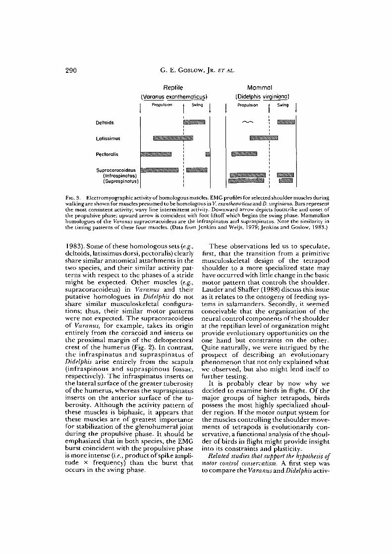
While analyzing the data from Varanus, wewere somewhat surprised when theseresults were comparable to those from theDidelphis study. We noted that manyhomologous muscles of the shouldershowed similar timing patterns of electricalactivity within a stride. (A stride may bedefined as a single, complete cycle of limbmovement and is comprised of a propulsivephase, during which the foot is in contact
with the substrate, and a swing phase, dur-ing which the foot is free of the substrate.)That is, for homologous sets of muscles,the onset, relative duration and cessationof EMG activity were equivalently timedwithin the stride cycle.
Consider, for example, the anatomy (Fig.2) and EMG profile (Fig. 3) of several mus-cles of Varanus and their presumed homo-logues in Didelphis (Jenkins and Goslow,
290 G. E. GOSLOW, JR. ET AL.
Reptile
(Varanus exanthemoticus)I Propulsion i Swing I I
Mammal
(Didelphis virginiona)Propulsion » Swing
Deltoids
Latissimus
Pectoralis
Supracoracoideus(Infraspinatus)(Suprospinotus)
FIG. 3. Electromyographic activity of homologous muscles. EMG profiles for selected shoulder muscles duringwalking are shown for muscles presumed to be homologous in V. exanthematicus and D. virginiana. Bars representthe most consistent activity; wavy line intermittent activity. Downward arrow depicts footstrike and onset ofthe propulsive phase; upward arrow is coincident with foot liftoff which begins the swing phase. Mammalianhomologues of the Varanus supracoracoideus are the infraspinatus and supraspinatus. Note the similarity inthe timing patterns of these four muscles. (Data from Jenkins and Weijs, 1979; Jenkins and Goslow, 1983.)
1983). Some of these homologous sets (e.g.,deltoids, latissimus dorsi, pectoralis) clearlyshare similar anatomical attachments in thetwo species, and their similar activity pat-terns with respect to the phases of a stridemight be expected. Other muscles (e.g.,supracoracoideus) in Varanus and theirputative homologues in Didelphis do notshare similar musculoskeletal configura-tions; thus, their similar motor patternswere not expected. The supracoracoideusof Varanus, for example, takes its originentirely from the coracoid and inserts onthe proximal margin of the deltopectoralcrest of the humerus (Fig. 2). In contrast,the infraspinatus and supraspinatus ofDidelphis arise entirely from the scapula(infraspinous and supraspinous fossae,respectively). The infraspinatus inserts onthe lateral surface of the greater tuberosityof the humerus, whereas the supraspinatusinserts on the anterior surface of the tu-berosity. Although the activity pattern ofthese muscles is biphasic, it appears thatthese muscles are of greatest importancefor stabilization of the glenohumeral jointduring the propulsive phase. It should beemphasized that in both species, the EMGburst coincident with the propulsive phaseis more intense (i.e., product of spike ampli-tude x frequency) than the burst thatoccurs in the swing phase.
These observations led us to speculate,first, that the transition from a primitivemusculoskeletal design of the tetrapodshoulder to a more specialized state mayhave occurred with little change in the basicmotor pattern that controls the shoulder.Lauder and Shaffer (1988) discuss this issueas it relates to the ontogeny of feeding sys-tems in salamanders. Secondly, it seemedconceivable that the organization of theneural control components of the shoulderat the reptilian level of organization mightprovide evolutionary opportunities on theone hand but constraints on the other.Quite naturally, we were intrigued by theprospect of describing an evolutionaryphenomenon that not only explained whatwe observed, but also might lend itself tofurther testing.
It is probably clear by now why wedecided to examine birds in flight. Of themajor groups of higher tetrapods, birdspossess the most highly specialized shoul-der region. If the motor output system forthe muscles controlling the shoulder move-ments of tetrapods is evolutionarily con-servative, a functional analysis of the shoul-der of birds in flight might provide insightinto its constraints and plasticity.
Related studies that support the hypothesis ofmotor control conservatism. A first step wasto compare the Varanus and Didelphis activ-
AVIAN SHOULDER FUNCTION 291
Supracorocotdtufl
Dorsal Cord/
Loii»itnua
Savannah monitor lizard(Voronus exonthemoticus)
Palm-Nut Vulture(Gypohierax anaolensis)
Domestic cat(Fehs domesticus)
rostral13
SEGMENT
14 15caudal16
Biceps
Flexor digitorum superficial -
Latissimus dorsi posterior —
Triceps
Extensor metacarpi ulnaris -
Pectoralis
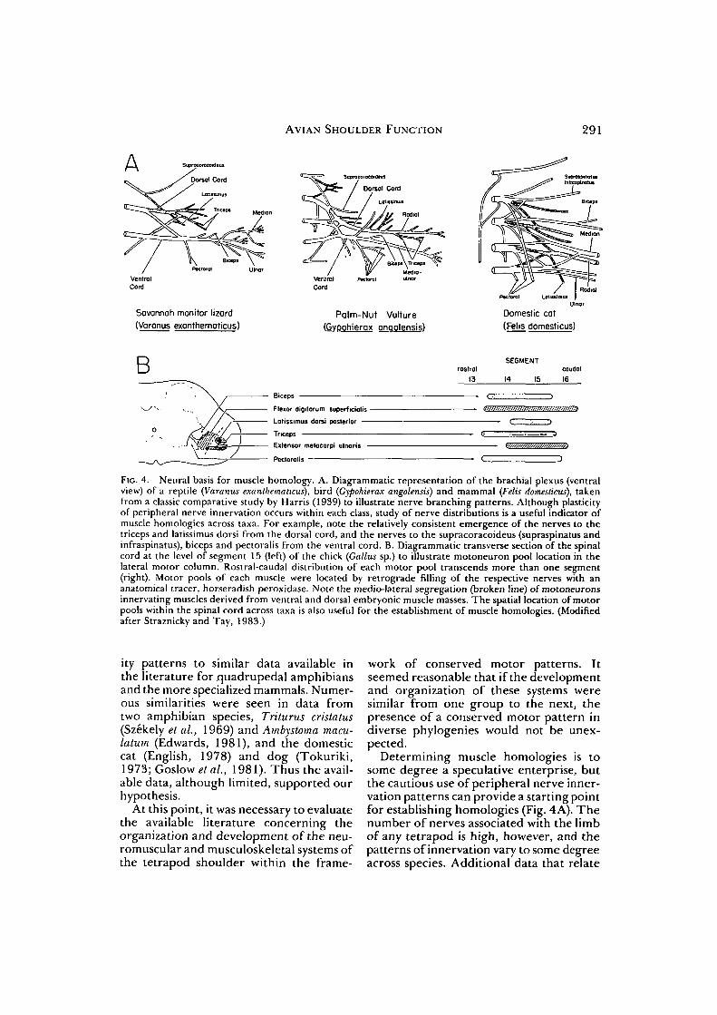
FIG. 4. Neural basis for muscle homology. A. Diagrammatic representation of the brachial plexus (ventralview) of a reptile (Varanus exanthematicus), bird {Gypohierax angolensis) and mammal (Felis domesticus), takenfrom a classic comparative study by Harris (1939) to illustrate nerve branching patterns. Although plasticityof peripheral nerve innervation occurs within each class, study of nerve distributions is a useful indicator ofmuscle homologies across taxa. For example, note the relatively consistent emergence of the nerves to thetriceps and latissimus dorsi from the dorsal cord, and the nerves to the supracoracoideus (supraspinatus andinfraspinatus), biceps and pectoralis from the ventral cord. B. Diagrammatic transverse section of the spinalcord at the level of segment 15 (left) of the chick (Gallus sp.) to illustrate motoneuron pool location in thelateral motor column. Rostral-caudal distribution of each motor pool transcends more than one segment(right). Motor pools of each muscle were located by retrograde filling of the respective nerves with ananatomical tracer, horseradish peroxidase. Note the medio-lateral segregation (broken line) of motoneuronsinnervating muscles derived from ventral and dorsal embryonic muscle masses. The spatial location of motorpools within the spinal cord across taxa is also useful for the establishment of muscle homologies. (Modifiedafter Straznicky and Tay, 1983.)
ity patterns to similar data available inthe literature for quadrupedal amphibiansand the more specialized mammals. Numer-ous similarities were seen in data fromtwo amphibian species, Triturus cristatus(Szekely et al., 1969) and Ambystoma macu-latum (Edwards, 1981), and the domesticcat (English, 1978) and dog (Tokuriki,1973; Goslow et al., 1981). Thus the avail-able data, although limited, supported ourhypothesis.
At this point, it was necessary to evaluatethe available literature concerning theorganization and development of the neu-romuscular and musculoskeletal systems ofthe tetrapod shoulder within the frame-
work of conserved motor patterns. Itseemed reasonable that if the developmentand organization of these systems weresimilar from one group to the next, thepresence of a conserved motor pattern indiverse phylogenies would not be unex-pected.
Determining muscle homologies is tosome degree a speculative enterprise, butthe cautious use of peripheral nerve inner-vation patterns can provide a starting pointfor establishing homologies (Fig. 4A). Thenumber of nerves associated with the limbof any tetrapod is high, however, and thepatterns of innervation vary to some degreeacross species. Additional data that relate

292 G. E. GOSLOW, JR. ET AL.
to development and spinal cord organiza-tion are therefore important. All tetrapodlimbs arise from dorsal and ventral pre-muscle masses of the limb bud which sub-sequently subdivide and separate into indi-vidual limb muscles (see Chen, 1935;Romer, 1944; Cheng, 1955; Sullivan, 1962;Shellswell and Wolpert, 1977). Each mus-cle of the shoulder receives innervationfrom specific levels of the spinal cord. Allof the motoneurons that innervate the limbmuscles are located within the lateral motorcolumns (Lamina IX) of the spinal cord(Fig. 4B). The population of motor neu-rons that innervate a given muscle com-prises the motor pool for that muscle.Motor pool maps have been obtained forselected tetrapod species using both ana-tomical tracing (retrograde transport ofhorseradish peroxidase) and electrophys-iological methods. In the tetrapods thathave been examined, the majority of moto-neurons belonging to a given motor pooltend to be situated close to one another,although in amphibians (Szekely and Czeh,1967; Cruce, 1974; Lamb, 1976) neuronsbelonging to different motor pools tend tobe intermingled more than in birds(Landmesser and Morris, 1975; Hollyday,l980a,b) and mammals (Romanes, 1951,1964; Weeks and English, 1987). Althoughnumerous details of development of thetetrapod forelimb remain to be deter-mined, we concluded that the general sim-ilarities from one group to the next notedhere are consistent with the conservedmotor pattern hypothesis.
Evolution of flight
Our decision to pursue an analysis of theavian shoulder during flight was alsodirected toward answering a longstandingand major morphological problem in ver-tebrate evolution: the interpretation of themusculoskeletal features that developedduring the transition from terrestrial loco-motion to flight.
Anatomical correlates of bird flight: A longhistory. The musculoskeletal structuresrelated to avian flight have been exten-sively studied; detailed treatments of theskeleton are available (Bellairs and Jenkin,1960). Particularly useful descriptions ofmusculature are given by Fiirbringer
(1902), Howell (1937a), Hudson and Lan-zillotti (1955) and George and Berger(1966). In addition, general biomechanicalprinciples that relate to wing movementhave been discussed by Bock (1969). Sev-eral workers have provided interpretiveanalyses of various aspects of flight, includ-ing Sy (1936), Fisher (1957), Savile (1957),Hartman (1961), Greenewalt (1962, 1975),Brown (1963), Pennycuick (1968, 1975),and Simpson (1983). From such studies hasbeen derived the generally accepted inter-pretation of wing muscles in terms of theiractions (summarized by Raikow, 1985).Features of wing movement have also beenelegantly demonstrated through the use ofhigh speed photography {e.g., Riippell,1977; Nachtigall, 1980).
In the paleontological literature theancestry of birds and the origin of flightare two persistently recurring themes (forreview see Hecht e/ al., 1985;Padian, 1986).The phylogenetic origin of birds is pres-ently unresolved. Ostrom (1975, 1976a)summarized the evidence that the closestrelatives of birds are to be found amongthe coelurosaurian dinosaurs. Walker(1972) has argued for a thecodont ances-tory, and other workers (Whetstone andMartin, 1979; Martin et al, 1980) haveadduced evidence of a close (sister group)relationship between birds and crocodiles.The question has been extensively debated(Tarsitano and Hecht, 1980; McGowan andBaker, 1981; Martin, 1983; Steadman,1983), but the preponderance of evidencefavors a relationship to coelurosaurs (Gau-thier, 1986). A second problem concernsthe origin of flight itself. Numerous sce-narios have been proposed over the lasthundred years, and each of these theoriesreconstructs a somewhat different loco-motor and behavioral intermediate stage.There are two major hypotheses: "arbo-real" and "cursorial" (reviewed by Ostrom,1974, 1979, 1986; Martin, 1983; Hecht etal, 1985; Bock, 1986). Regardless of whichparticular behavioral-ecological pathway iscorrect, a central issue in the origin of flightcontroversy is the difficulty of postulatinga set of transitional stages of the shoulderand forelimb that are adaptive at each evo-lutionary level.
Archaeopteryx, represented by six skele-

AVIAN SHOULDER FUNCTION 293
tal specimens from the Late Jurassic rocksof Germany, documents one stage in thereptilian-avian transition. Some aspects ofits anatomy provide insight into the prob-able evolutionary pathways to flight, butothers are controversial. For example,Archaeopteryx did not possess an ossified keelon its sternum. On the basis of the struc-ture of the sternum and shoulder girdle,Ostrom (1974, 1979) concluded that pow-ered flight was not possible at this evolu-tionary stage; others, however, hold a dif-ferent opinion (cf. Olson and Feduccia,1979; Feduccia, 1980; Martin, 1983; Pen-nycuick, 1986). Although it is unlikely thatthe intermediate stages that led to avianflight may be found in the fossil record,speculation about the accompanying struc-tural changes helps us to formulate ques-tions that can be addressed in extant rep-tilian and avian systems.
In modern birds, the supracoracoideusretains its primitive position deep to thepectoralis but its tendon of insertionattaches to the dorsal aspect of the humerus(Fig. 5A) rather than to its anterior aspectas in reptiles. After arising from parts ofthe sternum, coracoid and coracoclavicu-lar membrane, its fibers converge dorsallyon a tendon that passes upward throughthe foramen triosseum (formed by the cor-acoid laterally, furcula anteriorly and scap-ula posteriorly). Various explanations ofhow this arrangement may have evolvedcould be given, but here we presentOstrom's (19766) thoughtful outline whichis based on the premise that Archaeopteryxwas not capable of powered flight. He pro-posed that subsequent to the Archaeopteryxstage of evolution, the shape of the cora-coid underwent extensive change. Thischange was accompanied by alteration ofthe position and function of the principalhumeral extensor (coracobrachialis crani-alis) and forearm flexor (biceps brachii)which, in turn, converted the function ofthe supracoracoideus from a humeraldepressor to an elevator. Ostrom furtherproposed that the upward expansion of theavian coracoid (to form the acrocoracoid),an event basic to his hypothesis, may haveoccurred as a result of 1) elevation of theanterior part of the glenoid and rotationof the shoulder socket to face directly lat-
erally, thereby permitting unrestrictedtransverse (up and down) movements ofthe forelimb; 2) development of anenlarged buttress at the level of the glenoidto brace the furcula, thereby ensuringproper transverse separation of the shoul-der sockets, and 3) raising the levels ofhumeral extension and forearm flexion byelevating the sites of origin of the cora-cobrachialis and biceps. Figure 5B is takenfrom Ostrom (19766) to illustrate theseproposed changes in coracoid morphologyfrom Archaeopteryx to a modern bird likethe turkey vulture (Cathartes).
Implicit in Ostrom's argument, as wellas that of others, is the assumption that theflight muscles of birds have undergonesome fundamental alterations from theprimitive tetrapod pattern in phasic activ-ity or function with respect to limb move-ment. In our estimation, our ability to eval-uate the various reconstructions of theevolutionary stages in the origin of theavian flight apparatus, and the debate overwhether Archaeopteryx could fly, is limitedby our lack of an understanding of thefunctional anatomy of the avian shoulderin flight. For this reason we initiated stud-ies on the function of the shoulder ofpigeons {Columba livia) in free flight andEuropean starlings (Sturnus vulgaris) flyingin a wind tunnel.
FUNCTIONAL ANALYSIS OF THEBIRD SHOULDER
As so often happens, many of our pre-liminary observations were unexpected andopened numerous possibilities for futureresearch. Some of our early results are con-sidered relevant to an assessment of 1) thehypothesis that the motor pattern of shoul-der muscles has remained relatively con-servative throughout tetrapod evolution,and 2) an interpretation of the musculo-skeletal features that accompanied thetransition from terrestrial locomotion toflight.
Supracoracoideus and pectoralisactivity patterns
Our initial EMG studies focused uponthe supracoracoideus, the primary elevatorof the humerus during wing upstroke, andthe pectoralis, the major humeral depres-

294 G. E. GOSLOW, JR. ET AL.
Aleft corocoid
ocrocoracoidsupracoracoideustendon
right humerus
supracoracoideus
acrocoracoid
tendon alignment
musclebellyalignment
Cathartes Archaeopteryx
FIG. 5. Hypothetical transitional stages in supracoracoideus orientation from a primitive bird {Archaeopteryx)to a modern bird (Cathartes). A. Orientation of the supracoracoideus in a modern bird; note the elongatecoracoid, the acrocoracoid process, and the pulley-like arrangement of the supracoracoideus tendon as itpasses through the foramen triosseum to insert on the dorsal aspect of the humerus. The pectoralis has beenremoved. B. Arrow depicts the proposed orientation of the belly of the supracoracoideus and the line of pullof its tendon on the humerus of Archaeopteryx (right), hypothetical intermediates, and a modern bird, Cathartes(left). Note that the elongation of the coracoid and development of the acrocoracoid process facilitate theconversion of the supracoracoideus from a humeral protractor to a humeral elevator. (Modified after Ostrom,1976ft.)
sor. The muscle fascicles of the pectoralisoriginate on the anterolateral surface ofthe clavicle, along the entire surface of thesternal carina (keel), and from portions ofthe sternal body. Its fascicles converge toinsert on the cranial surface of the delto-pectoral crest of the humerus. A completedescription of the anatomy of the supra-coracoideus and pectoralis in pigeons isfound in George and Berger (1966).
Methods. We recorded EMGs in the pec-toralis and supracoracoideus from six adultpigeons during level, flapping flight. Flightbehavior was recorded synchronously on
16 mm film (64-200 fps). Two electrodeconfigurations and implantations wereemployed: 1) paired, Teflon-coated, 18-stranded, stainless steel wires (0.28 mmdiam.; 0.5 mm bared surface; intertip dis-tances 0.5 mm), and 2) paired, silver elec-trodes (100 Mm diam.; 0.5 mm bared tips;intertip distances < 0.5 mm). Electrodeswere sutured to muscle fascia and directedsubcutaneously to an exit point betweenthe scapulae and connected to anAmphenol plug. EMGs were recorded byboth telemetry and direct wire connectionsto the plug, amplified and stored on FM
AVIAN SHOULDER FUNCTION 295
Pectoralis
Supracoracoideus }0.5mV
t 100 ms
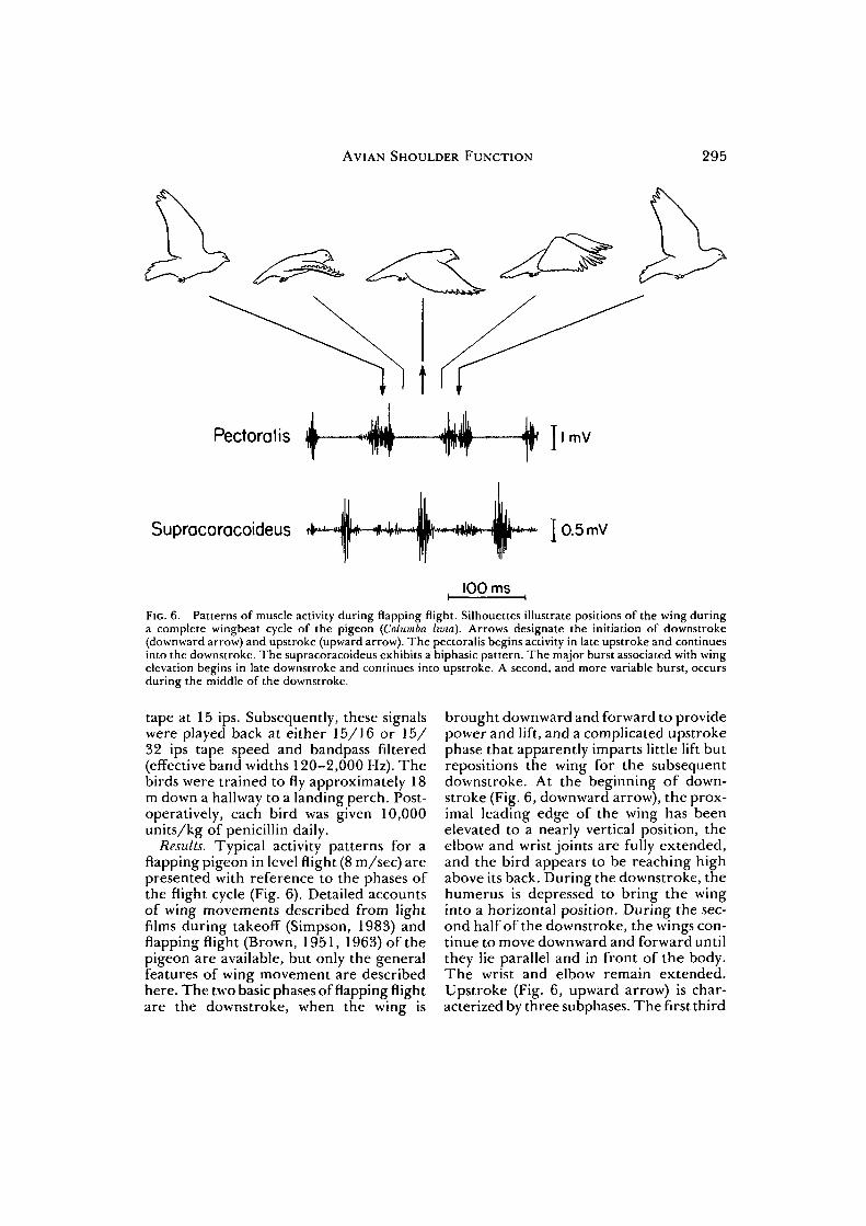
Fie. 6. Patterns of muscle activity during flapping flight. Silhouettes illustrate positions of the wing duringa complete wingbeat cycle of the pigeon (Columba hvia). Arrows designate the initiation of downstroke(downward arrow) and upstroke (upward arrow). The pectoralis begins activity in late upstroke and continuesinto the downstroke. The supracoracoideus exhibits a biphasic pattern. The major burst associated with wingelevation begins in late downstroke and continues into upstroke. A second, and more variable burst, occursduring the middle of the downstroke.
tape at 15 ips. Subsequently, these signalswere played back at either 15/16 or 15 /32 ips tape speed and bandpass filtered(effective band widths 120-2,000 Hz). Thebirds were trained to fly approximately 18m down a hallway to a landing perch. Post-operatively, each bird was given 10,000units/kg of penicillin daily.
Results. Typical activity patterns for aflapping pigeon in level flight (8 m/sec) arepresented with reference to the phases ofthe flight cycle (Fig. 6). Detailed accountsof wing movements described from lightfilms during takeoff (Simpson, 1983) andflapping flight (Brown, 1951, 1963) of thepigeon are available, but only the generalfeatures of wing movement are describedhere. The two basic phases of flapping flightare the downstroke, when the wing is
brought downward and forward to providepower and lift, and a complicated upstrokephase that apparently imparts little lift butrepositions the wing for the subsequentdownstroke. At the beginning of down-stroke (Fig. 6, downward arrow), the prox-imal leading edge of the wing has beenelevated to a nearly vertical position, theelbow and wrist joints are fully extended,and the bird appears to be reaching highabove its back. During the downstroke, thehumerus is depressed to bring the winginto a horizontal position. During the sec-ond half of the downstroke, the wings con-tinue to move downward and forward untilthey lie parallel and in front of the body.The wrist and elbow remain extended.Upstroke (Fig. 6, upward arrow) is char-acterized by three subphases. The first third

296 G. E. GOSLOW, JR. ET AL.
of the upstroke is marked by a reversal ofthe humeral movement that occurs in thelast part of the downstroke and flexion ofthe elbow and wrist. A backward flick comesnext and is characterized by the rapidretraction and elevation of the humeruswhile the elbow and wrist remain flexed.The final phase of upstroke is character-ized by maximum wing elevation andextension.
As intuitively expected, electrical activ-ity of the pectoralis is most pronouncedduring the downstroke, while the supra-coracoideus is most active in upstroke (Fig.6). Note, however, that activity in the pec-toralis begins during late upstroke beforethe wing reaches its highest position andbegins its downward movement. Similarly,the largest burst of the supracoracoideuscommences in late downstroke prior to wingupstroke. In addition, the electrical activityin the supracoracoideus is often biphasic.A second, relatively small, burst begins inlate upstroke and continues into the earlypart of downstroke. This second burst wasclearly measurable in 48% of our record-ings and is coincident with the largest EMGburst of the pectoralis. The double burstpattern is variable; in some cases it appearsand then disappears within a single flightsequence, whereas in others it is consis-tently present or absent. No relation of thedouble burst to flight speed, body angle,or wingbeat amplitude was evident.
Discussion. In the following discussion, wewill be equating the "upstroke" and"downstroke" phases of flight to the"swing" and "propulsive" phases respec-tively of terrestrial locomotion. The onsetof the contraction of the pectoralis andsupracoracoideus (as indicated by the EMGrecords) prior to the point where the mus-cle shortens to move the humerus is notunlike the pattern of contraction measuredin the flexors and extensors of various ver-tebrate limbs during terrestrial locomo-tion. The pattern has implications for ourunderstanding of muscle mechanics inoscillating limb systems and also for theenergetics of cyclic motion. In the case ofpigeon flight, this issue has been discussedby Dial ^ a/. (1987).
The supracoracoideus in birds is ahumeral elevator, and appears to functiondifferently from its homologue in livingreptiles and, presumably, in the earlyMesozoic reptiles from which birds arose.Certainly the putative supracoracoideushomologues in mammals, the supraspina-tus and infraspinatus, function differently;they serve to stabilize the glenohumeraljoint during the propulsive phase (in con-trast to birds, where the supracoracoideuslifts the humerus in the upstroke, which iscomparable to the swing or non-propulsivephase). How could the supracoracoideus inan ancestral tetrapod have given rise tosuch contrasting systems in birds and mam-mals? Can muscles change their function?Obviously this is possible through altera-tion of the musculoskeletal system whichis a framework of mechanical struts, leversand pulleys. However, another and oftenoverlooked aspect must change as well: thetiming of a muscle's activity in the loco-motor cycle. In cases where a muscle'sactivity is biphasic, there exists the possi-bility that in the course of evolution of themusculoskeletal system, one or the otherphase in a muscle's activity may assume acritical function. Such appears to have beenthe case for the supracoracoideus and itshomologues in higher tetrapods. Througha process that we may call "neuromuscularcanalization," the upstroke (=swing) com-ponent of the biphasic activity cycle for thesupracoracoideus is most important inbirds, whereas the propulsive ^down-stroke) component of the homologoussupraspinatus and infraspinatus muscles isessential in mammals. Yet in both birds andmammals the primitive organization of theneural control components still persists, forin both groups we find evidence of abiphasic pattern.
Although our conclusions on this issueare only tentative (because the data are asyet so few), we may pose some additionalquestions. Do the motoneurons whichreflect the biphasic activity pattern seen inthe EMG represent one population or two?If two, is one population only active duringthe swing phase, and the other only duringpropulsion? This might be the case, as evi-
AVIAN SHOULDER FUNCTION 297
dence is now available that the sartoriusmuscle of the domestic cat hindlimb iscontrolled by three populations of moto-neurons, each programmed for a specificlocomotor task (Hoffer et al, 1987). Alter-natively, does each supracoracoideusmotoneuron undergo two periods of exci-tation during each locomotor cycle? Dur-ing the evolution from ancestral to derivedforms, does the amplification of one of thetwo bursts {i.e., in swing phase) and thediminution of the other (i.e., propulsivephase) reflect a change in the number orkinds of motoneurons in the pool, or achange in synaptic connectivity? Why didwe only observe a distinct biphasic patternin the pigeon in 48% of the recordings? Atpresent we can only speculate about answersto these questions; much work needs to bedone.
SKELETAL DYNAMICS DURING FLIGHT
In order to pursue some of the abovequestions and to further our insight intothe general features of wing evolutionamong birds, we constructed a wind tun-nel. A wind tunnel undoubtedly inducescertain constraints on the performance ofa flying bird (Butler et al, 1977), but thebenefits of recording a series of successivewingbeats are many. Critical to our anal-yses is a clear understanding of wing move-ments in relation to muscle activity. There-fore we employed the wind tunnel inconjunction with a cineradiographic sys-tem which records X-ray images of a bird'sskeleton. The demands of the cineradio-graphic apparatus are such that the size ofthe flight chamber of the wind tunnel isrestricted to a relatively small volume.Hence, because pigeons are slightly largefor the wind tunnel, we have begun studieson a relatively small bird, the Europeanstarling (Sturnus vulgaris), for which somepreliminary results are available.
Methods. Four starlings were trained tofly in the variable speed wind tunnel. Theplexiglass flight box measured 61 cm squareand 91.5 cm long. Birds flew from 9 to 20m/sec. The visibility of bony landmarks onradiographic film was enhanced by theinsertion of tiny metallic markers. The
D
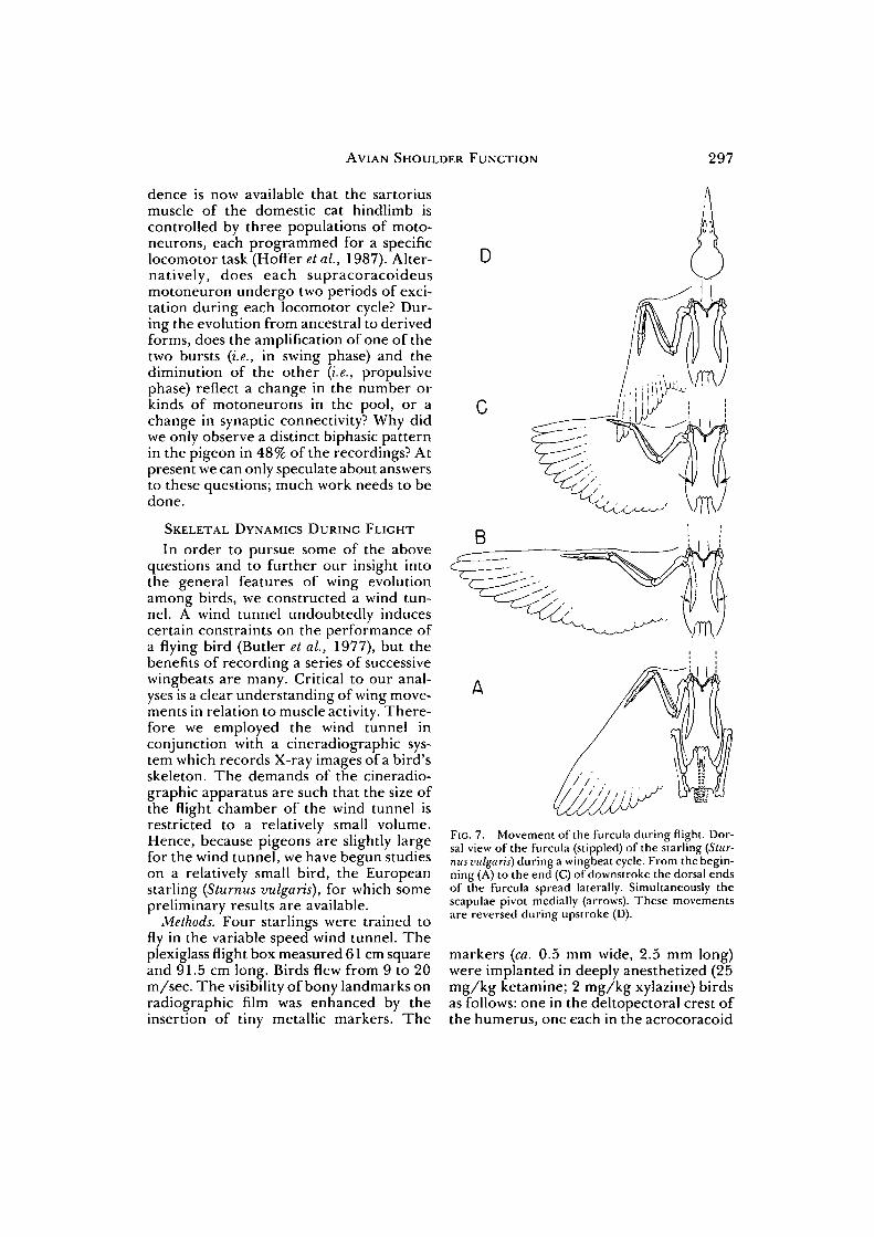
FIG. 7. Movement of the furcula during flight. Dor-sal view of the furcula (stippled) of the starling (Stur-nus vulgaris) during a wingbeat cycle. From the begin-ning (A) to the end (C) of downstroke the dorsal endsof the furcula spread laterally. Simultaneously thescapulae pivot medially (arrows). These movementsare reversed during upstroke (D).
markers (ca. 0.5 mm wide, 2.5 mm long)were implanted in deeply anesthetized (25mg/kg ketamine; 2 mg/kg xylazine) birdsas follows: one in the deltopectoral crest ofthe humerus, one each in the acrocoracoid

298 G. E. GOSLOW, JR. ET AL.
; 20
163o13
12
60S
1i
i 2 5 m s i
FIG. 8. Displacement of the furcula during flight of Sturnus vulgaris. Instantaneous distances between thetwo dorsal ends of the furcula are shown for three wingbeats. Resting distance is shown as a dashed line.Upward arrows indicate the beginning of wing upstroke, downward arrows the beginning of downstroke.Two sets of vertical calibrations are provided. The left scale is in absolute millimeters. The right scale illustratesthe percentage of displacement relative to resting distance.
processes of each of the coracoids, and twoalong the keel of the sternum. A marker1 cm in length was glued between the scap-ulae for scale.
Siemens cineradiographic apparatus,including a grid-controlled tube with 0.06mm focal spot and a 27.94 cm Sireconimage intensification system, was posi-tioned for lateral or dorsoventral projec-tion radiography. An Eclair GV16 highspeed cine camera, mounted on the imageintensifier and operated at approximately200 fps, recorded each sequence on 16 mmKodak Plus-X Reversal film at the sametime that the image was monitored on a 43cm television screen. Approximately sevenhundred feet of film was taken of each bird;analysis with a Vanguard M-CIIP FilmAnalyzer is in progress. The data pre-sented here are selective.
As an aid to kinematic analysis, the birdswith marker implants were injected withT-61 (Euthanasia Solution) and their skel-etons mounted to duplicate structural rela-tions observed radiographically. This pro-cedure permitted verification of thefollowing: 1) lateral and medial displace-ment of the acrocoracoids, as well as thedorsal ends of the furcula (wishbone), mea-sured from the sagittal plane; 2) antero-posterior excursion of the humerus, mea-sured at the intersection of the humeralaxis (which bisects the humeral head anda line between the epicondyles) and thesagittal plane; and 3) elevation or depres-sion of the humerus, measured by the loca-
tion of the humeral axis with respect to ahorizontal plane through the shoulderjoint.
Results. Our observations reveal that thedorsal ends of the "V" shaped furcula,which contact the coracoids dorsally, shiftlaterally during the downstroke (Fig. 7). Inthe subsequent upstroke they move medi-ally, presumably by elastic recoil. At rest,the distance between the dorsal ends of thefurcula is typically about 11-12 mm. At aflight speed of 17 m/sec, intrafurcular dis-tance is maximal (20 mm) at the end ofdownstroke (Fig. 8).
Discussion. The furcula is such a uniquestructure that biologists have speculated asto its function. For example, Ostrom(19766) suggested that the furcula servesto maintain a set distance between theshoulders, whereas Norberg (1985) men-tioned the furcula's potential role forenergy storage. The furcula may, in fact,serve both these functions but heretoforethere simply has been no way to make directobservations of its movements. Our initialkinematic analysis of some mechanicalproperties of isolated furculae provideindirect support for an additional hypoth-esis which relates to respiration (Jenkins etal., 1988). Coupled with movements of thesternum that also occur with each wing-beat, the furcula may play a role in cyclingair between the air sacs and lungs duringflight. Such a pattern might serve theincreased demands of flight. Clearly, addi-tional observations and experimental

AVIAN SHOULDER FUNCTION 299
manipulations are necessary in order toclarify the furcula's role not only in star-lings, but in other species of birds withvarying flight modes and furcular geome-try.
CONCLUDING REMARKS
Reconstruction of the evolutionaryevents that occurred during the develop-ment of avian flight continues to challengestudents of evolution and vertebrate loco-motion. Numerous evolutionary pathwayshave been proposed, and each has neces-sarily made assumptions about the formand function of the forelimb of the reptil-ian ancestor, protobird, or modern birdcapable of flight. We are fortunate at lastto possess the kind of technology necessaryfor precise documentation of tetrapod limbfunction in general, and bird wing kine-matics and neuromuscular control specif-ically. The data from such studies shouldallow us to reassess existing interpretationsof the evolution of bird flight and, in addi-tion, to test the premises on which inter-pretations of form-function relationshipshave been based.
ACKNOWLEDGMENTS
We thank L. L. Meszoly for illustrations,A. H. Coleman for photography, and L. L.W. Maloney for secretarial assistance. Wealso extend our appreciation to S. R. Kap-lan for technical assistance and to K. Hep-worth for the use of his camera. This workwas supported by N.S.F. Grant BSR85-11867 and by the Northern Arizona Uni-versity Organized Research Committee.
REFERENCES
Bellairs, A. d'A. and C. R. Jenkin. 1960. The skel-eton of birds. In A. J. Marshall (ed.), Biology andcomparative physiology of birds, Vol. 1, pp. 241 - 3 0 0 .Academic Press, New York.
Bock, W.J. 1969. The origin and radiation of birds.Ann. N.Y. Acad. Sci. 167:147-155.
Bock, W.J. 1986. The arboreal origin of avian flight.In K. Padian (ed.), The origin of birds and the evo-lution of flight, pp. 57-72. California Academy ofSciences, San Francisco.
Brown, R. H. J. 1951. Flapping flight. The Ibis 93:333-359.
Brown, R. H. J. 1963. The flight of birds. Biol.Reviews 38:460-489.
Butler, P. J., N. H. West, and D. R. Jones. 1977.Respiratory and cardiovascular responses of thepigeon to sustained, level flight in a wind-tunnel.J. Exp. Biol. 71:7-26.
Carroll, R. L. 1970. The ancestry of reptiles. Phil.Trans. Royal Soc. London 257:267-308.
Chen, H. K. 1935. Development of the pectoral limbof Xecturus maculosus. 111. Biol. Monogr. 14:1-71.
Cheng, C. 1955. The development of the shoulderregion of the opossum, Didelphis virgtniana, withspecial reference to the musculature. J. Morph.97:415-471.
Cruce, W. L. 1974. The anatomical organization ofhindlimb motoneurons in the lumbar spinal cordof the frog, Rana catesbiana. J. Comp. Neurol.153:59-76.
Dial, K. P., S. R. Kaplan, G. E. Goslow, Jr., and F. A.Jenkins, Jr. 1987. The structure and neural con-trol of the pectoralis in pigeons: Implications forflight mechanics. Anat. Rec. 218:284-287.
Edwards, J. L. 1981. An electromyographic analysisof the forelimb muscles of the spotted salaman-der, Ambystoma maculatum. Amer. Zool. 21:938.
English, A. W. 1978. Functional analysis of the shoul-der girdle of cats during locomotion. J. Morph.156:279-292.
Feduccia, A. 1980. The age of birds. Harvard Univer-sity Press, Cambridge, Massachusetts.
Fisher, H. 1. 1957. Bony mechanisms of automaticflexion and extension in the pigeon's wing. Sci-ence 126:446.
Fiirbringer, M. 1902. Zur vergleichenden anatomiedes brustschulter-apparates und der schulter-muskeln. Jena Zeit Naturwiss. 36:289-736.
Gauthier, J. 1986. Saurischian monophyly and theorigin of birds. In K. Padian (ed.), The origin ofbirds and the evolution of flight, pp. 1-55. CaliforniaAcademy of Sciences, San Francisco.
George, J. C. and A. J. Berger. 1966. Avian myology.Academic Press, New York.
Goslow, G. E.,Jr., H. J. Seeherman, C. R. Taylor, M.N. McCutchin, and N. C. Heglund. 1981. Elec-trical activity and relative length changes of doglimb muscles as a function of speed and gait. J.Exp. Biol. 94:15-42.
Greenewak, C. H. 1962. Dimensional relationshipsfor flying animals. Smithsonian Misc. Coll. 144(2):1-46.
Greenewalt, C. H. 1975. The flight of birds. Trans.Amer. Phil. Soc. 65(4):l-67.
Harris, W. 1939. The morphology of the brachial plexus.Oxford University Press, London.
Hartman, F. A. 1961. Locomotor mechanisms ofbirds. Smithsonian Misc. Coll. 143(1): 1-91.
Hecht, M. K., J. H. Ostrom, G. Viohl, and P. Welln-hofer. (eds.) 1985. The beginniiigsof birds. Freundedes Jura-Museums Eichstatt, Willibaldsburg.
Hotter, J. A., G. E. Loeb, N. Sugano, W. B. Marks,M. J. O'Donovan, and C. A. Pratt. 1987. Cathindlimb motoneurons during locomotion. III.Functional segregation in sartorius. J. Neuro-physiol. 57:554-562.
Hollyday, M. 1980a. Motoneuron histogenesis and

300 G. E. GOSLOW, JR. ET AL.
development of limb innervation. In R. K. Hunt(ed.), Current topics in development biology, Vol. 15,pp. 181-215. Academic Press, New York.
Hollyday, M. 19806. Organization of motor pools inthe chick lumbar lateral motor column. J. Comp.Neurol. 194:143-170.
Howell, A. B. 1936. Morphogenesis of the shoulderarchitecture: Part IV. Reptilia. Q. Rev. Biol. 11:183-208.
Howell, A. B. 1937a. Morphogenesis of the shoulderarchitecture: Part V. Aves. Auk 54:364-375.
Howell, A. B. 19376. Morphogenesis of the shoulderarchitecture: Part VI. Therian Mammalia. Q. Rev.Biol. 12:440-463.
Hudson, G. E. and P. J. Lanzillotti. 1955. Gross anat-omy of the wing muscles in the family Corvidae.Amer. Midi. Nat. 53:1-44.
Jenkins, F. A., Jr. 1971. The postcranial skeleton ofAfrican cynodonts. Bull. 36, Peabody Museum ofNat. Hist., Yale University, New Haven.
Jenkins, F. A., Jr., K. P. Dial, and G. E. Goslow, Jr.1988. A cineradiographic analysis of bird flight:The wishbone is a spring. Science 241:1495-1498.
Jenkins, F. A., Jr. and G. E. Goslow, Jr. 1983. Thefunctional anatomy of the shoulder of the Savan-nah monitor lizard Varanus exanthemalicus. J.Morph. 175:195-216.
Jenkins, F. A., Jr. and W. A. Weijs. 1979. The func-tional anatomy of the shoulder of the Virginiaopossum Didelphis virginiana J. Zool. 188:379-410.
Lamb, A. B. 1976. The projection patterns to theventral horn to the hind limb during develop-ment. Dev. Biol. 54:82-99.
Landmesser, L. and D. G. Morris. 1975. The devel-opment of functional innervation in the hind limbof the chick embryo. J. Physiol. (London) 249:301-326.
Lauder, G. V. and H. B. Shaffer. 1988. Ontogenyand functional design in tiger salamanders{Ambystoma ligrinum): Are motor patterns con-served during major morphological transforma-tions?J. Morph. 197:249-268.
Martin, L. D. 1983. The origin and early radiationof birds. In A. H. Brush and G. A. Clark, Jr. (eds.),Perspectives in ornithology, pp. 291-353. Cam-bridge University Press, Cambridge.
Martin, L. D., J. D. Stewart, and K. N. Whetstone.1980. The origin of birds: Structure of the tarsusand teeth. The Auk 97:86-93.
McGowan, C. and A.J. Baker. 1981. Common ances-try for birds and crocodiles? Nature (London)289:97-98.
Nachtigall, W. 1980. Bird flight: Kinematics of wingmovement and aspects of aerodynamics. In R.Nohring (ed.), Acta XVII Congressus Interna-tionalis Ornithologici, Vol. I, pp. 377-383. Ver-lag der Deutschen Ornithologen-Gesellschaft,Berlin.
Norberg, U. M. 1985. Flying, gliding and soaring.In M. Hildebrand, D. M. Bramble, K. F. Liem,and D. B. Wake (eds.), Functional vertebrate mor-phology, pp. 129-158. The Belknap Press of Har-vard University Press, Cambridge, Massachusettsand London, England.
Olson, S. L. and A. Feduccia. 1979. Flight capabilityand the pectoral girdle of Archaeopteryx. Nature(London) 278:247-248.
Ostrom, J. H. 1974. Archaeopteryx and the origin offlight. Q. Rev. Biol. 49:27-47.
Ostrom, J. H. 1975. On the origin of Archaeopteryxand the ancestry of birds. In Problemes actuels depaleontologie-evolution des vertebres. Coll. Inter.C.N.R.S. 218:519-532.
Ostrom, J. H. 1976a. Archaeopteryx and the origin ofbirds. Biol. Jour. Linn. Soc. 8(2):91-182.
Ostrom, J. H. 19766. Some hypothetical anatomicalstages in the evolution of avian flight. In S. L.Olson (ed.), Collected papers in avian paleontologyhonoring the 90th birthday of Alexander Wetmore.Smithsonian Contr. Paleobiol. 27:1-21.
Ostrom, J. H. 1978. The osteology of Campsognathuslongipes Wagner. Zitteliana Abhandlungen derBayerischen Staatssammlung fur Palaontologieund historische Geologie 4:73-118.
Ostrom, J. H. 1979. Bird flight: How did it begin?American Scientist 67:46-56.
Ostrom, J. H. 1986. The cursorial origin of avianflight. In K. Padian (ed.), The origin of birds andthe evolution of flight, pp. 73—81. California Acad-emy of Sciences, San Francisco.
Padian, K. (ed.) 1986. The origin of birds and the evo-lution of flight. California Academy of Sciences,San Francisco.
Pennycuick, C.J. 1968. Power requirements for hor-izontal flight in the pigeon Columba livia. J. Exp.Biol. 49:527-555.
Pennycuick, C.J. 1975. Mechanics of flight. In D. S.Farner and J. R. King, Jr. (eds.), Avian biology,Vol. 5, pp. 1-75. Academic Press, New York.
Pennycuick, C.J. 1986. Mechanical constraints onthe evolution of flight. In K. Padian (ed.), Theorigin of birds and the evolution of flight, pp. 83-98.California Academy of Sciences, San Francisco.
Raikow, R. 1985. Locomotor system. In A. S. Kingand J. McLelland (eds.), Form and function in birds,Vol. Ill, pp. 57-147. Academic Press, London.
Romanes, G.J. 1951. The motor cell columns of thelumbo-sacral spinal cord of the cat. J. Comp. Neu-rol. 94:313-363.
Romanes, G.J. 1964. The motor pools of the spinalcord. Progr. Brain Res. 11:93-119.
Romer, A. S. 1922. The locomotor apparatus of cer-tain primitive and mammal-like reptiles. Bull. Am.Mus. Nat. Hist. 46:517-606.
Romer, A. S. 1944. The development of tetrapodlimb musculature—the shoulder region oiLacerta.J. Morph. 74:1-41.
Ruppell, G. 1977. Bird flight. Van Nostrand ReinholdCompany, New York.
Savile, D. B. O. 1957. Adaptive evolution in the avianwing. Evolution 11:212-224.
Shellswell, G. B. and L. Wolpert. 1977. The patternof muscle and tendon development in the chickwing. In]. R. Hinchcliffe and M. Balls (eds.), Ver-tebrate limb and somite morphogenesis. CambridgeUniversity Press, Cambridge.
Simpson, S. F. 1983. The flight mechanism of thepigeon Columba livia during take-off. J. Zool.(London) 200:435-443.

AVIAN SHOULDER FUNCTION 301
Steadman, D. W. 1983. Commentary. In A. H. Brushand G. A. Clark, Jr. (eds.), Perspectives in orni-thology, pp. 338-344. Cambridge University Press,Cambridge.
Straznicky, C. and D. Tay. 1983. The localization ofmotoneuron pools innervating wing muscles inthe chick. Anat. Embryol. 100:209-218.
Sullivan, G. E. 1962. Anatomy and embryology ofthe wing musculature of the domestic fowl Gallus.Aust.J. Zool. 10:458-518.
Sy, M. 1936. Funktionell-anatomische Untersuch-ungen am Vogelflugel. Ornithologie 84:199-296.
Szekely, G. and G. Czeh. 1967. Localization of moto-neurons in the limb moving spinal cord segmentsof Ambystoma. Acta Physiol. Acad. Sci. Hung. 32:3-18. '
Szekeley, G.,G. Czeh, and G. Voros. 1969. The activ-
ity pattern of limb muscles in freely moving nor-mal and deafferented newts. Exp. Br. Res. 9:55-62.
Tarsitano, S. and M. K. Hecht. 1980. A reconsider-ation of the reptilian relationships of Archaeop-teryx. Zool. J. Linn. Soc. 69:149-182.
Tokuriki, M. 1973. Electromyographic and joint-mechanical studies in quadrupedal locomotion.I. Walk. Jap. J. Vet. Sci. 35:433-448.
Walker, A. D. 1972. New light on the origin of birdsand crocodiles. Nature 237:257-263.
Weeks, O. I. and A. W. English. 1987. Cat tricepssurae motor nuclei are organized topologically.Exp. Neurol. 96:163-177.
Whetstone, K. N. and L. D. Martin. 1979. New lookat the origin of birds and crocodiles. Nature 279:234-236.