the autonomic nervous system
DESCRIPTION
The Autonomic Nervous System. Proprioception pathways posterior column& Spinocerebellar Pathways. Assess Prof. Fawzia Al-Rouq Department of Physiology College of Medicine King Saud University. OBJECTIVES. Pathways of proprioception - PowerPoint PPT PresentationTRANSCRIPT
The The AutonomicAutonomic Nervous Nervous System System
Assess Prof. Fawzia Al-Rouq Department of PhysiologyCollege of MedicineKing Saud University
Proprioception pathways
posterior column& Spinocerebellar
Pathways
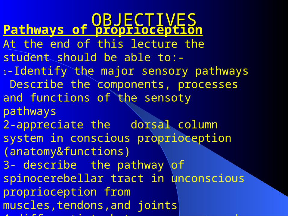
OBJECTIVESOBJECTIVES
Pathways of proprioceptionAt the end of this lecture the student should be able to:-
1-Identify the major sensory pathways Describe the components, processes and functions of the sensoty pathways2-appreciate the dorsal column system in conscious proprioception (anatomy&functions)3- describe the pathway of spinocerebellar tract in unconscious proprioception from muscles,tendons,and joints4-differentiate between sensory and motor ataxia

Introductionmajor sensory pathways dorsal column system spinocerebellar tract sensory and motor ataxia
INTRODUCTIONINTRODUCTION

ProprioceptionProprioceptionfrom Latin proprius, meaning "one's own",
"individual" and perception, is the sense of the relative position of neighbouring parts of the body and strength of effort being employed in movement.[It is distinguished from exteroception, by which one perceives the outside world, and interoception, by which one perceives pain, hunger, etc., and the movement of internal organs.
The initiation of proprioception is the activation of a proprioreceptor in the periphery.[The proprioceptive sense is believed to be composed of information from sensory neurons located in the inner ear (motion and orientation) and in the stretch receptors located in the muscles and the joint-supporting ligaments (stance).
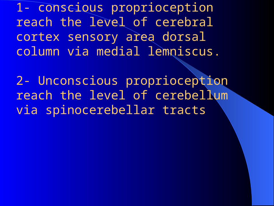
Types of proprioception:-1- conscious proprioception reach the level of cerebral cortex sensory area via dorsal column tract.
2- Unconscious proprioception reach the level of cerebellum via spinocerebellar tracts
Where is the location of these tracts?

Organization of the Organization of the Nervous SystemNervous System
2 big initial divisions:Central Nervous System
The brain + the spinal cordThe center of integration and control
Peripheral Nervous SystemThe nervous system outside of the brain and
spinal cordConsists of:
31 Spinal nervesCarry info to and from the spinal cord
12 Cranial nerves Carry info to and from the brain
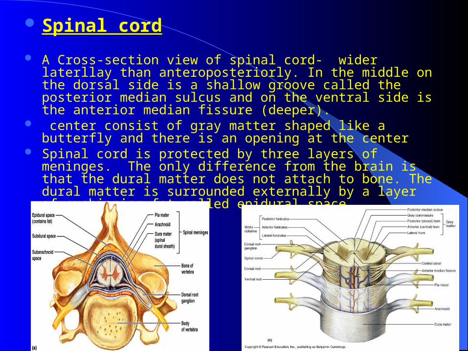
Spinal cord
A Cross-section view of spinal cord- wider laterllay than anteroposteriorly. In the middle on the dorsal side is a shallow groove called the posterior median sulcus and on the ventral side is the anterior median fissure (deeper).
center consist of gray matter shaped like a butterfly and there is an opening at the center
Spinal cord is protected by three layers of meninges. The only difference from the brain is that the dural matter does not attach to bone. The dural matter is surrounded externally by a layer of cushioning fat called epidural space.
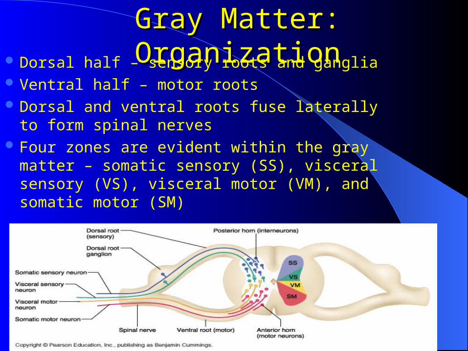
Gray Matter: OrganizationGray Matter: Organization Dorsal half – sensory roots and ganglia Ventral half – motor roots Dorsal and ventral roots fuse laterally to form
spinal nerves Four zones are evident within the gray matter –
somatic sensory (SS), visceral sensory (VS), visceral motor (VM), and somatic motor (SM)
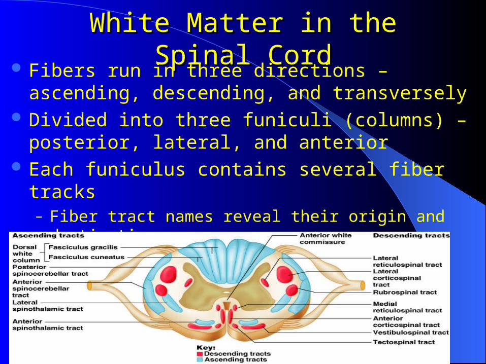
White Matter in the Spinal CordWhite Matter in the Spinal Cord Fibers run in three directions – ascending, descending,
and transversely Divided into three funiculi (columns) – posterior, lateral,
and anterior Each funiculus contains several fiber tracks
– Fiber tract names reveal their origin and destination– Fiber tracts are composed of axons with similar functions

Introductionmajor sensory pathways dorsal column system spinocerebellar tract sensory and motor ataxia
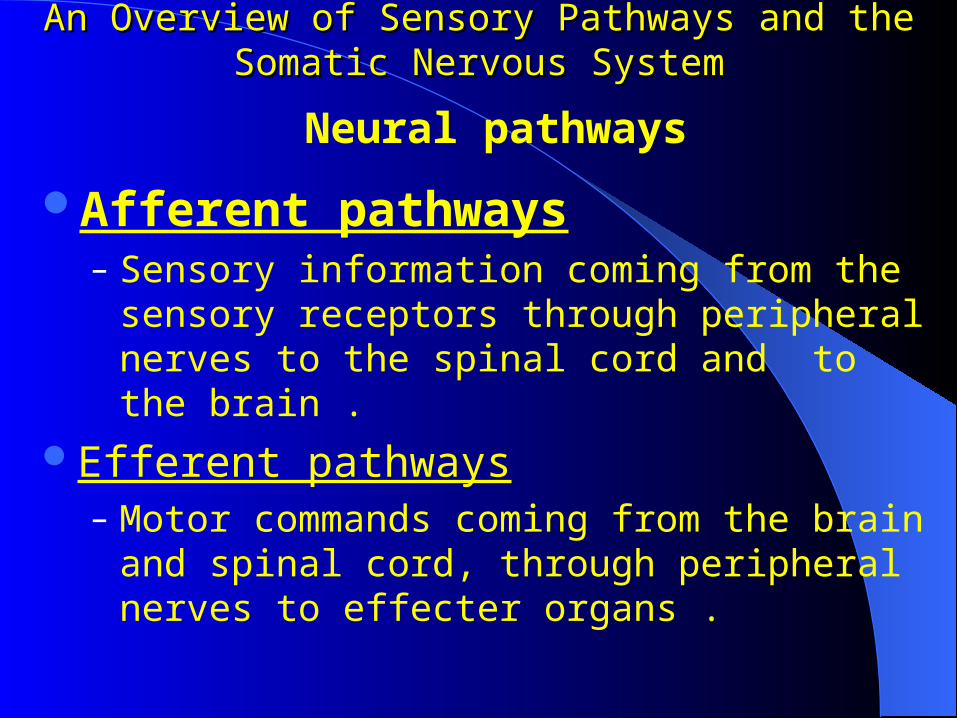
An Overview of Sensory Pathways and the Somatic An Overview of Sensory Pathways and the Somatic Nervous SystemNervous System
Afferent pathways– Sensory information coming from the sensory
receptors through peripheral nerves to the spinal cord and to the brain .
Efferent pathways– Motor commands coming from the brain and spinal
cord, through peripheral nerves to effecter organs .
Neural pathways
Sensory pathwaysSensory pathways
Sensory pathwaysSensory pathways
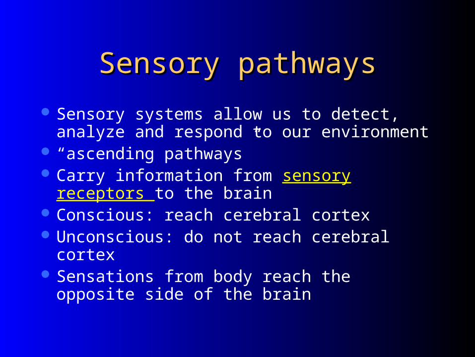
Sensory systems allow us to detect, analyze and respond to our environment
“ascending pathways” Carry information from sensory receptors to the
brain Conscious: reach cerebral cortex Unconscious: do not reach cerebral cortex Sensations from body reach the opposite side of
the brain

Sensory ReceptorsSensory Receptors
Copyright © 2005 Pearson Education, Inc., publishing as Benjamin Cummings
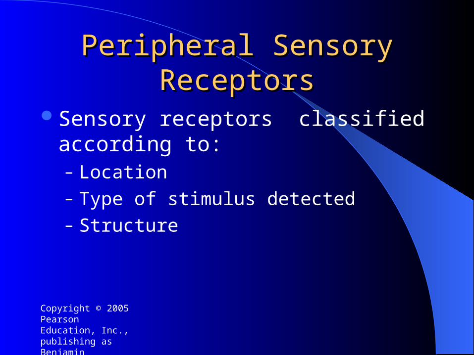
Peripheral Sensory ReceptorsPeripheral Sensory Receptors
Sensory receptors classified according to: – Location– Type of stimulus detected– Structure

Copyright © 2005 Pearson Education, Inc., publishing as Benjamin Cummings
Unencapsulated Nerve Unencapsulated Nerve EndingsEndings
Copyright © 2005 Pearson Education, Inc., publishing as Benjamin Cummings
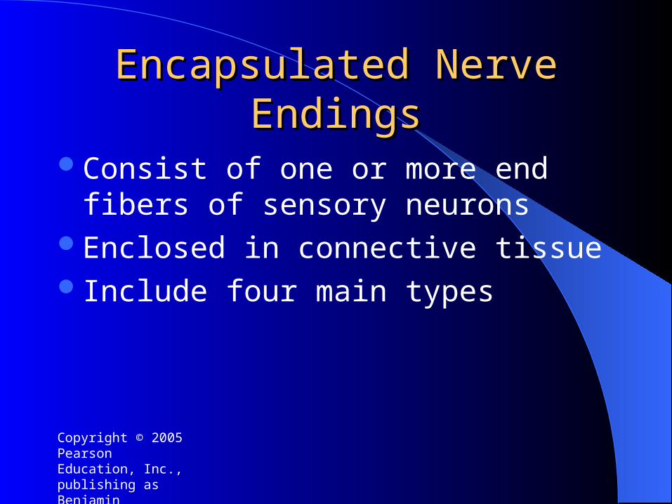
Encapsulated Nerve EndingsEncapsulated Nerve Endings
Consist of one or more end fibers of sensory neurons
Enclosed in connective tissueInclude four main types
Copyright © 2005 Pearson Education, Inc., publishing as Benjamin Cummings
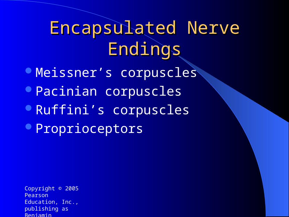
Encapsulated Nerve EndingsEncapsulated Nerve Endings
Meissner’s corpuscles Pacinian corpusclesRuffini’s corpusclesProprioceptors

Types of proprioception:-1- conscious proprioception reach the level of cerebral cortex sensory area dorsal column via medial lemniscus.
2- Unconscious proprioception reach the level of cerebellum via spinocerebellar tracts
The initiation of proprioception is the activation of a proprioreceptor in the periphery.[The proprioceptive sense is believed to be composed of information from sensory neurons located in the inner ear (motion and orientation) was dicussed befor.
and in the stretch receptors located in the muscles and the joint-supporting ligaments (stance).

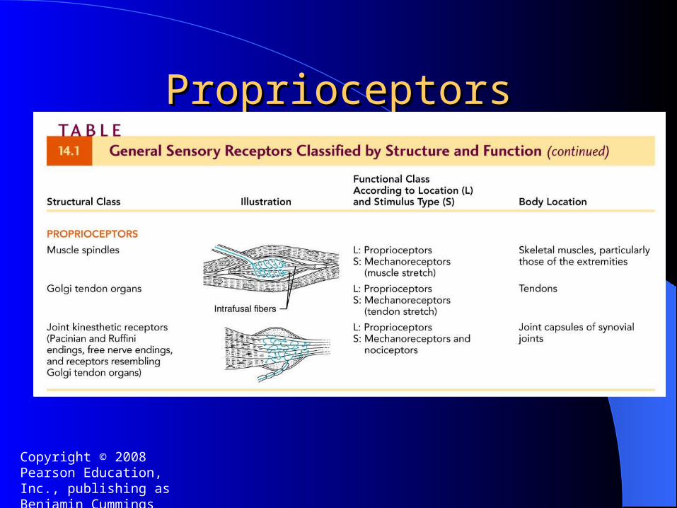
ProprioceptorsProprioceptors
– Encapsulated Nerve Endings– Monitor stretch in locomotory organs– Three types of proprioceptors
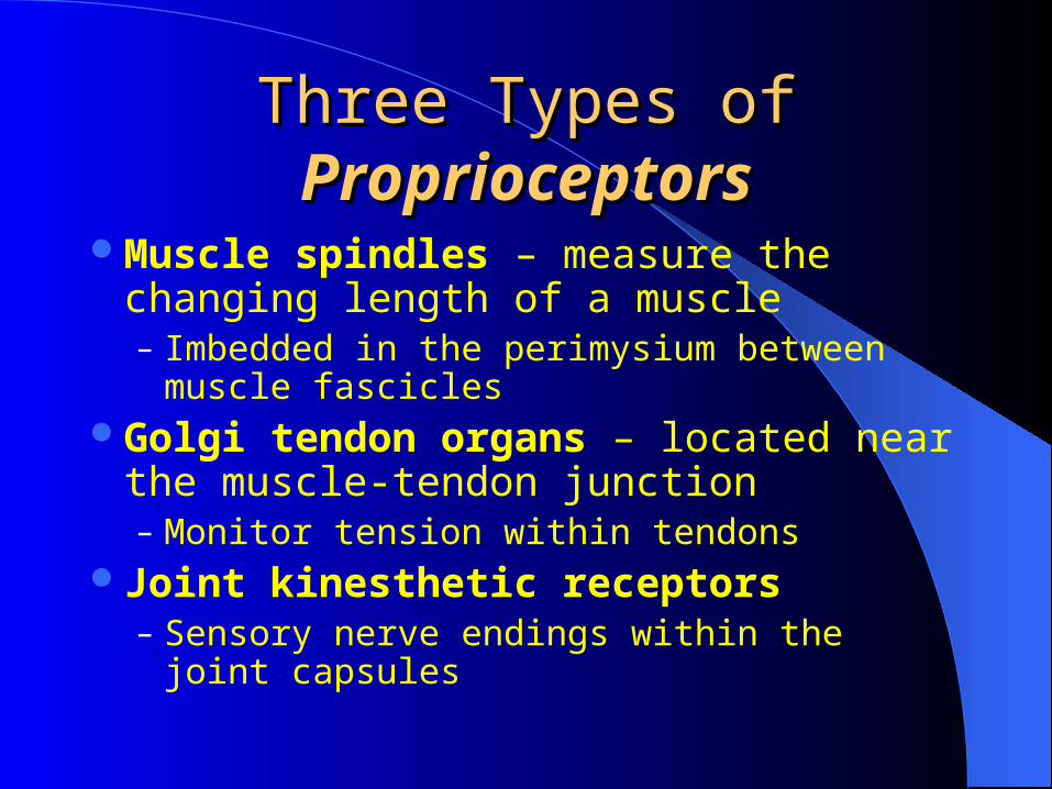
Three Types of Three Types of ProprioceptorsProprioceptors
Muscle spindles – measure the changing length of a muscle– Imbedded in the perimysium between muscle
fasciclesGolgi tendon organs – located near the
muscle-tendon junction– Monitor tension within tendons
Joint kinesthetic receptors – Sensory nerve endings within the joint capsules
Copyright © 2008 Pearson Education, Inc., publishing as Benjamin Cummings
ProprioceptorsProprioceptors
Copyright © 2008 Pearson Education, Inc., publishing as Benjamin Cummings
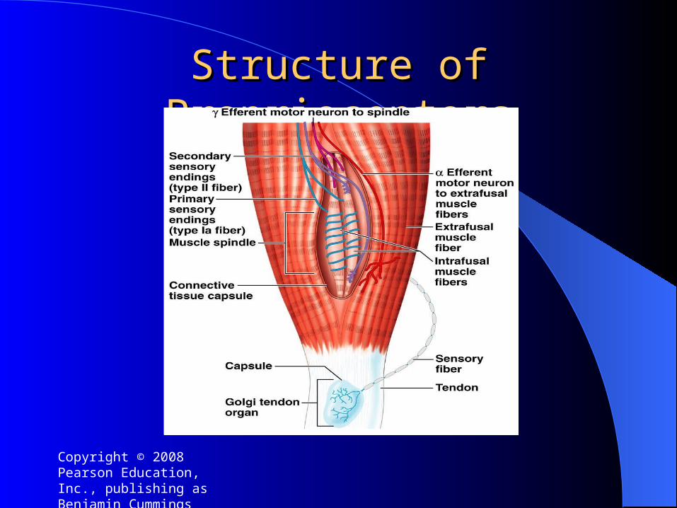
Structure of ProprioceptorsStructure of Proprioceptors
Introductionmajor sensory pathways dorsal column system spinocerebellar tract sensory and motor ataxia
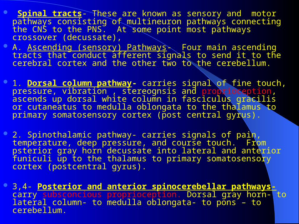
Spinal tracts- These are known as sensory and motor pathways consisting of multineuron pathways connecting the CNS to the PNS. At some point most pathways crossover (decussate),
A. Ascending (sensory) Pathways- Four main ascending tracts that conduct afferent signals to send it to the cerebral cortex and the other two to the cerebellum.
1. Dorsal column pathway- carries signal of fine touch, pressure, vibration , stereognsis and proprioception, ascends up dorsal white column in fasciculus gracilis or cutaneatus to medulla oblongata to the thalamus to primary somatosensory cortex (post central gyrus).
2. Spinothalamic pathway- carries signals of pain, temperature, deep pressure, and course touch. From psterior gray horn decussate into lateral and anterior funiculi up to the thalamus to primary somatosensory cortex (postcentral gyrus).
3,4- Posterior and anterior spinocerebellar pathways- carry subsconcious proprioception. Dorsal gray horn- to lateral column- to medulla oblongata- to pons – to cerebellum.
Sensory pathways:Sensory pathways: 3 neurons 3 neurons
1st: enters spinal cord from periphery
2nd: crosses over (decussates), ascends
in spinal cord to thalamus
3rd: projects to somatosensory cortex
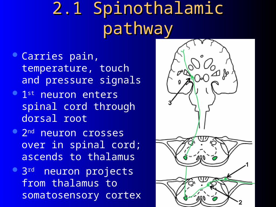
2.1 Spinothalamic pathway2.1 Spinothalamic pathway
Carries pain, temperature, touch and pressure signals
1st neuron enters spinal cord through dorsal root
2nd neuron crosses over in spinal cord; ascends to thalamus
3rd neuron projects from thalamus to somatosensory cortex
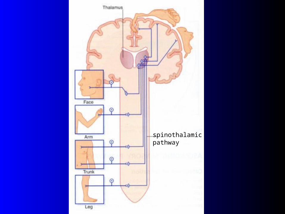
spinothalamicpathway
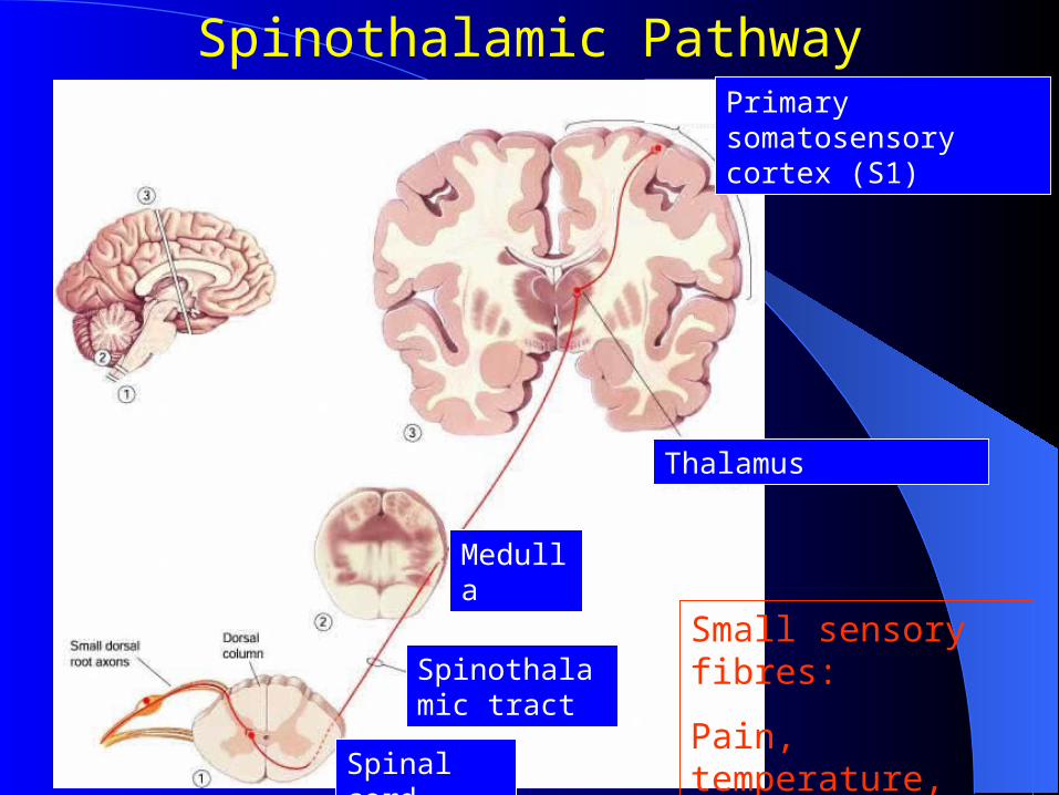
Spinothalamic Pathway
Small sensory fibres:
Pain, temperature, some touch
Primary somatosensory cortex (S1)
Thalamus
Medulla
Spinal cord
Spinothalamic tract

Spinothalamic damageSpinothalamic damagespinothalamic pathway
Leftspinal cord injury
Loss of sense of:•Touch•Pain•Warmth/coldin right leg

Introductionmajor sensory pathways dorsal column system spinocerebellar tract sensory and motor ataxia
Types of proprioception:-1- conscious proprioception reach the level of cerebral cortex sensory area dorsal column via medial lemniscus.
2- Unconscious proprioception reach the level of cerebellum via spinocerebellar tracts
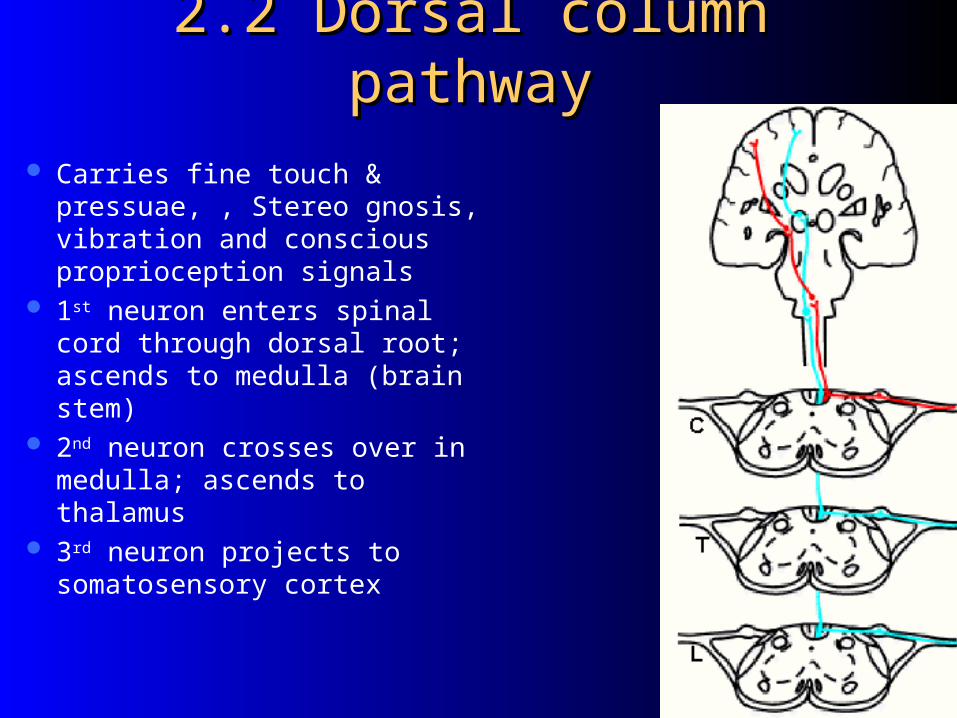
2.2 Dorsal column pathway2.2 Dorsal column pathway
Carries fine touch & pressuae, , Stereo gnosis, vibration and conscious proprioception signals
1st neuron enters spinal cord through dorsal root; ascends to medulla (brain stem)
2nd neuron crosses over in medulla; ascends to thalamus
3rd neuron projects to somatosensory cortex

Two-Point DiscriminationTwo-Point Discrimination
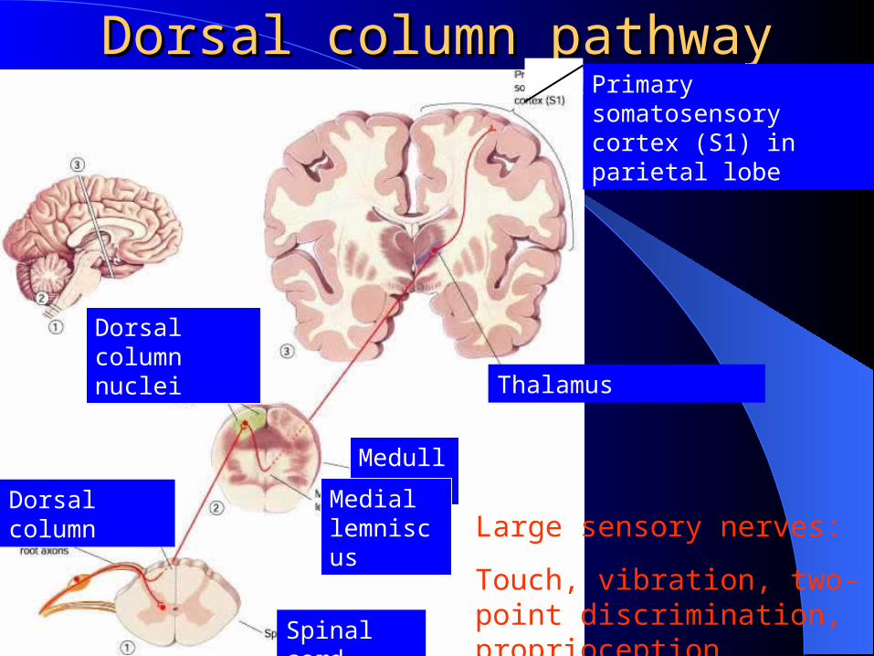
Dorsal column pathwayDorsal column pathway
Large sensory nerves:
Touch, vibration, two-point discrimination, proprioception
Primary somatosensory cortex (S1) in parietal lobe
Thalamus
Medulla
Mediallemniscus
Spinal cord
Dorsal column
Dorsal columnnuclei
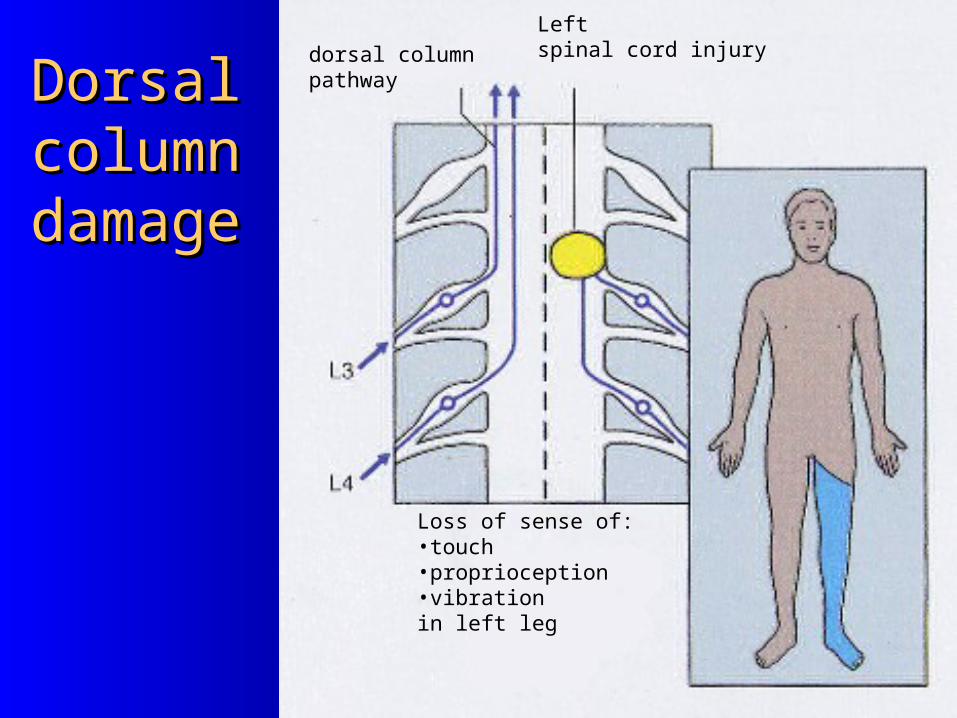
Dorsal Dorsal column column damagedamage
dorsal column pathway
Leftspinal cord injury
Loss of sense of:•touch•proprioception•vibrationin left leg
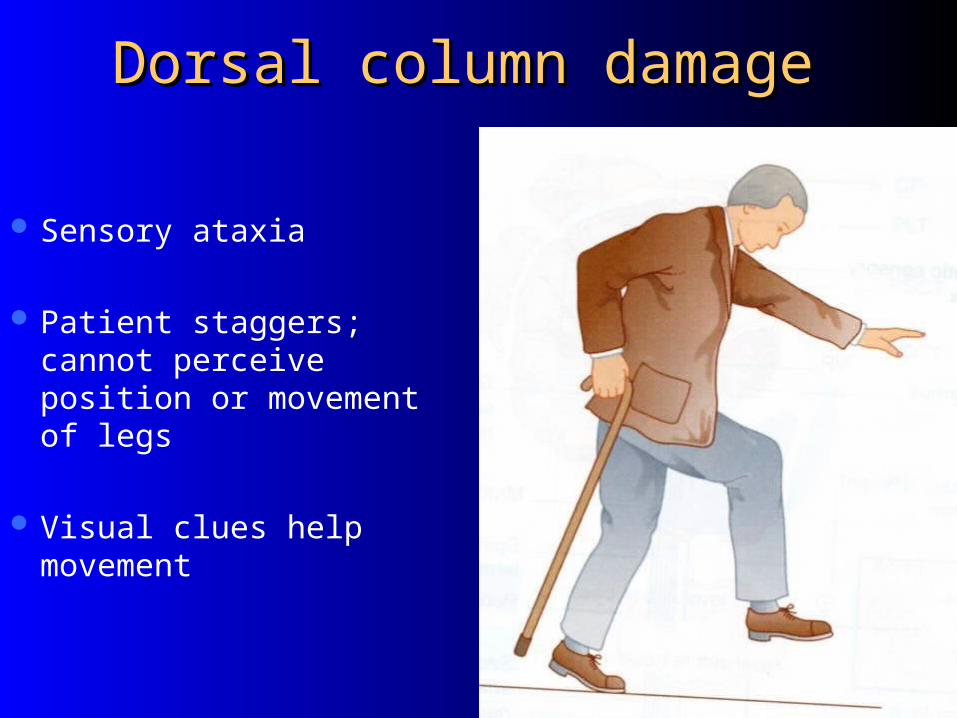
Dorsal column damageDorsal column damage
Sensory ataxia
Patient staggers; cannot perceive position or movement of legs
Visual clues help movement
Introductionmajor sensory pathways dorsal column system spinocerebellar tract sensory and motor ataxia
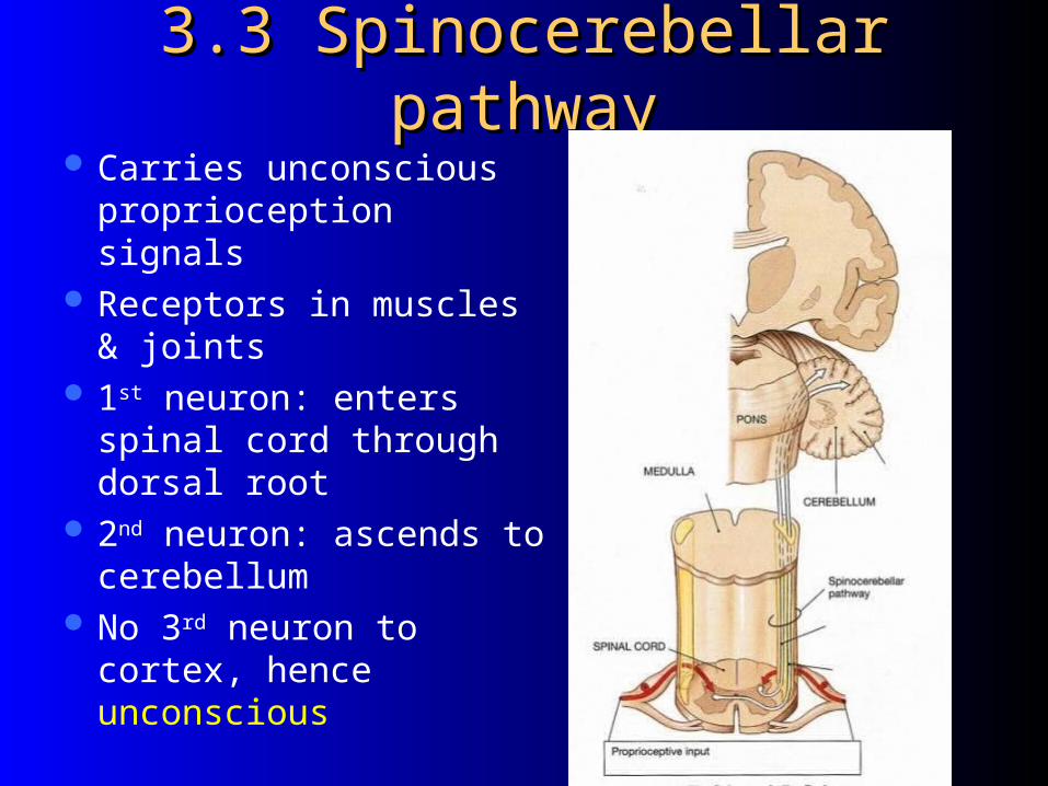
3.3 Spinocerebellar pathway3.3 Spinocerebellar pathway Carries unconscious
proprioception signals Receptors in muscles &
joints 1st neuron: enters spinal
cord through dorsal root 2nd neuron: ascends to
cerebellum No 3rd neuron to cortex,
hence unconscious
Spinocerebellar tract damageSpinocerebellar tract damage
Cerebellar ataxiaClumsy movementsIncoordination of the limbs (intention
tremor)Wide-based, reeling gait (ataxia)Alcoholic intoxication produces similar
effects!
Introductionmajor sensory pathways dorsal column system spinocerebellar tract sensory and motor ataxia
Motor & Sensory Ataxia
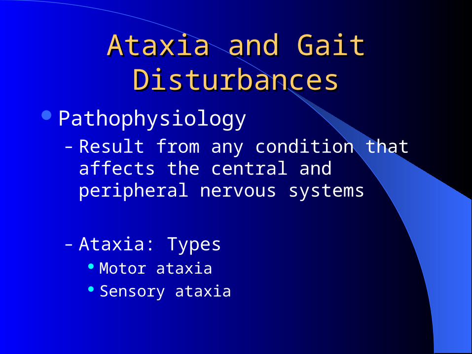
Ataxia and Gait DisturbancesAtaxia and Gait Disturbances
Pathophysiology– Result from any condition that affects the
central and peripheral nervous systems
– Ataxia: Types Motor ataxia Sensory ataxia
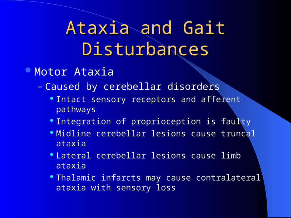
Ataxia and Gait DisturbancesAtaxia and Gait Disturbances
Motor Ataxia– Caused by cerebellar disorders
Intact sensory receptors and afferent pathways Integration of proprioception is faulty Midline cerebellar lesions cause truncal ataxia Lateral cerebellar lesions cause limb ataxia Thalamic infarcts may cause contralateral ataxia
with sensory loss
Ataxia and Gait DisturbancesAtaxia and Gait Disturbances
Sensory Ataxia– Failure of proprioceptive information to the CNS
– May be due to disorders of spinal cord or peripheral nerves
– Can be compensated for by visual inputs

Extra informationsExtra informations
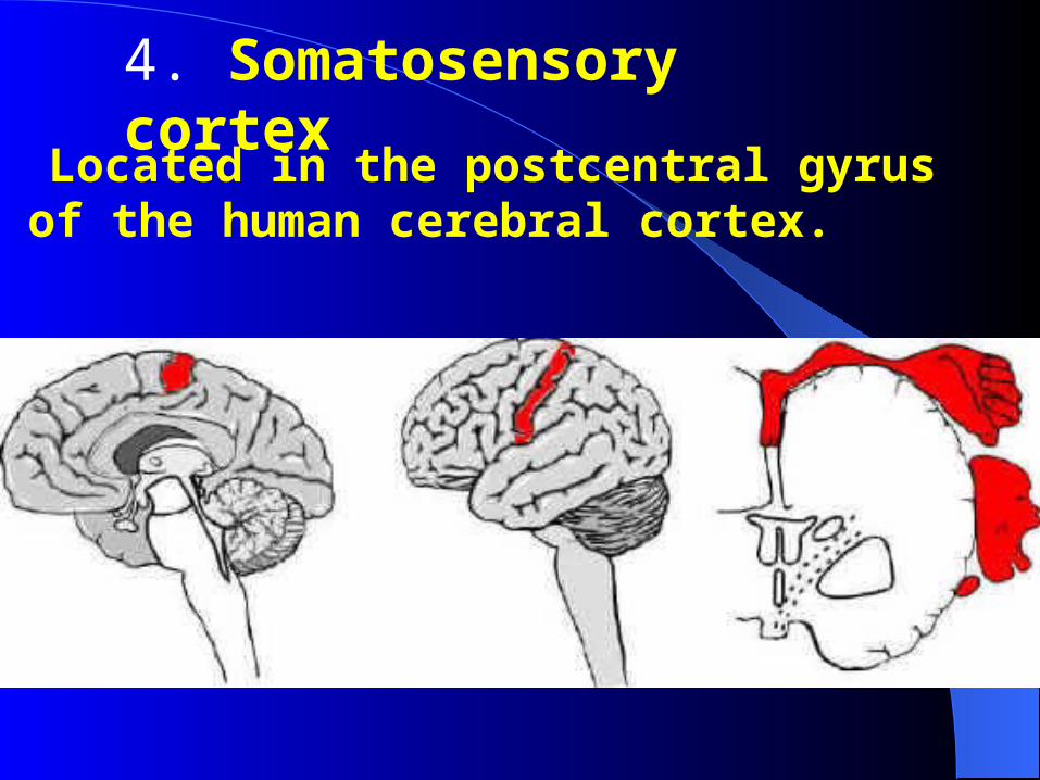
4. Somatosensory cortex
Located in the postcentral gyrus of the human cerebral cortex.
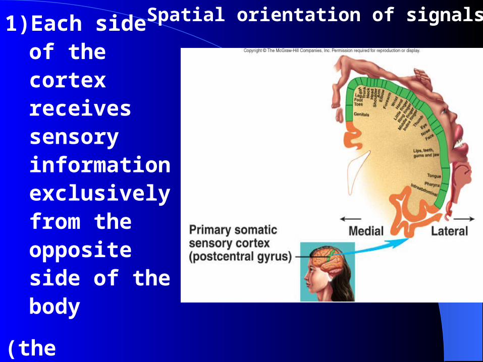
Spatial orientation of signals.1) Each side of the cortex receives sensory information exclusively from the opposite side of the body
(the exception: the same side of the face).

Spatial orientation of signals.2)The lips, face and thumb are represented by large areas in the somatic cortex,
whereas the trunk and lower part of the body, relatively small area.
3)The head in the most lateral portion, and the lower body is presented medially