the art of botany: the effect of drought on plant anatomy
TRANSCRIPT

Mānoa Horizons
Volume 3 | Issue 1 Article 19
11-15-2018
The Art of Botany: The Effect of Drought on PlantAnatomyAmanda WongUniversity of Hawaiʻi at Mānoa
Follow this and additional works at: https://kahualike.manoa.hawaii.edu/horizons
Part of the Botany Commons
This Article is brought to you for free and open access by Kahualike. It has been accepted for inclusion in Mānoa Horizons by an authorized editor ofKahualike. For more information, please contact [email protected].
Recommended CitationWong, Amanda (2018) "The Art of Botany: The Effect of Drought on Plant Anatomy," Mānoa Horizons: Vol. 3 : Iss. 1 , Article 19.Available at: https://kahualike.manoa.hawaii.edu/horizons/vol3/iss1/19
Mānoa Horizons, Vol. 3, 2018, pp. 100–104Copyright © 2018 by the University of Hawaiʻi at Mānoa
100
The Art of BotanyThe Effect of Drought on Plant Anatomy
Amanda Wong
Botany 420 (Plant Form and Function)Mentor: Dr. Kasey Barton
Plants are negatively affected by water deficiencies, and water stress is expected to increase due to more frequent and prolonged droughts from climate change. I investigated the effect of drought on the internal anatomy of the invasive Syzygium cumini plant. In the greenhouse, S. cumini plants were grown, such that half of the plants were watered daily, while the remaining number of plants did not receive any water for the entire experiment. After nine weeks, the base of the main stem was cut into thin disks, stained, and the center was viewed through a microscope for the pith cells that provide structural support, conduct water, and store starch. The regularly watered plants had circular shaped piths with expanded, rigid cells filled with water and starch, while the non-watered plants had pinched and elongated piths with shriveled cells filled with air, light staining, and a lack of starch. S. cumini demonstrated exceptional drought tolerance with no mortality and only slight wilting; however, the lack of water and the energy storage starch in the pith cells indicates that the plants were stressed. This experiment provides insight into the ability of the drought-tolerant S. cumini to become more invasive under climate change-induced drought in Hawai‘i.
Introduction
Earth is home to a diversity of plant species that exhibit re-markable variation in their internal and external structure. These structural differences within individuals of a species are not only attributed to genetic variation but are also influenced by interactions with herbivores, microbes, and their environ-ments. Water deficiency due to drought is a major environ-mental stressor on plants and is expected to increase under
climate change (Jaleel et al. 2009; Chadwick et al. 2016). In Hawai‘i, climate change-induced drought will increase in du-ration with less frequent rainfall and less rainfall overall (Chen and Chu 2014; Chu et al. 2010).
Drought stress negatively influences plant development that is apparent through a decrease in leaf size, plant height, and overall plant size. The visible reduction in plant devel-opment is driven by a decrease in turgor pressure, or fluid pressure, throughout the plant due to water stress at the cel-lular level that inhibits cell expansion and division (Jaleel et al.
I am a senior majoring in global environmental science with a concentration in sustainability science and a botany minor. After graduating from the University of Hawai‘i at Mānoa, I plan on earning my M.S. and Ph.D. in ecology or environmental science to gain and apply new knowledge and skills to make Hawai‘i a better place. As a current SEEDS fellow through the Ecological Society of America, I aspire to become a leader in my community by finding and implementing solutions to environmental problems that affect the people of Hawai‘i, my home. This research project was conducted as a section of my final research project for my Botany 420 class, Plant Form and Function. With my project’s colorful plant cell visuals, I hope to bridge the gap and connect with people from all backgrounds and disciplines to science.
Manoa Horizons Online 3.indb 100 10/10/2018 10:53:40 PM
Wong The Art of Botany 101
2009). Pith cells are essential for maintaining turgor pressure, water balance, and structural support throughout the whole plant through water storage at the center of the plant’s stem, water redistribution to water-stressed cells, and the presence of rigid lignin in the pith cell walls (Chabannes et al. 2001; Knip-fer et al. 2017). Here, the cellular stem anatomy of the drought- tolerant Syzygium cumini (Myrtaceae), commonly known as java plum, is examined and exposed to regular watering or drought through a greenhouse experiment.
Species Description: Syzygium cumini
S. cumini is a large tree (approximately 60 feet tall) with pale bark, ovate, opposite aromatic leaves, red/pink/orange young leaves and petioles, small clustered white flowers, and small glossy, purple/dark purple/black oval berries with one seed per fruit (Motooka et al. 2003; ISSG 2018). It is native to South Asia, and records indicate that S. cumini were cultivated in Ha-wai‘i by 1871 (Motooka et al. 2003). S. cumini was introduced not only in Hawai‘i but across the Pacific for its ornamental features, timber, and fruit and has become invasive on many islands through the dispersal of the seeds by frugivorous birds and even pigs (ISSG 2018). In Hawai‘i, S. cumini invades me-sic valleys along the stream edges and displaces native species by preventing their reestablishment (Motooka et al. 2003; ISSG 2018). S. cumini flourishes along the stream banks, but can also tolerate flooding and even drought (ISSG 2018). The Weed Risk Assessment score of 9 with a “High Risk” classifi-cation (PIER 2018) and the ability for S. cumini to tolerate not only flooding but also drought, makes S. cumini a species of interest for this experiment.
Methods
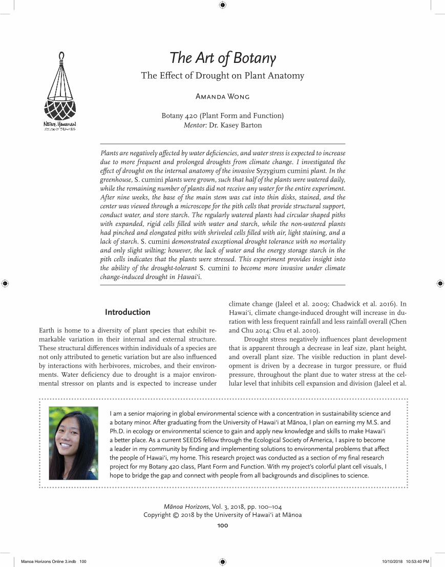
S. cumini fruits were collected from McBryde Garden, Kauai, and the seeds were sown in Dr. Barton’s Botany lab at the University of Hawai‘i at Mānoa (UHM) in November 2017. After the seedlings germinated and developed two true leaves, the seedlings were transplanted into 2-gallon pots with 4 kg of soil and slow-release Osmocote fertilizer in the Pope Greenhouse at UHM (Figure 1). The experimental wa-tering treatments were initiated immediately following seed-ling transplant such that the “control” treatment plants were watered daily, while the “drought” treatment plants received no water at all for the duration of the experiment. At the con-clusion of the nine-week treatment phase, the plant shoots (stems and leaves) were harvested at the soil interface where the shoot and root intersect.
In the lab, the base of the main stem closest to the cut was cut into thin, circular cross sections with a razor blade dipped in water. The cross sections were placed in one drop
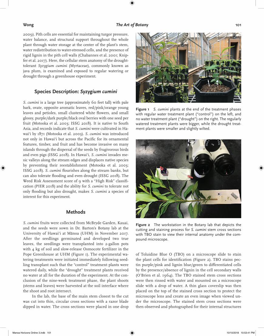
of Toluidine Blue O (TBO) on a microscope slide to stain the plant cells for identification (Figure 2). TBO stains pec-tin purple/pink and lignin blue/green to differentiated cells by the presence/absence of lignin in the cell secondary walls (O’Brien et al. 1964). The TBO stained stem cross sections were then rinsed with water and mounted on a microscope slide with a drop of water. A thin glass coverslip was then placed on the top of the stained cross section to protect the microscope lens and create an even image when viewed un-der the microscope. The stained stem cross sections were then observed and photographed for their internal structures
Figure 1 S. cumini plants at the end of the treatment phases with regular water treatment plant (“control”) on the left, and no water treatment plant (“drought”) on the right. The regularly watered treatment plants were bigger, while the drought treat-ment plants were smaller and slightly wilted.
Figure 2 The workstation in the Botany lab that depicts the cutting and staining process for S. cumini stem cross sections with TBO stain to view their internal anatomy under the com-pound microscope.
Manoa Horizons Online 3.indb 101 10/10/2018 10:53:41 PM
102 Mānoa Horizons Vol. 3, Fall 2018
through the compound microscope at a magnification of 10x and 40x.
Additional stem cross sections were cut from extra S. cumini that were not included in the experimental treatment groups but germinated from the same batch of seeds and were regularly watered. These cross sections were stained with one drop of potassium iodide solution (IKI or Lugol’s iodine) to identify the contents of the pith cells. IKI stains amylose starch blue and amylopectin starch red/purple in the starch storage compartments of the plant cells or amyloplasts (Whelan 1958; Zobel 1988). The IKI stained stem cross sections were mount-ed on a microscope slide with a drop of water and a coverslip. The IKI stained starch was viewed through the compound mi-croscope and photographed at 10x and 40x magnification.
Results
S. cumini control plants that received water daily were visibly larger in leaf number, leaf size, plant height, stem thickness, and overall plant size, compared to the drought plants that did not receive any water for nine weeks. In terms of the cellu-lar organization of the TBO stained piths, regularly watered S. cumini stem cross sections had circularly shaped piths with green and blue lignified pith cells (Figure 3), while the plants exposed to drought had piths that were elongated and pinched with pink and purple non-lignified pith cells (Figure 4). The TBO stained individual pith cells of the regularly watered plants were fully expanded with clearly defined cell walls and contained multiple amyloplasts with starch (Figure 5), while the pith cells of the plants exposed to the drought treatment were shriveled with indistinct cell walls, lightly stained, and lacking in starch (Figure 6). The IKI stain dyed the starch pur-ple at the center of the stem in the pith with surrounding bands of starch and inside the multiple amyloplasts within each pith cell (Figure 7).
Discussion
Despite evidence that drought influenced the internal anato-my of the S. cumini plants, this species demonstrated amazing drought tolerance with no mortality, and only slight wilting, and maintained slower growth during a nine-week drought with no watering. Water stress induced by the drought treat-ment appeared to lead to a decrease in plant growth and turgor pressure or fluid pressure throughout the plant that was evi-dent through the smaller and slightly wilted plants (Beauzamy et al. 2014). The effects of water stress through the decrease in turgor pressure were also apparent in the internal anatomy of the plants.
The plants exposed to drought and water stress exhibited elongated and pinched piths compared to the circular shaped
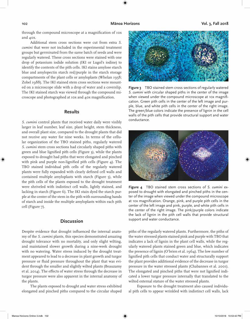
Figure 3 TBO stained stem cross sections of regularly watered S. cumini with circular shaped piths in the center of the image when viewed under the compound microscope at 10x magnifi-cation. Green pith cells in the center of the left image and pur-ple, blue, and white pith cells in the center of the right image. The green/blue colors indicate the presence of lignin in the cell walls of the pith cells that provide structural support and water conductance.
Figure 4 TBO stained stem cross sections of S. cumini ex-posed to drought with elongated and pinched piths in the cen-ter of the image when viewed under the compound microscope at 10x magnification. Orange, pink, and purple pith cells in the center of the left image and pink, purple, and white pith cells in the center of the right image. The pink/purple colors indicate the lack of lignin in the pith cell walls that provide structural support and water conductance.
piths of the regularly watered plants. Furthermore, the piths of the water stressed plants stained pink and purple with TBO that indicates a lack of lignin in the plant cell walls, while the reg-ularly watered plants stained green and blue, which indicates the presence of lignin (O’brien et al. 1964). The low number of lignified pith cells that conduct water and structurally support the plant provides additional evidence of the decrease in turgor pressure in the water stressed plants (Chabannes et al. 2001). The elongated and pinched piths that were not lignified indi-cated a lower turgor pressure internally that translated to the wilted external stature of the water stressed plants.
Exposure to the drought treatment also caused individu-al pith cells to appear wrinkled with indistinct cell walls, lack
Manoa Horizons Online 3.indb 102 10/10/2018 10:53:42 PM

Wong The Art of Botany 103
color from the TBO stain, and to contain a low abundance of energy storing starch (Jaleel et al. 2009). The light color inten-sity from the TBO stain may be attributed to pith cell autoly-sis or cell death by their enzymes that form air pockets within the pith cells (Knipfer et al. 2017). The pith cells filled with air instead of water do not allow the TBO stain to become effec-tively absorbed by the cell, resulting in a lack of color. The low abundance of amyloplasts containing starch indicates that the plants were allocating their energy towards maintaining plant function under drought conditions instead of storing energy in the form of starch (Zobel 1988). On the other hand, the purple color of the starch in the watered plants suggests that most of the starch was amylopectin versus amylose because amylopec-
tin stains red/purple and amylose stains blue (Whelan 1958; Zobel 1988). Although the IKI stain was applied to addition-al S. cumini and not the treatment plants dyed with the TBO stain, the starch in the treatment plants were also expected to stain purple based on these results.
Amongst the differences in appearance, the starch abun-dance and amount of stain absorbance were the most consis-tent characteristics that distinguished the regularly watered and drought treatment plants. All of the plants within each treatment group did not possess the exact same features dis-cussed but exhibited similarities between the two treatment groups and differences within the treatments. This variation within the S. cumini plants is inherently natural to the diversity expressed in botany. Even though the drought treatment plants appeared to tolerate drought with only slight wilting under a decrease in turgor pressure, the pinched, non-lignified piths, shriveled pith cells filled with air, lack in stain absorbance, and low starch abundance indicate that the plants were stressed. Overall, the effects of drought stress on S. cumini were evident through the visible differences in external and internal plant structure when compared to the regularly watered S. cumini. However, S. cumini’s ability to maintain growth and function over the nine-week duration of the drought treatment indicates the drought tolerance of this species.
Conclusion
Drought already negatively impacts plant growth through wa-ter stress, and is expected to increase in duration and frequen-cy under climate change. This experiment investigated the effect of drought stress on cellular plant anatomy of S. cumini
Figure 5 TBO stained stem cross sections of regularly watered S. cumini from Figure 3 with fully expanded pith cells filled with water that contain starch when viewed under the compound mi-croscope at 40x magnification. Blue, green, and white pith cells in the left image and purple, blue beige pith cells in the right image that both contain starch in the amyloplasts.
Figure 6 TBO stained stem cross sections of S. cumini ex-posed to drought from Figure 4 with shriveled pith cells that have obscure cell walls and do not contain starch when viewed under the compound microscope at 40x magnification. Beige, purple, and blue pith cells in the left image and beige, blue, and purple pith cells in the right image that are filled with air and do not contain amyloplasts with starch. The light colors indicate the inability for the pith cells to absorb the stain due to the lack of water in the cell.
Figure 7 Stem cross sections of additional, regularly watered S. cumini with amyloplasts containing the purple stained starch from the IKI stain that were viewed under the compound mi-croscope. The image on the left viewed at 10x magnification de-picts the abundance of purple stained starch in the pith at the center of the stem with bands of starch surrounding the pith. The image on the right viewed at 40x magnification shows mul-tiple amyloplasts with stained purple starch in each pith cell.
Manoa Horizons Online 3.indb 103 10/10/2018 10:53:44 PM

104 Mānoa Horizons Vol. 3, Fall 2018
stem cross sections and found that the majority of plants ex-posed to drought had pinched, non-lignified piths, shriveled pith cells filled with air, faint stain absorbance, and low starch abundance. Although the drought tolerance of S. cumini was favorable for this experiment, the results of this experiment could indicate the ability of S. cumini to become more invasive even under climate change-induced drought. Even though S. cumini exhibited high drought tolerance, its exceptional perfor-mance in drought is not indicative of the performance of other plant species under climate change-induced drought. There-fore, future work should experimentally investigate how oth-er plant species respond to climate change-induced drought, and also incorporate how drought influences the interactions between other plants and herbivores, especially the interac-tions between native and invasive plant species. I propose a greenhouse experiment that investigates the competitive in-teractions between the native Syzygium sandwicensis and the non-native Syzygium cumini under climate change-induced drought.
References
Beauzamy, L., Nakayama, N., and Boudaoud, A. (2014) Flowers under pressure: ins and outs of turgor regulation in de-velopment. Annals of Botany, 114(7), 1517–1533.
Chabannes, M., Ruel, K., Yoshinaga, A., Chabbert, B., Jauneau, A., Joseleau, J. P., and Boudet, A. M. (2001) In situ anal-ysis of lignins in transgenic tobacco reveals a differential impact of individual transformations on the spatial pat-terns of lignin deposition at the cellular and subcellular levels. The Plant Journal, 28(3), 271–282.
Chadwick, R., Good, P., Martin, G., and Rowell, D. P. (2016) Large rainfall changes consistently projected over sub-stantial areas of tropical land. Nature Climate Change, 6(2), 177.
Chen, Y. R., and Chu, P. S. (2014) Trends in precipitation ex-tremes and return levels in the Hawaiian Islands under a changing climate. International Journal of Climatolo-gy, 34(15), 3913–3925.
Chu, P. S., Chen, Y. R., and Schroeder, T. A. (2010) Changes in precipitation extremes in the Hawaiian Islands in a warming climate. Journal of Climate, 23(18), 4881–4900.
[ISSG] Invasive Species Specialist Group. (2018) Species Pro-file: Syzygium cumini. Global Invasive Species Database.
Jaleel C.A., Manivannan P.A., Wahid A., Farooq M., Al-Juburi H.J., Somasundaram R.A., and Panneerselvam R. (2009) Drought stress in plants: a review on morphological char-acteristics and pigments composition. International Jour-nal of Agriculture and Biology, 11(1), 100–5.
Knipfer, T., Cuneo, I.F., Earles, J.M., Reyes, C., Brodersen, C.R. and McElrone, A.J. (2017) Storage compartments for capillary water rarely refill in an intact woody plant. Plant Physiology, 175(4), 1649–1660.
Motooka, P., et. al. (2003) Syzygium cumini. Weeds of Hawai‘i’s Pastures and Natural Areas: An Identification and Manage-ment Guide.
O’brien, T.P., Feder, N. and McCully, M.E. (1964) Polychromat-ic staining of plant cell walls by toluidine blue O. Proto-plasma, 59(2), 368–373.
[PIER] Pacific Island Ecosystems at Risk. (2018) Syzygium cum-ini. Hawaiian Ecosystems at Risk Project (HEAR).
Whelan, W.J., (1958) Starch and similar polysaccharides. For-mation-Storage-Mobilization and Transformation of Carbo-hydrates, pp. 154–240. Springer, Berlin, Heidelberg.
Zobel, H. F. (1988) Molecules to granules: a comprehensive starch review. Starch, 40(2), 44–50.
Manoa Horizons Online 3.indb 104 10/10/2018 10:53:44 PM