the antioxidative role of natural compounds from a green
TRANSCRIPT
Research ArticleThe Antioxidative Role of Natural Compounds from a GreenCoconut Mesocarp Undeniably Contributes to Control DiabeticComplications as Evidenced by the Associated Genes andBiochemical Indexes
Rickta Rani Das ,1 Md. Atiar Rahman ,1 Salahuddin Qader Al-Araby ,1
Md. Shahidul Islam ,1 Md. Mamunur Rashid ,1 Nouf Abubakr Babteen ,2
Afnan M. Alnajeebi ,2 Hend Faisal H. Alharbi ,3 Philippe Jeandet ,4
Md. Khalid Juhani Rafi ,1 Tanvir Ahmed Siddique,1 Md. Nazim Uddin ,5
and Zainul Amiruddin Zakaria 6
1Department of Biochemistry and Molecular Biology, University of Chittagong, Chittagong 4331, Bangladesh2Department of Biochemistry, Collage of Science, University of Jeddah, Jeddah 80203, Saudi Arabia3Department of Food Science and Human Human Nutrition, Collage of Agriculture and Veterinary Medicine, Qassim University,Buraydah, Saudi Arabia4Department of Biology and Biochemistry, Faculty of Sciences, University of Reims, PO Box 1039, Reims, France5Institute of Food Science and Technology, Bangladesh Council of Scientific and Industrial Research, Dhaka 1205, Bangladesh6Department of Biomedical Science, Faculty of Medicine and Health Sciences, Universiti Putra Malaysia (UPM), Serdang,43400 Selangor, Malaysia
Correspondence should be addressed to Md. Atiar Rahman; [email protected] and Zainul Amiruddin Zakaria; [email protected]
Received 26 May 2021; Accepted 5 July 2021; Published 28 July 2021
Academic Editor: Cristina Cosentino
Copyright © 2021 Rickta Rani Das et al. This is an open access article distributed under the Creative Commons Attribution License,which permits unrestricted use, distribution, and reproduction in any medium, provided the original work is properly cited.
The purpose of this study was to look into the effects of green coconut mesocarp juice extract (CMJE) on diabetes-related problemsin streptozotocin- (STZ-) induced type 2 diabetes, as well as the antioxidative functions of its natural compounds in regulating theassociated genes and biochemical markers. CMJE’s antioxidative properties were evaluated by the standard antioxidant assays of1,1-diphenyl-2-picrylhydrazyl (DPPH), superoxide radical, nitric oxide, and ferrous ions along with the total phenolic andflavonoids content. The α-amylase inhibitory effect was measured by an established method. The antidiabetic effect of CMJEwas assayed by fructose-fed STZ-induced diabetic models in albino rats. The obtained results were verified by bioinformatics-based network pharmacological tools: STITCH, STRING, GSEA, and Cytoscape plugin cytoHubba bioinformatics tools. Theresults showed that GC-MS-characterized compounds from CMJE displayed a very promising antioxidative potential. In ananimal model study, CMJE significantly (P < 0:05) decreased blood glucose, serum alanine aminotransferase (ALT), aspartateaminotransferase (AST), creatinine, uric acid, and lipid levels and increased glucose tolerance as well as glucose homeostasis(HOMA-IR and HOMA-b scores). The animal’s body weights and relative organ weights were found to be partially restored.Tissue architectures of the pancreas and the kidney were remarkably improved by low doses of CMJE. Compound-proteininteractions showed that thymine, catechol, and 5-hydroxymethylfurfural of CMJE interacted with 84 target proteins. Of the top15 proteins found by Cytoscape 3.6.1, 8, CAT and OGG1 (downregulated) and CASP3, COMT, CYP1B1, DPYD, NQO1, andPTGS1 (upregulated), were dysregulated in diabetes-related kidney disease. The data demonstrate the highly prospective use ofCMJE in the regulation of tubulointerstitial tissues of patients with diabetic nephropathy.
HindawiOxidative Medicine and Cellular LongevityVolume 2021, Article ID 9711176, 22 pageshttps://doi.org/10.1155/2021/9711176

1. Introduction
Diabetes mellitus (DM), a metabolic disorder characterizedby hyperglycemia induced by insulin secretion deficiencyand/or resistance to its action, affects more than millions ofpeople across the world [1]. DM impairs several nonenzy-matic and enzymatic antioxidative defense mechanisms thatlead to cause oxidative stress as well as tissue damage inDM-associated comorbidities such as cataracts, neuropathy,nephropathy, and retinopathy [2]. Until now, no single effec-tive treatment for DM has been developed in medicine, andthe current therapeutic supports such as biguanides, sulfo-nylureas, meglitinides, thiazolidinediones, dipeptidyl pepti-dase IV inhibitors, and α-glucosidase inhibitors and theiranalogs have many side effects, such as weight gain, hypogly-cemia, gastrointestinal disorders, liver and kidney damage,and hypersensitivity reactions [2, 3].
The above-mentioned side effects suggest that furtherdevelopment of new, safer, and more powerful oral antihy-perglycemic agents, particularly in long-term therapy, isneeded. In this context, medicinal plants have emerged aspromising adjuvants to treat chronic, oxidative stress-mediated disorders [3]. Several medicinal plants recom-mended for the treatment of DM have been shown to protectβ-cells, increase insulin secretion and glucose absorption bythe adipose tissue, and decrease glucose absorption in theintestines [2, 4]. Some experiments have shown in recentyears that most plants produce carotenoids, flavonoids, ter-penoids, alkaloids, glycosides, and anthocyanin that exert asignificant impact on diabetes and other chronic diseases aswell as minimize oxidative stress [5]. Treatments whichinvolve the use of medicinal plants provoking antioxidativeactions are therefore highly recommended [6, 7].
Different parts of coconut have long been used as one ofthe most popular edible foods in almost every part of theworld. Nutritional and medicinal values of coconuts havebeen investigated, and especially, their antibacterial, antihy-pertensive, oral microflora inhibitory, antiviral, antifungal,antidermatophytic, antiparasitic, hypoglycemic, immunosti-mulant, and hepatoprotective properties are reported bymany scientists [8–10]. Microminerals and nutrients of coco-nut water are essentially important for human health whilethe endocarp part is cited to contain high contents of pheno-lic and flavonoids. Coconut milk has also been shown effec-tive in the management of diabetes [10]. Interestingly, themesocarp part of coconut has not yet been studied, and wetried here to investigate the antioxidative effect of coconutmesocarp juice which eventually and undeniably contributesto the management of diabetes and renal diabetic complica-tions using a fructose-fed streptozotocin-induced diabeticrat model. The observed effect has been verified and net-worked with the genes linked in reducing oxidative stress inthe biological system and through bioinformatics-based net-work pharmacological approach in a computational model.
2. Materials and Methods
2.1. Collection of Coconut Mesocarp. Green coconut meso-carps were collected from the local green coconut seller
around the University of Chittagong. The mesocarp juicewas extracted using a mechanical sugarcane juicer machine(detailed in the extraction section) with the aid of a local sug-arcane juice seller. The mesocarp part of coconut has beenkeenly identified with the help of a plant scientist Dr. SheikhBokhtear Uddin, Professor, Department of Botany, Univer-sity of Chittagong. A sample specimen of collected mesocarphas been preserved in the institutional herbarium with anidentification number (MPSS2017/02).
2.2. Chemicals and Reagents. All the chemicals and reagentsused in this study were of analytical grade unless specified.ABTS (2,2′-azino-bis(3-ethylbenzthiazoline-6-sulfonic acid)),dinitrosalicylic acid, Folin-Ciocalteu reagent, dimethyl sulfox-ide (DMSO), 1,1-diphenyl-2-picrylhydrazyl (DPPH), nitro bluetetrazolium (NBT), potassium ferric cyanide, sodium hydrox-ide, trichloroacetic acid (TCA), nitroprusside, N-(1-naphthyl)ethylene diamine dihydrochloride, O-phenanthroline, andα-amylase were procured from Sigma-Aldrich Co., St. Louis,USA. Butanol, n-hexane, methanol (absolute), ethanol(99.99%), and acetone were purchased from Sigma-Aldrich.
2.3. Preparation of Coconut Mesocarp Juice Extract (CMJE).Coconut mesocarp juice extract was prepared as previouslydescribed by Rahman et al. [11]. Briefly, the green coconut’smesocarp, which is also known as coir, is situated justbeneath the exocarp or outer skin of the fruit. The exocarppart was plucked, and the mesocarp was removed. The liquidsap of the mesocarp (coir) was then mechanically collected.The collected sap was filtered by using filter paper (Whatmanfilter paper #1) and a funnel. The filtered sap was then evap-orated by an electromantle at 45-50°C for several days. Thesample collected from the electromantle was further evapo-rated through a rotary evaporator (RE 200, Bibby SterilinLtd., UK) at 55-60°C, and the final extract was collected andstored in the refrigerator.
2.4. Screening for Phytochemical Content of CMJE
2.4.1. Total Flavonoid Content (TFC) and Total PhenolicContent (TPC) Determinations. The total flavonoid content(TFC) of CMJE was determined according to the methodestablished by Kumaran and Karunakaran [12]. The totalphenolic content (TPC) of the CMJE was measured accord-ing to a method described by Singleton and Rossi [13].
2.4.2. Gas Chromatography-Mass Spectroscopy (GC-MS)Analysis of CMJE. The crude CMJE was analyzed by GC-MSusing electron impact ionization (EI) with a gas chromato-graph (GC-17A, Shimadzu Corporation, Kyoto, Japan)coupled to a mass spectrometer (GC-MS TQ 8040, ShimadzuCorporation, Kyoto, Japan). A fused silica capillary column(Rxi-5ms; 0.25m film thickness) is coated with DB-1 (J&W).The inlet temperature of the capillary was set at 260°C,and the oven temperature was set at 70°C (0min), 10°C and150°C (5min), 12°C and 200°C (15min), and 12°C and 220°C(5min). The column flow rate was 0.6mL/min of helium gasat a constant pressure of 90kPa. The auxiliary (GC to MSinterface) temperature was set at 280°C. TheMSwas set in scanmode with a scanning range of 40-350amu. The mass range
2 Oxidative Medicine and Cellular Longevity
was set in the range of 50-550m/z. The prepared sample wasthen run for GC-MS analysis. The total GC-MS running timewas 35 minutes. All peak areas were compared with the data-base in the GC-MS library version NIST 08-S.
2.4.3. Estimation of Beta-Carotene and Lycopene Contents ofCMJE. Beta-carotene and lycopene contents of CMJE wereestimated using a slightly modified method of that describedby Kumari et al. [14]. Briefly, 100mg of the extract was mixedwith 10mL of the acetone-hexane mixture (4 : 6) for 1 minuteand filtered. The absorbance was measured at three differentwavelengths (453, 505, and 663nm).
The beta-carotene and lycopene contents were calculatedas follows:
Beta-carotene: ðmg/100mLÞ = ð0:216 × A663Þ − ð0:304 ×A505Þ + ð0:452 × A453Þ
Lycopene: ðmg/100mLÞ = −ð0:0458 × A663Þ + ð0:372 ×A505Þ − ð0:0806 × A453Þ.
2.4.4. Determination of the DPPH Free Radical ScavengingActivity of CMJE. The DPPH free radical scavenging effectwas measured according to the method of Shen et al. [15]supplemented with the established protocol described byBrand-Williams et al. [16]. Ascorbic acid was used as a refer-ence antioxidant agent in this experiment. The requiredamount (0.96mg) of ascorbic acid and CMJE sample wasindividually dissolved in 12mL methanol to prepare stocksolution. The stock solution of both CMJE and ascorbic acidwas diluted to the concentrations of 40, 20, 10, and 5μg/mL.Two milliliters of both CMJE solution and ascorbic acid solu-tion of different concentrations was taken as triplicate intotest tubes where 2mL of the freshly prepared DPPH solutionwas added. The reaction mixture was incubated in the darkfor 30min at room temperature, and the absorbance of thereaction mixture was measured at 517nm by using a visible
spectrophotometer. Control was prepared in similar mannerexcluding sample.
The percentage of inhibition was calculated by the fol-lowing equation:
%Inhibition = A0 − A1A0
� �× 100 ð1Þ
where A0 is the absorbance of control (freshly preparedDPPH solution) and A1 is the absorbance of extract/Std.Then, the percentage of scavenging activity or inhibitionwas plotted against the concentration, and IC50 was calcu-lated by the linear regression analysis from the graph.
2.4.5. Determination of ABTS Radical Scavenging Activity ofCMJE. The ABTS free radical scavenging activity of CMJEwas measured by using the ABTS (2,2′-azino-bis(3-ethyl-benzthiazoline-6-sulfonic acid)) radical cation decolorizationassay [17]. ABTS•+ was generated by reacting 7mM ABTSaqueous solution with 2.45mM potassium persulfate in thedark for 12-16 h at room temperature. At the beginning ofthe assay, this solution was diluted in ethanol (about 1 : 49,v/v) and equilibrated at 30 ± 2°C to give an absorbance of0:7 ± 0:02 at 734nm. The stock solution of the CMJE extractwas diluted to yield a concentration range of 50-8000μg/mL.The final concentration (0-15μM) was obtained by the addi-tion of 1mL of the diluted ABTS•+ solution to 62μL ofCMJE sample in ethanol. After 40 minutes of mixing, absor-bance was estimated at 25°C. Trolox and ethanol were used asa positive control and blank, respectively. At each dilution ofthe standard and sample, triplicate determinations weremade, and absorption was measured at 734nm in the UV-Vis spectrophotometer (UV-1200S UV-VIS 1200, ShimadzuCorporation, Japan). The ABTS•+ scavenging capability ofthe extract was compared with that of Trolox. The percentageinhibition calculated as follows:
2.4.6. Determination of Superoxide Scavenging Activity ofCMJE. The superoxide radical scavenging power of CMJEwas assessed by an updated protocol of Rana et al. [18].Using alkaline dimethyl sulfoxide (DMSO), the superoxideradical was formed by dissolving 250μL of 1M NaOH indouble-distilled water to 49.750μL of DMSO. With a NaOHconcentration of 5mM and a volume of 50mL, air bubbledthrough the mixture for 1 h and 30 minutes. The solutionof NBT (nitro blue tetrazolium) was prepared by dissolving12mg of NBT in 12mL of double-distilled water (pH7.4),
with a final concentration of NBT of 1mg/mL. The samplewas diluted, and each test tube received a volume of 43μLof each sample, where the sample concentration rangedfrom 25 to 800μg/mL. Furthermore, 143μL of alkalineDMSO and 14μL of NBT (1mg/mL) were added to each testtube, incubated for 20 minutes and read at 560nm for absor-bance. DMSO and ascorbic acid were used as negative andpositive controls, respectively. Triplicates were confirmedfor each experiment. The percentage inhibition calculatedas follows:
ABTS radical scavenging activity %ð Þ = Absorbance of control –Absorbance of the sampleð ÞAbsorbance of control
� �× 100 ð2Þ
Superoxide radical scavenging activity %ð Þ = sample Absorbance − control Absorbancesample Absorbance
� �× 100 ð3Þ
3Oxidative Medicine and Cellular Longevity

2.4.7. Estimation of Nitric Oxide Scavenging Activity of CMJE.The nitric oxide scavenging effect was estimated based on theprinciple of the analysis of nitrite ions which are generatedfrom sodium nitroprusside through nitric oxide in an aque-ous solution at a physiological pH [19]. Nitric oxide scaven-gers compete with oxygen, resulting in decreased nitrite ionproduction. For the experiment, 1.5mL of sodium nitroprus-side (10mM) in phosphate-buffered saline (pH7.4) wascombined with various 100μL volumes of water-dissolved
CMJE extracts and incubated for 150min at room tempera-ture. Without CMJE, the same reaction mixture, but an equalamount of water, served as control. 1.5mL of the Griessreagent, 1% sulfanilamide, 2 percent H3PO4, and 0.1 percentN-(1-naphthyl) ethylene diamine dihydrochloride, was addedafter the incubation time. At 546nm against the blank, theabsorbance of the chromophore formed was read. Ascorbicacid was used as a positive control. The nitric oxide radicalscavenging power was calculated by the following formula:
2.4.8. Determination of the Iron-Chelating Activity of CMJE.The iron-chelating activity of CMJE was measured usingthe method of Benzie and Strain [20]. The theory is basedon the formation of, and destruction of, the O-phenanthro-line-Fe2+ complex in the presence of chelating agents. A reac-tion mixture containing 1mL of 0.05% O-phenanthroline inmethanol, 2mL of fresh ferrous chloride (200μM), and 2mLof different CMJE concentrations was incubated at roomtemperature for 10min, and absorbance was measured at510nm. Experiments were performed in triplicate, and theoperation was correlated with the usual positive control,ascorbic acid.
Inhibition of Iron radical %ð Þ = A0 − A1½ �/A0 × 100 ð5Þ
where A0 is the test absorbance and A1 is the controlabsorbance.
2.5. Determination of the α-Amylase Inhibition Capacity ofCMJE. The α-amylase inhibitory action of CMJE was deter-mined by a modified procedure of McCue et al. [21, 22].Briefly, 4mg CMJE was dissolved in 5mL water to preparea stock solution of 800μg/mL, which was diluted to 50, 100,200, and 400μg/mL. Four milligrams of acarbose (standard)was dissolved in 5mL water to prepare similar concentra-tions of standard solutions as was done for CMJE sample.250μL of CMJE was mixed in a tube with 250μL of 0.02Msodium phosphate buffer (pH6.9) containing the α-amylasesolution (0.5mg/mL). This solution was preincubated at25°C for 10min, after which 250μL of 1% starch solution in0.02M sodium phosphate buffer (pH6.9) was added at timedintervals and then further incubated at 25°C for 10min. Ter-mination of the reaction was ensured by adding 500μL of thedinitrosalicylic acid (DNS) reagent. The assay mixtures werethen incubated for 5min in boiling water and cooled to roomtemperature. The reactionmixture was diluted with 5mL dis-tilled water, and the absorbance was measured at 540 nmusing a spectrophotometer (UV-1280, UV-Vis spectropho-tometer, Shimadzu Corporation, Japan). A control was pre-pared using the same procedure replacing the extract with
distilled water. The α-amylase inhibitory activity was calcu-lated as percentage inhibition:
%Inhibition = Abscontrol −Absextractð Þ/Abscontrol × 100 ð6Þ
2.6. Experimental Animals and their Maintenance. Twenty-five adult male (body weight 150-200 g, age 6-7 weeks) Wis-tar albino rats were purchased from BCSIR, Chittagong. Theanimals were randomly grouped into normal control (NC,animals received no treatment), diabetic control (DC,streptozotocin-induced and received no treatment), andtreatment group (CMJE50, CMJE100, and CMJE200mg/kgbw). The animals were individually housed in a polycarbo-nated cage bedded with wood husk at a temperature around22 ± 2°C and humidity 55-60% with a 12h light-dark cycle.All animals were supplied with a commercial pellet diet forthe entire intervention period. All animal experimentationswere carried out according to the guideline of the Institu-tional Animal Ethics Committee (EACUBS2018-4).
2.6.1. Acute Oral Toxicity Test. The acute oral toxicity testwas performed using standard laboratory conditions accord-ing to the “Organization for Environmental Control Devel-opment” guidelines (OECD: Guidelines 420; fixed-dosemethod). The allocated animals (n = 6) were administered asingle oral dose (500 to 2000mg/kg, body weight) of the testextract (CMJE). Before the administration of the extract, ratswere fasted overnight, and food was also delayed between 3and 4h. After administration, food was withheld for the next3-4 h. Experimental animals were observed individually dur-ing the first 30 minutes after dosing, periodically for the first24 minutes (special attention for the first 4 h), with particularmonitoring for possible unusual responses including allergicsyndromes (itching, swelling, skin, and rash), behavioralchanges, and mortality over the next 72 h. The median ther-apeutic effective dose was intervened as one-tenth of themedian lethal dose (LD50 > 5:0 g/kg) [23].
2.6.2. Induction of Diabetes Using Streptozotocin. Diabetesinduction was accomplished with slightly modifying of pro-tocol addressed by Al-Araby et al. [22]. Briefly, the animals
Scavenging activity of nitric oxide radical %ð Þ = Control OD − Sample ODControl OD
� �× 100 ð4Þ
4 Oxidative Medicine and Cellular Longevity

were randomly divided into control and treatment groupscomprising of 5 animals in each group. The normal controlgroup (NC) received vehicle only. Diabetic control (DC)was left untreated, and the treatment groups were adminis-tered with three different doses (CMJE50, CMJE100, andCMJE200mg/kg bw) of coconut mesocarp juice extracts.All animals except those of normal control (NC) were fedwith 10% fructose solutions before one week of intraperito-neal injection of streptozotocin (50mg/kg bw dissolved in0.1M citrate buffer, pH4.5) [24] to induce diabetes which isconfirmed with the fasting blood glucose level ≥ 16mmol/Lafter one week of injection (measured by glucometer, Accu-Chek, USA). Once the animals were confirmed diabetic afterSTZ injection, each animal of CMJE50 group has beentreated by the CMJE extract at the dose of 50mg/kg bw oncedaily; each animal of the CMJE100 group has been treatedwith the CMJE extract at the dose of 100mg/kg bw oncedaily, and each animal of CMJE200 group has been treatedwith CMJE extract at the dose of 200mg/kg bw once daily.The treatment was continued for three weeks.
2.6.3. Determination of BodyWeight, Blood Glucose, and OralGlucose Tolerance (OGT). Weekly body weights and bloodglucose levels of animals were measured and recorded. Bloodglucose was measured by tail prick method using a gluc-ometer as stated above. The glucose tolerance capacity ofeach animal was measured by the oral glucose tolerance test(OGTT) at the 3rd week of the intervention. Animals wereadministered a single dose of oral glucose solution (2 g/kgbody weight) and blood glucose levels were measured at 0(just before glucose ingestion), 30, 60, 90, and 120min afterthe glucose dose.
2.6.4. Animal’s Blood and Organ Collection. After 4 weeks ofintervention, animals were sacrificed, their blood being col-lected in heparinized test tubes as well as their kidney andpancreas. Blood samples were centrifuged at 3000 rpm for15min at 25-37°C to separate serum which was further ana-lyzed for hepatic enzymes (alanine aminotransferase, aspar-tate aminotransferase), insulin, lipid profile, uric acid, andcreatinine. The serum glucose level (mmol/L) was deter-mined using the glucose test kit based on the glucose oxidasemethod as described [25]. The pancreas and livers werewashed with 0.9% NaCl solution, wiped with tissue paper,and weighed to be preserved in 10% buffered formalin. Thekidney and the pancreas were used for histopathological inves-tigations. The homeostatic model assessment (HOMA-IR andHOMA-b) was estimated using serum insulin levels measuredat the end of the experiment using the following expression:
HOMA‐IR = Serum insulin U/Lð Þ × Blood glucose mg/dLð Þ22:5
,
HOMA − β − cell function =20 × Serum insulin inU/L
Blood glucose inmg/dL − 3:5:
ð7Þ
2.6.5. Histopathological Analyses. The pancreas and the kidneytissues were fixed with a buffered formalin solution for 48h,
dehydrated by passing through graded series of alcohol, andembedded in paraffin blocks [22]. The embedded tissues weresectioned at 5μm using a semiautomated rotator microtome(Biobase Bk-2258, Laboratory Manual Microtome, China).The tissue sections were then mounted on glass slides usingan incubator at 60-70°C for 30min. Afterwards, the tissue sec-tions were deparafinized with xylene and rehydrated by usingdifferent graded ethanol dilutions (100%, 90%, and 70%). Thesections were stained with hematoxylin and eosin (H&E). Allslides of kidney and pancreas were examined under the Olym-pus BX51 Microscope (Olympus Corporation, Tokyo, Japan),and the histopathological images were taken with the help ofthe Olympus DP20 system under a magnification of ×200.
2.7. Pharmacology-Based Analysis of Thymine, Catechol, and5-Hydroxymethylfurfural: AntidiabeticNephropathy Ingredients
2.7.1. Bioactive Compound-Target Protein NetworkConstruction. On the basis of the network pharmacology-based prediction, STITCH 5 (http://stitch.embl.de/, ver.5.0)was used to identify target proteins related to the identifiedbioactive phytochemicals [25]. The software calculates ascore for each pair of proteins-phytochemicals interactions.The SMILES structure of bioactive compounds (thymine,catechol, 5-hydroxymethylfurfural, 2,3-dihydro-3,5-dihy-droxy-6-methyl-4H-pyran-4-one, 2H-pyran-2-methanol, 2-hydroxy-4-methyl-benzaldehyde, and 2-methylbutanoicanhydride) was put into STITCH 5 singly to match theirpotential targets, with the organism selected as “Homo sapi-ens,” the medium required interaction score being ≥0.4. Wepredicted a total of 84 target proteins with a medium confi-dence score for thymine, catechol, and 5-hydroxymethylfur-fural, which were identified. The compound targets havingno relationship with the compound-proteins interactionswere not considered for further analysis.
2.7.2. Construction of the Protein-Protein Interaction (PPI)Network of the Predicted Genes. A PPI network of the pre-dicted genes was constructed by a search tool for the retrievalof interacting genes (STRING) database (https://string-db.org/cgi/input.pl; STRING-DB v11.0) [26]. The rank of thetarget proteins based on the degree of interactions in thePPI network was identified using the Cytoscape plugin cyto-Hubba [27]. The obtained protein interaction data of 84 tar-get proteins were imported into the Cytoscape 3.6.1 softwareto construct a PPI network [28].
2.7.3. Pathway Enrichment Analyses of the Target Proteins byGene Ontology (GO) and Kyoto Encyclopedia of Genes andGenomes (KEGG). To investigate the role of target proteinswhich interact with the selected phytoconstituents in genefunction and signaling pathway, the Database for Annota-tion, Visualization, and Integrated Discovery (DAVID,https://david.ncifcrf.gov/) v6.8 was employed [29]. TheKEGG [30] pathways significantly associated with the pre-dicted genes were identified. We analyzed the Gene Ontology(GO) function and KEGG pathway enrichment of proteins(for 75 target proteins) involved in the PPI network. A briefdescription was made for target proteins involved in the
5Oxidative Medicine and Cellular Longevity

cellular components (CC), molecular function (MF), biolog-ical process (BP), and the KEGG pathways. The adjusted Pvalue < 0:05, calculated by the Benjamini–Hochberg method[31], was considered significant.
2.7.4. Differential Expression of Hub Targets in the DiabeticNephropathy Cohort. The gene expression profiling datasetGSE30122 (https://www.ncbi.nlm.nih.gov/geo/query/acc.cgi?acc=GSE30122) for diabetic nephropathy was down-loaded from the NCBI gene expression omnibus (GEO) data-base (https://www.ncbi.nlm.nih.gov/geo/) [32]. This datasetcontains tubules of 10 patients and 24 controls. An interac-tive web tool GEO2R (http://www.ncbi.nlm.nih.gov/geo/geo2r) was applied to screen the differential expression. TheGEO2R tool used the GEOquery and limma R packages fromthe Bioconductor project (http://www.bioconductor.org/).To find out significant levels, the thresholds of P value <0:05 and ∣FC ∣ ðfold changeÞ > 0:50 were ensured.
2.8. Statistical Analysis. All the data are presented as amean ± SD. The data were analyzed by one-way ANOVA(analysis of variance) using the SPSS (Statistical Package forSocial Science) software (version 20.0, IBM Corporation,NY) followed by Tukey’s post hoc tests. The values at P <0:05 were considered significantly different.
3. Results
3.1. Total Flavonoid (TFC), Total Phenolic (TPC), Lycopene,and Beta-Carotene Contents of CMJE. TFC was estimatedby the standard rutin curve (y = 2:497x + 0:307, R2 = 0:979)and expressed as rutin equivalents per gram of the plantextract. The TFC of the sample was found to be 80.0mg rut-in/g while the total phenolic content using the Folin-Ciocalteu reagent method was found to be 102.0mg GAE/g.The beta-carotene content of the extract was found to be0.057056mg/g, and the lycopene content was 0.0300688mg/g.The data are presented in Table 1.
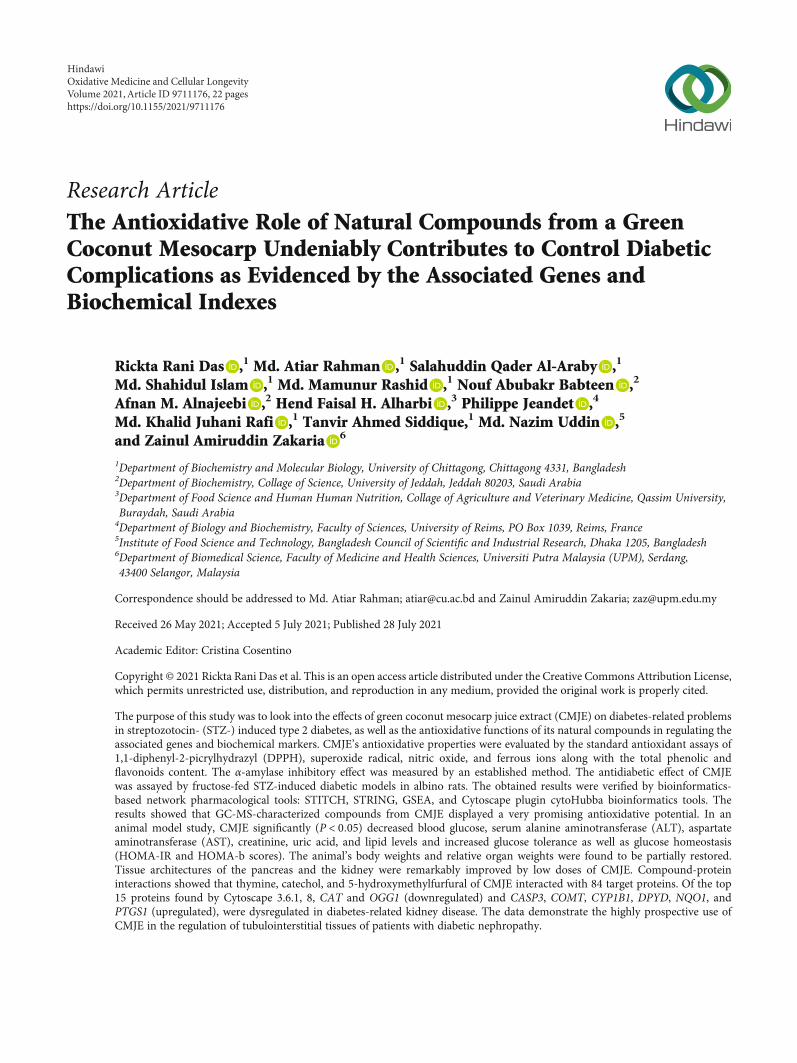
3.2. Antioxidative Capacity and α-Amylase Inhibitory Actionof CMJE. Antioxidative capacities were measured by theDPPH scavenging method, the superoxide scavengingmethod, the nitric oxide scavenging method, and the iron-chelating method. The reduction of DPPH in the scavengingassay was reflected through the decrease of absorbance. TheIC90 values of the sample (CMJE) and the standard (ascorbicacid) were found to be 123:02 ± 6:42 and 16:21 ± 2:34 μg/mL,respectively. The CMJE displayed the IC50 value 27:85 ± 1:32μg/mL in the superoxide scavenging assay and 284:40 ± 5:05μg/mL in the nitric oxide scavenging assays. The iron-chelating capacity in terms of IC50 was found to be 245:47 ±4:34μg/mL. ABTS assays showed a dose-dependent radicalscavenging capacity of CMJE. The results showed the inhibi-tion concentrations (IC50) 386:36 ± 1:22μg/mL for CMJEand 92:07 ± 3:21μg/mL for the standard Trolox. The compar-ative scavenging effect data are presented in Figure 1, and IC50values are summarized in Table 2.
The α-amylase inhibitory effect of CMJE is presented inFigure 2. Acarbose, an antidiabetic α-amylase inhibitorydrug, was used as a reference standard for this assay. The α-
amylase inhibitory activity of CMJE was significantly(P < 0:05) lower at each concentration of acarbose. The high-est inhibition for CMJE was achieved at the concentration of100μg/mL.
3.3. Effects of CMJE on Blood Glucose, Glucose Tolerance, andGlucose Homeostasis. CMJE was found nontoxic in the acutetoxicity study. Data regarding the effect of CMJE on animals’body weight and blood glucose levels are displayed inFigure 3. The body weight of the animals was not found tovary statistically among the treatment groups, but the weightof the CMJE50 group was close to that of the normal control(NC). Data reflected that the CMJE50 group has the bestglucose-lowering effect, which was statistically significant(P < 0:05) with the DC group.
Glucose tolerances were assessed by the oral glucose tol-erance test (OGTT) at the third week of the treatment period.Acquired data are presented in Figure 4. The glucose toler-ance of the DC group was significantly (P < 0:05) lower thanthat of the other groups. However, the CMJE50 groupshowed the highest tolerance of glucose load than the othergroup, which is consistent with the other parametersachieved by this group. Effects of CMJE extracts on the glu-cose homeostatic status are summarized in Table 3.
3.4. Effects of CMJE on Pancreas and Kidney Weights.Weights of the pancreas and kidney of treated animals arelisted in Table 4. Among the doses, CMJE100 in case of pan-creatic recovery and CMJE50 in case of kidney recovery werefound to be effective. The weight of kidneys was found to beremarkably recovered by CMJE50 while the weight of thepancreas was found to be better ameliorated by CMJE100.
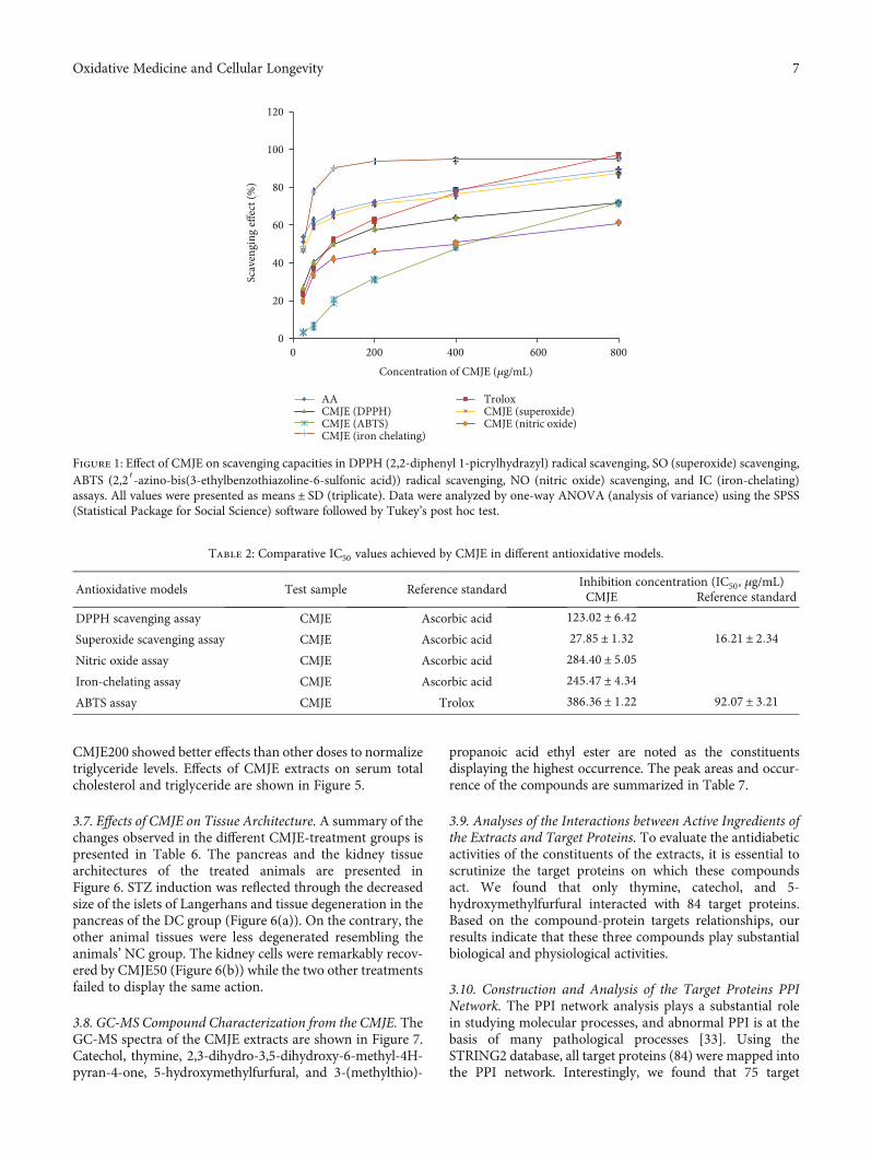
3.5. Effects of CMJE on ALT, AST, Uric Acid, and Creatinine.Changes in ALT, AST, uric acid, and creatinine levels at theend of the intervention are presented in Table 5. The ALTlevels of the different groups were not found to significantly(P < 0:05) differ from the NC group although the ALT levelof the DC group was somehow lower than that of all othergroups as well as that of the NC group. The AST level ofCMJE50 and CMJE100 group is lower than that of the DCgroup. The uric acid level of all treated groups is almost sim-ilar to that of the NC group. Lower creatinine levels for all thetreatments than DC were marked in the result.
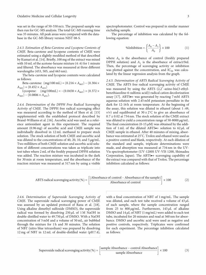
3.6. Effects of CMJE on Serum Total Cholesterol andTriglyceride Levels. Both serum total cholesterol and triglyc-eride levels for the treatment groups were consistently lowerthan those of the DC group while CMJE100 was found toreduce the total cholesterol more than other doses and
Table 1: Total phenolic, total flavonoid, lycopene, and carotenoidcontents of CMJE.
Phytochemical index Quantity
Total flavonoid 80.0mg rutin/g
Total phenolic content 102.0mg GAE/g
Lycopene 0.031mg/g
Total carotenoids 0.058mg/g
6 Oxidative Medicine and Cellular Longevity
CMJE200 showed better effects than other doses to normalizetriglyceride levels. Effects of CMJE extracts on serum totalcholesterol and triglyceride are shown in Figure 5.
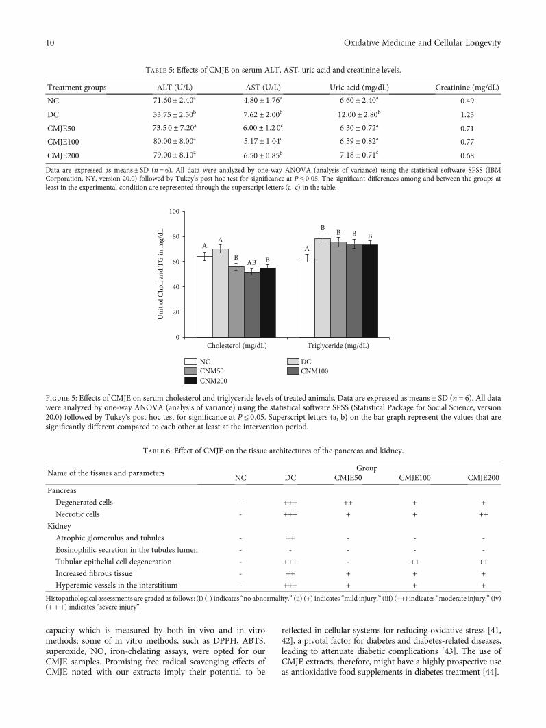
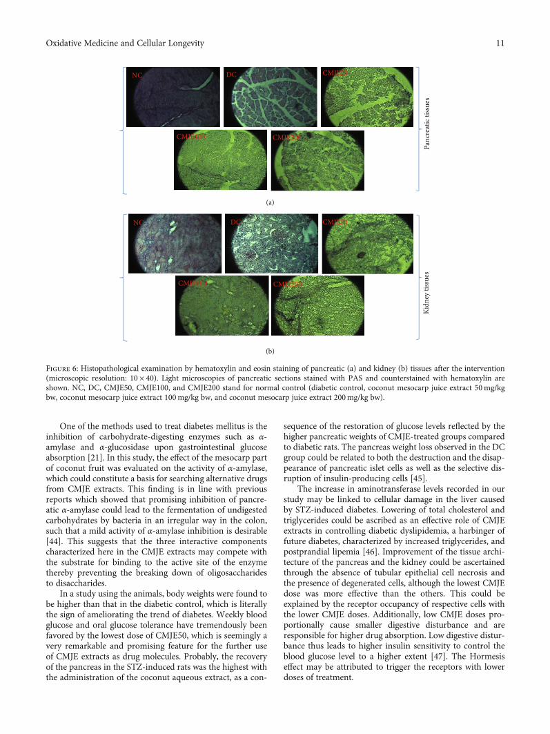
3.7. Effects of CMJE on Tissue Architecture. A summary of thechanges observed in the different CMJE-treatment groups ispresented in Table 6. The pancreas and the kidney tissuearchitectures of the treated animals are presented inFigure 6. STZ induction was reflected through the decreasedsize of the islets of Langerhans and tissue degeneration in thepancreas of the DC group (Figure 6(a)). On the contrary, theother animal tissues were less degenerated resembling theanimals’ NC group. The kidney cells were remarkably recov-ered by CMJE50 (Figure 6(b)) while the two other treatmentsfailed to display the same action.
3.8. GC-MS Compound Characterization from the CMJE. TheGC-MS spectra of the CMJE extracts are shown in Figure 7.Catechol, thymine, 2,3-dihydro-3,5-dihydroxy-6-methyl-4H-pyran-4-one, 5-hydroxymethylfurfural, and 3-(methylthio)-
propanoic acid ethyl ester are noted as the constituentsdisplaying the highest occurrence. The peak areas and occur-rence of the compounds are summarized in Table 7.
3.9. Analyses of the Interactions between Active Ingredients ofthe Extracts and Target Proteins. To evaluate the antidiabeticactivities of the constituents of the extracts, it is essential toscrutinize the target proteins on which these compoundsact. We found that only thymine, catechol, and 5-hydroxymethylfurfural interacted with 84 target proteins.Based on the compound-protein targets relationships, ourresults indicate that these three compounds play substantialbiological and physiological activities.
3.10. Construction and Analysis of the Target Proteins PPINetwork. The PPI network analysis plays a substantial rolein studying molecular processes, and abnormal PPI is at thebasis of many pathological processes [33]. Using theSTRING2 database, all target proteins (84) were mapped intothe PPI network. Interestingly, we found that 75 target
0
20
40
60
80
100
120
0 200 400 600 800
AA TroloxCMJE (DPPH) CMJE (superoxide)CMJE (ABTS) CMJE (nitric oxide)CMJE (iron chelating)
Concentration of CMJE (𝜇g/mL)
Scav
engi
ng eff
ect (
%)
Figure 1: Effect of CMJE on scavenging capacities in DPPH (2,2-diphenyl 1-picrylhydrazyl) radical scavenging, SO (superoxide) scavenging,ABTS (2,2′-azino-bis(3-ethylbenzothiazoline-6-sulfonic acid)) radical scavenging, NO (nitric oxide) scavenging, and IC (iron-chelating)assays. All values were presented as means ± SD (triplicate). Data were analyzed by one-way ANOVA (analysis of variance) using the SPSS(Statistical Package for Social Science) software followed by Tukey’s post hoc test.
Table 2: Comparative IC50 values achieved by CMJE in different antioxidative models.
Antioxidative models Test sample Reference standardInhibition concentration (IC50, μg/mL)CMJE Reference standard
DPPH scavenging assay CMJE Ascorbic acid 123:02 ± 6:42
Superoxide scavenging assay CMJE Ascorbic acid 27:85 ± 1:32 16:21 ± 2:34
Nitric oxide assay CMJE Ascorbic acid 284:40 ± 5:05
Iron-chelating assay CMJE Ascorbic acid 245:47 ± 4:34
ABTS assay CMJE Trolox 386:36 ± 1:22 92:07 ± 3:21
7Oxidative Medicine and Cellular Longevity

proteins were involved in PPI which have 235 edges, and anaverage node degree 5.88 with a PPI enrichment P value ofless than 1:0 × 10−16. In this PPI network, the larger the nodedegree, the stronger relationship between the proteins corre-sponding to the node, suggesting that the target proteinsplays a key role in the whole interaction network. Only ninetarget proteins were not included in PPI. We only got onesubnetwork in PPI and this subnetwork included 75 targetproteins. All target proteins with interactions with other pro-teins are illustrated in Figure 8. Cytoscape 3.6.1 was used toanalyze the interactions among the top 15 hub target proteins(degree of interaction with no less than 10). The hub targetproteins are TP53, CASP3, COMT, CYP1B1, DPYD, NQO1,
PTGS1, PTGS2, CAT, OGG1, GSTP1, MLH1, CYP1A1,TYMS, and TH.
3.11. Gene Ontology Analyses of the Interacting TargetProteins. The GO enrichment analysis of the interacting tar-get proteins (total 75 involved in PPI) which act with com-pounds of the CMJE extracts was performed by DAVID(https://david.ncifcrf.gov/). The significantly enriched termsin CC, BP, and MF categories were selected according tothe Benjamini–Hochberg-corrected P value < 0:05. A totalof 43 significant BP was listed in Table 8. In addition, two sig-nificant CC were also identified, as illustrated in Table 8.
A A AA
A
B B BB
B
0
20
40
60
80
50 100 200 400 800
SampleAcarbose
Perc
enta
ge o
f am
ylas
e inh
ibiti
on (%
)
Concentration of CMJE (𝜇g/mL)
Figure 2: Effects of CMJE on the α-amylase inhibitory activity. Acarbose was used as the reference standard. Data are presented asmeans ± SD(triplicate). All data were analyzed by one-way ANOVA (analysis of variance) using the statistical software SPSS (Statistical Package for SocialScience, version 20.0) followed by Tukey’s post hoc test. Superscript letters (a, b) over the graphical bars indicate the statistical difference betweeninhibitory effect of CMJE and acarbose.
0
50
100
150
200
250
Wee
kly
body
wei
ght (
g)
NC
DCNCM50CNM100CNM200
0 1 2 3 4Week
(a)
NC
DCNCM50CNM100CNM200
A A A A A
BC C C C
AA B A
C C C
CB
D
C C
0
10
20
30
40
50
Wee
kly
bloo
d gl
ucos
e lev
el (m
mol
/L)
0 1 2 3 4Week
(b)
Figure 3: Effects of CMJE extracts on body weight (a) and weekly blood glucose levels (b) of treated animals. Data are expressed asmeans ± SD(n = 6). All data were analyzed by one-way ANOVA (analysis of variance). Significance was confirmed at P < 0:05. Alphabets (a–c) over the linegraphs indicate the statistical differences among the groups.
8 Oxidative Medicine and Cellular Longevity

3.12. Target Proteins Are Associated with the Enrichment ofthe KEGG Pathways. To further elucidate the relationshipbetween target proteins and pathways, we identified 20KEGG pathways significantly associated with the targetproteins (Table 9), and they were found to be involvedin secretion (pancreatic secretion), metabolism, and cellu-lar signaling.
3.13. Validation of Hub Target Proteins in DiabeticNephropathy Cohort (GSE30122). We screened all 15 hubproteins in independent tubulointerstitial tissues of diabetickidney disease. Interestingly, we found that 8 targets are dys-regulated in this cohort. Among them, CAT and OGG1 aredownregulated, and CASP3, COMT, CYP1B1, DPYD,NQO1, and PTGS1 are upregulated in diabetic kidney dis-ease (DKD) tubuli (Figure 9).
4. Discussion
Coconut mesocarp juice extract (CMJE) was comprehen-sively studied for its antidiabetic effects which have beenevaluated in the light of CMJE’s phytoconstituent statusand antioxidative potential. It is believed that oxidative stressplays an important role in the development of vascular com-plications in diabetes particularly type 2 diabetes [34]. Freeradical formation in diabetes by nonenzymatic glycation ofproteins, glucose oxidation, and increased lipid peroxidationleads to damage of enzymes and cellular machinery andincreases insulin resistance [35]. Free radicals, therefore,through their aforesaid abilities play a major role in damag-ing lipids, proteins, and DNA [36] in the onset of diabeticcomplication. Additionally, the elevation of reactive oxygenspecies (ROS) decreases the production of biological antiox-idative enzymes such as catalase, superoxide dismutase(SOD), and glutathione peroxidase (GSH-Px) [37]. Varia-tions in the levels of these enzymes render the tissues suscep-tible to oxidative stress leading to the development of diabeticcomplications [38]. Our data are strongly supportive for theantidiabetic action of CMJE extracts because of their hightotal phenolic content, total flavonoid content, and caroten-oid and lycopene contents. Similar researches on the antidia-betic actions of plant phenolics especially flavonoids,triterpenoids, and saponins have been published recently[39, 40]. Phenolic compounds have attracted tremendousinterest due to their outstanding free radical scavenging
0
10
20
30
40
NC DCCNM50 CNM100CNM200
Bloo
d gl
ucos
e lev
el (m
mol
/L)
0 30 60 90 120Time after glucose ingestion (min)
Figure 4: Effects of CMJE on oral glucose tolerance (OGT) at the third week of intervention. Data are expressed as means ± SD (n = 6). Alldata were analyzed by one-way ANOVA (analysis of variance) using the statistical software SPSS (Statistical Package for Social Science,version 20.0) followed by Tukey’s post hoc test. Data significance was confirmed at P ≤ 0:05.
Table 3: Effects of CMJE on the glucose homeostatic status(HOMA-IR and HOMA-β).
Treatment groups HOMA-IR (mIU/L) HOMA-β (%)
NC 0.017 0.460
DC 0.116 0.036
CMJE50 0.052 0.240
CMJE100 0.155 0.045
CMJE200 0.111 0.082
HOMA-IR stands for homeostatic model assessment for insulin resistance,and HOMA-β represents the pancreatic beta cell function (%).
Table 4: Effects of CMJE on the relative weight of the pancreas andkidney of treated animals.
Tissue weight Pancreas weight (g) Kidney weight (g)
NC 0:569 ± 0:105a 1:794 ± 0:106a
DC 0:304 ± 0:047b 1:738 ± 0:093a
CMJE50 0:215 ± 0:044c 1:772 ± 0:127a
CMJE100 0:320 ± 0:050b 1:690 ± 0:031a
CMJE200 0:215 ± 0:035c 1:663 ± 0:199a
Data are expressed asmeans ± SD (n = 6). All data were analyzed by one-wayANOVA (analysis of variance) using the statistical software SPSS (StatisticalPackage for Social Science, version 20.0) followed by Tukey’s post hoc test.Data significance was confirmed at P ≤ 0:05. The superscript alphabets(a–c) in the table denote the reciprocal significance between and amongthe groups.
9Oxidative Medicine and Cellular Longevity
capacity which is measured by both in vivo and in vitromethods; some of in vitro methods, such as DPPH, ABTS,superoxide, NO, iron-chelating assays, were opted for ourCMJE samples. Promising free radical scavenging effects ofCMJE noted with our extracts imply their potential to be
reflected in cellular systems for reducing oxidative stress [41,42], a pivotal factor for diabetes and diabetes-related diseases,leading to attenuate diabetic complications [43]. The use ofCMJE extracts, therefore, might have a highly prospective useas antioxidative food supplements in diabetes treatment [44].
Table 5: Effects of CMJE on serum ALT, AST, uric acid and creatinine levels.
Treatment groups ALT (U/L) AST (U/L) Uric acid (mg/dL) Creatinine (mg/dL)
NC 71:60 ± 2:40a 4:80 ± 1:76a 6:60 ± 2:40a 0.49
DC 33:75 ± 2:50b 7:62 ± 2:00b 12:00 ± 2:80b 1.23
CMJE50 73:5 0 ± 7:20a 6:00 ± 1:2 0c 6:30 ± 0:72a 0.71
CMJE100 80:00 ± 8:00a 5:17 ± 1:04c 6:59 ± 0:82a 0.77
CMJE200 79:00 ± 8:10a 6:50 ± 0:85b 7:18 ± 0:71c 0.68
Data are expressed as means ± SD (n = 6). All data were analyzed by one-way ANOVA (analysis of variance) using the statistical software SPSS (IBMCorporation, NY, version 20.0) followed by Tukey’s post hoc test for significance at P ≤ 0:05. The significant differences among and between the groups atleast in the experimental condition are represented through the superscript letters (a–c) in the table.
A AA
B
B
B
AB
B
B
B
0
20
40
60
80
100
Cholesterol (mg/dL) Triglyceride (mg/dL)
NC DCCNM50 CNM100CNM200
Uni
t of C
hol.
and
TG in
mg/
dL
Figure 5: Effects of CMJE on serum cholesterol and triglyceride levels of treated animals. Data are expressed asmeans ± SD (n = 6). All datawere analyzed by one-way ANOVA (analysis of variance) using the statistical software SPSS (Statistical Package for Social Science, version20.0) followed by Tukey’s post hoc test for significance at P ≤ 0:05. Superscript letters (a, b) on the bar graph represent the values that aresignificantly different compared to each other at least at the intervention period.
Table 6: Effect of CMJE on the tissue architectures of the pancreas and kidney.
Name of the tissues and parametersGroup
NC DC CMJE50 CMJE100 CMJE200
Pancreas
Degenerated cells - +++ ++ + +
Necrotic cells - +++ + + ++
Kidney
Atrophic glomerulus and tubules - ++ - - -
Eosinophilic secretion in the tubules lumen - - - - -
Tubular epithelial cell degeneration - +++ - ++ ++
Increased fibrous tissue - ++ + + +
Hyperemic vessels in the interstitium - +++ + + +
Histopathological assessments are graded as follows: (i) (-) indicates “no abnormality.” (ii) (+) indicates “mild injury.” (iii) (++) indicates “moderate injury.” (iv)(+ + +) indicates “severe injury”.
10 Oxidative Medicine and Cellular Longevity
One of the methods used to treat diabetes mellitus is theinhibition of carbohydrate-digesting enzymes such as α-amylase and α-glucosidase upon gastrointestinal glucoseabsorption [21]. In this study, the effect of the mesocarp partof coconut fruit was evaluated on the activity of α-amylase,which could constitute a basis for searching alternative drugsfrom CMJE extracts. This finding is in line with previousreports which showed that promising inhibition of pancre-atic α-amylase could lead to the fermentation of undigestedcarbohydrates by bacteria in an irregular way in the colon,such that a mild activity of α-amylase inhibition is desirable[44]. This suggests that the three interactive componentscharacterized here in the CMJE extracts may compete withthe substrate for binding to the active site of the enzymethereby preventing the breaking down of oligosaccharidesto disaccharides.
In a study using the animals, body weights were found tobe higher than that in the diabetic control, which is literallythe sign of ameliorating the trend of diabetes. Weekly bloodglucose and oral glucose tolerance have tremendously beenfavored by the lowest dose of CMJE50, which is seemingly avery remarkable and promising feature for the further useof CMJE extracts as drug molecules. Probably, the recoveryof the pancreas in the STZ-induced rats was the highest withthe administration of the coconut aqueous extract, as a con-
sequence of the restoration of glucose levels reflected by thehigher pancreatic weights of CMJE-treated groups comparedto diabetic rats. The pancreas weight loss observed in the DCgroup could be related to both the destruction and the disap-pearance of pancreatic islet cells as well as the selective dis-ruption of insulin-producing cells [45].
The increase in aminotransferase levels recorded in ourstudy may be linked to cellular damage in the liver causedby STZ-induced diabetes. Lowering of total cholesterol andtriglycerides could be ascribed as an effective role of CMJEextracts in controlling diabetic dyslipidemia, a harbinger offuture diabetes, characterized by increased triglycerides, andpostprandial lipemia [46]. Improvement of the tissue archi-tecture of the pancreas and the kidney could be ascertainedthrough the absence of tubular epithelial cell necrosis andthe presence of degenerated cells, although the lowest CMJEdose was more effective than the others. This could beexplained by the receptor occupancy of respective cells withthe lower CMJE doses. Additionally, low CMJE doses pro-portionally cause smaller digestive disturbance and areresponsible for higher drug absorption. Low digestive distur-bance thus leads to higher insulin sensitivity to control theblood glucose level to a higher extent [47]. The Hormesiseffect may be attributed to trigger the receptors with lowerdoses of treatment.
Panc
reat
ic ti
ssue
sKi
dney
tiss
ues
DC CMJE50NC
CMJE100 CMJE200
CMJE200CMJE100
DC CMJE50NC
(a)
(b)
Figure 6: Histopathological examination by hematoxylin and eosin staining of pancreatic (a) and kidney (b) tissues after the intervention(microscopic resolution: 10 × 40). Light microscopies of pancreatic sections stained with PAS and counterstained with hematoxylin areshown. NC, DC, CMJE50, CMJE100, and CMJE200 stand for normal control (diabetic control, coconut mesocarp juice extract 50mg/kgbw, coconut mesocarp juice extract 100mg/kg bw, and coconut mesocarp juice extract 200mg/kg bw).
11Oxidative Medicine and Cellular Longevity

Chromatogram plant extract C:\GC-MSMS\DATA\2017\R & D\Atiar Sir (CU)\plant extract_R (rickta)_MeOH.qgd
min
25,552,756
4.0 10.0 20.0 25.0
TIC3.
536
3.62
63.
725
3.84
43.
994
4.08
34.
210
4.62
44.
751
4.83
35.
064
5.15
85.
253
5.35
85.
494
5.61
85.
823
6.29
1 6.35
36.
490
6.56
1 6.74
17.
002
7.36
47.
505
7.67
47.
750
7.93
38.
232
8.74
98.
808 8.
933
9.04
1
10.1
6810
.273 10
.948
11.1
5011
.213
12.6
58
21.4
59Instrument Name: GC-MS/MS
Model: GCMS-TQ8040
Column: Rxi-5ms
(a)
Figure 7: Continued.
12 Oxidative Medicine and Cellular Longevity

Most probable compounds:
S/N Ret. time Compound name
1 3.533 3-trans-methoxy-2-cis-methyl-1R-cyclohexanol
2 3.625 2,4 (1H,3H)-pyrimidinedione, dihydro-
3 3.725 Butanoic acid, 2-(hydroxymethyl)-, ethyl ester, (R)-
4 3.842 2-methylbutanoic anhydride
5 3.992 Thymine
6 4.083 Methyl 2-furoate
7 4.208 Trimethylaluminum
8 4.625 Ethanamine, N-ethyl-N-nitroso-
9 4.75 4H-pyran-4-one, 2,3-dihydro-3,5-dihydroxy-6-methyl-
10 4.833 3-(methylthio) propanoic acid ethyl ester
11 5.067 (S)-5-hydroxymethyl-2[5H]-furanone
12 5.158 4H-pyran-4-one, 3,5-dihydroxy-2-methyl-
13 5.25 Catechol
14 5.358 Butanedioic acid, 2-hydroxy-2-methyl-, dimethyl ester, (2R)-
15 5.492 2H-pyran-2-methanol, tetrahydro-
16 5.617 5-hydroxymethylfurfural
17 5.825 Butanedioic acid, hydroxy-, dimethyl ester
18 6.292 Undecenyl tiglate, 10-
19 6.35 1-(methylthio)-3-pentanone
20 6.492 3,4-hexanediol, 2,5-dimethyl-
21 6.558 Fumaric acid, monoamide, N,N-dimethyl-, 2-ethylhexyl ester
22 6.742 Heptyl caprylate
23 7 Butanedioic acid, 2-hydroxy-2-methyl-, (S)-
24 7.367 Phenol, 4-propyl-
25 7.508 1,2,3-benzenetriol
(b)
Figure 7: Continued.
13Oxidative Medicine and Cellular Longevity

26 7.675 Tetradecane
27 7.75 Benzene, 1-chloro-4-methoxy-
28 7.933 Butanamide, 2-hydroxy-N, 2,3,3-tetramethyl-
29 8.233 Benzaldehyde, 2-hydroxy-4-methyl-
30 8.75 Benzoic acid, 3-hydroxy-
31 8.808 (Z), (Z)-2, 5-Dimethyl-2, 4-hexadienedioic acid
32 8.933 Pentadecane
33 9.042 Benzoic acid, 4-hydroxy-
34 10.167 Hexadecane
35 10.275 1,3-benzenediol, 4-propyl-
36 10.95 3-deoxy-d-mannoic lactone
37 11.15 Butanoic acid, 2-methyl-, hexyl ester
38 11.217 3-deoxy-d-mannonic acid
39 12.658 2-hydroxy-5-methylisophthalaldehyde
(c)
Peak report TICPeak# R.time Area% Name 1 3.536 1.27 3-trans-methoxy-2-cis-methyl-1R-cyclohexanol 2 3.626 1.58 2, 4 (1H, 3H)-pyrimidinedione, dihydro- 3 3.725 0.52 Butanoic acid, 2-(hydroxymethyl)-, ethyl ester, (R)- 4 3.844 3.61 2-methylbutanoic anhydride 5 3.994 7.15 Thymine 6 4.083 1.73 Methyl 2-furoate 7 4.210 0.96 Trimethylaluminum 8 4.624 1.91 Ethanamine, N-ethyl-N-nitroso- 9 4.751 12.94 4H-pyran-4-one, 2,3-dihydro-3,5-dihydroxy-6-methyl- 10 4.833 7.24 3-(methylthio) propanoic acid ethyl ester 11 5.064 0.71 (S)-5-Hydroxymethyl-2[5H]-furanone 12 5.158 0.34 4H-pyran-4-one, 3,5-dihydroxy-2-methyl- 13 5.253 7.20 Catechol 14 5.358 1.00 Butanedioic acid, 2-hydroxy-2-methyl-, dimethyl ester, (2R)- 15 5.494 5.20 2H-pyran-2-methanol, tetrahydro- 16 5.618 14.41 5-Hydroxymethylfurfural 17 5.823 1.71 Butanedioic acid, hydroxy-, dimethyl ester 18 6.291 1.07 Undecenyl tiglate, 10- 19 6.353 1.86 1-(methylthio)-3-pentanone 20 6.490 1.26 3,4-hexanediol, 2,5-dimethyl- 21 6.561 0.78 Fumaric acid, monoamide, N, N-dimethyl-, 2-ethylhexyl ester 22 6.741 1.57 Heptyl caprylate 23 7.002 1.08 Butanedioic acid, 2-hydroxy-2-methyl-, (S)- 24 7.364 1.58 Phenol, 4-propyl- 25 7.505 2.44 1,2,3-benzenetriol 26 7.674 1.20 Tetradecane 27 7.750 0.42 Benzene, 1-chloro-4-methoxy- 28 7.933 1.04 Butanamide, 2-hydroxy-N,2,3,3-tetramethyl- 29 8.232 3.70 Benzaldehyde, 2-hydroxy-4-methyl- 30 8.749 2.01 Benzoic acid, 3-hydroxy- 31 8.808 1.34 (Z),(Z)-2,5-Dimethyl-2,4-hexadienedioic acid 32 8.933 1.50 Pentadecane 33 9.041 0.62 Benzoic acid, 4-hydroxy- 34 10.168 1.04 Hexadecane 35 10.273 0.81 1, 3-Benzenediol, 4-propyl- 36 10.948 2.49 3-Deoxy-d-mannoic lactone 37 11.150 0.40 Butanoic acid, 2-methyl-, hexyl ester 38 11.213 0.95 3-Deoxy-d-mannonic acid 39 12.658 0.87 2-Hydroxy-5-methylisophthalaldehyde 40 21.459 0.48 13-Docosenamide, (Z)-
100.00
(d)
Figure 7: GC-MS spectra of CMJE obtained from the mass spectrometer-electron impact ionization (EI) method (GC-MS TQ 8040,Shimadzu Corporation, Kyoto, Japan) coupled with a gas chromatograph (GC-17A, Shimadzu Corporation, Kyoto, Japan). A fused silicacapillary column with inlet temperature 260°C and oven temperature 70°C (0min) was programmed. The mass range was set in the rangeof 50-550m/z.
14 Oxidative Medicine and Cellular Longevity
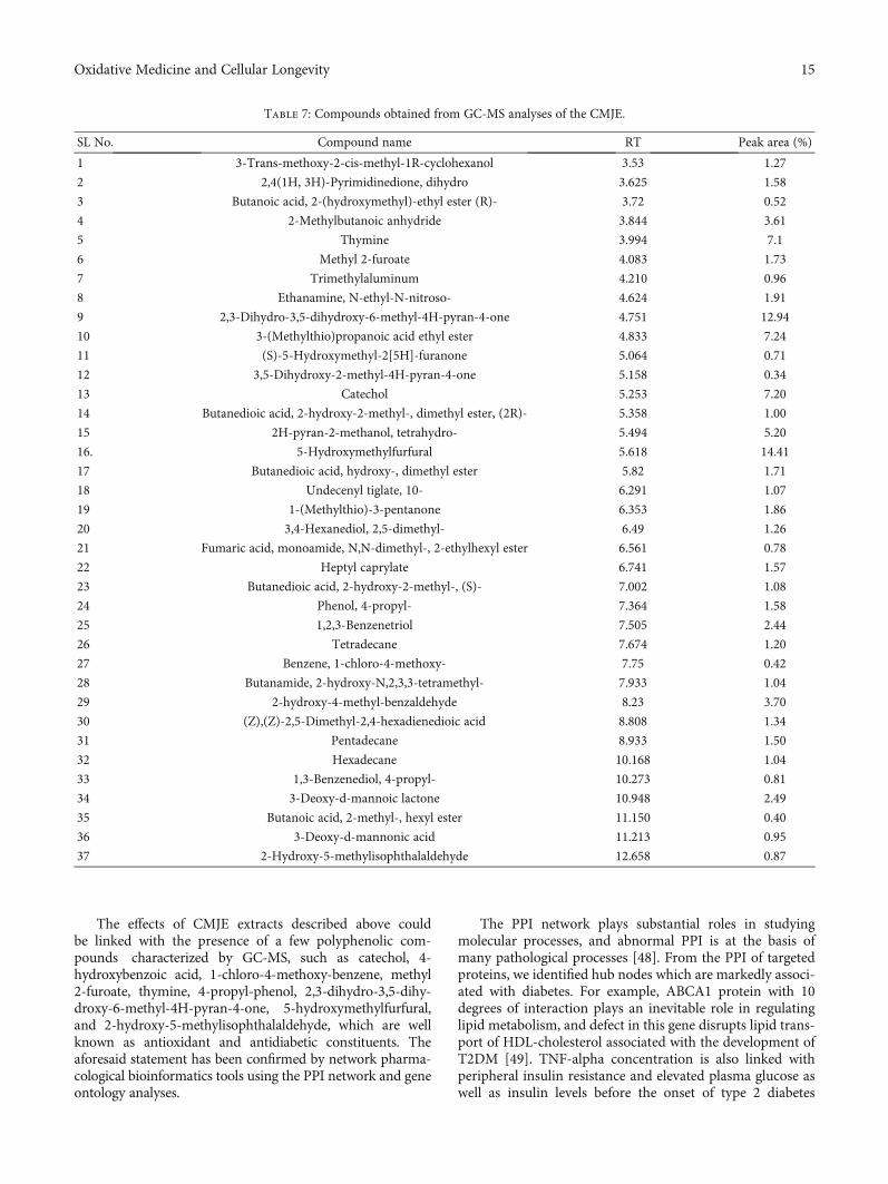
The effects of CMJE extracts described above couldbe linked with the presence of a few polyphenolic com-pounds characterized by GC-MS, such as catechol, 4-hydroxybenzoic acid, 1-chloro-4-methoxy-benzene, methyl2-furoate, thymine, 4-propyl-phenol, 2,3-dihydro-3,5-dihy-droxy-6-methyl-4H-pyran-4-one, 5-hydroxymethylfurfural,and 2-hydroxy-5-methylisophthalaldehyde, which are wellknown as antioxidant and antidiabetic constituents. Theaforesaid statement has been confirmed by network pharma-cological bioinformatics tools using the PPI network and geneontology analyses.
The PPI network plays substantial roles in studyingmolecular processes, and abnormal PPI is at the basis ofmany pathological processes [48]. From the PPI of targetedproteins, we identified hub nodes which are markedly associ-ated with diabetes. For example, ABCA1 protein with 10degrees of interaction plays an inevitable role in regulatinglipid metabolism, and defect in this gene disrupts lipid trans-port of HDL-cholesterol associated with the development ofT2DM [49]. TNF-alpha concentration is also linked withperipheral insulin resistance and elevated plasma glucose aswell as insulin levels before the onset of type 2 diabetes
Table 7: Compounds obtained from GC-MS analyses of the CMJE.
SL No. Compound name RT Peak area (%)
1 3-Trans-methoxy-2-cis-methyl-1R-cyclohexanol 3.53 1.27
2 2,4(1H, 3H)-Pyrimidinedione, dihydro 3.625 1.58
3 Butanoic acid, 2-(hydroxymethyl)-ethyl ester (R)- 3.72 0.52
4 2-Methylbutanoic anhydride 3.844 3.61
5 Thymine 3.994 7.1
6 Methyl 2-furoate 4.083 1.73
7 Trimethylaluminum 4.210 0.96
8 Ethanamine, N-ethyl-N-nitroso- 4.624 1.91
9 2,3-Dihydro-3,5-dihydroxy-6-methyl-4H-pyran-4-one 4.751 12.94
10 3-(Methylthio)propanoic acid ethyl ester 4.833 7.24
11 (S)-5-Hydroxymethyl-2[5H]-furanone 5.064 0.71
12 3,5-Dihydroxy-2-methyl-4H-pyran-4-one 5.158 0.34
13 Catechol 5.253 7.20
14 Butanedioic acid, 2-hydroxy-2-methyl-, dimethyl ester, (2R)- 5.358 1.00
15 2H-pyran-2-methanol, tetrahydro- 5.494 5.20
16. 5-Hydroxymethylfurfural 5.618 14.41
17 Butanedioic acid, hydroxy-, dimethyl ester 5.82 1.71
18 Undecenyl tiglate, 10- 6.291 1.07
19 1-(Methylthio)-3-pentanone 6.353 1.86
20 3,4-Hexanediol, 2,5-dimethyl- 6.49 1.26
21 Fumaric acid, monoamide, N,N-dimethyl-, 2-ethylhexyl ester 6.561 0.78
22 Heptyl caprylate 6.741 1.57
23 Butanedioic acid, 2-hydroxy-2-methyl-, (S)- 7.002 1.08
24 Phenol, 4-propyl- 7.364 1.58
25 1,2,3-Benzenetriol 7.505 2.44
26 Tetradecane 7.674 1.20
27 Benzene, 1-chloro-4-methoxy- 7.75 0.42
28 Butanamide, 2-hydroxy-N,2,3,3-tetramethyl- 7.933 1.04
29 2-hydroxy-4-methyl-benzaldehyde 8.23 3.70
30 (Z),(Z)-2,5-Dimethyl-2,4-hexadienedioic acid 8.808 1.34
31 Pentadecane 8.933 1.50
32 Hexadecane 10.168 1.04
33 1,3-Benzenediol, 4-propyl- 10.273 0.81
34 3-Deoxy-d-mannoic lactone 10.948 2.49
35 Butanoic acid, 2-methyl-, hexyl ester 11.150 0.40
36 3-Deoxy-d-mannonic acid 11.213 0.95
37 2-Hydroxy-5-methylisophthalaldehyde 12.658 0.87
15Oxidative Medicine and Cellular Longevity

[50]. It was observed that a CXCL8 antagonist amelioratesdiabetic nephropathy in diabetic male mice and attenuateshigh glucose-induced mesangial injury [51]. Caspase-3 pro-motes diabetic kidney disease [52]. The HMGCR gene, inpopulation studies, was also found to be associated withbodyweight gain and a higher risk of type 2 diabetes [53].Interleukin-10 (IL10), an anti-inflammatory cytokine, issupposed to play a type 2 diabetes (T2D) protective role[54]. Figure 9 shows that the immunological target proteinsincluding IL10, CXCL8, and TNF are located in the PPInetworks with a top degree of interaction, indicating thatthis PPI network is associated with immunological activi-ties. It was stated that humans’ chemokines have been asso-
ciated with, or implicated in, the pathogenesis of type 1diabetes [55]. Altogether, these compound-proteins interac-tions may be associated with the regulation of diabetespathophysiology.
In addition, we identified the GO and pathway enrich-ment of target proteins. The GO analysis indicated that thetarget proteins may bind with plasma membranes, chromo-somes, chromatin, regulatory regions of nucleic acids, or/andcellular receptors of cells for mediating metabolic, immuno-logical processes, signaling, and/or other activities, so as toexert signaling and antidiabetic potentials [56]. Pathwayanalysis revealed some pathways being also associated withdiabetes. Retinol metabolism is the most significantly
GCG
TYR
CASP9CASP8
ENSG00000253117
PTGS2MLH1
CAT
AKR1C4YJEFN3
TP53EPXCRY1
LPO
TH
SULT1A1
CYP19A1
TYMP
TDG
NTHL1
DHFR
MPO
XPA
UPP1
APOA1BP
HMBS
TET1
EDC3
SLC29A2
TET3
MCEE
LCN2
PXDN
PLA2G10
PRDX5
PLA2G2F
PLA2G2E
PLA2G5
TOP3B
SLC6A3
PLA2G2A
SULT1C2
PLA2G2D
DHDH
MBD4
TOP3A
DDC
UPP2
PLA2G1B
REV1
TYMS
CYP1B1
PTGS1
COMT
DPYD
CASP7 PMS2TPO
LRTOMTSULT1A3
POLHDPYS SDHACRY2
SULT1A2
AKR1C1
CYP1A1
NQO1
CASP3
OGG1GSTP1
PLA2G2C
LAD1
HBB
HBA2
Figure 8: The protein-protein interaction (PPI) network of the 75 target proteins.
16 Oxidative Medicine and Cellular Longevity

Table 8: Gene Ontology (GO) enrichment analysis of the interacting target proteins; 43 biological processes, 15 molecular functions, and 2cellular components.
Category TermBenjamini-corrected
P value
BP
GO:0042744, hydrogen peroxide catabolic process 5:78E − 12
GO:0098869, cellular oxidant detoxification 1:08E − 10
GO:0055114, oxidation-reduction process 6:90E − 10
GO:0050482, arachidonic acid secretion 1:03E − 09
GO:0036149, phosphatidylinositol acyl-chain remodeling 5:02E − 09
GO:0036148, phosphatidylglycerol acyl-chain remodeling 9:64E − 09
GO:0036150, phosphatidylserine acyl-chain remodeling 9:64E − 09
GO:0032355, response to estradiol 1:53E − 08
GO:0036152, phosphatidylethanolamine acyl-chain remodeling 5:14E − 08
GO:0006979, response to oxidative stress 6:62E − 08
GO:0036151, phosphatidylcholine acyl-chain remodeling 8:95E − 08
GO:0006654, phosphatidic acid biosynthetic process 4:34E − 07
GO:0006805, xenobiotic metabolic process 2:18E − 06
GO:0016042, lipid catabolic process 3:67E − 06
GO:0046135, pyrimidine nucleoside catabolic process 4:76E − 06
GO:0006644, phospholipid metabolic process 4:76E − 06
GO:0045471, response to ethanol 1:29E − 05
GO:0007568, aging 1:91E − 05
GO:0008202, steroid metabolic process 3:89E − 05
GO:0045008, depyrimidination 1:48E − 04
GO:0042493, response to drug 1:82E − 04
GO:0032496, response to lipopolysaccharide 1:97E − 04
GO:0006584, catecholamine metabolic process 1:91E − 04
GO:0046677, response to antibiotic 2:95E − 04
GO:0051923, sulfation 3:42E − 04
GO:0006284, base-excision repair 3:92E − 04
GO:0050427, 3′-phosphoadenosine 5′-phosphosulfate metabolic process 8:60E − 04
GO:0032025, response to cobalt ion 6:80E − 03
GO:0009635, response to herbicide 9:14E − 03
GO:0009636, response to toxic substance 1:11E − 02
GO:0009308, amine metabolic process 1:13E − 02
GO:0009812, flavonoid metabolic process 1:13E − 02GO:0008635, activation of cysteine-type endopeptidase activity involved in apoptotic process by
cytochrome c1:40E − 02
GO:0043066, negative regulation of apoptotic process 1:42E − 02
GO:0042416, dopamine biosynthetic process 1:64E − 02
GO:0043525, positive regulation of neuron apoptotic process 1:63E − 02
GO:0008210, estrogen metabolic process 1:89E − 02
GO:0043097, pyrimidine nucleoside salvage 2:19E − 02
GO:0042542, response to hydrogen peroxide 2:45E − 02
GO:0071407, cellular response to organic cyclic compound 3:62E − 02
GO:0097194, execution phase of apoptosis 3:61E − 02
17Oxidative Medicine and Cellular Longevity

Table 8: Continued.
Category TermBenjamini-corrected
P value
GO:0080111, DNA demethylation 3:61E − 02
GO:0033189, response to vitamin A 4:45E − 02
CCGO:0005829, cytosol 4:25E − 06
GO:0005739, mitochondrion 1:18E − 04
MF
GO:0004601, peroxidase activity 5:24E − 12
GO:0020037, heme binding 5:97E − 11
GO:0004623, phospholipase A2 activity 3:61E − 09
GO:0003684, damaged DNA binding 1:60E − 05
GO:0005506, iron ion binding 1:30E − 05
GO:0019825, oxygen binding 6:41E − 05
GO:0004062, aryl sulfotransferase activity 5:34E − 04
GO:0097153, cysteine-type endopeptidase activity involved in apoptotic process 6:06E − 04
GO:0008146, sulfotransferase activity 1:35E − 02
GO:0047498, calcium-dependent phospholipase A2 activity 1:48E − 02
GO:0019104, DNA N-glycosylase activity 1:67E − 02
GO:0030983, mismatched DNA binding 1:87E − 02GO:0016712, oxidoreductase activity, acting on paired donors, with incorporation or reduction of
molecular oxygen, reduced flavin or flavoprotein as one donor, and incorporation of one atom of oxygen3:23E − 02
GO:0004497, monooxygenase activity 3:21E − 02
GO:0004197, cysteine-type endopeptidase activity 3:46E − 02
Table 9: Enriched KEGG pathways significantly associated with target proteins in this study.
KEGG pathway Benjamini-corrected P value
hsa00592: alpha-linolenic acid metabolism 1:96E − 07
hsa00590: arachidonic acid metabolism 1:81E − 07
hsa00591: linoleic acid metabolism 2:06E − 07
hsa04975: fat digestion and absorption 1:42E − 06
hsa00565: ether lipid metabolism 3:21E − 06
hsa01100: metabolic pathways 5:47E − 05
hsa04972: pancreatic secretion 3:39E − 04
hsa00564: glycerophospholipid metabolism 3:42E − 04
hsa05204: chemical carcinogenesis 1:10E − 03
hsa04270: vascular smooth muscle contraction 1:05E − 03
hsa03460: fanconi anemia pathway 1:23E − 03
hsa00140: steroid hormone biosynthesis 1:74E − 03
hsa00350: tyrosine metabolism 2:51E − 03
hsa00980: metabolism of xenobiotics by cytochrome P450 4:69E − 03
hsa00983: drug metabolism—other enzymes 6:29E − 03
hsa00240: pyrimidine metabolism 1:66E − 02
hsa04210: apoptosis 1:69E − 02
hsa03410: base-excision repair 2:19E − 02
hsa04014: ras signaling pathway 2:74E − 02
hsa00380: tryptophan metabolism 3:39E − 02
18 Oxidative Medicine and Cellular Longevity

enriched pathway (FDR < 8:6 × 10⁻⁶). Retinoids and retinoid-related proteins are associated with signaling molecules link-ing obesity with the development of type 2 diabetes and in thepancreatic β-cell biology/insulin secretion [57]. Additionally,CAT and SOD1 genes/proteins are assumed to highly influ-ence the control of the PPI network because these are alsoknown as antioxidative enzymes having a very potent rolein diabetes and diabetes-related complications including dia-betic nephropathy [57]. Apart from these observations, the15 hub genes are targeted in independent diabetic kidney dis-ease (DKD). Interestingly, dysregulation of eight of thosegenes in this cohort complies with the fact that CAT andOGG1 are downregulated and CASP3, COMT, CYP1B1,DPYD, NQO1, and PTGS1 are upregulated in DKD, suggest-ing that the target compounds are clearly interacting withpotential genes associated with diabetic nephropathy [58].Moreover, metabolic, oxidative, oxidant detoxification, andinflammatory stresses are common features in diabeticnephropathy [59]. In streptozotocin-induced diabetic rats,modulation of xenobiotic metabolism and oxidative stressin various tissues may be related to altered metabolism[60]. Peroxidase activity, a top molecular function, isincreased in advanced diabetic nephropathy [61]. Phospholi-pase A2 activity is a risk factor in diabetic nephropathy [62].Altogether, these GO are clearly associated with the dysregu-lation of diabetic kidney disease (DKD).
The KEGG pathways were mainly involved in secretion(pancreatic secretion), metabolism, and cellular signaling.
In type 2 diabetes, alpha-linolenic acid has effects on thecontrol of the glycemic index [63]. Yan et al. revealed thatantidiabetic agents are associated with arachidonic acidmetabolism, glycerophospholipid metabolism, tryptophanmetabolism, and tyrosine metabolism [64]. Another path-way, apoptosis, is also critically associated with diabetes[65]. Collectively, these enriched pathways are associatedwith the DKD. In a diabetic nephropathy cohort study,CAT is a downregulated gene which may be associated withthe regulation of kidney functions in diabetes [66]. The geneCASP3 promotes the DKD through secondary necrosis [53].Another upregulated gene, CYP1B1, is also associated withthe damage and dysfunction of renal functions in mice.Increased expression levels of PTGS1 are associated withthe progression of diabetic nephropathy [67] suggesting thatthese are the potential genes, interacting with our target com-pounds, which are dysregulated in diabetic kidney diseases.
5. Conclusions
Our investigations revealed that the CMJE partially restoredbiochemical markers especially ALT, AST, creatinine, uricacid, and lipid profiles and improved glucose homeostasisas well as tissue architecture. The target compounds of CMJEdownregulated CAT and OGG1 genes and upregulatedCASP3, COMT, CYP1B1, DPYD, NQO1, and PTGS1 genesimplying to potentiate their antioxidative actions to protectthe dysregulation of the pancreas and the kidney of STZ-
–1.0
–0.5
Expr
essio
n le
vel o
f CA
SP3
(202
763_
at)
0.0
0.5
1.0
1.5
DKDT Control
–1.0Expr
essio
n le
vel o
f CO
MT
(208
81_a
t)
0.0
0.5
1.0
1.5
2.0
DKDT
COMT, FC = 1.00⁎⁎⁎CASP3, FC = 1.14⁎⁎⁎
Control
–1.5Expr
essio
n le
vel o
f CYP
1B1
(202
435_
s_at
)
–0.5
0.5
1.0
1.5
2.0
DKDT
CYP1B1, FC = 1.60⁎⁎⁎
Control
–2.0Expr
essio
n le
vel o
f DPY
D (2
0464
6_at
)
–1.0
0.0
0.5
1.0
DKDT
DPYD, FC = 0.79⁎⁎
Control
–0.5
0.0
0.5
1.0
1.5
2.0
Expr
essio
n le
vel o
f PTG
S1 (2
0512
8_x_
at)
DKDT
PTGS1, FC = 0.58⁎⁎
Control
–2
–1
0
1
Expr
essio
n le
vel o
f NQ
O1
(201
468_
s_at
)
DKDT
NQO1, FC = 0.70⁎
Control
–2
–1
0
1
Expr
essio
n le
vel o
f CAT
(201
432_
at)
DKDT
CAT, FC = –0.74⁎⁎
Control
–2
–1
0
1
Expr
essio
n le
vel o
f OG
G1
(205
760_
s_at
)
DKDT
OGG1, FC = –0.86⁎
Control
Figure 9: Dysregulation of hub targets (mRNA expression levels) in diabetic human kidney tubuli, when compared with control tubuli. FC:fold change, DKDT: diabetic kidney disease tubuli, control: control tubuli, ∗P < 0:05, ∗∗P < 0:01, and ∗∗∗P < 0:001.
19Oxidative Medicine and Cellular Longevity

diabetic animals. Therefore, the coconut mesocarp juiceextract is suggested to produce antidiabetic actions ininduced animal models. A further study of in vivo antioxida-tive effects both in enzymatic and nonenzymatic systemsmight confirm the use of CMJE as alternative therapeutic indiabetic complications.
Abbreviations
CMJE: Coconut mesocarp juice extractTFC: Total flavonoid contentTPC: Total phenolic contentSO: Superoxide scavenging effectDPPH-1: 1-Diphenyl, 2-picrylhydrazyl free radical
scavenging assayNO: Nitric oxide scavenging effectIrC: Iron-chelating effectABTS: (2,2′-azino-bis(3-ethylbenzothiazoline-6-sul-
fonic acid)ALT: Alanine aminotransferaseAST: Aspartate aminotransferaseHOMA-IR: Glucose homeostasisSPSS: Statistical Package for Social ScienceTCh: Total cholesterolTG: TriglyceridePPI: Protein-protein interactionGO: Gene OntologyKEGG: Kyoto Encyclopedia of Genes and GenomesBP: Biological processMF: Molecular functionCC: Cellular component groups.
Data Availability
The data used to support the findings of this study are avail-able from the corresponding authors upon request.
Additional Points
Institutional Review Board Statement. Animal handling andcare were ensured through the ethical guideline formed inaccord with the Helsinki protocol. All animal experimenta-tions were carried out according to the guideline of Institu-tional Animal Ethics Committee (EACUBS2018-4).
Disclosure
This research was submitted to Oxidative Medicine and Cel-lular Longevity.
Conflicts of Interest
The authors declare no conflict of interest.
Authors’ Contributions
MAR has designed the research, planned for research con-duction, arranged the funding for research, and introducedthe way of data collection. RRD, SQA,MMR,MSI, andMKJRhave collected sample, set the bench works, produced and
analyzed the data, and paid effort in manuscript preparation.MKJR and TAS particularly assessed the antioxidative effects.NAB, AMA, and HFHA along with MAR and PJ haveassisted in interpreting the data and made necessary stepsto harmonize the data. MNU has accomplished the networkpharmacological analyses. ZAZ has facilitated the funding forthis research.
Acknowledgments
This research was funded by the Malaysia Ministry of HigherEducation under the Fundamental Research Grant Scheme(FRGS; reference no: 04-01-18-1984FR). The APC wasfunded by the Universiti Putra Malaysia (UPM), Malaysia.The funders had no role in the design of the study; in the col-lection, analyses, or interpretation of data; in the writing ofthe manuscript; or in the decision to publish the results.The authors wish to thank Dr. Sheikh Bokhtear Uddin forshowing the strategy to minutely separate the coconutmesocarp.
Supplementary Materials
Table S1: list of compounds-targets identification. Table S2:list of targets involved in PPI with degree of interactions.Table S3: ontology (GO) enrichment analysis of the inter-acted target proteins. Table S4: the enriched KEGG pathwayswhich are significantly associated with target proteins. FigureS1: a comprehensive approach to display the effect of coconutmesocarp juice extract on the streptozotocin-induced diabetesand diabetes-related complications using in vitro, in vivo andcomputational models. (Supplementary Materials)
References
[1] F. G. Lupascu, S. E. Giusca, I. D. Caruntu, A. Anton, C. E.Lupușoru, and L. Profire, “The safety profile of new antidia-betic xanthine derivatives and their chitosan based formula-tions,” Europian Journal of Pharmaceutical Sciences, vol. 127,pp. 71–78, 2019.
[2] C. H. Lin, Z. Z. Shih, Y. H. Kuo, G. J. Huang, P. C. Tu, andC. C. Shih, “Antidiabetic and antihyperlipidemic effects ofthe flower extract of Eriobotrya japonica in streptozotocin-induced diabetic mice and the potential bioactive constituentsin vitro,” Journal of Functional Foods, vol. 49, no. 11, pp. 122–136, 2018.
[3] A. M. Al-Attar and F. A. Alsalmi, “Effect of Olea europaealeaves extract on streptozotocin induced diabetes in malealbino rats,” Saudi Journal of Biological Sciences, vol. 26,no. 1, pp. 118–128, 2019.
[4] H. Choudhury, M. Pandey, C. K. Hua et al., “An update onnatural compounds in the remedy of diabetes mellitus: a sys-tematic review,” Journal of Traditional and ComplementaryMedicine, vol. 8, no. 3, pp. 361–376, 2018.
[5] C. Forni, F. Facchiano, M. Bartoli et al., “Beneficial role of phy-tochemicals on oxidative stress and age-related diseases,”BioMed Research International, vol. 2019, Article ID8748253, 16 pages, 2019.
[6] W. Kooti, M. T. Moradi, and S. Ali-Akbari, “Therapeutic andpharmacological potential of Foeniculum vulgare Mill: a
20 Oxidative Medicine and Cellular Longevity

review,” Journal of HerbMed Pharmacology, vol. 4, no. 1,pp. 1–9, 2015.
[7] R. Afrisham, M. Aberomand, M. A. Ghaffari, A. Siahpoosh,and M. Jamalan, “Inhibitory Effect of Heracleum persicumand Ziziphus jujuba on Activity of Alpha-Amylase,” Journalof Botany, vol. 2015, Article ID 824683, 8 pages, 2015.
[8] Royal Botanic Gardens, “Cocos nucifera L,” inWorld Checklistof Selected Plant Families [Royal Botanic Gardens], Kew: RoyalBotanic Gardens, 2014.
[9] A. I. Airaodion, E. O. Ogbuagu, J. A. Ekenjoku, andU. Ogbuagu, “Antidiabetic effect of ethanolic extract of caricapapaya leaves in alloxan-induced diabetic rats,” AmericanJournal of Biomedical Science and Research, vol. 5, no. 3,pp. 227–234, 2019.
[10] B. O. Joshua and A. Muyiwa, “Effects of alkaloids of Cocosnucifera husk fibre on cardiovascular disease indices in albinomice,” Journal of Cardiac Pharmacology, vol. 8, 2019.
[11] M. A. Rahman, T. . Imran, and S. Islam, “Antioxidative, anti-microbial and cytotoxic effects of the phenolics of Leea indicaleaf extract,” Saudi Journal of Biological Sciences, vol. 20, no. 3,pp. 213–225, 2013.
[12] A. Kumaran and R. Joel Karunakaran, “In vitro antioxidantactivities of methanol extracts of five Phyllanthus species fromIndia,” LWT-Food Science and Technology, vol. 40, no. 2,pp. 344–352, 2007.
[13] V. L. Singleton and J. A. Rossi, “Colorimetry of total phenolicswith phosphomolybdic-phosphotungstic acid reagents,”American Journal of Enology and Viticulture, vol. 16, no. 3,pp. 144–158, 1965.
[14] R. Kumari, A. Meyyappan, P. Selvamani, J. Mukherjee, andP. Jaisankar, “Lipoxygenase inhibitory activity of crude barkextracts and isolated compounds from Commiphora berryi,”Journal of Ethnopharmacolgy, vol. 138, no. 1, pp. 256–259,2011.
[15] Q. Shen, B. Zhang, R. Xu, Y. Wang, X. Ding, and P. Li, “Anti-oxidant activity in vitro of the selenium-contained proteinfrom the Se- enriched Bifidobacterium animalis 01,” Anaerobe,vol. 16, no. 4, pp. 380–386, 2010.
[16] W. Brand-Williams, M. E. Cuvelier, and C. L. W. T. Berset,“Use of a free radical method to evaluate antioxidant activity,”LWT-Food Science and Technology, vol. 28, no. 1, pp. 25–30,1995.
[17] R. Re, N. Pellegrini, A. Proteggente, A. Pannala, M. Yang, andC. Rice-Evans, “Antioxidant activity applying an improvedABTS radical cation decolorization assay,” Free Radical Biol-ogy & Medicine, vol. 26, no. 9-10, pp. 1231–1237, 1999.
[18] M. G. Rana, R. V. Katbamna, and A. A. Padhya, “In vitro anti-oxidant and free radical scavenging studies of alcoholic extractof Medicago sativa L,” Romanian Journal of Biology-PlantBiology, vol. 55, no. 1, pp. 15–22, 2010.
[19] Sreejayan and M. N. A. Rao, “Nitric oxide scavenging by cur-cuminoids,” Journal of Pharmacy and Pharmacology, vol. 49,no. 1, pp. 105–107, 2011.
[20] I. F. Benzie and J. J. Strain, “The Ferric Reducing Ability ofPlasma (FRAP) as a Measure of "Antioxidant Power": TheFRAP Assay,” Annals of Biochemistry, vol. 239, no. 1, pp. 70–76, 1996.
[21] P. Mccue, Y. I. KWON, and K. Shetty, “Anti-amylase, anti-glucosidase and anti-angiotensin I-converting enzyme poten-tial of selected foods,” Journal of Food Biochemistry, vol. 29,no. 3, pp. 278–294, 2005.
[22] S. Q. al-Araby, M. A. Rahman, M. A. H. Chowdhury et al.,“Padina tenuis (marine alga) attenuates oxidative stress andstreptozotocin- induced type 2 diabetic indices in Wistaralbino rats,” South African Journal of Botany, vol. 128,pp. 87–100, 2020.
[23] A. Zaoui, Y. Cherrah, N. Mahassini, K. Alaoui, H. Amarouch,and M. Hassar, “Acute and chronic toxicity of Nigella sativafixed oil,” Phytomedicine, vol. 9, no. 1, pp. 69–74, 2002.
[24] S. K. Mitra, S. Gopumadhavan, T. S. Muralidhar, S. D. Antur-likar, and M. B. Sujatha, “Effect of a herbomineral preparationD-400 in streptozotocin-induced diabetic rats,” Journal of Eth-nopharmacology, vol. 54, no. 1, pp. 41–46, 1996.
[25] D. Szklarczyk, A. Santos, and C. V. Mering, “STITCH 5: aug-menting protein-chemical interaction networks with tissueand affinity data,” Nucleic Acids Research, vol. 44, no. D1,pp. D380–D384, 2016.
[26] D. Szklarczyk, A. L. Gable, D. Lyon et al., “STRING v11: pro-tein–protein association networks with increased coverage,supporting functional discovery in genome-wide experimentaldatasets,” Nucleic Acids Research, vol. 47, no. D1, pp. D607–D613, 2019.
[27] C. H. Chin, S. H. Chen, H. H. Wu, C. W. Ho, M. T. Ko, andC. Y. Lin, “cytoHubba: identifying hub objects and sub-networks from complex interactome,” BMC Systemic Biology,vol. 8, no. s4, p. S11, 2014.
[28] P. Shannon, A. Markiel, O. Ozier et al., “Cytoscape: a softwareenvironment for integrated models of biomolecular interac-tion networks,” Genome Research, vol. 13, no. 11, pp. 2498–2504, 2003.
[29] D. W. Huang, B. T. Sherman, and R. A. Lempicki, “Bioinfor-matics enrichment tools: paths toward the comprehensivefunctional analysis of large gene lists,” Nucleic Acids Research,vol. 37, no. 1, pp. 1–13, 2009.
[30] M. Kanehisa, M. Furumichi, M. Tanabe, Y. Sato, andK. Morishima, “KEGG: new perspectives on genomes, path-ways, diseases and drugs,” Nucleic Acids Research, vol. 45,no. D1, pp. D353–D361, 2017.
[31] Y. Benjamini and Y. Hochberg, “Controlling the false discov-ery rate: a practical and powerful approach to multiple test-ing,” Journal of the Royal Statistical Society Series B,Statistical methodology, vol. 57, no. 1, pp. 289–300, 1995.
[32] K. I. Woroniecka, A. S. D. Park, D.Mohtat, D. B. Thomas, J. M.Pullman, and K. Susztak, “Transcriptome analysis of humandiabetic kidney disease,” Diabetes, vol. 60, no. 9, pp. 2354–2369, 2011.
[33] F. H. Wong, C. Y. F. Huang, L. J. Su et al., “Combination ofmicroarray profiling and protein-protein interaction databasesdelineates the minimal discriminators as a metastasis networkfor esophageal squamous cell carcinoma,” International Jour-nal of Oncology, vol. 34, no. 1, pp. 117–128, 2009.
[34] R. Nasrallah, A. Landry, S. Singh, M. Sklepowicz, and R. L.Hébert, “Increased expression of cyclooxygenase-1 and -2 inthe diabetic rat renal medulla,” American Journal of Physiologyand Renal Physiology, vol. 285, no. 6, pp. F1068–F1077, 2003.
[35] L. A. Pham-Huy, H. He, and C. Pham-Huy, “Free radicals,antioxidants in disease and health,” International Journal ofBiomedical Sciences, vol. 4, no. 2, pp. 89–96, 2008.
[36] A. C. Maritim, R. A. Sanders, and J. B. Watkins, “Diabetes,oxidative stress, and antioxidants: a review,” Journal of Bio-chemical and Molecular Toxicology, vol. 17, no. 1, pp. 24–38, 2003.
21Oxidative Medicine and Cellular Longevity
[37] J. M. Matés, “Effects of antioxidant enzymes in the molecularcontrol of reactive oxygen species toxicology,” Toxicology,vol. 153, no. 1-3, pp. 83–104, 2000.
[38] O. R. Ayepola, N. L. Brooks, and O. O. Oguntibeju, “Oxidativestress and diabetic complications: the role of antioxidant vita-mins and flavonoids,” in In: Antioxidant-Antidiabeetic Agentsand Human Health, Oluwafemi Oguntibeju, pp. 25–58, IntechOpen, 2014.
[39] P. Mehta and V. Dhapte, “A comprehensive review on phar-macokinetic profile of some traditional chinese medicines,”New Journal of Science, vol. 2016, Article ID 7830367, 31 pages,2016.
[40] P. Mehta, R. Shah, S. Lohidasan, and K. R. Mahadik, “Pharma-cokinetic profile of phytoconstituent(s) isolated from medici-nal plants –a comprehensive review,” Journal of Traditionaland Complementary Medicine, vol. 5, no. 4, pp. 207–227, 2015.
[41] M. N. Sarian, Q. U. Ahmed, S. Z. Mat So’ad et al., “Antioxidantand antidiabetic effects of flavonoids: a structure-activity rela-tionship based study,” Biomed Research International,vol. 2017, Article ID 8386065, 14 pages, 2017.
[42] P. Trinder, “Determination of blood glucose using an oxidase-peroxidase system with a non-carcinogenic chromogen,” Jour-nal of Clinical Pathology, vol. 22, no. 2, pp. 158–161, 1969.
[43] B. Lipinski, “Pathophysiology of oxidative stress in diabetesmellitus,” The Journal of Diabetic Complications, vol. 15,no. 4, pp. 203–210, 2001.
[44] S. Golbidi, M. Badran, and I. Laher, “Antioxidant and anti-inflammatory effects of exercise in diabetic patients,” Experi-mental Diabetes Research, vol. 2012, Article ID 941868, 16pages, 2012.
[45] F. A. Matough, S. Budin, Z. Hamid, S. Louis, N. Alwahaibi, andJ. Mohamed, “Palm vitamin E reduces oxidative stress, andphysical and morphological alterations of erythrocyte mem-branes in streptozotocin-induced diabetic rats,” Oxidants andAntioxidants in Medical Sciences, vol. 1, no. 1, pp. 59–68, 2012.
[46] K. Yajima, A. Shimada, H. Hirose, A. Kasuga, and T. Saruta,“"Low dose" metformin improves hyperglycemia better thanacarbose in type 2 diabetics,” The Review of Diabetes Studies,vol. 1, no. 2, pp. 89–94, 2004.
[47] E. Apostolidis, Y. I. Kwon, and K. Shetty, “Inhibitory potentialof herb, fruit, and fungal-enriched cheese against key enzymeslinked to type 2 diabetes and hypertension,” Innovative FoodSciences and Emerging Technology, vol. 8, no. 1, pp. 46–54,2007.
[48] H. S. Jung, K. W. Chung, J. Won Kim et al., “Loss of autophagydiminishes pancreatic β cell mass and function with resultanthyperglycemia,” Cell Metabolism, vol. 8, no. 4, pp. 318–324,2008.
[49] I. J. Goldberg, “Diabetic dyslipidemia: causes and conse-quences,” Journal of Clinical Endocrinology and Metabolism,vol. 86, no. 3, pp. 965–971, 2001.
[50] B. S. Haerian, M. S. Haerian, A. Roohi, and H. Mehrad-Majd, “ABCA1 genetic polymorphisms and type 2 diabetesmellitus and its complications,” Meta Gene, vol. 13,pp. 104–114, 2017.
[51] Y. Miyazaki, R. Pipek, L. J. Mandarino, and R. A. DeFronzo,“Tumor necrosis factor α and insulin resistance in obese type2 diabetic patients,” International Journal of Obesity, vol. 27,no. 1, pp. 88–94, 2003.
[52] S. Cui, Y. Zhu, J. du et al., “CXCL8 antagonist improves dia-betic nephropathy in male mice with diabetes and attenuates
high glucose-induced mesangial injury,” Endocrinology,vol. 158, no. 6, pp. 1671–1684, 2017.
[53] S. Wen, Z. H. Wang, C. X. Zhang, Y. Yang, and Q. L. Fan,“Caspase-3 promotes diabetic kidney disease through gasder-min E-mediated progression to secondary necrosis duringapoptosis,” Diabetes Metabolic Syndrome and Obesity, vol. -Volume 13, pp. 313–323, 2020.
[54] D. I. Swerdlow, D. Preiss, K. B. Kuchenbaecker et al., “HMG-coenzyme A reductase inhibition, type 2 diabetes, and body-weight: evidence from genetic analysis and randomised trials,”The Lancet, vol. 385, no. 9965, pp. 351–361, 2015.
[55] J. C. Barry, S. Shakibakho, C. Durrer et al., “Hyporesponsive-ness to the anti-inflammatory action of interleukin-10 in type2 diabetes,” Scientific Reports, vol. 6, no. 1, 2016.
[56] S. A. Sarkar, C. E. Lee, F. Victorino et al., “Expression and reg-ulation of chemokines in murine and human type 1 diabetes,”Diabetes, vol. 61, no. 2, pp. 436–446, 2012.
[57] P. J. Brun, K. J. Z. Yang, S. A. Lee, J. J. Yuen, and W. S. Blaner,“Retinoids: potent regulators of metabolism,” Biofactors,vol. 39, no. 2, pp. 151–163, 2013.
[58] J. Mawa, M. A. Rahman, M. A. Hashem, and M. Juwel Hosen,“Leea macrophylla root extract upregulates the mRNA expres-sion for antioxidative enzymes and repairs the necrosis of pan-creatic β-cell and kidney tissues in fructose-fed Type 2 diabeticrats,” Biomedicine & Pharmacotherapy, vol. 110, pp. 74–84,2019.
[59] G. Manda, A. I. Checherita, M. V. Comanescu, and M. E.Hinescu, “Redox signaling in diabetic nephropathy: hypertro-phy versus death choices in mesangial cells and podocytes,”Mediators of Inflammation, vol. 2015, 604213 pages, 2015.
[60] H. Raza, I. Ahmed, A. John, and A. K. Sharma, “Modulation ofxenobiotic metabolism and oxidative stress in chronicstreptozotocin-induced diabetic rats fed with Momordicacharantia fruit extract,” Journal of Biochemistry and MolecularToxicology, vol. 14, no. 3, pp. 131–139, 2000.
[61] E. Lee and H. S. Lee, “Peroxidase expression is decreased bypalmitate in cultured podocytes but increased in podocytesof advanced diabetic nephropathy,” Journal of cellular andcomparative physiology, vol. 233, no. 12, pp. 9060–9069, 2018.
[62] Y. Hu, T. T. Li, W. Zhou et al., “Lipoprotein-associated phos-pholipase A2 is a risk factor for diabetic kidney disease,” Dia-betes Research and Clinical Practice, vol. 150, pp. 194–201,2019.
[63] E. Jovanovski, D. Li, H. V. Thanh Ho et al., “The effect ofalpha-linolenic acid on glycemic control in individuals withtype 2 diabetes: a systematic review and meta-analysis of ran-domized controlled clinical trials,” Medicine, vol. 96, no. 21,p. e6531, 2017.
[64] Z. Yan, H. Wu, H. Zhou et al., “Integrated metabolomics andgut microbiome to the effects and mechanisms of naoxintongcapsule on type 2 diabetes in rats,” Scientific Report, vol. 10,no. 1, p. 10829, 2020.
[65] P. A. J. Krijnen, S. Simsek, and H. W. M. Niessen, “Apoptosisin diabetes,” Apoptosis, vol. 14, no. 12, pp. 1387-1388, 2009.
[66] B. I. Freedman, M. Bostrom, P. Daeihagh, and D. W. Bowden,“Genetic factors in diabetic nephropathy,” Clinical Journal ofthe American Society of Nephrology, vol. 2, no. 6, pp. 1306–1316, 2007.
[67] V. Vallon and R. Komers, “Pathophysiology of the diabetickidney,” Comprehensive Physiology, vol. 1, no. 3, pp. 1175–1232, 2011.
22 Oxidative Medicine and Cellular Longevity