the acute abdomen-new mexico nurse practicioners council
TRANSCRIPT
4/29/12
1
The Acute Abdomen New Mexico Nurse Practitioner Council Annual Conference, 2012
Darra D. Kingsley, MD Associate Professor, Surgery, University of New Mexico School of Medicine Associate Chief of Staff, Education & Academic Affiliations, New Mexico VA Health Care System
Objectives
Describe embryologic origin of abdominal pain distribution.
Identify pertinent terminology in describing acute abdominal pain.
Describe 2 critical findings when evaluating abdominal pain.
Explain pertinent studies based on predicted etiology of abdominal symptoms.
**References
Overview
Visceral/splanchnic versus parietal/cerebrospinal/somatic pain
Since the embryonic gut and its appendages arise as midline organs, their splanchnic innervation is bilateral, and accordingly, visceral pain is perceived in the midline. – Foregut = epigastric – Midgut = periumbilical – Hindgut = lower midline
4/29/12
2
Overview
Cerebrospinal nerves/referred pain (dermatomes that supply afferent nerves to the same segments of the spinal cord as the affected organ or irritated nerve)
Phrenic (C3-C5), supraclavicular (Kehr’s sign)
Genitofemoral, perineum Obturator, medial thigh (Howship-Romberg’s
sign)
History
Onset (include mode) Duration (constant/intermittent) Character Location Radiation Factors that exacerbate or alleviate
symptoms Associated symptoms
History
Past medical and surgical history, including risk factors for cardiovascular disease and details of previous abdominal surgeries – Include over-the-counter medications
Menstrual (and contraceptive) history in women
4/29/12
3
Physical examination
Measurement of vital signs Examination for jaundice Auscultation chest Auscultation of the abdomen for bowel sounds Palpation of the abdomen for masses,
tenderness, and peritoneal signs Rectal examination Pelvic examination in women with lower
abdominal pain
Physical examination
Abdominal exam • Observation (distension, scars, patient position) • Auscultation • Percussion • Palpation • Elicitation of peritoneal signs
Physical examination
Abdominal signs • Cullen • Grey Turner • Kehr • Murphy • Romberg-Howship • Blumberg • Markle (heel jar) • Rovsing
4/29/12
4
Physical examination
Peritoneal signs • Guarding (voluntary-involuntary-rigidity) • Rebound • Heel jar/heel tap • Obturator test (pain in medial thigh with rotation) • Iliopsoas test (passive extension/active flexion) • Rovsing sign • Pain out of proportion to exam
Acute abdominal pain etiologies
Appendicitis Acute cholecystitis Acute pancreatitis Peptic ulcer disease Small bowel obstruction Diverticulitis Extra-abdominal conditions Abdominal emergencies
Acute abdominal pain etiologies
Appendicitis Acute cholecystitis Acute pancreatitis Peptic ulcer disease Small bowel obstruction Diverticulitis Extra-abdominal conditions Abdominal emergencies

4/29/12
5
Appendicitis
Incidence higher in males (1.5 : 1) compared to females
Incidence highest between ages 10-19, second highest for patient’s in their 20’s
Diagnostic difficulties in children < age 3, elderly and pregnancy
Peforation
Appendicitis
Pathophysiology of perforation in a hollow viscus
Classic presentation – Initial periumbilical pain, localizing to RLQ – Anorexia, possible nausea/emesis (follows pain) – Fever, leukocytosis (degree may correlate with
perforation) – McBurney's point tenderness – Rovsing's sign
Appendicitis
Diagnostic tools – Modified Alvarado scale
– Migratory right iliac fossa pain (1 point) – Anorexia (1 point) – Nausea/vomiting (1 point) – Tenderness in the right iliac fossa (2 points) – Rebound tenderness in the right iliac fossa (1 point) – Fever >37.5 degrees C (1 point) – Leukocytosis (2 points)
0-3, Discharge; 4-6, Observation; 7-9, Appendectomy
4/29/12
6
Appendicitis
Diagnostic tools – Computed tomography – Ultrasound
Children, elderly, women of childbearing age Diabetes, obesity, and immunocompromised patients
– Negative appendectomy rate
Appendicitis
Differential diagnosis – Diverticulitis (more common in Asia) – Ileitis (Campylobacter, Salmonella, Yersinia,) – Crohn’s disease – Meckel's diverticulitis – Ectopic pregnancy & pelvic inflammatory disease
Acute abdominal pain etiologies
Appendicitis Acute cholecystitis Acute pancreatitis Peptic ulcer disease Small bowel obstruction Diverticulitis Extra-abdominal conditions Abdominal emergencies
4/29/12
7
Acute cholecystitis
Manifestations – Biliary colic – Acute cholecystitis – Acute cholangitis – Gallstone pancreatitis
Acute cholecystitis
Classic presentation of acute cholecystitis – RUQ or epigastric pain, acute onset 1-2 hours
after ingestion of a fatty meal, constant pain lasting 4-6 hours
– Radiation of the pain to the mid-back or right scapula
– Fever, leukocytosis – Anorexia, nausea/ rarely emesis – Positive Murphy’s sign
Acute cholecystitis
Pathophysiology – Mucosal irritation/inflammation
Complications – Gangrenous cholecystitis 20%
Elderly, diabetic, delayed presentation – Perforation 2%

4/29/12
8
Acute cholecystitis
Diagnostic tests – CBC – Liver function tests
(elevation can imply more serious diagnosis, e.g. cholangitis, choledocholithiaisis, Mirizzi syndrome)
– Ultrasound – Cholescintigraphy (technetium labeled hepatic
iminodiacetic acid/HIDA scan) sensitivity 97% & specificity 90% false positive rate, e.g. liver disease/fasting
Acute cholecystitis
Differential diagnosis – Acute pancreatitis – Acute hepatitis – Peptic ulcer disease – Right-sided pneumonia – Fitz-Hugh-Curtis syndrome – Subhepatic or intraabdominal abscess – Cardiac ischemia – Herpes zoster
Acute abdominal pain etiologies
Appendicitis Acute cholecystitis Acute pancreatitis Peptic ulcer disease Small bowel obstruction Diverticulitis Extra-abdominal conditions Abdominal emergencies
4/29/12
9
Acute Pancreatitis
75% caused by gallstones or ETOH 5% mortality for acute pancreatitis 30% mortality of necrotizing pancreatitis
Acute Pancreatitis
Classic presentation – Acute onset of epigastric pain (98%) – Band distribution – Radiation to the mid-back – Nausea/emesis (90%) – Abdominal distension (ileus) – Tenderness/guarding disproportionate to pain – Systemic signs of toxicity (fever, tachycardia,
hypotension)
Acute Pancreatitis
Pathophysiology – Autodigestive injury of the gland
Blockade of secretion with continued pancreatic enzyme synthesis.
Intracellular/intraacinar activation of proteolytic enzymes.
Autodigestive injury to the gland.
4/29/12
10
Acute Pancreatitis
Diagnostic tests – Serum amylase
elevated within 6-12 hours (half-life 10 hours) not necessary for diagnosis, does not correlate with severity
– Serum lipase earlier elevation, increased specificity sensitivity 85-100%
– Liver function tests
Acute Pancreatitis
Diagnostic tests – Radiographs – Abdominal ultrasound – Computed tomography (CT Severity Index)
Grade A Normal Pancreas (0 points) Grade B Diffuse enlargement (1) Grade C Peripancreatic inflammation (2) Grade D Single fluid collection (3) Grade E Two or more fluid collections or air in the
pancreas or surrounding retroperitoneum (4)
Acute Pancreatitis
Diagnostic tests – Computed tomography (CT Severity Index)
No necrosis (0 points) < 33% necrosis (2) 33-50% necrosis (4) > 50% necrosis (6) 6 points or greater implies severe disease
4/29/12
11
Acute Pancreatitis
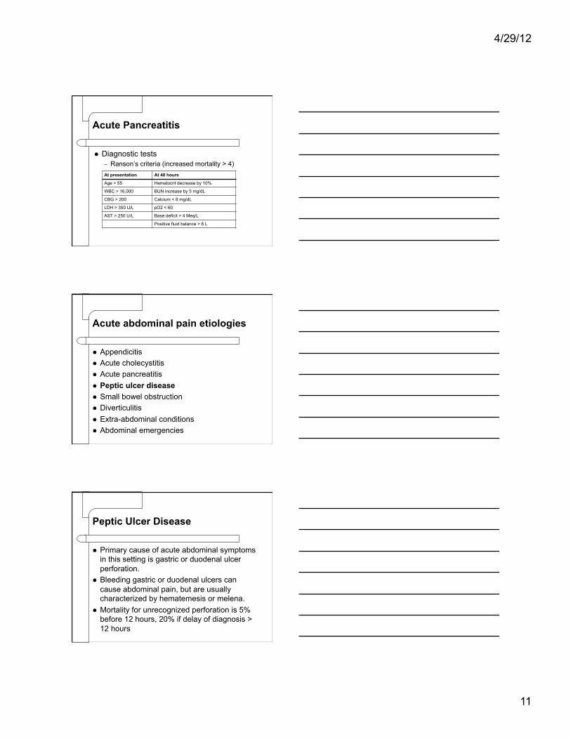
Diagnostic tests – Ranson’s criteria (increased mortality > 4) At presentation At 48 hours
Age > 55 Hematocrit decrease by 10%
WBC > 16,000 BUN increase by 5 mg/dL
CBG > 200 Calcium < 8 mg/dL
LDH > 350 U/L pO2 < 60
AST > 250 U/L Base deficit > 4 Meq/L
Positive fluid balance > 6 L
Acute abdominal pain etiologies
Appendicitis Acute cholecystitis Acute pancreatitis Peptic ulcer disease Small bowel obstruction Diverticulitis Extra-abdominal conditions Abdominal emergencies
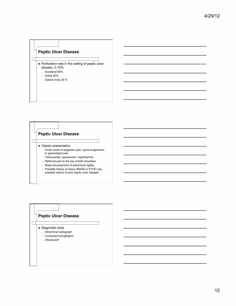
Peptic Ulcer Disease
Primary cause of acute abdominal symptoms in this setting is gastric or duodenal ulcer perforation.
Bleeding gastric or duodenal ulcers can cause abdominal pain, but are usually characterized by hematemesis or melena.
Mortality for unrecognized perforation is 5% before 12 hours, 20% if delay of diagnosis > 12 hours
4/29/12
12
Peptic Ulcer Disease
Perforation rate in the setting of peptic ulcer disease, 2-10% – Duodenal 60% – Antral 20% – Gastric body 20 %
Peptic Ulcer Disease
Classic presentation – Acute onset of epigastric pain, quick progression
to generalized pain – Tachycardia, hypotension, hypothermia – Referred pain to the top of both shoulders – Rapid development of abdominal rigidity – Possible history of heavy NSAID or ETOH use,
possible history of prior peptic ulcer disease
Peptic Ulcer Disease
Diagnostic tools – Abdominal radiograph – Computed tomography* – Ultrasound*
4/29/12
13
Acute abdominal pain etiologies
Appendicitis Acute cholecystitis Acute pancreatitis Peptic ulcer disease Small bowel obstruction Diverticulitis Extra-abdominal conditions Abdominal emergencies
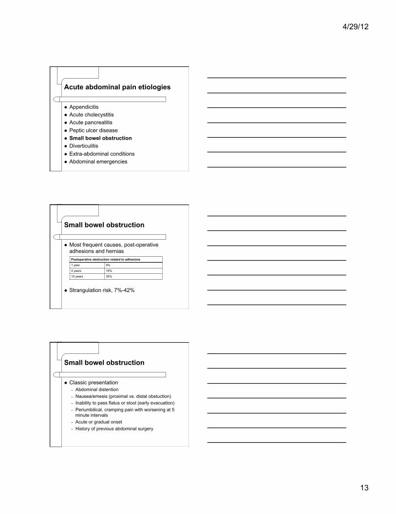
Small bowel obstruction
Most frequent causes, post-operative adhesions and hernias
Strangulation risk, 7%-42%
Postoperative obstruction related to adhesions
1 year 9%
4 years 19%
10 years 35%
Small bowel obstruction
Classic presentation – Abdominal distention – Nausea/emesis (proximal vs. distal obstuction) – Inability to pass flatus or stool (early evacuation) – Periumbilical, cramping pain with worsening at 5
minute intervals – Acute or gradual onset – History of previous abdominal surgery
4/29/12
14
Small bowel obstruction
Critical points on exam – Tympany – High pitched bowel sounds/rushes – Search for inguinal, femoral, obturator, umbilical,
and incisional hernias – Guarding
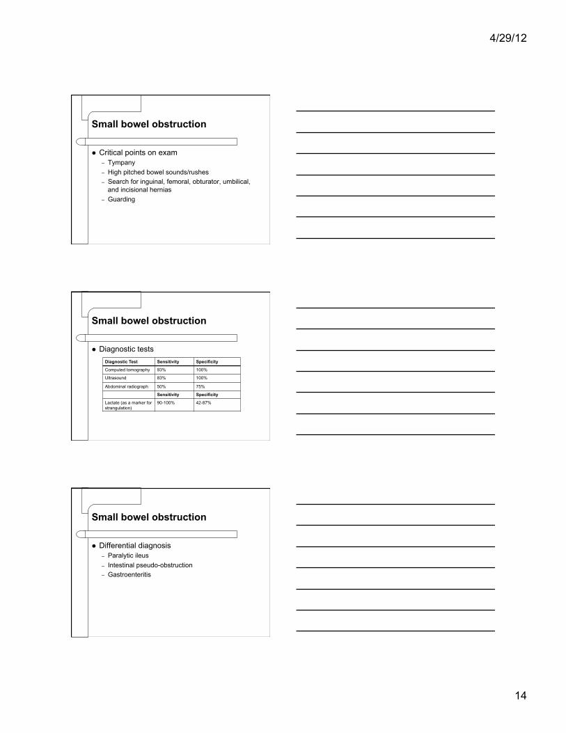
Small bowel obstruction
Diagnostic tests Diagnostic Test Sensitivity Specificity
Computed tomography 93% 100%
Ultrasound 83% 100%
Abdominal radiograph 50% 75%
Sensitivity Specificity
Lactate (as a marker for strangulation)
90-100% 42-87%
Small bowel obstruction
Differential diagnosis – Paralytic ileus – Intestinal pseudo-obstruction – Gastroenteritis
4/29/12
15
Acute abdominal pain etiologies
Appendicitis Acute cholecystitis Acute pancreatitis Peptic ulcer disease Small bowel obstruction Diverticulitis Extra-abdominal conditions Abdominal emergencies
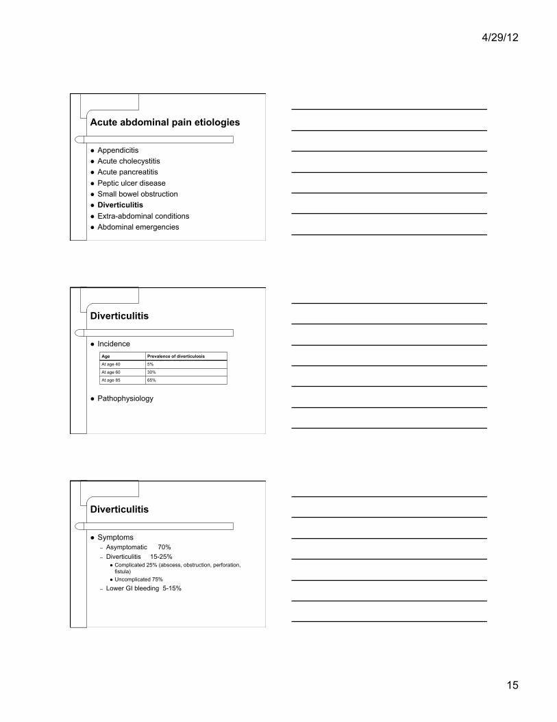
Diverticulitis
Incidence
Pathophysiology
Age Prevalence of diverticulosis
At age 40 5%
At age 60 30%
At age 85 65%
Diverticulitis
Symptoms – Asymptomatic 70% – Diverticulitis 15-25%
Complicated 25% (abscess, obstruction, perforation, fistula)
Uncomplicated 75%
– Lower GI bleeding 5-15%
4/29/12
16
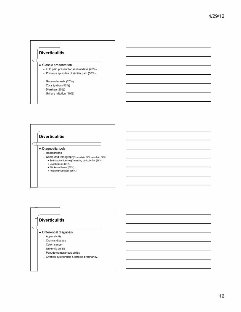
Diverticulitis
Classic presentation – LLQ pain present for several days (70%) – Previous episodes of similar pain (50%)
– Nausea/emesis (20%) – Constipation (50%) – Diarrhea (25%) – Urinary irritation (10%)
Diverticulitis
Diagnostic tools – Radiographs – Computed tomography (sensitivity 97%, specificity 98%)
Soft tissue thickening/stranding pericolic fat (98%) Diverticulosis (84%) Thickened bowel (70%) Phlegmon/abscess (35%)
Diverticulitis
Differential diagnosis – Appendicitis – Crohn's disease – Colon cancer – Ischemic colitis – Pseudomembranous colitis – Ovarian cyst/torsion & ectopic pregnancy.
4/29/12
17
Acute abdominal pain etiologies
Appendicitis Acute cholecystitis Acute pancreatitis Peptic ulcer disease Small bowel obstruction Diverticulitis Extra-abdominal conditions Abdominal emergencies
Extra-abdominal conditions
Pneumonia Acute myocardial infarction Diabetic ketoacidosis Herpes zoster Acute glaucoma
Acute abdominal pain etiologies
Appendicitis Acute cholecystitis Acute pancreatitis Peptic ulcer disease Small bowel obstruction Diverticulitis Extra-abdominal conditions Abdominal emergencies
4/29/12
18
Intra-abdominal emergencies
Mesenteric Ischemia/Infarction (rapid onset severe periumbilical abdominal pain, out of proportion
to exam, nausea/emesis/sudden bowel)
Ruptured Aneurysm (triad of pain, pulsatile abdominal mass, hypotension is
pathognomonic)
Diffuse peritonitis (generalized abdominal pain, rigidity, systemic toxicity)
Conversations with the surgical consultant
Questions
4/29/12
19
Objectives
Describe embryologic origin of abdominal pain distribution.
Identify pertinent terminology in describing acute abdominal pain.
Describe 2 critical findings when evaluating abdominal pain.
Explain pertinent studies based on predicted etiology of abdominal symptoms.
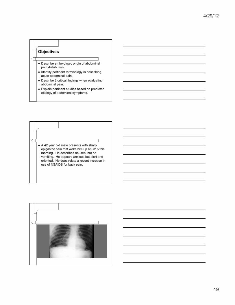
A 42 year old male presents with sharp epigastric pain that woke him up at 0315 this morning. He describes nausea, but no vomiting. He appears anxious but alert and oriented. He does relate a recent increase in use of NSAIDS for back pain.
4/29/12
20
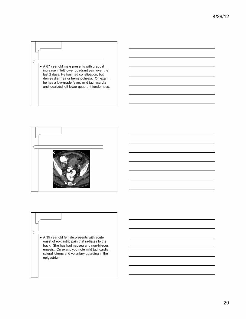
A 67 year old male presents with gradual increase in left lower quadrant pain over the last 2 days. He has had constipation, but denies diarrhea or hematochezia. On exam, he has a low-grade fever, mild tachycardia and localized left lower quadrant tenderness.
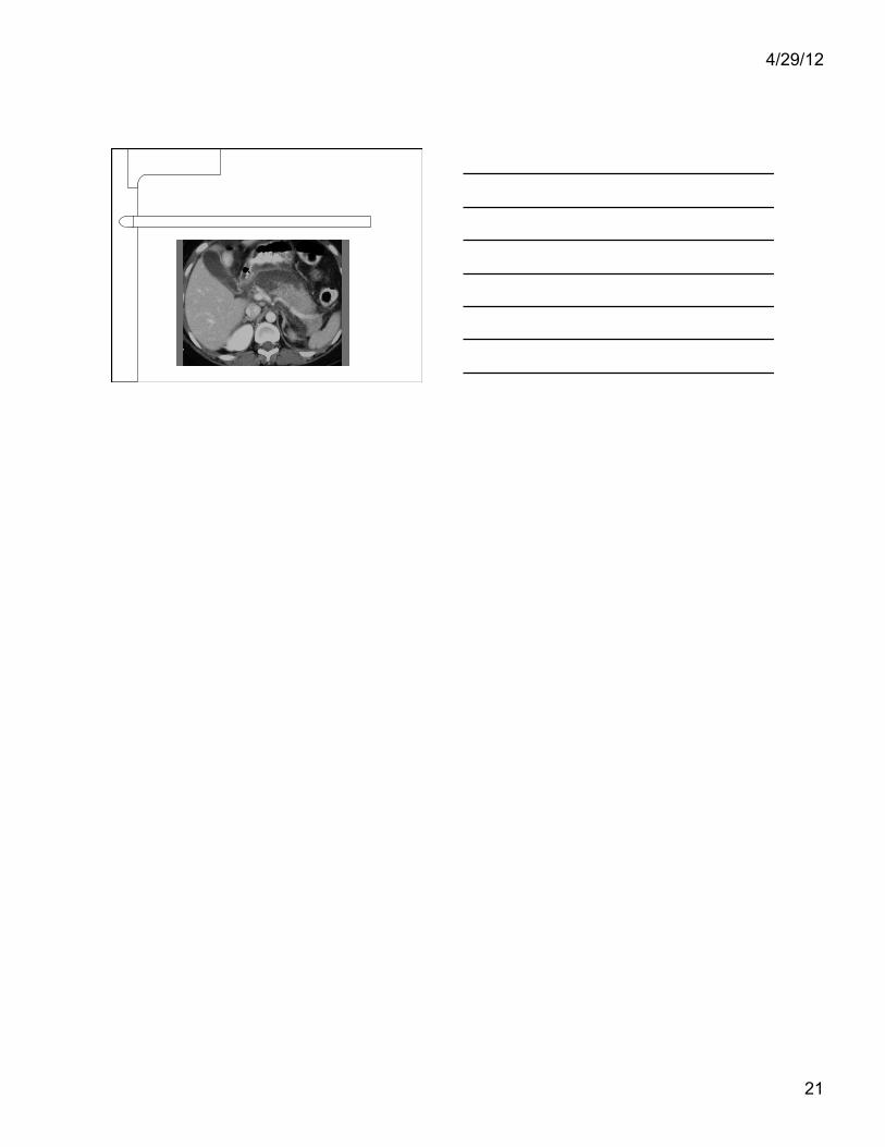
A 35 year old female presents with acute onset of epigastric pain that radiates to the back. She has had nausea and non-bileous emesis. On exam, you note mild tachcardia, scleral icterus and voluntary guarding in the epigastrium.
4/29/12
21