the 9-1-1 checkpoint clamp stimulates dna resection by ...authors.library.caltech.edu/48712/13/nucl....
TRANSCRIPT

10516–10528 Nucleic Acids Research, 2014, Vol. 42, No. 16 Published online 13 August 2014doi: 10.1093/nar/gku746
The 9-1-1 checkpoint clamp stimulates DNA resectionby Dna2-Sgs1 and Exo1Greg H.P. Ngo1*, Lata Balakrishnan2, Marion Dubarry1, Judith L. Campbell3 andDavid Lydall1,*
1Institute for Cell and Molecular Biosciences (ICaMB), Medical School, Newcastle University, Newcastle upon Tyne,NE2 4HH, UK, 2Department of Biochemistry and Biophysics, University of Rochester School of Medicine andDentistry, Rochester, NY 14642, USA and 3Divisions of Biology and Chemistry, Caltech, Braun Laboratories,Pasadena, CA 91125, USA
Received June 18, 2014; Revised July 17, 2014; Accepted August 1, 2014
ABSTRACT
Single-stranded DNA (ssDNA) at DNA ends is animportant regulator of the DNA damage response.Resection, the generation of ssDNA, affects DNAdamage checkpoint activation, DNA repair pathwaychoice, ssDNA-associated mutation and replicationfork stability. In eukaryotes, extensive DNA resectionrequires the nuclease Exo1 and nuclease/helicasepair: Dna2 and Sgs1BLM. How Exo1 and Dna2-Sgs1BLM coordinate during resection remains poorlyunderstood. The DNA damage checkpoint clamp (the9-1-1 complex) has been reported to play an impor-tant role in stimulating resection but the exact mech-anism remains unclear. Here we show that the human9-1-1 complex enhances the cleavage of DNA by bothDNA2 and EXO1 in vitro, showing that the resection-stimulatory role of the 9-1-1 complex is direct. Wealso show that in Saccharomyces cerevisiae, the 9-1-1 complex promotes both Dna2-Sgs1 and Exo1-dependent resection in response to uncapped telom-eres. Our results suggest that the 9-1-1 complex fa-cilitates resection by recruiting both Dna2-Sgs1 andExo1 to sites of resection. This activity of the 9-1-1complex in supporting resection is strongly inhibitedby the checkpoint adaptor Rad953BP1. Our results pro-vide important mechanistic insights into how DNA re-section is regulated by checkpoint proteins and haveimplications for genome stability in eukaryotes.
INTRODUCTION
DNA resection, the nucleolytic degradation of one strandof DNA ends, has emerged as an important regulator ofthe DNA damage response (1,2). The substrates for resec-tion, DNA ends, arise at uncapped telomeres, DNA double
strand breaks (DSBs) and stalled replication forks. DNA re-section generates single-stranded DNA (ssDNA) to triggerand maintain DNA damage checkpoint signaling, which in-duces cell cycle arrest, senescence (permanent cell cycle ar-rest) or apoptosis (cell death) (3,4). Furthermore, the ex-tent of DNA resection affects DNA repair pathway choiceby either homology directed repair or non-homologous endjoining (2,5). Extensive DNA resection can also be harmfulby increasing ssDNA-associated cancer-inducing mutationclusters (6–8). Furthermore, unregulated resection activitiesdegrade stalled replication forks or dysfunctional telomeres(9–13). Thus proper regulation of DNA resection has im-portant consequences on genome stability and cell viability.
In eukaryotes, DNA resection is carried out in two dis-tinct steps (1,2). In the first step, MRXMRN and Sae2CtIP
initiate the reaction by generating a short 3′ ssDNA over-hang (14,15). In the second step, the nuclease Exo1 and/ornuclease/helicase pair: Dna2 and Sgs1BLM carry out exten-sive DNA resection of up to 30 kb (9,14,15). Recent stud-ies suggest that initiation of DNA resection is tightly regu-lated by post-translational modifications (phosphorylation,acetylation and ubiquitination) of MRXMRN and Sae2CtIP
in budding yeast and higher eukaryotes (16–19). However,how Exo1 and Dna2-Sgs1 activities are coordinated to reg-ulate extensive DNA resection remains poorly understood.
The DNA damage checkpoint stimulates signal transduc-tion cascades that not only regulate cell cycle progressionbut also DNA repair (3,20). Several DNA damage check-point proteins play important, yet complex roles in regu-lating DNA resection. In budding yeast, Rad953BP1 inhibitsDNA resection through a mechanism largely independentof its role in activating checkpoint effector Rad53CHK2,but instead relies on its ability to bind to chromatin (21–24). 53BP1, the mammalian homolog of Rad953BP1, alsoinhibits DNA resection (25). On the other hand, the 9-1-1 checkpoint complex promotes DNA resection at un-capped telomeres, DNA DSBs and stalled replication forks,
*To whom correspondence should be addressed. Tel: +44 191 222 5318; Fax: +44 191 208 7424; Email: [email protected] may also be addressed to Greg Ngo. Tel: +44 191 222 6989; Fax: +44 191 208 7424; Email: [email protected]
C© The Author(s) 2014. Published by Oxford University Press on behalf of Nucleic Acids Research.This is an Open Access article distributed under the terms of the Creative Commons Attribution License (http://creativecommons.org/licenses/by/4.0/), whichpermits unrestricted reuse, distribution, and reproduction in any medium, provided the original work is properly cited.
by guest on Decem
ber 4, 2014http://nar.oxfordjournals.org/
Dow
nloaded from

Nucleic Acids Research, 2014, Vol. 42, No. 16 10517
but shows no nuclease activity in vitro and the biochemicalmechanism of control remains poorly understood (26–31).
To better understand how the 9-1-1 complex promotes re-section, we have used complementary biochemical and ge-netic techniques with human proteins and yeast cells. Weshow that the human 9-1-1 complex promotes DNA resec-tion by stimulating the DNA binding and nucleolytic activ-ity of both DNA2 and EXO1 in vitro. These roles are phys-iologically relevant and conserved, since in Saccharomycescerevisiae, 9-1-1 mutants are defective in both Dna2-Sgs1and Exo1-dependent resection. The activity of the 9-1-1complex in supporting resection is strongly inhibited byRad953BP1. Together these data illuminate the role of the9-1-1 complex in promoting extensive DNA resection andillustrate its role in the complex network that carefully reg-ulates the response to damaged DNA ends.
MATERIALS AND METHODS
Yeast techniques
All experiments were performed on S. cerevisiae. We usedthe W303 genetic background strains (Supplementary Ta-bles S1 and S2). Yeast strains with cdc13-1 cdc15-2 bar1 mu-tations were arrested in G1 with alpha-factor at 23◦C andreleased into 36◦C to induce telomere uncapping. Cell cyclepositions were scored using 4′,6-diamidino-2-phenylindole(DAPI) staining on a Nikon Eclipse 50i microscope. Quan-titative amplification of ssDNA (QAOS) analyses were car-ried out as previously described (32,33). Chromatin im-munoprecipitation (ChIP) were performed as previously de-scribed (34).
Immunoprecipitation
Yeast pellets were lysed in IP buffer (pH7.5, 20-mMHEPES, 140-mM NaCl, 1-mM ethylenediaminetetraaceticacid (EDTA), 1% Triton X 100, 0.1% sodium deoxycholate,2-mM phenylmethylsulfonyl fluoride (PMSF) and proteaseinhibitor cocktail) by bead beating. The cell lysates were in-cubated with anti HA (ab9110, Abcam) or anti Myc (ab32,Abcam) antibodies for 1 h and protein G Dynabeads (In-vitrogen) were added before incubation on a wheel at 4◦Covernight. The immunoprecipitates were washed four timesin IP buffer, boiled in laemmli buffer and subjected to west-ern blot analyses.
Yeast two-hybrid
Genes of interest were polymerase chain reaction (PCR)amplified and cloned in yeast by recombinational cloning.The Gal4 activation domain (prey AD-X; pMB29) vec-tor or Gal4 DNA binding domain (bait DB-Y; pMB27)vector (kind gifts from Mike Boxem) was linearized withSmaI and co-transformed into yeast with the PCR fragmentof interest (see Supplementary Table S4). Typically, vectorcontrol gave <10 colonies whereas co-transformation withPCR product gave >1000 colonies. cdc13-1 reporter strainsDLY7451 and DLY7452 (derived from Y8800 and Y8930)(35) transformed with bait and prey plasmids were selectedon SC-Trp or SC-Leu plates, respectively. Five colonies werepooled in SC-Trp or SC-Leu liquid media. Strains were
mated in yeast extract peptone dextrose (YPD) (+ adenine)liquid culture to construct a set of diploid two-hybrid com-bination reporter strains. These diploid strains were culti-vated to saturation in SC-Leu-Trp media before spotted at23, 25, 26, 27, 28 and 29oC for 4–7 days on the follow-ing plates: (i) SC-Leu-Trp – Cell growth control; (ii) SC-His-Leu-Trp – yeast two-hybrid (Y2H) interaction; (iii) SC-Ade-Leu-Trp – Y2H interaction; (iv) SC-His-Leu + 1mg/lCHX – Auto-activator detection and (v) SC-Ade-Leu +1mg/l CHX – Auto-activator detection. Oligonucleotidesused in two-hybrid experiments are listed in SupplementaryTable S4.
Purified human proteins
Recombinant human DNA2 was over-expressed usingpFastBac HTc vector in baculovirus High Five (H5) cellsand purified using a C-terminal FLAG tag as previouslydescribed (36). Rad9-Rad1-Hus1 (9-1-1) and RAD17-RFCwere expressed in H5 insect cells and isolated as previouslydescribed (37,38). Proliferating cell nuclear antigen (PCNA)was expressed and purified as previously described (39).EXO1 was expressed and purified as previously described(40,41).
Nuclease assays
Five fmol of substrate (flap, fork and 3′ ssDNA tailoverhang) were incubated with various concentrations ofDNA2, EXO1, 9-1-1 and PCNA in a reaction volume of20 �l at 37◦C for 10 min. For DNA2 nuclease assays, thereaction buffer consisted of 50-mM Tris HCl (pH 8.0), 2-mM dithiothreitol, 30-mM NaCl, 0.1-mg/ml bovine serumalbumin, 4-mM MgCl2 and 2-mM adenosine triphosphate(ATP). For EXO1 nuclease assays, the reaction buffer con-sisted of 40-mM Tris-HCl (pH 7.6), 2-mM glutathione, 0.1-mg/ml bovine serum albumin, 10-mM MgCl2 and 3-mMATP. The reactions were terminated using 2 X termina-tion dye (90% formamide (v/v), 10-mM EDTA, 0.01% bro-mophenol blue and 0.01% xylene cyanol). After termina-tion, samples were heated at 95◦C for 5 min and 20 �l ofreaction was loaded onto a denaturing gel (8-M urea)/18%polyacrylamide gel and electrophoresed for 1 h and 30 minat 80 W. Each experiment was performed at least in tripli-cate, and representative gels are shown in the figures. Gelswere analyzed as previously described (42).
Nuclease assay substrates
Synthetic oligonucleotides were synthesized by IntegratedDNA Technologies and subjected to 3′-end labeling as pre-viously described (43). Oligonucleotide sequences are listedin Supplementary Table S3. The downstream and upstreamprimer sequences are listed 5′ to 3′ and the template se-quences are listed 3′ to 5′ to facilitate visual alignment. Sub-strates were created by annealing oligonucleotides as listedbelow: Flap Substrate: U1:D1:T1; Fork Substrate: D1:T1;double-stranded DNA (dsDNA) Substrate: D2:T2; 3′ over-hang substrate: D3: T3.
by guest on Decem
ber 4, 2014http://nar.oxfordjournals.org/
Dow
nloaded from

10518 Nucleic Acids Research, 2014, Vol. 42, No. 16
RESULTS
Human 9-1-1 complex stimulates DNA2 and EXO1 cleavageactivity
Rad24, the checkpoint sliding clamp (9-1-1) loader, wasshown to promote resection in vivo nearly 20 years ago (26).Subsequent studies show that the 9-1-1 sliding clamp, itself,also promotes extensive telomere resection (23,24), but theinitial model that Rad17, a component of the sliding clampwas a nuclease, has proven to be incorrect (28). Therefore wetested an alternative hypothesis that the 9-1-1 sliding clampincreases the activity of nucleases important for extensiveDNA resection. Using recombinant human proteins, we ex-amined the effect of 9-1-1 on DNA2 and EXO1 activity,the two nucleases involved in long-range resection. We firsttested the activity of DNA2 on a fork structure, which mim-ics the unwound DNA substrate acted on by DNA2 dur-ing DNA resection (Figure 1A). Since the substrate is lin-ear, the 9-1-1 toroidal clamp can slide onto the DNA in theabsence of the RAD17/RFC clamp loader. DNA was la-beled at the 3′ end in order to be able to detect all productsformed (44). We found that DNA2 cleaved this substrate ef-ficiently (Figure 1A, lanes 2–4). Importantly, we found thatthe 9-1-1 complex stimulated the cleavage of the linear sub-strate by DNA2 (Figure 1A; compare lane 2 with lanes 6–8).The stimulation of DNA2 cleavage was specific to the 9-1-1complex, as addition of the related DNA replication clampPCNA, which has a similar toroidal structure to the 9-1-1 complex, failed to stimulate the nuclease reaction (Fig-ure 1A; compare lane 2 with lanes 10–12). The 9-1-1 com-plex or PCNA alone showed no nuclease or helicase activ-ities (Figure 1A, lanes 5 and 9, and Supplementary FigureS1A). We conclude that 9-1-1 stimulates DNA2 nuclease ac-tivity on resection-mimic-substrates in vitro.
We next tested stimulation of the activity of DNA2 ona double-flap structure, which is a preferred substrate ofDNA2 at replication forks (Figure 1B) (42). Importantly,similar to the result on the fork structure, addition of the9-1-1 complex (compare lane 2 with lanes 6–8), but notPCNA (lanes 10–12) stimulated the DNA2 nuclease activ-ity. Furthermore, we found that the 9-1-1 complex did notstimulate DNA2 degradation of a fully duplex DNA sub-strate or a gap substrate (Supplementary Figure S1B andC), suggesting 9-1-1 increases the activity of DNA2 on un-wound ssDNA ends. These data suggest that 9-1-1 onlystimulates the nuclease activity and not the helicase func-tion of DNA2, since we did not observe any unwinding andsubsequent cleavage of the dsDNA substrate (Supplemen-tary Figure S1B). We conclude that 9-1-1 directly stimu-lates DNA2 cleavage activity. The effect of 9-1-1 is differentto other stimulators of DNA2-Sgs1/BLM resection path-way (MRX/MRN and Top3-Rmi1), which do not stimu-late DNA2 cleavage activity in the absence of Sgs1/BLM(45,46). The observation that 9-1-1 did not affect the cleav-age pattern (Figure 1A and B) but lowered the concentra-tion of DNA2 required for cleavage suggests that 9-1-1 mayincrease the effective local concentration of DNA2 at thesubstrate.
We next examined whether 9-1-1 affects EXO1 activity.We used dsDNA substrate with a 3′ tail, which mimics the
structure formed during DNA resection. Use of a shortersubstrate compared to the traditionally longer substratesused in resection assays (45) allowed us to probe 9-1-1 spe-cific changes in the cleavage patterns of EXO1. We foundthat EXO1 cleaved this substrate inefficiently (Figure 1C;compare lanes 1 and 2). Interestingly, titrating in increasingamounts of 9-1-1 to the reaction resulted in increased cleav-age of the substrate by EXO1 (Figure 1C; compare lane 2with lanes 4 and 5). We found that PCNA also stimulatedEXO1 cleavage activity (Figure 1C; compare lane 2 withlanes 7 and 8), as has been reported recently (47). As EXO1also has flap endonuclease activity, we tested stimulation ofthe activity of EXO1 on a double-flap structure (Figure 1D).Importantly, similar to the result on the tailed structure, ad-dition of the 9-1-1 complex (compare lane 2 with lanes 7 and8) stimulated EXO1 activity. One interesting difference wasthat PCNA did not stimulate EXO1 on this substrate (Fig-ure 1D, lanes 11 and 12). The observation that 9-1-1 did notaffect the cleavage pattern of EXO1, but simply lowered theconcentration of EXO1 required for cleavage of these sub-strates, suggests that 9-1-1 may increase the effective localconcentration of EXO1 at the substrate. We conclude thatthe 9-1-1 complex promotes resection by directly stimulat-ing DNA2 and EXO1 nuclease activities.
Human 9-1-1 complex loads onto DNA to stimulate nucleaseactivity
The 9-1-1 complex is loaded onto the DNA substrate withthe help of the clamp loader RAD17/RFC and we notedthat a molar excess of 9-1-1 over DNA2 and EXO1 was re-quired for stimulation of the nucleases. Therefore, we rea-soned that the large excess of 9-1-1 required (Figure 1A–D) might be due to rapid binding, sliding and dissociationof 9-1-1 from the linear substrates in the absence of theclamp loader. To test this, we examined the effect of ATP-dependent RAD17/RFC clamp loader on DNA2 cleavageactivity. Importantly, RAD17/RFC allowed efficient, ATP-dependent stimulation of DNA2 by 9-1-1, reducing theamount of 9-1-1 required by 5–10-fold (Figure 2A; comparelanes 2 and 5 with lanes 9 and 10). At high concentrations of9-1-1, as expected, RAD17/RFC had no additive effect onDNA2 cleavage activity (Figure 2A; compare lanes 13 and16). This suggests that the 9-1-1 complex must be loadedonto the DNA to stimulate DNA2 nuclease activity. To di-rectly measure the interaction of the 9-1-1 complex withthe DNA substrate, we performed electromobility gel shiftassay in the presence and absence of RAD17/RFC. Lowamounts (25 fmol) of 9-1-1 could only bind the flap sub-strate stably in the presence of RAD17/RFC (Figure 2B;compare lanes 2 and 7). However, 250-fmol 9-1-1 could bindthe substrate on its own, in the absence of the clamp loader(Figure 2B, lanes 4 and 5).
To further test whether 9-1-1 needs to be loaded ontoDNA for its stimulation of nuclease activity, we examinedthe effect of blocking the DNA ends. Importantly, we foundthat blocking the entry of 9-1-1 into the double-strandedregion of the substrate by biotin-streptavidin stopped thestimulation of DNA2 by 9-1-1 (Figure 2C; compare lanes2 and 3 and lanes 4–7). We conclude that 9-1-1 stimula-tion of nuclease requires loading of the 9-1-1 complex onto
by guest on Decem
ber 4, 2014http://nar.oxfordjournals.org/
Dow
nloaded from

Nucleic Acids Research, 2014, Vol. 42, No. 16 10519
5’3’ 3’ Overhang Substrate
A BFlap Substrate5’
3’Fork Substrate
5’
3’*
DNA2
9-1-1 PCNA
100, 250, 500 100, 250, 500
12.5, 25, 50
Sub
stra
te A
lone
9-1-
1 (5
00 fm
ol)
PC
NA
(500
fmol
)
DNA2 (12.5 fmol)
DNA2 (12.5 fmol)
1 2 3 4 5 6 7 8 9 10 11 12Lanes
Fold stimulation
2.5 3.5 2.3 3.1 None
DNA2
9-1-1 PCNA
100, 250, 500 100, 250, 500
12.5, 25, 50
Sub
stra
te A
lone
9-1-
1 (5
00 fm
ol)
PC
NA
(500
fmol
)
DNA2 (12.5 fmol)
DNA2 (12.5 fmol)
1 2 3 4 5 6 7 8 9 10 11 12Lanes
Fold stimulation
2.1 3.6 2.9 3.4 None
*
9-1-1 PCNA
100, 250, 500 100, 250, 500
EX
O1
(20
fmol
)
EXO1 (20 fmol)
EXO1 (20 fmol) S
ubst
rate
Alo
ne
*C D
1 2 3 4 5 6 7 8 Lanes
Fold stimulation
3.2 2.3
(fmol)
(fmol) (fmol)
(fmol)
(fmol) (fmol)
(fmol) (fmol)
Flap Substrate5’3’ *
53
23
30
40
53
23
30
40
EXO1
9-1-1 PCNA
100, 250, 500 100, 250, 500
10, 20, 40
Sub
stra
te A
lone
9-1-
1 (5
00 fm
ol)
PC
NA
(500
fmol
)
EXO1 (10 fmol)
EXO1 (10 fmol)
(fmol)
(fmol) (fmol)
1 2 3 4 5 6 7 8 9 10 11 12Lanes
Fold stimulation
0 1.7 3.9 4.2 None
60
55
4535
53
23
30
40
Figure 1. Human 9-1-1 complex stimulates DNA2 and EXO1 activities. (A, B) DNA2 cleavage activities on a fork (A) and a flap (B) substrate in thepresence of the 9-1-1 complex or PCNA. Labeling of the substrate at the 3′ end allows the determination of the final products of the reaction. Products areanalyzed on alkaline gels as described in the Materials and Methods section. (C, D) EXO1 cleavage activities on a 3′overhang (C) and a flap (D) structurein the presence of 9-1-1 or PCNA. Substrates used for each panel of the experiment are depicted on the top of the gel with the asterisk indicating the siteof the 32P label. The flap is 30 nt long. The labeled strand in panel (C) is 60 nt long.
by guest on Decem
ber 4, 2014http://nar.oxfordjournals.org/
Dow
nloaded from

10520 Nucleic Acids Research, 2014, Vol. 42, No. 16
A
B C
5’3’ *
5’3’ *
5’
*B B
+ + + + + + + + + + + + + + + + - - -- - - 100 - - - 25 50 100 - - - 250 500 1000 25 50 100- - 500 - 25 50 75 25 25 25 100 250 500 100 250 500 25 50 75- + - - + + + + + + + + + + + + + + +
ATP (4 mM)RAD17/RFC (in fmol)
9-1-1 (in fmol)DNA2 (12.5 fmol)
Fold stimulation 2.3 2.9None 3.2 None
Substrate
1 2 3 4 5 6 7 8 9 10 11 12 13 14 15 16 17 18 19Lanes
1 2 3 4 5 6 7 8 9 10 11 12 Lanes
- 25 50 250 500 25 25 250 250 25 - - - - - - - 50 100 500 1000 100 1000 1000 + + + + + + + + + - - + ATP (4 nM)
RAD17/RFC (fmol) 9-1-1(fmol)
1 2 3 4 5 6 7 Lanes
DNA2(12.5 fmol)9-1-1(fmol)
- + + + + + + - - - 100 100 250 250
A B
Substrate + Protein
A: 9-1-1 complex is preincubated with substrate for 5 mins, then blocked with streptavidin and DNA2 is added. B: Substrate is preblocked with streptavidin and then 9-1-1 and DNA2 are added to the blocked substrate.
Figure 2. Human 9-1-1 complex loads onto DNA to stimulate nuclease activity. (A) DNA2 cleavage activity on a 5′ flap substrate in the presence of 9-1-1and RAD17/RFC. (B) Binding efficiency of 9-1-1 (low and high concentration) in the presence of the clamp loader RAD17/RFC on a 5′ flap substrate.(C) DNA2 nuclease activity in the presence of the 9-1-1 complex on a substrate containing blocked template ends and free 5′ flap. Substrates used for eachpanel of experiment are depicted on the top of the gel with the asterisk indicating the site of the 32P label.
by guest on Decem
ber 4, 2014http://nar.oxfordjournals.org/
Dow
nloaded from

Nucleic Acids Research, 2014, Vol. 42, No. 16 10521
DNA, and this 9-1-1 loading is likely dependent on theRAD17/RFC clamp loader complex in vivo.
The 9-1-1 complex promotes Dna2-Sgs1 and Exo1-dependentresection
We have shown that 9-1-1 increases both DNA2 and EXO1nuclease activity in vitro. These data are to some extent atodds with our previous in vivo data, which suggest that 9-1-1 stimulates an unidentified, Exo1-independent nucleasecalled ExoX (23). Therefore, to clarify whether our new bio-chemical experiments reflected resection mechanisms ac-tive in vivo, we used S. cerevisiae to re-examine resectionnear telomeres uncapped by the temperature sensitive al-lele cdc13-1. To examine DNA resection, we measured ss-DNA accumulation by QAOS (32) at loci on each right armtelomere of chromosomes VI and V, respectively (Figure 3Aand Supplementary Figure S2A). We arrested cdc13-1 cellsharboring various other mutations in G1 with alpha-factorat 23oC before releasing at 36oC to induce telomere uncap-ping. The strains also harbored a cdc15-2 mutation to keepany checkpoint-deficient strains in late anaphase and ensurea single round of DNA replication during the course of theexperiment.
Consistent with previous data (23,26), deletion of MEC3(encoding a component of the 9-1-1 complex) reduced ss-DNA accumulation following telomere uncapping (Fig-ure 3B and Supplementary Figure S2B; compare RAD+and mec3Δ), showing that 9-1-1 stimulates resection. Wealso examined the role of 9-1-1 in a rad9Δ background,where resection is increased (26). Consistent with an in-hibitory role for Rad953BP1, rad9Δ strains started to ac-cumulate 3′ ssDNA at loci distal to the telomeres earlierthan a RAD+ strain (Figure 3B and Supplementary Fig-ure S2B). Importantly, in the absence of 9-1-1, deletion ofRAD9 failed to affect ssDNA accumulation (Figure 3B;compare mec3Δ, rad9Δ and rad9Δ mec3Δ strains). Thesedata suggest that Rad953BP1 inhibits resection at uncappedtelomeres entirely by inhibiting 9-1-1-dependent nucleaseactivities. As expected, ssDNA was specific to the 3′ strands,not 5′ strands (Supplementary Figure S2C), and the qualityand quantity of DNA was similar in all samples (Supple-mentary Figure S2D).
A recent study shows that there is a Cdc13-independenttelomere defect in wild-type yeast strains at temperaturesabove 34oC (48) raising the possibility that ssDNA gener-ated was not due to cdc13-1. Although we have previouslyreported there is no significant accumulation of ssDNA attelomeres in CDC+ strains at 36◦C or 37◦C (11,26), we alsoperformed ssDNA measurement experiments at 32oC. Wefound that Mec3 and Rad9 played similar roles in resectionat 32oC as at 36◦C (Supplementary Figure S3A versus Fig-ure 3B). Together these data show that the 9-1-1 complexsupports DNA resection and that Rad953BP1 inhibits resec-tion. As both rad9Δ and mec3Δ mutants are completelyDNA damage checkpoint defective in response to telomereuncapping, as judged the fraction of cells arrested at medialnuclear division after 4 h at 36◦C (49), but show differentresection phenotypes, the role of the 9-1-1 complex in sup-porting extensive resection cannot be solely due to check-point signaling defects.
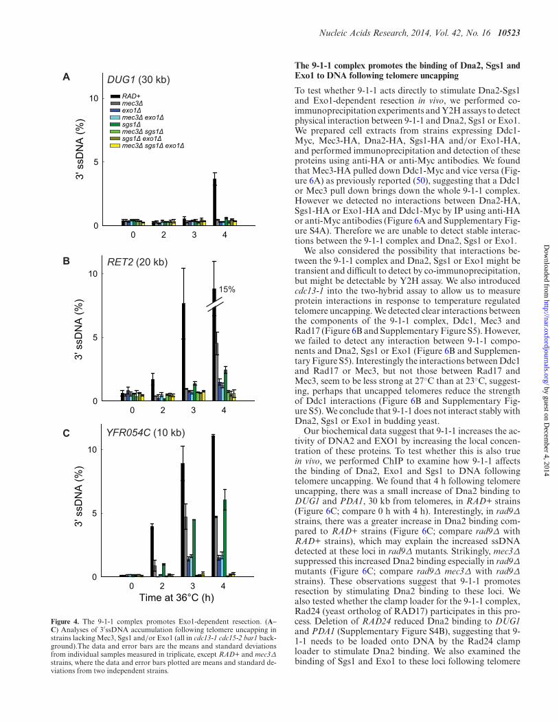
To begin to determine the interaction between 9-1-1 andDna2-Sgs1 and/or Exo1-dependent resection in vivo, wefirst examined the roles of Sgs1 and Exo1. We found thatdeletion of SGS1 or EXO1 partially reduced resection at locidistal to the telomeres (Figure 4; compare RAD+, exo1Δand sgs1Δ strains). Deletion of EXO1 reduced resectionmore than deletion of SGS1, showing that in this context,Exo1 contributes more to resection than Dna2-Sgs1 (Fig-ure 4B and C). Importantly, deletion of both SGS1 andEXO1 completely eliminated resection (Figure 4B and C;compare RAD+, exo1Δ, sgs1Δ and sgs1Δ exo1Δ strains).These results show that in exo1Δ strains, resection is totallydependent on Dna2-Sgs1 and in sgs1Δ strains, resection istotally dependent on Exo1. These results confirm that justlike at DSBs (2), extensive telomere resection in cdc13-1strains is totally dependent on Dna2-Sgs1 and Exo1 andthat it should be informative to examine the effects of 9-1-1on these nuclease activities.
To test whether 9-1-1 promotes Exo1-dependent resec-tion in vivo, we examined how mec3Δ affects ssDNA accu-mulation in sgs1Δ strains, where all the resection is due toExo1. Interestingly, mec3Δ completely eliminated ssDNAaccumulation in sgs1Δ strains (Figure 4B and C; comparesgs1Δ with mec3Δ sgs1Δ strains). Thus, 9-1-1 appears to bevery important for resection by Exo1. This result is consis-tent with our biochemical data reported above and showsthat 9-1-1 can indeed stimulate Exo1 in vivo, besides stimu-lating another Exo1-independent activity (23).
To determine whether 9-1-1 promotes Dna2-Sgs1-dependent resection, we examined how mec3Δ affects ss-DNA accumulation in exo1Δ strains, where all the resectionis due to Dna2-Sgs1. We found that mec3Δ did not signif-icantly affect resection in exo1Δ strains (Figure 4B and C;compare exo1Δ with mec3Δ exo1Δ strains). Thus, it seemsthat 9-1-1 is not important for Dna2-Sgs1-dependent resec-tion.
The results so far suggest that 9-1-1 is important forExo1-dependent but not for Dna2-Sgs1-dependent resec-tion. This is surprising given that our biochemical data sug-gest that 9-1-1 can stimulate both DNA2 and EXO1 invitro (Figure 1). Furthermore, our previous published datasuggest that 9-1-1 stimulated an Exo1-independent activ-ity (23). Therefore, we wondered whether the role of 9-1-1 in Dna2-Sgs1-dependent resection might be obscured bythe weaker contribution of Dna2-Sgs1 to resection com-pared to Exo1 (Figure 4C; compare sgs1Δ with exo1Δstrains). Therefore, we examined resection in rad9Δ back-ground strains, where Dna2-Sgs1-dependent resection issignificantly increased and where 9-1-1 has been inferred tostimulate an Exo1-independent activity (9,23).
As expected and consistent with results in RAD9+strains, deletion of SGS1 or EXO1 reduced resection inrad9Δ strains (Figure 5A). Interestingly, and in contrastto the result in RAD9+ strains, deletion of SGS1 reducedresection more than deletion of EXO1 (Figure 5A; com-pare rad9Δ exo1Δ with rad9Δ sgs1Δ strains at 2 h). How-ever, deletion of EXO1 reduced resection more than dele-tion of SGS1 at the late time point (Figure 5A; comparerad9Δ exo1Δ with rad9Δ sgs1Δ strains at 4 h). This resultshows that in the absence of Rad953BP1, Dna2-Sgs1 con-
by guest on Decem
ber 4, 2014http://nar.oxfordjournals.org/
Dow
nloaded from

10522 Nucleic Acids Research, 2014, Vol. 42, No. 16
5’
Ch VI R
3’DUG1 RET2
0 2 3 4
3' s
sDN
A (%
)
0
5
10
0 2 3 40
5
10
A
B 30 kb 20 kb
RAD+rad9Δmec3Δrad9Δ mec3Δ 14%
Time at 36°C (h) Time at 36°C (h)
Figure 3. The 9-1-1 complex promotes extensive DNA resection. (A) Maps of the right arms of chromosomes VI. (B) Analyses of 3′ssDNA accumulationfollowing telomere uncapping in strains lacking Mec3 and/or Rad9 (all in cdc13-1 cdc15-2 bar1 background). The data and error bars plotted are meansand standard deviations from two independent experiments.
tributes more to long-range resection than Exo1, whereasin the presence of Rad953BP1, the reverse is true.
Importantly, deletion of both SGS1 and EXO1 elimi-nated resection in rad9Δ strains (Figure 5A). These resultsshow that extensive resection observed in rad9Δ strains istotally dependent on Dna2-Sgs1 and Exo1, as in RAD9+strains. We conclude that deletion of RAD9 permits Dna2-Sgs1 to be more active than Exo1, but all resection in rad9Δstrains is still totally dependent on Dna2-Sgs1 or Exo1.
To determine whether 9-1-1 might promote Dna2-Sgs1-dependent resection in the absence of Rad953BP1, we ex-amined how mec3Δ affects ssDNA accumulation in rad9Δexo1Δ strains, where all resection is due to Dna2-Sgs1.Importantly, the mec3Δ mutation eliminated resection inrad9Δ exo1Δ strains at 2h and 3h time points (Figure 5A;compare rad9Δ exo1Δ with rad9Δ mec3Δ exo1Δ strains).This result shows that 9-1-1 is, in fact, important for Dna2-Sgs1-dependent resection, but this 9-1-1 effect is only re-vealed when RAD9 is deleted.
To test whether 9-1-1 promotes Exo1-dependent resec-tion in the absence of Rad953BP1, we examined how mec3Δaffects ssDNA accumulation in rad9Δ sgs1Δ strains, whereall resection is due to Exo1. Similar to the results obtainedin RAD9+ sgs1Δ strains, the mec3Δ mutation completelyeliminated resection in rad9Δ sgs1Δ strains (Figure 5A;compare rad9Δ sgs1Δ with rad9Δ mec3Δ sgs1Δ strains),confirming that 9-1-1 is important for Exo1-dependent re-section, whether or not Rad953BP1 is present. Importantly,in support of our hypothesis that Rad953BP1 inhibits re-
section at uncapped telomeres entirely by inhibiting 9-1-1-dependent nuclease activities (Figure 3), deletion of RAD9failed to affect ssDNA accumulation in mec3Δ exo1Δ ormec3Δ sgs1Δ strains (Figures 4A and B and 5A and Sup-plementary Figure S3B).
Finally to confirm that 9-1-1 promotes Exo1-dependentresection using a different mutation, we also examined howmec3Δ affects ssDNA accumulation in the absence of Dna2activity, using the dna2-1 temperature sensitive allele to in-activate Dna2 (because deletion of DNA2 is lethal). Im-portantly, the mec3Δ mutation completely eliminated ss-DNA accumulation in dna2-1 and rad9Δ dna2-1 strains(Figure 5B), confirming that 9-1-1 is important for Exo1-dependent resection. Together these data show that 9-1-1 isimportant for both Dna2-Sgs1 and Exo1-dependent resec-tion. However, Dna2-Sgs1 and Exo1 can also act indepen-dently of 9-1-1, as inactivating Sgs1, Dna2 or Exo1 reducedresection in rad9Δ mec3Δ strains (Figure 5A and B).
Collectively, all the data in Figures 3–5 show that the 9-1-1 complex stimulates extensive resection by both Dna2-Sgs1 and Exo1 in vivo and that Rad953BP1 inhibits this re-section stimulatory role of 9-1-1. Furthermore, our resultsshow that ExoX, a 9-1-1 stimulated nuclease, is in fact twonuclease activities, Exo1 and Dna2-Sgs1.
by guest on Decem
ber 4, 2014http://nar.oxfordjournals.org/
Dow
nloaded from
Nucleic Acids Research, 2014, Vol. 42, No. 16 10523
0 2 3 40
5
10
15%
3' s
sDN
A (%
)
0 2 3 40
5
10
0 2 3 4
3' s
sDN
A (%
)
0
5
10 RAD+mec3Δexo1Δmec3Δ exo1Δsgs1Δmec3Δ sgs1Δsgs1Δ exo1Δmec3Δ sgs1Δ exo1Δ
DUG1 (30 kb)3'
ssD
NA
(%)
Time at 36°C (h)
RET2 (20 kb)
YFR054C (10 kb)
A
B
C
Figure 4. The 9-1-1 complex promotes Exo1-dependent resection. (A–C) Analyses of 3′ssDNA accumulation following telomere uncapping instrains lacking Mec3, Sgs1 and/or Exo1 (all in cdc13-1 cdc15-2 bar1 back-ground).The data and error bars are the means and standard deviationsfrom individual samples measured in triplicate, except RAD+ and mec3Δ
strains, where the data and error bars plotted are means and standard de-viations from two independent strains.
The 9-1-1 complex promotes the binding of Dna2, Sgs1 andExo1 to DNA following telomere uncapping
To test whether 9-1-1 acts directly to stimulate Dna2-Sgs1and Exo1-dependent resection in vivo, we performed co-immunoprecipitation experiments and Y2H assays to detectphysical interaction between 9-1-1 and Dna2, Sgs1 or Exo1.We prepared cell extracts from strains expressing Ddc1-Myc, Mec3-HA, Dna2-HA, Sgs1-HA and/or Exo1-HA,and performed immunoprecipitation and detection of theseproteins using anti-HA or anti-Myc antibodies. We foundthat Mec3-HA pulled down Ddc1-Myc and vice versa (Fig-ure 6A) as previously reported (50), suggesting that a Ddc1or Mec3 pull down brings down the whole 9-1-1 complex.However we detected no interactions between Dna2-HA,Sgs1-HA or Exo1-HA and Ddc1-Myc by IP using anti-HAor anti-Myc antibodies (Figure 6A and Supplementary Fig-ure S4A). Therefore we are unable to detect stable interac-tions between the 9-1-1 complex and Dna2, Sgs1 or Exo1.
We also considered the possibility that interactions be-tween the 9-1-1 complex and Dna2, Sgs1 or Exo1 might betransient and difficult to detect by co-immunoprecipitation,but might be detectable by Y2H assay. We also introducedcdc13-1 into the two-hybrid assay to allow us to measureprotein interactions in response to temperature regulatedtelomere uncapping. We detected clear interactions betweenthe components of the 9-1-1 complex, Ddc1, Mec3 andRad17 (Figure 6B and Supplementary Figure S5). However,we failed to detect any interaction between 9-1-1 compo-nents and Dna2, Sgs1 or Exo1 (Figure 6B and Supplemen-tary Figure S5). Interestingly the interactions between Ddc1and Rad17 or Mec3, but not those between Rad17 andMec3, seem to be less strong at 27◦C than at 23◦C, suggest-ing, perhaps that uncapped telomeres reduce the strengthof Ddc1 interactions (Figure 6B and Supplementary Fig-ure S5). We conclude that 9-1-1 does not interact stably withDna2, Sgs1 or Exo1 in budding yeast.
Our biochemical data suggest that 9-1-1 increases the ac-tivity of DNA2 and EXO1 by increasing the local concen-tration of these proteins. To test whether this is also truein vivo, we performed ChIP to examine how 9-1-1 affectsthe binding of Dna2, Exo1 and Sgs1 to DNA followingtelomere uncapping. We found that 4 h following telomereuncapping, there was a small increase of Dna2 binding toDUG1 and PDA1, 30 kb from telomeres, in RAD+ strains(Figure 6C; compare 0 h with 4 h). Interestingly, in rad9Δstrains, there was a greater increase in Dna2 binding com-pared to RAD+ strains (Figure 6C; compare rad9Δ withRAD+ strains), which may explain the increased ssDNAdetected at these loci in rad9Δ mutants. Strikingly, mec3Δsuppressed this increased Dna2 binding especially in rad9Δmutants (Figure 6C; compare rad9Δ mec3Δ with rad9Δstrains). These observations suggest that 9-1-1 promotesresection by stimulating Dna2 binding to these loci. Wealso tested whether the clamp loader for the 9-1-1 complex,Rad24 (yeast ortholog of RAD17) participates in this pro-cess. Deletion of RAD24 reduced Dna2 binding to DUG1and PDA1 (Supplementary Figure S4B), suggesting that 9-1-1 needs to be loaded onto DNA by the Rad24 clamploader to stimulate Dna2 binding. We also examined thebinding of Sgs1 and Exo1 to these loci following telomere
by guest on Decem
ber 4, 2014http://nar.oxfordjournals.org/
Dow
nloaded from

10524 Nucleic Acids Research, 2014, Vol. 42, No. 16
0 2 3 4
3' s
sDN
A (%
)
0
5
10 rad9rad9 mec3rad9 exo1rad9 mec3 exo1rad9 sgs1rad9 mec3 sgs1rad9 sgs1 exo1rad9 mec3 sgs1 exo1
0 2 3 4
3' s
sDN
A (%
)
0
5
10 rad9rad9 mec3rad9 exo1rad9 exo1 mec3rad9 sgs1rad9 sgs1 mec3rad9 sgs1 exo1rad9 sgs1 exo1 mec3
3' s
sDN
A (%
)
0
5
10RAD+mec3dna2-1mec3 dna2-1rad9rad9 mec3rad9 dna2-1rad9 mec3 dna2-1
3' s
sDN
A (%
)
0
5
10RAD+mec3dna2-1mec3 dna2-1rad9rad9 mec3rad9 dna2-1rad9 mec3 dna2-1
RAD+
rad9Δ
mec3Δ
rad9Δ mec3Δ
dna2-1mec3Δ dna2-1
rad9Δ dna2-1rad9Δ mec3Δ dna2-1
Time at 36°C (h)0 2 3 4
Time at 36°C (h)0 2 3 4
A
B
rad9Δrad9Δ mec3Δrad9Δ exo1Δrad9Δ mec3Δ exo1Δrad9Δ sgs1Δrad9Δ mec3Δ sgs1Δrad9Δ sgs1Δ exo1Δrad9Δ mec3Δ sgs1Δ exo1Δ
DUG1 (30 kb) RET2 (20 kb)
Figure 5. The 9-1-1 complex promotes Exo1- and Dna2-Sgs1-dependent resection. (A) Analyses of 3′ssDNA accumulation following telomere uncappingin rad9Δ strains lacking Mec3, Sgs1 and/or Exo1 (all in cdc13-1 cdc15-2 bar1 background). The data and error bars plotted are means and standarddeviations from two independent experiments. (B) Analyses of 3′ssDNA accumulation following telomere uncapping in strains lacking Mec3, Rad9 andor Dna2 (all in cdc13-1 cdc15-2 bar1 background). The data and error bars are the means and standard deviations from individual samples measured intriplicate.
uncapping (Figure 6D and E). Importantly, we found that9-1-1 and Rad953BP1 also affected the binding of both Sgs1and Exo1 to these loci in a manner similar to Dna2 (Fig-ure 6D and E). These results suggest that 9-1-1 binding toDNA (Supplementary Figure S4C) stimulates the associa-tion of Dna2-Sgs1 and Exo1 with DNA. Collectively, ourChIP data support our biochemical and genetic data andsuggest that the 9-1-1 complex promotes extensive resectionof uncapped telomeres in vivo by stimulating the associationof Dna2-Sgs1 and Exo1 with DNA.
DISCUSSION
Here we combine biochemical and genetic analyses todemonstrate for the first time that the 9-1-1 checkpointclamp complex is an important stimulatory factor for bothDna2-Sgs1 and Exo1 during DNA resection. Our biochem-
ical experiments using purified human proteins and ourChIP experiments in budding yeast show that the 9-1-1complex performs a resection-stimulatory role by increas-ing the effective local concentration of Dna2-Sgs1 and Exo1on DNA (Figure 7A and B). The binding of 9-1-1 to DNAthus generates a positive feedback loop for DNA damagecheckpoint activation by promoting resection and ssDNAgeneration. Other mechanisms, such as Rad53-dependentExo1 phosphorylation, work in the opposite direction, toinhibit ssDNA accumulation (21).
We propose that the 9-1-1 complex, once loaded ontoDNA by the clamp loader, recruits Dna2-Sgs1 or Exo1 toDNA. Although we could detect no interaction between9-1-1 and these resection proteins, others have shown thatMec3 interacts physically with Exo1 (51). So it is possiblethat 9-1-1 directly interacts with Exo1 to facilitate its re-cruitment or prevent its disengagement from DNA. Alter-
by guest on Decem
ber 4, 2014http://nar.oxfordjournals.org/
Dow
nloaded from

Nucleic Acids Research, 2014, Vol. 42, No. 16 10525
Time at 36°C (h)0 4
Time at 36°C (h)0 4
IP (f
old)
02468
1012
DUG1 (30 kb Ch VI)
Time at 36°C (h)0 4
IP (f
old)
0
2468
1012
Time at 36°C (h)0 4
IP (f
old)
0
2
4
Dna2 ChIP
D
E
C
Sgs1 ChIP
Exo1 ChIP
PDA1 (30 kb Ch V)
Ddc1-Myc + - + - + - + - +Ddc1-Myc + - + - + - + - +
Mec3-HA - + + - - - - - -Exo1-HA - - - - - - - + +
Sgs1-HA - - - - - + + - -Dna2-HA - - - + + - - - -
Mec3-HA - + + - - - - - -Exo1-HA - - - - - - - + +
Sgs1-HA - - - - - + + - -Dna2-HA - - - + + - - - -
Ddc1-Myc
Mec3-HA
Dna2-HA Sgs1-HA
Exo1-HA
Ddc1-Myc
Mec3-HA
IP:anti HA IP:anti Myc
A
B
EX
O1
SG
S1
DN
A2
RA
D17
DD
C1
ME
C3
RAD17
MEC3
bait:
prey:
23°C 27°C No interactionInteraction
EX
O1
SG
S1
DN
A2
RA
D17
DD
C1
ME
C3
EX
O1
SG
S1
DN
A2
RA
D17
DD
C1
ME
C3
RAD+rad9Δmec3Δrad9Δ mec3Δ
RAD+rad9Δmec3Δrad9Δ mec3Δ
RAD+rad9Δmec3Δrad9Δ mec3Δ
Figure 6. The 9-1-1 complex promotes the binding of Dna2, Sgs1 andExo1 to DNA following telomere uncapping. (A) Co-immunoprecipitationexperiment to detect interaction between 9-1-1 and Dna2/Sgs1/Exo1. Pro-tein extract from cells expressing (+) or not expressing (−) the indicatedepitope-tagged proteins was subjected to immunoprecipitation with anti-HA (left panel) or anti-Myc (right panel) antibodies, before probing withanti-HA (top panels) or anti-Myc (bottom panels) antibodies. (B) Two-hybrid analysis in cdc13-1 reporter strains at 23◦C and 27◦C. Bait plasmidcontains either RAD17 or MEC3 and prey plasmids contain the sequenceof the genes indicated. (C–E) ChIP analyses of Dna2-Myc, Sgs1-Myc andExo1-Myc binding to DUG1 and PDA1 following telomere uncapping.The data plotted are fold increase over a control locus PAC2 and representthe means and standard deviations (error bars) from individual samplesmeasured in triplicate.
natively, 9-1-1 may alter DNA conformation to facilitate therecruitment/activation of Dna2-Sgs1 or Exo1. This modeof stimulation would be similar to how MRX and SOSS1
9-1-1
3’
Dna2-Sgs1BLM
Exo1Rad9
53BP1
A
9-1-1
3’
Dna2-Sgs1BLM
Exo1
B
Figure 7. The role of the 9-1-1 complex in stimulating DNA resection. (A,B) The 9-1-1 complex stimulates extensive DNA resection by recruitingDna2-Sgs1BLM and Exo1 to sites of resection. Following recruitment by9-1-1, Dna2-Sgs1BLM and Exo1 contribute differently to resection. In thepresence of Rad953BP1(A), extensive resection is more dependent on Exo1than Dna2-Sgs1BLM. In the absence of Rad953BP1(B), extensive resection ismore dependent on Dna2-Sgs1BLM than Exo1. The set of four filled ellipsesrepresent histone octamers.
stimulate Exo1/EXO1 without direct interactions betweenthe proteins (52,53).
We found that 9-1-1-dependent resection is strongly in-hibited by Rad953BP1. This is perhaps because Rad953BP1
binds to chromatin near DNA damage and thereby inhibitsDna2-Sgs1 and Exo1 (22,54). Interestingly, the effect of 9-1-1 in stimulating Dna2-Sgs1-dependent resection is most eas-ily observed in rad9Δ background strains. We believe thisis because in the presence of Rad953BP1, extensive resectionis more dependent on Exo1 than Dna2-Sgs1 (Figure 7A).In contrast, in the absence of Rad953BP1, extensive resectionis more dependent on Dna2-Sgs1 than Exo1 (Figure 7B).We propose that, in vivo, Exo1 and Dna2-Sgs1 have distinctproperties that are most clearly illustrated by the effect ofRad953BP1. If Rad953BP1 is present then Exo1 is the most im-portant nuclease, responsible for most extensive resection,and Dna2-Sgs1 is less important. However, when Rad953BP1
is absent the situation is reversed and Dna2-Sgs1 is respon-sible for the rapid resection that is observed in rad9Δ strains.Since Rad953BP1 binds to chromatin via methylated histoneH3K79 and phosphorylated histone H2A, we think thatone explanation for this is that the two nucleases have dif-ferent abilities to resect through Rad953BP1 containing chro-matin. We suggest that Exo1 is strong but slow whereasDna2-Sgs1 is fast but weak. We further suggest that thestrong nuclease activity of Exo1 is particularly importantfor resecting through ‘difficult’ Rad953BP1-dependent chro-
by guest on Decem
ber 4, 2014http://nar.oxfordjournals.org/
Dow
nloaded from

10526 Nucleic Acids Research, 2014, Vol. 42, No. 16
matin areas, whereas Dna2-Sgs1 can rapidly move throughless difficult areas. We propose that Rad953BP1 may bindwith different affinity to different regions of the genome dueto chromatin differences. If so, and if resection is to be ex-tensive and efficient, it will depend on both Exo1 and Dna2-Sgs1 nuclease activities. According to this model, 9-1-1 mayplay a critical role in the changeover between nuclease ac-tivities, helping to recruit nuclease(s) with different proper-ties at the ss/dsDNA junction, just as PCNA, the replicativesliding clamp that recruits different activities to the replica-tion fork (55).
53BP1, the mammalian homolog of Rad9, with its inter-acting proteins (RIF1 and PTIP) also inhibits DNA resec-tion, but the exact mechanism of inhibition remains unclear(56–61). It will be interesting to test whether 9-1-1 promotesresection by DNA2-BLM and EXO1 in mammalian cellsand if so whether 9-1-1 is responsible for increased resec-tion in cells lacking 53BP1.
In conclusion, we provide novel mechanistic insights intohow the important function of DNA resection is regulatedby the 9-1-1 complex. Our results have important implica-tions for the mechanisms that maintain genome stabilityand potentially shed light on the involvement of the 9-1-1 complex in many other processes like telomere mainte-nance, repair of stalled replication forks, homologous re-combination and cancer progression (51,62–67).
SUPPLEMENTARY DATA
Supplementary Data are available at NAR Online.
ACKNOWLEDGMENTS
We thank Mike Boxem and Matija Dreze for the generousgifts of strains and plasmids; Aziz Sancar, Robert Bambara,Laura Lindsey-Boltz and Paul Modrich for generously pro-viding purified proteins and advice; and members of the Ly-dall and Campbell labs for comments on the manuscript.
FUNDING
Wellcome Trust [075294, 093088 to D.L]; National In-stitutes of Health [GM098328 to L.B.; GM100186 toJ.L.C.]; European Molecular Biology Organization [EMBOALTF218-2012 to M.D.]. Funding for open access charge:Wellcome Trust [075294, 093088].Conflict of interest statement. None declared.
REFERENCES1. Huertas,P. (2010) DNA resection in eukaryotes: deciding how to fix
the break. Nat. Struct. Mol. Biol., 17, 11–16.2. Symington,L.S. and Gautier,J. (2011) Double-strand break end
resection and repair pathway choice. Annu. Rev. Genet., 45, 247–271.3. Ciccia,A. and Elledge,S.J. (2010) The DNA damage response: making
it safe to play with knives. Mol. Cell, 40, 179–204.4. Sperka,T., Wang,J. and Rudolph,K.L. (2012) DNA damage
checkpoints in stem cells, ageing and cancer. Nat. Rev. Mol. CellBiol., 13, 579–590.
5. Chapman,J.R., Taylor,M.R. and Boulton,S.J. (2012) Playing the endgame: DNA double-strand break repair pathway choice. Mol. Cell,47, 497–510.
6. Roberts,S.A., Sterling,J., Thompson,C., Harris,S., Mav,D., Shah,R.,Klimczak,L.J., Kryukov,G.V., Malc,E., Mieczkowski,P.A. et al.(2012) Clustered mutations in yeast and in human cancers can arisefrom damaged long single-strand DNA regions. Mol. Cell, 46,424–435.
7. Chan,K., Sterling,J.F., Roberts,S.A., Bhagwat,A.S., Resnick,M.A.and Gordenin,D.A. (2012) Base damage within single-strand DNAunderlies in vivo hypermutability induced by a ubiquitousenvironmental agent. PLoS Genet., 8, e1003149.
8. Taylor,B.J., Nik-Zainal,S., Wu,Y.L., Stebbings,L.A., Raine,K.,Campbell,P.J., Rada,C., Stratton,M.R. and Neuberger,M.S. (2013)DNA deaminases induce break-associated mutation showers withimplication of APOBEC3B and 3A in breast cancer kataegis. Elife, 2,e00534.
9. Ngo,H.P. and Lydall,D. (2010) Survival and growth of yeast withouttelomere capping by Cdc13 in the absence of Sgs1, Exo1, and Rad9.PLoS Genet., 6, e1001072.
10. Schlacher,K., Christ,N., Siaud,N., Egashira,A., Wu,H. and Jasin,M.(2011) Double-strand break repair-independent role for BRCA2 inblocking stalled replication fork degradation by MRE11. Cell, 145,529–542.
11. Maringele,L. and Lydall,D. (2002) EXO1-dependent single-strandedDNA at telomeres activates subsets of DNA damage and spindlecheckpoint pathways in budding yeast yku70Delta mutants. GenesDev., 16, 1919–1933.
12. Couch,F.B., Bansbach,C.E., Driscoll,R., Luzwick,J.W., Glick,G.G.,Betous,R., Carroll,C.M., Jung,S.Y., Qin,J., Cimprich,K.A. et al.(2013) ATR phosphorylates SMARCAL1 to prevent replication forkcollapse. Genes Dev., 27, 1610–1623.
13. Segurado,M. and Diffley,J.F. (2008) Separate roles for the DNAdamage checkpoint protein kinases in stabilizing DNA replicationforks. Genes Dev., 22, 1816–1827.
14. Zhu,Z., Chung,W.H., Shim,E.Y., Lee,S.E. and Ira,G. (2008) Sgs1helicase and two nucleases Dna2 and Exo1 resect DNAdouble-strand break ends. Cell, 134, 981–994.
15. Mimitou,E.P. and Symington,L.S. (2008) Sae2, Exo1 and Sgs1collaborate in DNA double-strand break processing. Nature, 455,770–774.
16. Steger,M., Murina,O., Huhn,D., Ferretti,L.P., Walser,R., Hanggi,K.,Lafranchi,L., Neugebauer,C., Paliwal,S., Janscak,P. et al. (2013)Prolyl isomerase PIN1 regulates DNA double-strand break repair bycounteracting DNA end resection. Mol. Cell, 50, 333–343.
17. Falck,J., Forment,J.V., Coates,J., Mistrik,M., Lukas,J., Bartek,J. andJackson,S.P. (2012) CDK targeting of NBS1 promotes DNA-endresection, replication restart and homologous recombination. EMBORep., 13, 561–568.
18. Kaidi,A., Weinert,B.T., Choudhary,C. and Jackson,S.P. (2010)Human SIRT6 promotes DNA end resection through CtIPdeacetylation. Science, 329, 1348–1353.
19. Huertas,P., Cortes-Ledesma,F., Sartori,A.A., Aguilera,A. andJackson,S.P. (2008) CDK targets Sae2 to control DNA-end resectionand homologous recombination. Nature, 455, 689–692.
20. Putnam,C.D., Jaehnig,E.J. and Kolodner,R.D. (2009) Perspectives onthe DNA damage and replication checkpoint responses inSaccharomyces cerevisiae. DNA Repair (Amst), 8, 974–982.
21. Morin,I., Ngo,H.P., Greenall,A., Zubko,M.K., Morrice,N. andLydall,D. (2008) Checkpoint-dependent phosphorylation of Exo1modulates the DNA damage response. EMBO J., 27, 2400–2410.
22. Lazzaro,F., Sapountzi,V., Granata,M., Pellicioli,A., Vaze,M.,Haber,J.E., Plevani,P., Lydall,D. and Muzi-Falconi,M. (2008)Histone methyltransferase Dot1 and Rad9 inhibit single-strandedDNA accumulation at DSBs and uncapped telomeres. EMBO J., 27,1502–1512.
23. Zubko,M.K., Guillard,S. and Lydall,D. (2004) Exo1 and Rad24differentially regulate generation of ssDNA at telomeres ofSaccharomyces cerevisiae cdc13–1 mutants. Genetics, 168, 103–115.
24. Jia,X., Weinert,T. and Lydall,D. (2004) Mec1 and Rad53 inhibitformation of single-stranded DNA at telomeres of Saccharomycescerevisiae cdc13–1 mutants. Genetics, 166, 753–764.
25. Panier,S. and Boulton,S.J. (2014) Double-strand break repair: 53BP1comes into focus. Nat. Rev. Mol. Cell Biol., 15, 7–18.
26. Lydall,D. and Weinert,T. (1995) Yeast checkpoint genes in DNAdamage processing: implications for repair and arrest. Science, 270,1488–1491.
by guest on Decem
ber 4, 2014http://nar.oxfordjournals.org/
Dow
nloaded from

Nucleic Acids Research, 2014, Vol. 42, No. 16 10527
27. Aylon,Y. and Kupiec,M. (2003) The checkpoint protein Rad24 ofSaccharomyces cerevisiae is involved in processing double-strandbreak ends and in recombination partner choice. Mol. Cell. Biol., 23,6585–6596.
28. Majka,J. and Burgers,P.M. (2003) Yeast Rad17/Mec3/Ddc1: asliding clamp for the DNA damage checkpoint. Proc. Natl. Acad. Sci.U.S.A., 100, 2249–2254.
29. Costelloe,T., Louge,R., Tomimatsu,N., Mukherjee,B., Martini,E.,Khadaroo,B., Dubois,K., Wiegant,W.W., Thierry,A., Burma,S. et al.(2012) The yeast Fun30 and human SMARCAD1 chromatinremodellers promote DNA end resection. Nature, 489, 581–584.
30. Blaikley,E.J., Tinline-Purvis,H., Kasparek,T.R., Marguerat,S.,Sarkar,S., Hulme,L., Hussey,S., Wee,B.Y., Deegan,R.S., Walker,C.A.et al. (2014) The DNA damage checkpoint pathway promotesextensive resection and nucleotide synthesis to facilitate homologousrecombination repair and genome stability in fission yeast. NucleicAcids Res., 42, 5644–5656.
31. Tsang,E., Miyabe,I., Iraqui,I., Zheng,J., Lambert,S.A. andCarr,A.M. (2014) The extent of error-prone replication restart byhomologous recombination is controlled by Exo1 and checkpointproteins. J. Cell Sci., 127, 2983–2994.
32. Holstein,E.M. and Lydall,D. (2012) Quantitative amplification ofsingle-stranded DNA. Methods Mol. Biol., 920, 323–339.
33. Dewar,J.M. and Lydall,D. (2012) Simple, non-radioactivemeasurement of single-stranded DNA at telomeric, sub-telomeric,and genomic loci in budding yeast. Methods Mol. Biol., 920, 341–348.
34. Addinall,S.G., Holstein,E.M., Lawless,C., Yu,M., Chapman,K.,Banks,A.P., Ngo,H.P., Maringele,L., Taschuk,M., Young,A. et al.(2011) Quantitative fitness analysis shows that NMD proteins andmany other protein complexes suppress or enhance distinct telomerecap defects. PLoS Genet., 7, e1001362.
35. Dreze,M., Monachello,D., Lurin,C., Cusick,M.E., Hill,D.E.,Vidal,M. and Braun,P. (2010) High-quality binary interactomemapping. Methods Enzymol., 470, 281–315.
36. Masuda-Sasa,T., Imamura,O. and Campbell,J.L. (2006) Biochemicalanalysis of human Dna2. Nucleic Acids Res., 34, 1865–1875.
37. Griffith,J.D., Lindsey-Boltz,L.A. and Sancar,A. (2002) Structures ofthe human Rad17-replication factor C and checkpoint Rad 9-1-1complexes visualized by glycerol spray/low voltage microscopy. J.Biol. Chem., 277, 15233–15236.
38. Lindsey-Boltz,L.A., Bermudez,V.P., Hurwitz,J. and Sancar,A. (2001)Purification and characterization of human DNA damage checkpointRad complexes. Proc. Natl Acad. Sci. U.S.A., 98, 11236–11241.
39. Podust,V.N., Podust,L.M., Goubin,F., Ducommun,B. andHubscher,U. (1995) Mechanism of inhibition of proliferating cellnuclear antigen-dependent DNA synthesis by the cyclin-dependentkinase inhibitor p21. Biochemistry, 34, 8869–8875.
40. Genschel,J. and Modrich,P. (2006) Analysis of the excision step inhuman DNA mismatch repair. Methods Enzymol., 408, 273–284.
41. Lindsey-Boltz,L.A., Kemp,M.G., Reardon,J.T., Derocco,V.,Iyer,R.R., Modrich,P. and Sancar,A. (2014) Coupling of humanDNA excision repair and the DNA damage checkpoint in a defined invitro system. J. Biol. Chem., 289, 5074–5082.
42. Gloor,J.W., Balakrishnan,L., Campbell,J.L. and Bambara,R.A.(2012) Biochemical analyses indicate that binding and cleavagespecificities define the ordered processing of human Okazakifragments by Dna2 and FEN1. Nucleic Acids Res., 40, 6774–6786.
43. Stewart,J.A., Campbell,J.L. and Bambara,R.A. (2009) Significance ofthe dissociation of Dna2 by flap endonuclease 1 to Okazaki fragmentprocessing in Saccharomyces cerevisiae. J. Biol. Chem., 284,8283–8291.
44. Kao,H.I., Veeraraghavan,J., Polaczek,P., Campbell,J.L. andBambara,R.A. (2004) On the roles of Saccharomyces cerevisiaeDna2p and Flap endonuclease 1 in Okazaki fragment processing. J.Biol. Chem., 279, 15014–15024.
45. Nimonkar,A.V., Genschel,J., Kinoshita,E., Polaczek,P.,Campbell,J.L., Wyman,C., Modrich,P. and Kowalczykowski,S.C.(2011) BLM-DNA2-RPA-MRN and EXO1-BLM-RPA-MRNconstitute two DNA end resection machineries for human DNAbreak repair. Genes Dev., 25, 350–362.
46. Cejka,P., Cannavo,E., Polaczek,P., Masuda-Sasa,T., Pokharel,S.,Campbell,J.L. and Kowalczykowski,S.C. (2010) DNA end resectionby Dna2-Sgs1-RPA and its stimulation by Top3-Rmi1 andMre11-Rad50-Xrs2. Nature, 467, 112–116.
47. Chen,X., Paudyal,S.C., Chin,R.I. and You,Z. (2013) PCNA promotesprocessive DNA end resection by Exo1. Nucleic Acids Res., 41,9325–9338.
48. Paschini,M., Toro,T.B., Lubin,J.W., Braunstein-Ballew,B.,Morris,D.K. and Lundblad,V. (2012) A naturally thermolabileactivity compromises genetic analysis of telomere function inSaccharomyces cerevisiae. Genetics, 191, 79–93.
49. Lydall,D. and Weinert,T. (1997) G2/M checkpoint genes ofSaccharomyces cerevisiae: further evidence for roles in DNAreplication and/or repair. Mol. Gen. Genet., 256, 638–651.
50. Kondo,T., Matsumoto,K. and Sugimoto,K. (1999) Role of a complexcontaining Rad17, Mec3, and Ddc1 in the yeast DNA damagecheckpoint pathway. Mol. Cell. Biol., 19, 1136–1143.
51. Karras,G.I., Fumasoni,M., Sienski,G., Vanoli,F., Branzei,D. andJentsch,S. (2013) Noncanonical role of the 9-1-1 clamp in theerror-free DNA damage tolerance pathway. Mol. Cell, 49, 536–546.
52. Nicolette,M.L., Lee,K., Guo,Z., Rani,M., Chow,J.M., Lee,S.E. andPaull,T.T. (2010) Mre11-Rad50-Xrs2 and Sae2 promote 5′ strandresection of DNA double-strand breaks. Nat. Struct. Mol. Biol., 17,1478–1485.
53. Yang,S.H., Zhou,R., Campbell,J., Chen,J., Ha,T. and Paull,T.T.(2013) The SOSS1 single-stranded DNA binding complex promotesDNA end resection in concert with Exo1. EMBO J., 32, 126–139.
54. Chen,X., Cui,D., Papusha,A., Zhang,X., Chu,C.D., Tang,J.,Chen,K., Pan,X. and Ira,G. (2012) The Fun30 nucleosomeremodeller promotes resection of DNA double-strand break ends.Nature, 489, 576–580.
55. Mailand,N., Gibbs-Seymour,I. and Bekker-Jensen,S. (2013)Regulation of PCNA-protein interactions for genome stability. Nat.Rev. Mol. Cell Biol., 14, 269–282.
56. Callen,E., Di Virgilio,M., Kruhlak,M.J., Nieto-Soler,M., Wong,N.,Chen,H.T., Faryabi,R.B., Polato,F., Santos,M., Starnes,L.M. et al.(2013) 53BP1 mediates productive and mutagenic DNA repairthrough distinct phosphoprotein interactions. Cell, 153, 1266–1280.
57. Chapman,J.R., Barral,P., Vannier,J.B., Borel,V., Steger,M.,Tomas-Loba,A., Sartori,A.A., Adams,I.R., Batista,F.D. andBoulton,S.J. (2013) RIF1 Is essential for 53BP1-dependentnonhomologous end joining and suppression of DNA double-strandbreak resection. Mol. Cell, 49, 858–871.
58. Di Virgilio,M., Callen,E., Yamane,A., Zhang,W., Jankovic,M.,Gitlin,A.D., Feldhahn,N., Resch,W., Oliveira,T.Y., Chait,B.T. et al.(2013) Rif1 prevents resection of DNA breaks and promotesimmunoglobulin class switching. Science, 339, 711–715.
59. Escribano-Diaz,C., Orthwein,A., Fradet-Turcotte,A., Xing,M.,Young,J.T., Tkac,J., Cook,M.A., Rosebrock,A.P., Munro,M.,Canny,M.D. et al. (2013) A cell cycle-dependent regulatory circuitcomposed of 53BP1-RIF1 and BRCA1-CtIP controls DNA repairpathway choice. Mol. Cell, 49, 872–883.
60. Panier,S. and Durocher,D. (2013) Push back to respond better:regulatory inhibition of the DNA double-strand break response. Nat.Rev. Mol. Cell Biol., 14, 661–672.
61. Zimmermann,M., Lottersberger,F., Buonomo,S.B., Sfeir,A. and deLange,T. (2013) 53BP1 regulates DSB repair using Rif1 to control 5′end resection. Science, 339, 700–704.
62. Broustas,C.G. and Lieberman,H.B. (2012) Contributions of Rad9 totumorigenesis. J. Cell. Biochem., 113, 742–751.
63. An,L., Wang,Y., Liu,Y., Yang,X., Liu,C., Hu,Z., He,W., Song,W. andHang,H. (2010) Rad9 is required for B cell proliferation andimmunoglobulin class switch recombination. J. Biol. Chem., 285,35267–35273.
64. Grandin,N. and Charbonneau,M. (2007) Control of the yeasttelomeric senescence survival pathways of recombination by the Mec1and Mec3 DNA damage sensors and RPA. Nucleic Acids Res., 35,822–838.
65. Pandita,R.K., Sharma,G.G., Laszlo,A., Hopkins,K.M., Davey,S.,Chakhparonian,M., Gupta,A., Wellinger,R.J., Zhang,J., Powell,S.N.et al. (2006) Mammalian Rad9 plays a role in telomere stability, S-and G2-phase-specific cell survival, and homologous recombinationalrepair. Mol. Cell. Biol., 26, 1850–1864.
66. Shinohara,M., Sakai,K., Ogawa,T. and Shinohara,A. (2003) Themitotic DNA damage checkpoint proteins Rad17 and Rad24 arerequired for repair of double-strand breaks during meiosis in yeast.Genetics, 164, 855–865.
by guest on Decem
ber 4, 2014http://nar.oxfordjournals.org/
Dow
nloaded from

10528 Nucleic Acids Research, 2014, Vol. 42, No. 16
67. Lyndaker,A.M., Lim,P.X., Mleczko,J.M., Diggins,C.E.,Holloway,J.K., Holmes,R.J., Kan,R., Schlafer,D.H., Freire,R.,Cohen,P.E. et al. (2013) Conditional inactivation of the DNA damageresponse gene Hus1 in mouse testis reveals separable roles for
components of the RAD9-RAD1-HUS1 complex in meioticchromosome maintenance. PLoS Genet., 9, e1003320.
by guest on Decem
ber 4, 2014http://nar.oxfordjournals.org/
Dow
nloaded from